Embed Size (px)
Citation preview
Abstract
Aim: Surgery is most important for the treatment of a hydatid cyst. All steps of conventional hydatid surgery are the same as in a minimally invasive laparoscopic approach. Laparoscopy management is the gold standard for hydatid liver disease. We report the use of a laparoscopic approach for hydatid cysts of the liver. Methods and Materials: Nine patients with ten liver hydatid cysts were operated on laparoscopi-cally. The mean operative time varied between 50 and 120 minutes. The mean duration of the hospital stay was between 3 and 9 days. There was no conversion to open surgery. Results: There was no major complication and mortality. To decrease the incidence of anaphylaxis, all patients were given 8mg of inj. dexamethasone, 60 minutes prior to surgery. There was no recur-rence during the follow-up period of 3 to 23 months. Conclusion: Minimal access surgery is a safe, effective, viable and feasible option for the manage-ment of selective patients with hydatidosis of the liver.
Key words: Anaphylaxis, hydatid cyst, laparoscopic.
Our Initial Experience of Laparoscopic Management of Liver Hydatid Disease in a Rural Medical College
Sunder Goyal1, Sanjay Pandit2, Rachana Raina3, Sidharth Maurya2
Original Article
1Department of SurgeryMuzaffarnagar Medical College
Muzaffarnagar (UP), India
2Department of Surgery3Department of Obstetrics
and GynaecologyMM Institute of
Medical Sciences and ResearchMullana, Ambala, India
Received: March 08, 2012 Accepted: May 01, 2012
Arch Clin Exp Surg 2013;2: 16-23DOI:10.5455/aces.20120501124612
Corresponding author:Sunder Goyal, MD
Department of SurgeryMuzaffarnagar Medical College
Muzaffarnagar (UP), [email protected]
IntroductionHydatid disease has been known to
mankind since ancient times. Hippocrates described ‘livers full of water.’ Hydatid is Greek for ‘drop of water,’ while echinococ-cosis means ‘hedgehog berry.’ Surgery is the mainstay of treatment for hydatid dis-ease of the liver. In a World Health Organi-zation study in the central Peruvian Andes,
the incidence of hydatid disease was re-ported in about 9.1% of human beings [1]. In humans, cysts occur mainly in the liver (50-75%), then in the lungs (25%), and 5-10% distribute along the arterial system to any organs from the great toe to crown of the head, except hair, nail and teeth [2]. In the last decade, laparoscopic treatment of hepatic hydatid disease has been in-
1Bauru Dental SchoolUniversity of São PauloBauru–SP, Brazil
2Araraquara Dental SchoolSão Paulo State UniversityAraraquara-SP, Brazil
Received: February 05, 2012 Accepted: February 29, 2012 Arch Clin Exp Surg 2012;X: X-XDOI: 10.5455/aces.20120229052919
Corresponding authorÉrica Dorigatti de AvilaDepartamento de Estomatologia da Faculdade de Odontologia de BauruUniversidade de São Paulo (USP)Avenida Alameda Octávio Pinheiro Brizola, 9-75, 17012-901 Bauru–SP, [email protected]
Original Article
Increased of Langerhans Cells in Smokeless Tobacco-Associated Oral Mucosal Lesions
Érica Dorigatti de Ávila1, Rafael Scaf de Molon2, Melaine de Almeida Lawall1, Renata Bianco Consolaro1, Alberto Consolaro1
Abstract
Objective: To evaluate the changes in the number of Langerhans Cells (LC) observed in the epithelium of smokeless tobacco (SLT-induced) lesions. Methods: Microscopic sections from biopsies carried out in the buccal mucosa of twenty patients, who were chronic users of smokeless tobacco (SLT), were utilized. For the control group, twenty non-SLT users of SLT with normal mucosa were selected. The sections were studied with routine coloring and were immunostained for S-100, CD1a, Ki-67 and p63. These data were statistically analyzed by the Student’s t-test to investigate the differences in the expression of immune markers in normal mucosa and in SLT-induced leukoplakia lesions. Results: There was a significant difference in the immunolabeling of all markers between normal mucosa and SLT-induced lesions (p<0.001). The leukoplakia lesions in chronic SLT users demonstrated a significant increase in the number of Langerhans cells and in the absence of epithelial dysplasia. Conclusion: The increase in the number of these cells represents the initial stage of leukoplakia. Key words: Smokeless tobacco, leukoplakic lesions, cancer, langerhans cells, chewing tobacco.
Introduction
Among tobacco users, there is a false be-lief that SLT is safe because it is not burned, which leads many people to quit cigarettes and start using SLT [1]. However, SLT con-tains higher concentrations of nicotine than cigarettes and, in addition, nearly 30 carci-nogenic substances, such as tobacco-specific N-nitrosamines (TSNA), which is formed during the aging process of the tobacco, [2-4] and which presents high carcinogenic poten-tial. Moreover, because the tobacco has direct
contact with the oral mucosa and creates a more alkaline environment, its products may even be more aggressive to tissue [5]. The percentage of SLT users is lower compared to cigarette users; however, usage is increasing among young individuals and it is therefore a significant and disturbing danger [6,7].
Initial studies on the effects of SLT on the oral mucosa demonstrated the formation of white lesions induced by chronic exposure to tobacco, characterized by epithelial thicken-ing, increased vascularization, collagen altera-
Archives of ClinicalExperimental Surgery
creasingly popular due to various advantages over open surgery. With increased use, the technique has been re-fined and standardized, and laparoscopic treatment has become the gold standard for management of hydatid liver disease. This study presents our experience in nine patients with ten hydatid cysts of the liver, treated with a laparoscopic technique in a rural medical college.
Methods and MaterialsFrom August 2009 to July 2011, nine patients with
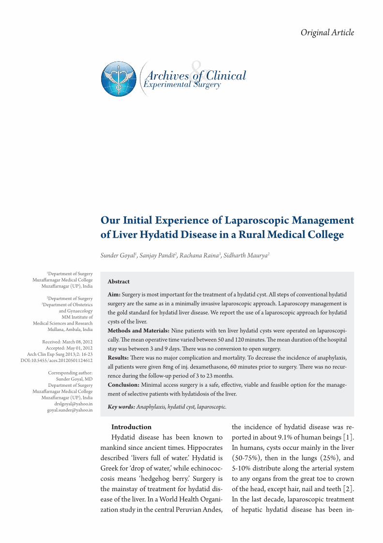
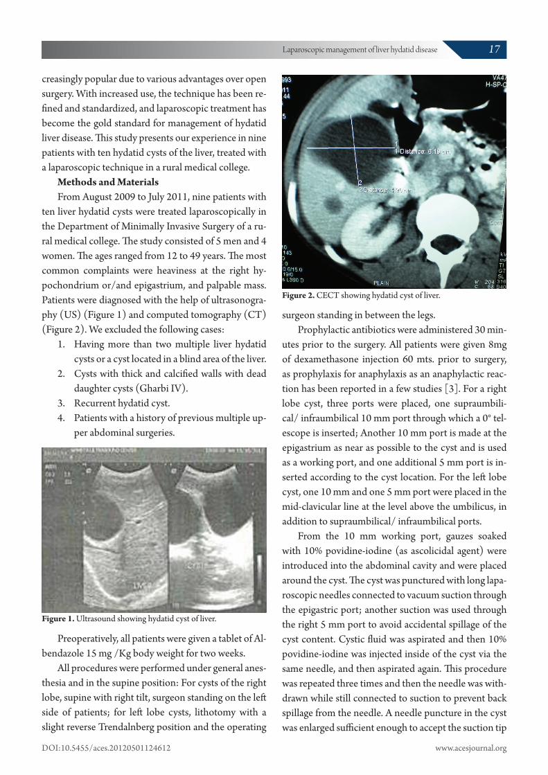
ten liver hydatid cysts were treated laparoscopically in the Department of Minimally Invasive Surgery of a ru-ral medical college. The study consisted of 5 men and 4 women. The ages ranged from 12 to 49 years. The most common complaints were heaviness at the right hy-pochondrium or/and epigastrium, and palpable mass. Patients were diagnosed with the help of ultrasonogra-phy (US) (Figure 1) and computed tomography (CT) (Figure 2). We excluded the following cases:
1. Having more than two multiple liver hydatid cysts or a cyst located in a blind area of the liver.
2. Cysts with thick and calcified walls with dead daughter cysts (Gharbi IV).
3. Recurrent hydatid cyst. 4. Patients with a history of previous multiple up-
per abdominal surgeries.
Figure 1. Ultrasound showing hydatid cyst of liver.
Preoperatively, all patients were given a tablet of Al-bendazole 15 mg /Kg body weight for two weeks.
All procedures were performed under general anes-thesia and in the supine position: For cysts of the right lobe, supine with right tilt, surgeon standing on the left side of patients; for left lobe cysts, lithotomy with a slight reverse Trendalnberg position and the operating
surgeon standing in between the legs.Prophylactic antibiotics were administered 30 min-
utes prior to the surgery. All patients were given 8mg of dexamethasone injection 60 mts. prior to surgery, as prophylaxis for anaphylaxis as an anaphylactic reac-tion has been reported in a few studies [3]. For a right lobe cyst, three ports were placed, one supraumbili-cal/ infraumbilical 10 mm port through which a 0° tel-escope is inserted; Another 10 mm port is made at the epigastrium as near as possible to the cyst and is used as a working port, and one additional 5 mm port is in-serted according to the cyst location. For the left lobe cyst, one 10 mm and one 5 mm port were placed in the mid-clavicular line at the level above the umbilicus, in addition to supraumbilical/ infraumbilical ports.
From the 10 mm working port, gauzes soaked with 10% povidine-iodine (as ascolicidal agent) were introduced into the abdominal cavity and were placed around the cyst. The cyst was punctured with long lapa-roscopic needles connected to vacuum suction through the epigastric port; another suction was used through the right 5 mm port to avoid accidental spillage of the cyst content. Cystic fluid was aspirated and then 10% povidine-iodine was injected inside of the cyst via the same needle, and then aspirated again. This procedure was repeated three times and then the needle was with-drawn while still connected to suction to prevent back spillage from the needle. A needle puncture in the cyst was enlarged sufficient enough to accept the suction tip
Figure 2. CECT showing hydatid cyst of liver.
17Laparoscopic management of liver hydatid disease
DOI:10.5455/aces.20120501124612 www.acesjournal.org
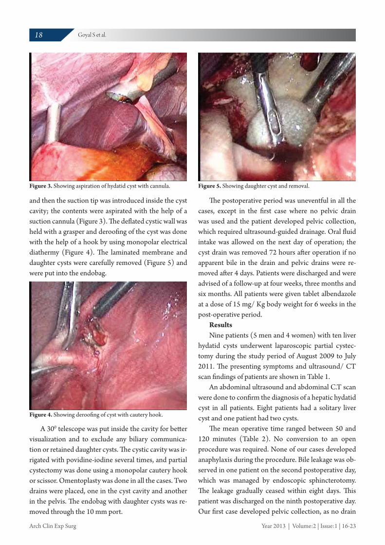
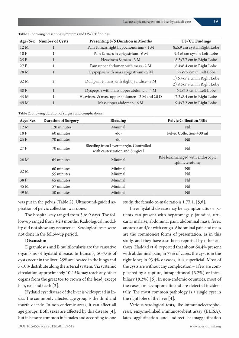
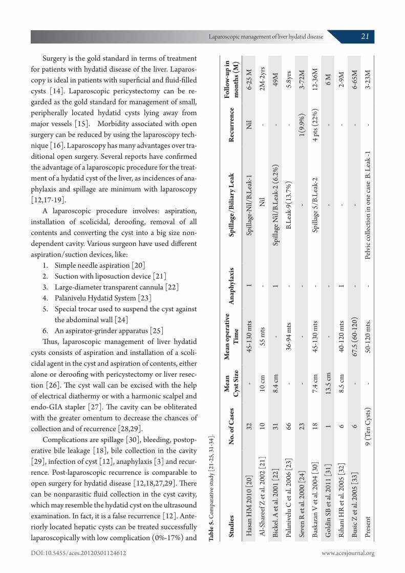
and then the suction tip was introduced inside the cyst cavity; the contents were aspirated with the help of a suction cannula (Figure 3). The deflated cystic wall was held with a grasper and deroofing of the cyst was done with the help of a hook by using monopolar electrical diathermy (Figure 4). The laminated membrane and daughter cysts were carefully removed (Figure 5) and were put into the endobag.
Figure 3. Showing aspiration of hydatid cyst with cannula.
A 300 telescope was put inside the cavity for better visualization and to exclude any biliary communica-tion or retained daughter cysts. The cystic cavity was ir-rigated with povidine-iodine several times, and partial cystectomy was done using a monopolar cautery hook or scissor. Omentoplasty was done in all the cases. Two drains were placed, one in the cyst cavity and another in the pelvis. The endobag with daughter cysts was re-moved through the 10 mm port.
Figure 4. Showing deroofing of cyst with cautery hook.
Figure 5. Showing daughter cyst and removal.
The postoperative period was uneventful in all the cases, except in the first case where no pelvic drain was used and the patient developed pelvic collection, which required ultrasound-guided drainage. Oral fluid intake was allowed on the next day of operation; the cyst drain was removed 72 hours after operation if no apparent bile in the drain and pelvic drains were re-moved after 4 days. Patients were discharged and were advised of a follow-up at four weeks, three months and six months. All patients were given tablet albendazole at a dose of 15 mg/ Kg body weight for 6 weeks in the post-operative period.
ResultsNine patients (5 men and 4 women) with ten liver
hydatid cysts underwent laparoscopic partial cystec-tomy during the study period of August 2009 to July 2011. The presenting symptoms and ultrasound/ CT scan findings of patients are shown in Table 1.
An abdominal ultrasound and abdominal C.T scan were done to confirm the diagnosis of a hepatic hydatid cyst in all patients. Eight patients had a solitary liver cyst and one patient had two cysts.
The mean operative time ranged between 50 and 120 minutes (Table 2). No conversion to an open procedure was required. None of our cases developed anaphylaxis during the procedure. Bile leakage was ob-served in one patient on the second postoperative day, which was managed by endoscopic sphincterotomy. The leakage gradually ceased within eight days. This patient was discharged on the ninth postoperative day. Our first case developed pelvic collection, as no drain
18 Goyal S et al.
Arch Clin Exp Surg Year 2013 | Volume:2 | Issue:1 | 16-23
Age/Sex Number of Cysts Presenting S/S Duration in Months US/CT Findings12 M 1 Pain & mass right hypochondrium - 1 M 8x5.9 cm cyst in Right Lobe18 F 1 Pain & mass in epigastrium - 6 M 9.4x6 cm cyst in Left Lobe25 F 1 Heaviness & mass - 3 M 8.5x7.7 cm in Right Lobe27 F 1 Pain upper abdomen with mass - 2 M 8.4x6.4 cm in Right Lobe28 M 1 Dyspepsia with mass epigastrium - 5 M 8.7x9.7 cm in Left Lobe
32 M 2 Dull pain & mass with slight jaundice - 3 M1) 6.4x7.2 cm in Right Lobe2) 8.5x7.3 cm in Right Lobe
38 F 1 Dyspepsia with mass upper abdomen - 4 M 6.2x7.3 cm in Left Lobe45 M 1 Heaviness & mass upper abdomen - 3 M and 20 D 7.2x8.4 cm in Right Lobe49 M 1 Mass upper abdomen - 6 M 9.4x7.2 cm in Right Lobe
Table 1. Showing presenting symptoms and US/CT findings.
Age/ Sex Duration of Surgery Bleeding Pelvic Collection/Bile 12 M 120 minutes Minimal Nil18 F 60 minutes -do- Pelvic Collection-400 ml25 F 70 minutes -do- Nil
27 F 70 minutes Bleeding from Liver margin. Controlled with cauterization and Surgicel Nil
28 M 65 minutes Minimal Bile leak managed with endoscopic sphincterotomy
32 M 60 minutes55 minutes
MinimalMinimal
NilNil
38 F 65 minutes Minimal Nil45 M 57 minutes Minimal Nil49 M 50 minutes Minimal Nil
Table 2. Showing duration of surgery and complications.
was put in the pelvis (Table 2). Ultrasound-guided as-piration of pelvic collection was done.
The hospital stay ranged from 3 to 9 days. The fol-low-up ranged from 3-23 months. Radiological modal-ity did not show any recurrence. Serological tests were not done in the follow-up period.
DiscussionE granulosus and E multilocularis are the causative
organisms of hydatid disease. In humans, 50-75% of cysts occur in the liver; 25% are located in the lungs and 5-10% distribute along the arterial system. Via systemic circulation, approximately 10-15% may reach any other organs from the great toe to crown of the head, except hair, nail and teeth [2].
Hydatid cyst disease of the liver is widespread in In-dia. The commonly affected age group is the third and fourth decade. In non-endemic areas, it can affect all age groups. Both sexes are affected by this disease [4], but it is more common in females and according to one
study, the female-to-male ratio is 1.77:1. [5,6].Liver hydatid disease may be asymptomatic or pa-
tients can present with hepatomegaly, jaundice, urti-caria, malaise, abdominal pain, abdominal mass, fever, anorexia and/or with cough. Abdominal pain and mass are the commonest forms of presentation, as in this study, and they have also been reported by other au-thors. Haddad et al. reported that about 64.4% present with abdominal pain; in 77% of cases, the cyst is in the right lobe; in 93.4% of cases, it is superficial. Most of the cysts are without any complication – a few are com-plicated by a rupture, intraperitoneal (3.2%) or intra-biliary (8.2%) [6]. In non-endemic countries, most of the cases are asymptomatic and are detected inciden-tally. The most common pathology is a single cyst in the right lobe of the liver [4].
Various serological tests, like immunoelectropho-resis, enzyme-linked immunosorbent assay (ELISA), latex agglutination and indirect haemagglutination
19Laparoscopic management of liver hydatid disease
DOI:10.5455/aces.20120501124612 www.acesjournal.org

Type I Pure fluidType II Fluid collection, split-wall floating membrane
Type III Fluid collection with septa, daughter cysts and honeycomb image
Type IV Heterogeneous echographic patternType V Reflecting thick walls
(IHA), are carried out for the diagnosis, screening and for postoperative follow-up for recurrence. However, these tests are often negative because the capsule iso-lates the parasite from the host’s immune system.
Ultrasonography (USG) and Computed tomogra-phy (CT) are both valuable imaging methods for the diagnosis of liver hydatid disease. USG is particularly useful for the recognition of cystic membranes, septa, and hydatid sand, while CT exhibits cyst wall calcifica-tion and cyst infection.
Gharbi et al. [7]. did ultrasonographic classifica-tion of hepatic hydatid disease, as shown in Table 3.
Table 3. Gharbi classification of hepatic echinococcal cysts [7].
The World Health Organization (WHO) Informal Working Group on Echinococcosis [8] modified their International Classification System, which was based on Gharbi’s ultrasonographic classification system, to identify the functional state of parasites to define treat-ment, as shown in Table 4.
Magnetic Resonance Imaging (MRI) or Endoscop-ic Retrograde Cholangiography (ERC) may confirm biliary involvement. Preoperatively, mistaking a hy-datid liver cyst for a simple liver cyst remains at about 5%, despite improved imaging modalities [9].
Endoscopic Retrograde Cholangiopancreaticogra-
Type of Cyst Status Ultrasound Features Remarks
CL Active Signs no pathognomonic, unilocular and no cyst wall usually early stage, not fertile and differential diagnosis necessary
CE 1 Active Cyst wall, hydatid sand usually fertileCE 2 Active Multivesicular, cyst wall and “rosette-like” usually fertile
CE 3 Transitional Detachment of laminated membrane, “water-lily sign”, less round-decreased intracystic pressure
starting to degenerate, may produce daugter cysts
CE 4 Inactive Heterogeneous hypo or hyperechogenic degenerative contents, no daughter cysts
usually no living protoscoleces D/D necessary
CE 5 Inactive Thick calcified wall, calcification partial to complete, and not pathognomonic but highly suggestive of diagnosis usually no living protoscoleces
Table 4. International classification of ultrasound images in cystic echinococcosis for application in clinical and field epidemiological setting [8].
phy (ERCP) is used for complicated cases of hydatid liver disease and the routine use of ERCP is only rec-ommended if surgery is planned [10]. Another recent study suggested that a cyst diameter independently predicts the risk of biliary-cyst communication in asymptomatic patients. In an asymptomatic patient, if the cyst size is more than 10.2 cm in diameter, the chances of biliary communication and bile leakage are high. Therefore, ERCP is usually done in asymptomatic patients with a large cyst [11]. An intraoperative chol-angiogram is not required in all cases and it also increas-es morbidity [12]. By inspecting the cyst cavity with a 300 telescope, all major bile leaks can be identified. If the diameter of a ruptured bile duct is less than 5 mm, it can be ligated; if the size is bigger than 5 mm, an in-traoperative cholangiogram is done to assess the com-mon bile duct (CBD) for debris, which requires CBD exploration with T-tube placement. 5mm is regarded as a cutoff size, as bile ducts smaller than this rarely trans-mit particulate matter to the CBD, while 65% of bile ducts 5mm or larger allow a passage of material into the CBD [13].
The role of preoperative drug therapy is to decrease the size of a cyst as well as to reduce the risk of intra-operative spillage-induced spread. A drug, like Alben-dazole, can also be used as chemotherapy in patients who are unfit for surgery and also for invasive hydatid disease. Adult dosage is 400 mg orally, twice a day for 1–5 months and a pediatric dosage is 15 mg/kg/day (max. of 800 mg) for 1–6 months. An alternative to al-bendazole is mebendazole at a dosage of 40 to 50 mg/kg/day for at least 3 to 6 months.
20 Goyal S et al.
Arch Clin Exp Surg Year 2013 | Volume:2 | Issue:1 | 16-23
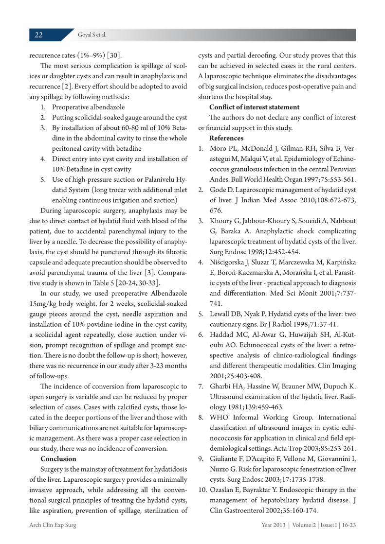
Surgery is the gold standard in terms of treatment for patients with hydatid disease of the liver. Laparos-copy is ideal in patients with superficial and fluid-filled cysts [14]. Laparoscopic pericystectomy can be re-garded as the gold standard for management of small, peripherally located hydatid cysts lying away from major vessels [15]. Morbidity associated with open surgery can be reduced by using the laparoscopy tech-nique [16]. Laparoscopy has many advantages over tra-ditional open surgery. Several reports have confirmed the advantage of a laparoscopic procedure for the treat-ment of a hydatid cyst of the liver, as incidences of ana-phylaxis and spillage are minimum with laparoscopy [12,17-19].
A laparoscopic procedure involves: aspiration, installation of scolicidal, deroofing, removal of all contents and converting the cyst into a big size non-dependent cavity. Various surgeon have used different aspiration/suction devices, like:
1. Simple needle aspiration [20] 2. Suction with liposuction device [21] 3. Large-diameter transparent cannula [22] 4. Palanivelu Hydatid System [23] 5. Special trocar used to suspend the cyst against
the abdominal wall [24] 6. An aspirator-grinder apparatus [25]Thus, laparoscopic management of liver hydatid
cysts consists of aspiration and installation of a scoli-cidal agent in the cyst and aspiration of contents, either alone or deroofing with pericystectomy or liver resec-tion [26]. The cyst wall can be excised with the help of electrical diathermy or with a harmonic scalpel and endo-GIA stapler [27]. The cavity can be obliterated with the greater omentum to decrease the chances of collection and of recurrence [28,29].
Complications are spillage [30], bleeding, postop-erative bile leakage [18], bile collection in the cavity [29], infection of cyst [12], anaphylaxis [3] and recur-rence. Post-laparoscopic recurrence is comparable to open surgery for hydatid disease [12,18,27,29]. There can be nonparasitic fluid collection in the cyst cavity, which may resemble the hydatid cyst on the ultrasound examination. In fact, it is a false recurrence [12]. Ante-riorly located hepatic cysts can be treated successfully laparoscopically with low complication (0%-17%) and St
udie
sN
o. o
f Cas
esM
ean
Cys
t Siz
eM
ean
oper
ativ
e T
ime
Ana
phyl
axis
Spill
age/
Bili
ary
Leak
Rec
urre
nce
Follo
w-u
p in
m
onth
s (M
)
Has
an H
M 2
010
[20]
32-
45-1
30 m
ts 1
Spill
age-
Nil/
B.Le
ak-1
Nil
6-25
M
Al-S
hare
ef Z
et al
. 200
2 [2
1]10
10 cm
55 m
ts-
N
il-
2M
-2yr
s
Bick
el. A
et al
. 200
1 [2
2]31
8.4
cm-
1Sp
illag
e Nil/
B.Le
ak-2
(6.2
%)
-49
M
Pala
nive
lu C
et al
. 200
6 [2
3]66
-36
-94
mts
-B.
Leak
-9(1
3.7%
)-
5.8y
rs
Seve
n R
et al
. 200
0 [2
4]23
--
--
1(9.
9%)
3-72
M
Bask
aran
V et
al. 2
004
[30]
187.
4 cm
45-1
30 m
ts-
Spill
age 5
/B.L
eak-
24
pts (
22%
)12
-36M
Gol
din
SB et
al. 2
011
[31]
113
.5 cm
--
--
6 M
Rih
ani H
R et
al. 2
005
[32]
68.
5 cm
40-1
20 m
ts1
--
2-9M
Busic
Z et
al. 2
005
[33]
6-
67.5
(60-
120)
--
-6-
65M
Pres
ent
9 (T
en C
ysts)
-50
-120
mts
.-
Pelv
ic co
llect
ion
in o
ne ca
se B
. Lea
k -1
-3-
23M
Tabl
e 5.
Com
para
tive s
tudy
[21-
25, 3
1-34
].
21Laparoscopic management of liver hydatid disease
DOI:10.5455/aces.20120501124612 www.acesjournal.org
recurrence rates (1%–9%) [30].The most serious complication is spillage of scol-
ices or daughter cysts and can result in anaphylaxis and recurrence [2]. Every effort should be adopted to avoid any spillage by following methods:
1. Preoperative albendazole 2. Putting scolicidal-soaked gauge around the cyst 3. By installation of about 60-80 ml of 10% Beta-
dine in the abdominal cavity to rinse the whole peritoneal cavity with betadine
4. Direct entry into cyst cavity and installation of 10% Betadine in cyst cavity
5. Use of high-pressure suction or Palanivelu Hy-datid System (long trocar with additional inlet enabling continuous irrigation and suction)
During laparoscopic surgery, anaphylaxis may be due to direct contact of hydatid fluid with blood of the patient, due to accidental parenchymal injury to the liver by a needle. To decrease the possibility of anaphy-laxis, the cyst should be punctured through its fibrotic capsule and adequate precaution should be observed to avoid parenchymal trauma of the liver [3]. Compara-tive study is shown in Table 5 [20-24, 30-33].
In our study, we used preoperative Albendazole 15mg/kg body weight, for 2 weeks, scolicidal-soaked gauge pieces around the cyst, needle aspiration and installation of 10% povidine-iodine in the cyst cavity, a scolicidal agent repeatedly, close suction under vi-sion, prompt recognition of spillage and prompt suc-tion. There is no doubt the follow-up is short; however, there was no recurrence in our study after 3-23 months of follow-ups.
The incidence of conversion from laparoscopic to open surgery is variable and can be reduced by proper selection of cases. Cases with calcified cysts, those lo-cated in the deeper portions of the liver and those with biliary communications are not suitable for laparoscop-ic management. As there was a proper case selection in our study, there was no incidence of conversion.
Conclusion Surgery is the mainstay of treatment for hydatidosis
of the liver. Laparoscopic surgery provides a minimally invasive approach, while addressing all the conven-tional surgical principles of treating the hydatid cysts, like aspiration, prevention of spillage, sterilization of
cysts and partial deroofing. Our study proves that this can be achieved in selected cases in the rural centers. A laparoscopic technique eliminates the disadvantages of big surgical incision, reduces post-operative pain and shortens the hospital stay.
Conflict of interest statementThe authors do not declare any conflict of interest
or financial support in this study.References
1. Moro PL, McDonald J, Gilman RH, Silva B, Ver-astegui M, Malqui V, et al. Epidemiology of Echino-coccus granulosus infection in the central Peruvian Andes. Bull World Health Organ 1997;75:553-561.
2. Gode D. Laparoscopic management of hydatid cyst of liver. J Indian Med Assoc 2010;108:672-673, 676.
3. Khoury G, Jabbour-Khoury S, Soueidi A, Nabbout G, Baraka A. Anaphylactic shock complicating laparoscopic treatment of hydatid cysts of the liver. Surg Endosc 1998;12:452-454.
4. Niścigorska J, Sluzar T, Marczewska M, Karpińska E, Boroń-Kaczmarska A, Morańska I, et al. Parasit-ic cysts of the liver - practical approach to diagnosis and differentiation. Med Sci Monit 2001;7:737-741.
5. Lewall DB, Nyak P. Hydatid cysts of the liver: two cautionary signs. Br J Radiol 1998;71:37-41.
6. Haddad MC, Al-Awar G, Huwaijah SH, Al-Kut-oubi AO. Echinococcal cysts of the liver: a retro-spective analysis of clinico-radiological findings and different therapeutic modalities. Clin Imaging 2001;25:403-408.
7. Gharbi HA, Hassine W, Brauner MW, Dupuch K. Ultrasound examination of the hydatic liver. Radi-ology 1981;139:459-463.
8. WHO Informal Working Group. International classification of ultrasound images in cystic echi-nococcosis for application in clinical and field epi-demiological settings. Acta Trop 2003;85:253-261.
9. Giuliante F, D’Acapito F, Vellone M, Giovannini I, Nuzzo G. Risk for laparoscopic fenestration of liver cysts. Surg Endosc 2003;17:1735-1738.
10. Ozaslan E, Bayraktar Y. Endoscopic therapy in the management of hepatobiliary hydatid disease. J Clin Gastroenterol 2002;35:160-174.
22 Goyal S et al.
Arch Clin Exp Surg Year 2013 | Volume:2 | Issue:1 | 16-23

11. Kilic M, Yoldas O, Koc M, Keskek M, Karakose N, Ertan T, et al. Can biliary-cyst communication be predicted before surgery for hepatic hydatid dis-ease: does size matter? Am J Surg 2008;196:732-735.
12. Ramachandran CS, Goel D, Arora V. Laparoscopic surgery in hepatic hydatid cysts: a technical im-provement. Surg Laparosc Endosc Percutan Tech 2001;11:14-18.
13. Ozmen MM, Coskun F. New technique for finding the ruptured bile duct into the liver cysts: scope in the cave technique. Surg Laparosc Endosc Percu-tan Tech 2002;12:187-189.
14. Sayek I, Cakmakci M. Laparoscopic management of echinococcal cysts of the liver. Zentralbl Chir 1999;124:1143-1146.
15. Bickel A, Loberant N, Singer-Jordan J, Goldfeld M, Daud G, Eitan A. The laparoscopic approach to ab-dominal hydatid cysts: a prospective nonselective study using the isolated hypobaric technique. Arch Surg 2001;136:789-795.
16. Misra MC, Khan RN, Bansal VK, Jindal V, Kumar S, Noba AL, et al. Laparoscopic pericystectomy for hydatid cyst of the liver. Surg Laparosc Endosc Per-cutan Tech 2010;20:24-26.
17. Sharma D, Babu R, Borgharia S, Baruah D, Thomas S, Kumar A. Laparoscopy for liver hydatid disease: where do we stand today? Surg Laparosc Endosc Percutan Tech 2009;19:419-423.
18. Ertem M, Uras C, Karahasanoglu T, Erguney S, Alemdaroglu K. Laparoscopic approach to hepatic hydatid disease. Dig Surg 1998;15:333-336.
19. Manterola C, Fernández O, Muñoz S, Vial M, Losada H, Carrasco R, et al. Laparoscopic peri-cystectomy for liver hydatid cysts. Surg Endosc 2002;16:521-524.
20. Hassan HM, El-Sayed OM. Laparoscopic treat-ment of liver hydatid cyst. Journal of Medicine and Biomedical Sciences 2010;1:47-51.
21. Al-Shareef Z, Hamour OA, Al-Shlash S, Ahmed I, Mohamed AA. Laparoscopic treatment of he-patic hydatid cysts with a liposuction device. JSLS
2002;6:327-330. 22. Bickel A, Eitan A. The use of a large, transparent
cannula, with a beveled tip, for safe laparoscopic management of hydatid cysts of liver. Surg Endosc 1995;9:1304-1305.
23. Palanivelu C, Jani K, Malladi V, Senthilkumar R, Rajan PS, Sendhilkumar K, et al. Laparoscopic management of hepatic hydatid disease. JSLS 2006;10:56-62.
24. Seven R, Berber E, Mercan S, Eminoglu L, Budak D. Laparoscopic treatment of hepatic hydatid cysts. Surgery 2000;128:36-40.
25. Alper A, Emre A, Acarli K, Bilge O, Ozden I, Ari-ogul O. Laparoscopic treatment of hepatic hydatid disease. J Laparoendosc Surg 1996;6:29-33.
26. Alonso Casado O, Moreno González E, Loinaz Se-gurola C, Gimeno Calvo A, González Pinto I, Pérez Saborido B, et al. Results of 22 years of experience in radical surgical treatment of hepatic hydatid cysts. Hepatogastroenterology 2001;48:235-243.
27. Sinha R, Sharma N. Abdominal hydatids: a mini-mally invasive approach. JSLS 2001;5:237-240.
28. Altinli E, Saribeyoglu K, Pekmezci S, Uras C, Tasçi H, Akçal T. An effective omentoplasty technique in laparoscopic surgery for hydatid disease of the liver. JSLS 2002;6:323-326.
29. Ertem M. False recurrence of laparoscopically treated hydatid cysts. Surgery 2001;129:383.
30. Baskaran V, Patnaik PK. Feasibility and safety of laparoscopic management of hydatid disease of the liver. JSLS 2004;8:359-363.
31. Goldin SB, Mateka JJ, Schnaus MJ, Dahal S. Laparoscopic drainage of a hepatic echinococcal cyst: a case report. Case Rep Gastrointest Med 2011;2011:107087.
32. Rihani HR, El-Nabulsi BA, Ziadat AAM, Al-Jarrah BR. Laparoscopic approach to liver hydatid cyst. Is it safe? JRMS 2005;12:69-71.
33. Busić Z, Lemac D, Stipancić I, Busić V, Cavka M, Martić K. Surgical treatment of liver echinococ-cosis - the role of laparoscopy. Acta Chir Belg 2006;106:688-691.
© GESDAVThis is an open access article licensed under the terms of the Creative Commons Attribution Non-Commercial License (http://creativecommons.org/licenses/by-nc/3.0/) which permits unrestricted, non-commercial use,
distribution and reproduction in any medium, provided the work is properly cited.
23Laparoscopic management of liver hydatid disease
DOI:10.5455/aces.20120501124612 www.acesjournal.org