Embed Size (px)
Citation preview

Increased Fibulin-5 and Elastin in S100A4/Mts1 Mice WithPulmonary Hypertension
Sandra L. Merklinger, Roger A. Wagner, Edda Spiekerkoetter, Aleksander Hinek, Russell H. Knutsen,M. Golam Kabir, Kavin Desai, Shelby Hacker, Lingli Wang, Gordon M. Cann,
Noona S. Ambartsumian, Eugene Lukanidin, Daniel Bernstein, Mansoor Husain, Robert P. Mecham,Barry Starcher, Hiromi Yanagisawa, Marlene Rabinovitch
Abstract—Transgenic mice overexpressing the calcium binding protein, S100A4/Mts1, occasionally develop severepulmonary vascular obstructive disease. To understand what underlies this propensity, we compared the pulmonaryvascular hemodynamic and structural features of S100A4/Mts1 with control C57Bl/6 mice at baseline, following a2-week exposure to chronic hypoxia, and after 1 and 3 months “recovery” in room air. S100A4/Mts1 mice had greaterright ventricular systolic pressure and right ventricular hypertrophy at baseline, which increased further with chronichypoxia and was sustained after 3 months “recovery” in room air. These findings correlated with a heightened responseto acute hypoxia and failure to vasodilate with nitric oxide or oxygen. S100A4/Mts1 mice, when compared with C57Bl/6mice, also had impaired cardiac function judged by reduced ventricular elastance and decreased cardiac output. Despitehigher right ventricular systolic pressures with chronic hypoxia, S100A4/Mts1 mice did not develop more severe PVD,but in contrast to C57Bl/6 mice, these features did not regress on return to room air. Microarray analysis of lung tissueidentified a number of genes differentially upregulated in S100A4/Mts1 versus control mice. One of these, fibulin-5, isa matrix component necessary for normal elastin fiber assembly. Fibulin-5 was localized to pulmonary arteries andassociated with thickened elastic laminae. This feature could underlie attenuation of pulmonary vascular changes inresponse to elevated pressure, as well as impaired reversibility. (Circ Res. 2005;97:0-0.)
Key Words: elastin � fibulin-5 � hypoxia � mouse � pulmonary hypertension � smooth muscle cells� S100 proteins � vascular smooth muscle cells � vascular disease
Familial idiopathic pulmonary arterial hypertension (PAH)is associated with mutations in bone morphogenetic
receptor II (BMPR-II), but the inheritance pattern is that of adominant gene with low penetrance, in that only �20% ofaffected family members develop the disease.1,2 This coupledto the fact that genetically engineered mice with abnormali-ties in BMPR-II have pulmonary hypertension but fail toreproduce the pathology seen in patients with pulmonaryvascular obstructive disease (PVD)3,4 indicates that multiplegenetic and environmental cofactors may be necessary for thedisease to develop. A potential modifier gene is the calcium-binding protein, S100A4/Mts1, because a small sub-population (�5%) of transgenic mice �1 year of age, thatoverexpress S100A4/Mts1, develops PVD.5 Furthermore,heightened expression of S100A4/Mts1 is observed in theneointima and adventitia of occlusive and early plexiform
lesions in patients with a congenital heart defect or idiopathicPAH.5 S100A4/Mts1 is induced in malignant metastaticbreast cancer6 and although its intracellular properties havebeen implicated in the motility of cancer cells,7 it is alsosecreted and can stimulate.8 Moreover, recombinant S100A4/Mts1 promotes proliferation and migration of cultured pul-monary artery (PA) SMC (our unpublished studies). Al-though these data suggest possible mechanisms underlyingabnormal cellular responses associated with PVD in S100A4/Mts1 overexpressing mice, they do not address why thepathology only occurs in a small percentage of these animals.We hypothesized that other factors suppress the cellulareffects of S100A4/Mts1 in the overexpressing mice, and thathypoxia-mediated vasoconstriction might derepress this pro-tective mechanism. We therefore subjected S100A4/Mts1 andage- and gender-matched control C57Bl/6 background (non-
Original received February 3, 2005; resubmission received June 27, 2005; revised resubmission received August 1, 2005; accepted August 9, 2005.From the Departments of Pediatrics (S.L.M., E.S., K.D., L.W., G.M.C., D.B., M.R.) and Medicine (R.A.W.), Stanford University School of Medicine,
Calif; the Cardiovascular Research Program (A.H.), The Hospital for Sick Children, and the Department of Laboratory Medicine and Pathobiology andCenter for Cardiovascular Research (M.G.K., M.H.), The Toronto Hospital Network, Division of Cardiology, Department of Medicine, University ofToronto, Canada; the Department of Medicine (R.H.K., R.P.M.), Washington University School of Medicine, St. Louis, Mo; the Department of MolecularBiology (S.H., H.Y.), University of Texas Southwestern Medical Center at Dallas; the Department of Molecular Cancer (N.S.A., E.L.), Institute of CancerBiology, Danish Cancer Society, Copenhagen, Denmark; and the Department of Biomedical Research (B.S.), University of Texas Health Science Centerat Tyler.
Correspondence to Dr Marlene Rabinovitch, The Vera Moulton Wall Center for Pulmonary Vascular Disease, Stanford University School of Medicine,CCSR Bldg, Room 2245a, 269 Campus Dr, Stanford, CA 94305-5162. E-mail [email protected]
© 2005 American Heart Association, Inc.
Circulation Research is available at http://circres.ahajournals.org DOI: 10.1161/01. RES.0000182425.49768.8a
1
by guest on July 12, 2018http://circres.ahajournals.org/
Dow
nloaded from
by guest on July 12, 2018http://circres.ahajournals.org/
Dow
nloaded from
by guest on July 12, 2018http://circres.ahajournals.org/
Dow
nloaded from
by guest on July 12, 2018http://circres.ahajournals.org/
Dow
nloaded from
by guest on July 12, 2018http://circres.ahajournals.org/
Dow
nloaded from
littermate) mice to 2 weeks of hypoxia and also followed their“recovery” in room air for up to 3 months.
Materials and MethodsExperimental DesignTransgenic mice overexpressing S100A4/Mts1 were generated asdescribed.9 For chronic hypoxia, oxygen levels were maintained withthe ProOx 110 system (BioSpherix) and carbon dioxide levelsmonitored continuously using the TelAire 7001 monitor (TelAire).All studies were performed under a protocol approved by the AnimalCare Committee at Stanford University following the guidelines ofthe American Physiological Society.
Hemodynamic MeasurementsRight ventricular systolic pressure (RVSP) was measured in nonven-tilated mice as described10 and systemic blood pressure determinedby direct carotid catheterization, both using the PowerLab/4SPrecording unit (AD Instruments).
Right Ventricular HypertrophyRighte ventricular hypertrophy (RVH) was measured as described10
by the weight of the right ventricle relative to body weight andrelative to left ventricle�septum.
Morphometric AnalysisTransverse lung sections were stained by Movat pentachrome or withan alpha-smooth muscle actin antibody (DakoCytomation, Carpin-teria, Calif). From all mice, we took the same full section in themid-portion of the lung parallel to the hilum, and embedded it in thesame manner. Muscularization was assessed by comparing fully andpartially muscularized alveolar wall and duct arteries to totalperipheral PAs. The total number of peripheral arteries was calcu-lated as a ratio of number of arteries/100 alveoli in each field andverified using sections in which the endothelial cells were stained byan antibody to von Willebrand factor.11 All morphometric analyseswere performed by one blinded observer.
Cardiac Output MeasurementCardiac output (CO) was assessed using a Siemens Sequoia C512ultrasound machine and calculated by ([Pi�(Aod)2�VTI�HR]/4),where Aod is aortic diameter, VTI is velocity time integral and HRis heart rate.
Micro CT ImagingThe eXplore Locus Micro CT Scanner (GE Medical Systems) wasused to acquire nondestructing 3D images of barium-infused12 wholelung specimens. The barium was infused by hand with similar grossand microscopic end points of precapillary filling of all small vesselsat alveolar duct and wall level. Images were scanned at 45 �mresolution using eXplore Evolver software. Qualitative assessment ofdistal arborization was performed by 2 independent blinded observ-ers. Left PA measurements were made using eXplore ReconstructionUtility software.
Pulmonary Vascular ReactivityContinuous RVSP measurements were obtained in ventilated, anes-thetized (1% to 2% isoflurane) mice at baseline (40% oxygen),during 5 minutes of acute hypoxia (10% oxygen), followed by theaddition of inhaled nitric oxide (NO) (40ppm) for 5 minutes, andwith return to 40% oxygen.
Vascular ComplianceCompliance measurements of the left-common carotid artery, leftbranch PA, and ascending aorta were made as previously describedwith assessments of external diameter of the vessel for each level ofintravascular pressure applied.13
Ventricular Function (Elastance)Ventricular elastance of anesthetized (1% to 2% isoflurane) micewas assessed as previously described14 using the Aria System (MillarInstruments). Signals from the catheter were digitized using thePowerlab system and stored for offline analysis using the PVANsoftware (Pressure-Volume Analysis, Millar Instruments).
Microarray AnalysisMicroarray studies of whole lung tissue, data acquisition, analysis,and statistical analysis were performed as previously described.15
Quantitative RT-PCR AnalysisRNA was extracted from mice whole lung tissue or from humanPA-SMCs by Trizol preparation. RNA was reverse-transcribed usingSuperscript II (Invitrogen) as per manufacturer’s instructions. Geneexpression levels were quantified using preverified Assays-on-Demand TaqMan primer/probe sets (Applied Biosystems). Expres-sion level of each gene was normalized to 18S ribosomal DNA usingthe comparative delta-CT method.
Western Immunoblots for Fibulin-5and S100A4/Mts1Proteins were resolved on 4% to 12% Bis-Tris gels with a 1:100concentration of the rabbit polyclonal antibody, BSYN1923, raisedagainst rat fibulin-5 polypeptide (supplied by Dr Yanagisawa) or a1:2000 concentration of rabbit polyclonal S100A4/Mts1 antibody(supplied by Dr Ambartsumian). The specificity of the antibody wasdetermined by its failure to cross-react with other S100 proteins5 andby negative immunostaining of sections from the knockout mice(unpublished observations). The specificity of the fibulin antibody isalso evident by failure of immunodetection in the knockout mouse.16
Normalization for protein was performed by reprobing the mem-brane with the control house-keeping gene, S6 (Cell SignalingTechnology, Beverly, Mass).
ImmunohistochemistryImmunohistochemistry, using techniques previously described,10
was performed on lung sections perfused with saline and fixed in10% formalin. Polyclonal fibulin-5 antibody (1:100) and a species-specific secondary reagent were used based on the avidin-biotinperoxidase method (Vector Laboratories).
Cell CultureHuman PA SMCs (Cascade Biologics, Portland, Ore) were grown to70% confluence and cultured for 48 hours in media with 0.1% serumbefore stimulation with recombinant S100A4/Mts1 (500 ng/mL;supplied by Drs Ambartsumian and Lukanidin) for 0, 1, 6, 12, 24,and 48 hours. Quantitative RT-PCR for fibulin-5 was performed asdescribed above.
Electron MicroscopyTransmission EM was performed as described17 and measurementsof internal and external elastic laminae and medial width were madeof PAs at the respiratory bronchiolus level. Assessments were madeusing Open Laboratory software (Improvision) of 5 to 10 separateimages per sample acquired at equidistant points.
Elastin and Elastase AssaysTo measure desmosine content in the tissues as a marker of insolubleelastin, the lungs at the hilum, the central pulmonary artery frompulmonary valve to bifurcation, and the thoracic aorta (top of thearch to the diaphragm) of control C57Bl6 and Mts1 overexpressingmice were harvested. Tissues were hydrolyzed, and desmosinequantified by a ninhydrin based radioimmunoassay.18 Values wererelated to total protein, to the tissue segment, and to body weight.19
Elastase activity was measured in whole lung tissue by a modifica-tion of a method previously described19 using fluorescein-conjugatedbovine neck ligament elastin (EnzChek Elastase Assay Kit, Molec-ular Probes, Eugene, Ore).
2 Circulation Research September 16, 2005
by guest on July 12, 2018http://circres.ahajournals.org/
Dow
nloaded from
Statistical AnalysisValues from multiple experiments are expressed as mean�SEM.Statistical significance was determined using one-way analysis ofvariance followed by Fisher least significant difference test ofmultiple comparisons to establish differences between individualgroups. A P value of �0.05 was considered as significant. Thenumber in each group is indicated in the figure legends.
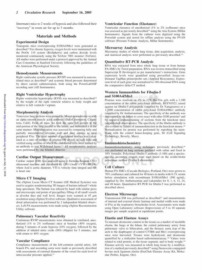
ResultsHemodynamic and Morphometric AssessmentsWe measured RVSP as an indication of pulmonary arterialpressure (PAP) and observed a mild elevation in the S100A4/Mts1 compared with C57Bl/6 mice under baseline room airconditions (P�0.01). Following 2 weeks of chronic hypoxia(10% oxygen), a greater elevation in RVSP was observed inthe S100A4/Mts1 compared with the C57Bl/6 control mice.Moreover, in contrast to C57Bl/6 mice, the elevated RVSP inthe S100A4/Mts1 mice did not regress even following returnto room air for 3 months (P�0.01) (Figure 1A). In keepingwith the elevation in RVSP, we also observed more severe rightventricular hypertrophy (RVH) in the S100A4/Mts1 versus
C57Bl/6 mice that was sustained during room air recovery onlyin the S100A4/Mts1mice (P�0.01) (Figure 1B).
Despite the more severe elevation in RVSP and RVH atbaseline and following hypoxia, S100A4/Mts1 and C57Bl/6mice showed a similar degree of muscularization of distalvessels at alveolar duct and alveolar wall level and a similarreduction in the number of peripheral arteries at alveolar ductand wall level, calculated relative to 100 alveoli. This featurewas confirmed using an antibody to von Willebrand factor tomark endothelial cells. However, consistent with the sus-tained elevation in RVSP in S100A4/Mts1 mice followingreturn to room air, there was persistent muscularization ofperipheral arteries (P�0.02) (Figure 1C), and a tendency fortheir number to remain reduced, compared with C57Bl/6mice, in which values returned to normal levels (Figure 1D).
We observed no qualitative difference in pulmonary arte-rial arborization in 3D reconstruction of micro-computedtomography (CT) imaging of barium-infused lung samplescomparing S100A4/Mts1 and control mice (Figure 2A and2B). However, measurements of lumen diameter along the
Figure 1. Pulmonary hypertension in S100A4/Mts1 mice. S100A4/Mts1 and control C57Bl/6 mice were exposed to hypoxia (10% oxy-gen) followed by room air “recovery” for 4 weeks and 3 months. RVSP measurements (A) and ratio of the weight of the RV to that ofLV plus septum (B) as an index of RVH. C, Representative photomicrograph of lung tissue stained by Movat pentachrome showing ahypoxia-induced increase in arterial muscularization (upper) and graphic representation of the presence and degree of muscularizationof alveolar duct Pas (lower). Arrows denote alveolar duct-associated arteries. D, Representative photomicrograph of lung tissue stainedby Movat pentachrome showing a hypoxia-induced decrease in arterial number (upper) and morphometric analysis of fully and partiallymuscularized alveolar duct and wall PAs relative to 100 alveoli (lower). Arrows denote alveolar duct and wall arteries. Bars representmean�SEM (A and B, n�12 to 20; C and D, n�8 to 12); *P�0.05 compared with C57Bl/6 normoxic mice; †P�0.05 compared withC57Bl/6 hypoxic mice; ‡P�0.05 compared with S100A4/Mts1 normoxic mice. (P values �0.05 are not distinguished to simplify theannotations.)
Merklinger et al Fibulin, Elastin, Mts1, and Pulmonary Hypertension 3
by guest on July 12, 2018http://circres.ahajournals.org/
Dow
nloaded from

length of the left PA revealed reduced values in the S100A4/Mts1 mice versus C57Bl/6 animals in normoxia (P�0.04)(Figure 2C, 2D, and 2E). A similar hypoxia-induced reduc-tion in lumen diameter was noted in both groups relative tonormoxia controls (P�0.04 and 0.02 respectively).
The increase in RVSP in the S100A4/Mts1 versus C57Bl/6mice at baseline was associated with a higher level of RVSPin response to acute hypoxia (10% oxygen for 5 minutes)(P�0.01), but a similar percent increase. However, S100A4/Mts1 mice showed no significant attenuation in RVSP inresponse to nitric oxide or 40% oxygen in contrast to theC57Bl/6 group where values returned to baseline levels(Figure 3A). There were no statistically significant differ-ences in the compliance of the branch pulmonary artery ofS100A4/Mts1 versus C57Bl/6 mice (Figure 3B) or in theother vessels studied (data not shown).
No significant difference in mean systemic arterial pres-sure was noted between the groups, and a comparablebaseline and hypoxia-induced elevation in hematocrit value
and reduction in body weight was observed. However,echocardiographic assessment revealed a reduced cardiacoutput (CO) in the S100A4/Mts1 versus C57Bl/6 mice both atbaseline (P�0.0001) and following chronic hypoxia(P�0.01) (supplemental Table I, available online at http://circres.ahajournals.com). These results suggested that therewas a greater elevation in pulmonary vascular resistance inthe S100A4/Mts1 compared with C57Bl/6 mice.
Consistent with the reduced CO in the S100A4/Mts1versus C57Bl/6 mice was a decrease in left ventricularfunction assessed by real-time continuous pressure-volumeanalysis of cardiac elastance14 (P�0.05) (Figure 3C through3E). End-diastolic pressure measurements were slightlyhigher in the S100A4/Mts1 versus C57Bl/6 mice (P�0.05)(supplemental Table II). Despite these indices of cardiacdysfunction that were measured under anesthesia, no frankevidence of heart failure judged by tachypnea or ascites, wasobserved in the S100A4/Mts1 mice at baseline or followinghypoxia, and trichrome staining of heart tissue sectionsshowed no increase in fibrosis relative to controls (data notshown).
Microarray Analysis of Lung Gene ExpressionDifferences in gene expression in S100A4/Mts1 versus con-trol C57Bl/6 mice were assessed to explain the more severePAH but relative attenuation of vascular disease duringhypoxia, as well as the impaired regression following returnto normoxia. There were 390 features exclusively upregulatedin hypoxic S100A4/Mts1 versus C57Bl/6 mice, 226 knowngenes and 164 expressed sequence tags or poorly annotatedfeatures. A relatively smaller number of known genes, 147,were significantly upregulated in S100A4/Mts1 versusC57Bl/6 mice under normoxic conditions. SupplementalTable III summarizes some of the genes that were upregu-lated in S100A4/Mts1 versus C57Bl/6 mice under hypoxicconditions (P�0.05 for all comparisons) and selected forfollow-up study for their functional relevance to vascularremodeling, eg, proteases, matrix components, growth andsurvival factors, or vasoreactive molecules. We were able toconfirm significant elevation of the transcript in 2 of the 6genes, and trends in 3 of the 6 by qRT-PCR. We alsoconfirmed significant suppression of one transcript, bonemorphogenetic protein receptor 1 (BMP-RI) by qRT-PCR(data not shown). We did not pursue the elevation in matrixmetalloproteinase 2 in S100A4/Mts1 versus control miceduring hypoxia, because the elevation in the activity of theenzyme by gelatin zymography was comparable in bothgroups in hypoxia relative to room air controls (data notshown).
Among the annotated genes that distinguished the S100A4/Mts1 from the C57Bl/6 mouse, only one, fibulin-5 (DANCEor EVEC), was significantly increased on microarray, bothunder normoxia and with chronic hypoxia. Fibulin-5, acalcium-dependent elastin-binding protein, is a necessarycomponent of normal elastin fiber assembly.16,20 We specu-lated that an increase in fibulin-5, could cause deposition ofmore elastin, altering the hemodynamic characteristics of thepulmonary vessel wall and also limit remodeling, particularlySMC proliferation.21,22
Figure 2. Micro CT imaging of barium-filled pulmonary arteries.Representative photomicrographs of micro CT images of wholelungs infused with barium gelatin from a C57Bl/6 (A) or aS100A4/Mts1 (B) mouse exposed to 2 weeks of hypoxia. 3Dreconstructions of the pulmonary arterial trees were obtainedusing a threshold of 7000 arbitrary digital units. C and D,Images captured at a less sensitive threshold (14 000 arbitrarydigital units). E, Direct diameter measurements along the lengthof the left PA from hilum to periphery in S100A4/Mts1 vsC57Bl/6 mice at baseline and after hypoxia. Diameters wereobtained using the GE eXplore Reconstruction software. Barsrepresent mean�SEM (n�6); *P�0.04 compared with C57Bl/6normoxic mice; †P�0.02 compared with Mts1 normoxic mice.
4 Circulation Research September 16, 2005
by guest on July 12, 2018http://circres.ahajournals.org/
Dow
nloaded from

Fibulin-5 and ElastinAnalysis by qRT-PCR indicated only a trend toward height-ened fibulin-5 mRNA expression in the lung at baseline andfollowing hypoxia in S100A4/Mts1 versus C57Bl/6 controlmice (Figure 4A). Densitometric analyses of the Westernimmunoblots of whole lung tissue, revealed a trend towardhigher levels of fibulin-5 in S100A4/Mts1 versus C57Bl/6mice in normoxia, but a significant difference between thegroups in hypoxia (P�0. 03) (Figure 4B). Although the dataare not quantitative, we were able to localize heightened
immunoreactivity for fibulin-5 to the PA muscular media asa consistent feature in vessels of different airway levels inS100A4/Mts1 versus C57Bl/6 mice in normoxia. Hypoxia perse appeared to induce a slight increase in fibulin-5 immuno-reactivity, a feature accentuated in the S100A4/Mts1 mice(Figure 4C). To determine whether S100A4/Mts1 couldinduce expression of fibulin-5, we incubated human PA-SMCs with recombinant S100A4/Mts1 for 48 hours anddetected elevated fibulin-5 mRNA levels by qRT-PCR with apeak at 24 hours (P�0.05) (Figure 4D).
Figure 3. Vasoreactivity, arterial compliance ventricular elastance. A, RVSP measurements in S100A4/Mts1 vs C57Bl/6 mice at baseline(40% oxygen), during acute hypoxia (10% oxygen), hypoxia plus NO (40 ppm), and a return to 40% oxygen. B, Compliance measure-ments of the large arteries. Pressure-volume loops were made in C57Bl/6 (C) and S100A4/Mts1 (D) mice during baseline normoxic con-ditions. E, Maximum left ventricular elastance derived from the pressure-volume loop analysis was decreased in S100A4/Mts1 micecompared with C57Bl/6 animals. Bars represent mean�SEM, (A, C through E, n�8; B, n�6); *P�0.05 compared with C57Bl/6 nor-moxic mice; †P�0.05 compared with C57Bl/6 hypoxic mice; ‡P�0.05 compared with S100A4/Mts1 normoxic mice. (P values �0.05are not distinguished to simplify the annotations.)
Merklinger et al Fibulin, Elastin, Mts1, and Pulmonary Hypertension 5
by guest on July 12, 2018http://circres.ahajournals.org/
Dow
nloaded from

The elevation in fibulin-5 in S100A4/Mts1 versus C57Bl/6mice, at baseline and following exposure to chronic hypoxia,was associated with increased thickness of the internal(Figure 5B) and external (Figure 5C) elastic laminae, asassessed by transmission EM in arteries at the level of therespiratory bronchiolus (P�0.05 for all comparisons). More-over, an increase in medial width, associated with hypertro-phy of the SMCs, was also noted in the hypoxic S100A4/Mts1 relative to control mice (P�0.05) (Figure 5D). Incontrast to previous reports,23 we did not observe fragmen-tation of elastin with hypoxia, but this might reflect the levelof vessel being assessed. In addition, in room air mice, wemeasured total lung elastin by desmosine radioimmunoassayand values were increased in S100A4/Mts1 versus theC57Bl/6 group (P�0.001) (Figure 5E), when normalized tobody weight. Normalization of elastin in the central pulmo-nary artery or in the aorta to tissue segment rather than to totalprotein determines an absolute increase in elastin unobscuredby elevation of other proteins in a hypertrophied organ,19 butdifferences were not significant when normalizing the elastinto segment or to total protein (data not shown).
We determined whether the increased elastin could also beattributed to suppression of vascular elastase activity. Com-pared with a persistent steady-state lung elastase activity incontrol room air mice, an increase in elastase activity wasnoted in the C57Bl/6 mice after 2 days of hypoxia (P�0.02),that returned to control values by day 14 (Figure 5F). Nosignificant increase in elastase activity relative to baselinewas evident in S100A4/Mts1 mice after 2 days of hypoxia,but values were also not significantly different from C57Bl/6mice at this time point. At 14 days after hypoxia, values inS100A4/Mts1 mice were reduced relative to C57Bl/6 mice(P�0.01).
DiscussionS100A4/Mts1 mice had greater RVSP and RVH at baseline,in response to acute and chronic hypoxia, and after “recov-ery” in room air when compared with C57Bl/6 controls.Pulmonary vascular changes were, however, similar in bothgroups in room air and after chronic hypoxia, but regressionwas attenuated in the S100A4/Mts1 mice. We related thesefeatures to reduced cardiac function and to increased fibulin-5
Figure 4. Fibulin-5 expression in S100A4/Mts1 mice and cultured pulmonary artery smooth muscle cells. A, Quantitative RT-PCR forfibulin-5mRNA in RNA from whole lung tissue of S100A4/Mts1 and C57Bl/6 mice exposed to room air or to 2 weeks of hypoxia.Expression levels of each gene were normalized to 18S ribosomal RNA and expressed as a fold change in gene expression comparedwith the control C57Bl/6 normoxic animals. B, Representative Western immunoblot showing expression of fibulin-5 protein in wholelung tissues of S100A4/Mts1 vs C57Bl/6 mice exposed to room air or hypoxia (top) and densitometric analysis of fibulin-5 protein, nor-malized to the internal housekeeping gene, S6, is shown in the bar graph (bottom). C, Representative photomicrographs of paraffin-embedded mouse lung tissue sections after immunoperoxidase staining for fibulin-5. Strong positive staining was noted particularly inthe media of pulmonary arteries (arrows) of S100A4/Mts1 mice at baseline and after chronic hypoxia. D, Quantitative RT-PCR forfibulin-5mRNA in RNA extracted from human PA-SMCs stimulated with recombinant S100A4/Mts1 at indicated time points. Data repre-sent mean�SEM, (A, C, and D, n�4; B, n�3); †P�0.05 compared with all groups. *P�0.03 vs C57BLl/6 hypoxia.
6 Circulation Research September 16, 2005
by guest on July 12, 2018http://circres.ahajournals.org/
Dow
nloaded from

and thicker elastic laminae, that could alter vascular tone,attenuate the severity of the vascular changes21,22 expected forthe level of pulmonary artery pressure and resistance,24 andimpair reversibility.
Our studies underscore the importance of considering theimpact of altered cardiac function, on the pulmonary hyper-tensive phenotype. In severe PAH, the dilated RV can lead toimpaired LV filling and reduced CO.25 In the S100A4/Mts1mice, a reduced CO accompanied a very mild elevation inRVSP. Thus, we pursued a load-independent assessment ofcardiac function and documented reduced ventricular elas-tance through pressure-volume curves. The S100 family ofcalcium-binding proteins, are expressed in cardiac myo-
cytes,26 and although S100A4/Mts1 and S100A11 are ele-vated in response to myocardial injury,27 their selectiveimpact on cardiac function is not known. Although themeasurements of left ventricular dysfunction were madeunder anesthesia, all mice were similarly treated. In anattempt to further evaluate the cause of the impaired LVfunction, we assessed elastin in the aorta, but found nosignificant differences in S100A4/Mts1 versus control mice.
Although measurements were not made at the same time,the mildly elevated left ventricular end-diastolic pressurederived from the pressure volume loops might explain theincrease in RVSP in the S100A4/Mts1 mice at baseline and inresponse to acute and chronic hypoxia. However, this would
Figure 5. Elastin quantification, medial hypertrophy, and elastase activity. A, Representative electron photomicrographs of S100A4/Mts1 and C57Bl/6 PAs at the respiratory bronchiolus level (16 to 18 �m) as seen by transmission EM, at �10 0000 magnification.Computed width of both the internal (B) and external (C) elastic laminae at baseline and in association with chronic hypoxia. D, Mea-surement of medial wall thickness as seen by EM. E, Desmosines were measured in whole lung of room air S100A4/Mts1 and C57Bl/6mice and normalized per weight of the animal. F, Elastase activity in S100A4/Mts1 and C57Bl/6 mice exposed to room air or hypoxia atindicated time points. Elastase activity was assayed in triplicate from whole lung tissue using fluorogenic elastin. Data representmean�SEM, (A through D, n�3; E, n�6; F, n�4 to 6); *P�0.05 compared with C57Bl/6 normoxic mice; †P�0.05 compared withC57Bl/6 hypoxic mice; ‡P�0.05 compared with S100A4/Mts1 normoxic mice; #P�0.001 compared with C57Bl/6 mice.
Merklinger et al Fibulin, Elastin, Mts1, and Pulmonary Hypertension 7
by guest on July 12, 2018http://circres.ahajournals.org/
Dow
nloaded from

be unlikely to account for the lack of vasodilatation inresponse to nitric oxide or oxygen28 and the failure to returnto baseline levels of RVSP despite 3 months of “recovery” inroom air. Thus we turned our attention to features that mightexplain a difference in vascular tone as well as attenuatedremodeling relative to that expected for the elevation in PApressure and estimated pulmonary vascular resistance.24
Normal assembly of elastin fibers prevents SMC prolifer-ation,22 and impaired elastin fiber assembly promotes thisresponse.21 In transgenic mice lacking the elastin gene, excessproliferation of SMCs was identified as causing occlusion ofcoronary arteries resulting in the animals’ demise in theimmediate postnatal period.29 Along the same lines, ourgroup has shown that there is degradation of elastin in thepathogenesis of pulmonary vascular disease,19 and that elas-tase inhibitors can suppress smooth muscle cell proliferation,and associated pulmonary hypertension.10,12,30 Thick elasticlaminae, documented by electron microscopy, were associ-ated with elevated desmosine levels S100A4Mts1 versuscontrol mice even in room air, and reduced elastase activitywas documented in chronic hypoxia. It is interesting that thethick elastic laminae still permit hypertrophy of the SMCsduring hypoxia, likely in response to the elevated pressure.Although these SMC were immunoreactive for alpha-smoothmuscle actin, it would be interesting to assess other markersof differentiation. We could not document an increase inelastin in the large central pulmonary artery nor could we finda change in compliance in those vessels, suggesting that thefeatures are limited to intraparenchymal vessels. Although theincrease in elastin could have been related to airways in thelung tissue, this was not evident on microscopic analysisusing a stain that recognizes elastin.
We speculate that in the S100A4/Mts1 mice, the increasein elastin limits the extent of abnormal muscularization ofdistal PAs,24 by suppressing pericyte proliferation and differ-entiation to SMCs. Fibulin-5 stabilization of the elastic fibers,through its ability to promote cell attachment via cell surfaceintegrins.31 might also prevent endothelial cell apoptosis,accounting for reduced loss of vessels32 relative to theelevation in pressure.24 This feature could also repress smoothmuscle apoptosis and regression of muscularization of distalvessels following return to room air. Only a tendency towardimpaired “recovery” of the number of peripheral vessels wasapparent in the S100A4/Mts1 compared with C57Bl/6 mice,but it is possible that the antiproliferative effect of elastinmight also suppress the endothelial cell proliferation neces-sary for vascular regrowth. Lack of regression may, however,simply be a feature of failure of vasodilatation on return toroom air.33 We did not assess capillary surface area which isincreased in hypoxia,34 but our findings of loss of peripheralarteries is similar to that noted by other groups.23
Although there may be a link between lack of reversibilityof vascular disease, and the propensity of a small subset ofolder S100A4/Mts1 mice to develop PVD,5 we were unableto show that a “second hit” with chronic hypoxia inducedthese lesions (data not shown). It is possible that a differentkind of insult or some property of aging is required.
Despite the reduced lumen diameter of the intrapulmonaryarteries on microCT and the lack of vasodilation in response
to NO or oxygen, we were unable to document that theincrease in elastin correlated with a change in compliance ofthe large central PAs. A mouse hemizygous for elastin alsoshowed normal compliance of these vessels,29 so evaluationof the intrapulmonary arteries may be necessary to detectabnormalities.
Microarray studies also revealed genes that, in addition tofibulin-5, might be of further interest in explaining howS100A4/Mts1 could induce PVOD in the small subset ofmice previously described.5 For example, we observed adecrease in BMP-RI expression, in keeping with recentobservations in lungs of patients with acquired idiopathicPAH.35
It is interesting that attenuation of vascular disease relativeto the severity of pulmonary hypertension, is observed in thetransgenic mouse with dominant negative3 or haplo insuffi-ciency BMP-RII.4 Asymptomatic carriers of the idiopathicPAH disease haplotype (mutation in BMP-RII), have abnor-mally elevated PA systolic pressures in response to exer-cise,36 but the structure of their pulmonary vessels isunknown.
In conclusion, our studies underscore the complexity ofdetermining how environmental and genetic factors ulti-mately lead to the development of PVD. The low penetranceof BMP-RII abnormalities in producing severe disease,1,2
may be related to our observation that altered expression of agene can trigger a compensatory mechanism. It is, perhaps,through understanding why these compensatory mechanismsfail, that we will identify critical risk factors and modifiergenes that can be targeted in developing new approaches totreatment.
AcknowledgmentsSupported by grant number 1-R01-HL074186-01 from the NationalInstitutes of Health and the Dwight and Vera Dunlevie EndowedProfessorship to M.R. S.M. was supported in part by a University ofToronto Fellowship. E.S. was supported by a fellowship from theAmerican Heart Association/Pulmonary Hypertension Association.
References1. Lane KB, Machado RD, Pauciulo MW, Thomson JR, Phillips JA 3rd,
Loyd JE, Nichols WC, Trembath RC. Heterozygous germline mutationsin BMPR2, encoding a TGF-beta receptor, cause familial primary pul-monary hypertension. The International PPH Consortium. NatGenet. 2000;26:81–84.
2. Deng Z, Morse JH, Slager SL, Cuervo N, Moore KJ, Venetos G,Kalachikov S, Cayanis E, Fischer SG, Barst RJ, Hodge SE, Knowles JA.Familial primary pulmonary hypertension (gene PPH1) is caused bymutations in the bone morphogenetic protein receptor-II gene. Am J HumGenet. 2000;67:737–744.
3. West J, Fagan K, Steudel W, Fouty B, Lane K, Harral J, Hoedt-Miller M,Tada Y, Ozimek J, Tuder R, Rodman DM. Pulmonary hypertension intransgenic mice expressing a dominant-negative BMPRII gene in smoothmuscle. Circ Res. 2004;94:1109–1114.
4. Beppu H, Ichinose F, Kawai N, Jones RC, Yu PB, Zapol WM, MiyazonoK, Li E, Bloch KD. BMPR-II heterozygous mice have mild pulmonaryhypertension and an impaired pulmonary vascular remodeling response toprolonged hypoxia. Am J Physiol Lung Cell Mol Physiol. 2004;287:L1241–L1247.
5. Greenway S, van Suylen RJ, Du Marchie Sarvaas G, Kwan E, Ambart-sumian N, Lukanidin E, Rabinovitch M. S100A4/Mts1 produces murinepulmonary artery changes resembling plexogenic arteriopathy and isincreased in human plexogenic arteriopathy. Am J Pathol. 2004;164:253–262.
8 Circulation Research September 16, 2005
by guest on July 12, 2018http://circres.ahajournals.org/
Dow
nloaded from

6. Albertazzi E, Cajone F, Leone BE, Naguib RN, Lakshmi MS, SherbetGV. Expression of metastasis-associated genes h-mts1 (S100A4) andnm23 in carcinoma of breast is related to disease progression. DNA CellBiol. 1998;17:335–342.
7. Kriajevska M, Tarabykina S, Bronstein I, Maitland N, Lomonosov M,Hansen K, Georgiev G, Lukanidin E. Metastasis-associated Mts1(S100A4) protein modulates protein kinase C phosphorylation of theheavy chain of nonmuscle myosin. J Biol Chem. 1998;273:9852–9856.
8. Ambartsumian N, Klingelhofer J, Grigorian M, Christensen C, KriajevskaM, Tulchinsky E, Georgiev G, Berezin V, Bock E, Rygaard J, Cao R, CaoY, Lukanidin E. The metastasis-associated Mts1(S100A4) protein couldact as an angiogenic factor. Oncogene. 2001;20:4685–4695.
9. Ambartsumian N, Klingelhofer J, Grigorian M, Karlstrom O, Sidenius N,Georgiev G, Lukanidin E. Tissue-specific posttranscriptional downregu-lation of expression of the S100A4(mts1) gene in transgenic animals.Invasion Metastasis. 1998;18:96–104.
10. Zaidi SH, You XM, Ciura S, Husain M, Rabinovitch M. Overexpressionof the serine elastase inhibitor elafin protects transgenic mice fromhypoxic pulmonary hypertension. Circulation. 2002;105:516–521.
11. Rabinovitch M, Andrew M, Thom H, Trusler GA, Williams WG, RoweRD, Olley PD. Abnormal endothelial factor VIII associated with pulmo-nary hypertension and congenital heart defects. Circulation. 1987;76:1043–1052.
12. Ye CL, Rabinovitch M. Inhibition of elastolysis by SC-37698 reducesdevelopment and progression of monocrotaline pulmonary hypertension.Am J Physiol. 1991;261:H1255–H1267.
13. Faury G, Pezet M, Knutsen RH, Boyle WA, Heximer SP, McLean SE,Minkes RK, Blumer KJ, Kovacs A, Kelly DP, Li DY, Starcher B,Mecham RP. Developmental adaptation of the mouse cardiovascularsystem to elastin haploinsufficiency. J Clin Invest. 2003;112:1419–1428.
14. Georgakopoulos D, Mitzner WA, Chen CH, Byrne BJ, Millar HD, HareJM, Kass DA. In vivo murine left ventricular pressure-volume relationsby miniaturized conductance micromanometry. Am J Physiol. 1998;274:H1416–H1422.
15. Tabibiazar R, Wagner RA, Liao A, Quertermous T. Transcriptional pro-filing of the heart reveals chamber-specific gene expression patterns. CircRes. 2003;93:1193–1201.
16. Yanagisawa H, Davis EC, Starcher BC, Ouchi T, Yanagisawa M, Rich-ardson JA, Olson EN. Fibulin-5 is an elastin-binding protein essential forelastic fibre development in vivo. Nature. 2002;415:168–171.
17. Hinek A, Mecham RP, Keeley F, Rabinovitch M. Impaired elastin fiberassembly related to reduced 67-kD elastin-binding protein in fetal lambductus arteriosus and in cultured aortic smooth muscle cells treated withchondroitin sulfate. J Clin Invest. 1991;88:2083–2094.
18. Starcher B. A ninhydrin-based assay to quantitate the total protein contentof tissue samples. Anal Biochem. 2001;292:125–129.
19. Todorovich-Hunter L, Dodo H, Ye C, McCready L, Keeley FW, Rabin-ovitch M. Increased pulmonary artery elastolytic activity in adult rats withmonocrotaline-induced progressive hypertensive pulmonary vasculardisease compared with infant rats with nonprogressive disease. Am RevRespir Dis. 1992;146:213–223.
20. Nakamura T, Lozano PR, Ikeda Y, Iwanaga Y, Hinek A, Minamisawa S,Cheng CF, Kobuke K, Dalton N, Takada Y, Tashiro K, Ross J Jr, HonjoT, Chien KR. Fibulin-5/DANCE is essential for elastogenesis in vivo.Nature. 2002;415:171–175.
21. Urban Z, Riazi S, Seidl TL, Katahira J, Smoot LB, Chitayat D, Boyd CD,Hinek A. Connection between elastin haploinsufficiency and increasedcell proliferation in patients with supravalvular aortic stenosis andWilliams-Beuren syndrome. Am J Hum Genet. 2002;71:30–44.
22. Karnik SK, Brooke BS, Bayes-Genis A, Sorensen L, Wythe JD, SchwartzRS, Keating MT, Li DY. A critical role for elastin signaling in vascularmorphogenesis and disease. Development. 2003;130:411–423.
23. Levi M, Moons L, Bouche A, Shapiro SD, Collen D, Carmeliet P.Deficiency of urokinase-type plasminogen activator-mediated plasmingeneration impairs vascular remodeling during hypoxia-induced pulmo-nary hypertension in mice. Circulation. 2001;103:2014–2020.
24. Rabinovitch M, Gamble W, Nadas AS, Miettinen OS, Reid L. Ratpulmonary circulation after chronic hypoxia: hemodynamic and structuralfeatures. Am J Physiol. 1979;236:H818–H827.
25. Marcus JT, Vonk Noordegraaf A, Roeleveld RJ, Postmus PE, HeethaarRM, Van Rossum AC, Boonstra A. Impaired left ventricular filling due toright ventricular pressure overload in primary pulmonary hypertension:noninvasive monitoring using MRI. Chest. 2001;119:1761–1765.
26. Tsoporis JN, Marks A, Zimmer DB, McMahon C, Parker TG. Themyocardial protein S100A1 plays a role in the maintenance of normalgene expression in the adult heart. Mol Cell Biochem. 2003;242:27–33.
27. Inamoto S, Murao S, Yokoyama M, Kitazawa S, Maeda S. Isoproterenol-induced myocardial injury resulting in altered S100A4 and S100A11protein expression in the rat. Pathol Int. 2000;50:480–485.
28. Adatia I, Perry S, Landzberg M, Moore P, Thompson JE, Wessel DL.Inhaled nitric oxide and hemodynamic evaluation of patients with pul-monary hypertension before transplantation. J Am Coll Cardiol. 1995;25:1656–1664.
29. Li DY, Brooke B, Davis EC, Mecham RP, Sorensen LK, Boak BB,Eichwald E, Keating MT. Elastin is an essential determinant of arterialmorphogenesis. Nature. 1998;393:276–280.
30. Maruyama K, Ye CL, Woo M, Venkatacharya H, Lines LD, Silver MM,Rabinovitch M. Chronic hypoxic pulmonary hypertension in rats andincreased elastolytic activity. Am J Physiol. 1991;261:H1716–H1726.
31. Nakamura T, Ruiz-Lozano P, Lindner V, Yabe D, Taniwaki M, FurukawaY, Kobuke K, Tashiro K, Lu Z, Andon NL, Schaub R, Matsumori A,Sasayama S, Chien KR, Honjo T. DANCE, a novel secreted RGD proteinexpressed in developing, atherosclerotic, and balloon-injured arteries.J Biol Chem. 1999;274:22476–22483.
32. Zhao YD, Campbell AI, Robb M, Ng D, Stewart DJ. Protective role ofangiopoietin-1 in experimental pulmonary hypertension. Circ Res. 2003;92:984–991.
33. O’Blenes SB, Fischer S, McIntyre B, Keshavjee S, Rabinovitch M.Hemodynamic unloading leads to regression of pulmonary vasculardisease in rats. J Thorac Cardiovasc Surg. 2001;121:279–289.
34. Howell K, Preston RJ, McLoughlin P. Chronic hypoxia causes angio-genesis in addition to remodelling in the adult rat pulmonary circulation.J Physiol. 2003;547:133–145.
35. Du L, Sullivan CC, Chu D, Cho AJ, Kido M, Wolf PL, Yuan JX, DeutschR, Jamieson SW, Thistlethwaite PA. Signaling molecules in nonfamilialpulmonary hypertension. N Engl J Med. 2003;348:500–509.
36. Grunig E, Janssen B, Mereles D, Barth U, Borst MM, Vogt IR, Fischer C,Olschewski H, Kuecherer HF, Kubler W. Abnormal pulmonary arterypressure response in asymptomatic carriers of primary pulmonary hyper-tension gene. Circulation. 2000;102:1145–1150.
Merklinger et al Fibulin, Elastin, Mts1, and Pulmonary Hypertension 9
by guest on July 12, 2018http://circres.ahajournals.org/
Dow
nloaded from

Barry Starcher, Hiromi Yanagisawa and Marlene RabinovitchS. Ambartsumian, Eugene Lukanidin, Daniel Bernstein, Mansoor Husain, Robert P. Mecham,
Knutsen, M. Golam Kabir, Kavin Desai, Shelby Hacker, Lingli Wang, Gordon M. Cann, Noona Sandra L. Merklinger, Roger A. Wagner, Edda Spiekerkoetter, Aleksander Hinek, Russell H.Increased Fibulin-5 and Elastin in S100A4/Mts1 Mice With Pulmonary Hypertension
Print ISSN: 0009-7330. Online ISSN: 1524-4571 Copyright © 2005 American Heart Association, Inc. All rights reserved.is published by the American Heart Association, 7272 Greenville Avenue, Dallas, TX 75231Circulation Research
published online August 18, 2005;Circ Res.
http://circres.ahajournals.org/content/early/2005/08/18/01.RES.0000182425.49768.8a.citationWorld Wide Web at:
The online version of this article, along with updated information and services, is located on the
http://circres.ahajournals.org/content/suppl/2005/08/18/01.RES.0000182425.49768.8a.DC1Data Supplement (unedited) at:
http://circres.ahajournals.org//subscriptions/
is online at: Circulation Research Information about subscribing to Subscriptions:
http://www.lww.com/reprints Information about reprints can be found online at: Reprints:
document. Permissions and Rights Question and Answer about this process is available in the
located, click Request Permissions in the middle column of the Web page under Services. Further informationEditorial Office. Once the online version of the published article for which permission is being requested is
can be obtained via RightsLink, a service of the Copyright Clearance Center, not theCirculation Researchin Requests for permissions to reproduce figures, tables, or portions of articles originally publishedPermissions:
by guest on July 12, 2018http://circres.ahajournals.org/
Dow
nloaded from

Online Data Supplement Merklinger et al.
Online Table 1
Final body weight, mean arterial pressure, hematocrit and cardiac output measurements
FBW (g)
MAP
(mmHg)
Hematocrit
(%)
Cardiac Output
(ml/min)
Normoxia
C57Bl/6
24±1.8
74.8±2.9
42.2±0.4
17.5±2.6
S100A4/Mts1 24.5±1.1 70±3.7 43.3±0.5 12±2.0*
Hypoxia
C57Bl/6
20.4±1.2
63±2.1*
61.3±0.4*
22.1±2.5
S100A4/Mts1 17.6±1.1‡ 64±2.6‡ 62.6±1.3* 14±0.8†
FBW indicates final body weight. MAP indicates mean arterial blood pressure.
*P<0.0001 compared to C57Bl/6 normoxia; †P<0.01 compared to C57Bl/6 hypoxia; ‡P<0.002
compared to S100A4/Mts1 normoxia.

Online Data Supplement Merklinger et al.
1
Online Table 2
Heart rate, end-diastolic pressure and dP/dt obtained from pressure/volume loops
Heart Rate
End-Diastolic Pressure,
(mmHg)
dP/dtmax
(mmHg/s)
dP/dtmin
(mmHg/s)
C57Bl/6
344±11.1
3.9±0.2
6591±428
-4884±485
S100A4/Mts1
356±13.3
7.7±1.6*
5428±554
-4792±606
*P<0.05 compared to C57Bl/6 mice.

Online Data Supplement Merklinger et al.
2
Online Table 3
Selected microarray data for S100A4/Mts1 vs. Control C57Bl/6 mice after chronic hypoxia
Function Elevated Gene Fold Increase
Proteases Matrix metalloproteinase 2 1.58
Matrix * Fibulin-5 1.21
Growth ** Tumor necrosis factor-α 1.31
** Stromal-derived factor 1 1.41
* Ephrin receptor B6 1.35
Vasoreactive * Endothelin 3 1.11
† Prostaglandin Synthase 1.23
*Trend by qRT-PCR, **Significant by qRT-PCR (P<0.05),
†Not confirmed by qRT-PCR.


![S100A4 over-expression underlies lymph node metastasis and ...€¦ · ing Manual (6th edition, 2002)[18]. For the screening and confirmation study, 32 primary CRC tissue samples](https://img.pdfslide.us/doc/110x75/5fbc593ae950797b7b2bee54/s100a4-over-expression-underlies-lymph-node-metastasis-and-ing-manual-6th-edition.jpg)