Embed Size (px)
Citation preview

In vivo imaging of alphaherpesvirus infection revealssynchronized activity dependent on axonal sortingof viral proteinsAndrea E. Granstedta,b, Jens B. Bossea,b, Stephan Y. Thibergeb,c, and Lynn W. Enquista,b,1
aDepartment of Molecular Biology, bPrinceton Neuroscience Institute, and cLewis-Sigler Institute for Integrative Genomics, Princeton University, Princeton,NJ 08544
Edited by Elliott Kieff, Harvard Medical School and Brigham and Women’s Hospital, Boston, MA, and approved August 2, 2013 (received for reviewJune 22, 2013)
A clinical hallmark of human alphaherpesvirus infections isperipheral pain or itching. Pseudorabies virus (PRV), a broad hostrange alphaherpesvirus, causes violent pruritus in many differentanimals, but the mechanism is unknown. Previous in vitro studieshave shown that infected, cultured peripheral nervous system(PNS) neurons exhibited aberrant electrical activity after PRVinfection due to the action of viral membrane fusion proteins,yet it is unclear if such activity occurs in infected PNS ganglia inliving animals and if it correlates with disease symptoms. Usingtwo-photon microscopy, we imaged autonomic ganglia in livingmice infected with PRV strains expressing GCaMP3, a geneticallyencoded calcium indicator, and used the changes in calcium flux tomonitor the activity of many neurons simultaneously with single-cell resolution. Infection with virulent PRV caused these PNSneurons to fire synchronously and cyclically in highly correlatedpatterns among infected neurons. This activity persisted evenwhen we severed the presynaptic axons, showing that infection-induced firing is independent of input from presynaptic brainstemneurons. This activity was not observed after infections with anattenuated PRV recombinant used for circuit tracing or with PRVmutants lacking either viral glycoprotein B, required for membranefusion, or viral membrane protein Us9, required for sorting virionsand viral glycoproteins into axons. We propose that the viral fusionproteins produced by virulent PRV infection induce electrical cou-pling in unmyelinated axons in vivo. This action would then giverise to the synchronous and cyclical activity in the ganglia and con-tribute to the characteristic peripheral neuropathy.
Alphaherpesviruses are pathogens of the mammalian nervoussystem that often evoke manifestations of peripheral pain or
itching after infection. The severity and extent of symptoms varydepending on the herpesvirus strain and the host. Herpes sim-plex virus (HSV) and varicella-zoster virus (VZV) infections inhumans characteristically cause lesions that are preceded bysensations of localized pain, which also sometimes lasts longafter lesions have been cleared (1, 2). Pseudorabies virus (PRV)is a swine alphaherpesvirus that is related to HSV and VZV.Whereas in its natural porcine host PRV establishes latency inthe peripheral sensory ganglia, in nonnative animal hosts, in-cluding laboratory mice, PRV causes a severe acute peripheralneuropathy called the “mad itch” (3). This neuropathy is char-acterized by a strong impulse to bite and scratch the skin of theinfected dermatome feverishly and incessantly, which results insevere self-inflicted wounds (4). PRV has proved useful forelucidating pathways involved in the manifestation of theseclinical symptoms. Specific PRV gene products have been iden-tified as virulence factors and these research efforts have led tosuccessful live vaccines, such as the strain PRV Bartha and itsderivatives (see ref. 5 for review). Despite this effort, the mo-lecular basis for the pronounced peripheral neuropathies pro-duced by PRV and the other alpha herpesviruses remain an activearea of research.
In the 1950s, Dempsher and colleagues recorded spontaneousbursts of neuronal activity in rat sympathetic superior cervicalganglia (SCG) infected with virulent PRV and the onset corre-lated with the mad itch symptomology (6). Other in vivo studiesshowed that the activity induced by PRV infection was reversiblyblocked by curare and was cholinergic in origin (7, 8). De-nervation of the preganglionic nerve before infection did notaffect mad itch symptoms, yet no spontaneous electrical dis-charges were recorded in the ganglia (8). These studies suggestedthat increased infection-induced electrical activity originated inthe presynaptic terminals within the ganglia (8, 9). A recentpublication examined the electrical activity of homogenous cul-tures of peripheral nervous system (PNS) neurons from the SCGinfected with virulent or attenuated PRV strains in vitro usingpatch clamp recordings (10). They found that neurons infectedwith a virulent PRV strain had increased and sustained rates ofaction potential firing late in infection that were synchronizedamong the neuronal population. Infection by the attenuatedPRV strain Bartha exhibited a marked delay in the onset ofabnormal firing. Importantly, these studies showed that the PRVmembrane fusion protein glycoprotein B (gB) was required forelevated firing rates and fusion of infected neuron cell bodies.The authors concluded that fusion pores were formed duringPRV infection, which enabled ions to flow between neurons tocause direct electrical coupling (10). As the pores increased insize, spontaneous action potentials spread into coupled neuronsand throughout the network, causing firing events to synchronize
Significance
Pseudorabies virus (PRV), an alphaherpesvirus related to hu-man herpes simplex and varicella-zoster viruses, causes thecharacteristic “mad itch” in which infected animals scratch anafflicted area until self-mutilation. The mechanism by whichPRV induces these symptoms is still poorly understood, andmost work has been done in limited systems in vitro. Here wereport studies performed in living mice. We observed that in-fection with a virulent PRV strain induces synchronous andcyclical neural activity, but this activity disappears during in-fection with a vaccine strain that does not cause mad itch. Wedemonstrate the aberrant activity depends on a viral fusionprotein, and its trafficking specifically into axons. These find-ings correlate the disrupted physiology to the known neurop-athy in vivo.
Author contributions: A.E.G. and L.W.E. designed research; A.E.G., J.B.B., and S.Y.T. per-formed research; A.E.G. and J.B.B. contributed new reagents/analytic tools; A.E.G., J.B.B.,and S.Y.T. analyzed data; and A.E.G., J.B.B., S.Y.T., and L.W.E. wrote the paper.
The authors declare no conflict of interest.
This article is a PNAS Direct Submission.1To whom correspondence should be addressed. E-mail: [email protected].
This article contains supporting information online at www.pnas.org/lookup/suppl/doi:10.1073/pnas.1311062110/-/DCSupplemental.
E3516–E3525 | PNAS | Published online August 26, 2013 www.pnas.org/cgi/doi/10.1073/pnas.1311062110
and resonate in the culture. This synchronous firing phenomenonalso manifests itself as a continuous and rapid calcium flux ininfected cultured neurons that can lead to other dramatic changes,including changes in mitochondrial shape and dynamics (11).Therefore, the formations of viral-induced excitatory synapticconnections that allow for continuous propagation of actionpotentials do not reflect normal chemical transmission at conven-tional synapses, but rather an abnormal coupling that requires viralDNA replication and that can be strain dependent (12, 13).Whereas the results with cultured neurons suggested a mech-
anism by which PRV may induce neuropathy, the relativelysimplified in vitro system still leaves unanswered questions aboutwhat happens during infection of animals. In particular, studiesusing the mouse flank scarification model showed that virulentand attenuated PRV strains, such as PRV Bartha, have twodifferent mechanisms of neuroinvasion and lethality in mice (4).Specifically, the virulent strain induces the classic symptomologyof violent pruritus with rapid onset of death, little viral antigen inthe brain, and no signs of CNS pathology. In contrast, miceinfected with PRV Bartha survive much longer, do not exhibitany peripheral neuropathy, have abundant infectious virus in thebrain, but do present CNS abnormalities at very late times afterinfection and still succumb (4). Therefore, it appears that duringinfection with virulent PRV strains, but not attenuated PRVBartha strains, the infected peripheral neural circuits are theprimary players in determining the severe clinical manifestations.Our previous study in living mice showed that a PRV Bartharecombinant, expressing a genetically encoded calcium indicator,did not induce synchronous activity among neurons in an auto-nomic ganglia (14), However, we did not examine neuronal ac-tivity after virulent PRV infection. The differences between invitro and in vivo infections are obvious, such as the organizationof innervated tissues, structure of ganglia, and the involvement ofglial cells, but their impact on infection and symptomology afterinfection with virulent or attenuated PRV strains is not clear.In this report, we examined neuronal activity during a virulent
PRV infection of anesthetized mice by imaging infected PNSganglia directly in vivo. To do so, we constructed virulent andmutant PRV recombinants that expressed the genetically enco-ded fluorescent calcium indicator protein GCaMP3 (15). Thiscalcium sensor fluoresces in the presence of calcium transientsand is therefore a useful correlate of neuronal activity (16).Calcium imaging enabled us to monitor the activity in vivo ofmultiple infected neurons simultaneously with single-cell reso-lution using two-photon microscopy. Interestingly, 2 d after in-fection of the salivary glands by virulent PRV, infected neuronsin the submandibular ganglia (SMG) flashed synchronously andcyclically. There were no qualitative signs of fusion betweensoma in the ganglia. When we severed the axons between thebrainstem and the ganglia in vivo and continued imaging, theflashing phenotype continued unabated, suggesting that the im-petus to fire did not arise in the presynaptic brainstem neurons.In contrast, infection by a replicating, but attenuated PRV-Bartha–derived recombinant that does not cause violent pruritusshowed no signs of synchronous or cyclical firing even at latertimes after infection. In addition, mutants that do not expresseither viral gB involved in membrane fusion or viral membraneprotein Us9 required for sorting and transport of virions andviral glycoproteins such as gB in axons, showed no signs of syn-chronous or cyclical firing. These data validate in vivo the in vitroobservation that virulent PRV infection induces electrical cou-pling via fusion events. Importantly, we further propose that invivo, this electrical coupling may occur in the infected, un-myelinated axons that bundle together as they project to thesalivary gland, and therefore the spatial organization of naturalsynaptic partners in vivo would contribute to the specific phe-notype, which cannot be distinguished in vitro. Our in vivo evi-dence supports the hypothesis that the peripheral neuropathy
observed during a virulent PRV infection in living mice resultsfrom sustained, aberrant firing of infected PNS neurons.
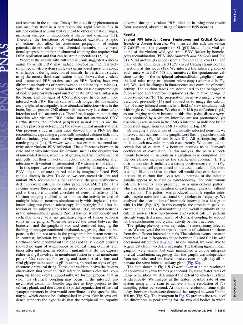
ResultsVirulent PRV Infection Causes Synchronous and Cyclical CalciumTransients Among Neurons. We inserted the calcium sensorG-CaMP3 into the glycoprotein G (gG) locus of the viral ge-nome of the virulent wild-type strain PRV Becker by homolo-gous recombination (PRV 468; Materials and Methods and Fig.S1). Viral protein gG is not essential for spread in vivo (17), andmany of the commonly used PRV circuit tracing strains containinsertions at this locus (18). We infected the salivary glands ofadult mice with PRV 468 and monitored the spontaneous cal-cium activity in the peripheral submandibular ganglia of anes-thetized mice using two-photon microscopy (schematic in Fig.1A). We used the changes in fluorescence as a correlate of neuralactivity. The calcium traces are normalized to the backgroundfluorescence and therefore displayed as the relative change influorescence (ΔF/F). The preparation for in vivo imaging was asdescribed previously (14) and allowed us to image the calciumflux of many infected neurons in a field of view simultaneouslywith single-cell resolution. We chose 40–48 h postinfection (h.p.i.)as our imaging window because at this time point, disease symp-toms produced by a virulent infection are not pronounced, yetessentially every neuron in the SMG is infected, as indicated by thefluorescent signal from GCaMP3 (picture in Fig. 1B).By imaging a population of individually infected neurons, we
observed that neurons in the ganglia were flashing synchronouslyand cyclically (Fig. 1B and Movie S1). All participating cellsinitiated each new calcium peak concurrently. We quantified thecorrelation of calcium flux between neurons using Pearson’scoefficients of correlation. In this analysis, the pairwise coef-ficients between all neurons in a ganglion were computed, andthe correlation increases as the coefficients approach 1. Thedistribution clearly indicated a strong positive correlation (Fig.1C): when one cell experienced an increase in calcium flux, thereis a high likelihood that another cell would also experience anincrease in calcium flux. As a result, neurons of the infectedganglia appear to be flashing synchronously. The synchronouscalcium transients also presented in a quasicyclical pattern,which persisted for the duration of each imaging session withoutinterruption. The pattern was produced at fairly regular inter-vals, despite noise and occasional short delays in frequency. Weanalyzed the distribution of interpeak intervals in a histogramwith 1-s bins (Fig. 1D). In this example, the prominent peak oc-curred at 10 and 11 s, demonstrating the overall regularity of thecalcium pulses. These synchronous and cyclical calcium patternsstrongly suggested a mechanism of electrical coupling to accountfor the synchronous and cyclical activity between neurons.The spiking phenotype was reproducible across many infected
mice. We analyzed the interpeak intervals of calcium transientsfrom five different infected animals. The calcium events occurredevery 5–15 s or in frequency range between 0.1 and 0.2 Hz, withoccasional differences (Fig. S2). In one animal, we were able toacquire data from two different ganglia. The flashing signals in eachganglion were similar, but each maintained a unique interpeakinterval distribution, suggesting that the ganglia are independentfrom each other and not interconnected even though they all in-nervate the same infected salivary gland (Fig. S2).The data shown in Fig. 1 B–D were taken at a time resolution
of approximately two frames per second. By using faster rates ofimage acquisition, we determined the extent to which cells firedsynchronously. We imaged at the fastest possible rate of oursystem using a line scan to achieve a time resolution of 758sampling points per second. At this time resolution, some slightdelays between traces were detected that ranged between 50 and100 ms (Fig. S3). The histogram in Fig. S3 presents the results ofthe differences in peak timing for the two cell bodies in which
Granstedt et al. PNAS | Published online August 26, 2013 | E3517
MICRO
BIOLO
GY
PNASPL
US

a segment of their measured traces are displayed. Twenty-ninesuccessive peaks are included in this distribution, which showsa mean difference of 68 ms. These slight delays suggest that ifelectrical coupling is occurring in the salivary circuit, the cou-pling likely exists at some distance from the neuron cell bodiesthat were imaged.
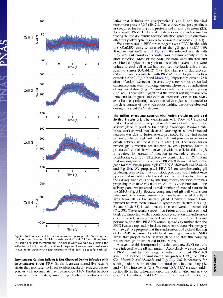
Each Cell Has a Unique Calcium Peak Shape. We noticed that al-though neurons presented synchronous and cyclical calciumtransients, the calcium traces of individual cells were not iden-tical in amplitude and duration. In fact, for the duration of theobservations, each synchronously flashing cell appeared to ex-hibit a unique calcium peak shape profile. We analyzed thisphenomenon at faster frame rates for data acquisition using theline scan parameters noted above. Fig. 2 shows four differentcells in which the calcium peaks of each cell were superimposed.Each cell displayed a calcium peak of unique shape, amplitude,and duration that are remarkably consistent. For example, cell 1displays very weak fluorescence changes that are slightly abovethe background noise. Cell 2 exhibits calcium flashes that aver-age 150% above background at their peak, and these calciumlevels plateau for a full second before beginning to decrease andreturn to background. Cell 3 also peaks around 150% abovebackground, but the calcium levels immediately begin to de-crease after peaking. Cell 4 displays much higher changes incalcium flux, averaging close to 500% above background. Theunique calcium traces of individual neurons suggest that elec-trical coupling of cell bodies in the ganglion is unlikely, andtherefore coupling could be taking place in distant axons.
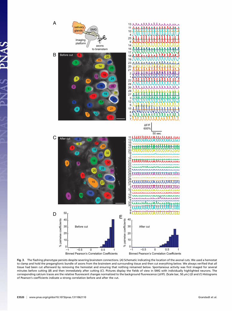
The Spiking Phenotype Does Not Require Connections from CNSNeurons That Project to the SMG. As previously mentioned, theclose synchrony of calcium flux between SMG neurons was remi-niscent of electrical coupling as reported in published work in vitro(10, 11) and therefore suggested that a similar mechanism could beoccurring somewhere within the salivary circuit, although to dateviral-induced fusion pores have not been demonstrated in vivo. Ifelectrical coupling were to occur in the salivary circuit, we rea-soned that possible sites would include the neurons of the superiorsalivatory nucleus (SSN) in the brainstem that are presynaptic tothe SMG, the cell bodies of neurons in the SMG, or in the SMGaxons that project to the salivary glands.The SMG ganglia receive input from the SSN, which lies in the
lateral reticular formation of the pons in the brainstem. Thepreganglionic axons branch to innervate one of more neurons inthe SMG, but each neuron in the SMG receives input from onlyone cell in the SSN (19). These brainstem neurons are in closeproximity to one another and not ensheathed by glial cells andtherefore could possibly become electrically coupled by viral-induced fusion events causing synchronous firing in each SMG.We tested this possibility by severing SSN axonal inputs to theSMG (schematic in Fig. 3A shows the location of axon cuts). Wefirst prepared infected animals for imaging as before and imagedthe synchronous and cyclical calcium activity for several minutes(Fig. 3B and Movie S2). We then clamped and held the pre-ganglionic bundle of axons and surrounding tissue with a hemo-stat and cut the axons and tissue below. We immediately resumedimaging and observed that the synchronous and cyclical flashingcontinued unabated (Fig. 3C and Movie S3). We quantified thecorrelation of calcium flux between neurons and the distributionsclearly indicated a strong positive correlation (Fig. 3 D and E).These experiments demonstrate that brainstem nuclei are notessential to elicit the spiking phenotype.Our data also suggest coupling is unlikely to occur among
infected SMG cell bodies for several reasons. We observed cy-clical and synchronous flashing in cells that were not adjacentand we never observed any overt fusion or syncytial events be-tween SMG cell bodies. Furthermore, there were slight delaysbetween the onset of the calcium peaks across cells and individualinfected neurons had unique calcium peak profiles. Finally,because each SMG neuron is ensheathed by one or more satellitecells, the SMG cell bodies are physically insulated from any fusionevents. The next possibility is that coupling occurs in SMG axonsthat project to the salivary gland, which we further explored usingattenuated and mutant PRV recombinants.
5
1
10
11
12
13
15
16
17
18
19
2
20
22
23
24
25
26
27
3
4
68
9
6
8
5
4
1
25
13
27
23
262
19
17
12
16
9
3
11
18
10
20
22
15
24
Virulent PRV-GCaMP3 (PRV 468)
A
B
% p
airw
ise
coef
ficie
nts
Pearson’s Correlation Coefficients
30
25
20
15
10
5
0
35
0 0.5 1-0.5-1Inter-peak interval (s)
Num
ber o
f pea
k in
terv
als
0 5 10 15 20 25 30
70
60
50
40
30
20
10
0
CD
∆F/F400%
50 sec
Pre-synaptic axonsSMG nerve
Salivary duct
Salivary glands
Initiateinfection
here
Image SMG neurons here
to brainstem
imagingplatform
Fig. 1. Neurons infected with PRV 468 display a synchronous flashingphenotype. (A) Simplified schematic of in vivo infection and imaginglocations (red arrows and red box) within the salivary circuit. Key com-ponents of salivary circuit are outlined, not to scale. The salivary glandsare inoculated with PRV and 2 d later, the submandibular ganglia (SMG)are imaged in living mice. (B) Calcium traces from neurons infected withvirulent PRV 468 expressing GCaMP3. Schematic shows the setup for invivo imaging in which the SMG ganglia are lifted on an imaging plat-form. Picture displays a representative field of view in which a pop-ulation of individually infected neurons in the SMG was imagedsimultaneously. The superimposed colored areas highlight the regions ofinterest within each cell from which the corresponding normalized ΔF/Fcalcium traces (numbered) were computed. The relative fluorescencechange (ΔF/F) and timescale are indicated. (Scale bar, 50 μm.) (C ) Quan-tification of the correlation of calcium flux between neurons usinga histogram of Pearson’s coefficients of correlation. Pairwise coefficientsbetween neurons in a ganglion were computed and coefficients binnedin 0.1 increments to generate a distribution. All features of the calciumtraces, such as amplitude, shape, and noise, were included in the analysisbetween each pair. The histogram indicates a strong correlation. (D) Distri-bution of interpeak intervals in a histogram with 1-s bins.
E3518 | www.pnas.org/cgi/doi/10.1073/pnas.1311062110 Granstedt et al.
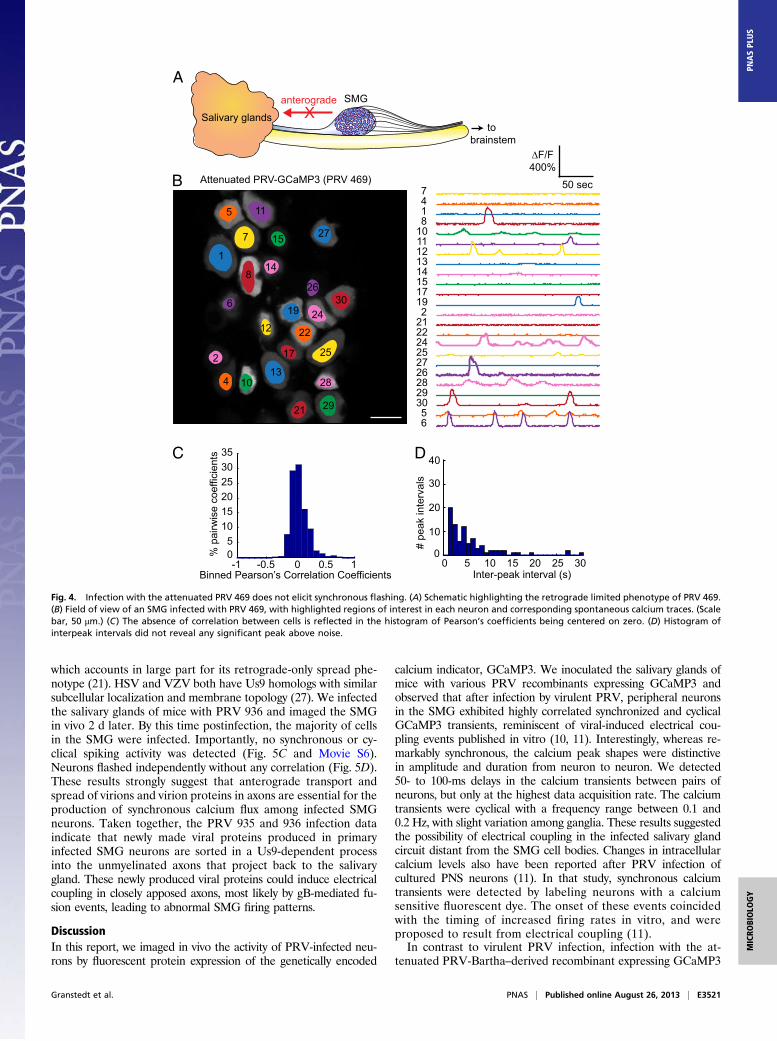
Spontaneous Calcium Spiking Is Not Observed During Infection withan Attenuated Strain. PRV Bartha is an attenuated live vaccinestrain that replicates well, yet exhibits markedly reduced patho-genesis with no mad itch symptomology. PRV Bartha harborsmany mutations in its genome; in particular, it contains a de-
letion that includes the glycoproteins E and I, and the viralmembrane protein Us9 (20, 21). These three viral gene productsare required for sorting viral proteins and virions into axons (22).As a result, PRV Bartha and its derivatives are widely used intracing neuronal circuitry because infection spreads unidirection-ally from postsynaptic neurons to presynaptic neurons (Fig. 4A).We constructed a PRV strain isogenic with PRV Bartha with
the GCaMP3 cassette inserted in the gG gene (PRV 469;Materials and Methods and Fig. S1). We infected animals withPRV 469 and monitored spontaneous calcium activity at 72 hafter infection. Most of the SMG neurons were infected andexhibited complex but asynchronous calcium events that wereunique to each cell as we had reported previously using a lesssensitive sensor (GCaMP2) (14). The changes in fluorescence(ΔF/F) in neurons infected with PRV 469 were bright and oftenexceeded 200% (Fig. 4B and Movie S4). Importantly, even at 72 hafter infection, we never observed any synchronous or cyclicalcalcium spiking activity among neurons. There was no indicationof any correlation (Fig. 4C) and no evidence of cyclical spiking(Fig. 4D). These data suggest that the axonal sorting of viral pro-teins and anterograde transport of infectious virus in the SMGaxon bundles projecting back to the salivary glands are crucial tothe development of the synchronous flashing phenotype observedduring a virulent PRV infection.
The Spiking Phenotype Requires Viral Fusion Protein gB and ViralSorting Protein Us9. The experiments with PRV 469 indicatedthat viral proteins were required in SMG axons that project to thesalivary gland to produce the spiking phenotype. Previous pub-lished work showed that electrical coupling in cultured infectedneurons was due to fusion events promoted by the viral fusionprotein gB, because gB null mutants did not promote microfusionevents between neuronal soma in vitro (10). The virion fusionprotein gB is essential for infection by virus particles where itpromotes fusion of the viral envelope with the cell. In addition, gBis required for spread of infection to secondary neurons orneighboring cells (23). Therefore, we constructed a PRV mutantthat was isogenic with the virulent PRV 468 strain, but lacked thegene for viral fusion protein gB (PRV 935; Materials and Methodsand Fig. S4). We propagated PRV 935 on complementing gBproducing cells so that the virus stock produced could infect onceupon initial inoculation to the salivary glands, either by infectingthe salivary gland cells or by infecting directly the axon terminalsprojecting from the SMG neurons. After PRV 935 infection of thesalivary gland, we observed a small number of infected neurons inthe SMG (Fig. 5A). Because complemented gB null virions caninfect only once, these neurons must have been infected directly ataxon terminals in the salivary gland. However, among thoseinfected neurons, none showed a synchronous calcium flux (Fig.5A and Movie S5). In addition, the transients were not correlated(Fig. 5B). These results suggest that fusion and spread promotedby gB are important to the spontaneous generation of synchronouscalcium activity among infected neurons in the SMG. It is im-portant to note that PRV 935 cannot spread any further than theSMG because replication in those neurons produces viral progenywith no gB. We propose that the synchronous and cyclical flashingof GCaMP3 is caused by electrical coupling of infected SMGaxons that project to the salivary gland and that this couplingresults from gB-driven axonal fusion events.A caveat to this interpretation is that very few SMG neurons
were infected by the gB null mutant. Accordingly, we constructeda PRV mutant that was isogenic with the virulent PRV 468strain, but lacked the viral membrane protein Us9 gene (PRV936; Materials and Methods and Fig. S4). Us9 is necessary forsorting and transport of viral structural proteins, including gB,and virions into axons (24). Without Us9, PRV spreads unidi-rectionally in the retrograde direction both in vitro and in vivo(25, 26). The attenuated PRV Bartha strain lacks the Us9 gene,
−1 0 1 2 3−50
0
50
100
−1 0 1 2 3−100
0
100
200
300
−1 0 1 2 3−100
0
100
200
300
−1 0 1 2 3−500
0
500
1000
Cell 1
Cell 2
Cell 3
Cell 4
Time (s)
Time (s)
Time (s)
Time (s)
% Δ
F/F
% Δ
F/F
% Δ
F/F
% Δ
F/F
Fig. 2. Each infected cell has a unique calcium peak profile. Superimposedcalcium traces from four individual cells are displayed. All four cells are fromthe same line scan measurement. The peaks were overlaid by aligning theinflection points in the rising portion of the peaks. Averaged peak profiles areshown in red. Data show a superimposition of at least 16 peaks for each cell.
Granstedt et al. PNAS | Published online August 26, 2013 | E3519
MICRO
BIOLO
GY
PNASPL
US
1
1011
12
13
14
15
16171819
2
2021
23
25
2224262827
3
4
5
6
897
ΔF/F600%
50 sec
10
1113151617
18
192
20
212223242526272829
331
32
34
5
63330
798
A
B
C
E40
30
20
10
0% p
airw
ise
coef
ficie
nts
Binned Pearson’s Correlation CoefficientsBinned Pearson’s Correlation Coefficients
50
40
30
20
10
0% p
airw
ise
coef
ficie
nts
axonsto brainstem
−1 −0.5 0 0.5 1 −1 −0.5 0 0.5 1
D
Before cut
13
11
10
4
25
6
14
16
17
18
19
23
20
21
2
1
22
24
26
28
27
3
12
5
15
8
9
7
After cut
8
9
7
17 26
15
11
16
18
19
2
20
21
22
23
24
25
27
28
29
313
32
34
10
5
6
33
30
13
imagingplatform
salivaryglands SMG
Before cut After cut
Fig. 3. The flashing phenotype persists despite severing brainstem connections. (A) Schematic indicating the location of the axonal cuts. We used a hemostatto clamp and hold the preganglionic bundle of axons from the brainstem and surrounding tissue and then cut everything below. We always verified that alltissue had been cut afterward by removing the hemostat and ensuring that nothing remained below. Spontaneous activity was first imaged for severalminutes before cutting (B) and then immediately after cutting (C). Pictures display the fields of view in SMG with individually highlighted neurons. Thecorresponding calcium traces are the relative fluorescent changes normalized to the background fluorescence (ΔF/F). (Scale bar, 50 μm.) (D and E) Histogramsof Pearson’s coefficients indicate a strong correlation before and after the cut.
E3520 | www.pnas.org/cgi/doi/10.1073/pnas.1311062110 Granstedt et al.
which accounts in large part for its retrograde-only spread phe-notype (21). HSV and VZV both have Us9 homologs with similarsubcellular localization and membrane topology (27). We infectedthe salivary glands of mice with PRV 936 and imaged the SMGin vivo 2 d later. By this time postinfection, the majority of cellsin the SMG were infected. Importantly, no synchronous or cy-clical spiking activity was detected (Fig. 5C and Movie S6).Neurons flashed independently without any correlation (Fig. 5D).These results strongly suggest that anterograde transport andspread of virions and virion proteins in axons are essential for theproduction of synchronous calcium flux among infected SMGneurons. Taken together, the PRV 935 and 936 infection dataindicate that newly made viral proteins produced in primaryinfected SMG neurons are sorted in a Us9-dependent processinto the unmyelinated axons that project back to the salivarygland. These newly produced viral proteins could induce electricalcoupling in closely apposed axons, most likely by gB-mediated fu-sion events, leading to abnormal SMG firing patterns.
DiscussionIn this report, we imaged in vivo the activity of PRV-infected neu-rons by fluorescent protein expression of the genetically encoded
calcium indicator, GCaMP3. We inoculated the salivary glands ofmice with various PRV recombinants expressing GCaMP3 andobserved that after infection by virulent PRV, peripheral neuronsin the SMG exhibited highly correlated synchronized and cyclicalGCaMP3 transients, reminiscent of viral-induced electrical cou-pling events published in vitro (10, 11). Interestingly, whereas re-markably synchronous, the calcium peak shapes were distinctivein amplitude and duration from neuron to neuron. We detected50- to 100-ms delays in the calcium transients between pairs ofneurons, but only at the highest data acquisition rate. The calciumtransients were cyclical with a frequency range between 0.1 and0.2 Hz, with slight variation among ganglia. These results suggestedthe possibility of electrical coupling in the infected salivary glandcircuit distant from the SMG cell bodies. Changes in intracellularcalcium levels also have been reported after PRV infection ofcultured PNS neurons (11). In that study, synchronous calciumtransients were detected by labeling neurons with a calciumsensitive fluorescent dye. The onset of these events coincidedwith the timing of increased firing rates in vitro, and wereproposed to result from electrical coupling (11).In contrast to virulent PRV infection, infection with the at-
tenuated PRV-Bartha–derived recombinant expressing GCaMP3
18
10111213141517192
21222425
2627
2829
4
3056
7
7
4
1
8
10
11
12
13
14
15
17
19
2
21
22
24
25
27
26
28
29
30
5
6
∆F/F400%
50 secAttenuated PRV-GCaMP3 (PRV 469)
A
B
0 0.5 1
% p
airw
ise
coef
ficie
nts
Binned Pearson’s Correlation Coefficients
3025201510
50
35
-0.5-1
# pe
ak in
terv
als
40
30
20
10
0
Inter-peak interval (s)0 5 10 15 20 25 30
C
Salivary glandsto
brainstem
SMGanterogradeX
D
Fig. 4. Infection with the attenuated PRV 469 does not elicit synchronous flashing. (A) Schematic highlighting the retrograde limited phenotype of PRV 469.(B) Field of view of an SMG infected with PRV 469, with highlighted regions of interest in each neuron and corresponding spontaneous calcium traces. (Scalebar, 50 μm.) (C) The absence of correlation between cells is reflected in the histogram of Pearson’s coefficients being centered on zero. (D) Histogram ofinterpeak intervals did not reveal any significant peak above noise.
Granstedt et al. PNAS | Published online August 26, 2013 | E3521
MICRO
BIOLO
GY
PNASPL
US

showed no signs of synchronous or cyclical firing despite efficientinfection of the ganglia. Rather, the neurons exhibited randomcalcium transients with no correlation between neurons. Theseresults are consistent with our previous published work using aPRV Bartha recombinant expressing the less sensitive indicatorGCaMP2 (14). Other in vivo transsynaptic analysis using PRVBartha derivatives as circuit tracers also have reported no dramaticalteration in the physiological properties of infected neurons (17).Our data showed that the CNS projections to the SMG were
not involved, that each neuron cell body displayed a uniquecalcium peak profile, and that the attenuated PRV-Bartha–derived recombinant, which spreads only from postsynaptic topresynaptic neurons, did not display the synchronous and cyclicalflashing phenotype. These data suggested that electrical couplingwas occurring at some distance from the ganglia, most likely inthe axons that project from the SMG to the salivary glands. Totest this hypothesis, we imaged the SMG after infection withPRV mutants lacking either the viral glycoprotein B, involved inmembrane fusion, or the viral membrane protein Us9, requiredfor sorting and transport of virions in axons. We focused on thesetwo viral proteins based on previous work; other viral proteinsare also involved in membrane fusion, such as the gH/gL com-plex, or anterograde spread, such as the glycoprotein E (gE)/glycoprotein I (gI) complex, but we did not explore their po-tential role in the firing phenotype in these experiments. Neitherthe gB or Us9 null mutant PRV infections showed any signs ofsynchronous or cyclical firing. Because Us9 is essential for sort-ing gB into axons (24), and the attenuated PRV-Bartha–derivedrecombinant expresses gB but not Us9, we propose that thetransport of newly made virions and virion proteins into SMGaxons that project back to the gland may be essential for the gen-eration of synchronous and cyclical calcium flux. The synchronous
and cyclic firing in the ganglia would occur by virally induced fusionin the infected, unmyelinated bundled axons that project to thesalivary glands.The concept of a functional interaction between closely ap-
posed infected axons has been noted before for PRV. Usingmodified Campenot chambers and cultured SCG neurons, Ch’ngand colleagues showed that PRV infection could spread betweenneurons at sites of axoaxonal contact (28). Furthermore, invarious in vitro models, virions can egress at axonal varicosities(23, 29). Other axon–axon interactions in the absence of in-fection have been described in the hippocampus for ultrafastneuronal communication (30). One computational analysis ofthis CNS phenomenon revealed that electrical coupling betweenaxons induces synchronous oscillations with cell body actionpotentials evoked antidromically from the axon (31). In the PNS,ultrastructural studies have demonstrated sites of axoaxonal syn-apses between sympathetic and parasympathetic nerves innervatingthe same peripheral target (32), and these sites of contact havefunctional significance (33). Sensory and autonomic postganglionicaxons are usually unmyelinated, and groups of axons are sur-rounded by Schwann cells in troughs termed “Remak bundles” (34,35). These unmyelinated sites could facilitate infection-inducedaxoaxonal coupling in the salivary circuit.The model developed and characterized here was the salivary
circuit, where infection is initiated at the salivary glands, and theperipheral parasympathetic SMG are imaged. The glands alsoreceive motor sympathetic efferent input from the superiorcervical ganglia, as well as somatic sensory afferent innervationsfrom the trigeminal ganglia (36). The afferent supply is consid-ered to be of the same origin as the trigeminal sensory inner-vations of the oral and nasal mucosa, and therefore this model is
123456789
10
∆F/F400%
50 sec
PRV gB null - GCaMP3 (PRV 935)
PRV Us9 null - GCaMP3 (PRV 936)
7
6
10 12 17
14 19
16 23
20
13 18
1522
26
25 28
27
30
2124
29 31
1 4
2
3 9
511
8
987654
313
3029282726252423222120
218191716151413121110
1
Binned Pearson’s Correlation Coefficients
% p
airw
ise
coef
ficie
nts
% p
airw
ise
coef
ficie
nts
0 0.5 1-0.5-1
30
25
20
15
10
50
35
404550
Binned Pearson’s Correlation Coefficients0 0.5 1-0.5-1
0
30
25
20
15
10
5
35
40
14
83
6
52
9
10
7
A B
C D
Fig. 5. Infection with either a gB null strain or a Us9 null strain does not elicit synchronous flashing. (A) Few neurons are infected after inoculation with thegB null strain PRV 935, but no spontaneous synchronous activity is detected among these cells. (Scale bar, 50 μm.) (B) Histogram of Pearson’s coefficientsindicated no correlation between cells infected with PRV 935. (C) The majority of cells are infected after inoculation of the Us9 null strain PRV 936, yet thespontaneous calcium signals are not synchronous. (Scale bar, 50 μm.) (D) Histogram of Pearson’s coefficients indicated no correlation between cells infectedwith PRV 936.
E3522 | www.pnas.org/cgi/doi/10.1073/pnas.1311062110 Granstedt et al.

also relevant to disease models of HSV-1, which naturally infectsthe trigeminal ganglia.A further important aspect of in vivo PRV infection in the
SMG model is the concept of “round-trip” reseeding and am-plification of infection in the ganglia. By imaging acute explantsof the SMG and surrounding tissues after in vivo PRV infectionof the salivary glands, we have recently reported that newly madeparticles in infected SMG neurons are transported back to theglands where the infection was initiated (37). These particlesthen infect more axons in the gland and return to infect additionalneurons in the ganglia, which in turn leads to more infection in thegland. This reseeding phenomenon occurs only with a virulentvirus infection and not with PRV Bartha or Us9 null infections(37). It is likely that reseeding of the infected gland contributes tomassive invasion of the innervating PNS ganglia and involvementof more axons in electrical coupling. We suggest that this round-trip infection process contributes significantly to the characteristicperipheral neuropathy of PRV infection.Our current observations may explain the known symptomol-
ogy of PRV infection, particularly the characteristic neuropathyof the mad itch. We suggest that the major cause is infection-induced, synchronous, and cyclical firing in PNS ganglia thatproject to the infected tissues, which results from fusion poreformation in unmyelinated axons of infected neurons. In thismodel, a normal action potential is “short circuited” by leakingions into fusion-coupled axons, which in turn leads to back-propagated signals that induce more firing. These sites of gB-mediated microfusion and current leakage may occur at vari-cosities or other places of functional interaction among axons.Fusion events could occur between PNS ganglia cell bodies asshown with cultured SCG neurons in vitro (10). However, in theSMG, and in other PNS ganglia, cell bodies are sheathed bysatellite glial cells that restrict direct contact of neurons andprobably inhibit viral spread between neuronal cell bodies (34).Importantly, we found that infection by the PRV Bartha
recombinant, which does not induce any peripheral neuropathy,also does not induce synchronized or cyclical firing in neurons.We suggest that PRV Bartha cannot induce electrical coupling inthe unmyelinated SMG axons because it lacks the Us9 proteinrequired for sorting virion proteins into axons. We have beenable to propose a model that accounts for the differences be-tween the virulent and attenuated strains because we used an invivo system in which the spatial organization of tissues andsynaptic partners are preserved.There is circumstantial evidence in the literature to suggest
that virulent alphaherpesviruses may spread laterally in periph-eral sensory ganglia (see ref. 38 for review). In the mouse flankmodel, herpes simplex virus and PRV inoculations at a single siteresult in spread throughout the innervated dermatome, suggest-ing disseminated spread in the sensory dorsal root ganglia (DRG)(4, 39). Fusion of neurons and satellite cells has been demon-strated in human DRG xenografts in mice infected with varicella-zoster virus (40). In addition, virulent PRV infection of the mouseflank results in substantial viral titers recoverable in visceralorgans, but not in an infection with attenuated PRV Bartha (4);infection of sensory neurons that project to internal organs couldbe accounted for by lateral spread in the DRG because thesevisceral afferent neurons reside concomitantly in sensory ganglia(38). Our model could account for these observations.The violent pruritus induced by virulent PRV infection has
several components. First, the animal appears to perceive a con-sistent and continuous irritation that stimulates itching motoractivity. The animal is then consumed by this behavior, withoutstopping to eat, drink, or groom (4). By observing the calciumprofiles of many SMG neurons simultaneously with single-cellresolution, we characterized synchronous and cyclical patterns ofactivity that were highly correlated among infected neurons. Thisphenotype is reminiscent of the spontaneous bursting activity in
infected sympathetic ganglia associated with behavioral pruritus(6). Our studies suggest that common mechanisms may exist thatinduce alterations in PNS physiology leading to severe neuro-pathic pain caused by human alphaherpesvirus infections (e.g.,disseminated herpes zoster or postherpetic neuralgia).
Materials and MethodsCells and Virus Construction. PRV 468 and PRV 469 were constructed by ho-mologous recombination between a plasmid containing a GCaMP3 expres-sion cassette and the gG locus of the PRV Becker and PRV Bartha genomes,respectively. The plasmid pN1-GCaMP3 was a gift of Charles Zuker (ColumbiaUniversity, New York, NY) and contains the coding sequence of GCaMP3 inplace of EGFP in the pEGFP-N1 plasmid, under the control of a cytomegalo-virus immediate-early (CMVIE) promoter (Clontech; GenBank accession no.U55762). The pN1-GaMP3 plasmid was linearized and cotransfected intoswine epithelial (PK15) cells with DNA from PRV 616, a PRV Becker de-rivative, or PRV 614, a PRV Bartha derivative, and both these parental strainshave mRFP1 inserted into the gG locus (18). Homologous recombination ofthe GCaMP3 cassette into the gG locus of PRV 616 or PRV 614 was directedbetween the CMVIE promoter and the simian virus 40 (SV40) poly-adenylation sequences, replacing the original 2.3-kb mRFP cassette at thislocus with the 2.5-kb GCaMP3 cassette. Resulting recombinants were plaquepurified on PK15 cells by selection of nonred plaques. The recombinantswere crossed with a PRV Becker or PRV Bartha derivative that expressed anmRFP capsid fusion protein (PRV 180 and PRV 765, respectively). Doublerecombinants were plaque purified three times so that the final strains PRV468 and PRV 469 expressed diffusible GCaMP3 as well as a red capsid tag.These recombinants enabled us to independently confirm infection of cellsand expression of GCaMP3. Restriction fragment length polymorphism(RFLP) analysis using SalI was used to compare PRV Becker, PRV Bartha, PRV468, and PRV 469, confirming the correct integration of the GCaMP3 cas-sette (Fig. S1A). Nucleocapsid DNA was prepared using standard protocols(41). To check for robust protein expression of the inserted cassette, weperformed a Western blot using the following antibodies: polyclonal rabbitanti-GFP (Invitrogen; A11122), mouse anti major capsid protein VP5 (42), andthe monoclonal mouse anti–beta-actin (Sigma; A1978) (Fig. S1B). For thesingle-step growth curve, PK15 cells were infected at a multiplicity of in-fection of 10, with input virus removed by saline washes after 1 h. Each timepoint was performed in triplicate and titered in duplicate (Fig. S1C).
PRV 935 was constructed by homologous recombination between pHF22(43), which has a gB deletion and NsiI/PvuI linearized pII-GCaMP3, a derivateof pII (17) in gB-complementing cells. pII-GCaMP3 was made by exchangingthe EGFP coding DNA sequences (CDS) of pII with the GCaMP3 CDS ofplasmid GCaMP3, provided by Loren Looger (Janelia Farm Research Campus,Ashburn, VA) (15) through Addgene (Addgene; plasmid 22692). Briefly,a 1370-bp fragment of the plasmid GCamp3 ranging from its BglII to theNotI site (position 628–1998) coding for the complete GCaMP3 CDS wasrecombined with pII at its EcoRI and NotI sites (position 1544–2316) by theproprietary cloneEZ technology (Genscript).
PRV 936 was constructed by homologous recombination similar to theconstruction of PRV 935. Nucleocapsid DNA of PRV 325 (26, 44) that carriesa mRFP-VP26 fusion and a Us9 deletion was recombined with HindIII line-arized pUC-gG-GCaMP3 as described above. pUC-gG-GCaMP3 is similar to pIIbut is smaller and genetically more stable. It is based on pUC57-gG-multiplecloning site (MCS), which was made by inserting a synthetic construct be-tween the NdeI and HindIII sites of pUC57 (Genscript). The inserted syntheticconstruct consists of 850 bp of homology to the surroundings of the PstIsite in the US4 locus of PRV Becker, into which a pCMV-IE-MCS-SV40 pol-yadenylation sequences cassette was inserted. The GCaMP3 CDS of GCaMP3(Addgene; plasmid 22692) was then subcloned into the MCS of pUC57-gG-MSbetween the BglII/NotI sites (Genscript).
PRV 935 was characterized by Western blotting using the monoclonalantibody M2 against gB (45) (Fig. S4A). For the characterization of PRV 936we used polyclonal rabbit antisera directed against Us9, gE, and gI (46–48) toprobe for the expression of Us9, gE, and gI, respectively (Fig. S4B).
Infection of Mouse SMG. All animal procedures were performed in accordancewith the guidelines of the National Institutes of Health andwere approved bylocal authorities (Princeton University Institutional Animal Care and UseCommittee). This preparation for mouse SMG infection was previouslypublished (49). Briefly, adult male mice aged roughly 7–10 wk were anes-thetized with a mixture of ketamine (100 mg/kg) and xylazine (10 mg/kg) byi.p. administration. A midline incision was made along the neck, exposingthe salivary glands. Using a Hamilton syringe, an aliquot of concentrated
Granstedt et al. PNAS | Published online August 26, 2013 | E3523
MICRO
BIOLO
GY
PNASPL
US
PRV inoculum (1 × 10⁹ pfu/mL) was injected into both submandibular glands(5 μL/side). The incision was sutured and the mice were given bupre-norphrine (100 μg/kg concentration) for postsurgical pain. The mice wereallowed to recover for various times after infection.
In Vivo Calcium Imaging. The protocol for imaging infected SMG in living micewas repeated from our previous publication (14), and more details areprovided in the published protocol (49). Briefly, mice were anesthetized inan isofluorane chamber and then transferred in supine position to a custom-built stage with heating pad, rectal temperature probe, and nose cone forisoflurane inhalation. A ventral midline incision was made along the neckand the skin was pulled back with retractors. The SMG were exposed andlifted on a small custom-made metallic platform, controlled by a microma-nipulator. The area was perfused with warmed artificial cerebral spinal fluidcontaining (in millimoles): 127 NaCl, 25 NaHCO3, 1.25 NaH2PO4, 2.5 KCl,1 MgCl2, and 25 glucose. The animal’s temperature was maintained at 35 °C.
Live imaging was performed using a custom-built two-photon scanningmicroscope (50) built around an upright Olympus BX51. Fluorescence pho-tons were collected through a 40×, 0.8 N.A. Olympus water immersion ob-jective LUMPlanFl/IR, and detected with high quantum efficiency GaAsPphotomultiplier tubes (H10770PA-40; Hamamatsu). The microscope wasoperated by the Matlab software ScanImage (51). Images were taken withan excitation wavelength of 920 nm provided by a tunable Ti:Sapphire laser(Chameleon; Coherent Inc.). Movies were recorded at various scan rates.
Image Analysis. The two-photon raw data were analyzed directly in Matlabusing custom-written code. To correct for the lateral movements induced bythe animal, the images were first aligned and registered automatically. Thenthe software drew regions of interest (ROI) around each infected cell andthese ROI were subsequently confirmed manually. ROI for cells bodies missedby the software were added manually. The mean pixel intensity F(t) in eachROI was then extracted for every time point to obtain a time-series fluo-rescence trace for each ROI (ΔF/F). Slow timescale changes in the fluorescencetraces (e.g., due to photobleaching or small drift in the z focal position) wereremoved by determining the distribution of fluorescence values in a 20-sinterval around each sample time point and subtracting the eighth per-centile value. The value of 20 s was chosen because it is much longer than
the duration of a calcium transient observed. The eighth percentile valuewas found to be appropriate to isolate the baseline F0. The normalizedbaseline-subtracted fluorescence traces [(F(t) − F0)/F0, noted in text as ΔF/F]for each ROI were then further analyzed to measure the correlation be-tween the fluorescent signals of different neurons. Correlations were com-puted from Pearson’s coefficients of correlation. The pairwise correlationcoefficients of a given ganglion were displayed in histograms of 0.1-widthbins. Interpeak interval analysis on low time resolution data only (moviestaken at 2 ms per line, 256 × 256, 256 × 128, and 256 × 64 pixels) was per-formed using a threshold method. ΔF/F traces were smoothed using a high-frequency filter and times at which ΔF/F reached the threshold value weremeasured. Subtractions of successive time values gave the interpeak inter-vals. The results for each cell were displayed in histograms of 1-s bins. Weperformed high time resolution experiments by scanning single lines (linescans at t = 1 and 2 ms) crossing usually three or four cell bodies. Regularimages of the whole field of view were recorded before and after the ex-periment to check for possible drift along the optical axis. The data werediscarded in the rare cases this drift was significant. The segments of the lineassociated with each cell body were then treated as ROIs and processed inMatlab, as described above. The peak timing analysis relying on arbitrarythreshold values was not appropriate for measuring the calcium transientdelays between cells. To address the timing of an individual cell’s calciumtransients, a method independent of the actual ΔF/F values was necessary,and the onset of the signal or the inflection point was measured. We choseto quantify the timing of the calcium transient by measuring the time valueswhen the second derivative of ΔF/F cancels (while going from positive tonegative value, i.e., in the rising portion of the fluorescence signal). Thesetime values were then used to measure interpeak intervals and cell-to-celldelays and to realign all peaks from a given cell body.
ACKNOWLEDGMENTS. We thank the members of the L.W.E. laboratory fortheir input and discussions, and particularly M. L. Szpara and O. Koyuncu fortheir detailed feedback on the manuscript. This research was funded byNational Institutes of Health (NIH) Grants NS033506 and NS060699. TheImaging Core Facility at the Lewis-Sigler Institute is funded by NIH NationalInstitute of General Medical Sciences Center Grant PM50 GM071508.
1. Cohen JI, Straus SE, Arvin AM (2007) Varicella-zoster virus replication, pathogenesis,and management. Fields Virology, eds Knipe DM, Howley PM (Lippincott Williams &Wilkins, Philadelphia), 5th Ed, Vol 2, pp 2773–2818.
2. Pellett PE, Roizman B (2007) The family Herpesviridae: A brief introduction. FieldsVirology, eds Knipe DM, Howley PM (Lippincott Williams & Wilkins, Philadelphia), 5thEd, Vol 2, pp 2479–2499.
3. Mettenleiter TC, Ehlers B, Muller T, Yoon K-J, Teifke JP (2012) Herpesviruses. Diseasesof Swine, eds Zimmerman JJ, Karriker LA, Ramirez A, Schwartz KJ, Stevenson GW(Wiley, New York), 10th Ed, pp 421–446.
4. Brittle EE, Reynolds AE, Enquist LW (2004) Two modes of pseudorabies virusneuroinvasion and lethality in mice. J Virol 78(23):12951–12963.
5. Mettenleiter TC (2000) Aujeszky’s disease (pseudorabies) virus: The virus andmolecular pathogenesis—state of the art, June 1999. Vet Res 31(1):99–115.
6. Dempsher J, Larrabee MG, Bang FB, Bodian D (1955) Physiological changes insympathetic ganglia infected with pseudorabies virus. Am J Physiol 182(1):203–216.
7. Dolivo M, Honegger P, George C, Kiraly M, Bommeli W (1979) Enzymatic activity,ultrastructure and function in ganglia infected with a neurotropic virus. Prog BrainRes 51:51–57.
8. Dempsher J, Riker WK (1957) The role of acetylcholine in virus-infected sympatheticganglia. J Physiol 139(1):145–156.
9. Kiraly M, Dolivo M (1982) Alteration of the electrophysiological activity insympathetic ganglia infected with a neurotropic virus. I. Presynaptic origin of thespontaneous bioelectric activity. Brain Res 240(1):43–54.
10. McCarthy KM, Tank DW, Enquist LW (2009) Pseudorabies virus infection altersneuronal activity and connectivity in vitro. PLoS Pathog 5(10):e1000640.
11. Kramer T, Enquist LW (2012) Alphaherpesvirus infection disrupts mitochondrialtransport in neurons. Cell Host Microbe 11(5):504–514.
12. Mayer ML, James MH, Russell RJ, Kelly JS, Pasternak CA (1986) Changes in excitabilityinduced by herpes simplex viruses in rat dorsal root ganglion neurons. J Neurosci 6(2):391–402.
13. Mayer ML, et al. (1985) Spontaneous electrical activity induced by herpes virusinfection in rat sensory neuron cultures. Brain Res 341(2):360–364.
14. Granstedt AE, Szpara ML, Kuhn B, Wang SS, Enquist LW (2009) Fluorescence-basedmonitoring of in vivo neural activity using a circuit-tracing pseudorabies virus. PLoSONE 4(9):e6923.
15. Tian L, et al. (2009) Imaging neural activity in worms, flies and mice with improvedGCaMP calcium indicators. Nat Methods 6(12):875–881.
16. Hires SA, Tian L, Looger LL (2008) Reporting neural activity with genetically encodedcalcium indicators. Brain Cell Biol 36(1–4):69–86.
17. Smith BN, et al. (2000) Pseudorabies virus expressing enhanced green fluorescentprotein: A tool for in vitro electrophysiological analysis of transsynaptically labeled
neurons in identified central nervous system circuits. Proc Natl Acad Sci USA 97(16):9264–9269.
18. Banfield BW, Kaufman JD, Randall JA, Pickard GE (2003) Development of pseudorabiesvirus strains expressing red fluorescent proteins: new tools for multisynaptic labelingapplications. J Virol 77(18):10106–10112.
19. Lichtman JW (1980) On the predominantly single innervation of submandibularganglion cells in the rat. J Physiol 302:121–130.
20. Szpara ML, et al. (2011) A wide extent of inter-strain diversity in virulent and vaccinestrains of alphaherpesviruses. PLoS Pathog 7(10):e1002282.
21. Pomeranz LE, Reynolds AE, Hengartner CJ (2005) Molecular biology of pseudorabiesvirus: Impact on neurovirology and veterinary medicine.Microbiol Mol Biol Rev 69(3):462–500.
22. Ekstrand MI, Enquist LW, Pomeranz LE (2008) The alpha-herpesviruses: Molecularpathfinders in nervous system circuits. Trends Mol Med 14(3):134–140.
23. Curanovic D, Enquist LW (2009) Virion-incorporated glycoprotein B mediates transneuronalspread of pseudorabies virus. J Virol 83(16):7796–7804.
24. Tomishima MJ, Enquist LW (2001) A conserved alpha-herpesvirus protein necessaryfor axonal localization of viral membrane proteins. J Cell Biol 154(4):741–752.
25. Brideau AD, Card JP, Enquist LW (2000) Role of pseudorabies virus Us9, a type IImembrane protein, in infection of tissue culture cells and the rat nervous system.J Virol 74(2):834–845.
26. Lyman MG, Feierbach B, Curanovic D, Bisher M, Enquist LW (2007) Pseudorabies virusUs9 directs axonal sorting of viral capsids. J Virol 81(20):11363–11371.
27. Lyman MG, Kemp CD, Taylor MP, Enquist LW (2009) Comparison of the pseudorabiesvirus Us9 protein with homologs from other veterinary and human alphaherpesviruses.J Virol 83(14):6978–6986.
28. Ch’ng TH, Spear PG, Struyf F, Enquist LW (2007) Glycoprotein D-independent spreadof pseudorabies virus infection in cultured peripheral nervous system neurons ina compartmented system. J Virol 81(19):10742–10757.
29. De Regge N, et al. (2006) Alpha-herpesvirus glycoprotein D interaction with sensoryneurons triggers formation of varicosities that serve as virus exit sites. J Cell Biol174(2):267–275.
30. Schmitz D, et al. (2001) Axo-axonal coupling. A novel mechanism for ultrafastneuronal communication. Neuron 31(5):831–840.
31. Maex R, De Schutter E (2007) Mechanism of spontaneous and self-sustainedoscillations in networks connected through axo-axonal gap junctions. Eur J Neurosci25(11):3347–3358.
32. Ehinger B, Falck B, Sporrong B (1970) Possible axo-axonal synapses betweenperipheral adrenergic and cholinergic nerve terminals. Z Zellforsch Mikrosk Anat107(4):508–521.
E3524 | www.pnas.org/cgi/doi/10.1073/pnas.1311062110 Granstedt et al.
33. Smith PG, Warn JD, Steinle JJ, Krizsan-Agbas D, Hasan W (2002) Modulation ofparasympathetic neuron phenotype and function by sympathetic innervation. AutonNeurosci 96(1):33–42.
34. Hanani M (2010) Satellite glial cells in sympathetic and parasympathetic ganglia: Insearch of function. Brain Res Brain Res Rev 64(2):304–327.
35. Campana WM (2007) Schwann cells: Activated peripheral glia and their role inneuropathic pain. Brain Behav Immun 21(5):522–527.
36. Chibuzo GA, Cummings JF (1980) Motor and sensory centers for the innervation ofmandibular and sublingual salivary glands: A horseradish peroxidase study in the dog.Brain Res 189(2):301–313.
37. Granstedt AE, Brunton BW, Enquist LW (2013) Imaging the transport dynamics ofsingle alphaherpesvirus particles in intact peripheral nervous system explants frominfected mice. mBio 4(3):e00358–00313.
38. Smith G (2012) Herpesvirus transport to the nervous system and back again. Annu RevMicrobiol 66:153–176.
39. Weeks BS, Ramchandran RS, Hopkins JJ, Friedman HM (2000) Herpes simplex virustype-1 and -2 pathogenesis is restricted by the epidermal basement membrane. ArchVirol 145(2):385–396.
40. Reichelt M, Zerboni L, Arvin AM (2008) Mechanisms of varicella-zoster virusneuropathogenesis in human dorsal root ganglia. J Virol 82(8):3971–3983.
41. Szpara ML, Tafuri YR, Enquist LW (2011) Preparation of viral DNA from nucleocapsids.J Vis Exp (54):3151.
42. Ch’ng TH, Enquist LW (2005) Efficient axonal localization of alphaherpesvirus structuralproteins in cultured sympathetic neurons requires viral glycoprotein E. J Virol 79(14):8835–8846.
43. Favoreel HW, Van Minnebruggen G, Nauwynck HJ, Enquist LW, Pensaert MB (2002) Atyrosine-based motif in the cytoplasmic tail of pseudorabies virus glycoprotein B isimportant for both antibody-induced internalization of viral glycoproteins andefficient cell-to-cell spread. J Virol 76(13):6845–6851.
44. Taylor MP, Kramer T, Lyman MG, Kratchmarov R, Enquist LW (2012) Visualization ofan alphaherpesvirus membrane protein that is essential for anterograde axonalspread of infection in neurons. mBio 3(2).
45. Hampl H, Ben-Porat T, Ehrlicher L, Habermehl KO, Kaplan AS (1984) Characterizationof the envelope proteins of pseudorabies virus. J Virol 52(2):583–590.
46. Brideau AD, Banfield BW, Enquist LW (1998) The Us9 gene product of pseudorabiesvirus, an alphaherpesvirus, is a phosphorylated, tail-anchored type II membraneprotein. J Virol 72(6):4560–4570.
47. Tirabassi RS, Enquist LW (2000) Role of the pseudorabies virus gI cytoplasmic domainin neuroinvasion, virulence, and posttranslational N-linked glycosylation. J Virol 74(8):3505–3516.
48. Whealy ME, et al. (1993) Specific pseudorabies virus infection of the rat visual systemrequires both gI and gp63 glycoproteins. J Virol 67(7):3786–3797.
49. Granstedt AE, Kuhn B, Wang SS, Enquist LW (2010) Calcium imaging of neuronalcircuits in vivo using a circuit-tracing pseudorabies virus. Cold Spring Harb Protoc2010(4):5410.
50. Denk W, Strickler JH, Webb WW (1990) Two-photon laser scanning fluorescencemicroscopy. Science 248(4951):73–76.
51. Pologruto TA, Sabatini BL, Svoboda K (2003) ScanImage: Flexible software for operatinglaser scanning microscopes. Biomed Eng Online 2:13.
Granstedt et al. PNAS | Published online August 26, 2013 | E3525
MICRO
BIOLO
GY
PNASPL
US