Embed Size (px)
Citation preview

P1: FLI/FGP P2: FLI/FDR
August 7, 1999 9:27 Annual Reviews AR089-05
?Annu. Rev. Microbiol. 1999. 53:129–54
Copyright c© 1999 by Annual Reviews. All rights reserved
IN VIVO GENETIC ANALYSIS OF BACTERIAL
VIRULENCE
Su L. Chiang,1 John J. Mekalanos,1
and David W. Holden21Department of Microbiology and Molecular Genetics and Shipley Instituteof Medicine, Harvard Medical School, Boston, Massachusetts 02115;e-mail: [email protected], [email protected] of Infectious Diseases, Imperial College of Science, Technology andMedicine, Hammersmith Hospital, London W12 ONN, United Kingdom;e-mail: [email protected]
Key Words in vivo expression technology, signature-tagged mutagenesis,differential fluorescence induction, pathogenesis, GAMBIT
■ Abstract In vitro assays contribute greatly to our understanding of bacterialpathogenesis, but they frequently cannot replicate the complex environment encoun-tered by pathogens during infection. The information gained from such studies istherefore limited. In vivo models, on the other hand, can be difficult to use, and thishas to some extent diminished the incentive to perform studies in living animals. How-ever, several recently developed techniques permit in vivo examination of many genessimultaneously. Most of these methods fall into two broad classes: in vivo expres-sion technology and signature-tagged mutagenesis. In vivo expression technology is apromoter-trap strategy designed to identify genes whose expression is induced in a spe-cific environment, typically that encountered in a host. Signature-tagged mutagenesisuses comparative hybridization to isolate mutants unable to survive specified environ-mental conditions and has been used to identify genes critical for survival in the host.Both approaches have so far been used exclusively for investigating pathogen-hostinteractions, but they should be easily adaptable to the study of other processes.
CONTENTS
Introduction. . . . . . . . . . . . . . . . . . . . . . . . . . . . . . . . . . . . . . . . . . . . . . . . . . . . 130Early Screens for In Vivo-Induced Genes. . . . . . . . . . . . . . . . . . . . . . . . . . . . 131In Vivo Expression Technology. . . . . . . . . . . . . . . . . . . . . . . . . . . . . . . . . . . . 132
Auxotrophic Selection. . . . . . . . . . . . . . . . . . . . . . . . . . . . . . . . . . . . . . . . . . . . 133Antibiotic Selection . . . . . . . . . . . . . . . . . . . . . . . . . . . . . . . . . . . . . . . . . . . . . 133Genetic Recombination as a Reporter for In Vivo Activity. . . . . . . . . . . . . . . . . . 135
Signature-Tagged Mutagenesis. . . . . . . . . . . . . . . . . . . . . . . . . . . . . . . . . . . . . 138Pool Complexity. . . . . . . . . . . . . . . . . . . . . . . . . . . . . . . . . . . . . . . . . . . . . . . . 142
0066-4227/99/1001-0129$08.00 129
P1: FLI/FGP P2: FLI/FDR
August 7, 1999 9:27 Annual Reviews AR089-05
?130 CHIANG ■ MEKALANOS ■ HOLDEN
Inoculum Dose. . . . . . . . . . . . . . . . . . . . . . . . . . . . . . . . . . . . . . . . . . . . . . . . . 142Route of Inoculum Administration. . . . . . . . . . . . . . . . . . . . . . . . . . . . . . . . . . . 142Duration of Infection . . . . . . . . . . . . . . . . . . . . . . . . . . . . . . . . . . . . . . . . . . . . 143
Advantages and Disadvantages of IVET and STM. . . . . . . . . . . . . . . . . . . . . 145GAMBIT . . . . . . . . . . . . . . . . . . . . . . . . . . . . . . . . . . . . . . . . . . . . . . . . . . . . . . 147Concluding Remarks. . . . . . . . . . . . . . . . . . . . . . . . . . . . . . . . . . . . . . . . . . . . 149
INTRODUCTION
The virulence of bacterial pathogens (their ability to produce morbidity and mor-tality in a host) is a complex, multifactorial process requiring the coordinatedactivity of many bacterial gene products. Infections may be described generallyas proceeding in a sequence that begins with attachment to and colonization of thehost, followed in the case of some pathogens by invasion of host tissues or cells.To multiply and persist within the host, a pathogen must then be able to circum-vent the host’s immune system and obtain nutrients for itself. Exit from the hostand transmission to new hosts are subsequent stages in the infectious cycle, and apathogen may at any point during infection produce factors that cause damage tothe host (25, 38).
A variety of in vitro systems have been developed that simulate certain aspectsof the infectious process, enabling the development of screens to study bacterialgene expression and the behavior of mutant strains in physiological conditionsthat reflect the situation in vivo (24). These include the use of specific cultureconditions to mimic the host environment and tissue culture assays for adhesion,invasion, or cytotoxicity. For example, studies of bacterial responses to changesin pH (55), temperature (44, 49), and iron levels (10, 29, 39), and analysis of hostcell invasion (27, 51) and survival in macrophages (7, 12, 13, 23, 26) have all beenused to identify and characterize bacterial virulence determinants.
In vitro assays have been enormously useful and continue to provide muchinformation on the mechanisms of bacterial pathogenesis, but it is obvious thatthey cannot accurately reproduce all aspects of the host-pathogen interaction. Apathogen may encounter several radically different environments in the host, and itmay therefore have very different requirements at various points during infection,particularly in the context of a developing immune response. Consequently, agene that seems important in in vitro studies may not be important in vivo, andgenes that appear unimportant in an in vitro assay may play a critical role duringa natural infection.
For these reasons, in vivo experimental models are highly desirable. Theypermit direct assessment of a pathogen’s ability to colonize and survive in a livinghost and to cause disease or damage. Animal models nevertheless have their ownlimitations, being generally labor intensive, expensive, and otherwise unwieldy,and these issues present a considerable barrier to undertaking large-scale in vivoexperiments. This is perhaps one reason that many searches for novel virulence

P1: FLI/FGP P2: FLI/FDR
August 7, 1999 9:27 Annual Reviews AR089-05
?IN VIVO ANALYSIS OF VIRULENCE 131
determinants have focused on identifying factors that are coregulated with knownvirulence determinants, rather than attempting to conduct generalized screens inanimals. Nevertheless, some screening of individual mutant strains for alteredvirulence has been carried out on a limited scale with animal infection models,using either randomly chosen transposon mutants (11) or strains affected in cellsurface or extracellular proteins (48).
Recently, however, several methods have been developed that greatly simplifyin vivo analysis of large numbers of strains. A number of these can be classified asin vivo expression technology (IVET) methods. These are promoter-trap strategiesdesigned to identify promoters that are specifically activated in the host, and manyIVET procedures permit positive selection for such promoters. Another methodthat has been used to perform in vivo analysis is signature-tagged mutagenesis(STM), which relies on comparative hybridization to identify mutants unable tosurvive in the host. In this review, we summarize adaptations of these techniquesthat have been used in different bacteria, compare the genes identified by IVET andSTM inSalmonella typhimurium, Staphylococcus aureus, andVibrio cholerae, anddiscuss the advantages and disadvantages of the two approaches. Possible futuredevelopment and applications of these and several recently developed methods arealso considered.
EARLY SCREENS FOR IN VIVO-INDUCED GENES
Upon entering the host, many pathogenic organisms find themselves in a situationthat must differ significantly from any encountered in the environmental reservoir.Bacteria respond to this change in circumstances by modulating their patterns ofgene expression accordingly, downregulating the expression of genes that are nolonger necessary, and upregulating those that are specifically required for survivalin the host (e.g. nutrient acquisition or evasion of host defenses). It thereforeseemed probable that at least some in vivo-induced genes would play a criticalrole in pathogenesis, and several promoter-trap strategies were used to identifygenetic loci whose expression is induced in host environments.
Conceptually, finding in vivo-induced genes is potentially a simple process ifit is possible to generate appropriately selectable or screenable gene fusions andobtain a host organism that is amenable to brute-force screening. For example, oneof the first screens for host-induced genes was carried out in the plant pathogenXanthomonas campestris(56). A library ofX. campestrisDNA fusions to a pro-moterless chloramphenicol resistance gene was generated and introduced intoX. campestrison plasmids. Eleven hundred of the resultant strains were individ-ually tested for ability to produce disease symptoms in chloramphenicol-treatedturnip seedlings. Of the 19 strains found to be virulent in treated seedlings, 14 werealso highly sensitive to chloramphenicol in vitro, indicating that these 14 strainsharbored plasmids carrying host-inducible fusions. Mutations were subsequentlycreated in and around two of the host-inducible genes, and the mutant strains were
P1: FLI/FGP P2: FLI/FDR
August 7, 1999 9:27 Annual Reviews AR089-05
?132 CHIANG ■ MEKALANOS ■ HOLDEN
tested for effects on pathogenicity. Mutations in one of the genes led to delayedsymptom expression in plants (57), whereas mutations in the other gene had nodiscernible phenotype (58).
Another example of brute-force screens for in vivo-induced genes involvedindividual scoring of 2550Listeria monocytogenesTn917-lacmutants for genesexpressed at higher levels in macrophage-like cells than in laboratory medium (37).This resulted in the identification of five genes that had as much as 100-fold induc-tion within macrophages. Three of these genes were nucleotide biosynthetic genes,one (arpJ) was involved in arginine uptake, and the fifth wasplcA, the gene encod-ing the previously identified virulence factor phosphatidylinositol-phospholipase C(16). Mutations in the nucleotide biosynthetic genes did not result in increasedLD50 values in mice, although the purine mutation tested did reduce the number ofbacteria recoverable from the liver. ThearpJ mutation caused a twofold increasein LD50 as well as a decrease in bacterial load in the liver, whereas theplcAmutantshowed a 25-fold increase in LD50 (46) and decreased bacterial load in both liverand spleen.
These results provide excellent examples of how promoter-trap screens canidentify in vivo-induced genes in host-pathogen models amenable to selection orbiochemical assay. However, since each strain carrying a given gene fusion wastested individually, the procedures described above were relatively laborious. Thiswas especially true in theXanthomonasstudy, in which each strain was inoculatedby hand into antibiotic-treated seedlings. Therefore, although the goal of identi-fying host-inducible genes was achieved in both cases, it was clear that significantreductions in labor would represent a critical advance in the development of similarmethods for studying bacterial gene expression in the host.
IN VIVO EXPRESSION TECHNOLOGY
In the last five years, a variety of additional methods have been formulated toisolate genes whose expression is induced in the host, all of which have increasedefficiency compared with the examples discussed above. Such techniques weregenerally termed “in vivo expression technology” (IVET) methods, and althoughinitial interest was understandably focused on host-induced genes, IVET couldpresumably be adapted to study the induction of microbial genes in response toany condition.
The first IVET methods used promoterless reporter genes whose products con-fer a phenotype that can be positively selected in the host (42). Both auxotrophicand antibiotic selections were used to this end. The recently developed differentialfluorescence induction (DFI) method (67) also permits positive selection for host-induced promoters, although the selection is carried out by fluorescence-activatedcell sorting (FACS) of organisms recovered from host cells or animals. ResolvaseIVET uses genetic recombination as a reporter activity and requires screeningfor host-induced promoters after bacteria are recovered from host tissues. Its

P1: FLI/FGP P2: FLI/FDR
August 7, 1999 9:27 Annual Reviews AR089-05
?IN VIVO ANALYSIS OF VIRULENCE 133
advantage is that it can in theory detect promoters that are only weakly or tran-siently induced during infection (14, 15).
Auxotrophic Selection
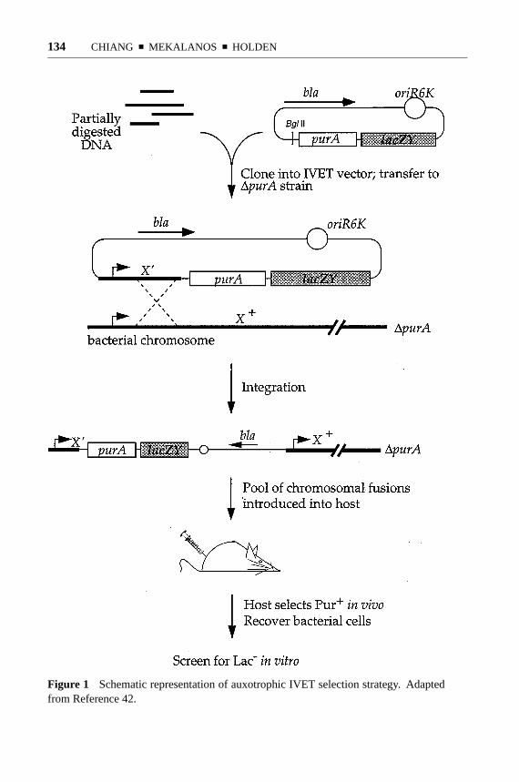
The original IVET selection (42) was performed by creating transcriptional fusionsof random fragments of theS. typhimuriumchromosome with a promoterlesspurAgene and introducing this library onto the chromosome of anS. typhimurium1purAstrain via homologous recombination at the chromosomal fragment (Figure 1).Because purines are limiting for growth ofS. typhimuriumin the mouse, only thosestrains expressingpurAfrom fused promoters would survive. It should be noted thatthe integration event resulting from a single crossover does not lead to disruptionof the wild-type locus on the chromosome, thereby permitting analysis of genesessential for growth in vivo. Bacteria representing the pool of chromosomal fusionswere then injected intraperitoneally into BALB/c mice, and the surviving poolswere recovered 3 days later and screened on laboratory medium for clones withlow promoter activity. Several strains carrying promoters meeting the criteria of invivo expression and in vitro inactivity were, on subsequent analysis, found to havesevere virulence defects as assayed by oral LD50, thereby validating the ability ofIVET to isolate virulence genes.
Similar IVET selections were carried out inPseudomonas aeruginosa, both invivo in BALB/c mice (70) and in vitro to find genes induced by respiratory mucuscollected from cystic fibrosis (CF) patients (69). The latter study was undertaken onthe premise thatP. aeruginosaisolates from CF patients are phenotypically differ-ent from isolates from natural environments and that CF respiratory mucus mightcontain substances that induce expression of CF patient-specific virulence factors.Both selections successfully identified novel loci specifically induced under theirrespective conditions, including genes with no known homologs. Two specificloci were identified independently by both studies. One of these genes encodedthe proposed virulence determinant FptA, a protein involved in iron acquisition(5), and the gene product of the other,np20, was similar to ferric uptake regula-tory proteins. Insertional disruption ofnp20 in a wild-type genetic backgroundcaused an∼100-fold increase in LD50. The other mouse-inducedP. aeruginosaloci displaying homology to known proteins appeared to fall into the two generalcategories of gene regulation and amino acid biosynthesis. Further informationregarding the virulence contributions of these genes and the contributions of themouseivi genes without known homologs is not yet available.
Antibiotic Selection
The IVET method described above obviously requires the existence of an atten-uating and complementable auxotrophy, which unfortunately may not be readilyavailable in all microbial systems. However, a variation on the basic principle wasestablished in which expression of the reporter gene provided resistance to theantibiotic chloramphenicol, which could be administered to the host. Using this
P1: FLI/FGP P2: FLI/FDR
August 7, 1999 9:27 Annual Reviews AR089-05
?134 CHIANG ■ MEKALANOS ■ HOLDEN
Figure 1 Schematic representation of auxotrophic IVET selection strategy. Adaptedfrom Reference 42.

P1: FLI/FGP P2: FLI/FDR
August 7, 1999 9:27 Annual Reviews AR089-05
?IN VIVO ANALYSIS OF VIRULENCE 135
method, it should be possible to carry out selection forivi genes in any tissue inwhich the antibiotic concentration can be made sufficiently high to select againststrains not expressing the resistance gene. Adjustment of the antibiotic dosagemay permit isolation ofivi promoters with different levels of activity, and varia-tion of the timing of antibiotic administration might allow investigators to identifyivi genes that are expressed at a particular time or place during infection.
The first application of antibiotic-basedivi selection was also carried out inS. typhimurium, in both BALB/c mice and in cultured macrophages (43). Takentogether, thepurA (42) and antibiotic IVET selections identified>100 ivi genesin S. typhimurium. Several of these were known virulence determinants, but morethan half had either no homologs or none with known function. For example, oneof the in vivo-induced genes identified by Heithoff et al (31) wasphoP, which isknown to autoregulate its own expression as well as the expression of multiple vir-ulence genes that are induced after invasion into macrophages (31, 50). Mutationsin many of theivi genes had no significant effect on LD50, but some mutant strainsshowed reduced ability to persist in the spleen (31).
Thirty of theS. typhimurium ivigenes identified to date are located in regions ofatypical base composition. Hybridization analysis showed that theseivi-containingregions are specific to theSalmonellaebut that several are serovar specific. Al-though some were present in all salmonellae, others were present only in broadhost-range serovars (S. typhimuriumandS. newport), and a few were found inall serovars except the host-adapted serovarS. typhi. Two of the regions alsocontain mobile genetic elements or insertionlike sequences, and deletion of cer-tain regions resulted in colonization defects as assessed by competition assays inBALB/c mice. These observations raise the possibility that these regions mighthave been acquired by horizontal transmission and may have contributed to theevolution of serovars with different host and tissue specificities (21).
Antibiotic-based IVET selection was also successfully used to detectivi genes orhost-responsive elements (hre) in Yersinia enterocolitica(71). Selection was per-formed in the Peyer’s patches of chloramphenicol-treated mice after peroral infec-tion, and the subset of prototrophic strains that were unable to grow on laboratorymedium containing chloramphenicol was retained for further analysis. The fusionsin these 404 strains were defined ashre fusions and were found to fall into 61 dif-ferent allelic groups. Sequence analysis of 48hregenes showed that about half hadsignificant similarity to known genes, a few were similar to genes with unknownfunction, and 18 had no similarity to any sequence in public DNA and proteindatabases. Insertion mutations were constructed in fourhregenes, and these mu-tants demonstrated increased LD50, decreased persistence in host tissues, or both.
Genetic Recombination as a Reporter for In Vivo Activity
The paramount advantage of the preceding IVET variations (auxotrophic comple-mentation or antibiotic selection) is the use of positive selection to isolateivi genefusions from a pool of fusion strains, thereby largely circumventing the labor-

P1: FLI/FGP P2: FLI/FDR
August 7, 1999 9:27 Annual Reviews AR089-05
?136 CHIANG ■ MEKALANOS ■ HOLDEN
intensive nature of individually screening for such loci. However, both methodsfavor the identification of genes that are expressed at high levels throughout the in-fection, because the stringent selective pressures would tend to prevent the survivalof strains with fusions to promoters that are expressed weakly or only transientlyduring infection. Stringent selections might also favor the isolation of promotersthat had mutated to higher activity during infection.
To address this problem, an IVET system was developed in which the reporter isγ δ resolvase, which catalyzes irreversible recombination between specific DNAsequences, termedres sites. By constructing a system in which resolvase ac-tivity results in permanent excision from the chromosome of a tetracycline re-sistance gene flanked byres sites (Figure 2), this method permits detection ofpromoter activity even if the promoter is active only briefly during infection. Anyexpression of the resolvase reporter results in a heritable change (i.e. conversionfrom tetracycline resistance to tetracycline sensitivity) that can be detected byreplica plate screening after the bacteria are recovered from the animal (14). Al-though the resolvase IVET method does not have the benefits of positive selection,theoretically it should be much more sensitive than the previous IVET systems.On the other hand, it may not be able to distinguish between strong and weakinduction.
The application of resolvase IVET inV. choleraeled to the identification of13 ivi fusions (15). Analysis of the sequences fused to the resolvase reporterdetermined that some were homologous to genes known to be involved in aminoacid biosynthesis and general metabolism, whereas others either had homologswith unknown function or no homologs at all. Twoivi fusions appeared to be toantisense transcripts whose gene products are involved in cell motility. Insertionmutants of all 13 loci were tested in infant mouse competition assays, and threeivi mutants demonstrated moderate but reproducible colonization defects.
Resolvase IVET was also used in the gram-positive bacteriumStaphylococcusaureus(40). Owing to the lack of a suitable stable integrating plasmid, the fu-sion library was not recombined onto the chromosome. A total of 45ivi geneswere identified by using the murine renal abscess model. Several were previouslyknown staphylococcal genes, includingagrA, which is involved in regulation ofseveral virulence factors and is known to be autoregulated (35, 53). The remain-ing ivi genes either had similarity to nonstaphylococcal genes or had no knownsimilarities. Elevenivi genes, representing all three classes, were mutated in theparental genetic background and tested for virulence, and seven of these mutantsshowed reduced ability to persist in the mouse. Six of these seven attenuatingmutations were in genes without homologs in public databases.
Differential Fluorescence Induction A promising new method for identifyinggenes induced during infection is DFI (66, 67). Developed in theS. typhimuriumsystem, DFI uses expression of green fluorescent protein as the reporter for pro-moter activity and relies on FACS to carry out the selection for active gene fusions.Random fragments of chromosomalS. typhimuriumDNA were cloned upstream

P1: FLI/FGP P2: FLI/FDR
August 7, 1999 9:27 Annual Reviews AR089-05
?IN VIVO ANALYSIS OF VIRULENCE 137
Figure 2 Schematic representation of resolvase IVET strategy. Adapted from Ref-erence 15.

P1: FLI/FGP P2: FLI/FDR
August 7, 1999 9:27 Annual Reviews AR089-05
?138 CHIANG ■ MEKALANOS ■ HOLDEN
of a promoterlessgfp gene, and the resultant library was introduced intoS.typhimurium. To reduce technical difficulties, the library of fusions was main-tained on plasmids and not recombined onto the chromosome. After the libraryof clones was used to infect macrophages, FACS was used to isolate macrophagescontaining bacteria with activegfpfusions. These bacteria were recovered from themacrophages, grown in tissue culture medium, and then re-sorted to obtain cloneswith low fluorescence (Figure 3). As many as 50% of the promoters thus isolatedwere confirmed to have host cell-dependent activity on subsequent analysis.
Of 14 macrophage-inducible genes identified by DFI, 8 had bacterial homologsof known function, some of which had previously described roles in virulence. Theremaining six genes either had no known bacterial homologs or had homologs withno known function. At least two of these novel loci contribute to virulence, asdetermined in competition assays testing spleen colonization in BALB/c mice,and both of these loci were regulated by the PhoP/PhoQ two-component regu-latory system, which modulates the expression of several macrophage-induciblevirulence factors inS. typhimurium(4, 8, 30, 50).
SIGNATURE-TAGGED MUTAGENESIS
A different approach to studying bacterial pathogenesis in vivo is STM, a com-parative hybridization technique that uses a collection of transposons, each onemodified by the incorporation of a different DNA sequence tag. The tags are shortDNA segments that contain a 40-bp variable central region flanked by invariant“arms” that facilitate the coamplification and labeling of the central portions bypolymerase chain reaction (PCR). When the tagged transposons are used to mu-tagenize an organism, each individual mutant can in theory be distinguished fromevery other mutant based on the different tags carried by the transposons in itsgenome. The use of DNA tags to monitor the fate of different cells in a mixedpopulation was originally used to study the distribution of neuronal clones in thecerebral cortex, by employing retroviruses marked with DNA segments of differentsizes and restriction patterns (68).
In STM, mutagenized bacterial strains are stored individually in arrays (usuallyin the wells of microtiter dishes), and colony or dot blots are made from thesearrays. Pools of mutants are then subjected to a selective process such as infectionof an animal, and PCR is used to prepare labeled probes representing the tagspresent in the preselection (input) and postselection (output) pools. Hybridizationof the tags from the input and output pools to the colony or dot blots permitsthe identification of mutants that are unable to survive the selective process, be-cause the tags carried by these mutants will not be present in the output pools. Thesestrains can then be recovered from the original arrays (Figure 4), and the nucleotidesequence of DNA flanking the transposon insertion point can be determined.
In the original method, the suitability of tags was checked before use by am-plification, labeling, and hybridization to colony blots representing the tags used

P1: FLI/FGP P2: FLI/FDR
August 7, 1999 9:27 Annual Reviews AR089-05
?IN VIVO ANALYSIS OF VIRULENCE 139
Figure 3 Schematic representation of DFI strategy, as used for isolation of macrophage-induced genes. Adapted from material kindly supplied by T McDaniels and S Falkow(Stanford University).

P1: FLI/FGP P2: FLI/FDR
August 7, 1999 9:27 Annual Reviews AR089-05
?140 CHIANG ■ MEKALANOS ■ HOLDEN
Figure 4 Schematic representation of the original STM strategy.
to make the probes. Mutants whose tags failed to yield clear signals on autoradio-grams were discarded, and those that gave good signals were reassembled into newpools for animal infection studies (32). The method was subsequently modifiedto avoid this prescreening process (45). In this version of STM, a series of taggedtransposons is selected before mutagenesis, based on efficient tag amplificationand labeling and lack of cross-hybridization to other tags. These modified trans-posons are then used separately to generate a large number of bacterial mutantsthat are arrayed based on the tags they carry (Figure 5). Because the same tags canbe used to generate an infinite number of mutants, the need to prescreen mutantstrains for the suitability of the tags they carry is obviated. A second advantage isthat, because the identity of the tag in each mutant is known, hybridization anal-ysis can be done by plasmid or tag DNA dot blots rather than colony blots. Thisincreases the sensitivity of the assay and allows the use of nonradioactive detectionmethods (45).
STM relies on the ability of the pathogen in question to replicate in vivo as amixed population and can be expected to identify only virulence genes whose mu-tant phenotypes cannot be trans-complemented by other virulent strains present inthe same inoculum. When STM is applied to a bacterial pathogen for the first time,a number of parameters must be considered to obtain reproducible identificationof mutants attenuated in virulence from different animals inoculated with the samepool of mutants.

P1: FLI/FGP P2: FLI/FDR
August 7, 1999 9:27 Annual Reviews AR089-05
?IN VIVO ANALYSIS OF VIRULENCE 141
Figure 5 Schematic representation of the revised STM strategy.

P1: FLI/FGP P2: FLI/FDR
August 7, 1999 9:27 Annual Reviews AR089-05
?142 CHIANG ■ MEKALANOS ■ HOLDEN
Pool Complexity
As the complexity of the pool (the number of different mutant strains) increases,so must the probability that virulent mutants will fail to be recovered in sufficientnumbers to yield hybridization signals, and this could lead to false identificationof attenuated mutants. ForS. typhimuriuminoculated into mice by the intraperi-toneal route, pools of 96 different mutants gave reproducible hybridization signals,whereas pools of 192 did not (32). WithV. cholerae, even pools of 96 differentstrains did not give reproducible results, and it was necessary to reduce the poolcomplexity to 48 strains (17).
Inoculum Dose
If the inoculum dose is too low, there may be insufficient cells of any one virulentmutant to initiate a successful infection. For instance, a given input containingtwo differentially marked, wild-type strains can yield markedly different outputratios of the two strains after an infection cycle initiated by a small inoculum. Suchevents are reflective of a “bottleneck” in the infection process that selects individualcells stochastically that then grow out as the infection proceeds. On the otherhand, if the dose is too high, the animal’s immune defenses may be overwhelmed,resulting in the growth of mutant strains that would otherwise be attenuated. InS.typhimurium, it was found that, with a pool of 96 different mutants, an inoculumof 104 cells (∼100 cells/mutant) gave variable hybridization patterns from animalto animal (DWH, unpublished observations), whereas an inoculum of 105 cells(∼1000 cells/mutant) gave reproducible hybridization patterns and an attenuatedvirulence frequency of∼4% (32). These results are consistent with studies ofS.typhimuriumandS. paratyphiin mice, which showed (more than 30 years ago) thatbacterial cells cause infection by independent rather than synergistic action (47).
Route of Inoculum Administration
The route of administration of bacterial inoculum also influences the numbers ofbacterial strains that reach the target organ(s) and tissues, hence the reproducibil-ity of tag hybridization signals. For example, if inoculated by the intraperitonealroute, 105 S. typhimuriumcells representing a pool of 96 mutants yield repro-ducible hybridization signals for the vast majority of strains recovered from thespleens of infected animals. If the same inoculum is given orally, however, only asmall percentage of mutants are subsequently found in the spleens, and the identityof these varies from animal to animal (J Shea, DW Holden, unpublished obser-vations). Evidently, the majority of cells in the inoculum fail to cross the gutepithelium, either because they are rapidly cleared from the small intestine orbecause the M cells of Peyer’s patches, through which the majority of bacteriaare thought to gain access to the deeper tissues of the host (19, 20, 62), representan infection bottleneck, and only a relatively small number of bacteria proceedto cause systemic disease. These observations suggest that, apart from its use in

P1: FLI/FGP P2: FLI/FDR
August 7, 1999 9:27 Annual Reviews AR089-05
?IN VIVO ANALYSIS OF VIRULENCE 143
studies of bacterial virulence, STM might also prove to be useful in studies of thepopulation dynamics of virulent strains during the course of infection. These typesof studies have hitherto been restricted by the small number of markers availablefor strain identification (47, 52).
Duration of Infection
Another important aspect of the STM screening process concerns the postinocu-lation time point at which bacteria are recovered to prepare tags for hybridizationanalysis. If this time period is short, virulent cells may have had insufficient timeto outgrow the attenuated strains to a degree that is reflected in a clear difference inhybridization signal intensity of tags on the blots. On the other hand, if the periodis too long, there may be a risk that some virulent strains may simply outgrowother virulent strains in a nonspecific manner.
The parameters described above are obviously interrelated and must be op-timized empirically for each pathogen-host interaction, to obtain reproduciblehybridization patterns with tags recovered from at least two animals infected withthe same pool of mutants.
From IVET, STM, and earlier studies (47), it is clear that, if the inoculum doseis sufficiently high, systemicS. typhimuriuminfection of the mouse involves multi-plication of many of the cells present in the inoculum, rather than clonal expansionfrom one or a small number of cells in the inoculum. By comparing the results ofSTM with results from virulence tests with individual mutants at lower doses (11), itis possible to determine whether trans-complementation of mutant defects by viru-lent mutant strains occurs to a significant degree and whether inoculation with a mu-tant pool at a dose several orders of magnitude higher than the wild-type LD50(<10cells by the intraperitoneal route) overwhelms the immune response and resultsin the growth of strains that would otherwise be attenuated. The virulence of 330individual MudJ transposon mutants was tested by intraperitoneal inoculations at adose of 103 bacteria, and it was found that 1.2% had LD50s>1000-fold higher thanthat of the parental strain (11). In the initial STM screen using mTn5 mutagenesisof the sameS. typhimuriumstrain in the same mouse strain, 3.4% of 1152 mutantswere identified as attenuated, and the LD50s of>70% of these strains are>1000-fold higher than that of the parental strain. There is therefore no evidence fromtheS. typhimurium-mouse interaction that mixed infections of virulent and atten-uated strains inoculated at high dosages lead to a lower level of attenuated-mutantrecovery than would be observed with single-strain infections at a lower dose.
The original application of STM inS. typhimurium(32) by intraperitoneal in-oculation of mice resulted in the identification of a new pathogenicity island, SPI2,containing at least 31 genes predicted to encode proteins of a type III secretionsystem that is specific to the salmonellae (33, 60). Genes in SPI2 were indepen-dently identified by a genome comparison approach (54) and by DFI (67), andthe SPI2 type III secretion system appears to be required for replication of bacte-rial cells in macrophages (18, 33, 54). Two of the SPI2 mutants were inoculated

P1: FLI/FGP P2: FLI/FDR
August 7, 1999 9:27 Annual Reviews AR089-05
?144 CHIANG ■ MEKALANOS ■ HOLDEN
by the peroral route and were shown to be severely attenuated as evidenced bysignificantly increased LD50 values (60). This result, along with the recovery ofknown virulence factors by STM (32), shows that althoughSalmonellainfectionsare not acquired intraperitoneally in nature, this route of inoculation does provideinformation relevant to natural infection. By the same token, it is not surprisingthat genes important for survival in the gut and for translocation across the gutepithelium (28) were not identified by STM screening.
Virulence inStaphylococcus aureushas been studied by using the modifiedSTM methodology described above (22, 45, 59). In the study by Mei et al (45),Tn917mutants were tested in a murine model of bacteremia. The majority of locifrom 50 mutants that were identified as attenuated were predicted by sequence sim-ilarity to be involved in cell surface metabolism (e.g. peptidoglycan cross-linkingand transport functions), nutrient biosynthesis, and cellular repair processes, butmost of the remainder had no known function. A slightly larger signature-taggedmutant bank was constructed by using the same transposon and tested in models ofbacteremia, abscess and wound formation, and endocarditis (22). This enabled theidentification of various genes affecting growth and virulence in specific diseasestates, as well as 18 that are important in at least three of the infection models.Many of these genes appear to be involved in the same kinds of processes as thoseidentified in the earlier study (45); indeed, seven of the genes identified by Meiet al (45) were also found by Coulter et al (22).
STM was also used to isolate colonization-defective mutants ofV. cholerae(17),and the screen resulted in the identification of a number of genetic loci critical forcolonization of the infant mouse intestine. As expected, several of these geneswere previously known to be involved in biogenesis of the toxin coregulated pilus,which is absolutely required for efficient colonization in both infant mice andhumans (6, 34, 36, 64, 65). Mutations in purine, biotin, and lipopolysaccharidebiosynthetic genes were also found to cause severe colonization defects. Twoloci identified by STM appear to encode phosphotransferases, and mutations inthese genes affect coordinate regulation of virulence factors inV. cholerae. Otheridentified loci had no previously known function in pathogenesis, and one had nohomology to any known genes.
A further modification of the basic STM method involves hybridization oftags to high-density arrays, in an approach termed molecular bar coding (61). Itspotential feasibility was demonstrated in a pilot study with 11 auxotrophicSac-charomyces cerevisiaedeletion strains to monitor the depletion of some of thesestrains in media lacking the relevant metabolite. Molecular bar coding appearsto be quantitative, and it may be particularly useful for studying mutant strainswith subtle phenotypic defects. It may also be capable of processing very largenumbers of strains simultaneously because the tag population is monitored by hy-bridization to a high-density oligonucleotide array, but it should be noted that, al-though this could potentially permit simultaneous analysis of thousands of strains,pool complexity would still be subject to biological constraints such as in vivobottlenecks.

P1: FLI/FGP P2: FLI/FDR
August 7, 1999 9:27 Annual Reviews AR089-05
?IN VIVO ANALYSIS OF VIRULENCE 145
ADVANTAGES AND DISADVANTAGES OF IVET AND STM
The studies described above demonstrate that IVET is quite capable of findingnovel virulence genes, although the rate of success rather depends on the definitionof virulence gene. Not allivi mutations result in pronounced virulence defects asevidenced by vastly increased LD50 values or complete inability to survive in thehost, but many do cause decreased ability to persist in host tissues. They couldalso be responsible for damage to the host, which has not been assessed in mostIVET studies. It may be that manyivi loci make small individual contributionsto virulence, and their effects may be additive or synergistic. There have beenno published studies examining the effects of multipleivi mutations in a singlestrain, presumably owing to technical considerations, but this could be a fruitfulapproach eventually.
The most significant disadvantage of IVET is that, in most of its current incar-nations, it discriminates perhaps too strongly against genes that are expressed invitro. These are almost invariably removed from the pool at some point, althoughthere is no reason to expect that in vitro-expressed genes would not be importantfor either survival in the host or to cause damage to the host.
With IVET methods, it is necessary to bear in mind that the in vitro conditionsmay have a profound influence on the nature of the genes isolated. For example,if essential biosynthetic genes are induced in response to the lack of a particularnutrient, then the presence or absence of that nutrient in the in vitro situationmay determine whether these biosynthetic genes are identified by IVET as hostinducible. Growth on minimal media would cause such genes to be expressed invitro, which in turn would lead to their elimination from consideration. On richmedia, however, the genes might be expressed at a low level, and they would beidentified asivi loci if their expression were subsequently induced in the nutrient-limited host. A similar argument applies to any gene, and because many virulencegenes are already known to be regulated by environmental signals, the choice ofin vitro conditions becomes a major consideration when using IVET to search forvirulence factors.
To date, no published IVET strategy has attempted to identify genes whoseexpression must be downregulated during infection, although this could be a valu-able approach. InBordetella bronchiseptica, for instance, it appears that flagellaare not produced during infection in rat and rabbit models, and forced expres-sion of flagella during rat infection in fact results in decreased colonization (1).Although flagella are not required for successful infection in rats and so perhapswould not commonly be described as virulence factors, the knowledge that ectopicproduction of flagella reduces colonization surely increases our understanding ofthe infectious process.
The preceding discussion makes it clear that, although most current IVET meth-ods aim to detect increases in promoter activity, ideally IVET should be capableof studying both increases and decreases in promoter activity. It would also bedesirable to be able to quantitate such changes in expression level. Studies of this

P1: FLI/FGP P2: FLI/FDR
August 7, 1999 9:27 Annual Reviews AR089-05
?146 CHIANG ■ MEKALANOS ■ HOLDEN
nature may be possible with antibiotic IVET selections, which theoretically permitidentification ofivi promoters with different levels of activity through variationof antibiotic levels and administration. No such studies have yet been carried out,and it has not been determined whether the levels of antibiotic can be controlledat a sufficiently fine level both in vitro and in vivo for this method to be im-plemented easily. However, DFI provides a simpler way to accomplish the samegoals, becausegfpfusions are not required for survival during infection and strainscarrying such fusions can be efficiently and arbitrarily sorted by their green fluo-rescent protein activity. Even more exciting is the finding that activegfp fusionscan be detected by FACS analysis of homogenized tissue from infected animals(67). This indicates that DFI is useful not only in cell culture models of virulence,but that it might also be used to isolate active fusions directly from animals.
Every strain identified by STM is by definition attenuated for survival underthe specified conditions, regardless of the expression patterns of the gene mutatedin that strain. It is therefore a more direct method than IVET for isolating genesrequired for survival in the host, because genes identified by IVET must be mutatedsubsequently to demonstrate their requirement for virulence. On the other hand,STM does not select positively for mutants bearing the desired traits. The hostanimal selects against the interesting mutants, but these can be identified onlypostinfection by hybridization screening. Therefore, although STM is generallymuch less laborious than traditional “one-mutant, one-animal” screens because ofits ability to screen mutants in pools, STM is not as straightforward a selectionmethod as IVET.
The majority of mutant strains identified by STM inS. typhimurium, S. aureus,andV. choleraehave subsequently been shown by LD50 tests or competition anal-ysis to be important for growth in vivo. Very occasionally, however, strains havebeen isolated with weak output hybridization signals but for which no apparentvirulence defect could be demonstrated (R Mundy & DW Holden, unpublishedobservations). The reason for this is not known.
Not surprisingly, only a subset of the genes identified by IVET as host-inducedwere found to have a substantial role in virulence as assessed by LD50 or com-petition assays. InS. typhimurium, IVET identified previously known virulencegenes (such asphoP) and several novel genes whose inactivation did not produce anoticeable virulence defect in LD50assays (31). Of 11S. aureus ivigenes that weremutated, 7 of the corresponding mutant strains had a virulence defect (40). InV.cholerae, 3 of the 13 identifiedivi genes had a demonstrable role in colonization(15).
It is curious that, whereas many of the genes identified by IVET and STMin S. typhimuriumare clearly “virulence determinants” in the classical sense ofthe term, many of theS. aureusgenes identified by IVET and STM appear tohave more fundamental roles in bacterial metabolism. Although IVET success-fully identified theS. aureusvirulence factoragrA, genes for other known viru-lence determinants, such as toxins and extracellular matrix-binding proteins (41),have not yet been identified by these screens (22, 40, 45). There are several possi-ble explanations for this. First, it may be that these factors are expressed at too high

P1: FLI/FGP P2: FLI/FDR
August 7, 1999 9:27 Annual Reviews AR089-05
?IN VIVO ANALYSIS OF VIRULENCE 147
a level in vitro to qualify as in vivo-induced. Second, for STM, transposon muta-genesis is not fully random and may have favored mutation of certain areas of thechromosome over others. Third, mutation of genes encoding toxins may result intrans-complementable phenotypes. Fourth, no STM screen to date has examinedmore than∼1500 different mutants, so the screens are probably not saturating. Itshould also be noted that, depending on the sensitivity of the PCR/hybridizationprotocol for detecting such changes in tag populations, STM might not identifymutations causing small or even moderate reductions in survival. This is a partic-ularly important consideration because this category includes mutations in genesthat are critical for causing disease but do not appreciably affect survival of thebacterium in the host. For example, it is the action of cholera toxin that is primarilyresponsible for the lethality of cholera (9), but deletion of the cholera toxin genesdoes not affect the bacterium’s ability to colonize the host (64).
The types of genes that could in theory be identified by IVET and STM can besummarized as follows: STM should identify a subset of genes that are requiredfor growth in vivo; IVET should identify some genes that are required for growthin vivo and others that are not, because not all genes that are expressed in vivoare required for survival in vivo, and some genes that are required for growth invivo may also be expressed in vitro; STM would not be expected to identify genesthat are essential for bacterial growth, nor would IVET unless the genes wereexpressed at a sufficiently low level in vitro. The results of the IVET and STMstudies reported to date support these predictions. Moreover, based on the studiesin S. typhimurium, S. aureus, andV. cholerae, there seems to be little overlapbetween the genes identified by IVET and STM, so these two approaches appearto be genuinely complementary.
GAMBIT
Essential genes are by definition required for growth or viability in vitro. Becauseone would expect such genes to be expressed under all conditions, they would notbe identified by most IVET methods unless they were expressed at extremely lowlevels in vitro. This particular category ofivi genes may have been documentedby Lowe et al (40) in that they identified severalS. aureusgenes that were in vivo-induced but that could not be disrupted in subsequent analyses. STM is equallyunable to assess the role of essential genes in pathogenesis, because transposoninsertions in these loci would be expected almost invariably to be lethal. This is acritical issue because essential genes are prime targets for antimicrobial strategies.For these reasons, a systematic and efficient means of studying essential genes iscertain to contribute greatly to our understanding of pathogenic processes.
The recently developed GAMBIT method was designed to identify essentialgenes in naturally transformable organisms whose genomes have been sequenced(2). The name GAMBIT stands for “genomic analysis and mapping by in vitrotransposition,” and the procedure is outlined in Figure 6. A specific region of thechromosome is amplified by extended-length PCR, and the product is subjected to

P1: FLI/FGP P2: FLI/FDR
August 7, 1999 9:27 Annual Reviews AR089-05
?148 CHIANG ■ MEKALANOS ■ HOLDEN
Figure 6 Schematic diagram of the two steps required for GAMBIT. (a) Strategy for produc-tion of chromosomal mutations by in vitro transposon mutagenesis. (b) Genetic footprintingfor detection of essential genes. Reprinted from Reference 2.

P1: FLI/FGP P2: FLI/FDR
August 7, 1999 9:27 Annual Reviews AR089-05
?IN VIVO ANALYSIS OF VIRULENCE 149
in vitro transposon mutagenesis. The resultant pool of mutagenized DNA is thentransformed into bacteria, which are then grown under selective conditions (e.g.on defined medium or in an animal). PCR is then performed on the postselectionpool using a transposon-specific primer and a primer to a known location on thechromosome. Subsequent analysis of the PCR products allows determination ofwhich genes in that region of the chromosome are required for survival under thoseselective conditions. This type of PCR analysis of transposon insertions has beentermed “genetic footprinting” and was first tested inSaccharomyces cerevisiae(63).
The ability of GAMBIT to identify in vitro essential genes was confirmed inbothHaemophilus influenzaeandStreptococcus pneumoniae(2), and GAMBIThas already been applied to the problem of identifying genes essential for growthof H. influenzaein a mouse model (3). Like STM, the use of GAMBIT in animalmodels constitutes a negative selection in which certain mutants are eliminated byselection in the animal. These mutants are recognized by the loss of PCR productscorresponding to insertions in the in vivo essential genes that are represented inthe preinfection inoculum. A particularly attractive aspect of GAMBIT analysisis its ability to target specific genes or regions of the chromosome. Although itis necessary to design quite a large number of PCR primers (∼130 primers perMb of target DNA) to apply GAMBIT to entire genomes, the facility with whichthis method defines essential genes should make it enormously useful in boththe study of microbial pathogenesis and the development of antimicrobial drugs.Finally, the development of efficient DNA transformation methods should enablethe adaptation of this system for the analysis of bacteria that are not naturallycompetent.
CONCLUDING REMARKS
In recent years, sequences of entire bacterial genomes have been obtained withgreater rapidity and ease than thought possible only a short time ago, and this isrevolutionizing our understanding of and experimental approaches to the study ofbacterial virulence. With good annotation, genomic sequences will constitute apowerful genetic “infrastructure” capable of providing not only the sequence ofall of an organism’s genes but also functional information for some of them. It isnevertheless clear that, for the majority of genes, it is not possible to determinethe biochemical function of their products from their DNA sequences. Therefore,a continuing need exists for gene expression and mutational studies to providephenotypes that can be used to characterize the functions of these genes. Suchstudies are also necessary for genes with known function that were not previouslysuspected to play a role in pathogenesis.
The value of the IVET and STM methods is that they allow these types of analy-sis to be performed simultaneously on a relatively large number of genes during anactual infection. STM is most useful for determining outright which loci contribute

P1: FLI/FGP P2: FLI/FDR
August 7, 1999 9:27 Annual Reviews AR089-05
?150 CHIANG ■ MEKALANOS ■ HOLDEN
strongly to survival in the host, whereas IVET strategies are capable of providingmore subtle information regarding the expression patterns of genes during infec-tion. Comparison of the IVET and STM results inS. typhimurium, S. aureus, andV. choleraeshows that they are complementary approaches. The integrated useof these approaches is already well underway in a variety of organisms, becausecurrent work in this field includes STM analyses inStreptococcus pneumoniae,P. aeruginosa, Y. enterocolitica, andLegionella pneumophilaand the applicationof DFI in L. pneumophilaandBartonella henselae. The information obtained fromsuch studies will undoubtedly contribute to a more comprehensive understandingof bacterial pathogenesis.
ACKNOWLEDGMENTS
Work in the Holden laboratory was supported by the MRC (U.K.), WellcomeTrust (U.K.), Pharmacia and Upjohn, Inc., and SmithKline Beecham plc. Workin the Mekalanos laboratory was supported by National Institutes of Health grantsAI18045 and AI26289 (to JJM).
Visit the Annual Reviews home page at http://www.AnnualReviews.org.
LITERATURE CITED
1. Akerley BJ, Cotter PA, Miller JF. 1995.Ectopic expression of the flagellar regulonalters development of the Bordetella-hostinteraction.Cell 80:611–20
2. Akerley BJ, Rubin EJ, Camilli A, LampeDJ, Robertson HM, et al. 1998. Systematicidentification of essential genes by in vitromariner mutagenesis.Proc. Natl. Acad. Sci.USA95:8927–32
3. Akerley BJ, Rubin EJ, Lampe DJ, Meka-lanos JJ. 1998.PCR-mediated detection ofgrowth-attenuated mutants in large poolsgenerated by in vitro transposon mutagene-sis. Presented at Am. Soc. Microbiol., Gen.Meet., 98th, Atlanta
4. Alpuche Aranda CM, Swanson JA, LoomisWP, Miller SI. 1992. Salmonella ty-phimuriumactivates virulence gene trans-cription within acidified macrophage phago-somes.Proc. Natl. Acad. Sci. USA89:10079–83
5. Ankenbauer RG, Quan HN. 1994. FptA, theFe(III)-pyochelin receptor ofPseudomonasaeruginosa: a phenolate siderophore recep-
tor homologous to hydroxamate siderophorereceptors.J. Bacteriol.176:307–19
6. Attridge SR, Voss E, Manning PA. 1993. Therole of toxin-coregulated pili in the patho-genesis ofVibrio choleraeO1 El Tor. Mi-crob. Pathog.15:421–31
7. Baorto DM, Gao Z, Malaviya R, DustinML, van der Merwe A, et al. 1997. Sur-vival of FimH-expressing enterobacteria inmacrophages relies on glycolipid traffic.Na-ture389:636–39
8. Belden WJ, Miller SI. 1994. Further char-acterization of the PhoP regulon: identifi-cation of new PhoP-activated virulence loci.Infect. Immun.62:5095–101
9. Bennish ML. 1994. Cholera: pathophysi-ology, clinical features, and treatment. InVibrio cholerae and Cholera: Molecular toGlobal Perspectives, ed. IK Wachsmuth, PABlake, Ø Olsvik, pp. 229–255. Washington,DC: Am. Soc. Microbiol.
10. Bockmann R, Dickneite C, Middendorf B,Goebel W, Sokolovic Z. 1996. Specific bind-ing of theListeria monocytogenestranscrip-

P1: FLI/FGP P2: FLI/FDR
August 7, 1999 9:27 Annual Reviews AR089-05
?IN VIVO ANALYSIS OF VIRULENCE 151
tional regulator PrfA to target sequences re-quires additional factor(s) and is influencedby iron.Mol. Microbiol. 22:643–53
11. Bowe F, Lipps CJ, Tsolis RM, Groisman E,Heffron F, et al. 1998. At least four percentof theSalmonella typhimuriumgenome isrequired for fatal infection of mice.Infect.Immun.66:3372–77
12. Buchmeier NA, Heffron F. 1989. Intra-cellular survival of wild-typeSalmonellatyphimurium and macrophage-sensitivemutants in diverse populations of macro-phages.Infect. Immun.57:1–7
13. Buchmeier NA, Heffron F. 1990. Inductionof Salmonellastress proteins upon infec-tion of macrophages.Science248:730–32
14. Camilli A, Beattie D, Mekalanos J. 1994.Use of genetic recombination as a reporterof gene expression.Proc. Natl. Acad. Sci.USA91:2634–38
15. Camilli A, Mekalanos JJ. 1995. Use of re-combinase gene fusions to identifyVibriocholeraegenes induced during infection.Mol. Microbiol. 18:671–83
16. Camilli A, Tilney L, Portnoy D. 1993. Dualroles of plcA in Listeria monocytogenespathogenesis.Mol. Microbiol. 8:143–57
17. Chiang SL, Mekalanos JJ. 1998. Use ofsignature-tagged transposon mutagenesisto identify Vibrio choleraegenes criticalfor colonization.Mol. Microbiol. 27:797–806
18. Cirillo DM, Valdivia RH, Monack DM,Falkow S. 1998. Macrophage-dependentinduction of theSalmonellapathogenicityisland 2 type III secretion system and itsrole in intracellular survival.Mol. Micro-biol. 30:175–88
19. Clark MA, Hirst BH, Jepson MA. 1998.Inoculum composition and Salmonellapathogenicity island 1 regulate M-cell in-vasion and epithelial destruction bySalmo-nella typhimurium. Infect. Immun.66:724–31
20. Clark MA, Jepson MA, Simmons NL,Hirst BH. 1994. Preferential interactionof Salmonella typhimuriumwith mouse
Peyer’s patch M cells.Res. Microbiol.145:543–52
21. Conner CP, Heithoff DM, Julio SM, Sin-sheimer RL, Mahan MJ. 1998. Differentialpatterns of acquired virulence genes dis-tinguish Salmonellanstrains.Proc. Natl.Acad. Sci. USA95:4641–45
22. Coulter SN, Schwan WR, Ng EYW,Langhorne MH, Ritchie HD, et al. 1998.Staphylococcus aureusgenetic loci im-pacting growth and survival in multipleinfection environments.Mol. Microbiol.30:393–404
22a. Darwin AJ, Miller VL. 1999. Identificationof Yersinia enterocoliticagenes affectingsurvival in an animal host using signature-tagged transposon mutagenesis.Mol. Mi-crobiol. 32:51–62
23. De Groote MA, Ochsner UA, ShilohMU, Nathan C, McCord JM, et al. 1997.Periplasmic superoxide dismutase protectsSalmonellafrom products of phagocyteNADPH-oxidase and nitric oxide synthase.Proc. Natl. Acad. Sci. USA94:13997–4001
24. DiRita VJ, Mekalanos JJ. 1989. Geneticregulation of bacterial virulence.Annu.Rev. Genet.23:455–82
25. Falkow S. 1996. The evolution of patho-genicity in Escherichia, Shigella, andSalmonella. In Escherichia coli and Sal-monella: Cellular and Molecular Biology,ed. FC Neidhardt, R Curtiss III, JL Ingra-ham, ECC Lin, KB Low, et al, pp. 2723–29.Washington, DC: Am. Soc. Microbiol.
26. Fields PI, Swanson RV, Haidaris CG,Heffron F. 1986. Mutants ofSalmonellatyphimurium that cannot survive withinthe macrophage are avirulent.Proc. Natl.Acad. Sci. USA83:5189–93
27. Gahring LC, Heffron F, Finlay BB, FalkowS. 1990. Invasion and replication ofSalmonella typhimuriumin animal cells.Infect. Immun.58:443–48
28. Galan JE. 1996. Molecular genetic basesof Salmonellaentry into host cells.Mol.Microbiol. 20:263–71
29. Goldberg MB, DiRita VJ, Calderwood SB.

P1: FLI/FGP P2: FLI/FDR
August 7, 1999 9:27 Annual Reviews AR089-05
?152 CHIANG ■ MEKALANOS ■ HOLDEN
1990. Identification of an iron-regulatedvirulence determinant inVibrio cholerae,using TnphoAmutagenesis.Infect. Immun.58:55–60
30. Groisman EA, Chiao E, Lipps CJ, HeffronF. 1989.Salmonella typhimurium phoPvir-ulence gene is a transcriptional regula-tor. Proc. Natl. Acad. Sci. USA86:7077–81
31. Heithoff DM, Conner CP, Hanna PC, JulioSM, Hentschel U, et al. 1997. Bacterial in-fection as assessed byin vivogene expres-sion.Proc. Natl. Acad. Sci. USA94:934–39
32. Hensel M, Shea JE, Gleeson C, Jones MD,Dalton E, et al. 1995. Simultaneous iden-tification of bacterial virulence genes bynegative selection.Science269:400–3
33. Hensel M, Shea JE, Waterman SR, MundyR, Nikolaus T, et al. 1998. Genes encodingputative effector proteins of the type III se-cretion system ofSalmonellapathogenic-ity island 2 are required for bacterial vir-ulence and proliferation in macrophages.Mol. Microbiol. 30:163–74
34. Herrington DA, Hall RH, Losonsky G,Mekalanos JJ, Taylor RK, et al. 1988.Toxin, toxin-coregulated pili, and thetoxRregulon are essential forVibrio choleraepathogenesis in humans.J. Exp. Med.168:1487–92
35. Ji G, Beavis R, Novick RP. 1997. Bacterialinterference caused by autoinducing pep-tide variants.Science276:2027–30
36. Kaufman MR, Taylor RK. 1994. The toxin-coregulated pilus: biogenesis and func-tion. In Vibrio cholerae and Cholera:Molecular to Global Perspectives, ed. IKWachsmuth, PA Blake, Ø Olsvik, pp. 187–202. Washington, DC: Am. Soc. Micro-biol.
37. Klarsfeld AD, Goossens PL, Cossart P.1994. FiveListeria monocytogenesgenespreferentially expressed in infected mam-malian cells:plcA, purH, purD, pyrE andan arginine ABC transporter gene,arpJ.Mol. Microbiol. 13:585–97
38. Lipsitch M, Moxon ER. 1997. Virulence
and transmissibility of pathogens: what isthe relationship?Trends Microbiol.5:31–37
39. Litwin CM, Calderwood SB. 1993. Role ofiron in regulation of virulence genes.Clin.Microbiol. Rev.6:137–49
40. Lowe AM, Beattie DT, Deresiewicz RL.1998. Identification of novel staphylococ-cal virulence genes byin vivo expressiontechnology.Mol Microbiol 27:967–976
41. Lowy FD. 1998.Staphylococcus aureusin-fections.N. Engl. J. Med.339:520–32
42. Mahan MJ, Slauch JM, Mekalanos JJ.1993. Selection of bacterial virulencegenes that are specifically induced in hosttissues.Science259:686–88
43. Mahan MJ, Tobias JW, Slauch JM, HannaPC, Collier RJ, et al. 1995. Antibiotic-based IVET selection for bacterial viru-lence genes that are specifically inducedduring infection of a host.Proc. Natl. Acad.Sci. USA92:669–73
44. Maurelli AT. 1989. Temperature regulationof virulence genes in pathogenic bacteria:a general strategy for human pathogens?Microb. Pathog.7:1–10
45. Mei J-M, Nourbakhsh F, Ford CW, HoldenDW. 1997. Identification ofStaphylococ-cus aureusvirulence genes in a murinemodel of bacteremia using signature-tagged mutagenesis.Mol. Microbiol. 26:399–407
46. Mengaud J, Dramsi S, Gouin E, Vazquez-Boland JA, Milon G, et al. 1991. Plei-otropic control ofListeria monocytogenesvirulence factors by a gene which is au-toregulated.Mol. Microbiol. 5:2273–83
47. Meynell GG, Stocker BAD. 1957. Somehypotheses on the aetiology of fatal in-fections in partially resistant hosts andtheir application to mice challenged withSalmonella paratyphi-Bor Salmonella ty-phimuriumby intraperitoneal injection.J.Gen. Microbiol.16:38–58
48. Miller I, Maskell D, Hormaeche C, John-son K, Pickard D, et al. 1989. Isolation oforally attenuatedSalmonella typhimurium

P1: FLI/FGP P2: FLI/FDR
August 7, 1999 9:27 Annual Reviews AR089-05
?IN VIVO ANALYSIS OF VIRULENCE 153
following TnphoAmutagenesis.Infect. Im-mun.57:2758–63
49. Miller JF, Mekalanos JJ, Falkow S. 1989.Coordinate regulation and sensory trans-duction in the control of bacterial viru-lence.Science243:916–22
50. Miller SI, Kukral AM, Mekalanos JJ.1989. A two-component regulatory sys-tem (phoP phoQ) controlsSalmonella ty-phimurium virulence. Proc. Natl. Acad.Sci. USA86:5054–58
51. Miller VL. 1995. Tissue-culture invasion:fact or artefact?Trends Microbiol.3:69–71
52. Moxon RE, Murphy PA. 1978.Haemo-philus influenzae bacteremia resultingfrom survival of a single organism.Proc.Natl. Acad. Sci. USA75:1534–36
53. Novick RP, Projan SJ, Kornblum J, RossHF, Ji G, et al. 1995. Theagr P2 operon:an autocatalytic sensory transduction sys-tem in Staphylococcus aureus. Mol. Gen.Genet.248:446–58
54. Ochman H, Soncini FC, Solomon F,Groisman EA. 1996. Identification of apathogenicity island required forSalmo-nella survival in host cells.Proc. Natl.Acad. Sci. USA93:7800–4
55. Olson ER. 1993. Influence of pH on bacte-rial gene expression.Mol. Microbiol. 8:5–14
56. Osbourn AE, Barber CE, Daniels MJ.1987. Identification of plant-induced genesof the bacterial pathogenXanthomonascampestrispathovar campestrisusing apromoter-probe plasmid.EMBO J.6:23–28
57. Osbourn AE, Barber CE, Daniels MJ.1988. Identification and analysis ofplant-induced genes ofXanthomonascampestris. In Molecular Genetics ofPlant-Microbe Interactions, ed. R Pala-cios, DPS Verma, pp. 237–74. St. Paul,MN: Am. Phytopathol. Soc.
58. Osbourn AE, Clarke BR, Daniels MJ.1990. Identification and DNA sequenceof a pathogenicity gene ofXanthomonas
campestris. Mol. Plant-Microbe Interact.3:280–85
59. Schwan WR, Coulter SN, Ng EYW,Langhorne MH, Ritchie HD, et al. 1998.Identification and characterization of thePutP proline permease that contributes toin vivo survival ofStaphylococcus aureusin animal models.Infect. Immun.66:567–72
60. Shea JE, Hensel M, Gleeson C, HoldenDW. 1996. Identification of a virulence lo-cus encoding a second type III secretionsystem inSalmonella typhimurium. Proc.Natl. Acad. Sci. USA93:2593–97
61. Shoemaker DD, Lashkari DA, Morris D,Mittmann M, Davis RW. 1996. Quantita-tive phenotypic analysis of yeast deletionmutants using a highly parallel molecularbar-coding strategy.Nat. Genet.14:450–56
62. Siebers A, Finlay BB. 1996. M cells andthe pathogenesis of mucosal and systemicinfections.Trends Microbiol4:22–29
63. Smith V, Botstein D, Brown PO. 1995.Genetic footprinting: a genomic strategyfor determining a gene’s function givenits sequence.Proc. Natl. Acad. Sci. USA92:6479–83
64. Taylor RK, Miller VL, Furlong DB,Mekalanos JJ. 1987. Use ofphoAgene fu-sions to identify a pilus colonization factorcoordinately regulated with cholera toxin.Proc. Natl. Acad. Sci. USA84:2833–37
65. Thelin KH, Taylor RK. 1996. Toxin-coregulated pilus, but not mannose-sensi-tive hemagglutinin, is required for coloni-zation byVibrio choleraeO1 El Tor biotypeand O139 strains.Infect. Immun.64:2853–56
66. Valdivia RH, Falkow S. 1996. Bacterial ge-netics by flow cytometry: rapid isolationof Salmonella typhimuriumacid-induciblepromoters by differential fluorescence in-duction.Mol. Microbiol. 22:367–78
67. Valdivia RH, Falkow S. 1997. Fluores-cence-based isolation of bacterial genesexpressed within host cells.Science277:2007–11

P1: FLI/FGP P2: FLI/FDR
August 7, 1999 9:27 Annual Reviews AR089-05
?154 CHIANG ■ MEKALANOS ■ HOLDEN
68. Walsh C, Cepko CL. 1992. Widespreaddispersion of neuronal clones across func-tional regions of the cerebral cortex.Sci-ence255:434–40
69. Wang J, Lory S, Ramphal R, Jin S. 1996.Isolation and characterization ofPseudo-monas aeruginosagenes inducible by res-piratory mucus derived from cystic fibrosispatients.Mol. Microbiol. 22:1005–12
70. Wang J, Mushegian A, Lory S, Jin S. 1996.Large-scale isolation of candidate viru-lence genes ofPseudomonas aeruginosaby in vivoselection.Proc. Natl. Acad. Sci.USA93:10434–39
71. Young GM, Miller VL. 1997. Identifica-tion of novel chromosomal loci affectingYersinia enterocoliticapathogenesis.Mol.Microbiol. 25:319–28