Embed Size (px)
Citation preview
In Vivo Demonstration of Red Cell-Endothelial Interaction, Sickling and AlteredMicrovascular Response to Oxygen in the Sickle Transgenic MouseDhananjaya K. Kaul,* Mary E. Fabry,* Frank Costantini,$ Edward M. Rubin, and Ronald L. Nagel**Division of Hematology, Albert Einstein College of Medicine and Montefiore Medical Center, New York 10461; *Department ofGenetics and Development, Columbia University, New York 10027; and 'Cell and Molecular Biology Division, Lawrence BerkeleyLaboratory, Berkeley, California 94720
Abstract
Intravascular sickling, red cell-endothelium interaction,and altered microvascular responses have been suggestedto contribute to the pathophysiology of human sickle celldisease, but have never been demonstrated under in vivoflow. To address this issue, we have examined a transgenicmouse line, a ~psps-''[.8 I which has a combined high(78% ) expression of Bs and fJS-Atinles globins.
In vivo microcirculatory studies using the cremastermuscle preparation showed adhesion of red cells, restrictedto postcapillary venules, in transgenic mice but not in con-
trol mice. Electron microscopy revealed distinct contactsbetween the red cell membrane and the endothelium sur-
face. Some red cells exhibiting sickling were regularly ob-served in the venular flow. Infusion of transgenic mouse redcells into the ex vivo mesocecum vasculature also showedadhesion of mouse red cells exclusively in venules.
Under resting conditions (po2, 15-20 mmHg), therewere no differences in the cremaster microvascular diame-ters of control and transgenic mice; however, transgenicmice showed a drastic reduction in microvascular red cellvelocities (Vrbc) with maximal Vrbc decrease (> 60%) oc-
curring in venules, the sites of red cell adhesion and sickling.Local, transient hyperoxia (P02, 150 mmHg) resulted instriking differences between control and transgenic mice.In controls, oxygen caused a 69% arteriolar constriction,accompanied by 75% reduction in Vrbc. In contrast, intransgenic mice, hyperoxia resulted in only 8% decrease inthe arteriolar diameter and in 68% increase in Vrbc; thelatter is probably due to an improved flow behavior of redcells as a consequence of unsickling.
In summary, the high expression of human sickle hemo-globin in the mouse results not only in intravascular sicklingbut also red cell-endothelium interaction. The altered mi-crovascular response to oxygen could be secondary to bloodrheological changes, although possible intrinsic differences
This work was presented in part at the annual meeting of the AmericanSociety of Hematology, Nashville, TN, 2-5 December 1994, and hasappeared in abstract form (1994. Blood. 81:220a).
Address correspondence to Dhananjaya K. Kaul, Ph.D., Departmentof Medicine, U-917, Albert Einstein College of Medicine, 1300 MorrisPark Avenue, Bronx, NY 10461.
Receivedfor publication 14 June 1995 and accepted in revisedform28 August 1995.
in the endothelial cell/vascular smooth muscle function inthe transgenic mouse may also contribute. These sickletransgenic mice could serve as a useful model to investigatevasoocclusive mechanisms, as well as to test potential thera-pies. (J. Clin. Invest. 1995. 96:2845-2853.) Key words: mi-crocirculation * red cell adhesion * sickling * oxygen tension- vessel diameter
Introduction
Sickle cell anemia (SS)' is the consequence of a single aminoacid substitution (,66Glut-Val) in the hemoglobin (Hb) mole-cule, resulting in the polymerization of HbS molecules andsickling of red cells under deoxy conditions. The effect of thissingle-point mutation is not limited to the ability of red cells tosickle, but results in pleiotropic abnormalities such as hemoly-sis, red cell heterogeneity, increased red cell adhesivity, vascularendothelial injury, and multiple organ damage (1). AlthoughHbS polymerization is central to the pathophysiology of vasooc-clusion, the role of multiple factors (both primary and second-ary) in the initiation of vasoocclusion has not been investigatedunder in vivo flow conditions. Among these factors, adhesionof SS cells to the vascular endothelium has been suggested toplay an important role in vasoocclusion. Increased adhesion ofSS cells was first demonstrated by Hebbel and coworkers (2,3) using endothelial cell cultures, and later confirmed by othersin both static and flow systems (4-6). Finally, adhesion wasdemonstrated in an ex vivo microcirculatory bed (7-9) andfound to be restricted to venules. Increased adhesion of SS cellsin microcirculation could lead to an increased red cell capillarytransit time and intravascular sickling.
We have previously used a trans-species ex vivo microvas-culature to understand vasoocclusive behavior of human SScells (7-9). Although these ex vivo studies have advanced ourunderstanding of adhesive and obstructive behavior of heteroge-nous SS red cell classes, as well as of microvascular sites (ve-nules) of adhesion (7-9), the results of these acute trans-species infusion experiments need to be verified in vivo, prefera-bly in an intraspecies model. Transgenic mouse models, inparticular, provide with an opportunity to investigate potentialvasoocclusive mechanisms and to resolve the question if mousered cells containing human sickle hemoglobin could adhere.Although several transgenic mouse lines expressing humansickle hemoglobin have been well characterized ( 10-15 ), therehas been no intravital study to address these issues.
1. Abbreviations used in this paper: Hb, hemoglobin; Hct, hematocrit;MCHC, mean corpuscular hemoglobin concentration; PRU, peripheralresistance unit; Q, estimated volumetric flow rate; SS, sickle cell anemia;TEM, transmission electron microscopy; Tpf, pressure-flow recoverytime; Vrbc, red cell velocity.
In Vivo Microvascular Flow in Sickle Transgenic Mouse 2845
J. Clin. Invest.© The American Society for Clinical Investigation, Inc.0021-9738/95/12/2845/09 $2.00Volume 96, December 1995, 2845-2853
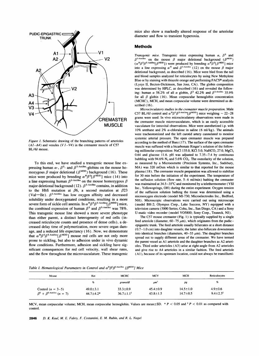
Figure 1. Schematic drawing of the branching patterns of arterioles(Al-A4) and venules (Vl-V4) in the cremaster muscle of C57BL/6J mouse.
To this end, we have studied a transgenic mouse line ex-pressing human a-, ,3s- and ,l3S-Antilles-globins on the mouse ho-mozygous /3 major deletional (3MDD) background (16). Thesemice were produced by breeding a'H#s[/6MDDI mice (14) intoa line expressing human / S-Antilles on the mouse homozygous /major deletional background ( 12). S-Antilles contains, in additionto the HbS mutation at 636, a second mutation at /323(Val-Ile). 5S-Antilles has low oxygen affinity and decreasedsolubility under deoxygenated conditions, resulting in a moresevere form of sickle cell anemia. In a H3 S1 S-Antilles[l MDDI mice,the combined expression of human /3s and / S-Antilles was 78%.This transgenic mouse line showed a more severe phenotypethan either parent, a distinct heterogeneity of red cells (in-creased reticulocyte counts and presence of dense cells), a de-creased delay time of polymerization, more severe organ dam-age, and a reduced life expectancy (16). Now, we demonstratethat aHp S/ S-Antilles[# MDD1 mouse red cells are not only moreprone to sickling, but also to adhesion under in vivo dynamicflow conditions. Furthermore, adhesion and sickling have sig-nificant consequences for red cell velocity, wall shear rates,and the flow throughout the microvasculature. These transgenic
mice also show a markedly altered response of the arteriolardiameter and flow to transient hyperoxia.
Methods
Transgenic mice. Transgenic mice expressing human a, /3s and/3S-Antilles on the mouse /3 major deletional background (0MDD)(aH SoS]Antilles[/MDD) were produced by breeding acH/S[3MDD] miceinto a line expressing a H and / S-Antilles (12) on the mouse / majordeletional background, as described (16). Mice were bled from the tailand blood samples analyzed for reticulocytes by using New MethyleneBlue or by staining with thiazole orange and performing FACS® analysis(Lysys II, Becton-Dickinson, San Jose, CA). The globin compositionwas determined by HPLC, as described ( 16) and revealed the follow-ing: human a 58.2% of all a globin, 3is 42.2% and /3S-Antilles 35.9%for all /3 globin (16). Mean corpuscular hemoglobin concentration(MCHC), MCH, and mean corpuscular volume were determined as de-scribed ( 16).
Microcirculatory studies in the cremaster muscle preparation. MaleC57 BL/6J control and a HospsAntilles[I3MDDI mice weighing - 22-28grams were used. In vivo microcirculatory observations were made inthe cremaster muscle microvasculature, which is an easily accessiblevasculature for intravital observations. Mice were anesthetized i.p. with10% urethane and 2% a-chloralose in saline (6 ml/kg). The animalswere tracheotomized and the left carotid artery cannulated to monitorsystemic arterial pressure. The open cremaster muscle was preparedaccording to the method of Baez ( 17). The surface of the open cremastermuscle was suffused with a bicarbonate Ringer's solution of the follow-ing millimolar composition: NaCl 135.0, KCl 5.0, NaHCO3 27.0, MgCl20.64 and glucose 11.6. pH was adjusted to 7.35-7.4 by continuousbubbling with 94.6% N2 and 5.6% CO2. The osmolarity of the solution,as measured by a Microosmette (Precision Systems, Inc., Sudsbury,MA) was 320 mOsm which is similar to that reported for the mouseplasma ( 18 ). The cremaster muscle preparation was allowed to stabilizefor 30 min before the initiation of the experiment. The temperature ofthe suffusion solution (flow rate, 5-6 ml/min) bathing the cremasterwas maintained at 34.5-35°C and monitored by a telethermometer (YSIInc., Yellowsprings, OH) during the entire experiment. Oxygen tensionof the suffusion solution bathing the tissue was determined using amicrooxygen electrode (model MI-730; Microelectrodes Inc., Bedford,NH). Microscopic observations were carried out using microscope(model BH-2; Olympus Corp., Lake Success, NY) equipped with atelevision camera (5000 Series; Cohu, Inc., San Diego, CA) and a SonyU-matic video recorder (model V05800; Sony Corp., Teaneck, NJ).
The C57 mouse cremaster (Fig. 1) is typically supplied by a singlefeed arteriole (diameter, 60-75 jm), which originates from the pudic-epigastric trunk. The feed arteriole usually bifurcates at a short distance(0.7-1.0 cm) into daughter vessels; the latter also bifurcate downstreaminto identical branches (diameters, 40-55 ym). The daughter branchesspread out to supply different areas of the cremaster. We have termedthe parent vessel as Al arteriole and the daughter branches as A2 arteri-oles. Third order arterioles (A3) arise at right-angle from A2 arteriolesand give rise to A4 arterioles in a similar fashion. The feed arteriole(Al), because of its upstream location, could not always be transillumi-
Table L Hematological Parameters in Control and aHfs's-Anhi11es [J3MDD] Mice
Mouse Hct MCHC MCV MCH Reticulocytes
'%o grams/dl jim3 pg %
Control (n = 3-5) 49.0±3.1 33.3±0.9 45.4±0.9 14.5±1.0 4.9±0.6PS + /3s-Antilles (n = 7) 44.7±4.2* 36.7±1.1P 43.8+1.5 14.7±0.5 9.4±2.31
MCV, mean corpuscular volume; MCH, mean corpuscular hemoglobin. Values are mean±SD. * P < 0.05 and t P < 0.01 as compared withcontrol.
2846 D. K. Kaul, M. E. Fabry, F. Costantini, E. M. Rubin, and R. L. Nagel
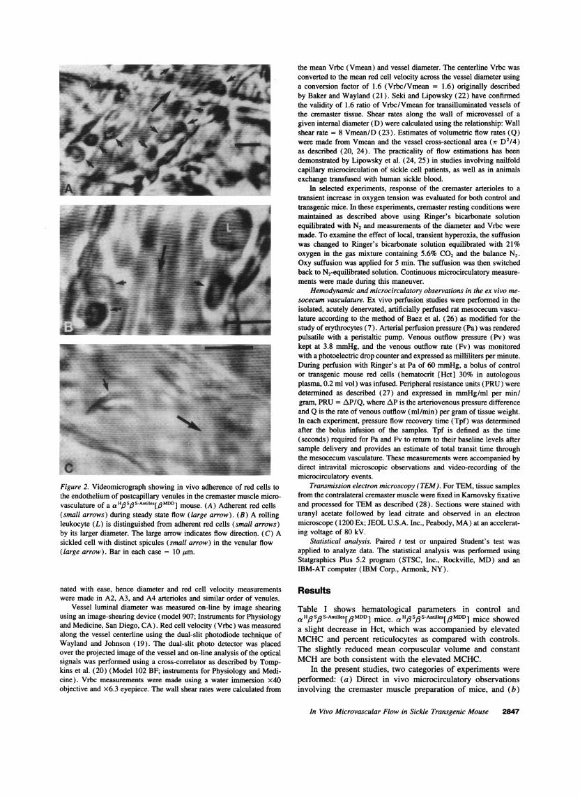
Figure 2. Videomicrograph showing in vivo adherence of red cells tothe endothelium of postcapillary venules in the cremaster muscle micro-vasculature of a a'H#3S#sfAntiIIs[/6MDD] mouse. (A) Adherent red cells(small arrows) during steady state flow (large arrow). (B) A rollingleukocyte (L) is distinguished from adherent red cells (small arrows)by its larger diameter. The large arrow indicates flow direction. (C) Asickled cell with distinct spicules (small arrow) in the venular flow(large arrow). Bar in each case = 10 Ism.
nated with ease, hence diameter and red cell velocity measurementswere made in A2, A3, and A4 arterioles and similar order of venules.
Vessel luminal diameter was measured on-line by image shearingusing an image-shearing device (model 907; Instruments for Physiologyand Medicine, San Diego, CA). Red cell velocity (Vrbc) was measuredalong the vessel centerline using the dual-slit photodiode technique ofWayland and Johnson (19). The dual-slit photo detector was placedover the projected image of the vessel and on-line analysis of the opticalsignals was performed using a cross-correlator as described by Tomp-kins et al. (20) (Model 102 BF; instruments for Physiology and Medi-cine). Vrbc measurements were made using a water immersion x40objective and x6.3 eyepiece. The wall shear rates were calculated from
the mean Vrbc (Vmean) and vessel diameter. The centerline Vrbc wasconverted to the mean red cell velocity across the vessel diameter usinga conversion factor of 1.6 (Vrbc/Vmean = 1.6) originally describedby Baker and Wayland (21). Seki and Lipowsky (22) have confirmedthe validity of 1.6 ratio of Vrbc/Vmean for transilluminated vessels ofthe cremaster tissue. Shear rates along the wall of microvessel of agiven internal diameter (D) were calculated using the relationship: Wallshear rate = 8 Vmean/D (23). Estimates of volumetric flow rates (Q)were made from Vmean and the vessel cross-sectional area (7r D2/4)as described (20, 24). The practicality of flow estimations has beendemonstrated by Lipowsky et al. (24, 25) in studies involving nailfoldcapillary microcirculation of sickle cell patients, as well as in animalsexchange transfused with human sickle blood.
In selected experiments, response of the cremaster arterioles to atransient increase in oxygen tension was evaluated for both control andtransgenic mice. In these experiments, cremaster resting conditions weremaintained as described above using Ringer's bicarbonate solutionequilibrated with N2 and measurements of the diameter and Vrbc weremade. To examine the effect of local, transient hyperoxia, the suffusionwas changed to Ringer's bicarbonate solution equilibrated with 21%oxygen in the gas mixture containing 5.6% CO2 and the balance N2.Oxy suffusion was applied for 5 min. The suffusion was then switchedback to N2-equilibrated solution. Continuous microcirculatory measure-ments were made during this maneuver.
Hemodynamic and microcirculatory observations in the ex vivo me-socecum vasculature. Ex vivo perfusion studies were performed in theisolated, acutely denervated, artificially perfused rat mesocecum vascu-lature according to the method of Baez et al. (26) as modified for thestudy of erythrocytes (7). Arterial perfusion pressure (Pa) was renderedpulsatile with a peristaltic pump. Venous outflow pressure (Pv) waskept at 3.8 mmHg, and the venous outflow rate (Fv) was monitoredwith a photoelectric drop counter and expressed as milliliters per minute.During perfusion with Ringer's at Pa of 60 mmHg, a bolus of controlor transgenic mouse red cells (hematocrit [Hct] 30% in autologousplasma, 0.2 ml vol) was infused. Peripheral resistance units (PRU) weredetermined as described (27) and expressed in mmHg/ml per min/gram, PRU = AP/Q, where AP is the arteriovenous pressure differenceand Q is the rate of venous outflow (ml/min) per gram of tissue weight.In each experiment, pressure flow recovery time (Tpf) was determinedafter the bolus infusion of the samples. Tpf is defined as the time(seconds) required for Pa and Fv to return to their baseline levels aftersample delivery and provides an estimate of total transit time throughthe mesocecum vasculature. These measurements were accompanied bydirect intravital microscopic observations and video-recording of themicrocirculatory events.
Transmission electron microscopy (TEM). For TEM, tissue samplesfrom the contralateral cremaster muscle were fixed in Karnovsky fixativeand processed for TEM as described (28). Sections were stained withuranyl acetate followed by lead citrate and observed in an electronmicroscope (1200 Ex; JEOL U.S.A. Inc., Peabody, MA) at an accelerat-ing voltage of 80 kV.
Statistical analysis. Paired t test or unpaired Student's test wasapplied to analyze data. The statistical analysis was performed usingStatgraphics Plus 5.2 program (STSC, Inc., Rockville, MD) and anIBM-AT computer (IBM Corp., Armonk, NY).
Results
Table I shows hematological parameters in control anda H3 S# S-Antilles[ / MDD ] mice. aHo So S-Antilles [ MDDI] mice showeda slight decrease in Hct, which was accompanied by elevatedMCHC and percent reticulocytes as compared with controls.The slightly reduced mean corpuscular volume and constantMCH are both consistent with the elevated MCHC.
In the present studies, two categories of experiments wereperformed: (a) Direct in vivo microcirculatory observationsinvolving the cremaster muscle preparation of mice, and (b)
In Vivo Microvascular Flow in Sickle Transgenic Mouse 2847
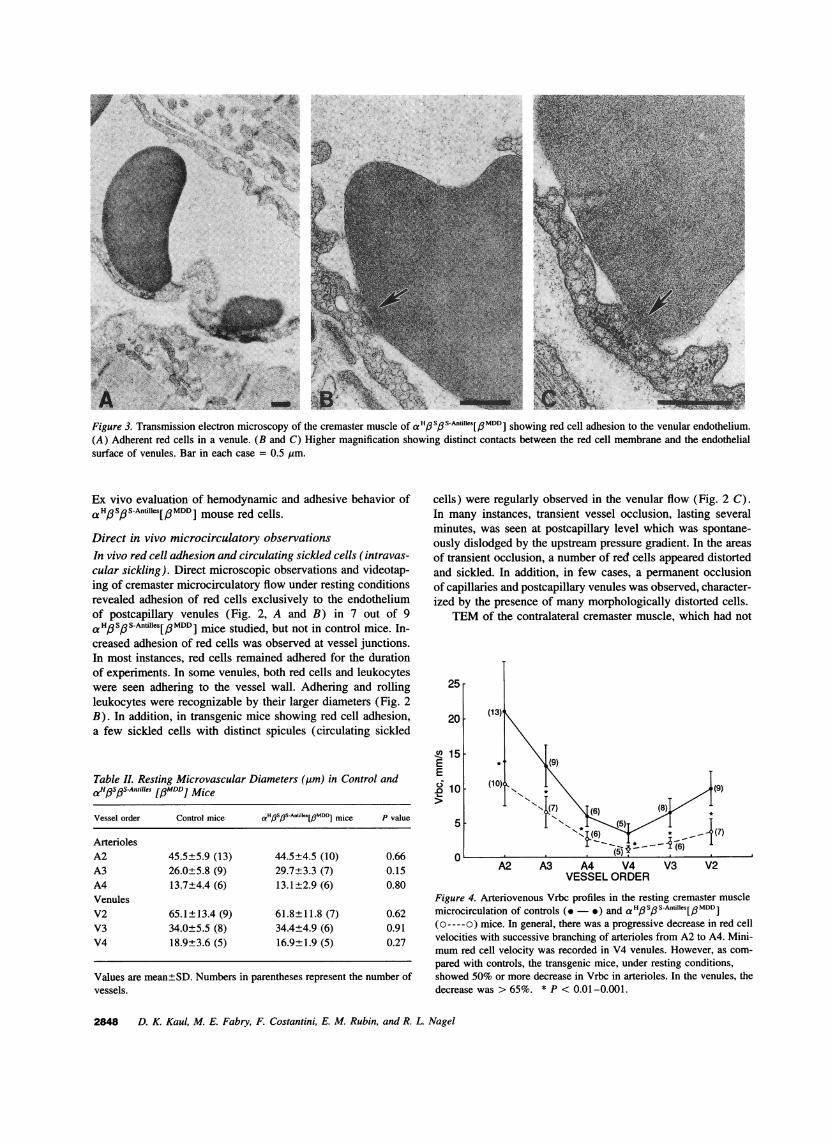
Figure 3. Transmission electron microscopy of the cremaster muscle of aH#S#S-Anfies[#MDD] showing red cell adhesion to the venular endothelium.(A) Adherent red cells in a venule. (B and C) Higher magnification showing distinct contacts between the red cell membrane and the endothelialsurface of venules. Bar in each case = 0.5 Mm.
Ex vivo evaluation of hemodynamic and adhesive behavior ofaHoSpS-Antilles[/PMDD I mouse red cells.
Direct in vivo microcirculatory observationsIn vivo red cell adhesion and circulating sickled cells (intravas-cular sickling). Direct microscopic observations and videotap-ing of cremaster microcirculatory flow under resting conditionsrevealed adhesion of red cells exclusively to the endotheliumof postcapillary venules (Fig. 2, A and B) in 7 out of 9aH#S#SAm11es[#MDD ] mice studied, but not in control mice. In-creased adhesion of red cells was observed at vessel junctions.In most instances, red cells remained adhered for the durationof experiments. In some venules, both red cells and leukocyteswere seen adhering to the vessel wall. Adhering and rollingleukocytes were recognizable by their larger diameters (Fig. 2B). In addition, in transgenic mice showing red cell adhesion,a few sickled cells with distinct spicules (circulating sickled
Table I. Resting Microvascular Diameters (Mm) in Control andaHfs/3s-A"ni1es [3MDD] Mice
Vessel order Control mice aH6SpS-AnfiIks[#MDDi mice P value
ArteriolesA2 45.5±5.9 (13) 44.5±4.5 (10) 0.66A3 26.0±5.8 (9) 29.7±3.3 (7) 0.15A4 13.7±4.4 (6) 13.1±2.9 (6) 0.80VenulesV2 65.1±13.4 (9) 61.8±11.8 (7) 0.62V3 34.0±5.5 (8) 34.4±4.9 (6) 0.91V4 18.9±3.6 (5) 16.9±1.9 (5) 0.27
Values are mean±SD. Numbers in parentheses represent the number ofvessels.
cells) were regularly observed in the venular flow (Fig. 2 C).In many instances, transient vessel occlusion, lasting severalminutes, was seen at postcapillary level which was spontane-ously dislodged by the upstream pressure gradient. In the areasof transient occlusion, a number of red cells appeared distortedand sickled. In addition, in few cases, a permanent occlusionof capillaries and postcapillary venules was observed, character-ized by the presence of many morphologically distorted cells.
TEM of the contralateral cremaster muscle, which had not
25
20 (13)'
4 15E * (9)E
10 (10)*(9)07) (6) (8)
o~~~~~~* (5)
A2 A3 A4 V4 V3 V2VESSEL ORDER
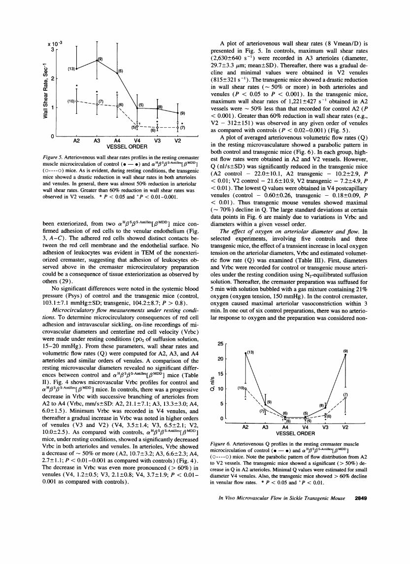
Figure 4. Arteriovenous Vrbc profiles in the resting cremaster musclemicrocirculation of controls (.- *) and aHs,3s-AntiIles[f3MDD](o----o) mice. In general, there was a progressive decrease in red cellvelocities with successive branching of arterioles from A2 to A4. Mini-mum red cell velocity was recorded in V4 venules. However, as com-pared with controls, the transgenic mice, under resting conditions,showed 50% or more decrease in Vrbc in arterioles. In the venules, thedecrease was > 65%. * P < 0.01-0.001.
2848 D. K. Kaul, M. E. Fabry, F. Costantini, E. M. Rubin, and R. L Nagel
x lo-33 r
U0a)
co
=c 1
0A2 A3 A4 V4
VESSEL ORDERV3 V.
Figure 5. Arteriovenous wall shear rates profiles in the resting C
muscle microcirculation of control (.- ) and a HoSoS-Antille(o----o) mice. As is evident, during resting conditions, the tbmice showed a drastic reduction in wall shear rates in both aiand venules. In general, there was almost 50% reduction in awall shear rates. Greater than 60% reduction in wall shear ralobserved in V2 vessels. * P < 0.05 and +P < 0.01-0.001.
been exteriorized, from two a H3SPS-Antilles[3MDDI mifirmed adhesion of red cells to the venular endotheliu3, A-C). The adhered red cells showed distinct conttween the red cell membrane and the endothelial surfadhesion of leukocytes was evident in TEM of the noorized cremaster, suggesting that adhesion of leukoc3served above in the cremaster microcirculatory prelcould be a consequence of tissue exteriorization as obseothers (29).
No significant differences were noted in the systemipressure (Psys) of control and the transgenic mice (103.1±7.1 mmHg±SD; transgenic, 104.2±8.7; P > 0.
Microcirculatory flow measurements under restinAtions. To determine microcirculatory consequences ofadhesion and intravascular sickling, on-line recording-crovascular diameters and centerline red cell velocitywere made under resting conditions (P02 of suffusion s15-20 mmHg). From these parameters, wall shear ravolumetric flow rates (Q) were computed for A2, A3,arterioles and similar orders of venules. A comparisoiresting microvascular diameters revealed no significanences between control and a HpSp SAntilles[/PMDD] miceII). Fig. 4 shows microvascular Vrbc profiles for cona Ho ,S Antilles[,a MDD] mice. In controls, there was a proldecrease in Vrbc with successive branching of arteriolA2 to A4 (Vrbc, mn/s±SD: A2, 21.1+7.1; A3, 13.3:+6.0±1.5). Minimum Vrbc was recorded in V4 venu]thereafter a gradual increase in Vrbc was noted in higheof venules (V3 and V2) (V4, 3.5±1.4; V3, 6.5±210.0+2.5). As compared with controls, a H,6SpS-Anfillesmice, under resting conditions, showed a significantly deVrbc in both arterioles and venules. In arterioles, Vrbca decrease of - 50% or more (A2, 10.7+3.2; A3, 6.6+'2.7 1.1;P < 0.01-0.001 as compared with controls) (The decrease in Vrbc was even more pronounced (>(venules (V4, 1.2+0.5; V3, 2.1±0.8; V4, 3.7±1.9;PP0.001 as compared with controls).
A plot of arteriovenous wall shear rates (8 Vmean/D) ispresented in Fig. 5. In controls, maximum wall shear rates(2,630+640 s-') were recorded in A3 arterioles (diameter,29.7+3.3 gim; mean±SD). Thereafter, there was a gradual de-cline and minimal values were obtained in V2 venules(815±321 s '). The transgenic mice showed a drastic reductionin wall shear rates (- 50% or more) in both arterioles andvenules (P < 0.05 to P < 0.001). In the transgenic mice,maximum wall shear rates of 1,221±427 s-' obtained in A2vessels were - 50% less than that recorded for control A2 (P
(9) < 0.001). Greater than 60% reduction in wall shear rates (e.g.,* V2 - 312±151) was observed in any given order of venules
.(7) as compared with controls (P < 0.02-0.001) (Fig. 5).A plot of averaged arteriovenous volumetric flow rates (Q)
2in the resting microvasculature showed a parabolic pattern inboth control and transgenic mice (Fig. 6). In each group, high-
cremaster est flow rates were obtained in A2 and V2 vessels. However,s[/3MDD] Q (nl/s±SD) was significantly reduced in the transgenic miceransgenic (A2 control - 22.0+ 10.1, A2 transgenic - 10.2±2.9, Prteriolesrteriolar < 0.01; V2 control - 21.6±10.9, V2 transgenic - 7.2±4.9, Ptes was < 0.01). The lowest Q values were obtained in V4 postcapillary
venules (control - 0.60+0.26, transgenic - 0.18±0.09, P< 0.01). Thus transgenic mouse venules showed maximal(- 70%) decline in Q. The large standard deviations at certaindata points in Fig. 6 are mainly due to variations in Vrbc and
ice con- diameters within a given vessel order.m (Fig. The effect of oxygen on arteriolar diameter and flow. Inacts be- selected experiments, involving five controls and threeFace. No transgenic mice, the effect of a transient increase in local oxygennexteri- tension on the arteriolar diameters, Vrbc and estimated volumet-ytes ob- ric flow rate (Q) was examined (Table Ill). First, diametersparation and Vrbc were recorded for control or transgenic mouse arteri-,rved by oles under the resting condition using N2-equilibrated suffusion
solution. Thereafter, the cremaster preparation was suffused foric blood 5 min with solution bubbled with a gas mixture containing 21%control, oxygen (oxygen tension, 150 mmHg). In the control cremaster,.8). oxygen caused maximal arteriolar vasoconstriction within 3g condi- min. In one out of six control preparations, there was no arterio-red cell lar response to oxygen and the preparation was considered non-s of mi-(Vrbc)
;olution,ates andand A4n of theIt differ-(Table
trol andgressiveles from3.0; A4,les, andr orders'.1; V2,s[ MDD ]
creasedshowed2.3; A4,Fig. 4).60%) in- 0.01-
25
20
15cn
Co5
0
,(9)
A4 V4VESSEL ORDER
V3 V2
Figure 6. Arteriovenous Q profiles in the resting cremaster musclemicrocirculation of control (- *) and a'H,6s,6s3 Antifes[QMDD](o----o) mice. Note the parabolic pattern of flow distribution from A2to V2 vessels. The transgenic mice showed a significant (> 50%) de-crease in Q in A2 arterioles. Minimal Q values were estimated for smalldiameter V4 venules. Also, the transgenic mice showed > 60% declinein venular flow rates. * P < 0.05 and +P < 0.01.
In Vivo Microvascular Flow in Sickle Transgenic Mouse 2849
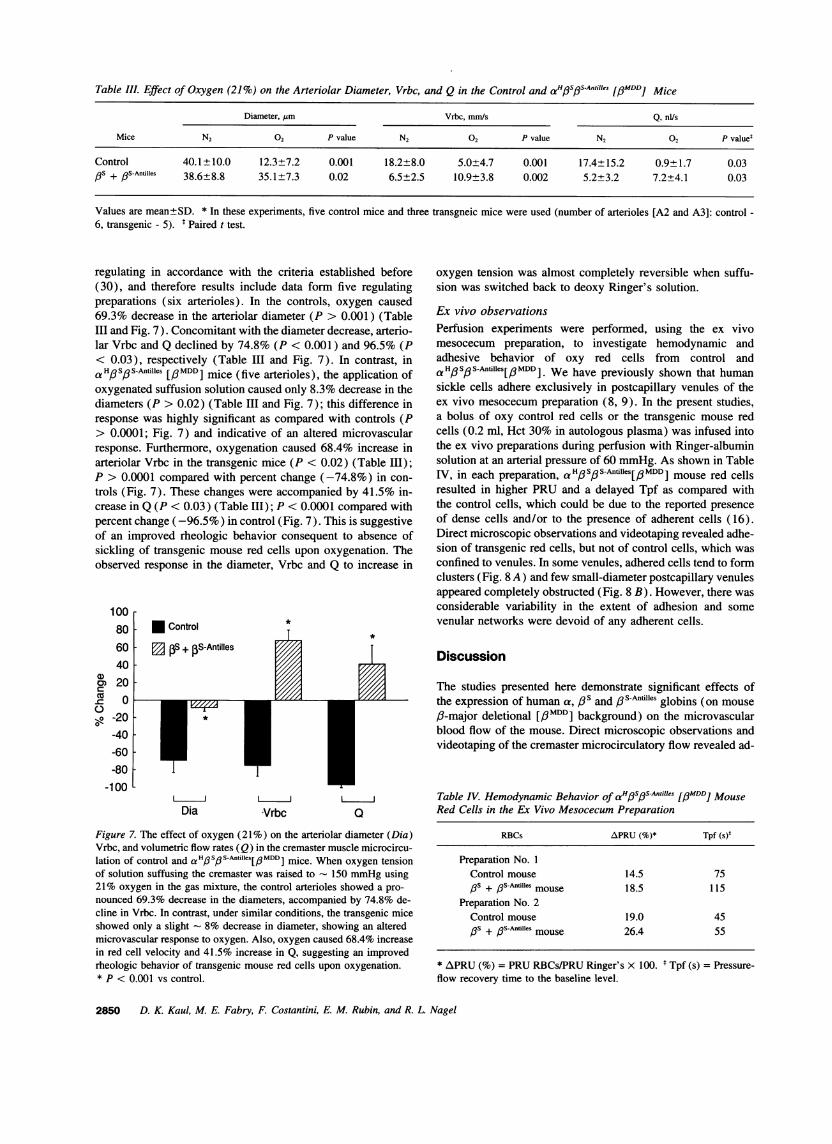
Table III. Effect of Oxygen (21%) on the Arteriolar Diameter, Vrbc, and Q in the Control and aHfSfS-Antilles [pMDD] Mice
Diameter, pm Vrbc, mm/s Q, nUs
Mice N2 02 P value N2 02 P value N2 02 P valuet
Control 40.1±10.0 12.3±7.2 0.001 18.2±8.0 5.0±4.7 0.001 17.4±15.2 0.9±1.7 0.03PS + /3S-Antilles 38.6±8.8 35.1±7.3 0.02 6.5±2.5 10.9±3.8 0.002 5.2±3.2 7.2±4.1 0.03
Values are mean±SD. * In these experiments, five control mice and three transgneic mice were used (number of arterioles [A2 and A3]: control -6, transgenic - 5). t Paired t test.
regulating in accordance with the criteria established before(30), and therefore results include data form five regulatingpreparations (six arterioles). In the controls, oxygen caused69.3% decrease in the arteriolar diameter (P > 0.001) (TableIII and Fig. 7). Concomitant with the diameter decrease, arterio-lar Vrbc and Q declined by 74.8% (P < 0.001) and 96.5% (P< 0.03), respectively (Table III and Fig. 7). In contrast, ina HpSoS-Antilles [/#MDD] mice (five arterioles), the application ofoxygenated suffusion solution caused only 8.3% decrease in thediameters (P > 0.02) (Table III and Fig. 7); this difference inresponse was highly significant as compared with controls (P> 0.0001; Fig. 7) and indicative of an altered microvascularresponse. Furthermore, oxygenation caused 68.4% increase inarteriolar Vrbc in the transgenic mice (P < 0.02) (Table III);P > 0.0001 compared with percent change (-74.8%) in con-trols (Fig. 7). These changes were accompanied by 41.5% in-crease in Q (P < 0.03) (Table III); P < 0.0001 compared withpercent change (-96.5%) in control (Fig. 7). This is suggestiveof an improved rheologic behavior consequent to absence ofsickling of transgenic mouse red cells upon oxygenation. Theobserved response in the diameter, Vrbc and Q to increase in
1008060
0
-20
-40
-60
-80
-100
* Control
[ PS + pS-Antilles
L
Dia Vrbc
oxygen tension was almost completely reversible when suffu-sion was switched back to deoxy Ringer's solution.
Ex vivo observationsPerfusion experiments were performed, using the ex vivomesocecum preparation, to investigate hemodynamic andadhesive behavior of oxy red cells from control andaHo/SoS-Antilles[/PMDD]. We have previously shown that humansickle cells adhere exclusively in postcapillary venules of theex vivo mesocecum preparation (8, 9). In the present studies,a bolus of oxy control red cells or the transgenic mouse redcells (0.2 ml, Hct 30% in autologous plasma) was infused intothe ex vivo preparations during perfusion with Ringer-albuminsolution at an arterial pressure of 60 mmHg. As shown in TableIV, in each preparation, a H/So/ S-Antilles[oMDDI mouse red cellsresulted in higher PRU and a delayed Tpf as compared withthe control cells, which could be due to the reported presenceof dense cells and/or to the presence of adherent cells (16).Direct microscopic observations and videotaping revealed adhe-sion of transgenic red cells, but not of control cells, which wasconfined to venules. In some venules, adhered cells tend to formclusters (Fig. 8 A) and few small-diameter postcapillary venulesappeared completely obstructed (Fig. 8 B). However, there wasconsiderable variability in the extent of adhesion and somevenular networks were devoid of any adherent cells.
Discussion
Q
Figure 7. The effect of oxygen (21%) on the arteriolar diameter (Dia)Vrbc, and volumetric flow rates (Q) in the cremaster muscle microcircu-lation of control and aH3SpfS-Antilles[aMDD] mice. When oxygen tensionof solution suffusing the cremaster was raised to 150 mmHg using21% oxygen in the gas mixture, the control arterioles showed a pro-nounced 69.3% decrease in the diameters, accompanied by 74.8% de-cline in Vrbc. In contrast, under similar conditions, the transgenic miceshowed only a slight 8% decrease in diameter, showing an altered
microvascular response to oxygen. Also, oxygen caused 68.4% increasein red cell velocity and 41.5% increase in Q, suggesting an improvedrheologic behavior of transgenic mouse red cells upon oxygenation.* P < 0.001 vs control.
The studies presented here demonstrate significant effects ofthe expression of human a, 13s and # S-Antilles globins (on mouse/-major deletional [/3i"DD] background) on the microvascularblood flow of the mouse. Direct microscopic observations andvideotaping of the cremaster microcirculatory flow revealed ad-
Table IV. Hemodynamic Behavior of aH/3s3s#Anti~es [/3MDD] MouseRed Cells in the Ex Vivo Mesocecum Preparation
RBCs APRU (%)* Tpf (s)5
Preparation No. 1Control mouse 14.5 75,3s + ,3S-Antifles mouse 18.5 115
Preparation No. 2Control mouse 19.0 45
)6S + 0S-Antilles mouse 26.4 55
* APRU (%) = PRU RBCs/PRU Ringer's x 100. t Tpf (s) = Pressure-flow recovery time to the baseline level.
2850 D. K. Kaul, M. E. Fabry, F. Costantini, E. M. Rubin, and R. L. Nagel
CZ
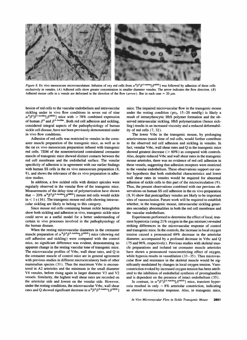
Figure 8. Ex vivo mesocecum microvasculature. Infusion of oxy red cells from aH#S3S-Anfi1es[J3MDD] was followed by adhesion of these cellsexclusively in venules. (A) Adhered cells show greater concentration in smaller diameter venules. The arrow indicates the flow direction. (B)Adhered mouse cells in a venule are deformed in the direction of the flow (arrow). Bar in each case = 20 jLm.
hesion of red cells to the vascular endothelium and intravascularsickling under in vivo flow conditions in seven out of nineaHoSos-Antilles[PMDD] mice with > 78% combined expressionof human 13s and 63S-Antilles. Both red cell adhesion and sickling,considered integral aspects of the pathophysiology of humansickle cell disease, have not been previously demonstrated underin vivo flow conditions.
Adhesion of red cells was restricted to venules in the crem-aster muscle preparation of the transgenic mice, as well as inthe rat ex vivo mesocecum preparation infused with transgenicred cells. TEM of the nonexteriorized contralateral cremastermuscle of transgenic mice showed distinct contacts between thered cell membrane and the endothelial surface. The venularspecificity of adhesion is in agreement with our earlier findingswith human SS cells in the ex vivo mesocecum preparation (8,9), and shows the relevance of the ex vivo preparation in adhe-sion studies.
In addition, a few sickled cells with distinct spicules wereregularly observed in the venular flow of the transgenic mice.Measurements of the delay time of polymerization have shownthat 20% aHo3SoS-Anflilies[I3MDD] mouse red cells would sicklein < 1 s ( 16). The transgenic mouse red cells showing intravas-cular sickling are likely to belong to this category.
Since mouse red cells containing human sickle hemoglobinshow both sickling and adhesion in vivo, transgenic sickle micecould serve as a useful model for a better understanding ofcertain in vivo processes involved in the pathophysiology ofthe human disease.
When the resting microvascular diameters in the cremastermuscle preparation of aHoIS3 SAntilles[PMDD] mice (showing redcell adhesion and sickling) were compared with the controlmice, no significant difference was evident, demonstrating noapparent change in the resting vascular tone of transgenic mice.The microvascular profiles of Vrbc, wall shear rates, and Q inthe cremaster muscle of control mice are in general agreementwith previous studies in different microcirculatory beds of othermammalian species (31). Thus the maximum Vrbc is encoun-tered in A2 arterioles and the minimum in the small diameterV4 venules, before rising again in larger diameter V3 and V2vessels. Similarity, the highest wall shear rates are recorded onthe arteriolar side and lowest on the venular side. However,under the resting conditions, the microvascular Vrbc, wall shearrates and Q showed significant decrease in aH'3SoS/3sAntifes[/PMDD]
mice. The impaired microvascular flow in the transgenic mouseunder the resting condition (po2, 15-20 mmHg) is likely aresult of intraeythrocytic HbS polymer formation and the ob-served intravascular sickling. HbS polymerization (hence sick-ling) results in an increased viscosity and a reduced deformabil-ity of red cells (7, 32).
The lower Vrbc in the transgenic mouse, by prolongingarteriovenous transit time of red cells, would further contributeto the observed red cell adhesion and sickling in venules. Infact, venular Vrbc, wall shear rates and Q in the transgenic miceshowed greatest decrease (> 60%) as compared with controls.Also, despite reduced Vrbc and wall shear rates in the transgenicmouse arterioles, there was no evidence of red cell adhesion inthese vessels, suggesting that adhesion receptors were restrictedto the venular endothelium. These observations support our ear-lier hypothesis that both endothelial characteristics and lowerwall shear rates in venules would be required for abnormaladhesion of sickle cells to this part of the microcirculation (9).Thus, the present observations combined with our previous ob-servations on human SS cell adhesion in the ex vivo preparation(8, 9) show that postcapillary venules are likely to be importantsites of vasoocclusion. Future work will be required to establishwhether, in the transgenic mouse, intravascular sickling gener-ates secondary abnormalities in both the red cell membrane andthe vascular endothelium.
Experiments performed to determine the effect of local, tran-sient hyperoxia (using 21% oxygen in the gas mixture) revealedstriking differences in the microvascular response of controland transgenic mice. In the controls, the increase in local oxygentension caused a pronounced 69% decrease in the arteriolardiameter, accompanied by a profound decrease in Vrbc and Q(75 and 96%, respectively). Previous studies with skeletal mus-cle preparations and isolated rat cremaster muscle arterioleshave shown a pronounced vasoconstricting effect of oxygen,while hypoxia results in vasodilation (33-35). Thus microvas-cular flow and resistance in the skeletal muscle would be sig-nificantly modulated by changes in local oxygen tension. Vaso-constriction evoked by increased oxygen tension has been attrib-uted to the inhibition of endothelial synthesis of prostaglandinsand is dependent on the presence of intact endothelium (35).
In contrast, in a 1H, Sp S-Antilles[pMDD ] mice, transient hyper-oxia resulted in only - 8% arteriolar constriction, indicatingan altered microvascular response. Also, in transgenic mice,
In Vivo Microvascular Flow in Sickle Transgenic Mouse 2851
hyperoxia induced a significant net increase in the arteriolarVrbc and Q (68 and 41.5%, respectively) (Fig. 7). In view ofthe observed intravascular sickling during resting conditions,the absence of sickling following the application of oxygenwould indicate unsickling of red cells due to depolymerizationof HbS and HbSAntiles. In general, vasoconstriction results in adecrease in the flow (as seen in control mice) while an increasein the flow is observed after vasodilation (36). In transgenicmice, the net increase in Vrbc and Q during transient hyperoxia,despite a slight vasoconstriction, is likely to be a consequenceof unsickling and a decrease in the bulk viscosity. The bulkviscosity of transgenic mouse red cells is significantly higherin the deoxygenated state (data not shown), which is in accor-dance with our studies on the viscosity behavior of human SScells (7).
On the other hand, the altered vessel diameter response tooxygen in transgenic mice may be in part a consequence of anincrease in the flow. Although the exact mechanism remains tobe elucidated, this response could involve either release of spe-cific endothelial molecules (prostaglandins and/or nitric oxide)or vessel diameter compliance secondary to an increased bloodflow as suggested for flow-mediated vasodilation in large arter-ies (37-39). Blood rheological factors have been implicatedin the flow intermittency observed in sickle cell patients (40),as well as in a depressed postocclusive reactive hyperemia dur-ing the crisis (24). However, there could also be intrinsic differ-ences in the endothelial cell and/or vascular smooth musclefunction between controls and transgenic mice. In particular,additional contributions from possible endothelial abnormalities(inflicted by circulating dense cells, adhesion and intravascularsickling) which could induce, for example, increased prosta-cyclin production (41 ) cannot be ruled out in the altered vascu-lar tone response to oxygen.
To conclude, the transgenic mice expressing human Ps and,S-Antilles on the mouse /-major deletional background exhibitred cell endothelial interaction and intravascular sickling, bothof which have been implicated in the pathophysiology of thehuman sickle cell disease, but never been demonstrated beforein vivo under dynamic flow conditions. In addition, we find anovel phenomenon, an altered microvascular tone, and flowresponse to oxygen in the transgenic mice. One implication ofthis finding is that if an incompletely obstructed vessel couldbe reached to provide adequate level of oxygenation, some ofthe vasoocclusion could be averted. Taken as a whole, the mi-crovascular flow abnormalities would contribute to the observedorgan damage and a reduced life span in these transgenic mice.Finally, the transgenic mouse models present a unique opportu-nity for further investigations of vasoocclusive mechanisms andto test potential therapies in vivo.
Acknowledgments
We would like to thank Xiao-du Liu and Sandra Suzuka for their excel-lent technical assistance.
This work was supported by National Institutes of Health grantsHL-45931, HL-37212, and Al 34064, and by the American Heart Asso-ciation (New York Affiliate).
References
1. Kaul, D. K., and R. L. Nagel. 1993. Sickle cell vasoocclusion: many issuesand some answers. (Base!). 49:5-15.
2. Hebbel, R. P., 0. Yamada, C. F. Moldow, H. S. Jacob, J. G. White, and
J. W. Eaton. 1981. Abnormal adherence of sickle erythrocytes to cultures vascularendothelium: possible mechanism for microvascular occlusion in sickle cell dis-ease. J. Clin. Invest. 65:154-160.
3. Hebbel, R. P., C. F. Moldow, and M. H. Steinberg. 1981. Modulation oferythrocyte-endothelial interactions and the vasoocclusive severity of sicklingdisorders. Blood. 58:947-952.
4. Hoover, R., R. Rubin, G. Wise, and R. Warren. 1985. Adhesion of normaland sickle erythrocytes to endothelial monolayers. Blood. 54:872-876.
5. Mohandas, N., and E. Evans. 1985. Sickle erythrocyte adherence to thevascular endothelium: morphological correlates and the requirement for divalentcations and collagen-binding plasma proteins. J. Clin. Invest. 76:1605-1612.
6. Barabino, G. A., L. V. McIntire, S. G. Eskin, D. A. Sears, and M. Udden.1987. Endothelial interactions with sickle cells, sickle trait, mechanically injured,and normal erythrocytes under controlled flow. Blood. 70:152-157.
7. Kaul, D. K., M. E. Fabry, P. Windisch, S. Baez, and R. L. Nagel. 1983.Erythrocytes in sickle cell anemia are heterogeneous in their rheological andhemodynamic properties. J. Clin. Invest. 72:22-31.
8. Kaul, D. K., M. E. Fabry, and R. L. Nagel. 1989. Microvascular sitesand characteristics of sickle cell adhesion to vascular endothelium in shear flowconditions: pathophysiological implications. Proc. Nat!. Acad. Sci. USA. 86:3356-3360.
9. Kaul, D. K., D. Chen, and J. Zhan. 1994. Adhesion of sickle cells tovascular endothelium is critically dependent on changes in density and shape ofthe cells. Blood. 83:3006-3017.
10. Greaves, D. R., P. Fraser, M. A. Vidal, M. A. Hedges, D. Ropers, L.Luzzatto, and F. Grosveld. 1990. A transgenic mouse model of sickle cell disorder.Nature (Lond.). 343:183-185.
11. Ryan, T. M., T. M. Townes, M. P. Reilly, T. Asakura, R. P. Palmiter, andR. R. Behringer. 1990. Human sickle hemoglobin in transgenic mice. Science(Wash. DC). 247:566-568.
12. Rubin, E. M., H. E. Witkowska, E. Spangler, P. Curtin, N. Mohandas,and B. H. Lubin. 1991. Hypoxia-induced in vivo sickling of transgenic mousered cells. J. Clin. Invest. 87:639-647.
13. Trudel, M., M. E. De Paepe, N. Chretien, S. Nacera, J. Jacmain, M.Sorette, T. Hoang, and Y. Beuzard. 1994. Sickle cell disease of transgenic SADmice. Blood. 84:3189-3197.
14. Fabry, M. E., R. L. Nagel, A. Pachnis, S. M. Suzuka, and F. Costantini.1992. High expression of human Ps3 and a-globin chains in transgenic mice:hemoglobin composition and hematological consequences. Proc. Natl. Acad. Sci.USA. 89:12150-12154.
15. Fabry, M. E., F. Costantini, A. Pachnis, S. M. Suzuka, N. Bank, H. S.Aynedjian, S. M. Factor, and R. L. Nagel. 1992. High expression of human ,#S-and a-globin chains in transgenic mice: erythrocyte abnormalities, organ damage,and the effect of hypoxia. Proc. Natl. Acad. Sci. USA. 89:12155-12159.
16. Fabry, M. E., A. Sengupta, S. M. Suzuka, F. Costantini, E. M. Rubin, J.Hoftrichter, G. Christoph, E. Mancie, D. Culberson, S. M. Factor, and R. L.Nagel. A second generation of transgenic mouse model expressing HbS and HbS-Antilles results in increased phenotypic severity. Blood. 86:2419-2428.
17. Baez, S. 1973. An open cremaster muscle preparation for the study ofblood vessels by in vivo microscopy. Microvasc. Res. 5:384-394.
18. Chen, D., and D. K. Kaul. 1994. Rheological and hemodynamic character-istics of red cells of mouse, rat and human. Biorheology. 31:103-113.
19. Wayland, H., and P. Johnson. 1967. Erythrocyte velocity measurementsin microvessels by a two-slit method. J. Appl. Physiol. 22:333-337.
20. Tompkins, W., R. R. Monti, and M. Intaglietta. 1974. Velocity measure-ments by self-tracking correlator. Rev. Sci. Instrum. 45:647-649.
21. Baker, M., and H. Wayland. 1974. On-line volume flow rate and velocityprofile measurement for blood in microvessels. Microvasc. Res. 7:131-143.
22. Seki, J., and H. H. Lipowsky. 1987. In vivo and in vitro measurementsof red cell velocity under epifluorescent microscopy. Microvasc. Res. 38:110-124.
23. Lipowsky, H. H., S. Usami, and S. Chien. 1980. In vivo measurementsof "apparent viscosity" and microvessel hematocrit in the mesentery of the cat.Microvasc. Res. 19:297-319.
24. Lipowsky, H. H., N. U. Sheikh, and D. M. Katz. 1987. Intravital micros-copy of capillary hemodynamics in sickle cell disease. J. Clin. Invest. 80:117-127.
25. Lipowsky, H. H., S. Usami, and S. Chien. 1982. Human SS red cellrheological behavior in the microcirculation of cremaster muscle. Blood Cells(Berl.). 8:113-126.
26. Baez, S., H. Lamport, and A. Baez. 1960. Pressure effects in livingmicroscopic vessels. In Flow Properties of Blood and Other Biological Systems.A. L. Copley and G. Stainsby, editors. Pergamon Press, London. 122-136.
27. Green, H. D., C. E. Rapela, and M. D. Conard. 1963. Resistance (conduc-tance) and capicitance phenomena in terminal vascular beds. In Handbook ofPhysiology, Section 2 Circulation, Vol. II. W. F. Hamilton and P. Dow, editors.American Physiological Society, Bethesda, MD. 935-960.
28. Kaul, D. K., R. L. Nagel, J. F. Llena, and H. L. Shear. 1994. Cerebralmalaria in mice: demonstration of cytoadherence of infected red cells and micror-
2852 D. K. Kaul, M. E. Fabry, F. Costantini, E. M. Rubin, and R. L. Nagel
heologic correlates. Am. J. Trop. Med. Hyg. 50:512-521.29. Fiebig, E., K. Ley, and K. E. Arfors. 1991. Rapid leukocyte accumulation
by "spontaneous" rolling and adhesion in the exteriorized rabbit mesentery. Int.J. Microcirc. Clin. Exp. 10:127-144.
30. Prewitt, R. L., and P. C. Johnson. 1976. The effect of oxygen on arteriolarred cell velocity and capillary density in the rat cremaster muscle. Microvasc.Res. 12:59-70.
31. Zweifach, B. W., and H. H. Lipowsky. 1984. Pressure-flow relationsin blood and lymph microcirculation. In Handbook of Physiology, Section 2:Cardiovascular System, Vol. IV, Microcirculation (Part I). E. M. Renkin andC. C. Michen, editors. American Physiological Society, Bethesda, MD. 251-308.
32. Nash, G. B., C. S. Johnson, and H. J. Meiselman. 1986. Influence ofoxygen tension on the viscoelastic behavior of red blood cells in sickle celldisease. Blood. 67:110-118.
33. Duling, B. R. 1972. Microvascular responses to alterations in oxygentension. Circ. Res. 31:481-489.
34. Hutchins, P. M., R. F. Bond, and H. D. 1974. Participation of oxygen inthe local control of skeletal muscle microvasculature. Circ. Res. 34:85-93.
35. Messina, E. J., D. Sun, A. Koller, M. S. Wolin, and G. Kaley. 1994.
Increases in oxygen tension evoke arteriolar constriction by inhibiting endothelialprostaglandin synthesis. Microvasc. Res. 48:151-160.
36. Ballard, S. T., M. A. Hill, and C. A. Meininger. 1991. Effect of vasodilationand vasoconstriction on microvascular pressures in skeletal muscle. Microcirc.Endothelium Lymphatics. 7:109-131.
37. Bevan, J. A., E. H. Joyce, and G. C. Wellman. 1988. Flow-dependentvasodilation in a resistance artery still occurs after endothelial removal. Circ. Res.63:980-985.
38. Cooke, J. P., J. Stamler, N. Andon, P. F. Davies, G. McKinely, J. andLoscalzo. 1990. Flow stimulates endothelial cells to release a nitrovasodilator thatis potentiated by reduced thiol. Am. J. Physiol. 259 (Heart Circ. Physiol.28) :H804-H812.
39. Fujii, K., D. D. Heistad, and F. M. Faraci. 1991. Flow-mediated dilationof the basilar artery in vivo. Circ. Res. 69:697-705.
40. Rodgers, G. P., A. N. Schechter, C. T. Noguchi, H. G. Klein, A. W.Neinhuis, and R. F. Bonner. 1984. Periodic microcirculatory flow in patients withsickle-cell disease. N. Engl. J. Med. 311:1534-1538.
41. Chappey, O., M. P. Wautier-Pepin, and J. L. Wautier. 1994. Adhesion oferythrocytes to endothelium in pathological situations: a review article. Nouv.Rev. Fr. Hematol. 36:281-288.
In Vivo Microvascular Flow in Sickle Transgenic Mouse 2853


![[CANCER RESEARCH 59, 5012–5016, October 1, 1999] In Vivo ... · [CANCER RESEARCH 59, 5012–5016, October 1, 1999] In Vivo Prediction of Vascular Susceptibility to Vascular Endothelial](https://img.pdfslide.us/doc/110x75/5f246678cd32495f5b1ca216/cancer-research-59-5012a5016-october-1-1999-in-vivo-cancer-research.jpg)