Embed Size (px)
Citation preview
94 Copyright © 2015 Korean Academy of Periodontology
pISSN 2093-2278eISSN 2093-2286In vivo comparison between the effects
of chemically modified hydrophilic and anodically oxidized titanium surfaces on initial bone healingHyo-Jung Lee1,†, Il-Hyung Yang2,†, Seong-Kyun Kim3, In-Sung Yeo3, Taek-Ka Kwon4,*1Department of Periodontology, Section of Dentistry, Seoul National University Bundang Hospital, Seongnam, Korea2Department of Orthodontics and Dental Research Institute, Seoul National University School of Dentistry, Seoul, Korea3Department of Prosthodontics and Dental Research Institute, Seoul National University School of Dentistry, Seoul, Korea4Department of Prosthetic Dentistry, St. Vincent Hospital, Catholic University of Korea, Suwon, Korea
Research ArticleJ Periodontal Implant Sci 2015;45:94-100http://dx.doi.org/10.5051/jpis.2015.45.3.94
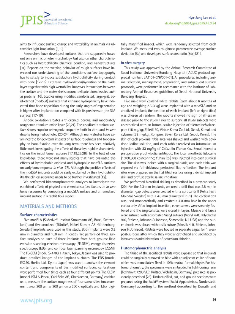
Purpose: The aim of this study was to investigate the combined effects of physical and chemical surface factors on in vivo bone responses by comparing chemically modified hy-drophilic sandblasted, large-grit, acid-etched (modSLA) and anodically oxidized hydropho-bic implant surfaces.Methods: Five modSLA implants and five anodized implants were inserted into the tibiae of five New Zealand white rabbits (one implant for each tibia). The characteristics of each surface were determined using field emission scanning electron microscopy, energy disper-sive spectroscopy, and confocal laser scanning microscopy before the installation. The ex-perimental animals were sacrificed after 1 week of healing and histologic slides were pre-pared from the implant-tibial bone blocks removed from the animals. Histomorphometric analyses were performed on the light microscopic images, and bone-to-implant contact (BIC) and bone area (BA) ratios were measured. Nonparametric comparison tests were ap-plied to find any significant differences (P<0.05) between the modSLA and anodized sur-faces.Results: The roughness of the anodized surface was 1.22±0.17 μm in Sa, which was within the optimal range of 1.0-2.0 μm for a bone response. The modSLA surface was significantly rougher at 2.53±0.07 μm in Sa. However, the modSLA implant had significantly higher BIC than the anodized implant (P=0.02). Furthermore, BA ratios did not significantly differ be-tween the two implants, although the anodized implant had a higher mean value of BA (P>0.05).Conclusions: Within the limitations of this study, the hydrophilicity of the modSLA surface may have a stronger effect on in vivo bone healing than optimal surface roughness and surface chemistry of the anodized surface.
Keywords: Animal experimentation; Dental implants; Histology; Osseointegration.
Received: Apr. 5, 2015Accepted: Apr. 29, 2015
*Correspondence: Taek-Ka KwonDepartment of Prosthetic Dentistry, St. Vincent Hospital, the Catholic University of Korea, 93 Jungbu-daero, Paldal-gu, Suwon 422-723, KoreaE-mail: [email protected]: +82-31-249-7670Fax: +82-31-258-3352
†Hyo-Jung Lee and Il-Hyung Yang contributed equally to this study.
INTRODUCTION
Successful osseointegration of titanium (Ti) implants is partly determined by how the im-planted materials influence bone responses at the cell-biomaterial interface [1,2]. Such events occurring between the bone and implant surface are influenced by a variety of spe-cific surface properties, including topography, structure, chemistry, surface charge, and wet-tability [3-6]. Of these, surface topography has been particularly well studied. Researchers have developed numerous additive and subtractive surface modification techniques to im-prove osseointegration by altering implant surface topography, thus enhancing bone-to-im-plant contact (BIC) and increasing biomechanical interlocking with bone [7,8]. The clinical introduction of a novel implant surface has also helped to advance the field [9]. This product
This is an Open Access article distributed under the terms of the Creative Commons Attribution Non-Commercial License (http://creativecommons.org/licenses/by-nc/3.0/).
Hyo-Jung Lee et al.
dx.doi.org/10.5051/jpis.2015.45.3.94
www.jpis.org 95
aims to influence surface charge and wettability in animals via ul-traviolet light irradiation [9,10].
Researchers have developed surfaces that are supposedly based not only on micrometre morphology, but also on other characteris-tics such as hydrophilicity, chemical bonding, and nanostructures [11]. Reports on the wetting behavior of rough surfaces have in-creased our understanding of the conditions surface topography has to satisfy to induce satisfactory hydrophilicity during contact with bone [12-15]. Extensive hydroxylation/hydration of the oxide layer, together with high wettability, improves interactions between the surface and the water shells around delicate biomolecules such as proteins [16]. Studies using modified sandblasted, large-grit, ac-id-etched (modSLA) surfaces that enhance hydrophilicity have indi-cated that bone apposition during the early stages of regeneration is higher after implantation compared with its predecessor (the SLA surface) [17-19].
Anodic oxidation creates a thickened, porous, and moderately roughened titanium oxide layer [20,21]. The anodized titanium sur-face shows superior osteogenic properties both in vitro and in vivo despite being hydrophobic [20-24]. Although many studies have ex-amined the longer-term impacts of surface roughness and topogra-phy on bone fixation over the long term, there has been relatively little work investigating the effects of these hydrophilic characteris-tics on the initial bone response [17,19,25,26]. To the best of our knowledge, there were not many studies that have evaluated the effects of hydrophobic oxidized and hydrophilic modSLA surfaces on early bone response in vivo [27]. Although the positive effects of the modSLA implants could be easily explained by their hydrophilic-ity, the clinical relevance needs to be further investigated [12].
We performed histomorphometric analyses to investigate the combined effects of physical and chemical surface factors on in vivo bone responses by comparing a modSLA surface and an anodized implant surface in a rabbit tibia model.
MATERIALS AND METHODS
Surface characteristicsFive modSLA (SLActive®, Institut Straumann AG, Basel, Switzer-
land) and five anodized (TiUnite®, Nobel Biocare AB, Göthenburg, Sweden) implants were used in this study. Both implants were 3.3 mm in diameter and 10.0 mm in length. We performed three sur-face analyses on each of three implants from both groups: field emission scanning electron microscopy (FE-SEM), energy dispersive spectroscopy (EDS), and confocal laser scanning microscopy (CLSM). The FE-SEM (model S-4700, Hitachi, Tokyo, Japan) was used to pro-duce detailed images of the implant surfaces. The EDS (model EX220, Horiba Ltd., Kyoto, Japan) was used to analyze the element content and components of the modified surfaces; calibrations were performed four times each at four different points. The CLSM (model LSM 5-Pascal, Carl Zeiss AG, Oberkochen, Germany) enabled us to measure the surface roughness of four screw sides (measure-ment area: 300 μm × 300 μm on a 200× optically and 1.5× digi-
tally magnified image), which were randomly selected from each implant. We measured two roughness parameters: average surface deviation (Sa) and developed surface area ratio (Sdr) [21].
In vivo surgeryThis study was approved by the Animal Research Committee of
Seoul National University Bundang Hospital (IACUC protocol ap-proval number: BA1101-076/001-01). All procedures, including ani-mal selection, management, preparation, and subsequent surgical protocols, were performed in accordance with the Institute of Lab-oratory Animal Resources guidelines of Seoul National University Bundang Hospital.
Five male New Zealand white rabbits (each about 6 months of age and weighing 2.5-3 kg) were implanted with a modSLA and an anodized implant; the location of each implant (left or right tibia) was chosen at random. The rabbits showed no sign of illness or disease prior to the study. Prior to surgery, all study subjects were anesthetized with an intramuscular injection of tiletamine/zolaze-pam (15 mg/kg; Zoletil 50, Virbac Korea Co. Ltd., Seoul, Korea) and xylazine (33 mg/kg; Rompun, Bayer Korea Ltd., Seoul, Korea). The skin of each proximal tibia area was shaved and washed with povi-done iodine solution, and each rabbit received an intramuscular injection with 33 mg/kg of Cefazolin (Yuhan Co., Seoul, Korea), a preoperative prophylactic antibiotic. The local anesthetic lidocaine (1:100,000 epinephrine; Yuhan Co.) was injected into each surgical site. The skin was incised with a surgical blade, and each tibia was exposed via full-thickness periosteal flap reflection. The implant sites were prepared on the flat tibial surface using a dental implant drill and profuse sterile saline irrigation.
We performed bicortical drilling as described in a previous study [20]. For the 3.3-mm implants, we used a drill that was 2.8 mm in diameter; gap defects were created with a cortical drill (Astra Tech, Mölndal, Sweden) with a 4.0 mm diameter (Fig. 1). The cortical drill was used monocortically and created a 4.0-mm hole in the upper cortex only. After implant insertion, cover screws were securely fas-tened and the surgical sites were closed in layers. Muscle and fascia were sutured with absorbable Vicryl sutures (Vicryl 4-0, Polyglactin 910, Ethicon, Johnson & Johnson, Somerville, NJ, USA) and the out-er dermis was closed with a silk suture (Mersilk 4-0, Ethicon, John-son & Johnson). Rabbits were housed in separate cages for 1 week post-surgery, after which they were anesthetized and sacrificed by intravenous administration of potassium chloride.
Histomorphometric analysisThe tibiae of the sacrificed rabbits were exposed so that implants
could be surgically removed en bloc with an adjacent collar of bone, which was immediately fixed in 10% neutral formaldehyde. For his-tomorphometry, the specimens were embedded in light-curing resin (Technovit 7200 VLC, Kultzer, Wehrheim, Germany) prepared as pre-viously described [28]. Undecalcified, cut, and ground sections were prepared using the Exakt® system (Exakt Apparatebau, Norderstedt, Germany) according to the method described by Donath and
Bone healing of implant with modified hydrophilic surface
dx.doi.org/10.5051/jpis.2015.45.3.94
www.jpis.org96
Sdr) between the test and control implants. The Wilcoxon signed-rank test was used to determine statistically significant differences in BIC and BA between the groups. Significance was defined as P<0.05.
RESULTS
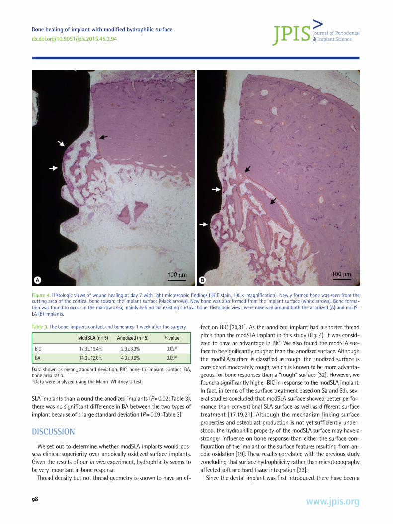
Analysis of surface characteristicsThe modSLA surface comprised 70.3% titanium, 29.3% oxide,
and no phosphorous, while the anodized surface was 26.1% titani-um, 69.8% oxide, and 3.5% phosphorus (Table 1). The FE-SEM im-ages of each surface are shown in Fig. 3. Magnification at 5,000× revealed the anodized surface to be scattered with many volcano-like porous structures. The modSLA surface had a sharp, irregular pattern produced by the sandblasting and acid-etching processes. At 50,000× magnification, the anodized implant was characterized by a relatively smooth surface composed of large micro-pores and small nanopores. This contrasted with the relatively rough surface observed on the hydrophilic modSLA implant, which exhibited beadings of approximately 1-2 μm diameter, along with 0.1-0.2 μm
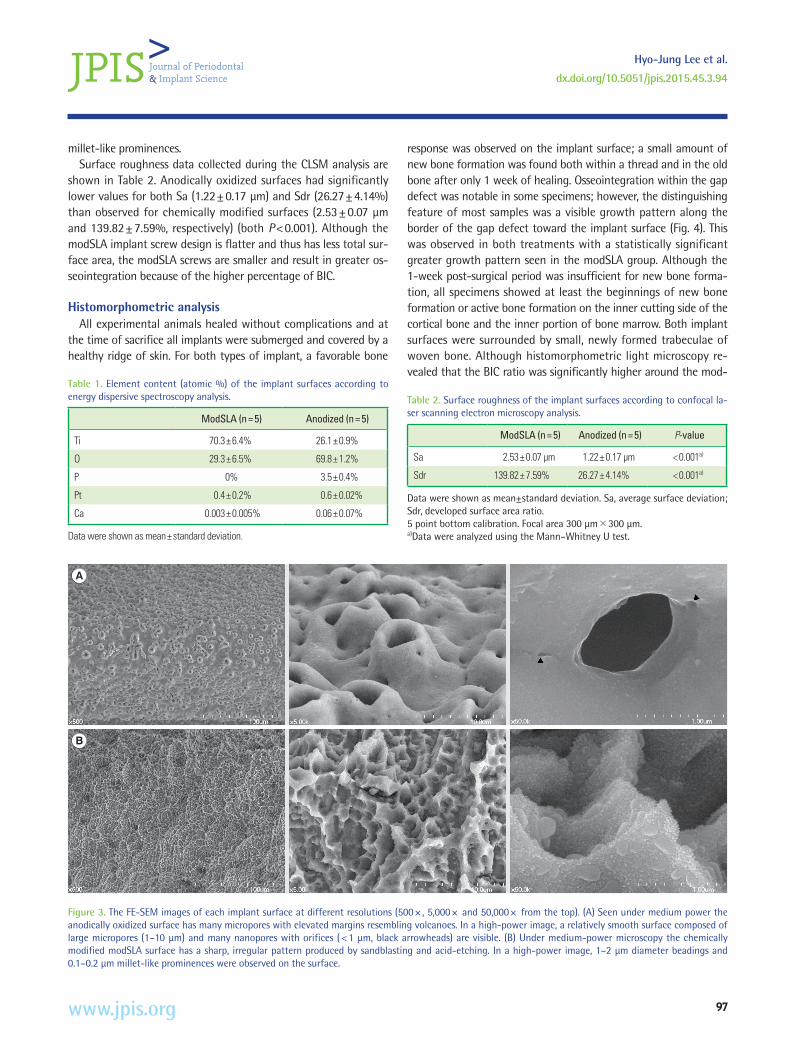
Breuner [29]. The specimens were ground to a thickness of approxi-mately 50 μm and stained with hematoxylin and eosin (H&E). His-tological examinations of specimens were performed under a light microscope (Olympus BX, Olympus, Tokyo, Japan). BIC and BA per-centages were defined and measured in the range of 2 mm below the upper bone crest, as shown in Fig. 2. Histomorphometric analy-ses were performed on both the right and left sides of each speci-men using image analysis software (Kappa PS30C Imagebase, Kappa Opto-electronics GmbH, Gleichen, Germany).Statistical analyses
The Mann-Whitney U test was used to assess the statistical sig-nificance of the difference in surface roughness parameters (Sa and
200 μm
Figure 2. A light microscopic image for histomorphometric analyses (H&E staining, 75×magnification). Note the gap space (blue area) that was inten-tionally made between the implant surface and the cortical bone.
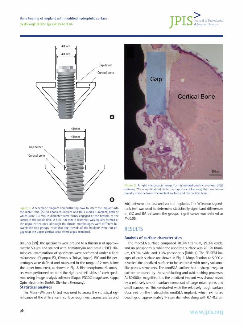
Figure 1. A schematic diagram demonstrating how to insert the implant into the rabbit tibia. (A) An anodized implant and (B) a modSLA implant, both of which were 3.3 mm in diameter, were firmly engaged at the bottom of the cortex in the rabbit tibia. A hole, 4.0 mm in diameter, was equally formed at the upper cortex only, although the thread morphologies were different be-tween the two groups. Note that the threads of the implants were not en-gaged at the upper cortical area where a gap remained.
4.0 mm
Gap defect
Cortical bone
4.0 mm
A
Gap defect
Cortical bone
4.0 mm
4.0 mm
B
Hyo-Jung Lee et al.
dx.doi.org/10.5051/jpis.2015.45.3.94
www.jpis.org 97
millet-like prominences.Surface roughness data collected during the CLSM analysis are
shown in Table 2. Anodically oxidized surfaces had significantly lower values for both Sa (1.22±0.17 μm) and Sdr (26.27±4.14%) than observed for chemically modified surfaces (2.53±0.07 μm and 139.82±7.59%, respectively) (both P<0.001). Although the modSLA implant screw design is flatter and thus has less total sur-face area, the modSLA screws are smaller and result in greater os-seointegration because of the higher percentage of BIC.
Histomorphometric analysisAll experimental animals healed without complications and at
the time of sacrifice all implants were submerged and covered by a healthy ridge of skin. For both types of implant, a favorable bone
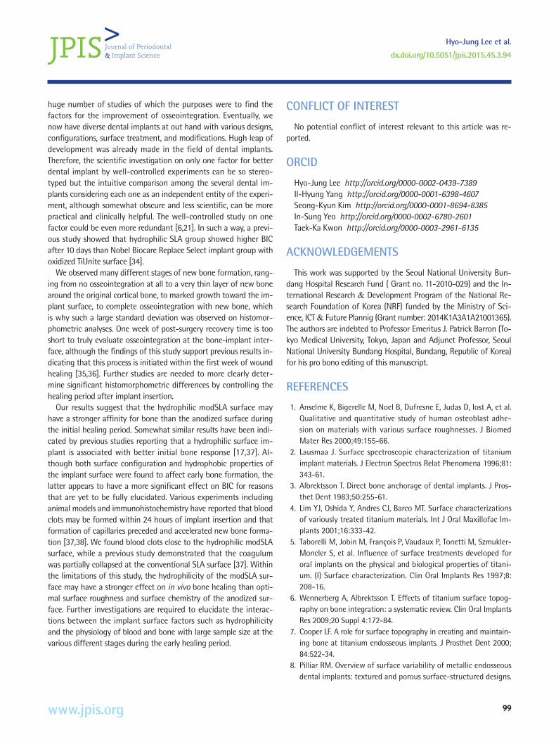
response was observed on the implant surface; a small amount of new bone formation was found both within a thread and in the old bone after only 1 week of healing. Osseointegration within the gap defect was notable in some specimens; however, the distinguishing feature of most samples was a visible growth pattern along the border of the gap defect toward the implant surface (Fig. 4). This was observed in both treatments with a statistically significant greater growth pattern seen in the modSLA group. Although the 1-week post-surgical period was insufficient for new bone forma-tion, all specimens showed at least the beginnings of new bone formation or active bone formation on the inner cutting side of the cortical bone and the inner portion of bone marrow. Both implant surfaces were surrounded by small, newly formed trabeculae of woven bone. Although histomorphometric light microscopy re-vealed that the BIC ratio was significantly higher around the mod-
Table 1. Element content (atomic %) of the implant surfaces according to energy dispersive spectroscopy analysis.
ModSLA (n=5) Anodized (n=5)
Ti 70.3±6.4% 26.1±0.9%
O 29.3±6.5% 69.8±1.2%
P 0% 3.5±0.4%
Pt 0.4±0.2% 0.6±0.02%
Ca 0.003±0.005% 0.06±0.07%
Data were shown as mean±standard deviation.
A
B
Figure 3. The FE-SEM images of each implant surface at different resolutions (500× , 5,000× and 50,000× from the top). (A) Seen under medium power the anodically oxidized surface has many micropores with elevated margins resembling volcanoes. In a high-power image, a relatively smooth surface composed of large micropores (1–10 μm) and many nanopores with orifices (<1 μm, black arrowheads) are visible. (B) Under medium-power microscopy the chemically modified modSLA surface has a sharp, irregular pattern produced by sandblasting and acid-etching. In a high-power image, 1–2 μm diameter beadings and 0.1–0.2 μm millet-like prominences were observed on the surface.
Table 2. Surface roughness of the implant surfaces according to confocal la-ser scanning electron microscopy analysis.
ModSLA (n=5) Anodized (n=5) P-value
Sa 2.53±0.07 μm 1.22±0.17 μm <0.001a)
Sdr 139.82±7.59% 26.27±4.14% <0.001a)
Data were shown as mean±standard deviation. Sa, average surface deviation; Sdr, developed surface area ratio.5 point bottom calibration. Focal area 300 μm×300 μm.a)Data were analyzed using the Mann–Whitney U test.
Bone healing of implant with modified hydrophilic surface
dx.doi.org/10.5051/jpis.2015.45.3.94
www.jpis.org98
SLA implants than around the anodized implants (P=0.02; Table 3), there was no significant difference in BA between the two types of implant because of a large standard deviation (P=0.09; Table 3).
DISCUSSION
We set out to determine whether modSLA implants would pos-sess clinical superiority over anodically oxidized surface implants. Given the results of our in vivo experiment, hydrophilicity seems to be very important in bone response.
Thread density but not thread geometry is known to have an ef-
fect on BIC [30,31]. As the anodized implant had a shorter thread pitch than the modSLA implant in this study (Fig. 4), it was consid-ered to have an advantage in BIC. We also found the modSLA sur-face to be significantly rougher than the anodized surface. Although the modSLA surface is classified as rough, the anodized surface is considered moderately rough, which is known to be more advanta-geous for bone responses than a "rough" surface [32]. However, we found a significantly higher BIC in response to the modSLA implant. In fact, in terms of the surface treatment based on Sa and Sdr, sev-eral studies concluded that modSLA surface showed better perfor-mance than conventional SLA surface as well as different surface treatment [17,19,21]. Although the mechanism linking surface properties and osteoblast production is not yet sufficiently under-stood, the hydrophilic property of the modSLA surface may have a stronger influence on bone response than either the surface con-figuration of the implant or the surface features resulting from an-odic oxidation [19]. These results correlated with the previous study concluding that surface hydrophilicity rather than microtopography affected soft and hard tissue integration [33].
Since the dental implant was first introduced, there have been a
Table 3. The bone-implant-contact and bone area 1 week after the surgery.
ModSLA (n=5) Anodized (n=5) P-value
BIC 17.9±19.4% 2.9±8.3% 0.02a)
BA 14.0±12.0% 4.0±9.0% 0.09a)
Data shown as mean±standard deviation. BIC, bone-to-implant contact; BA, bone area ratio.a)Data were analyzed using the Mann–Whitney U test.
A B
Figure 4. Histologic views of wound healing at day 7 with light microscopic findings (H&E stain, 100× magnification). Newly formed bone was seen from the cutting area of the cortical bone toward the implant surface (black arrows). New bone was also formed from the implant surface (white arrows). Bone forma-tion was found to occur in the marrow area, mainly behind the existing cortical bone. Histologic views were observed around both the anodized (A) and modS-LA (B) implants.
Hyo-Jung Lee et al.
dx.doi.org/10.5051/jpis.2015.45.3.94
www.jpis.org 99
huge number of studies of which the purposes were to find the factors for the improvement of osseointegration. Eventually, we now have diverse dental implants at out hand with various designs, configurations, surface treatment, and modifications. Hugh leap of development was already made in the field of dental implants. Therefore, the scientific investigation on only one factor for better dental implant by well-controlled experiments can be so stereo-typed but the intuitive comparison among the several dental im-plants considering each one as an independent entity of the experi-ment, although somewhat obscure and less scientific, can be more practical and clinically helpful. The well-controlled study on one factor could be even more redundant [6,21]. In such a way, a previ-ous study showed that hydrophilic SLA group showed higher BIC after 10 days than Nobel Biocare Replace Select implant group with oxidized TiUnite surface [34].
We observed many different stages of new bone formation, rang-ing from no osseointegration at all to a very thin layer of new bone around the original cortical bone, to marked growth toward the im-plant surface, to complete osseointegration with new bone, which is why such a large standard deviation was observed on histomor-phometric analyses. One week of post-surgery recovery time is too short to truly evaluate osseointegration at the bone-implant inter-face, although the findings of this study support previous results in-dicating that this process is initiated within the first week of wound healing [35,36]. Further studies are needed to more clearly deter-mine significant histomorphometric differences by controlling the healing period after implant insertion.
Our results suggest that the hydrophilic modSLA surface may have a stronger affinity for bone than the anodized surface during the initial healing period. Somewhat similar results have been indi-cated by previous studies reporting that a hydrophilic surface im-plant is associated with better initial bone response [17,37]. Al-though both surface configuration and hydrophobic properties of the implant surface were found to affect early bone formation, the latter appears to have a more significant effect on BIC for reasons that are yet to be fully elucidated. Various experiments including animal models and immunohistochemistry have reported that blood clots may be formed within 24 hours of implant insertion and that formation of capillaries preceded and accelerated new bone forma-tion [37,38]. We found blood clots close to the hydrophilic modSLA surface, while a previous study demonstrated that the coagulum was partially collapsed at the conventional SLA surface [37]. Within the limitations of this study, the hydrophilicity of the modSLA sur-face may have a stronger effect on in vivo bone healing than opti-mal surface roughness and surface chemistry of the anodized sur-face. Further investigations are required to elucidate the interac-tions between the implant surface factors such as hydrophilicity and the physiology of blood and bone with large sample size at the various different stages during the early healing period.
CONFLICT OF INTEREST
No potential conflict of interest relevant to this article was re-ported.
ORCID
Hyo-Jung Lee http://orcid.org/0000-0002-0439-7389Il-Hyung Yang http://orcid.org/0000-0001-6398-4607Seong-Kyun Kim http://orcid.org/0000-0001-8694-8385In-Sung Yeo http://orcid.org/0000-0002-6780-2601Taek-Ka Kwon http://orcid.org/0000-0003-2961-6135
ACKNOWLEDGEMENTS
This work was supported by the Seoul National University Bun-dang Hospital Research Fund ( Grant no. 11-2010-029) and the In-ternational Research & Development Program of the National Re-search Foundation of Korea (NRF) funded by the Ministry of Sci-ence, ICT & Future Plannig (Grant number: 2014K1A3A1A21001365). The authors are indebted to Professor Emeritus J. Patrick Barron (To-kyo Medical University, Tokyo, Japan and Adjunct Professor, Seoul National University Bundang Hospital, Bundang, Republic of Korea) for his pro bono editing of this manuscript.
REFERENCES
1. Anselme K, Bigerelle M, Noel B, Dufresne E, Judas D, Iost A, et al. Qualitative and quantitative study of human osteoblast adhe-sion on materials with various surface roughnesses. J Biomed Mater Res 2000;49:155-66.
2. Lausmaa J. Surface spectroscopic characterization of titanium implant materials. J Electron Spectros Relat Phenomena 1996;81: 343-61.
3. Albrektsson T. Direct bone anchorage of dental implants. J Pros-thet Dent 1983;50:255-61.
4. Lim YJ, Oshida Y, Andres CJ, Barco MT. Surface characterizations of variously treated titanium materials. Int J Oral Maxillofac Im-plants 2001;16:333-42.
5. Taborelli M, Jobin M, François P, Vaudaux P, Tonetti M, Szmukler-Moncler S, et al. Influence of surface treatments developed for oral implants on the physical and biological properties of titani-um. (I) Surface characterization. Clin Oral Implants Res 1997;8: 208-16.
6. Wennerberg A, Albrektsson T. Effects of titanium surface topog-raphy on bone integration: a systematic review. Clin Oral Implants Res 2009;20 Suppl 4:172-84.
7. Cooper LF. A role for surface topography in creating and maintain-ing bone at titanium endosseous implants. J Prosthet Dent 2000; 84:522-34.
8. Pilliar RM. Overview of surface variability of metallic endosseous dental implants: textured and porous surface-structured designs.
Bone healing of implant with modified hydrophilic surface
dx.doi.org/10.5051/jpis.2015.45.3.94
www.jpis.org100
Implant Dent 1998;7:305-14.9. Kilpadi DV, Lemons JE. Surface energy characterization of unal-
loyed titanium implants. J Biomed Mater Res 1994;28:1419-25.10. Wang R, Hashimoto K, Fujishima A, Chikuni M, Kojima E, Kita-
mura A, et al. Light-induced amphiphilic surfaces. Nature 1997; 388:431-2.
11. Wennerberg A, Albrektsson T. Suggested guidelines for the topo-graphic evaluation of implant surfaces. Int J Oral Maxillofac Im-plants 2000;15:331-44.
12. Bico J, Thiele U, Quéré D. Wetting of textured surfaces. Colloids Surf A Physicochem Eng Asp 2002;206:41-6.
13. Rupp F, Scheideler L, Eichler M, Geis-Gerstorfer J. Wetting behav-ior of dental implants. Int J Oral Maxillofac Implants 2011;26: 1256-66.
14. Rupp F, Scheideler L, Olshanska N, de Wild M, Wieland M, Geis-Gerstorfer J. Enhancing surface free energy and hydrophilicity through chemical modification of microstructured titanium im-plant surfaces. J Biomed Mater Res A 2006;76:323-34.
15. Rupp F, Scheideler L, Rehbein D, Axmann D, Geis-Gerstorfer J. Roughness induced dynamic changes of wettability of acid etched titanium implant modifications. Biomaterials 2004;25:1429-38.
16. Textor M, Sittig C, Frauchiger V, Tosatti S, Brunette DM. Proper-ties and biological significance of natural oxide films on titani-um and its alloys. In: Brunette DM, Tengvall P, Textor M, Thom-sen P, editors. Titanium in medicine: material science, surface science, engineering, biological responses, and medical applica-tions. Berlin: Springer; 2001. p.171-230.
17. Buser D, Broggini N, Wieland M, Schenk RK, Denzer AJ, Cochran DL, et al. Enhanced bone apposition to a chemically modified SLA titanium surface. J Dent Res 2004;83:529-33.
18. Junker R, Dimakis A, Thoneick M, Jansen JA. Effects of implant surface coatings and composition on bone integration: a system-atic review. Clin Oral Implants Res 2009;20 Suppl 4:185-206.
19. Schwarz F, Ferrari D, Herten M, Mihatovic I, Wieland M, Sager M, et al. Effects of surface hydrophilicity and microtopography on early stages of soft and hard tissue integration at non-submerged titanium implants: an immunohistochemical study in dogs. J Peri-odontol 2007;78:2171-84.
20. Choi JY, Lee HJ, Jang JU, Yeo IS. Comparison between bioactive fluoride modified and bioinert anodically oxidized implant sur-faces in early bone response using rabbit tibia model. Implant Dent 2012;21:124-8.
21. Wennerberg A, Albrektsson T. On implant surfaces: a review of current knowledge and opinions. Int J Oral Maxillofac Implants 2010;25:63-74.
22. Iwai-Yoshida M, Shibata Y, Wurihan, Suzuki D, Fujisawa N, Tani-moto Y, et al. Antioxidant and osteogenic properties of anodically oxidized titanium. J Mech Behav Biomed Mater 2012;13:230-6.
23. Le Guehennec L, Lopez-Heredia MA, Enkel B, Weiss P, Amouriq Y, Layrolle P. Osteoblastic cell behaviour on different titanium im-plant surfaces. Acta Biomater 2008;4:535-43.
24. Schuler RF, Janakievski J, Hacker BM, O'Neal RB, Roberts FA. Ef-
fect of implant surface and grafting on implants placed into simulated extraction sockets: a histologic study in dogs. Int J Oral Maxillofac Implants 2010;25:893-900.
25. Ferguson SJ, Broggini N, Wieland M, de Wild M, Rupp F, Geis-Ger-storfer J, et al. Biomechanical evaluation of the interfacial strength of a chemically modified sandblasted and acid-etched titanium surface. J Biomed Mater Res A 2006;78:291-7.
26. Wall I, Donos N, Carlqvist K, Jones F, Brett P. Modified titanium surfaces promote accelerated osteogenic differentiation of mes-enchymal stromal cells in vitro. Bone 2009;45:17-26.
27. Hong J, Kurt S, Thor A. A hydrophilic dental implant surface ex-hibits thrombogenic properties in vitro. Clin Implant Dent Relat Res 2013;15:105-12.
28. Yeo IS, Han JS, Yang JH. Biomechanical and histomorphometric study of dental implants with different surface characteristics. J Biomed Mater Res B Appl Biomater 2008;87:303-11.
29. Donath K, Breuner G. A method for the study of undecalcified bones and teeth with attached soft tissues. The Säge-Schliff (saw-ing and grinding) technique. J Oral Pathol 1982;11:318-26.
30. Roberts WE, Smith RK, Zilberman Y, Mozsary PG, Smith RS. Osse-ous adaptation to continuous loading of rigid endosseous im-plants. Am J Orthod 1984;86:95-111.
31. Steigenga J, Al-Shammari K, Misch C, Nociti FH Jr, Wang HL. Ef-fects of implant thread geometry on percentage of osseointe-gration and resistance to reverse torque in the tibia of rabbits. J Periodontol 2004;75:1233-41.
32. Albrektsson T, Wennerberg A. Oral implant surfaces: Part 2--re-view focusing on clinical knowledge of different surfaces. Int J Prosthodont 2004;17:544-64.
33. Schwarz F, Herten M, Sager M, Wieland M, Dard M, Becker J. Bone regeneration in dehiscence-type defects at chemically modified (SLActive) and conventional SLA titanium implants: a pilot study in dogs. J Clin Periodontol 2007;34:78-86.
34. Gottlow J, Barkarmo S, Sennerby L. An experimental comparison of two different clinically used implant designs and surfaces. Clin Implant Dent Relat Res 2012;14 Suppl 1:e204-12.
35. Abrahamsson I, Berglundh T, Linder E, Lang NP, Lindhe J. Early bone formation adjacent to rough and turned endosseous implant surfaces. An experimental study in the dog. Clin Oral Implants Res 2004;15:381-92.
36. Berglundh T, Abrahamsson I, Lang NP, Lindhe J. De novo alveolar bone formation adjacent to endosseous implants. Clin Oral Im-plants Res 2003;14:251-62.
37. Schwarz F, Herten M, Sager M, Wieland M, Dard M, Becker J. His-tological and immunohistochemical analysis of initial and early osseous integration at chemically modified and conventional SLA titanium implants: preliminary results of a pilot study in dogs. Clin Oral Implants Res 2007;18:481-8.
38. Schmid J, Wallkamm B, Hämmerle CH, Gogolewski S, Lang NP. The significance of angiogenesis in guided bone regeneration. A case report of a rabbit experiment. Clin Oral Implants Res 1997; 8:244-8.