Embed Size (px)
Citation preview
http://www.bio-protocol.org/e1762 Vol 6, Iss 6, Mar 20, 2016
Copyright © 2016 The Authors; exclusive licensee Bio-protocol LLC. 1
In vivo Bioluminescence Imaging of Luciferase-labeled Cancer Cells
Marc Carceles-Cordon, Irene Rodriguez-Fernandez, Veronica Rodriguez-Bravo, Carlos
Cordon-Cardo and Josep Domingo-Domenech*
Department of Pathology, Icahn School of Medicine at Mount Sinai, New York, USA *For correspondence: [email protected]
[Abstract] Over the past decade, in vivo bioluminescent imaging has emerged as a
non-invasive and sensitive tool for studying ongoing biological processes within living
organisms (Contag et al., 1997; Contag et al., 1998). Based on the detection and quantitation
of the photons produced by the oxidation of luciferin by luciferase enzymes (Harvey, 1927),
this technique has proved to be particularly useful in analyzing cancerous cells and monitoring
tumor growth (Edinger et al., 1999; Sweeney et al., 1999; Vidal et al., 2015), providing a
cost-effective insight into how the disease progresses in vivo, without the need of serial
sacrifice of animals. This protocol describes in detail the procedure of obtaining
luciferase-tagged tumors in immunocompromised mice that can be studied by bioluminescent
imaging through the use of an IVIS Spectrum imager.
Materials and Reagents
1. Falcon™ Standard 10 cm2 Tissue Culture Dishes (Corning, catalog number: 353003) 2. Syringe Filter 0.45 μm strainer (Corning, catalog number: CLS431225)
3. 22Rv1 prostate cancer cells (ATCC, catalog number: CRL-2505) 4. NOD.Cg-Prkdcscid Il2rgtm1Wjl/SzJ (NSG™) mice (Jackson Lab, catalog number: 00557)
5. Phoenix-Ampho cells (ATCC, catalog number: CRL-3213) 6. pLenti CMV Puro LUC (w168-1) (Addgene, catalog number: 17477) 7. Helper pCMV-VSV-G (Addgene, catalog number: 8454) and pMD2.G (Addgene,
catalog number: 12259) 8. One Shot Stbl3 Competent E. coli (Thermo Fisher Scientific, Invitrogen™, catalog
number: C7373-03)
9. LB Broth Medium (Thermo Fisher Scientific, BioReagents™, catalog number:
BP1426-2)
10. Ampicillin (Sigma-Aldrich, catalog number: A9393)
11. QIAprep Spin Miniprep Kit (QIAGEN, catalog number: 27106) 12. RPMI 1640 Medium (Thermo Fisher Scientific, Gibco™, catalog number: 11875119)
13. Dulbecco’s Phosphate-Buffered Saline (DPBS) 1x (Corning, catalog number: 21-031)
14. jetPEI® DNA Transfection Agent (Polyplus-transfection, catalog number: 101-10N) 15. Penicillin-Streptomycin antibiotic (PenStrep) (Thermo Fisher Scientific, Gibco™,
catalog number: 15140122)
http://www.bio-protocol.org/e1762 Vol 6, Iss 6, Mar 20, 2016
Copyright © 2016 The Authors; exclusive licensee Bio-protocol LLC. 2
16. Polybrene® (Santa Cruz Biotechnology, catalog number: 134220)
17. Isoflurane (Baxter, catalog number: 1001936040)
18. Matrigel® Growth Factor Reduced (GFR) Basement Membrane Matrix (Corning,
catalog number: 354230)
19. D-luciferin (PerkinElmer, catalog number: 770504)
Equipment
1. Thermo-block (Thermo Fisher Scientific, model: Isotemp™ Digital and Analog Dry
Bath Incubator) 2. Shaker incubator (Eppendorf AG, New Brunswick Scientific, model: Innova 44R
Incubator/Shaker)
3. Table-top microcentrifuge (Eppendorf AG, model: centrifuge 5415R) 4. Spectrophotometer (Thermo Fisher Scientific, model: SPECTRONIC™ 200
Spectrophotometer) 5. Incubator (Thermo Fisher Scientific, model: Heracell 150i)
6. Vortex (Scientific Industries, model: Vortex Genie 2) 7. IVIS Spectrum imager (PerkinElmer, model: IVIS Spectrum Preclinical In Vivo Imaging
System) Software
1. Caliper LifeScience Living Image® in vivo imaging software (PerkinElmer, catalog
number: 128110) (Lumina/Kinetic/XR/100, Living Image V4.1)
http://www.bio-protocol.org/e1762 Vol 6, Iss 6, Mar 20, 2016
Copyright © 2016 The Authors; exclusive licensee Bio-protocol LLC. 3
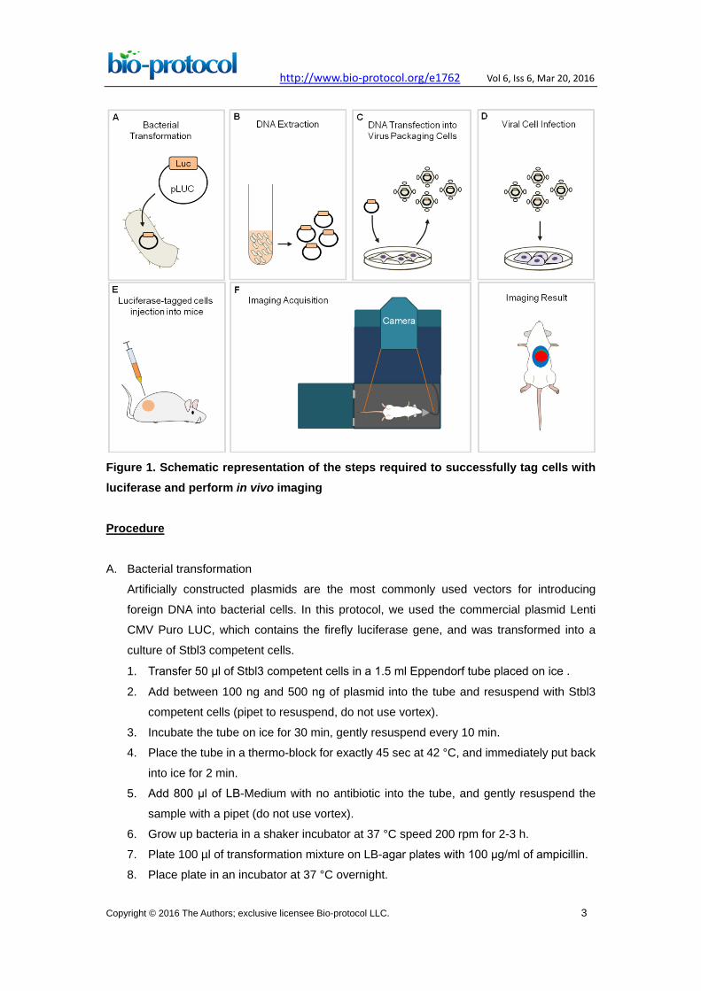
Figure 1. Schematic representation of the steps required to successfully tag cells with luciferase and perform in vivo imaging Procedure A. Bacterial transformation
Artificially constructed plasmids are the most commonly used vectors for introducing
foreign DNA into bacterial cells. In this protocol, we used the commercial plasmid Lenti
CMV Puro LUC, which contains the firefly luciferase gene, and was transformed into a
culture of Stbl3 competent cells.
1. Transfer 50 μl of Stbl3 competent cells in a 1.5 ml Eppendorf tube placed on ice . 2. Add between 100 ng and 500 ng of plasmid into the tube and resuspend with Stbl3
competent cells (pipet to resuspend, do not use vortex).
3. Incubate the tube on ice for 30 min, gently resuspend every 10 min.
4. Place the tube in a thermo-block for exactly 45 sec at 42 °C, and immediately put back
into ice for 2 min.
5. Add 800 μl of LB-Medium with no antibiotic into the tube, and gently resuspend the
sample with a pipet (do not use vortex).
6. Grow up bacteria in a shaker incubator at 37 °C speed 200 rpm for 2-3 h.
7. Plate 100 µl of transformation mixture on LB-agar plates with 100 μg/ml of ampicillin.
8. Place plate in an incubator at 37 °C overnight.
http://www.bio-protocol.org/e1762 Vol 6, Iss 6, Mar 20, 2016
Copyright © 2016 The Authors; exclusive licensee Bio-protocol LLC. 4
9. Pick a single colony and inoculate a flask containing 50 ml of LB-Medium + 100 μg/ml
of ampicillin.
10. Incubate overnight at 37 °C in a shaker incubator at a speed of 200 rpm.
B. DNA extraction
There are several methods that can be used to purify plasmid DNA depending on the size
of the bacterial culture and its corresponding plasmid yield, and a handful of kits available
from varying manufacturers to do it. Here, we perform a minipreparation (miniprep) using
the QIAprep Spin Miniprep Kit, which allows the isolation of a small-scale sample of up to
20 μg plasmid DNA from the transformed bacteria. Briefly, in this protocol the isolation of
DNA from bacteria relies upon the use of columns and centrifugation steps in which DNA is
sheared, extracted, and precipitated. Follow protocol of the manufacturer.
C. Transfection of DNA into viral packaging cells
Depending on the plasmid, several packaging cells can be used to generate the desired
virus. In this protocol, we used Phoenix-Ampho cells, which are a second-generation
retrovirus producer cell line that is also capable of a long-term stable production of
lentiviruses when helper DNA (CMV and DMV) is present. Helper DNA encodes viral
genes that increase viral replication and packaging.
1. Seed 3 x 10⁶ Phoenix-Ampho cells in 8 ml of RPMI supplemented with 10% FBS and 1%
PenStrep in a 10 cm2 culture plate 24 h before transfection.
2. In a first 1.5 ml Eppendorf tube, add 6 μg of DNA, 3 μg of each helper (CMV and DMV)
and fill up to 250 μl of 150 mM NaCl (per sample to transfect).
3. In a second 1.5 ml tube add 24 μl of jetPEI and 226 μl of 150 mM NaCl (per sample to
transfect).
4. Vortex the tubes for 10 sec.
5. Add the Jetpei + NaCl into the Eppendorf tube with the DNA and vortex for 10 sec.
6. Incubate mixture for 15 min at room temperature.
7. Remove media of plate and add 9.5 ml of fresh RPMI supplemented with 10% FBS
and 1% PenStrep to Phoenix-Ampho cells (see step C1).
8. Add 500 μl drop by drop, of the mixture (step C5) to the plate and incubate overnight at
37 °C.
9. After 24 h change the medium and add fresh RPMI supplemented with 10% FBS and
1% PenStrep.
10. After 48 h, collect and filter the medium with a 0.45 μm strainer into a 50 ml Falcon
tube. Note: Viral concentration can be measured after this step by serial dilution to
obtain quantitative information. Various techniques can be used to quantify virus
concentration which include traditional methods (e.g., plaque assay) and commercial
kits (e.g. ELISA or Q-PCR).
http://www.bio-protocol.org/e1762 Vol 6, Iss 6, Mar 20, 2016
Copyright © 2016 The Authors; exclusive licensee Bio-protocol LLC. 5
D. Viral particle cell infection
The viral particles produced in the previous step are used to infect cells. Virus will infect
cells which hold a receptor that the virus can bind to. Therefore, the efficiency of viral
infection will greatly depend on the cell type. In this protocol, we infected 70% confluent
22Rv1 prostate cancer cells growing in 10 cm2 culture dishes with 10 ml of media.
1. Add Polybrene to cultured cells to a final concentration of 10 μg/ml.
Note: Polybrene is an efficient infection reagent used to introduce viral vectors into
mammalian cells. Polybrene acts by neutralizing the charge repulsion between virions
and the cell surface (Davis et al., 2004).
2. Using a 5 ml pipet, add 4 ml of filtered viral particles to cultured cells.
3. Centrifuge 10 cm2 plate at 250 x g for 5 min.
Note: This step helps the physical contact of viruses with surface of cells increasing
infection efficiency.
4. Place cells in 37 °C, 5% CO2 incubator for 18-24 h.
Note: Infection efficiency can be affected by incubation time. Troubleshooting using
different incubation times can be performed to identify most efficient incubation time
for a specific cell type.
5. Aspirate the medium and add 10 ml of fresh RPMI supplemented with 10% FBS and 1%
PenStrep. Incubate at 37 °C for 24 h.
6. Select infected 22Rv1 prostate cancer cells by adding Puromycin at a final
concentration of 2 µg/ml to culture media.
Note: Puromycin concentration will depend on cell type.
7. Replace medium containing Puromycin every 3 days. Cells that have incorporated the
pLenti CMV Puro LUC will grow up under antibiotic selection conditions.
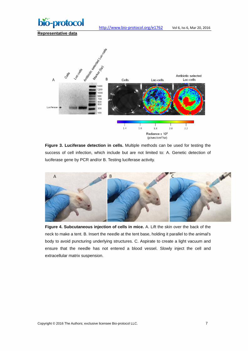
8. Check that the cells have incorporated the luciferase plasmid.
Note: Multiple methods can be used to test successful luciferase. Examples are
detection of luciferase by PCR or measuring luciferase activity upon D-Luciferin
exposure (see Figure 2).
E. Injection of luciferase-tagged cells into immunocompromised mice
In this protocol, we performed subcutaneous injection of luciferase-tagged tumor cells.
However, alternative injection sites such as the mouse prostate (orthotropic), which
promotes a prostate microenvironment, and subrenal capsule injection, which enhances
successful engraftment and preserves tumor heterogeneity, can be performed based on
investigator preference (Hidalgo et al., 2014; Siolas et al., 2013).
Note: Conduct all animal procedures in compliance with protocols approved by the
institutional animal care committee. This protocol has been conducted at our institution
under a specific Animal Care Committee in accordance and compliance with all relevant
regulatory and institutional agencies, regulations and guidelines.
http://www.bio-protocol.org/e1762 Vol 6, Iss 6, Mar 20, 2016
Copyright © 2016 The Authors; exclusive licensee Bio-protocol LLC. 6
1. Harvest attached cells by removing culture media, washing cells with 10 ml 1x DBPS
two times and trypsinizing cells using 2 ml 0.05% Trypsin. Recover 2 ml trypsinized
cells to 15 ml tube containing 4 ml RPM1 supplemented with 10% FBS and 1%
PenStep.
2. Count cells using a hemocytometer.
3. Mix 106 cells suspended in 100 μl of RPMI supplemented with 100 μl of extracellular
matrix (1:1 ratio) and place on ice.
4. Anesthetize mice prior to subcutaneous injection in a chamber supplying 5% (v/v)
inhaled isoflurane in 1 L/min of oxygen.
5. Using a 25 gauge needle and a 1 ml syringe, inject 250 μl of cell and extracellular
matrix suspension subcutaneously into the flanks of immunodeficient NSG mice.
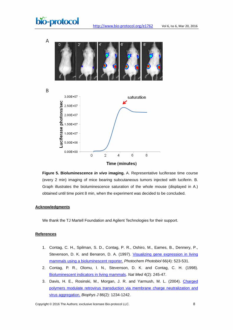
F. Bioluminescence imaging
The IVIS Spectrum imager expresses the bioluminescent signal in photons per second
and displays it as an intensity map. The luminescence, which is the consequence of the
photon flux emitted by the luciferase-expressing cells, directly correlates to the size of the
tumor and can be measured at the site of injection using a region of interest (ROI) tool.
The optical emission image can be adjusted to provide optimal contrast and resolution
without affecting quantitation using the Living Image© in vivo imaging software.
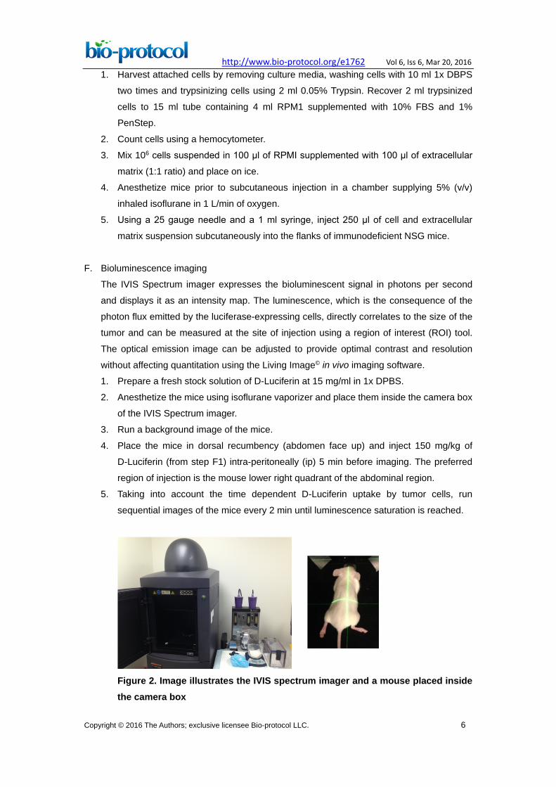
1. Prepare a fresh stock solution of D-Luciferin at 15 mg/ml in 1x DPBS.
2. Anesthetize the mice using isoflurane vaporizer and place them inside the camera box
of the IVIS Spectrum imager.
3. Run a background image of the mice.
4. Place the mice in dorsal recumbency (abdomen face up) and inject 150 mg/kg of
D-Luciferin (from step F1) intra-peritoneally (ip) 5 min before imaging. The preferred
region of injection is the mouse lower right quadrant of the abdominal region.
5. Taking into account the time dependent D-Luciferin uptake by tumor cells, run
sequential images of the mice every 2 min until luminescence saturation is reached.
Figure 2. Image illustrates the IVIS spectrum imager and a mouse placed inside the camera box
http://www.bio-protocol.org/e1762 Vol 6, Iss 6, Mar 20, 2016
Copyright © 2016 The Authors; exclusive licensee Bio-protocol LLC. 7
Representative data
Figure 3. Luciferase detection in cells. Multiple methods can be used for testing the
success of cell infection, which include but are not limited to: A. Genetic detection of
luciferase gene by PCR and/or B. Testing luciferase activity.
Figure 4. Subcutaneous injection of cells in mice. A. Lift the skin over the back of the
neck to make a tent. B. Insert the needle at the tent base, holding it parallel to the animal’s
body to avoid puncturing underlying structures. C. Aspirate to create a light vacuum and
ensure that the needle has not entered a blood vessel. Slowly inject the cell and
extracellular matrix suspension.
http://www.bio-protocol.org/e1762 Vol 6, Iss 6, Mar 20, 2016
Copyright © 2016 The Authors; exclusive licensee Bio-protocol LLC. 8
Figure 5. Bioluminescence in vivo imaging. A. Representative luciferase time course
(every 2 min) imaging of mice bearing subcutaneous tumors injected with luciferin. B.
Graph illustrates the bioluminescence saturation of the whole mouse (displayed in A.)
obtained until time point 8 min, when the experiment was decided to be concluded. Acknowledgments
We thank the TJ Martell Foundation and Agilent Technologies for their support.
References
1. Contag, C. H., Spilman, S. D., Contag, P. R., Oshiro, M., Eames, B., Dennery, P.,
Stevenson, D. K. and Benaron, D. A. (1997). Visualizing gene expression in living
mammals using a bioluminescent reporter. Photochem Photobiol 66(4): 523-531.
2. Contag, P. R., Olomu, I. N., Stevenson, D. K. and Contag, C. H. (1998).
Bioluminescent indicators in living mammals. Nat Med 4(2): 245-47.
3. Davis, H. E., Rosinski, M., Morgan, J. R. and Yarmush, M. L. (2004). Charged
polymers modulate retrovirus transduction via membrane charge neutralization and
virus aggregation. Biophys J 86(2): 1234-1242.
http://www.bio-protocol.org/e1762 Vol 6, Iss 6, Mar 20, 2016
Copyright © 2016 The Authors; exclusive licensee Bio-protocol LLC. 9
4. Edinger, M., Sweeney, T. J., Tucker, A. A., Olomu, A. B., Negrin, R. S. and Contag, C.
H. (1999). Noninvasive assessment of tumor cell proliferation in animal models.
Neoplasia 1(4): 303-10.
5. Harvey, E. N. (1927). The oxidation-reduction potential of the luciferin-oxyluciferin
system. J Gen Physiol 10(3): 385-393.
6. Hidalgo, M., Amant, F., Biankin, A. V., Budinska, E., Byrne, A. T., Caldas, C., Clarke,
R. B., de Jong, S., Jonkers, J., Maelandsmo, G. M., Roman-Roman, S., Seoane, J.,
Trusolino, L. and Villanueva, A. (2014). Patient-derived xenograft models: an
emerging platform for translational cancer research. Cancer Discov 4(9): 998-1013.
7. Siolas, D. and Hannon, G. J. (2013). Patient-derived tumor xenografts: transforming
clinical samples into mouse models. Cancer Res 73(17): 5315-19.
8. Sweeney, T. J., Mailander, V., Tucker, A. A., Olomu, A. B., Zhang, W., Cao, Y., Negrin,
R. S. and Contag, C. H. (1999). Visualizing the kinetics of tumor-cell clearance in living
animals. Proc Natl Acad Sci U S A 96(21): 12044-12049.
9. Vidal, S. J., Rodriguez-Bravo, V., Quinn, S. A., Rodriguez-Barrueco, R., Lujambio, A.,
Williams, E., Sun, X., de la Iglesia-Vicente, J., Lee, A., Readhead, B., Chen, X.,
Galsky, M., Esteve, B., Petrylak, D. P., Dudley, J. T., Rabadan, R., Silva, J. M.,
Hoshida, Y., Lowe, S. W., Cordon-Cardo, C. and Domingo-Domenech, J. (2015). A
targetable GATA2-IGF2 axis confers aggressiveness in lethal prostate cancer. Cancer
Cell 27(2): 223-239.