Embed Size (px)
Citation preview
IN VITRO TRACER STUDY OF A CHEMICAL CARCINOGEN
A Thesis
Submitted to
The Division of Biological Sciences
Emporia State University
In Partial Fulfillment
of the Requirements for the Degree
Master of Science
by
Noboru Nakamichi
August 1980
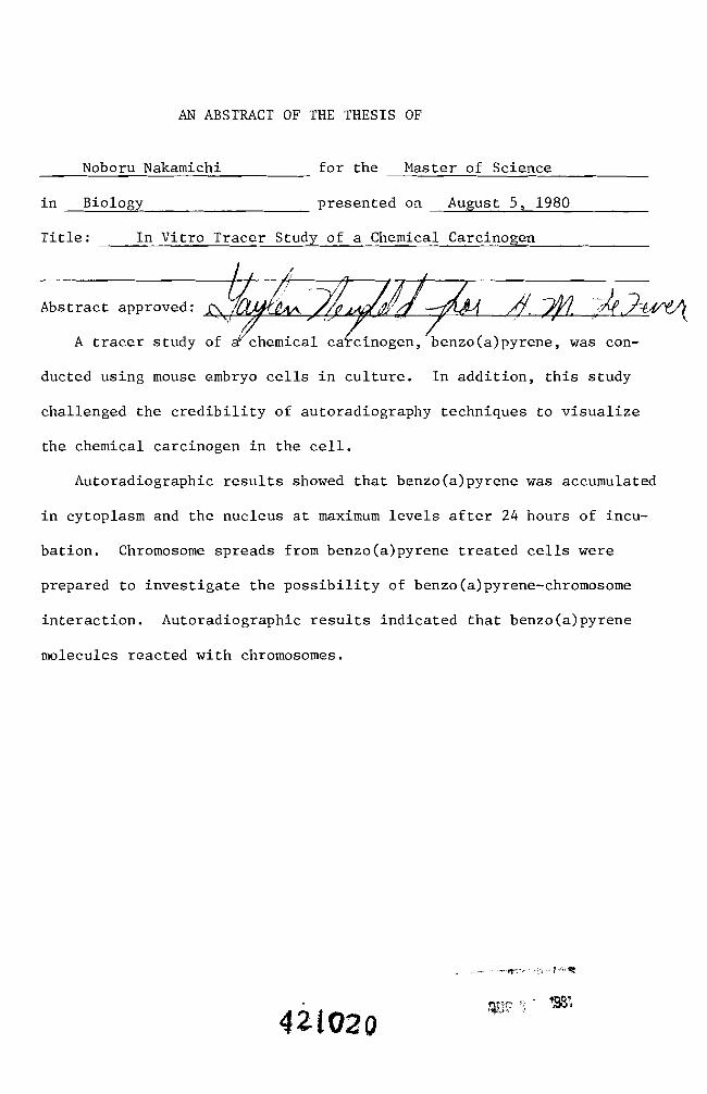
AN ABSTRACT OF THE THESIS OF
in Biology presented on August 5 1980
Title In Vitro Tracer Study of a Chemical Carcinogen
_ __~___ 4
~TJC j - 198
Master of Sciencefor the
421020
Noboru Nakamichi
Abstract approved ~
A tracer study of ~ chemlcal ca~ClnOgen benzotaJpyrene was conshy
ducted using mouse embryo cells in culture In addition this study
challenged the credibility of autoradiography techniques to visualize
the chemical carcinogen in the cell
Autoradiographic results showed that benzo(a)pyrene was accumulated
in cytoplasm and the nucleus at maximum levels after 24 hours of incushy
bation Chromosome spreads from benzo(a)pyrene treated cells were
prepared to investigate the possibility of benzo(a)pyrene-chromosome
interaction Autoradiographic results indicated that benzo(a)pyrene
molecules reacted with chromosomes
iv
ACKNOWLEDGEMENT
I would like to express my deepest appreciation to Dr Michael
LeFever for his guidance and constructive help during this research
Furthermore I would like to thank him for his patience and conshy
tinuous help during the time required for the completion of the
investigation
Sincere appreciation is also extended to my committee members
including Dr Gaylen Neufeld and Dr Helen McElree
Deep appreciation goes to Mr Mel Fuqua my friend and colleague
who was of great help in the preparation of this thesis
v
TABLE OF CONTENTS
Introduction bullbullbullbullbull 1
Materials and Methods 9
Results 15
Discussion 62
Summarybullbullbullbullbullbullbull 70
Literature Cited bull 72
vi
LIST OF TABLES
Table 1 Polycyclic aromatic hydrocarbon carcinogens in foods 4
Table 2 Effect of B(a)P treated cells in adult mice bullbullbullbullbull 54
vii
LIST OF FIGURES
Figure 1 Human exposure to hazardous environmental agents (From Kraybi11 1977) bullbullbullbullbullbullbullbullbullbullbullbullbullbullbull 2
Figure 2 A model of noncovalent interaction of B(a)P with DNA (From Arcos and Argus 1968) bullbullbullbullbullbullbullbullbull 7
Figure 4 Toxicity effect of B(a)P concentration bullbullbullbullbullbullbullbullbullbull 18
Figure 7 Six day old normal mouse embryo cells in culturebullbullbull 2l
Figure 8 Eight day old normal mouse embryo cells in culture bullbullbullbull 2l
Figure 11 Thirty-five day old normal mouse embryo cells in
Figure 3 Toxic effect of B(a)P on cultured mouse embryo cells bull 17
Figure 5 Two day old normal mouse embryo cells in culture bullbullbullbull 19
Figure 6 Four day old normal mouse embryo cells in culture bullbullbullbull 19
Figure 9 Thirteen day old normal mouse embryo cells in culture bullbull 23
Figure 10 Thirty day old normal mouse embryo cells in culture bullbullbull 23
cul ture bullbullbullbullbullbullbullbullbullbullbullbullbullbullbullbullbullbullbullbullbullbullbullbullbull 25
Figure 12 Forty day old normal mouse embryo cells in culture bullbullbull 25
Figure 17 Time-course of 3H- B(a)P accumulation in the cells from
Figure 13 Mouse embryo cells treated for one day with B(a)P bullbullbull 27
Figure 14 Mouse embryo cells treated for two days with B(a)P bullbullbull 27
Figure 15 Mouse embryo cells treated for six days with B(a)P bullbullbullbull 29
Figure 16 Mouse embryo cells treated for nine days with B(a)P bull 29
15 hours to 17 hours bullbullbullbullbullbullbullbullbullbullbullbullbullbullbullbull 32
Figure 18 Autoradiogram of the cells treated for 15 hours with 3H-B(a)P bullbullbullbullbullbullbullbullbullbullbullbullbullbullbullbullbullbullbullbullbull 34
Figure 19 Autoradiogram of the cells treated for 35 hours with 3H-B(a)P bullbullbullbullbullbullbullbullbullbullbullbullbullbullbullbull 34
Figure 20 Autoradiogram of the cells treated for 17 hours with 3H-B(a)P bullbullbullbullbullbullbullbullbullbullbullbullbullbullbullbullbull 36
Figure 21 Autoradiogram of the cells treated for 24 hours with 3H-B(a)P bullbullbullbullbullbullbullbullbullbullbullbullbullbullbullbullbull 36
viii
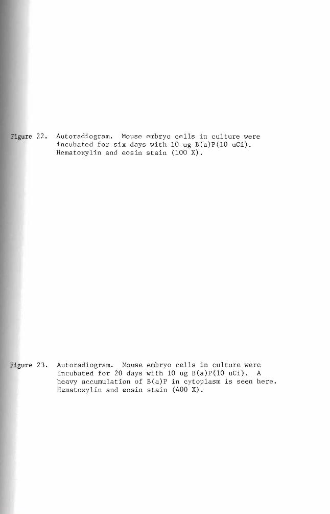
Figure 22
Figure 23
Figure 24
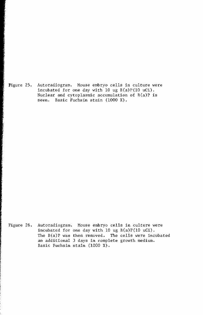
Figure 25
Figure 26
Figure 27
Figure 28
Figure 29
Figure 30
Figure 31
Figure 32
Figure 33
Figure 34
Autoradiogram of the cells treated for six days with 3H- B(a)P bullbullbullbullbullbullbullbullbullbullbullbullbullbullbullbull 38
Autoradiogram of the cells treated for 20 days with 3H- B(a)P bullbullbullbullbullbullbullbullbullbullbullbullbullbull 38
Time-course of 3H- B(a)P accumulation in the cells f rom one day to 21 days bullbullbullbullbullbullbullbullbullbull 40
Autoradiogram of the cells treated for one day with 3H-B (a)P bullbullbullbullbullbullbullbullbullbullbullbullbullbullbullbullbull 41
Autoradiogram of the cells treated for one day with 3H- B(a)P 3H- B(a)P was replaced by complete growth medium and the cells were incubated three addi tiona1 days bullbullbullbullbullbullbullbullbullbullbullbullbullbull 41
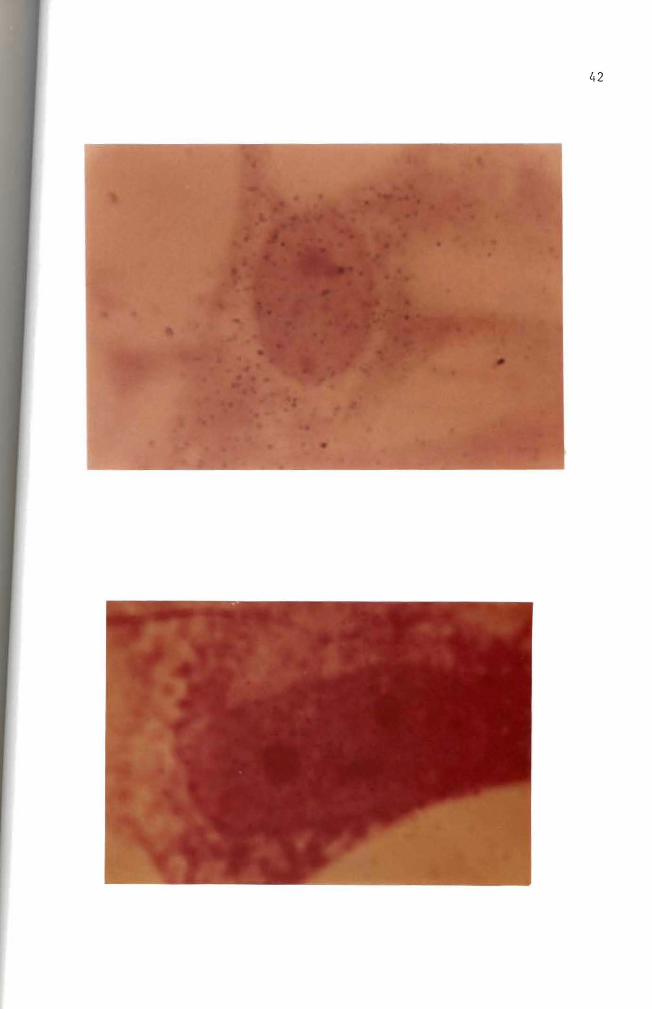
Autoradiogram of the cells treated for one day with 3H-B(a)P 3H-B(a)P was replaced by complete growth medium and the cells were incubated 11 additional days bullbullbullbullbullbullbullbullbullbullbullbullbullbullbullbull 43
Autoradiogram of the cells treated for one day with 3H- B(a)P 3H-B(a)P was replaced by complete growth medium and the cells were incubated 21 additional days bullbullbullbullbullbullbullbullbullbullbullbullbullbullbullbullbullbullbull 43
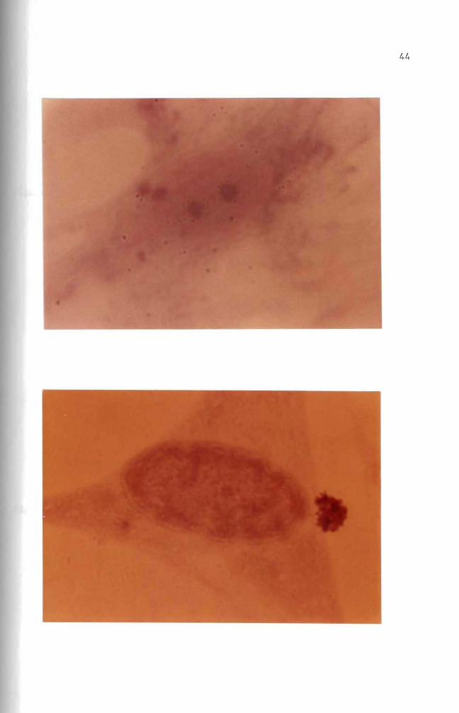
Autoradiogram of the cells treated for four days with 3H-B(a)P bullbullbullbullbullbullbullbullbullbullbullbullbullbullbullbullbullbullbullbullbullbullbullbullbullbullbullbullbull 45
Autoradiogram of the cells treated for four days with 3H-B(a)P 3H-B(a)P was replaced by complete growth medium and the cells were incubated six additional days bullbullbullbullbullbullbullbullbullbullbullbullbullbullbullbullbullbullbullbullbullbullbullbullbull 45
Autoradiogram of the cells treated for four days with 3H-B(a)P 3H-B(a)P was replaced by complete growth medium and the cells were incubated 11 additional days bullbullbullbullbullbullbullbullbullbullbullbullbullbullbullbullbullbullbullbullbullbullbullbullbull 47
Autoradiogram of the cells treated for four days with 3H- B(a)P 3H- B(a)P was replaced by complete growth medium and the cells were incubated 23 additional days bullbullbullbullbullbullbullbullbullbullbullbullbullbullbullbullbullbullbull 47
Autoradiogram of chromosomes prepared from cells treated with 3H-B(a)P for one day bullbullbullbull 49
Autoradiogram of chromosomes prepared from cells treated for two days with 3H-B(a)P bullbullbullbullbullbullbullbull 49
ix
Figure 35 Autoradiogram of chromosomes prepared from the cells treated for 3 days with 3H-B(a)P 5l
Figure 36 Normal male mous e bullbullbullbullbullbullbullbullbullbullbullbullbullbullbullbullbullbull 51
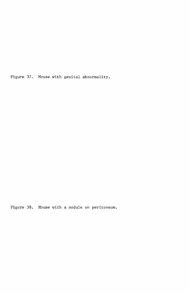
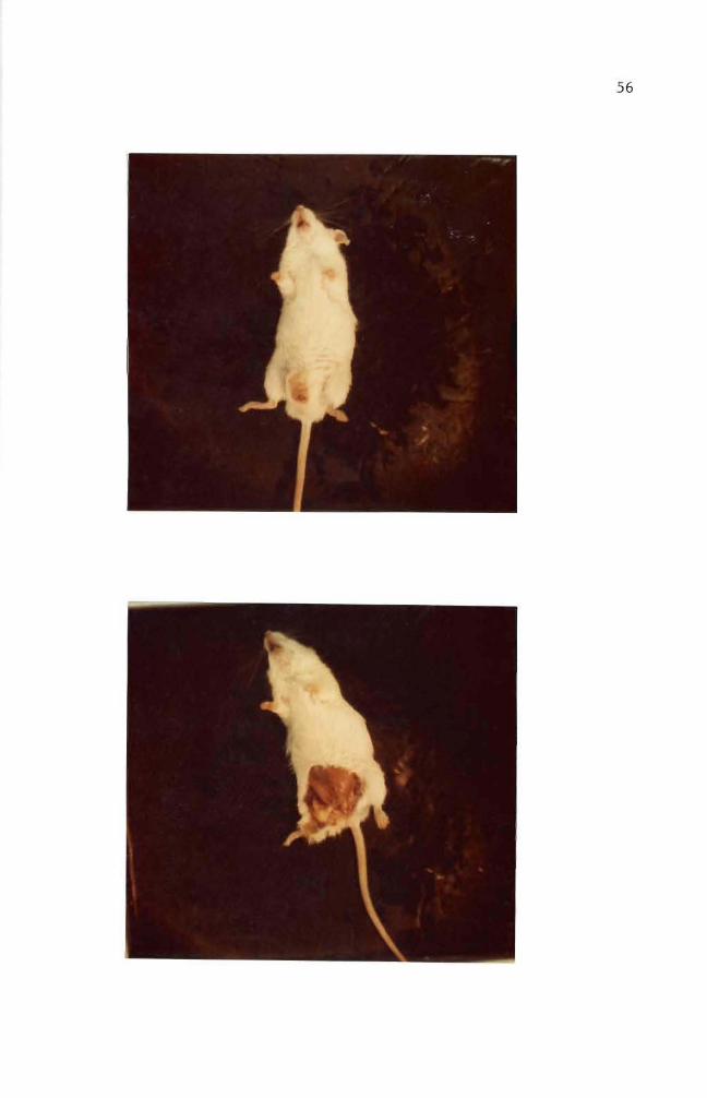
Figure 37 Mouse with genital abnormality 55
Figure 38 Mouse with a nodule on peritoneum 55
Figure 39 Vertical section of testis to show anatomical locations of the seminiferous tubules and the ductus epididymis bullbullbullbullbullbullbullbullbullbullbullbullbullbullbullbullbullbullbullbullbullbullbullbullbull 57
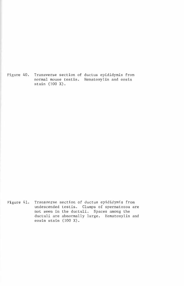
Figure 40 Transverse section of the ductus epididymis from a normal mouse testis bullbullbullbullbullbullbullbullbullbullbullbullbullbullbullbullbullbullbullbullbullbull 58
Figure 41 Transverse section of the ductus epididymis from undescended testis bullbullbullbullbullbullbullbullbullbullbullbullbullbullbullbullbullbullbullbullbullbullbull 58
Figure 42 Transverse section of seminiferous tubules from normal mouse testis bullbullbullbullbullbullbullbullbullbullbullbullbullbullbullbullbullbull 60
Figure 43 Transverse section of seminiferous tubules from undescended testis bullbullbullbullbullbullbullbullbullbullbullbullbullbullbullbullbullbull 60
INTRODUCTION
All living organisms including human beings are exposed to both
external and internal environmental factors (see diagram on page 3)
A close study of these environmental factors in relationship to cancer
led investigators to conclude that many human cancers have chemicals
as their causative agents (Boyland and Sims 1965 Van Cantfort et al
1975 Hecht et al 1974 Henderson et al 1975) Although the existshy
ence of cancer has been known for many years intensive investigations
into its origins were not conducted until the 19th century
A Japanese research group performed one of the initial studies in
cancer research They repeatedly applied coal tar to rabbit ears for
100-580 days and a consequent development of skin cancer was observed
(Yamagiwa and Ichikawa 1918)
This study stimulated many investigators to search for cancer
causing chemicals Among the cancer causing chemicals found
benzo(a)pyrene was isolated as well as with other chemical constituents
from coal tar (Cook et al 1932 1933) Benzo(a)pyrene has also been
isolated from numerous materials found in environment (Table 1 Hirono
et al 1970 19721973 Pamukcu et al 1968 Falk et al 1958
Evans and Mason 1965)
Recent investigations have classified chemical carcinogens by
chemical structures such as a) carcinogenic aromatic hydrocarbons (see
diagram on page 3) b) carcinogenic aromatic amines c) carcinogenic
compounds (amino azo dyes nitroso compounds alkylating agents) and
d) inorganic carcinogens (Weisburger 1977)
2
BIOLOGICAL
AGENTS
J
POPULATION-shy -
OCCUPATIONAL
Figure 1 Human exposure to hazardous environmental agents (Kraybill 1977)
3
Disputed (33 or less)
Chrysene
Benz (a) anthracene
Dibenz(ac)anthrashycene(1234shy
Dibenzonthracene)
Moderate (33 - 66)
(11UJ
Benzo(c)phenanthrene
Dibenz(ah)anthracene
Dibenzshy(aj)anthracene
Benzo(c)chrysene-
High activity (66 or higher)
~
Benzo(a)pyrene
Dibenzo(ah)pyrene
Dibenzo(al)pyrene
Dibenzo(ai)pyrene (1256-dibenzphenanthrene)
4
Table 1 Polycyclic aromatic hydrocarbons in food
Benzo(a) pyrene (ugKg)
Benz(a) anthracene
(ugKg)
Charcoal broiled meat 8 45 Lijinsky and Shubik 1964
Smoked fish 12-37 91-189 Masuda and Kuratune 1971
Singed sheep head Coal raw Coal cooked
21 9
15 5
Thorsteinsson and Thordarson 1968
Singed sea bird Coal raw Coal cooked
99 96
69 50
Smoked food Mutton Trout Cod Red Fish
13 21 05 03
-shy-shy
Bailey and Dungal 1958
Barbecued ribs 105 36 Lijinsky and Shubik 1965
Coffee 4 -- Kuratsune and Hueper 1960
5
Researchers attention up to this point was devoted to isolating
chemical carcinogens from our environment and to characterizing the
chemical carcinogens Some researchers Allison and Mallucci (1964)
Shires et ale (1966) Brooks and Lawley (1964) Wang et ale (1972)
started to investigate the mechanisms involved in chemical carcinoshy
genesis
Allison and Mallucci (1964) utilized fluorescence microscopy
phase contrast microscopy and autoradiography techniques to locate
carcinogenic hydrocarbons in mammalian cell cultures These investishy
gators concluded that the hydrocarbon carcinogens were not concentrated
in either mitochondria or the nuclei They were however found to be
within lysosomes Allison and Paton (1965) Allison and Dingle (1966)
and Allison (1968) centered their studies on lysosomes and their
relationship to cancer
A study similar to the one mentioned above was carried out two
years later by Shires and Richter (1966) These workers exposed mammashy
lian cells in vitro to carcinogenic hydrocarbons for periods ranging
from 12 hours to 4 days The treatment was terminated at different
intervals for microscopic observations The researchers were unable to
detect any incorporation of benzo(a)pyrene into the nuclei or chromoshy
somes during a complete generation cycle
On the other hand Brooks and Lawley (1964) tried to show the
binding of carcinogenic hydrocarbons to the nucleic acids in mouse skin
They did so by applying the hydrocarbon carcinogens to shaved regions on
the backs of mice The mice were sacrificed at various times and the
treated areas of skin were removed and used for deoxyribonucleic acid
(DNA) and ribonucleic acid (RNA) isolation This study indicated that
6
DNA reacts with the hydrocarbon carcinogens tested Experimental
results did not indicate how the hydrocarbon carcinogens are bound to
the nucleic acids
Lerman (1960) investigated the way in which polynuclear hydroshy
carbons react with DNA He used mutagenic acridine dye derivatives
for his study This investigation showed that the dyes are intershy
calating into the DNA helix through extension of DNA backbone This
information enhanced the idea that polycyclic aromatic hydrocarbons
require structural planality for the carcinogenic potency A similar
mechanism of the intercalation reaction between hydrocarbon carcinoshy
gens and DNA was proposed by Liquori (1962) Liquoris findings were
based upon the increased solubility of benzo(a)pyrene in DNA aqueous
solutions This is significant because benzo(a)pyrene is virtually
insoluble in water
Boyland and Green (1964) expressed a slightly different opinion
They showed that carcinogenic hydrocarbon molecules react with DNA by
slipping into the sites where the DNA is already stretched Such
stretching of DNA can only occur during DNA replication or RNA transhy
scription An expanded idea was postulated by Arcos and Argus (1968)
They indicated that the solubilization of carcinogenic hydrocarbons by
DNA can be accomplished by either intercalation or through external
binding mechanisms (Figure 2)
Van Duuren et al (1969) suggested a reconsideration of the
intercalation mechanism This was proposed because a) solubilization
of detergent micelles of sodium lauryl sulfate and aqueous DNA solution
caused the same results which were found with hydrocarbon carcinogens
7
e oI I I I I I IIgt I I
I
A B
Figure 2 Noncova1ent interactions of polycyclic aromatic hydroshycarbons (illustrated with 34-benzpyrene) with DNA A Intercalation into DNA double helix B External binding to DNA (From Arcos and Argus 1968)
8
and aqueous DNA solution and b) potent carcinogens such as 712shy
dimethy1benz(a)anthracene and 3-methy1cho1anthrene are thicker than
the distance between adjacent base pairs in DNA
Because previous investigations had been unable to prove whether
or not particular parent carcinogenic hydrocarbon molecules react with
DNA research interest was directed to the isolation and characterization
of intermediate metabolites Benzo(a)pyrene-45-oxide (K-region
epoxide) proved to be one of the highly reactive metabolites of
benzo(a)pyrene (Wang et a1 1972 Selkirk et a1 1971) Another
metabolite 78-dihydro-78-dihydroxybenzo(a)pyrene-910-oxide has been
investigated for its metabolism and mutagenicity It was confirmed to
be carcinogenic (Neubo1d and Brooks 1976 Sims et a1 1974)
Thus far accumulated data favor the theory of hydrocarbon carcinoshy
gens binding to DNA On the other hand the significance of 1ysosomes
in regard to carcinogenesis cannot be ignored Both Allison and Ma11ucci
(1964) and Shires and Richter (1966) utilized fluorescence and phase
microscopies and concluded that 1ysosomes were somehow involved In
addition to optical systems Allison and Ma11ucci (1964) used autoshy
radiography techniques for localization of hydrocarbon carcinogens in
mammalian cell cultures They concluded that the amount present if
any in cellular organelles other than 1ysosomes is too small to detect
by these methods
It was the purpose of this investigation to challenge the credishy
bility of autoradiographic techniques as an indicator of
benzo(a)pyrene-DNA incorporation and to consider the problems that
were encountered with this technique
MATERIALS AND METHODS
Experimental animals
Heterogenic white mice were obtained from the Emporia State
University research animal facility and from the Mid-Continent
Research Company (Kansas City Kansas) They were maintained in the
animal facility at Emporia State University
Chemicals
Benzo(a)pyrene (B(a)P) obtained from Sigma Chemical Company
was first dissolved in acetone (10 mgm1) and then dispersed into complete
growth medium with a Pyrex disposable syringe to give a concentration of
100 ugm1 of medium (Berwald and Sachs 1965) This B(a)P suspension was
used as stock solution from which dilutions were made Dilutions were
01 ug 10 ug and 50 ug B(a)P per milliliter of medium The stock solushy
tion was stored at OdegC The desired concentrations of B(a)P solution
were applied to cells in culture
Tissue culture medium
Du1beccos modified Eagles medium containing 45 gl glucose was
purchased from GIBCO The growth medium was supplemented with 10 fetal
bovine serum (GIBCO) and 05 m1 combiotics This complete growth medium
was used to grow and maintain the cells in vitro GKN (calcium and
magnesium free saline) was used for cell harvest and cell transfer with
025 trypsin and trypsin-versene (0002-005) solutions respectively
Stock combiotic solutions were prepared by combing penicillin G
sodium (1650 Umg) and streptomycin sulfate (740 mggm) in 1X Hanks
BSS Stock solutions were sterilized using Mi11ipore filters One
milliliter a1iquots were prepared and stored frozen until needed Cell
10
harvesting solutions were prepared by adding 05 ml of the combiotic to
every 100 ml of GKN Likewise cell maintenance solutions were preshy
pared by adding 05 ml of combiotic stock solution to every 100 ml of
growth medium
Fetal bovine serum which was mycoplasma tested and virus screened
was obtained from GIBCO and used throughout this study
Carbon dioxide gas prepared by mixing calcium carbonate and
sulfuric acid was used to maintain the desired pH of the medium
Glassware
Glassware was cleaned by soaking in an acid bath (17 mixture of
concentrated hydrochloric acid and concentrated sulfuric acid) overshy
night followed by washing with Alconox solution and repetitive rinsing
with tap water All glassware was rinsed with deionized distilled
water after being washed
Microscope coverslips (22 X 50 mm) were used to make cell growing
covers lips (22 X 8 mm) These were immersed in an Alconox solution and
rinsed with running tap water for 10-13 hours They were rinsed individshy
ually 10 times in three consecutive deionized distilled water baths and
one 100 ethanol bath then stored in an absolute ethanol bath until
use
Cell culture
Minced whole embryos of heterogenic white mice served as the source
of normal cells Pregnant mice (15-18 days in term) were sacrificed by
cervical dislocation Embryos were then removed aseptically from the
mouse and transferred to a petri dish containing GKN-combiotics mixture
Embryos were minced and placed into a trypsinizing flask The tissue
11
chunks were subjected to gentle stirring while being trypsinized at
room temperature for 30 minutes Trypsinized tissue was filtered
through four layers of cheese cloth The filtrate was washed with one
change of GKN and one change of complete growth medium Cell popushy
lations were adjusted with complete growth medium to a concentration
6of 3 X 10 cellsmI Three milliliter aliquots were planted in 25
2 cm polystyrene culture flasks and incubated at 37degC for one to three
days (primary cell culture) Primary cell cultures were treated with
trypsin-versene solution for two minutes washed centrifuged and
resuspended to give a concentration of 6 X 10⁵ cellsmI They were
inoculated in test tubes containing covers lips (7 X 22 mm) for 24 hours
at 37degC (secondary culture) prior to usage in the studies Secondary
cell cultures were treated with either a colloidal suspension of B(a)P
(10 ugml) and 3H- B(a)P (10 uCiml) or 3H- B(a)P (10 uCiml) alone
Autoradiography
Tritium-labeled B(a)P treated cells growing on the coverslips were
removed from the test tubes rinsed with two changes of GKN solution and
fixed in an absolute methanol bath for five minutes The covers lips
were mounted on a slide with the cell side up using Pro-texx mounting
medium The slides were dried overnight on a heating plate at 37degC
Ilford emulsion (1-4 Polysciences Inc) was melted in a 40degC
water bath Ten milliliters of melted emulsion were diluted with an
equal volume of deionized distilled water and any bubbles resulting in
the mixing operation were removed through a layer of cheese cloth The
slides were dipped into the diluted emulsion and immediately removed
Residual emulsion was cleaned from the back of the slides and the
12
slides held in a vertical position for 15 minutes to allow the emulsion
to dry Slides were sealed in a light tight slide box by wrapping the
box with aluminum foil The slides were exposed for 10 days at OdegC
The exposed slides were developed in Dekto1 (D-17) for two
minutes rinsed in a deionized distilled water stop bath and fixed in
acid fixer for three minutes After washing for 15 minutes in slowly
running tap water the slides were dried and stained for four minutes
in 005 basic fuchsin solution The slides were dehydrated in a
series of ethanol baths and immersed in a mixture of cedar wood oil
and absolute ethanol (1 1) for one hour followed by infiltration of
a Pro-texx mounting medium and xylene mixture (1 1) for one hour
The slides were examined for the appearance of silver grains
(black dots) located within the cells thereby indicating B(a)P uptake
by the cell
Cell viability test
The toxic effect of B(a)P on cultured cells was examined by adding
5 m1 of B(a)P at different concentrations Concentrations used ranged
from 01 to 50 ug B(a)P per milliliter of growth medium At regular
intervals B(a)P-growth medium mixtures were collected from culture
flasks and centrifuged for five minutes at 800 rpm The supernatant
was siphoned off and discarded Pellets were resuspended with 1 m1
growth medium and 01 m1 of erythrosin (04) was added and stained for
two minutes Dead cells staining pink were counted on a hemocytometer
Numbers of dead cells were plotted against the durations of B(a)P treatshy
ment
13
Inoculation of B(a)P treated cells into animals
Cells treated with B(a)P were harvested with a trypsin-versene
solution for three minutes Cell harvest was made at preselected inshy
tervals of time The cell population was adjusted to 10 X 106 ce11sm1
with complete growth medium From this solution 02 m1 were injected
into a group of five male mice Injections were made subcutaneously on
the left thigh (modified from Evans et a1 1957) Inoculation sites
were checked every 14 days for tumor development until termination of
the experiment
Histological study
Twenty-eight days after inoculation of B(a)P treated cells mice
which developed tumors were sacrificed for histological studies The
tumor tissue was excised and fixed overnight in Bouins fixative
Several solution changes of 70 ethanol saturated with lithium carbonate
were made to eliminate any fixative in the tissue Dehydration of fixed
tissue was accomplished in a series of ethanol baths of one hour each
The tissue was then immersed for one hour in a mixture of absolute
ethanol and xylene (1 1) It was soaked in a xylene bath overnight
and then transferred to a mixture of xylene and paraffin (56 0 tissuemat)
for 15 minutes Infiltration of hot tissuemat was carried out in four
changes of tissuemat solution Tissue was then embedded in a plastic
ice cube tray with hot tissuemat and cooled to room temperature The
blocks were sectioned on the microtome at a thickness of seven microns
The sections were stained by standard hematoxylin and eosin procedures
The slides were then carefully examined for histological abnormalities
14
Chromosome preparation
Cells in culture were mitotically blocked by lyophilized colcemid
006 ugml (GIBCO) for two hours Harvesting of cells was accomplished
by trypsinization and centrifuging for five minutes at 800 rpm The
pellet was resuspended in 1 ml of complete growth medium with a Pasteur
pipette Hypotonic solution (3 ml deionized distilled water) was added
to this suspension for seven minutes at room temperature One milliliter
of freshly prepared methanol-acetic acid (3 1) fixative was introduced
to the hypotonic solution for 10 minutes The suspension was centri shy
fuged and fixed by repeated dispersion at 10 minute intervals in three
changes of 31 methanol-acetic acid fixative During these fixation
periods microscope slides were washed in an Alconox solution and stored
in deionized distilled water at 4degC After the third fixation the
pellet was resuspended in 03-1 ml (depending on size of the pellet)
fixative solution Several drops of the final cell suspension were
applied to the slides The backs of the slides were wiped dry with
tissue paper A film of water on the upper surface of the slides was
left to facilitate spreading of the cells The slides were air-dried
and stained for 15 minutes in modified Giemsa stain Modification
was made according to Gude et al (1955) For the autoradiographic
study the slides were stained for 15 minutes after the completion of
photographic processes
RESULTS
Cell viability test
The toxic effect of B(a)P was tested for nine days Results are
summarized in Figure 3 From the data linear regression curves were
prepared for each concentration The slope of the untreated cell line
is 48 The slopes of the B(a)P treated cell lines are 65 for 01 ug
B(a)P treated cells 70 for 10 ug B(a)P treated cells and 76 for 50
ug B(a)P treated cells A statistical analysis did not indicate a
significant difference between the normal cell line and the B(a)P
treated cell lines Re-examination of the data used in Figure 3 will
help in understanding effect of treatment on cell viability for specific
periods of time In Figure 4 one and three day incubation periods
showed a smaller rate of increase (slope for one day 007 and slope
for three days 016) which implies that the B(a)P concentration did
not have a significant effect in terms of cell killing ability The dead
cell numbers in five day B(a)P treated cultures presented about 28-fold
increase from that of three day B(a)P treated cultures A statistical
analysis showed a significant difference indicating that the critical
factor in cell killing ability of B(a)P is dependent upon the exposure
period
Cell morphology in culture
Mouse embryo cells in culture were observed under the light microshy
scope for their morphology Embryonic cells were round immediately after
implantation into culture flasks Approximately three hours incubation
at 37degC after initial cell plantation some cells started to establish
their natural morphological appearance (elongation of cytoplasm) The
16
elongation process was an indication of cell attachment on the surface
of culture flasks Owens et a1 (1974) reported that in earlier days
of incubation two cell types epithelial cells and fibroblast cells
can be identified Epithelial cells from mono1ayered sheets which are
attached to each other (Figures 5 and 9) Vasi1iev and Ge1hand (1977)
reported that fibroblast cells form several morphological varieties in
culture mono1ayers of mutually oriented cells which are not firmly
attached to each other (Figures 6 and 8) multilayered sheets (Figure
7) or spherical aggregates (Figure 7 lower right and upper left
darkly stained aggregates) Fibroblasts andor fibroblast-like cells
became predominant throughout the entire cell population when cells were
cultured for over 30 days (Figures 10 11 and 12) These fibroblast
cells are characterized by elongated or stellate cytoplasmic structures
(Figures 10 11 and 12)
Mouse embryo cells were treated with 10 ug B(a)P for predesignated
exposure periods to observe an effect on cell morphology Some B(a)P
treated cells showed an abnormal spindle shape while the other cells
retained their normal morphological appearance (Figures 13 15 and 16)
In Figures 15 and 16 some cells were found to have vacuoles within their
cytoplasm Many of these also showed attenuated cytoplasms (Figure 16)
Autoradiography
Cultured cells were incubated with B(a)P for several different types
of treatment One treatment consisted of incubating cultured cells with
B(a)P at 10 ugm1 and 3H- B(a)P at 10 uCim1 until the termination of
experiment (full-term treatment) Other cells were partially exposed
to B(a)P at the same concentration and radioactivity level The exposure
17
60
-l S-r-
0 0 0 -l
gtlt
40-
Ul -l -l ltl) tJ
d Cll ltl) d
~
0 ltl)
20 l z
o o 5 10
Days
Figure 3 Toxic effect of B(a)P on cultured mouse embryo cells Untreated cells 0 Cells treated with 01 ug B(a)P e Cells treated with 10 ug B(a)P X Cells treated with 50 ug B(a) P
__
--------
18
60
-~ s
I
I ~ ~7 r 0 0 0 ~
x -
Ul ~ ~
eu 40u 0 ell eu 0
4-l 0
Ul H eu ~ l Ie z
20 3
9
_ ---41---------------- 1 o
o 20 40 60
B(a)P concentration (ugml)
Figure 4 Toxicity effect of B(a)P concentration The numbers 1 3 5 7 and 9 indicate the days of B(a)P treatment
Figure 5 Two day old normal mouse embryo cells in culture Epithelial cells in the central area of fibroshyblast cells surrounding the epithelial cells are shown here May-Grunwald and Giemsa stain (100 X)
Figure 6 Four day old normal mouse embryo cells in culture Large nucleated epithelial-like cell and fibroshyblast cells are shown here May-Grunwald and Giemsa stain (400 X)
Figure 7 Six day old normal mouse embryo cells in culture Multilayered fibroblast cells and two heavily stained aggregates are shown here May-Grunwald and Giemsa stain (100 X)
Figure 8 Eight day old normal mouse embryo cells in culture Fibroblast cells formed a monolayer sheet MayshyGrunwald and Giemsa stain (100 X)
Figure 9 Thirteen day old normal mouse embryo cells in culture Epithelial cells in the central and upper left areas are surrounded by well elongated fibroblast cells May-Grunwald and Giemsa stain (100 X)
Figure 10 Thirty day old normal mouse embryo cells in culture May-Grunwald and Giemsa stain (100 X)
Figure 11 Thirty five day old normal mouse embryo cells in culture Cell orientation is obscure and vacuoles are seen May-Grunwald and Giemsa stain (100 X)
Figure 12 Forty day old normal mouse embryo cells in culture Cell orientation is obscure and vacuoles are present May-Grunwald and Giemsa stain (100 X)
9Z
Figure 13 Mouse embryo cells incubated with 10 ug B(a)P for 1 day May-Grunwald stain (100 X)
Figure 14 Mouse embryo cells incubated with 10 ug B(a)P for 2 days May-Grunwald stain (100 X)
Figure 15 Mouse embryo cells treated with 10 ug B(a)P for 6 days May-Grunwald stain (100 X)
Figure 16 Mouse embryo cells treated with 10 ug B(a)P for 9 days Basic Fuchsin stain (100 X)
31
periods were 24 hours and four days
Figures 18 through 23 represent the full-term treatment and events
for the first 17 hours are summarized in Figure 17 After
hours exposure to 3H- B(a)P B(a)P accumulation in the nuclei
low Its cytoplasmic concentration was also minimal (Figure
3H-B(a)P uptake by the cells increased rather sharply after 35
of incubation (Figures 19 20 and 21) Long term incubation (six
and 20 days) showed a concentration of B(a)P in the cytoplasm while no
concentration of B(a)P was observed (Figures 22 and 23)
In other treatments cultured cells were partially exposed to
B(a)P at the same concentration Radioactivity levels were as in the
treatment The cells were exposed to B(a)P for one and four
days At the end of the exposure period the complete growth medium
was replaced for further incubation Figures 24 through 27 indicate
some of the results of 24 hour treated cells Overall results are
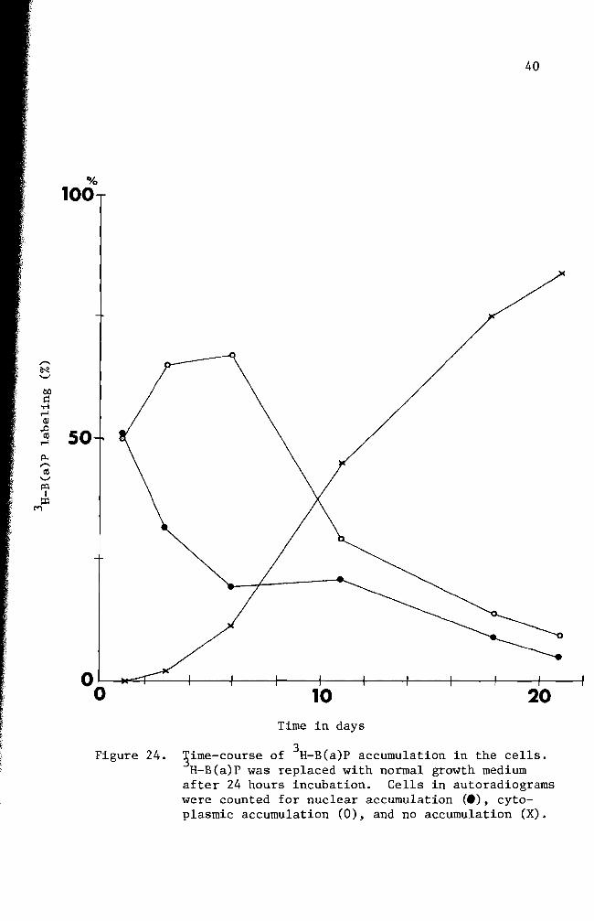
summarized in Figure 24 Nuclear accumulation of 3H-B(a)P was maximum
at 24 hours and decreased drastically when fresh complete growth medium
was introduced and incubation was continued (Figures 24 27 and 28)
The nuclear accumulation of B(a)P exceeded that of cytoplasm only at
24 hours incubation (Figure 24) Cytoplasmic accumulation of 3H-B(a)P
was at its highest peak on the 6th day of incubation and followed the
same pattern of decrease as the nuclear accumulation curve Extrashy
polation of the two curves suggests disappearance of B(a)P from the
cells occurs between the 24th and 26th day Some cells as exemplified
in Figure 28 lost nuclear B(a)P and most of the cytoplasmic B(a)P at
50
100
32
00
0 1 bull
O Hours
Figure 17 Time-course of 3H- B(a)P accumulation in the cells Cells in autoradiograms were counted for nuclear accumulation () cytoplasmic accumulation (0) and no accumulation (X)
I I Ii I Ii(I I ~ I I
2 4 6 8 10 12 14 16 18
33
21 days of incubation in the growth medium
Figures 29 through 32 represent the results of four day treated
Four (no additional incubation in growth medium) and 11 days
(additional six day incubation in growth medium) cells show nuclear and
cytoplasmic accumulation of B(a)P but not as intense as seen in the one
cells (Figures 29 and 30) Fifteen day incubated cells had
some B(a)P molecules in the cytoplasm The cells incubated for 27 days
of B(a)P in any part of the cells (Figure 32)
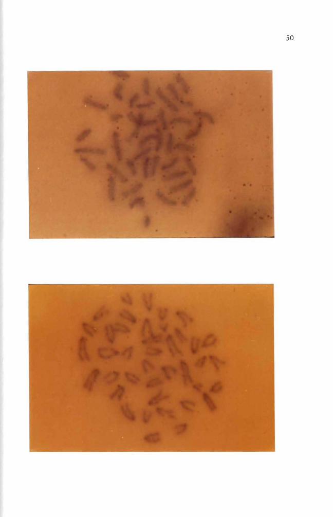
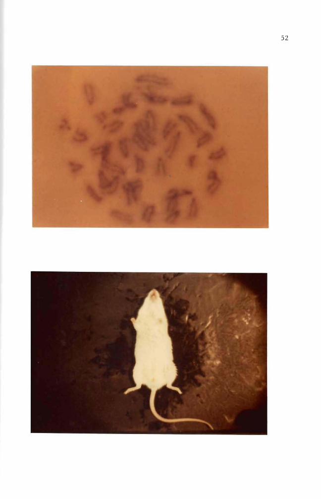
Figures 33 through 35 are the chromosome spreads from 3H- B(a)p
treated cells Silver grains (black dots) were seen on or near the chromoshy
somes in three autoradiograms (Figures 33 34 and 35) Autoradiograms of
chromosomes prepared from one two and three day treated cells with
3H- B(a)P (10 uCiml) were counted for use in labeling analysis Chroshy
mosome spreads from one and two day treated cells produced 936 and 920
labeled chromosome spreads respectively Total chromosome spreads examshy
ined were 94 and 100 Cells treated for three days gave 304 labeled
chromosome spreads
Figures 34 and 35 showed chromosome breakages
Induction of abnormalities and its histology
B(a)P treated cells were injected into adult mice The mouse in
Figure 36 was a normal male mouse The mouse in Figure 37 exemplifies
the abnormal genital inflammation characterized by many of the mice inshy
jected with the B(a)P treated cells From the 26 injected mice 31 deshy
veloped a genital inflammation In contrast only 38 of the control
mice exhibited genital abnormalities The controls consisted of three
groups of mice One group of 32 mice received no injections Another
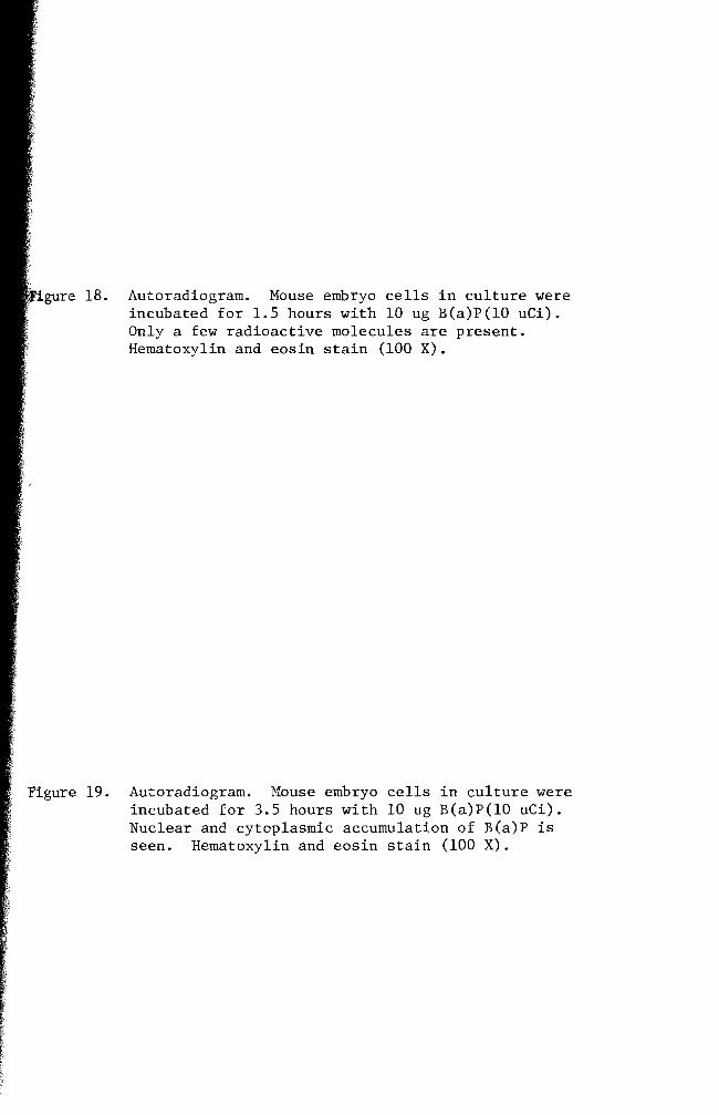
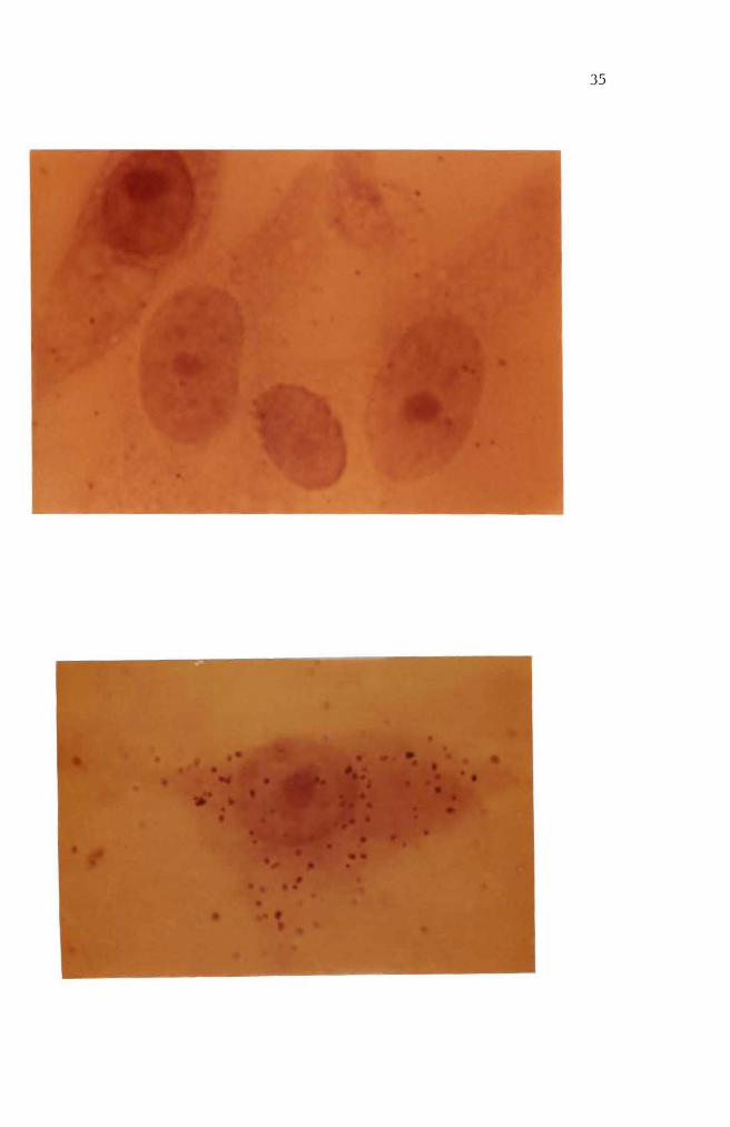
Autoradiogram Mouse embryo cells in culture were incubated for 15 hours with 10 ug B(a)P(lO uCi) Only a few radioactive molecules are present Hematoxylin and eosin stain (100 X)
Figure 19 Autoradiogram Mouse embryo cells in culture were incubated for 35 hours with 10 ug B(a)P(lO uCi) Nuclear and cytoplasmic accumulation of B(a)P is seen Hematoxylin and eosin stain (100 X)
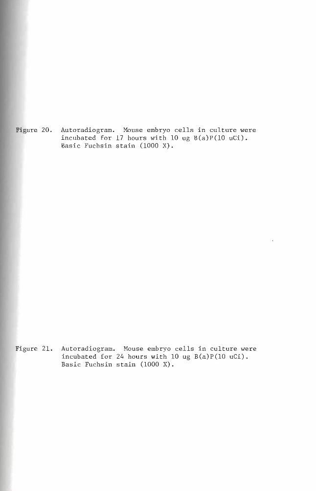
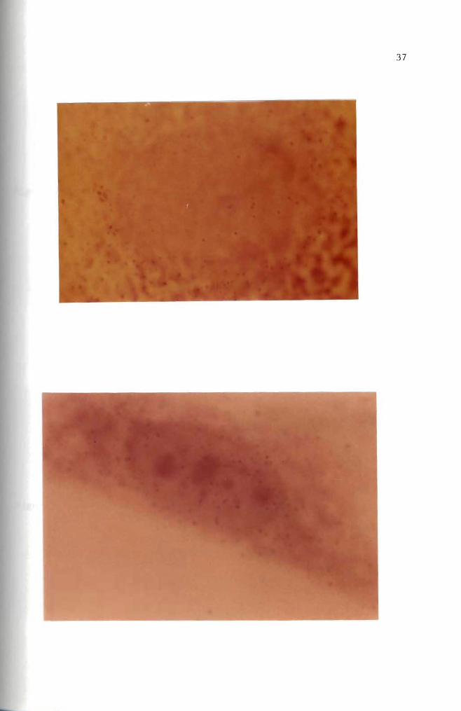
Figure 20 Autoradiogram Mouse embryo cells in culture were incubated for 17 hours with 10 ug B(a)P(10 uCi) Basic Fuchsin stain (1000 X)
Figure 21 Autoradiogram Mouse embryo cells in culture were incubated for 24 hours with 10 ug B(a)P(10 uCi) Basic Fuchsin stain (1000 X)
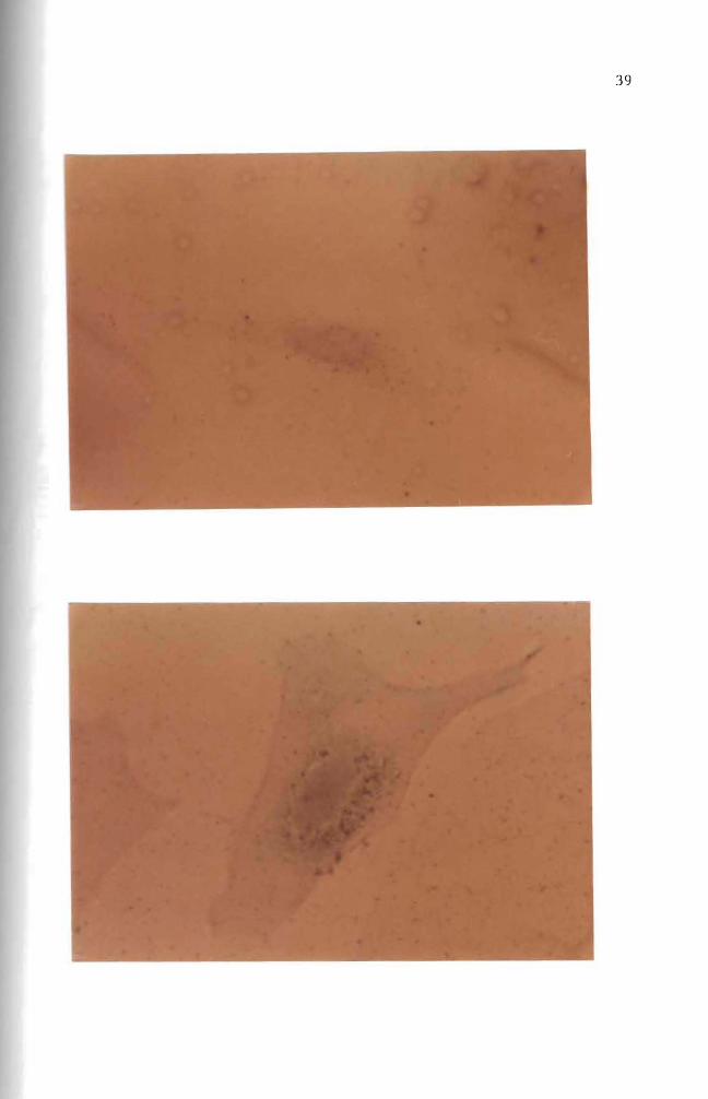
Figure 22 Autoradiogram Mouse embryo cells in culture were incubated for six days with 10 ug B(a)P(10 uCi) Hematoxylin and eosin stain (100 X)
Figure 23 Autoradiogram Mouse embryo cells in culture were incubated for 20 days with 10 ug B(a)P(10 uCi) A heavy accumulation of B(a)P in cytoplasm is seen here Hematoxylin and eosin stain (400 X)
-
6pound
40
00
100
~ -
00 c
-l M Q)
~ 50M
~
ro - lQI
z M
10 20 Time in days
Figure 24 3ime-course of 3H- B(a)P accumulation in the cells H-B(a)P was replaced with normal growth medium
after 24 hours incubation Cells in autoradiograms were counted for nuclear accumulation (e) cytoshyplasmic accumulation (0) and no accumulation (X)
00
Figure 25 Autoradiogram Mouse embryo cells in culture were incubated for one day with 10 ug B(a)P(lO uCi) Nuclear and cytoplasmic accumulation of B(a)P is seen Basic Fuchsin stain (1000 X)
Figure 26 Autoradiogram Mouse embryo cells in culture were incubated for one day with 10 ug B(a)P(lO uCi) The B(a)P was then removed The cells were incubated an additional 3 days in complete growth medium Basic Fuchsin stain (1000 X)
Figure 27 Autoradiogram Mouse embryo cells in culture were incubated for one day with 10 ug B(a)P(lO uCi) The B(a)P was then removed The cells were incubated an additional 11 days in complete growth medium Basic Fuchsin stain (1000 X)
Figure 28 Autoradiogram Mouse embryo cells in culture were incubated for one day with 10 ug B(a)P(lO uCi) The B(a)P mixture was then removed The cells were incubated an additional 21 days in complete growth medium Basic Fuchsin stain (1000 X)
shy
Figure 29 Autoradiogram Mouse embryo cells in culture were incubated for four days with 10 ug B(a)P(lO uCi) Negri body stain (1000 X)
Figure 30 Autoradiogram Mouse embryo cells in culture were incubated for four days with 10 ug B(a)P(lO uCi) The B(a)P mixture was then removed The cells were incubated an additional six days in complete growth medium Some silver grains are seen Basic Fuchsin stain (1000 X)
97
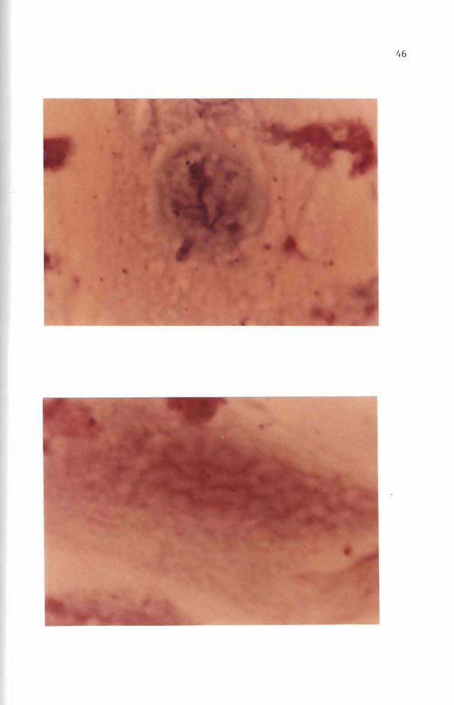
Figure 31 Autoradiogram Mouse embryo cells in culture were incubated for four days with 10 ug B(a)P(lO uCi) The B(a)P was then removed The cells were incubated an additional 11 days in complete growth medium Some silver grains were seen in the cytoplasm Basic Fuchsin stain (1000 X)
Figure 32 Autoradiogram Mouse embryo cells in culture were incubated for four days with 10 ug B(a)P(lO uei) The B(a)P mixture was then removed The cells were incubated an additional 23 days in complete growth medium Basic Fuchsin stain (1000 X)
81]
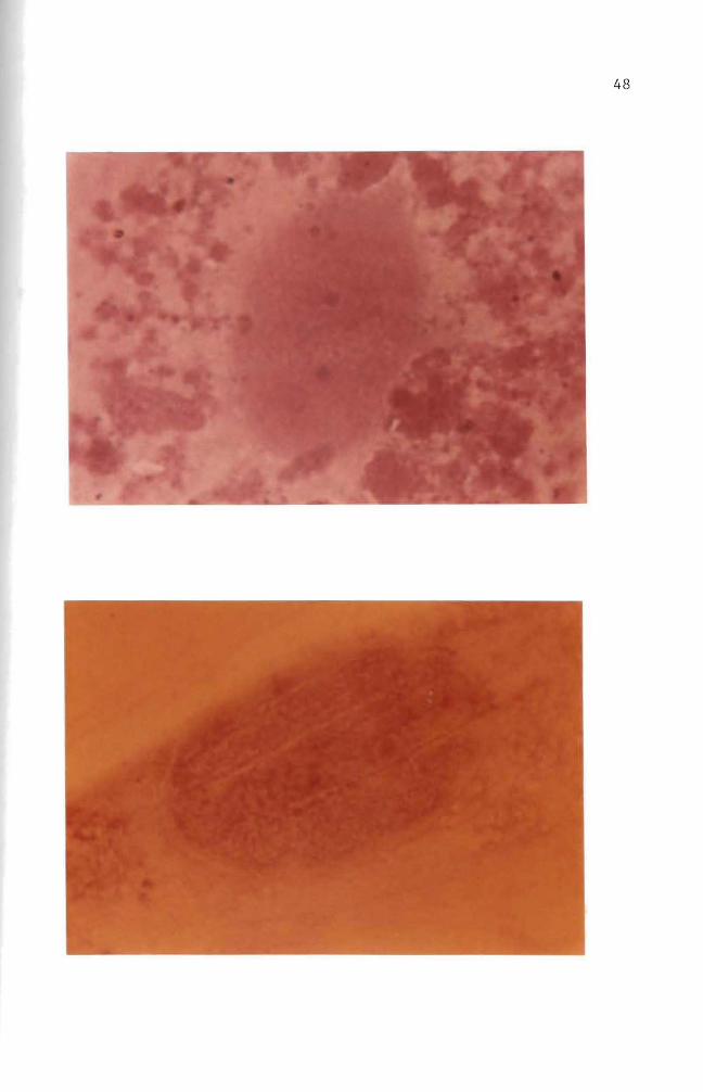
Figure 33 Autoradiogram of chromosomes Mouse embryo cells in culture were incubated for one day with 005 ug B(a)P(lO uCi) Giemsa stain (1000 X)
Figure 34 Autoradiogram of chromosomes Mouse embryo cells in culture were incubated for two days with 005 ug B(a)P(lO uCi) Giemsa stain (1000 X)
-
os
Figure 35 Autoradiogram of chromosomes Mouse embryo cells in culture were incubated for three days with 005 ug B(a)P(10 uCi) Giemsa stain (1000 X)
Figure 36 Normal male mouse
53
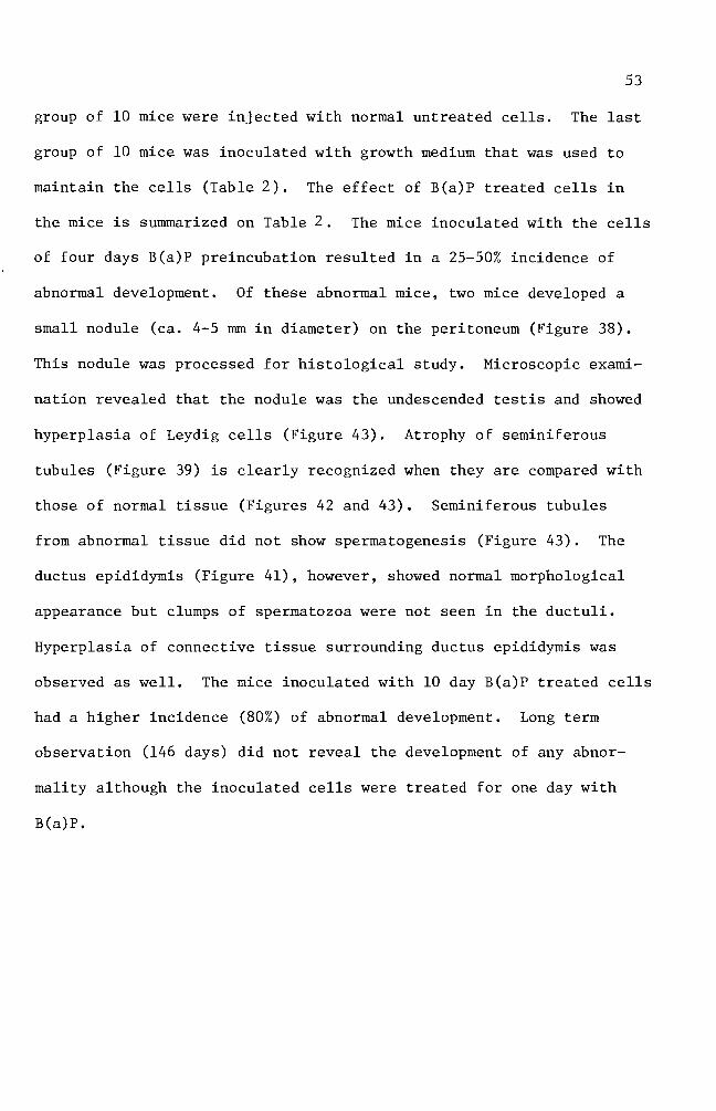
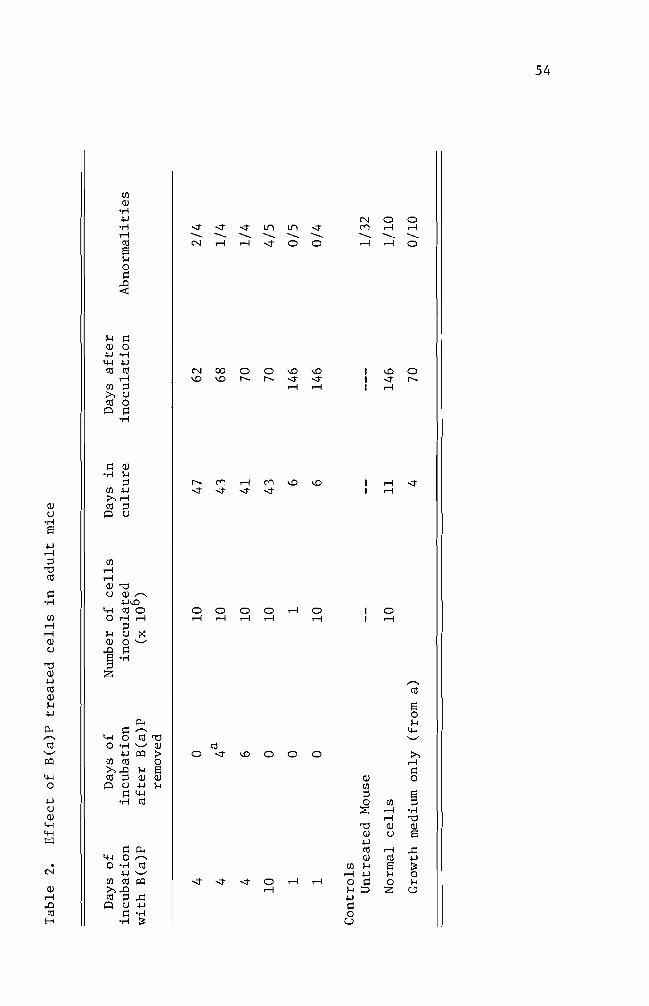
group of 10 mice were injected with normal untreated cells The last
group of 10 mice was inoculated with growth medium that was used to
maintain the cells (Table 2) The effect of B(a)P treated cells in
the mice is summarized on Table 2 The mice inoculated with the cells
of four days B(a)P preincubation resulted in a 25-50 incidence of
abnormal development Of these abnormal mice two mice developed a
small nodule (ca 4-5 rom in diameter) on the peritoneum (Figure 38)
This nodule was processed for histological study Microscopic examishy
nation revealed that the nodule was the undescended testis and showed
hyperplasia of Leydig cells (Figure 43) Atrophy of seminiferous
tubules (Figure 39) is clearly recognized when they are compared with
those of normal tissue (Figures 42 and 43) Seminiferous tubules
from abnormal tissue did not show spermatogenesis (Figure 43) The
ductus epididymis (Figure 41) however showed normal morphological
appearance but clumps of spermatozoa were not seen in the ductuli
Hyperplasia of connective tissue surrounding ductus epididymis was
observed as well The mice inoculated with 10 day B(a)P treated cells
had a higher incidence (80) of abnormal development Long term
observation (146 days) did not reveal the development of any abnorshy
mality although the inoculated cells were treated for one day with
B(a)P
Tab
le
2
Eff
ect
of
B(a
)P tr
eate
d cell
s in
ad
ult
mic
e
Day
s o
f D
ays
of
Num
ber
of
cell
s D
ays
in
Day
s aft
er
incu
bati
on
w
ith
B(a
)P
incu
bat
ion
aft
er
B(a
)P
ino
cu
late
d
(x
106
) cu
ltu
re
ino
cu
lati
on
A
bn
orm
alit
ies
rem
oved
4 0
10
47
62
24
4 4a
10
43
68
14
4 6
10
41
70
14
10
0 10
43
70
4
5
1 0
1 6
146
05
1 0
10
6 14
6 0
4
Co
ntr
ols
U
ntr
eate
d M
ouse
-shy
-shy
--shy
13
2
Nor
mal
cell
s 10
1
1
146
11
0
Gro
wth
med
ium
o
nly
(f
rom
a)
4 70
0
10
In
+shy
Figure 37 Mouse with genital abnormality
Figure 38 Mouse with a nodule on peritoneum
9~
Figure 40 Transverse section of ductus epididymis from normal mouse testis Hematoxylin and eosin stain (100 X)
Figure 41 Transverse section of ductus epididymis from undescended testis Clumps of spermatozoa are not seen in the ductu1i Spaces among the ductu1i are abnormally large Hematoxylin and eosin stain (100 X)
6S
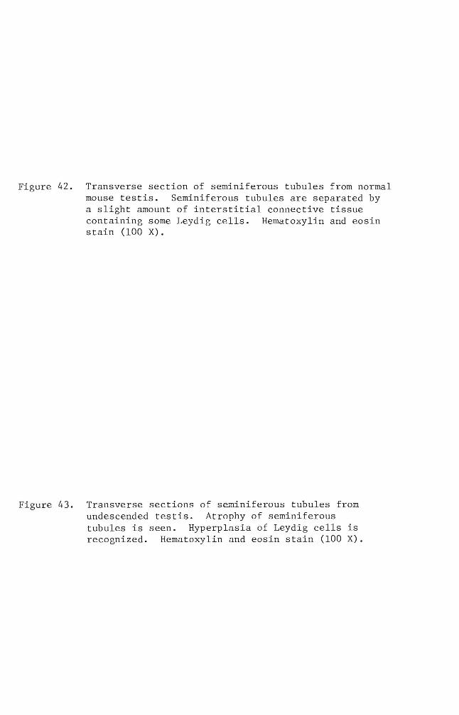
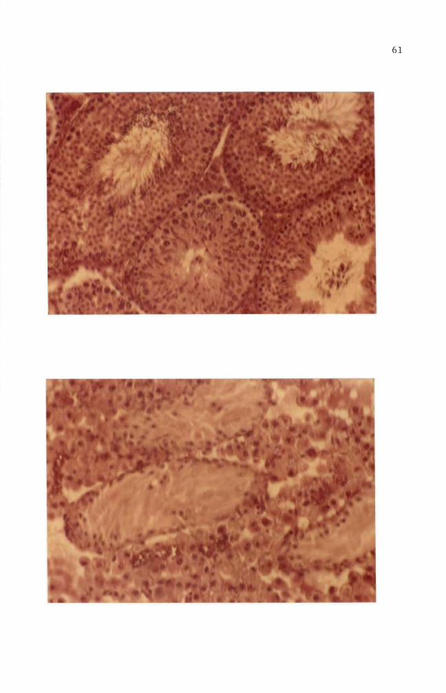
Figure 42 Transverse section of seminiferous tubules from normal mouse testis Seminiferous tubules are separated by a slight amount of interstitial connective tissue containing some Leydig cells Hematoxylin and eosin stain (100 X)
Figure 43 Transverse sections of seminiferous tubules from undescended testis Atrophy of seminiferous tubules is seen Hyperplasia of Leydig cells is recognized Hematoxylin and eosin stain (100 X)
19
DISCUSSION
Cook et a1 (1932 1933) isolated carcinogenic hydrocarbons from
coal tar pitch The melting point (mp) of one of the hydrocarbons
was determined to be 1755-1765degC This particular hydrocarbon species
was shown to be pure benzo(a)pyrene (B(a)P) by comparison with the
synthesized B(a)P B(a)P has alternative names such as 12-benzpyrene
and 34-benzpyrene It has the molecular weight of 25230 its
structure is shown in diagram on page 3
B(a)P and other closely related carcinogenic hydrocarbons (see
diagram on page 3) are being used to determine a mechanism of carshy
cinogenesis in several research systems B(a)P is known to be one of
the chemically inert carcinogens To be activated B(a)P must first be
taken up by the cells Thus invasion into the cell is the first step
in the mechanism B(a)P particles in the growth medium are taken into
the cells by endocytosis (Allison and Ma11ucci 1964) Engulfed B(a)P
particles then require enzymatic activations to acquire cytotoxic
mutagenic (Huberman et a1 1971 1976 Newbold and Brooks 1976) and
carcinogenic abilities in the cells (Wood et a1 1976 Wis10cki et a1
1976)
The greatly increased number of dead cells in cultures which had
been treated with B(a)P for five days suggested that enzymatic activations
of ingested B(a)P might have occurred during a one to three day incubation
period As a result B(a)P acquired cytotoxic potency to the cells
(Figure 4) Figures 17 and 24 indicate that the cytoplasmic accumushy
lation had maximum values between 35 hours and six days This sugshy
gested the possibility that B(a)P metabolism takes place between these
periods A reliable explanation for the cytotoxic ability of B(a)P was
63
made by several workers (Nebert and Gelboin 1968a 1968b Kasper
1971 Khandwala and Kasper 1973) There exists an enzyme aryl
hydrocarbon hydroxylase that functions in the metabolism of B(a)P
The metabolite is 3-hydroxybenzopyrene which also possesses cytotoxic
potency to the cell Benzo(a)pyrene-hydroxylase was shown to be preshy
sent at low concentrations in the nuclear membrane The existence of
inducible B(a)P-hydroxylase in a microsomal preparation was reported
as well The major difference between nuclear and microsomal memshy
branes is its specificity Nuclear membranes produce hydrocarbon
hydroxylase specifically by the stimulation of hydrocarbon carcinogens
while microsomal membranes produce hydrocarbon hydroxylase in response
to the addition of hydrocarbon carcinogens or phenobarbital
In addition to enzymatic activation in the cytoplasm B(a)P
molecules may be accumulated in one or some of the cellular organelles
To determine the ultimate B(a)P accumulation site in the cell it is
important to define the target organelle(s) for hydrocarbon carcinoshy
gens Several techniques were developed for this purpose and an autoshy
radiography technique was the one utilized in this study Autoradioshy
graphy techniques in the biomedical field are a powerful tool for
localization and visualization of desired chemicals in the cell or the
tissues Autoradiograms obtained in this study indicated that the
cytoplasmic accumulation of B(a)P occurred as early as 15 hours after
the initial application and lasted about 14 days (Figure 17 and 24)
The silver grains seen in Figures 19 20 and 27 indicate that B(a)P
molecules may be encapsulated as a phagosome perhaps independently
from lysosomes or fused to lysosomes where enzymatic alterations as
64
well as detoxification reactions take place Along with the cytoplasmic
accumulation a maximum nuclear accumulation (52 labeling) was observed
in one day (Figure 24) Although nuclear accumulation of B(a)P was
shown it does not clearly indicate the incorporation of B(a)P into
genetic material (DNA) To answer this question chromosome spreads
combined with autoradiography were prepared (Figures 33 34 and 35)
These figures indicate that an interaction of B(a)P and chromosomes
might have occurred in the nuclei This result is supported by the work
of Brooks and Lawley (1964) who isolated DNA and RNA from mouse skin to
which radioactive carcinogenic hydrocarbons were previously applied
One of their results is shown in Figure 44 which demonstrates that the
maximum binding of B(a)P to proteins occurred about 24 hours after
application of B(a)P This result suggests the possibility that B(a)P
molecules bind to DNA
Figures 34 and 35 demonstrated chromosome breakages A generally
accepted explanation for chromosome aberrations is that a direct
chemical or physical attack on DNA has occurred However Allison and
Patton (1965) proposed another possible mechanism which could cause
chromosome breakages The idea was that damage to 1ysosomes can cause
chromosomal damage It was demonstrated by De Duve and his colleagues
that 1ysosomes contain hydrolytic enzymes acid phosphatase acid DNase
acid RNase and acid protease (reviewed by Allison and Ma11ucci 1964)
Among these lysosomal enzymes DNase can break the DNA which is the
backbone of the uncoiled interphase chromatid Deoxyribonuclease must
therefore enter into the nucleus for chromosome breakages to occur
Widne11 and Tata (1964) reported a result which supports DNase migration
0
I
I
I
02
r-
U l
I
r- (]J CIl o
d shyI r- (]J
jJ o ~
01
~ o ~
jJ
r- gt r-
jJ U co U
r- ~
r- U
(]J
0shy
Cfl
o 5
0
150
25
0
Fig
ure
4
4
T
ime-
cou
rse
of
the
bin
din
g
of
(3H
) la
bell
ed
hy
dro
carb
on
s to
to
tal
pro
tein
s o
f m
ouse
sk
in
Resu
lts
are
ex
pre
ssed
as
specif
ic ra
dio
shyacti
vit
y
of
pro
tein
(u
Ci
g)
div
ided
by
th
e d
ose
(u
Ci
mo
use
) g
iven
at
zero
ti
me
plo
tted
ag
ain
st
tim
e
34
-ben
zp
yre
ne
(Part
iall
y
rep
rod
uce
d
from
B
roo
kes
an
d L
awle
y
19
64
)
V1
66
thus DNA-dependent RNA polymerase activity in isolated nuclei is
blocked by addition of DNase Newton et al (1962) also reported that
DNA breakdown occurred shortly after infection with herpes virus This
report implies that lysosomal damage by herpes virus induced a libershy
ation of DNase which in turn caused DNA breakage
The studies conducted so far do not give any indication concerning
mechanisms by which B(a)P binds to the nucleic acids Many investi shy
gators worked to clarify the steps involved as B(a)P migrates from the
cytoplasm to the nucleus B(a)P is enzymatically converted to at least
four phenols three dihydrodiols and three quinones and epoxides
(reviewed by Yung et al 1976) The active B(a)P metabolites which are
converted by microsomal hydroxylase within the cells possess a relatively
high tendency to react with the nucleophilic sites of the macromolecules
Thus the B(a)P metabolite has a rare chance of reacting with nuclear
DNA (Alexandrov and Thompson 1977) This report implies that further
metabolic activation of B(a)P metabolites by the other enzymes of microshy
somes or the nuclear envelope becomes necessary (Rogan et al 1976)
A similar interpretation of a two-step activation mechanism was reported
by several workers (Sims et al 1974 Weinstein et al 1976 Spelsberg
et al 1977) Diol-epoxide intermediates (7S-dihydro-7S-dihydroshy
xybenz(a)pyrene and 7S-dihydro-7S-dihydroxybenzo(a)pyrene-9lO-oxide)
were reported to be the metabolites responsible for DNA-hydrocarbon
binding (Brooks et al 1975 Sims et al 1974) Brooks et al (1975)
also indicated that the purine moieties of DNA react with diol-epoxide
intermediates A specific isomer of a diol-epoxide derivative of B(a)P
(plusmn)-7B Sa-dihydroxy-9a lOa-7S9lO-tetra-hydrobenzo(a)pyrene also
67
appeared to be a B(a)P metabolite in terms of covalent binding to
nucleic acids (Weinstein et a1 1976) Another isomeric form of a
dio1 epoxide derivative of B(a)P 7a 8B-dihydroxy-9B10B-epoxy-789
10-tetrahydrobenzo(a)pyrene was proved to be a highly mutagenic intershy
mediate of B(a)P metabolism (Wood et a1 1976 Huberman et a1 1976)
This metabolite is suspected to be the ultimate carcinogenic species
in B(a)P carcinogenesis The reactions mentioned above are the events
occurring as B(a)P molecules move into the nucleus of the cell It is
important to s~e the relationship between the duration of B(a)P treatshy
ment and cell transformation To determine the tumorigenicity of B(a)P
in the cells in vitro suspected cells must be injected into a host
animal to observe the behavior of the cells (Fedoroff 1967) In this
experiment several conditions for B(a)P treatments were designed and
inoculation of mice with B(a)P treated cells was carried out The
results are summarized in Table 3 Two mice developed palpable tumors
(Figure 38) while the remainder showed genital abnormality (Figure 37)
The mice inoculated with four day B(a)P treated cells developed 25-50
abnormality development Some four day B(a)P treated cells were incushy
bated additional days in a normal growth medium before inoculation
Abnormal development was not affected significantly by the additional
incubation period This is because during a four day incubation period
most of the cells may be transformed This idea can be supported by
the work of Berwald and Sachs (1965) They reported that two days of
incubation with B(a)P is necessary for cell transformation Ten day
B(a)P incubation created an 80 rate of abnormal development Higher
genital development in 10 day B(a)P treated cells was possibly due to
68
the increased probability of cell transformation Berwald and Sachs
(1965) reported that the normal mouse cells in vitro showed spontaneous
transformation sometime after 45 to 80 days in the culture As can be
seen on Table 3 the cells used for the inoculations were cultured from
41 to 47 days which is in the range of Berwalds figures Thus this
spontaneous transformation factor may be involved in the results shown
on Table 3 The mice inoculated with one day B(a)P treated cells
developed no abnormalities (Table 3) One reason for the absence of
abnormal development may be related to the age of the cells in culture
the cells used were cultured for six days while the abnorma1ityshy
producing cells were cultured from 41 to 47 days
A tumor development in host mice by the inoculation of suspected
cells must be further examined by histological means Figures 40 through
43 are transverse sections of the ductus epididymis and the seminiferous
tubules from abnormal and normal mouse testis Figures 40 and 42 from
the control mouse show a normal histological appearance The ductu1i
are well packed and contain aggregates of spermatozoa In contrast
Figures 41 and 43 show atrophy of ducts of the epididymis resulting in
the formation of large interstitial spaces Closer observations revealed
a deterioration of the steroci1ia as well Figures 40 and 42 show the
normal condition in which seminiferous tubules contain newly formed
spermatozoa primary spermatocytes and secondary spermatocytes Figures
41 and 43 show atrophy of seminiferous tubules with a few supporting
cells (Serto1i) located near the basement membrane An abnormal increase
in the number of interstitial Leydig cells was also seen Occasionally
binuc1eated Leydig cells were observed Detailed observation of the
excised nodule revealed it to be histologically abnormal It is however
69
difficult to determine if the abnormal nodule was caused by the direct
action of B(a)P treated cells
Allison and Mallucci (1964) reported that autoradiographic
techniques failed to visualize accumulation of chemical carcinogens in
the nucleus Through this study it was demonstrated that autoradioshy
graphic techniques could show the existence of B(a)P in the nucleus
Furthermore autoradiograms of chromosomes gave an indication that
interaction between B(a)P and chromosomes may have occurred
SUMMARY
1) Benzo(a)pyrene accumulation in the cell occurred after 24 hours
of incubation period
2) Autoradiograms of chromosomes prepared from benzo(a)pyrene
treated cells indicated that benzo(a)pyrene reacted with
chromosomes
3) Autoradiographic techniques were shown to have the capacity to
localize the sites of benzo(a)pyrene accumulation in the cell
4) Cytotoxicity studies of benzo(a)pyrene showed a time-dependent
effect on the cultured mouse embryo cells
5) A tumor induction study indicated that tumor inducibility in the
host animal seems to be affected by the length of cell culture
period
LITERATURE CITED
Allison A C and L Ma11ucci 1964 Uptake of hydrocarbon carcinogens by 1ysosomes Nature 2031024-1027
Allison A C and G R Paton 1965 Chromosome damage in human diploid cells following activation of lysosomal enzymes Nature 2091170-1173
Allison A C and J T Dingle 1966 Role of 1ysosomes in adrenal necrosis caused by dimethy1benzanthracene Nature 209303-304
Allison A C 1968 Lysosomes in relation to cancer induction and treatment Europ J Cancer 3481-490
Arcos C J and F M Argus 1968 Molecular geometry and carcinoshygenic activity pp 348-435 Advances in Cancer Research Vol 11 Academic Press Inc
A1exandrov K and H M Thompson 1977 Influence of inducers and inhibitors of mixed-function oxidase on benzo(a)pyrene binding to the DNA of rat nuclei Cancer Res 371443-1449
Bailey J E and N Dunga1 1958 Polycyclic hydrocarbons in icelandic smoked food Brit J Cancer 12348-350
Berwald Y and L Sachs 1965 In vitro transformation of normal cells to tumor cells by carcinogenic hydrocarbons J Nat1 Cancer Inst 35(4)641-661
Boyland E and B Green 1964 On the reported sedimentation of polycyclic hydrocarbons from aqueous solutions of DNA J Mol Bio1 9589-597
Boyland E and P Sims 1965 Induction of adrenal damage and cancer with metabolites of 712-dimethy1benz(a)anthracene Nature 207816-817
Brookes P and P D Lawley 1964 Evidence for the binding of polynuclear aromatic hydrocarbons to the nucleic acids of mouse skin Relation between carcinogenic power of hydrocarbons and their binding to deoxyribonucleic acid Nature 202781-784
Brookes P P Jones and J Amos 1975 The nature of the deoxyribonuc1eosides involved in the binding of carcinogenic hydrocarbons to the DNA of mouse embryo cells Int J Cancer 15912-917
Cook J W C Hewett and L Hieger 1932 Coal tar constituents and cancer Nature 130926
73
Cook J W 1933 The isolation of a cancer producing hydrocarbon from coal tar Chern Society J 398395-405
Dipple A 1976 Chemical Carcinogens ACS Monograph pp 245-314
Evans V J N M Hawkins B B Westfall and W R Earle 1957 Studies on culture lines derived from mouse liver parenchymatous cells grown in long term tissue culture Cancer Res 18261-273
Evans I A and J Mason 1965 Carcinogenic activity of bracken Nature 208913-914
Fa1k H L A Miller and P Kotin 1958 Elution of 34-benzpyrene and related hydrocarbons from soots by plasma proteins Science 127474-475
Fedoroff S 1967 Proposed usage of animal tissue culture terms J Nat1 Cancer Inst 38(4)607-611
Gude W D A C Upton and T T Odell 1955 Giemsa staining of autoradiograrns prepared with stripping film Stain Tech 30161-162
Hecht S S R M Ornaf and D Hoffman 1974 Chemical studies on tobacco smoke XXXIII N-nitrosonornicotine in tobacco Analysis of possible contributing factors and biologic implishycations J Nat1 Cancer Inst 54(5)1234-1244
Henderson E B J R Gordon H Menck J Soohoo P S Martin and C M Pike 1975 Original contributions Lung cancer and air pollution in South central Los Angeles county American J of Epidemiology 101(6)477-488
Hirono I C Shibuya K Fushimi and M Raga 1970 Studies on carcinogenic properties of bracken Pteridium aqui1iurn J Nat1 Cancer Inst 45(1)179-188
Hirono I C Shibuya M Shimizu and K Fushimi 1972 Carcinogenic activity of processed bracken used as human food J Nat1 Cancer Inst 48(4)1245-1249
Hirono I K Fushimi H Mori T Miwa and M Haga 1973 Comparative study of carcinogenic activity in each part of bracken J Natl Cancer Inst 50(5)1367-1371
Haberman E L Aspiras C Heidelberger P L Grover and P Sims 1971 Mutagenicity to mammalian cells of epoxides and other derivatives of polycyclic hydrocarbons Nat1 Acad Sci USA Proceeding 68(12)3195-3199
Kasper C B 1971 Biochemical distinctions between the nuclear and microsomal membranes from rat hepatocytes J Bio1 Chern 246(3)577-581
74
Khandwa1a A S and C B Kasper 1973 Preferential induction of aryl hydroxylase activity in rat liver nuclear envelope by 3-methy1cho1anthrene Biochem Biophys Res Commun 54(4)1241-1246
Kuratsune M and W C Hueper 1960 Polycyclic hydrocarbons in roasted coffee J Nat1 Cancer Inst 24(2)463-469
Kraybi11 H F 1977 Advances in Modern Toxicology Vol 3 pp 27shy29 Hemisphere Publishing Co Washington D C
Lerman L S 1960 Structural considerations in the interaction of DNA and acridines J Mol Bio1 318-30
Lijinsky W and P Shubik 1964 Benzo(a)pyrene and other polynuclear hydrocarbons in charcoal-broiled meat Science 14553-55
1965 Polynuclear hydrocarbon carcinogens in cooked meat and smoked food Indus Med Surge 34152-154
Liquori A M 1962 Interaction between DNA and polycyclic aromatic hydrocarbons J Mol Bio1 5521-526
Masuda Y and M Kuratsune 1971 Polycyclic aromatic hydrocarbons in smoked fish Katsuobushi Gann 6227-30
Nebert D W and H V Ge1boin 1968a Substrate-inducible microsomal aryl hydroxylase in mammalian cell culture I Assay and properties of induced enzyme J Bio1 Chern 243(23)6242-6249
1968b Substrate-inducible microsomal aryl hydroxylase in mammalian cell culture II Cellular responses during enzyme induction J Bio1 Chern 243(23)6250-6251
Newton A P P Dendy C L Smith and P Wi1dy 1962 A pool size problem associated with the use of tritiated thymidine Nature 194886-887
Newbold R F and P Brooks 1976 Exceptional mutagenicity of a benzo(a)pyrene dio1 epoxide in cultured mammalian cells Nature 26152-54
Owens R B H S Smith and A J Hackett 1974 Epithelial cell cultures from normal glandular tissue of mice J NatL Cancer Inst 53(1)261-269
Owens R B 1974 Brief communication Glandular epithelial cells from mice A method for selective cultivation J Nat1 Cancer Inst 52(4)1375-1378
75
Pamukcu A M and M J Price 1969 Induction of intestinal and urinary bladder cancer in rats by feeding bracken fern (Pteris aquilina) J Natl Cancer Inst 43(1)275-281
Rogan~ E G~ P Mailander and E Cavalieri 1976 Metabolic activation of aromatic hydrocarbons in purified rat liver nuclei Induction of enzyme activities and binding to DNA with and without monooxygenase-catalyzed formation of active oxygen Natl Acad Sci USA Proceeding 73(2)457-461
Selkirk J K E Huberman and C Heidelberger 1971 An epoxide is an intermediate in the microsomal metabolism of the chemical carcinogen dibenz(ah)anthracene Biochem Biophys Res Commun 43(5)1010-1016
Shires T K and K M Richter 1966 The distribution of 34shybenz(a)pyrene in replicating mammalian cells in vitro Exp Cell Res 44617-620 -------shy
Sims P P L Grover~ A Swaisland K Pal and A Hewer 1974 Metabolic activation of benzo(a)pyrene proceeds by a diol-epoxide Nature 252326-328
Spelsberg T C T H Zytkovicz and H L Moses 1977 Effects of metabolism on the binding of polycyclic hydrocarbons to nuclear subfractions of cultured AKR mouse embryo cells Cancer Res 371490-1496
Thorsteinsson T and G Thordarson 1968 Polycyclic hydrocarbons in singed food in ice land Cancer 21390-392
Van Cantfort J and J Gielen 1975 Organ specificity of aryl hydrocarbon hydroxylase induction by cigarette smoke in rats and mice Biochem Pharmacol 241253-1256
Van Duuren~ B L B M Goldschmidt and H H Seltzman 1969 The interaction of mutagenic and carcinogenic agents with nucleic acids Ann N Y Acad Sci 153744-757
Vasiliev~ J M and I M Gelhand 1977 Mechanisms of Morphogenesis in cell cultures International Review of Cytology Vol 50 pp 159-163 Academic Press~ Inc
Wang~ I Y~ R E Rasmussen and T T Crocker 1972 Isolation and characterization of an active DNA-binding of benzo(a)pyrene from hamster liver microsomal incubation system Biochem Biophys Res Commun 49(4)1142-1149
Weinstein~ B 1 A M Jeffery K W Jennette~ S H Blobstein R G Harvey~ C Harris H Autrup~ H Kasai and K Nakanishi 1976 Benzo(a)pyrene diol epoxides as intermediates in nucleic acid binding in vitro and in vivo Science 193592-595
76
Weisburger E K 1977 Cancer-causing chemicals Chemistry 50(1) 42-48
Widnell C C and J R Tata 1964 Evidence for two DNA-dependent RNA polymerase activities in isolated rat-liner nuclei Biochem J 92313-315
Wislocki P G A W Wood R L Chang W Levin H Yagi O Hernandez P M Dansette D M Jerina and A H Conney 1976 Mutagenicity and cytotoxicity of benzo(a)pyrene arene oxides phenols quinones and dihydrodiols in bacterial and mammalian cells Cancer Res 363350-3357
Wood A W P G Wislocki R L Chang W Levin Y Anthony H Lu H Yagi O Hernandez D M Jerina and A H Conney 1976 Mutagenicity and cytotoxicity of benzo(a)pyrene benzo-ring epoxides Cancer Res 36(9)3358-3366
Yamagiwa K and K Ichikawa 1918 Experimental study of the pathoshygenesis of carcinoma J Cancer Res 3(1)1-29
Yung S K D W McCourt P P Roller and H V Gelboin 1976 Enzymatic conversion of benzo(a)pyrene leading predominantly to the diol-epoxide r-7t-8-dihydroxy-t-910-oxy-789lOshytetrahydrobenzo(a)pyrene through a single enantiomer of r-7 t-8-dihydroxy-78-dihydro-benzo(a)pyrene Natl Acad Sci USA Proceeding 73(8)2594-2598
AN ABSTRACT OF THE THESIS OF
in Biology presented on August 5 1980
Title In Vitro Tracer Study of a Chemical Carcinogen
_ __~___ 4
~TJC j - 198
Master of Sciencefor the
421020
Noboru Nakamichi
Abstract approved ~
A tracer study of ~ chemlcal ca~ClnOgen benzotaJpyrene was conshy
ducted using mouse embryo cells in culture In addition this study
challenged the credibility of autoradiography techniques to visualize
the chemical carcinogen in the cell
Autoradiographic results showed that benzo(a)pyrene was accumulated
in cytoplasm and the nucleus at maximum levels after 24 hours of incushy
bation Chromosome spreads from benzo(a)pyrene treated cells were
prepared to investigate the possibility of benzo(a)pyrene-chromosome
interaction Autoradiographic results indicated that benzo(a)pyrene
molecules reacted with chromosomes
iv
ACKNOWLEDGEMENT
I would like to express my deepest appreciation to Dr Michael
LeFever for his guidance and constructive help during this research
Furthermore I would like to thank him for his patience and conshy
tinuous help during the time required for the completion of the
investigation
Sincere appreciation is also extended to my committee members
including Dr Gaylen Neufeld and Dr Helen McElree
Deep appreciation goes to Mr Mel Fuqua my friend and colleague
who was of great help in the preparation of this thesis
v
TABLE OF CONTENTS
Introduction bullbullbullbullbull 1
Materials and Methods 9
Results 15
Discussion 62
Summarybullbullbullbullbullbullbull 70
Literature Cited bull 72
vi
LIST OF TABLES
Table 1 Polycyclic aromatic hydrocarbon carcinogens in foods 4
Table 2 Effect of B(a)P treated cells in adult mice bullbullbullbullbull 54
vii
LIST OF FIGURES
Figure 1 Human exposure to hazardous environmental agents (From Kraybi11 1977) bullbullbullbullbullbullbullbullbullbullbullbullbullbullbull 2
Figure 2 A model of noncovalent interaction of B(a)P with DNA (From Arcos and Argus 1968) bullbullbullbullbullbullbullbullbull 7
Figure 4 Toxicity effect of B(a)P concentration bullbullbullbullbullbullbullbullbullbull 18
Figure 7 Six day old normal mouse embryo cells in culturebullbullbull 2l
Figure 8 Eight day old normal mouse embryo cells in culture bullbullbullbull 2l
Figure 11 Thirty-five day old normal mouse embryo cells in
Figure 3 Toxic effect of B(a)P on cultured mouse embryo cells bull 17
Figure 5 Two day old normal mouse embryo cells in culture bullbullbullbull 19
Figure 6 Four day old normal mouse embryo cells in culture bullbullbullbull 19
Figure 9 Thirteen day old normal mouse embryo cells in culture bullbull 23
Figure 10 Thirty day old normal mouse embryo cells in culture bullbullbull 23
cul ture bullbullbullbullbullbullbullbullbullbullbullbullbullbullbullbullbullbullbullbullbullbullbullbullbull 25
Figure 12 Forty day old normal mouse embryo cells in culture bullbullbull 25
Figure 17 Time-course of 3H- B(a)P accumulation in the cells from
Figure 13 Mouse embryo cells treated for one day with B(a)P bullbullbull 27
Figure 14 Mouse embryo cells treated for two days with B(a)P bullbullbull 27
Figure 15 Mouse embryo cells treated for six days with B(a)P bullbullbullbull 29
Figure 16 Mouse embryo cells treated for nine days with B(a)P bull 29
15 hours to 17 hours bullbullbullbullbullbullbullbullbullbullbullbullbullbullbullbull 32
Figure 18 Autoradiogram of the cells treated for 15 hours with 3H-B(a)P bullbullbullbullbullbullbullbullbullbullbullbullbullbullbullbullbullbullbullbullbull 34
Figure 19 Autoradiogram of the cells treated for 35 hours with 3H-B(a)P bullbullbullbullbullbullbullbullbullbullbullbullbullbullbullbull 34
Figure 20 Autoradiogram of the cells treated for 17 hours with 3H-B(a)P bullbullbullbullbullbullbullbullbullbullbullbullbullbullbullbullbull 36
Figure 21 Autoradiogram of the cells treated for 24 hours with 3H-B(a)P bullbullbullbullbullbullbullbullbullbullbullbullbullbullbullbullbull 36
viii
Figure 22
Figure 23
Figure 24
Figure 25
Figure 26
Figure 27
Figure 28
Figure 29
Figure 30
Figure 31
Figure 32
Figure 33
Figure 34
Autoradiogram of the cells treated for six days with 3H- B(a)P bullbullbullbullbullbullbullbullbullbullbullbullbullbullbullbull 38
Autoradiogram of the cells treated for 20 days with 3H- B(a)P bullbullbullbullbullbullbullbullbullbullbullbullbullbull 38
Time-course of 3H- B(a)P accumulation in the cells f rom one day to 21 days bullbullbullbullbullbullbullbullbullbull 40
Autoradiogram of the cells treated for one day with 3H-B (a)P bullbullbullbullbullbullbullbullbullbullbullbullbullbullbullbullbull 41
Autoradiogram of the cells treated for one day with 3H- B(a)P 3H- B(a)P was replaced by complete growth medium and the cells were incubated three addi tiona1 days bullbullbullbullbullbullbullbullbullbullbullbullbullbull 41
Autoradiogram of the cells treated for one day with 3H-B(a)P 3H-B(a)P was replaced by complete growth medium and the cells were incubated 11 additional days bullbullbullbullbullbullbullbullbullbullbullbullbullbullbullbull 43
Autoradiogram of the cells treated for one day with 3H- B(a)P 3H-B(a)P was replaced by complete growth medium and the cells were incubated 21 additional days bullbullbullbullbullbullbullbullbullbullbullbullbullbullbullbullbullbullbull 43
Autoradiogram of the cells treated for four days with 3H-B(a)P bullbullbullbullbullbullbullbullbullbullbullbullbullbullbullbullbullbullbullbullbullbullbullbullbullbullbullbullbull 45
Autoradiogram of the cells treated for four days with 3H-B(a)P 3H-B(a)P was replaced by complete growth medium and the cells were incubated six additional days bullbullbullbullbullbullbullbullbullbullbullbullbullbullbullbullbullbullbullbullbullbullbullbullbull 45
Autoradiogram of the cells treated for four days with 3H-B(a)P 3H-B(a)P was replaced by complete growth medium and the cells were incubated 11 additional days bullbullbullbullbullbullbullbullbullbullbullbullbullbullbullbullbullbullbullbullbullbullbullbullbull 47
Autoradiogram of the cells treated for four days with 3H- B(a)P 3H- B(a)P was replaced by complete growth medium and the cells were incubated 23 additional days bullbullbullbullbullbullbullbullbullbullbullbullbullbullbullbullbullbullbull 47
Autoradiogram of chromosomes prepared from cells treated with 3H-B(a)P for one day bullbullbullbull 49
Autoradiogram of chromosomes prepared from cells treated for two days with 3H-B(a)P bullbullbullbullbullbullbullbull 49
ix
Figure 35 Autoradiogram of chromosomes prepared from the cells treated for 3 days with 3H-B(a)P 5l
Figure 36 Normal male mous e bullbullbullbullbullbullbullbullbullbullbullbullbullbullbullbullbullbull 51
Figure 37 Mouse with genital abnormality 55
Figure 38 Mouse with a nodule on peritoneum 55
Figure 39 Vertical section of testis to show anatomical locations of the seminiferous tubules and the ductus epididymis bullbullbullbullbullbullbullbullbullbullbullbullbullbullbullbullbullbullbullbullbullbullbullbullbull 57
Figure 40 Transverse section of the ductus epididymis from a normal mouse testis bullbullbullbullbullbullbullbullbullbullbullbullbullbullbullbullbullbullbullbullbullbull 58
Figure 41 Transverse section of the ductus epididymis from undescended testis bullbullbullbullbullbullbullbullbullbullbullbullbullbullbullbullbullbullbullbullbullbullbull 58
Figure 42 Transverse section of seminiferous tubules from normal mouse testis bullbullbullbullbullbullbullbullbullbullbullbullbullbullbullbullbullbull 60
Figure 43 Transverse section of seminiferous tubules from undescended testis bullbullbullbullbullbullbullbullbullbullbullbullbullbullbullbullbullbull 60
INTRODUCTION
All living organisms including human beings are exposed to both
external and internal environmental factors (see diagram on page 3)
A close study of these environmental factors in relationship to cancer
led investigators to conclude that many human cancers have chemicals
as their causative agents (Boyland and Sims 1965 Van Cantfort et al
1975 Hecht et al 1974 Henderson et al 1975) Although the existshy
ence of cancer has been known for many years intensive investigations
into its origins were not conducted until the 19th century
A Japanese research group performed one of the initial studies in
cancer research They repeatedly applied coal tar to rabbit ears for
100-580 days and a consequent development of skin cancer was observed
(Yamagiwa and Ichikawa 1918)
This study stimulated many investigators to search for cancer
causing chemicals Among the cancer causing chemicals found
benzo(a)pyrene was isolated as well as with other chemical constituents
from coal tar (Cook et al 1932 1933) Benzo(a)pyrene has also been
isolated from numerous materials found in environment (Table 1 Hirono
et al 1970 19721973 Pamukcu et al 1968 Falk et al 1958
Evans and Mason 1965)
Recent investigations have classified chemical carcinogens by
chemical structures such as a) carcinogenic aromatic hydrocarbons (see
diagram on page 3) b) carcinogenic aromatic amines c) carcinogenic
compounds (amino azo dyes nitroso compounds alkylating agents) and
d) inorganic carcinogens (Weisburger 1977)
2
BIOLOGICAL
AGENTS
J
POPULATION-shy -
OCCUPATIONAL
Figure 1 Human exposure to hazardous environmental agents (Kraybill 1977)
3
Disputed (33 or less)
Chrysene
Benz (a) anthracene
Dibenz(ac)anthrashycene(1234shy
Dibenzonthracene)
Moderate (33 - 66)
(11UJ
Benzo(c)phenanthrene
Dibenz(ah)anthracene
Dibenzshy(aj)anthracene
Benzo(c)chrysene-
High activity (66 or higher)
~
Benzo(a)pyrene
Dibenzo(ah)pyrene
Dibenzo(al)pyrene
Dibenzo(ai)pyrene (1256-dibenzphenanthrene)
4
Table 1 Polycyclic aromatic hydrocarbons in food
Benzo(a) pyrene (ugKg)
Benz(a) anthracene
(ugKg)
Charcoal broiled meat 8 45 Lijinsky and Shubik 1964
Smoked fish 12-37 91-189 Masuda and Kuratune 1971
Singed sheep head Coal raw Coal cooked
21 9
15 5
Thorsteinsson and Thordarson 1968
Singed sea bird Coal raw Coal cooked
99 96
69 50
Smoked food Mutton Trout Cod Red Fish
13 21 05 03
-shy-shy
Bailey and Dungal 1958
Barbecued ribs 105 36 Lijinsky and Shubik 1965
Coffee 4 -- Kuratsune and Hueper 1960
5
Researchers attention up to this point was devoted to isolating
chemical carcinogens from our environment and to characterizing the
chemical carcinogens Some researchers Allison and Mallucci (1964)
Shires et ale (1966) Brooks and Lawley (1964) Wang et ale (1972)
started to investigate the mechanisms involved in chemical carcinoshy
genesis
Allison and Mallucci (1964) utilized fluorescence microscopy
phase contrast microscopy and autoradiography techniques to locate
carcinogenic hydrocarbons in mammalian cell cultures These investishy
gators concluded that the hydrocarbon carcinogens were not concentrated
in either mitochondria or the nuclei They were however found to be
within lysosomes Allison and Paton (1965) Allison and Dingle (1966)
and Allison (1968) centered their studies on lysosomes and their
relationship to cancer
A study similar to the one mentioned above was carried out two
years later by Shires and Richter (1966) These workers exposed mammashy
lian cells in vitro to carcinogenic hydrocarbons for periods ranging
from 12 hours to 4 days The treatment was terminated at different
intervals for microscopic observations The researchers were unable to
detect any incorporation of benzo(a)pyrene into the nuclei or chromoshy
somes during a complete generation cycle
On the other hand Brooks and Lawley (1964) tried to show the
binding of carcinogenic hydrocarbons to the nucleic acids in mouse skin
They did so by applying the hydrocarbon carcinogens to shaved regions on
the backs of mice The mice were sacrificed at various times and the
treated areas of skin were removed and used for deoxyribonucleic acid
(DNA) and ribonucleic acid (RNA) isolation This study indicated that
6
DNA reacts with the hydrocarbon carcinogens tested Experimental
results did not indicate how the hydrocarbon carcinogens are bound to
the nucleic acids
Lerman (1960) investigated the way in which polynuclear hydroshy
carbons react with DNA He used mutagenic acridine dye derivatives
for his study This investigation showed that the dyes are intershy
calating into the DNA helix through extension of DNA backbone This
information enhanced the idea that polycyclic aromatic hydrocarbons
require structural planality for the carcinogenic potency A similar
mechanism of the intercalation reaction between hydrocarbon carcinoshy
gens and DNA was proposed by Liquori (1962) Liquoris findings were
based upon the increased solubility of benzo(a)pyrene in DNA aqueous
solutions This is significant because benzo(a)pyrene is virtually
insoluble in water
Boyland and Green (1964) expressed a slightly different opinion
They showed that carcinogenic hydrocarbon molecules react with DNA by
slipping into the sites where the DNA is already stretched Such
stretching of DNA can only occur during DNA replication or RNA transhy
scription An expanded idea was postulated by Arcos and Argus (1968)
They indicated that the solubilization of carcinogenic hydrocarbons by
DNA can be accomplished by either intercalation or through external
binding mechanisms (Figure 2)
Van Duuren et al (1969) suggested a reconsideration of the
intercalation mechanism This was proposed because a) solubilization
of detergent micelles of sodium lauryl sulfate and aqueous DNA solution
caused the same results which were found with hydrocarbon carcinogens
7
e oI I I I I I IIgt I I
I
A B
Figure 2 Noncova1ent interactions of polycyclic aromatic hydroshycarbons (illustrated with 34-benzpyrene) with DNA A Intercalation into DNA double helix B External binding to DNA (From Arcos and Argus 1968)
8
and aqueous DNA solution and b) potent carcinogens such as 712shy
dimethy1benz(a)anthracene and 3-methy1cho1anthrene are thicker than
the distance between adjacent base pairs in DNA
Because previous investigations had been unable to prove whether
or not particular parent carcinogenic hydrocarbon molecules react with
DNA research interest was directed to the isolation and characterization
of intermediate metabolites Benzo(a)pyrene-45-oxide (K-region
epoxide) proved to be one of the highly reactive metabolites of
benzo(a)pyrene (Wang et a1 1972 Selkirk et a1 1971) Another
metabolite 78-dihydro-78-dihydroxybenzo(a)pyrene-910-oxide has been
investigated for its metabolism and mutagenicity It was confirmed to
be carcinogenic (Neubo1d and Brooks 1976 Sims et a1 1974)
Thus far accumulated data favor the theory of hydrocarbon carcinoshy
gens binding to DNA On the other hand the significance of 1ysosomes
in regard to carcinogenesis cannot be ignored Both Allison and Ma11ucci
(1964) and Shires and Richter (1966) utilized fluorescence and phase
microscopies and concluded that 1ysosomes were somehow involved In
addition to optical systems Allison and Ma11ucci (1964) used autoshy
radiography techniques for localization of hydrocarbon carcinogens in
mammalian cell cultures They concluded that the amount present if
any in cellular organelles other than 1ysosomes is too small to detect
by these methods
It was the purpose of this investigation to challenge the credishy
bility of autoradiographic techniques as an indicator of
benzo(a)pyrene-DNA incorporation and to consider the problems that
were encountered with this technique
MATERIALS AND METHODS
Experimental animals
Heterogenic white mice were obtained from the Emporia State
University research animal facility and from the Mid-Continent
Research Company (Kansas City Kansas) They were maintained in the
animal facility at Emporia State University
Chemicals
Benzo(a)pyrene (B(a)P) obtained from Sigma Chemical Company
was first dissolved in acetone (10 mgm1) and then dispersed into complete
growth medium with a Pyrex disposable syringe to give a concentration of
100 ugm1 of medium (Berwald and Sachs 1965) This B(a)P suspension was
used as stock solution from which dilutions were made Dilutions were
01 ug 10 ug and 50 ug B(a)P per milliliter of medium The stock solushy
tion was stored at OdegC The desired concentrations of B(a)P solution
were applied to cells in culture
Tissue culture medium
Du1beccos modified Eagles medium containing 45 gl glucose was
purchased from GIBCO The growth medium was supplemented with 10 fetal
bovine serum (GIBCO) and 05 m1 combiotics This complete growth medium
was used to grow and maintain the cells in vitro GKN (calcium and
magnesium free saline) was used for cell harvest and cell transfer with
025 trypsin and trypsin-versene (0002-005) solutions respectively
Stock combiotic solutions were prepared by combing penicillin G
sodium (1650 Umg) and streptomycin sulfate (740 mggm) in 1X Hanks
BSS Stock solutions were sterilized using Mi11ipore filters One
milliliter a1iquots were prepared and stored frozen until needed Cell
10
harvesting solutions were prepared by adding 05 ml of the combiotic to
every 100 ml of GKN Likewise cell maintenance solutions were preshy
pared by adding 05 ml of combiotic stock solution to every 100 ml of
growth medium
Fetal bovine serum which was mycoplasma tested and virus screened
was obtained from GIBCO and used throughout this study
Carbon dioxide gas prepared by mixing calcium carbonate and
sulfuric acid was used to maintain the desired pH of the medium
Glassware
Glassware was cleaned by soaking in an acid bath (17 mixture of
concentrated hydrochloric acid and concentrated sulfuric acid) overshy
night followed by washing with Alconox solution and repetitive rinsing
with tap water All glassware was rinsed with deionized distilled
water after being washed
Microscope coverslips (22 X 50 mm) were used to make cell growing
covers lips (22 X 8 mm) These were immersed in an Alconox solution and
rinsed with running tap water for 10-13 hours They were rinsed individshy
ually 10 times in three consecutive deionized distilled water baths and
one 100 ethanol bath then stored in an absolute ethanol bath until
use
Cell culture
Minced whole embryos of heterogenic white mice served as the source
of normal cells Pregnant mice (15-18 days in term) were sacrificed by
cervical dislocation Embryos were then removed aseptically from the
mouse and transferred to a petri dish containing GKN-combiotics mixture
Embryos were minced and placed into a trypsinizing flask The tissue
11
chunks were subjected to gentle stirring while being trypsinized at
room temperature for 30 minutes Trypsinized tissue was filtered
through four layers of cheese cloth The filtrate was washed with one
change of GKN and one change of complete growth medium Cell popushy
lations were adjusted with complete growth medium to a concentration
6of 3 X 10 cellsmI Three milliliter aliquots were planted in 25
2 cm polystyrene culture flasks and incubated at 37degC for one to three
days (primary cell culture) Primary cell cultures were treated with
trypsin-versene solution for two minutes washed centrifuged and
resuspended to give a concentration of 6 X 10⁵ cellsmI They were
inoculated in test tubes containing covers lips (7 X 22 mm) for 24 hours
at 37degC (secondary culture) prior to usage in the studies Secondary
cell cultures were treated with either a colloidal suspension of B(a)P
(10 ugml) and 3H- B(a)P (10 uCiml) or 3H- B(a)P (10 uCiml) alone
Autoradiography
Tritium-labeled B(a)P treated cells growing on the coverslips were
removed from the test tubes rinsed with two changes of GKN solution and
fixed in an absolute methanol bath for five minutes The covers lips
were mounted on a slide with the cell side up using Pro-texx mounting
medium The slides were dried overnight on a heating plate at 37degC
Ilford emulsion (1-4 Polysciences Inc) was melted in a 40degC
water bath Ten milliliters of melted emulsion were diluted with an
equal volume of deionized distilled water and any bubbles resulting in
the mixing operation were removed through a layer of cheese cloth The
slides were dipped into the diluted emulsion and immediately removed
Residual emulsion was cleaned from the back of the slides and the
12
slides held in a vertical position for 15 minutes to allow the emulsion
to dry Slides were sealed in a light tight slide box by wrapping the
box with aluminum foil The slides were exposed for 10 days at OdegC
The exposed slides were developed in Dekto1 (D-17) for two
minutes rinsed in a deionized distilled water stop bath and fixed in
acid fixer for three minutes After washing for 15 minutes in slowly
running tap water the slides were dried and stained for four minutes
in 005 basic fuchsin solution The slides were dehydrated in a
series of ethanol baths and immersed in a mixture of cedar wood oil
and absolute ethanol (1 1) for one hour followed by infiltration of
a Pro-texx mounting medium and xylene mixture (1 1) for one hour
The slides were examined for the appearance of silver grains
(black dots) located within the cells thereby indicating B(a)P uptake
by the cell
Cell viability test
The toxic effect of B(a)P on cultured cells was examined by adding
5 m1 of B(a)P at different concentrations Concentrations used ranged
from 01 to 50 ug B(a)P per milliliter of growth medium At regular
intervals B(a)P-growth medium mixtures were collected from culture
flasks and centrifuged for five minutes at 800 rpm The supernatant
was siphoned off and discarded Pellets were resuspended with 1 m1
growth medium and 01 m1 of erythrosin (04) was added and stained for
two minutes Dead cells staining pink were counted on a hemocytometer
Numbers of dead cells were plotted against the durations of B(a)P treatshy
ment
13
Inoculation of B(a)P treated cells into animals
Cells treated with B(a)P were harvested with a trypsin-versene
solution for three minutes Cell harvest was made at preselected inshy
tervals of time The cell population was adjusted to 10 X 106 ce11sm1
with complete growth medium From this solution 02 m1 were injected
into a group of five male mice Injections were made subcutaneously on
the left thigh (modified from Evans et a1 1957) Inoculation sites
were checked every 14 days for tumor development until termination of
the experiment
Histological study
Twenty-eight days after inoculation of B(a)P treated cells mice
which developed tumors were sacrificed for histological studies The
tumor tissue was excised and fixed overnight in Bouins fixative
Several solution changes of 70 ethanol saturated with lithium carbonate
were made to eliminate any fixative in the tissue Dehydration of fixed
tissue was accomplished in a series of ethanol baths of one hour each
The tissue was then immersed for one hour in a mixture of absolute
ethanol and xylene (1 1) It was soaked in a xylene bath overnight
and then transferred to a mixture of xylene and paraffin (56 0 tissuemat)
for 15 minutes Infiltration of hot tissuemat was carried out in four
changes of tissuemat solution Tissue was then embedded in a plastic
ice cube tray with hot tissuemat and cooled to room temperature The
blocks were sectioned on the microtome at a thickness of seven microns
The sections were stained by standard hematoxylin and eosin procedures
The slides were then carefully examined for histological abnormalities
14
Chromosome preparation
Cells in culture were mitotically blocked by lyophilized colcemid
006 ugml (GIBCO) for two hours Harvesting of cells was accomplished
by trypsinization and centrifuging for five minutes at 800 rpm The
pellet was resuspended in 1 ml of complete growth medium with a Pasteur
pipette Hypotonic solution (3 ml deionized distilled water) was added
to this suspension for seven minutes at room temperature One milliliter
of freshly prepared methanol-acetic acid (3 1) fixative was introduced
to the hypotonic solution for 10 minutes The suspension was centri shy
fuged and fixed by repeated dispersion at 10 minute intervals in three
changes of 31 methanol-acetic acid fixative During these fixation
periods microscope slides were washed in an Alconox solution and stored
in deionized distilled water at 4degC After the third fixation the
pellet was resuspended in 03-1 ml (depending on size of the pellet)
fixative solution Several drops of the final cell suspension were
applied to the slides The backs of the slides were wiped dry with
tissue paper A film of water on the upper surface of the slides was
left to facilitate spreading of the cells The slides were air-dried
and stained for 15 minutes in modified Giemsa stain Modification
was made according to Gude et al (1955) For the autoradiographic
study the slides were stained for 15 minutes after the completion of
photographic processes
RESULTS
Cell viability test
The toxic effect of B(a)P was tested for nine days Results are
summarized in Figure 3 From the data linear regression curves were
prepared for each concentration The slope of the untreated cell line
is 48 The slopes of the B(a)P treated cell lines are 65 for 01 ug
B(a)P treated cells 70 for 10 ug B(a)P treated cells and 76 for 50
ug B(a)P treated cells A statistical analysis did not indicate a
significant difference between the normal cell line and the B(a)P
treated cell lines Re-examination of the data used in Figure 3 will
help in understanding effect of treatment on cell viability for specific
periods of time In Figure 4 one and three day incubation periods
showed a smaller rate of increase (slope for one day 007 and slope
for three days 016) which implies that the B(a)P concentration did
not have a significant effect in terms of cell killing ability The dead
cell numbers in five day B(a)P treated cultures presented about 28-fold
increase from that of three day B(a)P treated cultures A statistical
analysis showed a significant difference indicating that the critical
factor in cell killing ability of B(a)P is dependent upon the exposure
period
Cell morphology in culture
Mouse embryo cells in culture were observed under the light microshy
scope for their morphology Embryonic cells were round immediately after
implantation into culture flasks Approximately three hours incubation
at 37degC after initial cell plantation some cells started to establish
their natural morphological appearance (elongation of cytoplasm) The
16
elongation process was an indication of cell attachment on the surface
of culture flasks Owens et a1 (1974) reported that in earlier days
of incubation two cell types epithelial cells and fibroblast cells
can be identified Epithelial cells from mono1ayered sheets which are
attached to each other (Figures 5 and 9) Vasi1iev and Ge1hand (1977)
reported that fibroblast cells form several morphological varieties in
culture mono1ayers of mutually oriented cells which are not firmly
attached to each other (Figures 6 and 8) multilayered sheets (Figure
7) or spherical aggregates (Figure 7 lower right and upper left
darkly stained aggregates) Fibroblasts andor fibroblast-like cells
became predominant throughout the entire cell population when cells were
cultured for over 30 days (Figures 10 11 and 12) These fibroblast
cells are characterized by elongated or stellate cytoplasmic structures
(Figures 10 11 and 12)
Mouse embryo cells were treated with 10 ug B(a)P for predesignated
exposure periods to observe an effect on cell morphology Some B(a)P
treated cells showed an abnormal spindle shape while the other cells
retained their normal morphological appearance (Figures 13 15 and 16)
In Figures 15 and 16 some cells were found to have vacuoles within their
cytoplasm Many of these also showed attenuated cytoplasms (Figure 16)
Autoradiography
Cultured cells were incubated with B(a)P for several different types
of treatment One treatment consisted of incubating cultured cells with
B(a)P at 10 ugm1 and 3H- B(a)P at 10 uCim1 until the termination of
experiment (full-term treatment) Other cells were partially exposed
to B(a)P at the same concentration and radioactivity level The exposure
17
60
-l S-r-
0 0 0 -l
gtlt
40-
Ul -l -l ltl) tJ
d Cll ltl) d
~
0 ltl)
20 l z
o o 5 10
Days
Figure 3 Toxic effect of B(a)P on cultured mouse embryo cells Untreated cells 0 Cells treated with 01 ug B(a)P e Cells treated with 10 ug B(a)P X Cells treated with 50 ug B(a) P
__
--------
18
60
-~ s
I
I ~ ~7 r 0 0 0 ~
x -
Ul ~ ~
eu 40u 0 ell eu 0
4-l 0
Ul H eu ~ l Ie z
20 3
9
_ ---41---------------- 1 o
o 20 40 60
B(a)P concentration (ugml)
Figure 4 Toxicity effect of B(a)P concentration The numbers 1 3 5 7 and 9 indicate the days of B(a)P treatment
Figure 5 Two day old normal mouse embryo cells in culture Epithelial cells in the central area of fibroshyblast cells surrounding the epithelial cells are shown here May-Grunwald and Giemsa stain (100 X)
Figure 6 Four day old normal mouse embryo cells in culture Large nucleated epithelial-like cell and fibroshyblast cells are shown here May-Grunwald and Giemsa stain (400 X)
Figure 7 Six day old normal mouse embryo cells in culture Multilayered fibroblast cells and two heavily stained aggregates are shown here May-Grunwald and Giemsa stain (100 X)
Figure 8 Eight day old normal mouse embryo cells in culture Fibroblast cells formed a monolayer sheet MayshyGrunwald and Giemsa stain (100 X)
Figure 9 Thirteen day old normal mouse embryo cells in culture Epithelial cells in the central and upper left areas are surrounded by well elongated fibroblast cells May-Grunwald and Giemsa stain (100 X)
Figure 10 Thirty day old normal mouse embryo cells in culture May-Grunwald and Giemsa stain (100 X)
Figure 11 Thirty five day old normal mouse embryo cells in culture Cell orientation is obscure and vacuoles are seen May-Grunwald and Giemsa stain (100 X)
Figure 12 Forty day old normal mouse embryo cells in culture Cell orientation is obscure and vacuoles are present May-Grunwald and Giemsa stain (100 X)
9Z
Figure 13 Mouse embryo cells incubated with 10 ug B(a)P for 1 day May-Grunwald stain (100 X)
Figure 14 Mouse embryo cells incubated with 10 ug B(a)P for 2 days May-Grunwald stain (100 X)
Figure 15 Mouse embryo cells treated with 10 ug B(a)P for 6 days May-Grunwald stain (100 X)
Figure 16 Mouse embryo cells treated with 10 ug B(a)P for 9 days Basic Fuchsin stain (100 X)
31
periods were 24 hours and four days
Figures 18 through 23 represent the full-term treatment and events
for the first 17 hours are summarized in Figure 17 After
hours exposure to 3H- B(a)P B(a)P accumulation in the nuclei
low Its cytoplasmic concentration was also minimal (Figure
3H-B(a)P uptake by the cells increased rather sharply after 35
of incubation (Figures 19 20 and 21) Long term incubation (six
and 20 days) showed a concentration of B(a)P in the cytoplasm while no
concentration of B(a)P was observed (Figures 22 and 23)
In other treatments cultured cells were partially exposed to
B(a)P at the same concentration Radioactivity levels were as in the
treatment The cells were exposed to B(a)P for one and four
days At the end of the exposure period the complete growth medium
was replaced for further incubation Figures 24 through 27 indicate
some of the results of 24 hour treated cells Overall results are
summarized in Figure 24 Nuclear accumulation of 3H-B(a)P was maximum
at 24 hours and decreased drastically when fresh complete growth medium
was introduced and incubation was continued (Figures 24 27 and 28)
The nuclear accumulation of B(a)P exceeded that of cytoplasm only at
24 hours incubation (Figure 24) Cytoplasmic accumulation of 3H-B(a)P
was at its highest peak on the 6th day of incubation and followed the
same pattern of decrease as the nuclear accumulation curve Extrashy
polation of the two curves suggests disappearance of B(a)P from the
cells occurs between the 24th and 26th day Some cells as exemplified
in Figure 28 lost nuclear B(a)P and most of the cytoplasmic B(a)P at
50
100
32
00
0 1 bull
O Hours
Figure 17 Time-course of 3H- B(a)P accumulation in the cells Cells in autoradiograms were counted for nuclear accumulation () cytoplasmic accumulation (0) and no accumulation (X)
I I Ii I Ii(I I ~ I I
2 4 6 8 10 12 14 16 18
33
21 days of incubation in the growth medium
Figures 29 through 32 represent the results of four day treated
Four (no additional incubation in growth medium) and 11 days
(additional six day incubation in growth medium) cells show nuclear and
cytoplasmic accumulation of B(a)P but not as intense as seen in the one
cells (Figures 29 and 30) Fifteen day incubated cells had
some B(a)P molecules in the cytoplasm The cells incubated for 27 days
of B(a)P in any part of the cells (Figure 32)
Figures 33 through 35 are the chromosome spreads from 3H- B(a)p
treated cells Silver grains (black dots) were seen on or near the chromoshy
somes in three autoradiograms (Figures 33 34 and 35) Autoradiograms of
chromosomes prepared from one two and three day treated cells with
3H- B(a)P (10 uCiml) were counted for use in labeling analysis Chroshy
mosome spreads from one and two day treated cells produced 936 and 920
labeled chromosome spreads respectively Total chromosome spreads examshy
ined were 94 and 100 Cells treated for three days gave 304 labeled
chromosome spreads
Figures 34 and 35 showed chromosome breakages
Induction of abnormalities and its histology
B(a)P treated cells were injected into adult mice The mouse in
Figure 36 was a normal male mouse The mouse in Figure 37 exemplifies
the abnormal genital inflammation characterized by many of the mice inshy
jected with the B(a)P treated cells From the 26 injected mice 31 deshy
veloped a genital inflammation In contrast only 38 of the control
mice exhibited genital abnormalities The controls consisted of three
groups of mice One group of 32 mice received no injections Another
Autoradiogram Mouse embryo cells in culture were incubated for 15 hours with 10 ug B(a)P(lO uCi) Only a few radioactive molecules are present Hematoxylin and eosin stain (100 X)
Figure 19 Autoradiogram Mouse embryo cells in culture were incubated for 35 hours with 10 ug B(a)P(lO uCi) Nuclear and cytoplasmic accumulation of B(a)P is seen Hematoxylin and eosin stain (100 X)
Figure 20 Autoradiogram Mouse embryo cells in culture were incubated for 17 hours with 10 ug B(a)P(10 uCi) Basic Fuchsin stain (1000 X)
Figure 21 Autoradiogram Mouse embryo cells in culture were incubated for 24 hours with 10 ug B(a)P(10 uCi) Basic Fuchsin stain (1000 X)
Figure 22 Autoradiogram Mouse embryo cells in culture were incubated for six days with 10 ug B(a)P(10 uCi) Hematoxylin and eosin stain (100 X)
Figure 23 Autoradiogram Mouse embryo cells in culture were incubated for 20 days with 10 ug B(a)P(10 uCi) A heavy accumulation of B(a)P in cytoplasm is seen here Hematoxylin and eosin stain (400 X)
-
6pound
40
00
100
~ -
00 c
-l M Q)
~ 50M
~
ro - lQI
z M
10 20 Time in days
Figure 24 3ime-course of 3H- B(a)P accumulation in the cells H-B(a)P was replaced with normal growth medium
after 24 hours incubation Cells in autoradiograms were counted for nuclear accumulation (e) cytoshyplasmic accumulation (0) and no accumulation (X)
00
Figure 25 Autoradiogram Mouse embryo cells in culture were incubated for one day with 10 ug B(a)P(lO uCi) Nuclear and cytoplasmic accumulation of B(a)P is seen Basic Fuchsin stain (1000 X)
Figure 26 Autoradiogram Mouse embryo cells in culture were incubated for one day with 10 ug B(a)P(lO uCi) The B(a)P was then removed The cells were incubated an additional 3 days in complete growth medium Basic Fuchsin stain (1000 X)
Figure 27 Autoradiogram Mouse embryo cells in culture were incubated for one day with 10 ug B(a)P(lO uCi) The B(a)P was then removed The cells were incubated an additional 11 days in complete growth medium Basic Fuchsin stain (1000 X)
Figure 28 Autoradiogram Mouse embryo cells in culture were incubated for one day with 10 ug B(a)P(lO uCi) The B(a)P mixture was then removed The cells were incubated an additional 21 days in complete growth medium Basic Fuchsin stain (1000 X)
shy
Figure 29 Autoradiogram Mouse embryo cells in culture were incubated for four days with 10 ug B(a)P(lO uCi) Negri body stain (1000 X)
Figure 30 Autoradiogram Mouse embryo cells in culture were incubated for four days with 10 ug B(a)P(lO uCi) The B(a)P mixture was then removed The cells were incubated an additional six days in complete growth medium Some silver grains are seen Basic Fuchsin stain (1000 X)
97
Figure 31 Autoradiogram Mouse embryo cells in culture were incubated for four days with 10 ug B(a)P(lO uCi) The B(a)P was then removed The cells were incubated an additional 11 days in complete growth medium Some silver grains were seen in the cytoplasm Basic Fuchsin stain (1000 X)
Figure 32 Autoradiogram Mouse embryo cells in culture were incubated for four days with 10 ug B(a)P(lO uei) The B(a)P mixture was then removed The cells were incubated an additional 23 days in complete growth medium Basic Fuchsin stain (1000 X)
81]
Figure 33 Autoradiogram of chromosomes Mouse embryo cells in culture were incubated for one day with 005 ug B(a)P(lO uCi) Giemsa stain (1000 X)
Figure 34 Autoradiogram of chromosomes Mouse embryo cells in culture were incubated for two days with 005 ug B(a)P(lO uCi) Giemsa stain (1000 X)
-
os
Figure 35 Autoradiogram of chromosomes Mouse embryo cells in culture were incubated for three days with 005 ug B(a)P(10 uCi) Giemsa stain (1000 X)
Figure 36 Normal male mouse
53
group of 10 mice were injected with normal untreated cells The last
group of 10 mice was inoculated with growth medium that was used to
maintain the cells (Table 2) The effect of B(a)P treated cells in
the mice is summarized on Table 2 The mice inoculated with the cells
of four days B(a)P preincubation resulted in a 25-50 incidence of
abnormal development Of these abnormal mice two mice developed a
small nodule (ca 4-5 rom in diameter) on the peritoneum (Figure 38)
This nodule was processed for histological study Microscopic examishy
nation revealed that the nodule was the undescended testis and showed
hyperplasia of Leydig cells (Figure 43) Atrophy of seminiferous
tubules (Figure 39) is clearly recognized when they are compared with
those of normal tissue (Figures 42 and 43) Seminiferous tubules
from abnormal tissue did not show spermatogenesis (Figure 43) The
ductus epididymis (Figure 41) however showed normal morphological
appearance but clumps of spermatozoa were not seen in the ductuli
Hyperplasia of connective tissue surrounding ductus epididymis was
observed as well The mice inoculated with 10 day B(a)P treated cells
had a higher incidence (80) of abnormal development Long term
observation (146 days) did not reveal the development of any abnorshy
mality although the inoculated cells were treated for one day with
B(a)P
Tab
le
2
Eff
ect
of
B(a
)P tr
eate
d cell
s in
ad
ult
mic
e
Day
s o
f D
ays
of
Num
ber
of
cell
s D
ays
in
Day
s aft
er
incu
bati
on
w
ith
B(a
)P
incu
bat
ion
aft
er
B(a
)P
ino
cu
late
d
(x
106
) cu
ltu
re
ino
cu
lati
on
A
bn
orm
alit
ies
rem
oved
4 0
10
47
62
24
4 4a
10
43
68
14
4 6
10
41
70
14
10
0 10
43
70
4
5
1 0
1 6
146
05
1 0
10
6 14
6 0
4
Co
ntr
ols
U
ntr
eate
d M
ouse
-shy
-shy
--shy
13
2
Nor
mal
cell
s 10
1
1
146
11
0
Gro
wth
med
ium
o
nly
(f
rom
a)
4 70
0
10
In
+shy
Figure 37 Mouse with genital abnormality
Figure 38 Mouse with a nodule on peritoneum
9~
Figure 40 Transverse section of ductus epididymis from normal mouse testis Hematoxylin and eosin stain (100 X)
Figure 41 Transverse section of ductus epididymis from undescended testis Clumps of spermatozoa are not seen in the ductu1i Spaces among the ductu1i are abnormally large Hematoxylin and eosin stain (100 X)
6S
Figure 42 Transverse section of seminiferous tubules from normal mouse testis Seminiferous tubules are separated by a slight amount of interstitial connective tissue containing some Leydig cells Hematoxylin and eosin stain (100 X)
Figure 43 Transverse sections of seminiferous tubules from undescended testis Atrophy of seminiferous tubules is seen Hyperplasia of Leydig cells is recognized Hematoxylin and eosin stain (100 X)
19
DISCUSSION
Cook et a1 (1932 1933) isolated carcinogenic hydrocarbons from
coal tar pitch The melting point (mp) of one of the hydrocarbons
was determined to be 1755-1765degC This particular hydrocarbon species
was shown to be pure benzo(a)pyrene (B(a)P) by comparison with the
synthesized B(a)P B(a)P has alternative names such as 12-benzpyrene
and 34-benzpyrene It has the molecular weight of 25230 its
structure is shown in diagram on page 3
B(a)P and other closely related carcinogenic hydrocarbons (see
diagram on page 3) are being used to determine a mechanism of carshy
cinogenesis in several research systems B(a)P is known to be one of
the chemically inert carcinogens To be activated B(a)P must first be
taken up by the cells Thus invasion into the cell is the first step
in the mechanism B(a)P particles in the growth medium are taken into
the cells by endocytosis (Allison and Ma11ucci 1964) Engulfed B(a)P
particles then require enzymatic activations to acquire cytotoxic
mutagenic (Huberman et a1 1971 1976 Newbold and Brooks 1976) and
carcinogenic abilities in the cells (Wood et a1 1976 Wis10cki et a1
1976)
The greatly increased number of dead cells in cultures which had
been treated with B(a)P for five days suggested that enzymatic activations
of ingested B(a)P might have occurred during a one to three day incubation
period As a result B(a)P acquired cytotoxic potency to the cells
(Figure 4) Figures 17 and 24 indicate that the cytoplasmic accumushy
lation had maximum values between 35 hours and six days This sugshy
gested the possibility that B(a)P metabolism takes place between these
periods A reliable explanation for the cytotoxic ability of B(a)P was
63
made by several workers (Nebert and Gelboin 1968a 1968b Kasper
1971 Khandwala and Kasper 1973) There exists an enzyme aryl
hydrocarbon hydroxylase that functions in the metabolism of B(a)P
The metabolite is 3-hydroxybenzopyrene which also possesses cytotoxic
potency to the cell Benzo(a)pyrene-hydroxylase was shown to be preshy
sent at low concentrations in the nuclear membrane The existence of
inducible B(a)P-hydroxylase in a microsomal preparation was reported
as well The major difference between nuclear and microsomal memshy
branes is its specificity Nuclear membranes produce hydrocarbon
hydroxylase specifically by the stimulation of hydrocarbon carcinogens
while microsomal membranes produce hydrocarbon hydroxylase in response
to the addition of hydrocarbon carcinogens or phenobarbital
In addition to enzymatic activation in the cytoplasm B(a)P
molecules may be accumulated in one or some of the cellular organelles
To determine the ultimate B(a)P accumulation site in the cell it is
important to define the target organelle(s) for hydrocarbon carcinoshy
gens Several techniques were developed for this purpose and an autoshy
radiography technique was the one utilized in this study Autoradioshy
graphy techniques in the biomedical field are a powerful tool for
localization and visualization of desired chemicals in the cell or the
tissues Autoradiograms obtained in this study indicated that the
cytoplasmic accumulation of B(a)P occurred as early as 15 hours after
the initial application and lasted about 14 days (Figure 17 and 24)
The silver grains seen in Figures 19 20 and 27 indicate that B(a)P
molecules may be encapsulated as a phagosome perhaps independently
from lysosomes or fused to lysosomes where enzymatic alterations as
64
well as detoxification reactions take place Along with the cytoplasmic
accumulation a maximum nuclear accumulation (52 labeling) was observed
in one day (Figure 24) Although nuclear accumulation of B(a)P was
shown it does not clearly indicate the incorporation of B(a)P into
genetic material (DNA) To answer this question chromosome spreads
combined with autoradiography were prepared (Figures 33 34 and 35)
These figures indicate that an interaction of B(a)P and chromosomes
might have occurred in the nuclei This result is supported by the work
of Brooks and Lawley (1964) who isolated DNA and RNA from mouse skin to
which radioactive carcinogenic hydrocarbons were previously applied
One of their results is shown in Figure 44 which demonstrates that the
maximum binding of B(a)P to proteins occurred about 24 hours after
application of B(a)P This result suggests the possibility that B(a)P
molecules bind to DNA
Figures 34 and 35 demonstrated chromosome breakages A generally
accepted explanation for chromosome aberrations is that a direct
chemical or physical attack on DNA has occurred However Allison and
Patton (1965) proposed another possible mechanism which could cause
chromosome breakages The idea was that damage to 1ysosomes can cause
chromosomal damage It was demonstrated by De Duve and his colleagues
that 1ysosomes contain hydrolytic enzymes acid phosphatase acid DNase
acid RNase and acid protease (reviewed by Allison and Ma11ucci 1964)
Among these lysosomal enzymes DNase can break the DNA which is the
backbone of the uncoiled interphase chromatid Deoxyribonuclease must
therefore enter into the nucleus for chromosome breakages to occur
Widne11 and Tata (1964) reported a result which supports DNase migration
0
I
I
I
02
r-
U l
I
r- (]J CIl o
d shyI r- (]J
jJ o ~
01
~ o ~
jJ
r- gt r-
jJ U co U
r- ~
r- U
(]J
0shy
Cfl
o 5
0
150
25
0
Fig
ure
4
4
T
ime-
cou
rse
of
the
bin
din
g
of
(3H
) la
bell
ed
hy
dro
carb
on
s to
to
tal
pro
tein
s o
f m
ouse
sk
in
Resu
lts
are
ex
pre
ssed
as
specif
ic ra
dio
shyacti
vit
y
of
pro
tein
(u
Ci
g)
div
ided
by
th
e d
ose
(u
Ci
mo
use
) g
iven
at
zero
ti
me
plo
tted
ag
ain
st
tim
e
34
-ben
zp
yre
ne
(Part
iall
y
rep
rod
uce
d
from
B
roo
kes
an
d L
awle
y
19
64
)
V1
66
thus DNA-dependent RNA polymerase activity in isolated nuclei is
blocked by addition of DNase Newton et al (1962) also reported that
DNA breakdown occurred shortly after infection with herpes virus This
report implies that lysosomal damage by herpes virus induced a libershy
ation of DNase which in turn caused DNA breakage
The studies conducted so far do not give any indication concerning
mechanisms by which B(a)P binds to the nucleic acids Many investi shy
gators worked to clarify the steps involved as B(a)P migrates from the
cytoplasm to the nucleus B(a)P is enzymatically converted to at least
four phenols three dihydrodiols and three quinones and epoxides
(reviewed by Yung et al 1976) The active B(a)P metabolites which are
converted by microsomal hydroxylase within the cells possess a relatively
high tendency to react with the nucleophilic sites of the macromolecules
Thus the B(a)P metabolite has a rare chance of reacting with nuclear
DNA (Alexandrov and Thompson 1977) This report implies that further
metabolic activation of B(a)P metabolites by the other enzymes of microshy
somes or the nuclear envelope becomes necessary (Rogan et al 1976)
A similar interpretation of a two-step activation mechanism was reported
by several workers (Sims et al 1974 Weinstein et al 1976 Spelsberg
et al 1977) Diol-epoxide intermediates (7S-dihydro-7S-dihydroshy
xybenz(a)pyrene and 7S-dihydro-7S-dihydroxybenzo(a)pyrene-9lO-oxide)
were reported to be the metabolites responsible for DNA-hydrocarbon
binding (Brooks et al 1975 Sims et al 1974) Brooks et al (1975)
also indicated that the purine moieties of DNA react with diol-epoxide
intermediates A specific isomer of a diol-epoxide derivative of B(a)P
(plusmn)-7B Sa-dihydroxy-9a lOa-7S9lO-tetra-hydrobenzo(a)pyrene also
67
appeared to be a B(a)P metabolite in terms of covalent binding to
nucleic acids (Weinstein et a1 1976) Another isomeric form of a
dio1 epoxide derivative of B(a)P 7a 8B-dihydroxy-9B10B-epoxy-789
10-tetrahydrobenzo(a)pyrene was proved to be a highly mutagenic intershy
mediate of B(a)P metabolism (Wood et a1 1976 Huberman et a1 1976)
This metabolite is suspected to be the ultimate carcinogenic species
in B(a)P carcinogenesis The reactions mentioned above are the events
occurring as B(a)P molecules move into the nucleus of the cell It is
important to s~e the relationship between the duration of B(a)P treatshy
ment and cell transformation To determine the tumorigenicity of B(a)P
in the cells in vitro suspected cells must be injected into a host
animal to observe the behavior of the cells (Fedoroff 1967) In this
experiment several conditions for B(a)P treatments were designed and
inoculation of mice with B(a)P treated cells was carried out The
results are summarized in Table 3 Two mice developed palpable tumors
(Figure 38) while the remainder showed genital abnormality (Figure 37)
The mice inoculated with four day B(a)P treated cells developed 25-50
abnormality development Some four day B(a)P treated cells were incushy
bated additional days in a normal growth medium before inoculation
Abnormal development was not affected significantly by the additional
incubation period This is because during a four day incubation period
most of the cells may be transformed This idea can be supported by
the work of Berwald and Sachs (1965) They reported that two days of
incubation with B(a)P is necessary for cell transformation Ten day
B(a)P incubation created an 80 rate of abnormal development Higher
genital development in 10 day B(a)P treated cells was possibly due to
68
the increased probability of cell transformation Berwald and Sachs
(1965) reported that the normal mouse cells in vitro showed spontaneous
transformation sometime after 45 to 80 days in the culture As can be
seen on Table 3 the cells used for the inoculations were cultured from
41 to 47 days which is in the range of Berwalds figures Thus this
spontaneous transformation factor may be involved in the results shown
on Table 3 The mice inoculated with one day B(a)P treated cells
developed no abnormalities (Table 3) One reason for the absence of
abnormal development may be related to the age of the cells in culture
the cells used were cultured for six days while the abnorma1ityshy
producing cells were cultured from 41 to 47 days
A tumor development in host mice by the inoculation of suspected
cells must be further examined by histological means Figures 40 through
43 are transverse sections of the ductus epididymis and the seminiferous
tubules from abnormal and normal mouse testis Figures 40 and 42 from
the control mouse show a normal histological appearance The ductu1i
are well packed and contain aggregates of spermatozoa In contrast
Figures 41 and 43 show atrophy of ducts of the epididymis resulting in
the formation of large interstitial spaces Closer observations revealed
a deterioration of the steroci1ia as well Figures 40 and 42 show the
normal condition in which seminiferous tubules contain newly formed
spermatozoa primary spermatocytes and secondary spermatocytes Figures
41 and 43 show atrophy of seminiferous tubules with a few supporting
cells (Serto1i) located near the basement membrane An abnormal increase
in the number of interstitial Leydig cells was also seen Occasionally
binuc1eated Leydig cells were observed Detailed observation of the
excised nodule revealed it to be histologically abnormal It is however
69
difficult to determine if the abnormal nodule was caused by the direct
action of B(a)P treated cells
Allison and Mallucci (1964) reported that autoradiographic
techniques failed to visualize accumulation of chemical carcinogens in
the nucleus Through this study it was demonstrated that autoradioshy
graphic techniques could show the existence of B(a)P in the nucleus
Furthermore autoradiograms of chromosomes gave an indication that
interaction between B(a)P and chromosomes may have occurred
SUMMARY
1) Benzo(a)pyrene accumulation in the cell occurred after 24 hours
of incubation period
2) Autoradiograms of chromosomes prepared from benzo(a)pyrene
treated cells indicated that benzo(a)pyrene reacted with
chromosomes
3) Autoradiographic techniques were shown to have the capacity to
localize the sites of benzo(a)pyrene accumulation in the cell
4) Cytotoxicity studies of benzo(a)pyrene showed a time-dependent
effect on the cultured mouse embryo cells
5) A tumor induction study indicated that tumor inducibility in the
host animal seems to be affected by the length of cell culture
period
LITERATURE CITED
Allison A C and L Ma11ucci 1964 Uptake of hydrocarbon carcinogens by 1ysosomes Nature 2031024-1027
Allison A C and G R Paton 1965 Chromosome damage in human diploid cells following activation of lysosomal enzymes Nature 2091170-1173
Allison A C and J T Dingle 1966 Role of 1ysosomes in adrenal necrosis caused by dimethy1benzanthracene Nature 209303-304
Allison A C 1968 Lysosomes in relation to cancer induction and treatment Europ J Cancer 3481-490
Arcos C J and F M Argus 1968 Molecular geometry and carcinoshygenic activity pp 348-435 Advances in Cancer Research Vol 11 Academic Press Inc
A1exandrov K and H M Thompson 1977 Influence of inducers and inhibitors of mixed-function oxidase on benzo(a)pyrene binding to the DNA of rat nuclei Cancer Res 371443-1449
Bailey J E and N Dunga1 1958 Polycyclic hydrocarbons in icelandic smoked food Brit J Cancer 12348-350
Berwald Y and L Sachs 1965 In vitro transformation of normal cells to tumor cells by carcinogenic hydrocarbons J Nat1 Cancer Inst 35(4)641-661
Boyland E and B Green 1964 On the reported sedimentation of polycyclic hydrocarbons from aqueous solutions of DNA J Mol Bio1 9589-597
Boyland E and P Sims 1965 Induction of adrenal damage and cancer with metabolites of 712-dimethy1benz(a)anthracene Nature 207816-817
Brookes P and P D Lawley 1964 Evidence for the binding of polynuclear aromatic hydrocarbons to the nucleic acids of mouse skin Relation between carcinogenic power of hydrocarbons and their binding to deoxyribonucleic acid Nature 202781-784
Brookes P P Jones and J Amos 1975 The nature of the deoxyribonuc1eosides involved in the binding of carcinogenic hydrocarbons to the DNA of mouse embryo cells Int J Cancer 15912-917
Cook J W C Hewett and L Hieger 1932 Coal tar constituents and cancer Nature 130926
73
Cook J W 1933 The isolation of a cancer producing hydrocarbon from coal tar Chern Society J 398395-405
Dipple A 1976 Chemical Carcinogens ACS Monograph pp 245-314
Evans V J N M Hawkins B B Westfall and W R Earle 1957 Studies on culture lines derived from mouse liver parenchymatous cells grown in long term tissue culture Cancer Res 18261-273
Evans I A and J Mason 1965 Carcinogenic activity of bracken Nature 208913-914
Fa1k H L A Miller and P Kotin 1958 Elution of 34-benzpyrene and related hydrocarbons from soots by plasma proteins Science 127474-475
Fedoroff S 1967 Proposed usage of animal tissue culture terms J Nat1 Cancer Inst 38(4)607-611
Gude W D A C Upton and T T Odell 1955 Giemsa staining of autoradiograrns prepared with stripping film Stain Tech 30161-162
Hecht S S R M Ornaf and D Hoffman 1974 Chemical studies on tobacco smoke XXXIII N-nitrosonornicotine in tobacco Analysis of possible contributing factors and biologic implishycations J Nat1 Cancer Inst 54(5)1234-1244
Henderson E B J R Gordon H Menck J Soohoo P S Martin and C M Pike 1975 Original contributions Lung cancer and air pollution in South central Los Angeles county American J of Epidemiology 101(6)477-488
Hirono I C Shibuya K Fushimi and M Raga 1970 Studies on carcinogenic properties of bracken Pteridium aqui1iurn J Nat1 Cancer Inst 45(1)179-188
Hirono I C Shibuya M Shimizu and K Fushimi 1972 Carcinogenic activity of processed bracken used as human food J Nat1 Cancer Inst 48(4)1245-1249
Hirono I K Fushimi H Mori T Miwa and M Haga 1973 Comparative study of carcinogenic activity in each part of bracken J Natl Cancer Inst 50(5)1367-1371
Haberman E L Aspiras C Heidelberger P L Grover and P Sims 1971 Mutagenicity to mammalian cells of epoxides and other derivatives of polycyclic hydrocarbons Nat1 Acad Sci USA Proceeding 68(12)3195-3199
Kasper C B 1971 Biochemical distinctions between the nuclear and microsomal membranes from rat hepatocytes J Bio1 Chern 246(3)577-581
74
Khandwa1a A S and C B Kasper 1973 Preferential induction of aryl hydroxylase activity in rat liver nuclear envelope by 3-methy1cho1anthrene Biochem Biophys Res Commun 54(4)1241-1246
Kuratsune M and W C Hueper 1960 Polycyclic hydrocarbons in roasted coffee J Nat1 Cancer Inst 24(2)463-469
Kraybi11 H F 1977 Advances in Modern Toxicology Vol 3 pp 27shy29 Hemisphere Publishing Co Washington D C
Lerman L S 1960 Structural considerations in the interaction of DNA and acridines J Mol Bio1 318-30
Lijinsky W and P Shubik 1964 Benzo(a)pyrene and other polynuclear hydrocarbons in charcoal-broiled meat Science 14553-55
1965 Polynuclear hydrocarbon carcinogens in cooked meat and smoked food Indus Med Surge 34152-154
Liquori A M 1962 Interaction between DNA and polycyclic aromatic hydrocarbons J Mol Bio1 5521-526
Masuda Y and M Kuratsune 1971 Polycyclic aromatic hydrocarbons in smoked fish Katsuobushi Gann 6227-30
Nebert D W and H V Ge1boin 1968a Substrate-inducible microsomal aryl hydroxylase in mammalian cell culture I Assay and properties of induced enzyme J Bio1 Chern 243(23)6242-6249
1968b Substrate-inducible microsomal aryl hydroxylase in mammalian cell culture II Cellular responses during enzyme induction J Bio1 Chern 243(23)6250-6251
Newton A P P Dendy C L Smith and P Wi1dy 1962 A pool size problem associated with the use of tritiated thymidine Nature 194886-887
Newbold R F and P Brooks 1976 Exceptional mutagenicity of a benzo(a)pyrene dio1 epoxide in cultured mammalian cells Nature 26152-54
Owens R B H S Smith and A J Hackett 1974 Epithelial cell cultures from normal glandular tissue of mice J NatL Cancer Inst 53(1)261-269
Owens R B 1974 Brief communication Glandular epithelial cells from mice A method for selective cultivation J Nat1 Cancer Inst 52(4)1375-1378
75
Pamukcu A M and M J Price 1969 Induction of intestinal and urinary bladder cancer in rats by feeding bracken fern (Pteris aquilina) J Natl Cancer Inst 43(1)275-281
Rogan~ E G~ P Mailander and E Cavalieri 1976 Metabolic activation of aromatic hydrocarbons in purified rat liver nuclei Induction of enzyme activities and binding to DNA with and without monooxygenase-catalyzed formation of active oxygen Natl Acad Sci USA Proceeding 73(2)457-461
Selkirk J K E Huberman and C Heidelberger 1971 An epoxide is an intermediate in the microsomal metabolism of the chemical carcinogen dibenz(ah)anthracene Biochem Biophys Res Commun 43(5)1010-1016
Shires T K and K M Richter 1966 The distribution of 34shybenz(a)pyrene in replicating mammalian cells in vitro Exp Cell Res 44617-620 -------shy
Sims P P L Grover~ A Swaisland K Pal and A Hewer 1974 Metabolic activation of benzo(a)pyrene proceeds by a diol-epoxide Nature 252326-328
Spelsberg T C T H Zytkovicz and H L Moses 1977 Effects of metabolism on the binding of polycyclic hydrocarbons to nuclear subfractions of cultured AKR mouse embryo cells Cancer Res 371490-1496
Thorsteinsson T and G Thordarson 1968 Polycyclic hydrocarbons in singed food in ice land Cancer 21390-392
Van Cantfort J and J Gielen 1975 Organ specificity of aryl hydrocarbon hydroxylase induction by cigarette smoke in rats and mice Biochem Pharmacol 241253-1256
Van Duuren~ B L B M Goldschmidt and H H Seltzman 1969 The interaction of mutagenic and carcinogenic agents with nucleic acids Ann N Y Acad Sci 153744-757
Vasiliev~ J M and I M Gelhand 1977 Mechanisms of Morphogenesis in cell cultures International Review of Cytology Vol 50 pp 159-163 Academic Press~ Inc
Wang~ I Y~ R E Rasmussen and T T Crocker 1972 Isolation and characterization of an active DNA-binding of benzo(a)pyrene from hamster liver microsomal incubation system Biochem Biophys Res Commun 49(4)1142-1149
Weinstein~ B 1 A M Jeffery K W Jennette~ S H Blobstein R G Harvey~ C Harris H Autrup~ H Kasai and K Nakanishi 1976 Benzo(a)pyrene diol epoxides as intermediates in nucleic acid binding in vitro and in vivo Science 193592-595
76
Weisburger E K 1977 Cancer-causing chemicals Chemistry 50(1) 42-48
Widnell C C and J R Tata 1964 Evidence for two DNA-dependent RNA polymerase activities in isolated rat-liner nuclei Biochem J 92313-315
Wislocki P G A W Wood R L Chang W Levin H Yagi O Hernandez P M Dansette D M Jerina and A H Conney 1976 Mutagenicity and cytotoxicity of benzo(a)pyrene arene oxides phenols quinones and dihydrodiols in bacterial and mammalian cells Cancer Res 363350-3357
Wood A W P G Wislocki R L Chang W Levin Y Anthony H Lu H Yagi O Hernandez D M Jerina and A H Conney 1976 Mutagenicity and cytotoxicity of benzo(a)pyrene benzo-ring epoxides Cancer Res 36(9)3358-3366
Yamagiwa K and K Ichikawa 1918 Experimental study of the pathoshygenesis of carcinoma J Cancer Res 3(1)1-29
Yung S K D W McCourt P P Roller and H V Gelboin 1976 Enzymatic conversion of benzo(a)pyrene leading predominantly to the diol-epoxide r-7t-8-dihydroxy-t-910-oxy-789lOshytetrahydrobenzo(a)pyrene through a single enantiomer of r-7 t-8-dihydroxy-78-dihydro-benzo(a)pyrene Natl Acad Sci USA Proceeding 73(8)2594-2598
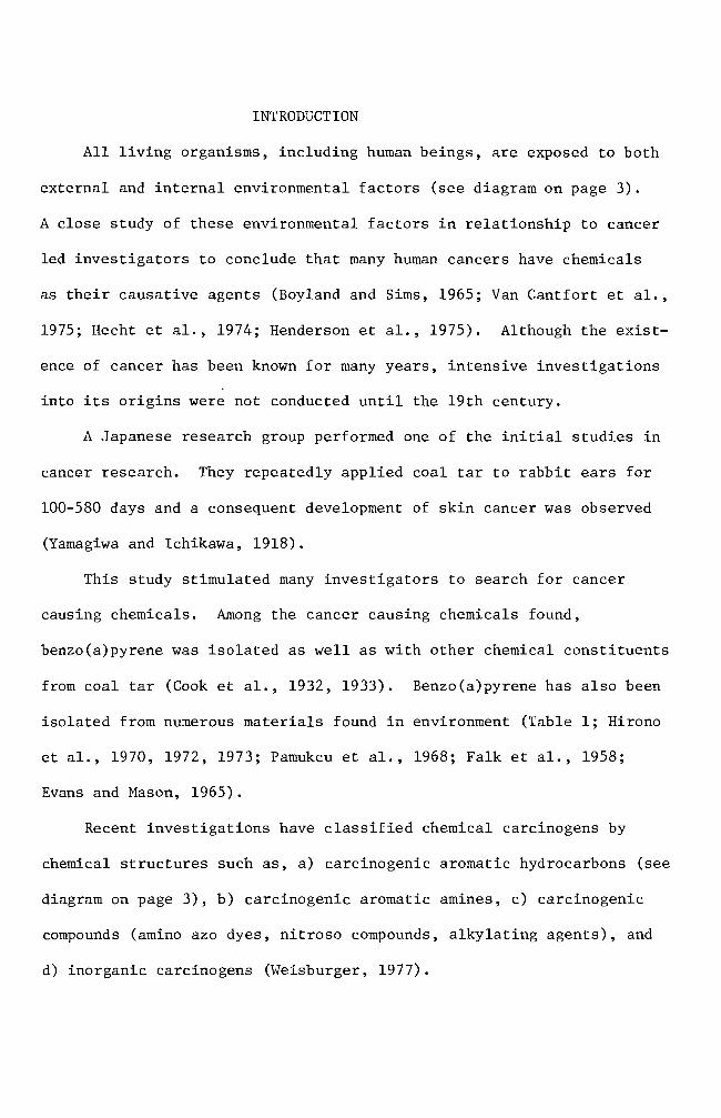
2
BIOLOGICAL
AGENTS
J
POPULATION-shy -
OCCUPATIONAL
Figure 1 Human exposure to hazardous environmental agents (Kraybill 1977)
3
Disputed (33 or less)
Chrysene
Benz (a) anthracene
Dibenz(ac)anthrashycene(1234shy
Dibenzonthracene)
Moderate (33 - 66)
(11UJ
Benzo(c)phenanthrene
Dibenz(ah)anthracene
Dibenzshy(aj)anthracene
Benzo(c)chrysene-
High activity (66 or higher)
~
Benzo(a)pyrene
Dibenzo(ah)pyrene
Dibenzo(al)pyrene
Dibenzo(ai)pyrene (1256-dibenzphenanthrene)
4
Table 1 Polycyclic aromatic hydrocarbons in food
Benzo(a) pyrene (ugKg)
Benz(a) anthracene
(ugKg)
Charcoal broiled meat 8 45 Lijinsky and Shubik 1964
Smoked fish 12-37 91-189 Masuda and Kuratune 1971
Singed sheep head Coal raw Coal cooked
21 9
15 5
Thorsteinsson and Thordarson 1968
Singed sea bird Coal raw Coal cooked
99 96
69 50
Smoked food Mutton Trout Cod Red Fish
13 21 05 03
-shy-shy
Bailey and Dungal 1958
Barbecued ribs 105 36 Lijinsky and Shubik 1965
Coffee 4 -- Kuratsune and Hueper 1960
5
Researchers attention up to this point was devoted to isolating
chemical carcinogens from our environment and to characterizing the
chemical carcinogens Some researchers Allison and Mallucci (1964)
Shires et ale (1966) Brooks and Lawley (1964) Wang et ale (1972)
started to investigate the mechanisms involved in chemical carcinoshy
genesis
Allison and Mallucci (1964) utilized fluorescence microscopy
phase contrast microscopy and autoradiography techniques to locate
carcinogenic hydrocarbons in mammalian cell cultures These investishy
gators concluded that the hydrocarbon carcinogens were not concentrated
in either mitochondria or the nuclei They were however found to be
within lysosomes Allison and Paton (1965) Allison and Dingle (1966)
and Allison (1968) centered their studies on lysosomes and their
relationship to cancer
A study similar to the one mentioned above was carried out two
years later by Shires and Richter (1966) These workers exposed mammashy
lian cells in vitro to carcinogenic hydrocarbons for periods ranging
from 12 hours to 4 days The treatment was terminated at different
intervals for microscopic observations The researchers were unable to
detect any incorporation of benzo(a)pyrene into the nuclei or chromoshy
somes during a complete generation cycle
On the other hand Brooks and Lawley (1964) tried to show the
binding of carcinogenic hydrocarbons to the nucleic acids in mouse skin
They did so by applying the hydrocarbon carcinogens to shaved regions on
the backs of mice The mice were sacrificed at various times and the
treated areas of skin were removed and used for deoxyribonucleic acid
(DNA) and ribonucleic acid (RNA) isolation This study indicated that
6
DNA reacts with the hydrocarbon carcinogens tested Experimental
results did not indicate how the hydrocarbon carcinogens are bound to
the nucleic acids
Lerman (1960) investigated the way in which polynuclear hydroshy
carbons react with DNA He used mutagenic acridine dye derivatives
for his study This investigation showed that the dyes are intershy
calating into the DNA helix through extension of DNA backbone This
information enhanced the idea that polycyclic aromatic hydrocarbons
require structural planality for the carcinogenic potency A similar
mechanism of the intercalation reaction between hydrocarbon carcinoshy
gens and DNA was proposed by Liquori (1962) Liquoris findings were
based upon the increased solubility of benzo(a)pyrene in DNA aqueous
solutions This is significant because benzo(a)pyrene is virtually
insoluble in water
Boyland and Green (1964) expressed a slightly different opinion
They showed that carcinogenic hydrocarbon molecules react with DNA by
slipping into the sites where the DNA is already stretched Such
stretching of DNA can only occur during DNA replication or RNA transhy
scription An expanded idea was postulated by Arcos and Argus (1968)
They indicated that the solubilization of carcinogenic hydrocarbons by
DNA can be accomplished by either intercalation or through external
binding mechanisms (Figure 2)
Van Duuren et al (1969) suggested a reconsideration of the
intercalation mechanism This was proposed because a) solubilization
of detergent micelles of sodium lauryl sulfate and aqueous DNA solution
caused the same results which were found with hydrocarbon carcinogens
7
e oI I I I I I IIgt I I
I
A B
Figure 2 Noncova1ent interactions of polycyclic aromatic hydroshycarbons (illustrated with 34-benzpyrene) with DNA A Intercalation into DNA double helix B External binding to DNA (From Arcos and Argus 1968)
8
and aqueous DNA solution and b) potent carcinogens such as 712shy
dimethy1benz(a)anthracene and 3-methy1cho1anthrene are thicker than
the distance between adjacent base pairs in DNA
Because previous investigations had been unable to prove whether
or not particular parent carcinogenic hydrocarbon molecules react with
DNA research interest was directed to the isolation and characterization
of intermediate metabolites Benzo(a)pyrene-45-oxide (K-region
epoxide) proved to be one of the highly reactive metabolites of
benzo(a)pyrene (Wang et a1 1972 Selkirk et a1 1971) Another
metabolite 78-dihydro-78-dihydroxybenzo(a)pyrene-910-oxide has been
investigated for its metabolism and mutagenicity It was confirmed to
be carcinogenic (Neubo1d and Brooks 1976 Sims et a1 1974)
Thus far accumulated data favor the theory of hydrocarbon carcinoshy
gens binding to DNA On the other hand the significance of 1ysosomes
in regard to carcinogenesis cannot be ignored Both Allison and Ma11ucci
(1964) and Shires and Richter (1966) utilized fluorescence and phase
microscopies and concluded that 1ysosomes were somehow involved In
addition to optical systems Allison and Ma11ucci (1964) used autoshy
radiography techniques for localization of hydrocarbon carcinogens in
mammalian cell cultures They concluded that the amount present if
any in cellular organelles other than 1ysosomes is too small to detect
by these methods
It was the purpose of this investigation to challenge the credishy
bility of autoradiographic techniques as an indicator of
benzo(a)pyrene-DNA incorporation and to consider the problems that
were encountered with this technique
MATERIALS AND METHODS
Experimental animals
Heterogenic white mice were obtained from the Emporia State
University research animal facility and from the Mid-Continent
Research Company (Kansas City Kansas) They were maintained in the
animal facility at Emporia State University
Chemicals
Benzo(a)pyrene (B(a)P) obtained from Sigma Chemical Company
was first dissolved in acetone (10 mgm1) and then dispersed into complete
growth medium with a Pyrex disposable syringe to give a concentration of
100 ugm1 of medium (Berwald and Sachs 1965) This B(a)P suspension was
used as stock solution from which dilutions were made Dilutions were
01 ug 10 ug and 50 ug B(a)P per milliliter of medium The stock solushy
tion was stored at OdegC The desired concentrations of B(a)P solution
were applied to cells in culture
Tissue culture medium
Du1beccos modified Eagles medium containing 45 gl glucose was
purchased from GIBCO The growth medium was supplemented with 10 fetal
bovine serum (GIBCO) and 05 m1 combiotics This complete growth medium
was used to grow and maintain the cells in vitro GKN (calcium and
magnesium free saline) was used for cell harvest and cell transfer with
025 trypsin and trypsin-versene (0002-005) solutions respectively
Stock combiotic solutions were prepared by combing penicillin G
sodium (1650 Umg) and streptomycin sulfate (740 mggm) in 1X Hanks
BSS Stock solutions were sterilized using Mi11ipore filters One
milliliter a1iquots were prepared and stored frozen until needed Cell
10
harvesting solutions were prepared by adding 05 ml of the combiotic to
every 100 ml of GKN Likewise cell maintenance solutions were preshy
pared by adding 05 ml of combiotic stock solution to every 100 ml of
growth medium
Fetal bovine serum which was mycoplasma tested and virus screened
was obtained from GIBCO and used throughout this study
Carbon dioxide gas prepared by mixing calcium carbonate and
sulfuric acid was used to maintain the desired pH of the medium
Glassware
Glassware was cleaned by soaking in an acid bath (17 mixture of
concentrated hydrochloric acid and concentrated sulfuric acid) overshy
night followed by washing with Alconox solution and repetitive rinsing
with tap water All glassware was rinsed with deionized distilled
water after being washed
Microscope coverslips (22 X 50 mm) were used to make cell growing
covers lips (22 X 8 mm) These were immersed in an Alconox solution and
rinsed with running tap water for 10-13 hours They were rinsed individshy
ually 10 times in three consecutive deionized distilled water baths and
one 100 ethanol bath then stored in an absolute ethanol bath until
use
Cell culture
Minced whole embryos of heterogenic white mice served as the source
of normal cells Pregnant mice (15-18 days in term) were sacrificed by
cervical dislocation Embryos were then removed aseptically from the
mouse and transferred to a petri dish containing GKN-combiotics mixture
Embryos were minced and placed into a trypsinizing flask The tissue
11
chunks were subjected to gentle stirring while being trypsinized at
room temperature for 30 minutes Trypsinized tissue was filtered
through four layers of cheese cloth The filtrate was washed with one
change of GKN and one change of complete growth medium Cell popushy
lations were adjusted with complete growth medium to a concentration
6of 3 X 10 cellsmI Three milliliter aliquots were planted in 25
2 cm polystyrene culture flasks and incubated at 37degC for one to three
days (primary cell culture) Primary cell cultures were treated with
trypsin-versene solution for two minutes washed centrifuged and
resuspended to give a concentration of 6 X 10⁵ cellsmI They were
inoculated in test tubes containing covers lips (7 X 22 mm) for 24 hours
at 37degC (secondary culture) prior to usage in the studies Secondary
cell cultures were treated with either a colloidal suspension of B(a)P
(10 ugml) and 3H- B(a)P (10 uCiml) or 3H- B(a)P (10 uCiml) alone
Autoradiography
Tritium-labeled B(a)P treated cells growing on the coverslips were
removed from the test tubes rinsed with two changes of GKN solution and
fixed in an absolute methanol bath for five minutes The covers lips
were mounted on a slide with the cell side up using Pro-texx mounting
medium The slides were dried overnight on a heating plate at 37degC
Ilford emulsion (1-4 Polysciences Inc) was melted in a 40degC
water bath Ten milliliters of melted emulsion were diluted with an
equal volume of deionized distilled water and any bubbles resulting in
the mixing operation were removed through a layer of cheese cloth The
slides were dipped into the diluted emulsion and immediately removed
Residual emulsion was cleaned from the back of the slides and the
12
slides held in a vertical position for 15 minutes to allow the emulsion
to dry Slides were sealed in a light tight slide box by wrapping the
box with aluminum foil The slides were exposed for 10 days at OdegC
The exposed slides were developed in Dekto1 (D-17) for two
minutes rinsed in a deionized distilled water stop bath and fixed in
acid fixer for three minutes After washing for 15 minutes in slowly
running tap water the slides were dried and stained for four minutes
in 005 basic fuchsin solution The slides were dehydrated in a
series of ethanol baths and immersed in a mixture of cedar wood oil
and absolute ethanol (1 1) for one hour followed by infiltration of
a Pro-texx mounting medium and xylene mixture (1 1) for one hour
The slides were examined for the appearance of silver grains
(black dots) located within the cells thereby indicating B(a)P uptake
by the cell
Cell viability test
The toxic effect of B(a)P on cultured cells was examined by adding
5 m1 of B(a)P at different concentrations Concentrations used ranged
from 01 to 50 ug B(a)P per milliliter of growth medium At regular
intervals B(a)P-growth medium mixtures were collected from culture
flasks and centrifuged for five minutes at 800 rpm The supernatant
was siphoned off and discarded Pellets were resuspended with 1 m1
growth medium and 01 m1 of erythrosin (04) was added and stained for
two minutes Dead cells staining pink were counted on a hemocytometer
Numbers of dead cells were plotted against the durations of B(a)P treatshy
ment
13
Inoculation of B(a)P treated cells into animals
Cells treated with B(a)P were harvested with a trypsin-versene
solution for three minutes Cell harvest was made at preselected inshy
tervals of time The cell population was adjusted to 10 X 106 ce11sm1
with complete growth medium From this solution 02 m1 were injected
into a group of five male mice Injections were made subcutaneously on
the left thigh (modified from Evans et a1 1957) Inoculation sites
were checked every 14 days for tumor development until termination of
the experiment
Histological study
Twenty-eight days after inoculation of B(a)P treated cells mice
which developed tumors were sacrificed for histological studies The
tumor tissue was excised and fixed overnight in Bouins fixative
Several solution changes of 70 ethanol saturated with lithium carbonate
were made to eliminate any fixative in the tissue Dehydration of fixed
tissue was accomplished in a series of ethanol baths of one hour each
The tissue was then immersed for one hour in a mixture of absolute
ethanol and xylene (1 1) It was soaked in a xylene bath overnight
and then transferred to a mixture of xylene and paraffin (56 0 tissuemat)
for 15 minutes Infiltration of hot tissuemat was carried out in four
changes of tissuemat solution Tissue was then embedded in a plastic
ice cube tray with hot tissuemat and cooled to room temperature The
blocks were sectioned on the microtome at a thickness of seven microns
The sections were stained by standard hematoxylin and eosin procedures
The slides were then carefully examined for histological abnormalities
14
Chromosome preparation
Cells in culture were mitotically blocked by lyophilized colcemid
006 ugml (GIBCO) for two hours Harvesting of cells was accomplished
by trypsinization and centrifuging for five minutes at 800 rpm The
pellet was resuspended in 1 ml of complete growth medium with a Pasteur
pipette Hypotonic solution (3 ml deionized distilled water) was added
to this suspension for seven minutes at room temperature One milliliter
of freshly prepared methanol-acetic acid (3 1) fixative was introduced
to the hypotonic solution for 10 minutes The suspension was centri shy
fuged and fixed by repeated dispersion at 10 minute intervals in three
changes of 31 methanol-acetic acid fixative During these fixation
periods microscope slides were washed in an Alconox solution and stored
in deionized distilled water at 4degC After the third fixation the
pellet was resuspended in 03-1 ml (depending on size of the pellet)
fixative solution Several drops of the final cell suspension were
applied to the slides The backs of the slides were wiped dry with
tissue paper A film of water on the upper surface of the slides was
left to facilitate spreading of the cells The slides were air-dried
and stained for 15 minutes in modified Giemsa stain Modification
was made according to Gude et al (1955) For the autoradiographic
study the slides were stained for 15 minutes after the completion of
photographic processes
RESULTS
Cell viability test
The toxic effect of B(a)P was tested for nine days Results are
summarized in Figure 3 From the data linear regression curves were
prepared for each concentration The slope of the untreated cell line
is 48 The slopes of the B(a)P treated cell lines are 65 for 01 ug
B(a)P treated cells 70 for 10 ug B(a)P treated cells and 76 for 50
ug B(a)P treated cells A statistical analysis did not indicate a
significant difference between the normal cell line and the B(a)P
treated cell lines Re-examination of the data used in Figure 3 will
help in understanding effect of treatment on cell viability for specific
periods of time In Figure 4 one and three day incubation periods
showed a smaller rate of increase (slope for one day 007 and slope
for three days 016) which implies that the B(a)P concentration did
not have a significant effect in terms of cell killing ability The dead
cell numbers in five day B(a)P treated cultures presented about 28-fold
increase from that of three day B(a)P treated cultures A statistical
analysis showed a significant difference indicating that the critical
factor in cell killing ability of B(a)P is dependent upon the exposure
period
Cell morphology in culture
Mouse embryo cells in culture were observed under the light microshy
scope for their morphology Embryonic cells were round immediately after
implantation into culture flasks Approximately three hours incubation
at 37degC after initial cell plantation some cells started to establish
their natural morphological appearance (elongation of cytoplasm) The
16
elongation process was an indication of cell attachment on the surface
of culture flasks Owens et a1 (1974) reported that in earlier days
of incubation two cell types epithelial cells and fibroblast cells
can be identified Epithelial cells from mono1ayered sheets which are
attached to each other (Figures 5 and 9) Vasi1iev and Ge1hand (1977)
reported that fibroblast cells form several morphological varieties in
culture mono1ayers of mutually oriented cells which are not firmly
attached to each other (Figures 6 and 8) multilayered sheets (Figure
7) or spherical aggregates (Figure 7 lower right and upper left
darkly stained aggregates) Fibroblasts andor fibroblast-like cells
became predominant throughout the entire cell population when cells were
cultured for over 30 days (Figures 10 11 and 12) These fibroblast
cells are characterized by elongated or stellate cytoplasmic structures
(Figures 10 11 and 12)
Mouse embryo cells were treated with 10 ug B(a)P for predesignated
exposure periods to observe an effect on cell morphology Some B(a)P
treated cells showed an abnormal spindle shape while the other cells
retained their normal morphological appearance (Figures 13 15 and 16)
In Figures 15 and 16 some cells were found to have vacuoles within their
cytoplasm Many of these also showed attenuated cytoplasms (Figure 16)
Autoradiography
Cultured cells were incubated with B(a)P for several different types
of treatment One treatment consisted of incubating cultured cells with
B(a)P at 10 ugm1 and 3H- B(a)P at 10 uCim1 until the termination of
experiment (full-term treatment) Other cells were partially exposed
to B(a)P at the same concentration and radioactivity level The exposure
17
60
-l S-r-
0 0 0 -l
gtlt
40-
Ul -l -l ltl) tJ
d Cll ltl) d
~
0 ltl)
20 l z
o o 5 10
Days
Figure 3 Toxic effect of B(a)P on cultured mouse embryo cells Untreated cells 0 Cells treated with 01 ug B(a)P e Cells treated with 10 ug B(a)P X Cells treated with 50 ug B(a) P
__
--------
18
60
-~ s
I
I ~ ~7 r 0 0 0 ~
x -
Ul ~ ~
eu 40u 0 ell eu 0
4-l 0
Ul H eu ~ l Ie z
20 3
9
_ ---41---------------- 1 o
o 20 40 60
B(a)P concentration (ugml)
Figure 4 Toxicity effect of B(a)P concentration The numbers 1 3 5 7 and 9 indicate the days of B(a)P treatment
Figure 5 Two day old normal mouse embryo cells in culture Epithelial cells in the central area of fibroshyblast cells surrounding the epithelial cells are shown here May-Grunwald and Giemsa stain (100 X)
Figure 6 Four day old normal mouse embryo cells in culture Large nucleated epithelial-like cell and fibroshyblast cells are shown here May-Grunwald and Giemsa stain (400 X)
Figure 7 Six day old normal mouse embryo cells in culture Multilayered fibroblast cells and two heavily stained aggregates are shown here May-Grunwald and Giemsa stain (100 X)
Figure 8 Eight day old normal mouse embryo cells in culture Fibroblast cells formed a monolayer sheet MayshyGrunwald and Giemsa stain (100 X)
Figure 9 Thirteen day old normal mouse embryo cells in culture Epithelial cells in the central and upper left areas are surrounded by well elongated fibroblast cells May-Grunwald and Giemsa stain (100 X)
Figure 10 Thirty day old normal mouse embryo cells in culture May-Grunwald and Giemsa stain (100 X)
Figure 11 Thirty five day old normal mouse embryo cells in culture Cell orientation is obscure and vacuoles are seen May-Grunwald and Giemsa stain (100 X)
Figure 12 Forty day old normal mouse embryo cells in culture Cell orientation is obscure and vacuoles are present May-Grunwald and Giemsa stain (100 X)
9Z
Figure 13 Mouse embryo cells incubated with 10 ug B(a)P for 1 day May-Grunwald stain (100 X)
Figure 14 Mouse embryo cells incubated with 10 ug B(a)P for 2 days May-Grunwald stain (100 X)
Figure 15 Mouse embryo cells treated with 10 ug B(a)P for 6 days May-Grunwald stain (100 X)
Figure 16 Mouse embryo cells treated with 10 ug B(a)P for 9 days Basic Fuchsin stain (100 X)
31
periods were 24 hours and four days
Figures 18 through 23 represent the full-term treatment and events
for the first 17 hours are summarized in Figure 17 After
hours exposure to 3H- B(a)P B(a)P accumulation in the nuclei
low Its cytoplasmic concentration was also minimal (Figure
3H-B(a)P uptake by the cells increased rather sharply after 35
of incubation (Figures 19 20 and 21) Long term incubation (six
and 20 days) showed a concentration of B(a)P in the cytoplasm while no
concentration of B(a)P was observed (Figures 22 and 23)
In other treatments cultured cells were partially exposed to
B(a)P at the same concentration Radioactivity levels were as in the
treatment The cells were exposed to B(a)P for one and four
days At the end of the exposure period the complete growth medium
was replaced for further incubation Figures 24 through 27 indicate
some of the results of 24 hour treated cells Overall results are
summarized in Figure 24 Nuclear accumulation of 3H-B(a)P was maximum
at 24 hours and decreased drastically when fresh complete growth medium
was introduced and incubation was continued (Figures 24 27 and 28)
The nuclear accumulation of B(a)P exceeded that of cytoplasm only at
24 hours incubation (Figure 24) Cytoplasmic accumulation of 3H-B(a)P
was at its highest peak on the 6th day of incubation and followed the
same pattern of decrease as the nuclear accumulation curve Extrashy
polation of the two curves suggests disappearance of B(a)P from the
cells occurs between the 24th and 26th day Some cells as exemplified
in Figure 28 lost nuclear B(a)P and most of the cytoplasmic B(a)P at
50
100
32
00
0 1 bull
O Hours
Figure 17 Time-course of 3H- B(a)P accumulation in the cells Cells in autoradiograms were counted for nuclear accumulation () cytoplasmic accumulation (0) and no accumulation (X)
I I Ii I Ii(I I ~ I I
2 4 6 8 10 12 14 16 18
33
21 days of incubation in the growth medium
Figures 29 through 32 represent the results of four day treated
Four (no additional incubation in growth medium) and 11 days
(additional six day incubation in growth medium) cells show nuclear and
cytoplasmic accumulation of B(a)P but not as intense as seen in the one
cells (Figures 29 and 30) Fifteen day incubated cells had
some B(a)P molecules in the cytoplasm The cells incubated for 27 days
of B(a)P in any part of the cells (Figure 32)
Figures 33 through 35 are the chromosome spreads from 3H- B(a)p
treated cells Silver grains (black dots) were seen on or near the chromoshy
somes in three autoradiograms (Figures 33 34 and 35) Autoradiograms of
chromosomes prepared from one two and three day treated cells with
3H- B(a)P (10 uCiml) were counted for use in labeling analysis Chroshy
mosome spreads from one and two day treated cells produced 936 and 920
labeled chromosome spreads respectively Total chromosome spreads examshy
ined were 94 and 100 Cells treated for three days gave 304 labeled
chromosome spreads
Figures 34 and 35 showed chromosome breakages
Induction of abnormalities and its histology
B(a)P treated cells were injected into adult mice The mouse in
Figure 36 was a normal male mouse The mouse in Figure 37 exemplifies
the abnormal genital inflammation characterized by many of the mice inshy
jected with the B(a)P treated cells From the 26 injected mice 31 deshy
veloped a genital inflammation In contrast only 38 of the control
mice exhibited genital abnormalities The controls consisted of three
groups of mice One group of 32 mice received no injections Another
Autoradiogram Mouse embryo cells in culture were incubated for 15 hours with 10 ug B(a)P(lO uCi) Only a few radioactive molecules are present Hematoxylin and eosin stain (100 X)
Figure 19 Autoradiogram Mouse embryo cells in culture were incubated for 35 hours with 10 ug B(a)P(lO uCi) Nuclear and cytoplasmic accumulation of B(a)P is seen Hematoxylin and eosin stain (100 X)
Figure 20 Autoradiogram Mouse embryo cells in culture were incubated for 17 hours with 10 ug B(a)P(10 uCi) Basic Fuchsin stain (1000 X)
Figure 21 Autoradiogram Mouse embryo cells in culture were incubated for 24 hours with 10 ug B(a)P(10 uCi) Basic Fuchsin stain (1000 X)
Figure 22 Autoradiogram Mouse embryo cells in culture were incubated for six days with 10 ug B(a)P(10 uCi) Hematoxylin and eosin stain (100 X)
Figure 23 Autoradiogram Mouse embryo cells in culture were incubated for 20 days with 10 ug B(a)P(10 uCi) A heavy accumulation of B(a)P in cytoplasm is seen here Hematoxylin and eosin stain (400 X)
-
6pound
40
00
100
~ -
00 c
-l M Q)
~ 50M
~
ro - lQI
z M
10 20 Time in days
Figure 24 3ime-course of 3H- B(a)P accumulation in the cells H-B(a)P was replaced with normal growth medium
after 24 hours incubation Cells in autoradiograms were counted for nuclear accumulation (e) cytoshyplasmic accumulation (0) and no accumulation (X)
00
Figure 25 Autoradiogram Mouse embryo cells in culture were incubated for one day with 10 ug B(a)P(lO uCi) Nuclear and cytoplasmic accumulation of B(a)P is seen Basic Fuchsin stain (1000 X)
Figure 26 Autoradiogram Mouse embryo cells in culture were incubated for one day with 10 ug B(a)P(lO uCi) The B(a)P was then removed The cells were incubated an additional 3 days in complete growth medium Basic Fuchsin stain (1000 X)
Figure 27 Autoradiogram Mouse embryo cells in culture were incubated for one day with 10 ug B(a)P(lO uCi) The B(a)P was then removed The cells were incubated an additional 11 days in complete growth medium Basic Fuchsin stain (1000 X)
Figure 28 Autoradiogram Mouse embryo cells in culture were incubated for one day with 10 ug B(a)P(lO uCi) The B(a)P mixture was then removed The cells were incubated an additional 21 days in complete growth medium Basic Fuchsin stain (1000 X)
shy
Figure 29 Autoradiogram Mouse embryo cells in culture were incubated for four days with 10 ug B(a)P(lO uCi) Negri body stain (1000 X)
Figure 30 Autoradiogram Mouse embryo cells in culture were incubated for four days with 10 ug B(a)P(lO uCi) The B(a)P mixture was then removed The cells were incubated an additional six days in complete growth medium Some silver grains are seen Basic Fuchsin stain (1000 X)
97
Figure 31 Autoradiogram Mouse embryo cells in culture were incubated for four days with 10 ug B(a)P(lO uCi) The B(a)P was then removed The cells were incubated an additional 11 days in complete growth medium Some silver grains were seen in the cytoplasm Basic Fuchsin stain (1000 X)
Figure 32 Autoradiogram Mouse embryo cells in culture were incubated for four days with 10 ug B(a)P(lO uei) The B(a)P mixture was then removed The cells were incubated an additional 23 days in complete growth medium Basic Fuchsin stain (1000 X)
81]
Figure 33 Autoradiogram of chromosomes Mouse embryo cells in culture were incubated for one day with 005 ug B(a)P(lO uCi) Giemsa stain (1000 X)
Figure 34 Autoradiogram of chromosomes Mouse embryo cells in culture were incubated for two days with 005 ug B(a)P(lO uCi) Giemsa stain (1000 X)
-
os
Figure 35 Autoradiogram of chromosomes Mouse embryo cells in culture were incubated for three days with 005 ug B(a)P(10 uCi) Giemsa stain (1000 X)
Figure 36 Normal male mouse
53
group of 10 mice were injected with normal untreated cells The last
group of 10 mice was inoculated with growth medium that was used to
maintain the cells (Table 2) The effect of B(a)P treated cells in
the mice is summarized on Table 2 The mice inoculated with the cells
of four days B(a)P preincubation resulted in a 25-50 incidence of
abnormal development Of these abnormal mice two mice developed a
small nodule (ca 4-5 rom in diameter) on the peritoneum (Figure 38)
This nodule was processed for histological study Microscopic examishy
nation revealed that the nodule was the undescended testis and showed
hyperplasia of Leydig cells (Figure 43) Atrophy of seminiferous
tubules (Figure 39) is clearly recognized when they are compared with
those of normal tissue (Figures 42 and 43) Seminiferous tubules
from abnormal tissue did not show spermatogenesis (Figure 43) The
ductus epididymis (Figure 41) however showed normal morphological
appearance but clumps of spermatozoa were not seen in the ductuli
Hyperplasia of connective tissue surrounding ductus epididymis was
observed as well The mice inoculated with 10 day B(a)P treated cells
had a higher incidence (80) of abnormal development Long term
observation (146 days) did not reveal the development of any abnorshy
mality although the inoculated cells were treated for one day with
B(a)P
Tab
le
2
Eff
ect
of
B(a
)P tr
eate
d cell
s in
ad
ult
mic
e
Day
s o
f D
ays
of
Num
ber
of
cell
s D
ays
in
Day
s aft
er
incu
bati
on
w
ith
B(a
)P
incu
bat
ion
aft
er
B(a
)P
ino
cu
late
d
(x
106
) cu
ltu
re
ino
cu
lati
on
A
bn
orm
alit
ies
rem
oved
4 0
10
47
62
24
4 4a
10
43
68
14
4 6
10
41
70
14
10
0 10
43
70
4
5
1 0
1 6
146
05
1 0
10
6 14
6 0
4
Co
ntr
ols
U
ntr
eate
d M
ouse
-shy
-shy
--shy
13
2
Nor
mal
cell
s 10
1
1
146
11
0
Gro
wth
med
ium
o
nly
(f
rom
a)
4 70
0
10
In
+shy
Figure 37 Mouse with genital abnormality
Figure 38 Mouse with a nodule on peritoneum
9~
Figure 40 Transverse section of ductus epididymis from normal mouse testis Hematoxylin and eosin stain (100 X)
Figure 41 Transverse section of ductus epididymis from undescended testis Clumps of spermatozoa are not seen in the ductu1i Spaces among the ductu1i are abnormally large Hematoxylin and eosin stain (100 X)
6S
Figure 42 Transverse section of seminiferous tubules from normal mouse testis Seminiferous tubules are separated by a slight amount of interstitial connective tissue containing some Leydig cells Hematoxylin and eosin stain (100 X)
Figure 43 Transverse sections of seminiferous tubules from undescended testis Atrophy of seminiferous tubules is seen Hyperplasia of Leydig cells is recognized Hematoxylin and eosin stain (100 X)
19
DISCUSSION
Cook et a1 (1932 1933) isolated carcinogenic hydrocarbons from
coal tar pitch The melting point (mp) of one of the hydrocarbons
was determined to be 1755-1765degC This particular hydrocarbon species
was shown to be pure benzo(a)pyrene (B(a)P) by comparison with the
synthesized B(a)P B(a)P has alternative names such as 12-benzpyrene
and 34-benzpyrene It has the molecular weight of 25230 its
structure is shown in diagram on page 3
B(a)P and other closely related carcinogenic hydrocarbons (see
diagram on page 3) are being used to determine a mechanism of carshy
cinogenesis in several research systems B(a)P is known to be one of
the chemically inert carcinogens To be activated B(a)P must first be
taken up by the cells Thus invasion into the cell is the first step
in the mechanism B(a)P particles in the growth medium are taken into
the cells by endocytosis (Allison and Ma11ucci 1964) Engulfed B(a)P
particles then require enzymatic activations to acquire cytotoxic
mutagenic (Huberman et a1 1971 1976 Newbold and Brooks 1976) and
carcinogenic abilities in the cells (Wood et a1 1976 Wis10cki et a1
1976)
The greatly increased number of dead cells in cultures which had
been treated with B(a)P for five days suggested that enzymatic activations
of ingested B(a)P might have occurred during a one to three day incubation
period As a result B(a)P acquired cytotoxic potency to the cells
(Figure 4) Figures 17 and 24 indicate that the cytoplasmic accumushy
lation had maximum values between 35 hours and six days This sugshy
gested the possibility that B(a)P metabolism takes place between these
periods A reliable explanation for the cytotoxic ability of B(a)P was
63
made by several workers (Nebert and Gelboin 1968a 1968b Kasper
1971 Khandwala and Kasper 1973) There exists an enzyme aryl
hydrocarbon hydroxylase that functions in the metabolism of B(a)P
The metabolite is 3-hydroxybenzopyrene which also possesses cytotoxic
potency to the cell Benzo(a)pyrene-hydroxylase was shown to be preshy
sent at low concentrations in the nuclear membrane The existence of
inducible B(a)P-hydroxylase in a microsomal preparation was reported
as well The major difference between nuclear and microsomal memshy
branes is its specificity Nuclear membranes produce hydrocarbon
hydroxylase specifically by the stimulation of hydrocarbon carcinogens
while microsomal membranes produce hydrocarbon hydroxylase in response
to the addition of hydrocarbon carcinogens or phenobarbital
In addition to enzymatic activation in the cytoplasm B(a)P
molecules may be accumulated in one or some of the cellular organelles
To determine the ultimate B(a)P accumulation site in the cell it is
important to define the target organelle(s) for hydrocarbon carcinoshy
gens Several techniques were developed for this purpose and an autoshy
radiography technique was the one utilized in this study Autoradioshy
graphy techniques in the biomedical field are a powerful tool for
localization and visualization of desired chemicals in the cell or the
tissues Autoradiograms obtained in this study indicated that the
cytoplasmic accumulation of B(a)P occurred as early as 15 hours after
the initial application and lasted about 14 days (Figure 17 and 24)
The silver grains seen in Figures 19 20 and 27 indicate that B(a)P
molecules may be encapsulated as a phagosome perhaps independently
from lysosomes or fused to lysosomes where enzymatic alterations as
64
well as detoxification reactions take place Along with the cytoplasmic
accumulation a maximum nuclear accumulation (52 labeling) was observed
in one day (Figure 24) Although nuclear accumulation of B(a)P was
shown it does not clearly indicate the incorporation of B(a)P into
genetic material (DNA) To answer this question chromosome spreads
combined with autoradiography were prepared (Figures 33 34 and 35)
These figures indicate that an interaction of B(a)P and chromosomes
might have occurred in the nuclei This result is supported by the work
of Brooks and Lawley (1964) who isolated DNA and RNA from mouse skin to
which radioactive carcinogenic hydrocarbons were previously applied
One of their results is shown in Figure 44 which demonstrates that the
maximum binding of B(a)P to proteins occurred about 24 hours after
application of B(a)P This result suggests the possibility that B(a)P
molecules bind to DNA
Figures 34 and 35 demonstrated chromosome breakages A generally
accepted explanation for chromosome aberrations is that a direct
chemical or physical attack on DNA has occurred However Allison and
Patton (1965) proposed another possible mechanism which could cause
chromosome breakages The idea was that damage to 1ysosomes can cause
chromosomal damage It was demonstrated by De Duve and his colleagues
that 1ysosomes contain hydrolytic enzymes acid phosphatase acid DNase
acid RNase and acid protease (reviewed by Allison and Ma11ucci 1964)
Among these lysosomal enzymes DNase can break the DNA which is the
backbone of the uncoiled interphase chromatid Deoxyribonuclease must
therefore enter into the nucleus for chromosome breakages to occur
Widne11 and Tata (1964) reported a result which supports DNase migration
0
I
I
I
02
r-
U l
I
r- (]J CIl o
d shyI r- (]J
jJ o ~
01
~ o ~
jJ
r- gt r-
jJ U co U
r- ~
r- U
(]J
0shy
Cfl
o 5
0
150
25
0
Fig
ure
4
4
T
ime-
cou
rse
of
the
bin
din
g
of
(3H
) la
bell
ed
hy
dro
carb
on
s to
to
tal
pro
tein
s o
f m
ouse
sk
in
Resu
lts
are
ex
pre
ssed
as
specif
ic ra
dio
shyacti
vit
y
of
pro
tein
(u
Ci
g)
div
ided
by
th
e d
ose
(u
Ci
mo
use
) g
iven
at
zero
ti
me
plo
tted
ag
ain
st
tim
e
34
-ben
zp
yre
ne
(Part
iall
y
rep
rod
uce
d
from
B
roo
kes
an
d L
awle
y
19
64
)
V1
66
thus DNA-dependent RNA polymerase activity in isolated nuclei is
blocked by addition of DNase Newton et al (1962) also reported that
DNA breakdown occurred shortly after infection with herpes virus This
report implies that lysosomal damage by herpes virus induced a libershy
ation of DNase which in turn caused DNA breakage
The studies conducted so far do not give any indication concerning
mechanisms by which B(a)P binds to the nucleic acids Many investi shy
gators worked to clarify the steps involved as B(a)P migrates from the
cytoplasm to the nucleus B(a)P is enzymatically converted to at least
four phenols three dihydrodiols and three quinones and epoxides
(reviewed by Yung et al 1976) The active B(a)P metabolites which are
converted by microsomal hydroxylase within the cells possess a relatively
high tendency to react with the nucleophilic sites of the macromolecules
Thus the B(a)P metabolite has a rare chance of reacting with nuclear
DNA (Alexandrov and Thompson 1977) This report implies that further
metabolic activation of B(a)P metabolites by the other enzymes of microshy
somes or the nuclear envelope becomes necessary (Rogan et al 1976)
A similar interpretation of a two-step activation mechanism was reported
by several workers (Sims et al 1974 Weinstein et al 1976 Spelsberg
et al 1977) Diol-epoxide intermediates (7S-dihydro-7S-dihydroshy
xybenz(a)pyrene and 7S-dihydro-7S-dihydroxybenzo(a)pyrene-9lO-oxide)
were reported to be the metabolites responsible for DNA-hydrocarbon
binding (Brooks et al 1975 Sims et al 1974) Brooks et al (1975)
also indicated that the purine moieties of DNA react with diol-epoxide
intermediates A specific isomer of a diol-epoxide derivative of B(a)P
(plusmn)-7B Sa-dihydroxy-9a lOa-7S9lO-tetra-hydrobenzo(a)pyrene also
67
appeared to be a B(a)P metabolite in terms of covalent binding to
nucleic acids (Weinstein et a1 1976) Another isomeric form of a
dio1 epoxide derivative of B(a)P 7a 8B-dihydroxy-9B10B-epoxy-789
10-tetrahydrobenzo(a)pyrene was proved to be a highly mutagenic intershy
mediate of B(a)P metabolism (Wood et a1 1976 Huberman et a1 1976)
This metabolite is suspected to be the ultimate carcinogenic species
in B(a)P carcinogenesis The reactions mentioned above are the events
occurring as B(a)P molecules move into the nucleus of the cell It is
important to s~e the relationship between the duration of B(a)P treatshy
ment and cell transformation To determine the tumorigenicity of B(a)P
in the cells in vitro suspected cells must be injected into a host
animal to observe the behavior of the cells (Fedoroff 1967) In this
experiment several conditions for B(a)P treatments were designed and
inoculation of mice with B(a)P treated cells was carried out The
results are summarized in Table 3 Two mice developed palpable tumors
(Figure 38) while the remainder showed genital abnormality (Figure 37)
The mice inoculated with four day B(a)P treated cells developed 25-50
abnormality development Some four day B(a)P treated cells were incushy
bated additional days in a normal growth medium before inoculation
Abnormal development was not affected significantly by the additional
incubation period This is because during a four day incubation period
most of the cells may be transformed This idea can be supported by
the work of Berwald and Sachs (1965) They reported that two days of
incubation with B(a)P is necessary for cell transformation Ten day
B(a)P incubation created an 80 rate of abnormal development Higher
genital development in 10 day B(a)P treated cells was possibly due to
68
the increased probability of cell transformation Berwald and Sachs
(1965) reported that the normal mouse cells in vitro showed spontaneous
transformation sometime after 45 to 80 days in the culture As can be
seen on Table 3 the cells used for the inoculations were cultured from
41 to 47 days which is in the range of Berwalds figures Thus this
spontaneous transformation factor may be involved in the results shown
on Table 3 The mice inoculated with one day B(a)P treated cells
developed no abnormalities (Table 3) One reason for the absence of
abnormal development may be related to the age of the cells in culture
the cells used were cultured for six days while the abnorma1ityshy
producing cells were cultured from 41 to 47 days
A tumor development in host mice by the inoculation of suspected
cells must be further examined by histological means Figures 40 through
43 are transverse sections of the ductus epididymis and the seminiferous
tubules from abnormal and normal mouse testis Figures 40 and 42 from
the control mouse show a normal histological appearance The ductu1i
are well packed and contain aggregates of spermatozoa In contrast
Figures 41 and 43 show atrophy of ducts of the epididymis resulting in
the formation of large interstitial spaces Closer observations revealed
a deterioration of the steroci1ia as well Figures 40 and 42 show the
normal condition in which seminiferous tubules contain newly formed
spermatozoa primary spermatocytes and secondary spermatocytes Figures
41 and 43 show atrophy of seminiferous tubules with a few supporting
cells (Serto1i) located near the basement membrane An abnormal increase
in the number of interstitial Leydig cells was also seen Occasionally
binuc1eated Leydig cells were observed Detailed observation of the
excised nodule revealed it to be histologically abnormal It is however
69
difficult to determine if the abnormal nodule was caused by the direct
action of B(a)P treated cells
Allison and Mallucci (1964) reported that autoradiographic
techniques failed to visualize accumulation of chemical carcinogens in
the nucleus Through this study it was demonstrated that autoradioshy
graphic techniques could show the existence of B(a)P in the nucleus
Furthermore autoradiograms of chromosomes gave an indication that
interaction between B(a)P and chromosomes may have occurred
SUMMARY
1) Benzo(a)pyrene accumulation in the cell occurred after 24 hours
of incubation period
2) Autoradiograms of chromosomes prepared from benzo(a)pyrene
treated cells indicated that benzo(a)pyrene reacted with
chromosomes
3) Autoradiographic techniques were shown to have the capacity to
localize the sites of benzo(a)pyrene accumulation in the cell
4) Cytotoxicity studies of benzo(a)pyrene showed a time-dependent
effect on the cultured mouse embryo cells
5) A tumor induction study indicated that tumor inducibility in the
host animal seems to be affected by the length of cell culture
period
LITERATURE CITED
Allison A C and L Ma11ucci 1964 Uptake of hydrocarbon carcinogens by 1ysosomes Nature 2031024-1027
Allison A C and G R Paton 1965 Chromosome damage in human diploid cells following activation of lysosomal enzymes Nature 2091170-1173
Allison A C and J T Dingle 1966 Role of 1ysosomes in adrenal necrosis caused by dimethy1benzanthracene Nature 209303-304
Allison A C 1968 Lysosomes in relation to cancer induction and treatment Europ J Cancer 3481-490
Arcos C J and F M Argus 1968 Molecular geometry and carcinoshygenic activity pp 348-435 Advances in Cancer Research Vol 11 Academic Press Inc
A1exandrov K and H M Thompson 1977 Influence of inducers and inhibitors of mixed-function oxidase on benzo(a)pyrene binding to the DNA of rat nuclei Cancer Res 371443-1449
Bailey J E and N Dunga1 1958 Polycyclic hydrocarbons in icelandic smoked food Brit J Cancer 12348-350
Berwald Y and L Sachs 1965 In vitro transformation of normal cells to tumor cells by carcinogenic hydrocarbons J Nat1 Cancer Inst 35(4)641-661
Boyland E and B Green 1964 On the reported sedimentation of polycyclic hydrocarbons from aqueous solutions of DNA J Mol Bio1 9589-597
Boyland E and P Sims 1965 Induction of adrenal damage and cancer with metabolites of 712-dimethy1benz(a)anthracene Nature 207816-817
Brookes P and P D Lawley 1964 Evidence for the binding of polynuclear aromatic hydrocarbons to the nucleic acids of mouse skin Relation between carcinogenic power of hydrocarbons and their binding to deoxyribonucleic acid Nature 202781-784
Brookes P P Jones and J Amos 1975 The nature of the deoxyribonuc1eosides involved in the binding of carcinogenic hydrocarbons to the DNA of mouse embryo cells Int J Cancer 15912-917
Cook J W C Hewett and L Hieger 1932 Coal tar constituents and cancer Nature 130926
73
Cook J W 1933 The isolation of a cancer producing hydrocarbon from coal tar Chern Society J 398395-405
Dipple A 1976 Chemical Carcinogens ACS Monograph pp 245-314
Evans V J N M Hawkins B B Westfall and W R Earle 1957 Studies on culture lines derived from mouse liver parenchymatous cells grown in long term tissue culture Cancer Res 18261-273
Evans I A and J Mason 1965 Carcinogenic activity of bracken Nature 208913-914
Fa1k H L A Miller and P Kotin 1958 Elution of 34-benzpyrene and related hydrocarbons from soots by plasma proteins Science 127474-475
Fedoroff S 1967 Proposed usage of animal tissue culture terms J Nat1 Cancer Inst 38(4)607-611
Gude W D A C Upton and T T Odell 1955 Giemsa staining of autoradiograrns prepared with stripping film Stain Tech 30161-162
Hecht S S R M Ornaf and D Hoffman 1974 Chemical studies on tobacco smoke XXXIII N-nitrosonornicotine in tobacco Analysis of possible contributing factors and biologic implishycations J Nat1 Cancer Inst 54(5)1234-1244
Henderson E B J R Gordon H Menck J Soohoo P S Martin and C M Pike 1975 Original contributions Lung cancer and air pollution in South central Los Angeles county American J of Epidemiology 101(6)477-488
Hirono I C Shibuya K Fushimi and M Raga 1970 Studies on carcinogenic properties of bracken Pteridium aqui1iurn J Nat1 Cancer Inst 45(1)179-188
Hirono I C Shibuya M Shimizu and K Fushimi 1972 Carcinogenic activity of processed bracken used as human food J Nat1 Cancer Inst 48(4)1245-1249
Hirono I K Fushimi H Mori T Miwa and M Haga 1973 Comparative study of carcinogenic activity in each part of bracken J Natl Cancer Inst 50(5)1367-1371
Haberman E L Aspiras C Heidelberger P L Grover and P Sims 1971 Mutagenicity to mammalian cells of epoxides and other derivatives of polycyclic hydrocarbons Nat1 Acad Sci USA Proceeding 68(12)3195-3199
Kasper C B 1971 Biochemical distinctions between the nuclear and microsomal membranes from rat hepatocytes J Bio1 Chern 246(3)577-581
74
Khandwa1a A S and C B Kasper 1973 Preferential induction of aryl hydroxylase activity in rat liver nuclear envelope by 3-methy1cho1anthrene Biochem Biophys Res Commun 54(4)1241-1246
Kuratsune M and W C Hueper 1960 Polycyclic hydrocarbons in roasted coffee J Nat1 Cancer Inst 24(2)463-469
Kraybi11 H F 1977 Advances in Modern Toxicology Vol 3 pp 27shy29 Hemisphere Publishing Co Washington D C
Lerman L S 1960 Structural considerations in the interaction of DNA and acridines J Mol Bio1 318-30
Lijinsky W and P Shubik 1964 Benzo(a)pyrene and other polynuclear hydrocarbons in charcoal-broiled meat Science 14553-55
1965 Polynuclear hydrocarbon carcinogens in cooked meat and smoked food Indus Med Surge 34152-154
Liquori A M 1962 Interaction between DNA and polycyclic aromatic hydrocarbons J Mol Bio1 5521-526
Masuda Y and M Kuratsune 1971 Polycyclic aromatic hydrocarbons in smoked fish Katsuobushi Gann 6227-30
Nebert D W and H V Ge1boin 1968a Substrate-inducible microsomal aryl hydroxylase in mammalian cell culture I Assay and properties of induced enzyme J Bio1 Chern 243(23)6242-6249
1968b Substrate-inducible microsomal aryl hydroxylase in mammalian cell culture II Cellular responses during enzyme induction J Bio1 Chern 243(23)6250-6251
Newton A P P Dendy C L Smith and P Wi1dy 1962 A pool size problem associated with the use of tritiated thymidine Nature 194886-887
Newbold R F and P Brooks 1976 Exceptional mutagenicity of a benzo(a)pyrene dio1 epoxide in cultured mammalian cells Nature 26152-54
Owens R B H S Smith and A J Hackett 1974 Epithelial cell cultures from normal glandular tissue of mice J NatL Cancer Inst 53(1)261-269
Owens R B 1974 Brief communication Glandular epithelial cells from mice A method for selective cultivation J Nat1 Cancer Inst 52(4)1375-1378
75
Pamukcu A M and M J Price 1969 Induction of intestinal and urinary bladder cancer in rats by feeding bracken fern (Pteris aquilina) J Natl Cancer Inst 43(1)275-281
Rogan~ E G~ P Mailander and E Cavalieri 1976 Metabolic activation of aromatic hydrocarbons in purified rat liver nuclei Induction of enzyme activities and binding to DNA with and without monooxygenase-catalyzed formation of active oxygen Natl Acad Sci USA Proceeding 73(2)457-461
Selkirk J K E Huberman and C Heidelberger 1971 An epoxide is an intermediate in the microsomal metabolism of the chemical carcinogen dibenz(ah)anthracene Biochem Biophys Res Commun 43(5)1010-1016
Shires T K and K M Richter 1966 The distribution of 34shybenz(a)pyrene in replicating mammalian cells in vitro Exp Cell Res 44617-620 -------shy
Sims P P L Grover~ A Swaisland K Pal and A Hewer 1974 Metabolic activation of benzo(a)pyrene proceeds by a diol-epoxide Nature 252326-328
Spelsberg T C T H Zytkovicz and H L Moses 1977 Effects of metabolism on the binding of polycyclic hydrocarbons to nuclear subfractions of cultured AKR mouse embryo cells Cancer Res 371490-1496
Thorsteinsson T and G Thordarson 1968 Polycyclic hydrocarbons in singed food in ice land Cancer 21390-392
Van Cantfort J and J Gielen 1975 Organ specificity of aryl hydrocarbon hydroxylase induction by cigarette smoke in rats and mice Biochem Pharmacol 241253-1256
Van Duuren~ B L B M Goldschmidt and H H Seltzman 1969 The interaction of mutagenic and carcinogenic agents with nucleic acids Ann N Y Acad Sci 153744-757
Vasiliev~ J M and I M Gelhand 1977 Mechanisms of Morphogenesis in cell cultures International Review of Cytology Vol 50 pp 159-163 Academic Press~ Inc
Wang~ I Y~ R E Rasmussen and T T Crocker 1972 Isolation and characterization of an active DNA-binding of benzo(a)pyrene from hamster liver microsomal incubation system Biochem Biophys Res Commun 49(4)1142-1149
Weinstein~ B 1 A M Jeffery K W Jennette~ S H Blobstein R G Harvey~ C Harris H Autrup~ H Kasai and K Nakanishi 1976 Benzo(a)pyrene diol epoxides as intermediates in nucleic acid binding in vitro and in vivo Science 193592-595
76
Weisburger E K 1977 Cancer-causing chemicals Chemistry 50(1) 42-48
Widnell C C and J R Tata 1964 Evidence for two DNA-dependent RNA polymerase activities in isolated rat-liner nuclei Biochem J 92313-315
Wislocki P G A W Wood R L Chang W Levin H Yagi O Hernandez P M Dansette D M Jerina and A H Conney 1976 Mutagenicity and cytotoxicity of benzo(a)pyrene arene oxides phenols quinones and dihydrodiols in bacterial and mammalian cells Cancer Res 363350-3357
Wood A W P G Wislocki R L Chang W Levin Y Anthony H Lu H Yagi O Hernandez D M Jerina and A H Conney 1976 Mutagenicity and cytotoxicity of benzo(a)pyrene benzo-ring epoxides Cancer Res 36(9)3358-3366
Yamagiwa K and K Ichikawa 1918 Experimental study of the pathoshygenesis of carcinoma J Cancer Res 3(1)1-29
Yung S K D W McCourt P P Roller and H V Gelboin 1976 Enzymatic conversion of benzo(a)pyrene leading predominantly to the diol-epoxide r-7t-8-dihydroxy-t-910-oxy-789lOshytetrahydrobenzo(a)pyrene through a single enantiomer of r-7 t-8-dihydroxy-78-dihydro-benzo(a)pyrene Natl Acad Sci USA Proceeding 73(8)2594-2598
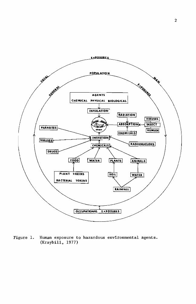
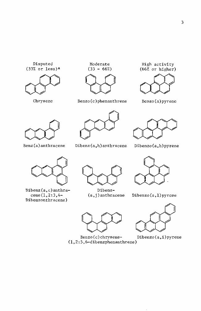
4
Table 1 Polycyclic aromatic hydrocarbons in food
Benzo(a) pyrene (ugKg)
Benz(a) anthracene
(ugKg)
Charcoal broiled meat 8 45 Lijinsky and Shubik 1964
Smoked fish 12-37 91-189 Masuda and Kuratune 1971
Singed sheep head Coal raw Coal cooked
21 9
15 5
Thorsteinsson and Thordarson 1968
Singed sea bird Coal raw Coal cooked
99 96
69 50
Smoked food Mutton Trout Cod Red Fish
13 21 05 03
-shy-shy
Bailey and Dungal 1958
Barbecued ribs 105 36 Lijinsky and Shubik 1965
Coffee 4 -- Kuratsune and Hueper 1960
5
Researchers attention up to this point was devoted to isolating
chemical carcinogens from our environment and to characterizing the
chemical carcinogens Some researchers Allison and Mallucci (1964)
Shires et ale (1966) Brooks and Lawley (1964) Wang et ale (1972)
started to investigate the mechanisms involved in chemical carcinoshy
genesis
Allison and Mallucci (1964) utilized fluorescence microscopy
phase contrast microscopy and autoradiography techniques to locate
carcinogenic hydrocarbons in mammalian cell cultures These investishy
gators concluded that the hydrocarbon carcinogens were not concentrated
in either mitochondria or the nuclei They were however found to be
within lysosomes Allison and Paton (1965) Allison and Dingle (1966)
and Allison (1968) centered their studies on lysosomes and their
relationship to cancer
A study similar to the one mentioned above was carried out two
years later by Shires and Richter (1966) These workers exposed mammashy
lian cells in vitro to carcinogenic hydrocarbons for periods ranging
from 12 hours to 4 days The treatment was terminated at different
intervals for microscopic observations The researchers were unable to
detect any incorporation of benzo(a)pyrene into the nuclei or chromoshy
somes during a complete generation cycle
On the other hand Brooks and Lawley (1964) tried to show the
binding of carcinogenic hydrocarbons to the nucleic acids in mouse skin
They did so by applying the hydrocarbon carcinogens to shaved regions on
the backs of mice The mice were sacrificed at various times and the
treated areas of skin were removed and used for deoxyribonucleic acid
(DNA) and ribonucleic acid (RNA) isolation This study indicated that
6
DNA reacts with the hydrocarbon carcinogens tested Experimental
results did not indicate how the hydrocarbon carcinogens are bound to
the nucleic acids
Lerman (1960) investigated the way in which polynuclear hydroshy
carbons react with DNA He used mutagenic acridine dye derivatives
for his study This investigation showed that the dyes are intershy
calating into the DNA helix through extension of DNA backbone This
information enhanced the idea that polycyclic aromatic hydrocarbons
require structural planality for the carcinogenic potency A similar
mechanism of the intercalation reaction between hydrocarbon carcinoshy
gens and DNA was proposed by Liquori (1962) Liquoris findings were
based upon the increased solubility of benzo(a)pyrene in DNA aqueous
solutions This is significant because benzo(a)pyrene is virtually
insoluble in water
Boyland and Green (1964) expressed a slightly different opinion
They showed that carcinogenic hydrocarbon molecules react with DNA by
slipping into the sites where the DNA is already stretched Such
stretching of DNA can only occur during DNA replication or RNA transhy
scription An expanded idea was postulated by Arcos and Argus (1968)
They indicated that the solubilization of carcinogenic hydrocarbons by
DNA can be accomplished by either intercalation or through external
binding mechanisms (Figure 2)
Van Duuren et al (1969) suggested a reconsideration of the
intercalation mechanism This was proposed because a) solubilization
of detergent micelles of sodium lauryl sulfate and aqueous DNA solution
caused the same results which were found with hydrocarbon carcinogens
7
e oI I I I I I IIgt I I
I
A B
Figure 2 Noncova1ent interactions of polycyclic aromatic hydroshycarbons (illustrated with 34-benzpyrene) with DNA A Intercalation into DNA double helix B External binding to DNA (From Arcos and Argus 1968)
8
and aqueous DNA solution and b) potent carcinogens such as 712shy
dimethy1benz(a)anthracene and 3-methy1cho1anthrene are thicker than
the distance between adjacent base pairs in DNA
Because previous investigations had been unable to prove whether
or not particular parent carcinogenic hydrocarbon molecules react with
DNA research interest was directed to the isolation and characterization
of intermediate metabolites Benzo(a)pyrene-45-oxide (K-region
epoxide) proved to be one of the highly reactive metabolites of
benzo(a)pyrene (Wang et a1 1972 Selkirk et a1 1971) Another
metabolite 78-dihydro-78-dihydroxybenzo(a)pyrene-910-oxide has been
investigated for its metabolism and mutagenicity It was confirmed to
be carcinogenic (Neubo1d and Brooks 1976 Sims et a1 1974)
Thus far accumulated data favor the theory of hydrocarbon carcinoshy
gens binding to DNA On the other hand the significance of 1ysosomes
in regard to carcinogenesis cannot be ignored Both Allison and Ma11ucci
(1964) and Shires and Richter (1966) utilized fluorescence and phase
microscopies and concluded that 1ysosomes were somehow involved In
addition to optical systems Allison and Ma11ucci (1964) used autoshy
radiography techniques for localization of hydrocarbon carcinogens in
mammalian cell cultures They concluded that the amount present if
any in cellular organelles other than 1ysosomes is too small to detect
by these methods
It was the purpose of this investigation to challenge the credishy
bility of autoradiographic techniques as an indicator of
benzo(a)pyrene-DNA incorporation and to consider the problems that
were encountered with this technique
MATERIALS AND METHODS
Experimental animals
Heterogenic white mice were obtained from the Emporia State
University research animal facility and from the Mid-Continent
Research Company (Kansas City Kansas) They were maintained in the
animal facility at Emporia State University
Chemicals
Benzo(a)pyrene (B(a)P) obtained from Sigma Chemical Company
was first dissolved in acetone (10 mgm1) and then dispersed into complete
growth medium with a Pyrex disposable syringe to give a concentration of
100 ugm1 of medium (Berwald and Sachs 1965) This B(a)P suspension was
used as stock solution from which dilutions were made Dilutions were
01 ug 10 ug and 50 ug B(a)P per milliliter of medium The stock solushy
tion was stored at OdegC The desired concentrations of B(a)P solution
were applied to cells in culture
Tissue culture medium
Du1beccos modified Eagles medium containing 45 gl glucose was
purchased from GIBCO The growth medium was supplemented with 10 fetal
bovine serum (GIBCO) and 05 m1 combiotics This complete growth medium
was used to grow and maintain the cells in vitro GKN (calcium and
magnesium free saline) was used for cell harvest and cell transfer with
025 trypsin and trypsin-versene (0002-005) solutions respectively
Stock combiotic solutions were prepared by combing penicillin G
sodium (1650 Umg) and streptomycin sulfate (740 mggm) in 1X Hanks
BSS Stock solutions were sterilized using Mi11ipore filters One
milliliter a1iquots were prepared and stored frozen until needed Cell
10
harvesting solutions were prepared by adding 05 ml of the combiotic to
every 100 ml of GKN Likewise cell maintenance solutions were preshy
pared by adding 05 ml of combiotic stock solution to every 100 ml of
growth medium
Fetal bovine serum which was mycoplasma tested and virus screened
was obtained from GIBCO and used throughout this study
Carbon dioxide gas prepared by mixing calcium carbonate and
sulfuric acid was used to maintain the desired pH of the medium
Glassware
Glassware was cleaned by soaking in an acid bath (17 mixture of
concentrated hydrochloric acid and concentrated sulfuric acid) overshy
night followed by washing with Alconox solution and repetitive rinsing
with tap water All glassware was rinsed with deionized distilled
water after being washed
Microscope coverslips (22 X 50 mm) were used to make cell growing
covers lips (22 X 8 mm) These were immersed in an Alconox solution and
rinsed with running tap water for 10-13 hours They were rinsed individshy
ually 10 times in three consecutive deionized distilled water baths and
one 100 ethanol bath then stored in an absolute ethanol bath until
use
Cell culture
Minced whole embryos of heterogenic white mice served as the source
of normal cells Pregnant mice (15-18 days in term) were sacrificed by
cervical dislocation Embryos were then removed aseptically from the
mouse and transferred to a petri dish containing GKN-combiotics mixture
Embryos were minced and placed into a trypsinizing flask The tissue
11
chunks were subjected to gentle stirring while being trypsinized at
room temperature for 30 minutes Trypsinized tissue was filtered
through four layers of cheese cloth The filtrate was washed with one
change of GKN and one change of complete growth medium Cell popushy
lations were adjusted with complete growth medium to a concentration
6of 3 X 10 cellsmI Three milliliter aliquots were planted in 25
2 cm polystyrene culture flasks and incubated at 37degC for one to three
days (primary cell culture) Primary cell cultures were treated with
trypsin-versene solution for two minutes washed centrifuged and
resuspended to give a concentration of 6 X 10⁵ cellsmI They were
inoculated in test tubes containing covers lips (7 X 22 mm) for 24 hours
at 37degC (secondary culture) prior to usage in the studies Secondary
cell cultures were treated with either a colloidal suspension of B(a)P
(10 ugml) and 3H- B(a)P (10 uCiml) or 3H- B(a)P (10 uCiml) alone
Autoradiography
Tritium-labeled B(a)P treated cells growing on the coverslips were
removed from the test tubes rinsed with two changes of GKN solution and
fixed in an absolute methanol bath for five minutes The covers lips
were mounted on a slide with the cell side up using Pro-texx mounting
medium The slides were dried overnight on a heating plate at 37degC
Ilford emulsion (1-4 Polysciences Inc) was melted in a 40degC
water bath Ten milliliters of melted emulsion were diluted with an
equal volume of deionized distilled water and any bubbles resulting in
the mixing operation were removed through a layer of cheese cloth The
slides were dipped into the diluted emulsion and immediately removed
Residual emulsion was cleaned from the back of the slides and the
12
slides held in a vertical position for 15 minutes to allow the emulsion
to dry Slides were sealed in a light tight slide box by wrapping the
box with aluminum foil The slides were exposed for 10 days at OdegC
The exposed slides were developed in Dekto1 (D-17) for two
minutes rinsed in a deionized distilled water stop bath and fixed in
acid fixer for three minutes After washing for 15 minutes in slowly
running tap water the slides were dried and stained for four minutes
in 005 basic fuchsin solution The slides were dehydrated in a
series of ethanol baths and immersed in a mixture of cedar wood oil
and absolute ethanol (1 1) for one hour followed by infiltration of
a Pro-texx mounting medium and xylene mixture (1 1) for one hour
The slides were examined for the appearance of silver grains
(black dots) located within the cells thereby indicating B(a)P uptake
by the cell
Cell viability test
The toxic effect of B(a)P on cultured cells was examined by adding
5 m1 of B(a)P at different concentrations Concentrations used ranged
from 01 to 50 ug B(a)P per milliliter of growth medium At regular
intervals B(a)P-growth medium mixtures were collected from culture
flasks and centrifuged for five minutes at 800 rpm The supernatant
was siphoned off and discarded Pellets were resuspended with 1 m1
growth medium and 01 m1 of erythrosin (04) was added and stained for
two minutes Dead cells staining pink were counted on a hemocytometer
Numbers of dead cells were plotted against the durations of B(a)P treatshy
ment
13
Inoculation of B(a)P treated cells into animals
Cells treated with B(a)P were harvested with a trypsin-versene
solution for three minutes Cell harvest was made at preselected inshy
tervals of time The cell population was adjusted to 10 X 106 ce11sm1
with complete growth medium From this solution 02 m1 were injected
into a group of five male mice Injections were made subcutaneously on
the left thigh (modified from Evans et a1 1957) Inoculation sites
were checked every 14 days for tumor development until termination of
the experiment
Histological study
Twenty-eight days after inoculation of B(a)P treated cells mice
which developed tumors were sacrificed for histological studies The
tumor tissue was excised and fixed overnight in Bouins fixative
Several solution changes of 70 ethanol saturated with lithium carbonate
were made to eliminate any fixative in the tissue Dehydration of fixed
tissue was accomplished in a series of ethanol baths of one hour each
The tissue was then immersed for one hour in a mixture of absolute
ethanol and xylene (1 1) It was soaked in a xylene bath overnight
and then transferred to a mixture of xylene and paraffin (56 0 tissuemat)
for 15 minutes Infiltration of hot tissuemat was carried out in four
changes of tissuemat solution Tissue was then embedded in a plastic
ice cube tray with hot tissuemat and cooled to room temperature The
blocks were sectioned on the microtome at a thickness of seven microns
The sections were stained by standard hematoxylin and eosin procedures
The slides were then carefully examined for histological abnormalities
14
Chromosome preparation
Cells in culture were mitotically blocked by lyophilized colcemid
006 ugml (GIBCO) for two hours Harvesting of cells was accomplished
by trypsinization and centrifuging for five minutes at 800 rpm The
pellet was resuspended in 1 ml of complete growth medium with a Pasteur
pipette Hypotonic solution (3 ml deionized distilled water) was added
to this suspension for seven minutes at room temperature One milliliter
of freshly prepared methanol-acetic acid (3 1) fixative was introduced
to the hypotonic solution for 10 minutes The suspension was centri shy
fuged and fixed by repeated dispersion at 10 minute intervals in three
changes of 31 methanol-acetic acid fixative During these fixation
periods microscope slides were washed in an Alconox solution and stored
in deionized distilled water at 4degC After the third fixation the
pellet was resuspended in 03-1 ml (depending on size of the pellet)
fixative solution Several drops of the final cell suspension were
applied to the slides The backs of the slides were wiped dry with
tissue paper A film of water on the upper surface of the slides was
left to facilitate spreading of the cells The slides were air-dried
and stained for 15 minutes in modified Giemsa stain Modification
was made according to Gude et al (1955) For the autoradiographic
study the slides were stained for 15 minutes after the completion of
photographic processes
RESULTS
Cell viability test
The toxic effect of B(a)P was tested for nine days Results are
summarized in Figure 3 From the data linear regression curves were
prepared for each concentration The slope of the untreated cell line
is 48 The slopes of the B(a)P treated cell lines are 65 for 01 ug
B(a)P treated cells 70 for 10 ug B(a)P treated cells and 76 for 50
ug B(a)P treated cells A statistical analysis did not indicate a
significant difference between the normal cell line and the B(a)P
treated cell lines Re-examination of the data used in Figure 3 will
help in understanding effect of treatment on cell viability for specific
periods of time In Figure 4 one and three day incubation periods
showed a smaller rate of increase (slope for one day 007 and slope
for three days 016) which implies that the B(a)P concentration did
not have a significant effect in terms of cell killing ability The dead
cell numbers in five day B(a)P treated cultures presented about 28-fold
increase from that of three day B(a)P treated cultures A statistical
analysis showed a significant difference indicating that the critical
factor in cell killing ability of B(a)P is dependent upon the exposure
period
Cell morphology in culture
Mouse embryo cells in culture were observed under the light microshy
scope for their morphology Embryonic cells were round immediately after
implantation into culture flasks Approximately three hours incubation
at 37degC after initial cell plantation some cells started to establish
their natural morphological appearance (elongation of cytoplasm) The
16
elongation process was an indication of cell attachment on the surface
of culture flasks Owens et a1 (1974) reported that in earlier days
of incubation two cell types epithelial cells and fibroblast cells
can be identified Epithelial cells from mono1ayered sheets which are
attached to each other (Figures 5 and 9) Vasi1iev and Ge1hand (1977)
reported that fibroblast cells form several morphological varieties in
culture mono1ayers of mutually oriented cells which are not firmly
attached to each other (Figures 6 and 8) multilayered sheets (Figure
7) or spherical aggregates (Figure 7 lower right and upper left
darkly stained aggregates) Fibroblasts andor fibroblast-like cells
became predominant throughout the entire cell population when cells were
cultured for over 30 days (Figures 10 11 and 12) These fibroblast
cells are characterized by elongated or stellate cytoplasmic structures
(Figures 10 11 and 12)
Mouse embryo cells were treated with 10 ug B(a)P for predesignated
exposure periods to observe an effect on cell morphology Some B(a)P
treated cells showed an abnormal spindle shape while the other cells
retained their normal morphological appearance (Figures 13 15 and 16)
In Figures 15 and 16 some cells were found to have vacuoles within their
cytoplasm Many of these also showed attenuated cytoplasms (Figure 16)
Autoradiography
Cultured cells were incubated with B(a)P for several different types
of treatment One treatment consisted of incubating cultured cells with
B(a)P at 10 ugm1 and 3H- B(a)P at 10 uCim1 until the termination of
experiment (full-term treatment) Other cells were partially exposed
to B(a)P at the same concentration and radioactivity level The exposure
17
60
-l S-r-
0 0 0 -l
gtlt
40-
Ul -l -l ltl) tJ
d Cll ltl) d
~
0 ltl)
20 l z
o o 5 10
Days
Figure 3 Toxic effect of B(a)P on cultured mouse embryo cells Untreated cells 0 Cells treated with 01 ug B(a)P e Cells treated with 10 ug B(a)P X Cells treated with 50 ug B(a) P
__
--------
18
60
-~ s
I
I ~ ~7 r 0 0 0 ~
x -
Ul ~ ~
eu 40u 0 ell eu 0
4-l 0
Ul H eu ~ l Ie z
20 3
9
_ ---41---------------- 1 o
o 20 40 60
B(a)P concentration (ugml)
Figure 4 Toxicity effect of B(a)P concentration The numbers 1 3 5 7 and 9 indicate the days of B(a)P treatment
Figure 5 Two day old normal mouse embryo cells in culture Epithelial cells in the central area of fibroshyblast cells surrounding the epithelial cells are shown here May-Grunwald and Giemsa stain (100 X)
Figure 6 Four day old normal mouse embryo cells in culture Large nucleated epithelial-like cell and fibroshyblast cells are shown here May-Grunwald and Giemsa stain (400 X)
Figure 7 Six day old normal mouse embryo cells in culture Multilayered fibroblast cells and two heavily stained aggregates are shown here May-Grunwald and Giemsa stain (100 X)
Figure 8 Eight day old normal mouse embryo cells in culture Fibroblast cells formed a monolayer sheet MayshyGrunwald and Giemsa stain (100 X)
Figure 9 Thirteen day old normal mouse embryo cells in culture Epithelial cells in the central and upper left areas are surrounded by well elongated fibroblast cells May-Grunwald and Giemsa stain (100 X)
Figure 10 Thirty day old normal mouse embryo cells in culture May-Grunwald and Giemsa stain (100 X)
Figure 11 Thirty five day old normal mouse embryo cells in culture Cell orientation is obscure and vacuoles are seen May-Grunwald and Giemsa stain (100 X)
Figure 12 Forty day old normal mouse embryo cells in culture Cell orientation is obscure and vacuoles are present May-Grunwald and Giemsa stain (100 X)
9Z
Figure 13 Mouse embryo cells incubated with 10 ug B(a)P for 1 day May-Grunwald stain (100 X)
Figure 14 Mouse embryo cells incubated with 10 ug B(a)P for 2 days May-Grunwald stain (100 X)
Figure 15 Mouse embryo cells treated with 10 ug B(a)P for 6 days May-Grunwald stain (100 X)
Figure 16 Mouse embryo cells treated with 10 ug B(a)P for 9 days Basic Fuchsin stain (100 X)
31
periods were 24 hours and four days
Figures 18 through 23 represent the full-term treatment and events
for the first 17 hours are summarized in Figure 17 After
hours exposure to 3H- B(a)P B(a)P accumulation in the nuclei
low Its cytoplasmic concentration was also minimal (Figure
3H-B(a)P uptake by the cells increased rather sharply after 35
of incubation (Figures 19 20 and 21) Long term incubation (six
and 20 days) showed a concentration of B(a)P in the cytoplasm while no
concentration of B(a)P was observed (Figures 22 and 23)
In other treatments cultured cells were partially exposed to
B(a)P at the same concentration Radioactivity levels were as in the
treatment The cells were exposed to B(a)P for one and four
days At the end of the exposure period the complete growth medium
was replaced for further incubation Figures 24 through 27 indicate
some of the results of 24 hour treated cells Overall results are
summarized in Figure 24 Nuclear accumulation of 3H-B(a)P was maximum
at 24 hours and decreased drastically when fresh complete growth medium
was introduced and incubation was continued (Figures 24 27 and 28)
The nuclear accumulation of B(a)P exceeded that of cytoplasm only at
24 hours incubation (Figure 24) Cytoplasmic accumulation of 3H-B(a)P
was at its highest peak on the 6th day of incubation and followed the
same pattern of decrease as the nuclear accumulation curve Extrashy
polation of the two curves suggests disappearance of B(a)P from the
cells occurs between the 24th and 26th day Some cells as exemplified
in Figure 28 lost nuclear B(a)P and most of the cytoplasmic B(a)P at
50
100
32
00
0 1 bull
O Hours
Figure 17 Time-course of 3H- B(a)P accumulation in the cells Cells in autoradiograms were counted for nuclear accumulation () cytoplasmic accumulation (0) and no accumulation (X)
I I Ii I Ii(I I ~ I I
2 4 6 8 10 12 14 16 18
33
21 days of incubation in the growth medium
Figures 29 through 32 represent the results of four day treated
Four (no additional incubation in growth medium) and 11 days
(additional six day incubation in growth medium) cells show nuclear and
cytoplasmic accumulation of B(a)P but not as intense as seen in the one
cells (Figures 29 and 30) Fifteen day incubated cells had
some B(a)P molecules in the cytoplasm The cells incubated for 27 days
of B(a)P in any part of the cells (Figure 32)
Figures 33 through 35 are the chromosome spreads from 3H- B(a)p
treated cells Silver grains (black dots) were seen on or near the chromoshy
somes in three autoradiograms (Figures 33 34 and 35) Autoradiograms of
chromosomes prepared from one two and three day treated cells with
3H- B(a)P (10 uCiml) were counted for use in labeling analysis Chroshy
mosome spreads from one and two day treated cells produced 936 and 920
labeled chromosome spreads respectively Total chromosome spreads examshy
ined were 94 and 100 Cells treated for three days gave 304 labeled
chromosome spreads
Figures 34 and 35 showed chromosome breakages
Induction of abnormalities and its histology
B(a)P treated cells were injected into adult mice The mouse in
Figure 36 was a normal male mouse The mouse in Figure 37 exemplifies
the abnormal genital inflammation characterized by many of the mice inshy
jected with the B(a)P treated cells From the 26 injected mice 31 deshy
veloped a genital inflammation In contrast only 38 of the control
mice exhibited genital abnormalities The controls consisted of three
groups of mice One group of 32 mice received no injections Another
Autoradiogram Mouse embryo cells in culture were incubated for 15 hours with 10 ug B(a)P(lO uCi) Only a few radioactive molecules are present Hematoxylin and eosin stain (100 X)
Figure 19 Autoradiogram Mouse embryo cells in culture were incubated for 35 hours with 10 ug B(a)P(lO uCi) Nuclear and cytoplasmic accumulation of B(a)P is seen Hematoxylin and eosin stain (100 X)
Figure 20 Autoradiogram Mouse embryo cells in culture were incubated for 17 hours with 10 ug B(a)P(10 uCi) Basic Fuchsin stain (1000 X)
Figure 21 Autoradiogram Mouse embryo cells in culture were incubated for 24 hours with 10 ug B(a)P(10 uCi) Basic Fuchsin stain (1000 X)
Figure 22 Autoradiogram Mouse embryo cells in culture were incubated for six days with 10 ug B(a)P(10 uCi) Hematoxylin and eosin stain (100 X)
Figure 23 Autoradiogram Mouse embryo cells in culture were incubated for 20 days with 10 ug B(a)P(10 uCi) A heavy accumulation of B(a)P in cytoplasm is seen here Hematoxylin and eosin stain (400 X)
-
6pound
40
00
100
~ -
00 c
-l M Q)
~ 50M
~
ro - lQI
z M
10 20 Time in days
Figure 24 3ime-course of 3H- B(a)P accumulation in the cells H-B(a)P was replaced with normal growth medium
after 24 hours incubation Cells in autoradiograms were counted for nuclear accumulation (e) cytoshyplasmic accumulation (0) and no accumulation (X)
00
Figure 25 Autoradiogram Mouse embryo cells in culture were incubated for one day with 10 ug B(a)P(lO uCi) Nuclear and cytoplasmic accumulation of B(a)P is seen Basic Fuchsin stain (1000 X)
Figure 26 Autoradiogram Mouse embryo cells in culture were incubated for one day with 10 ug B(a)P(lO uCi) The B(a)P was then removed The cells were incubated an additional 3 days in complete growth medium Basic Fuchsin stain (1000 X)
Figure 27 Autoradiogram Mouse embryo cells in culture were incubated for one day with 10 ug B(a)P(lO uCi) The B(a)P was then removed The cells were incubated an additional 11 days in complete growth medium Basic Fuchsin stain (1000 X)
Figure 28 Autoradiogram Mouse embryo cells in culture were incubated for one day with 10 ug B(a)P(lO uCi) The B(a)P mixture was then removed The cells were incubated an additional 21 days in complete growth medium Basic Fuchsin stain (1000 X)
shy
Figure 29 Autoradiogram Mouse embryo cells in culture were incubated for four days with 10 ug B(a)P(lO uCi) Negri body stain (1000 X)
Figure 30 Autoradiogram Mouse embryo cells in culture were incubated for four days with 10 ug B(a)P(lO uCi) The B(a)P mixture was then removed The cells were incubated an additional six days in complete growth medium Some silver grains are seen Basic Fuchsin stain (1000 X)
97
Figure 31 Autoradiogram Mouse embryo cells in culture were incubated for four days with 10 ug B(a)P(lO uCi) The B(a)P was then removed The cells were incubated an additional 11 days in complete growth medium Some silver grains were seen in the cytoplasm Basic Fuchsin stain (1000 X)
Figure 32 Autoradiogram Mouse embryo cells in culture were incubated for four days with 10 ug B(a)P(lO uei) The B(a)P mixture was then removed The cells were incubated an additional 23 days in complete growth medium Basic Fuchsin stain (1000 X)
81]
Figure 33 Autoradiogram of chromosomes Mouse embryo cells in culture were incubated for one day with 005 ug B(a)P(lO uCi) Giemsa stain (1000 X)
Figure 34 Autoradiogram of chromosomes Mouse embryo cells in culture were incubated for two days with 005 ug B(a)P(lO uCi) Giemsa stain (1000 X)
-
os
Figure 35 Autoradiogram of chromosomes Mouse embryo cells in culture were incubated for three days with 005 ug B(a)P(10 uCi) Giemsa stain (1000 X)
Figure 36 Normal male mouse
53
group of 10 mice were injected with normal untreated cells The last
group of 10 mice was inoculated with growth medium that was used to
maintain the cells (Table 2) The effect of B(a)P treated cells in
the mice is summarized on Table 2 The mice inoculated with the cells
of four days B(a)P preincubation resulted in a 25-50 incidence of
abnormal development Of these abnormal mice two mice developed a
small nodule (ca 4-5 rom in diameter) on the peritoneum (Figure 38)
This nodule was processed for histological study Microscopic examishy
nation revealed that the nodule was the undescended testis and showed
hyperplasia of Leydig cells (Figure 43) Atrophy of seminiferous
tubules (Figure 39) is clearly recognized when they are compared with
those of normal tissue (Figures 42 and 43) Seminiferous tubules
from abnormal tissue did not show spermatogenesis (Figure 43) The
ductus epididymis (Figure 41) however showed normal morphological
appearance but clumps of spermatozoa were not seen in the ductuli
Hyperplasia of connective tissue surrounding ductus epididymis was
observed as well The mice inoculated with 10 day B(a)P treated cells
had a higher incidence (80) of abnormal development Long term
observation (146 days) did not reveal the development of any abnorshy
mality although the inoculated cells were treated for one day with
B(a)P
Tab
le
2
Eff
ect
of
B(a
)P tr
eate
d cell
s in
ad
ult
mic
e
Day
s o
f D
ays
of
Num
ber
of
cell
s D
ays
in
Day
s aft
er
incu
bati
on
w
ith
B(a
)P
incu
bat
ion
aft
er
B(a
)P
ino
cu
late
d
(x
106
) cu
ltu
re
ino
cu
lati
on
A
bn
orm
alit
ies
rem
oved
4 0
10
47
62
24
4 4a
10
43
68
14
4 6
10
41
70
14
10
0 10
43
70
4
5
1 0
1 6
146
05
1 0
10
6 14
6 0
4
Co
ntr
ols
U
ntr
eate
d M
ouse
-shy
-shy
--shy
13
2
Nor
mal
cell
s 10
1
1
146
11
0
Gro
wth
med
ium
o
nly
(f
rom
a)
4 70
0
10
In
+shy
Figure 37 Mouse with genital abnormality
Figure 38 Mouse with a nodule on peritoneum
9~
Figure 40 Transverse section of ductus epididymis from normal mouse testis Hematoxylin and eosin stain (100 X)
Figure 41 Transverse section of ductus epididymis from undescended testis Clumps of spermatozoa are not seen in the ductu1i Spaces among the ductu1i are abnormally large Hematoxylin and eosin stain (100 X)
6S
Figure 42 Transverse section of seminiferous tubules from normal mouse testis Seminiferous tubules are separated by a slight amount of interstitial connective tissue containing some Leydig cells Hematoxylin and eosin stain (100 X)
Figure 43 Transverse sections of seminiferous tubules from undescended testis Atrophy of seminiferous tubules is seen Hyperplasia of Leydig cells is recognized Hematoxylin and eosin stain (100 X)
19
DISCUSSION
Cook et a1 (1932 1933) isolated carcinogenic hydrocarbons from
coal tar pitch The melting point (mp) of one of the hydrocarbons
was determined to be 1755-1765degC This particular hydrocarbon species
was shown to be pure benzo(a)pyrene (B(a)P) by comparison with the
synthesized B(a)P B(a)P has alternative names such as 12-benzpyrene
and 34-benzpyrene It has the molecular weight of 25230 its
structure is shown in diagram on page 3
B(a)P and other closely related carcinogenic hydrocarbons (see
diagram on page 3) are being used to determine a mechanism of carshy
cinogenesis in several research systems B(a)P is known to be one of
the chemically inert carcinogens To be activated B(a)P must first be
taken up by the cells Thus invasion into the cell is the first step
in the mechanism B(a)P particles in the growth medium are taken into
the cells by endocytosis (Allison and Ma11ucci 1964) Engulfed B(a)P
particles then require enzymatic activations to acquire cytotoxic
mutagenic (Huberman et a1 1971 1976 Newbold and Brooks 1976) and
carcinogenic abilities in the cells (Wood et a1 1976 Wis10cki et a1
1976)
The greatly increased number of dead cells in cultures which had
been treated with B(a)P for five days suggested that enzymatic activations
of ingested B(a)P might have occurred during a one to three day incubation
period As a result B(a)P acquired cytotoxic potency to the cells
(Figure 4) Figures 17 and 24 indicate that the cytoplasmic accumushy
lation had maximum values between 35 hours and six days This sugshy
gested the possibility that B(a)P metabolism takes place between these
periods A reliable explanation for the cytotoxic ability of B(a)P was
63
made by several workers (Nebert and Gelboin 1968a 1968b Kasper
1971 Khandwala and Kasper 1973) There exists an enzyme aryl
hydrocarbon hydroxylase that functions in the metabolism of B(a)P
The metabolite is 3-hydroxybenzopyrene which also possesses cytotoxic
potency to the cell Benzo(a)pyrene-hydroxylase was shown to be preshy
sent at low concentrations in the nuclear membrane The existence of
inducible B(a)P-hydroxylase in a microsomal preparation was reported
as well The major difference between nuclear and microsomal memshy
branes is its specificity Nuclear membranes produce hydrocarbon
hydroxylase specifically by the stimulation of hydrocarbon carcinogens
while microsomal membranes produce hydrocarbon hydroxylase in response
to the addition of hydrocarbon carcinogens or phenobarbital
In addition to enzymatic activation in the cytoplasm B(a)P
molecules may be accumulated in one or some of the cellular organelles
To determine the ultimate B(a)P accumulation site in the cell it is
important to define the target organelle(s) for hydrocarbon carcinoshy
gens Several techniques were developed for this purpose and an autoshy
radiography technique was the one utilized in this study Autoradioshy
graphy techniques in the biomedical field are a powerful tool for
localization and visualization of desired chemicals in the cell or the
tissues Autoradiograms obtained in this study indicated that the
cytoplasmic accumulation of B(a)P occurred as early as 15 hours after
the initial application and lasted about 14 days (Figure 17 and 24)
The silver grains seen in Figures 19 20 and 27 indicate that B(a)P
molecules may be encapsulated as a phagosome perhaps independently
from lysosomes or fused to lysosomes where enzymatic alterations as
64
well as detoxification reactions take place Along with the cytoplasmic
accumulation a maximum nuclear accumulation (52 labeling) was observed
in one day (Figure 24) Although nuclear accumulation of B(a)P was
shown it does not clearly indicate the incorporation of B(a)P into
genetic material (DNA) To answer this question chromosome spreads
combined with autoradiography were prepared (Figures 33 34 and 35)
These figures indicate that an interaction of B(a)P and chromosomes
might have occurred in the nuclei This result is supported by the work
of Brooks and Lawley (1964) who isolated DNA and RNA from mouse skin to
which radioactive carcinogenic hydrocarbons were previously applied
One of their results is shown in Figure 44 which demonstrates that the
maximum binding of B(a)P to proteins occurred about 24 hours after
application of B(a)P This result suggests the possibility that B(a)P
molecules bind to DNA
Figures 34 and 35 demonstrated chromosome breakages A generally
accepted explanation for chromosome aberrations is that a direct
chemical or physical attack on DNA has occurred However Allison and
Patton (1965) proposed another possible mechanism which could cause
chromosome breakages The idea was that damage to 1ysosomes can cause
chromosomal damage It was demonstrated by De Duve and his colleagues
that 1ysosomes contain hydrolytic enzymes acid phosphatase acid DNase
acid RNase and acid protease (reviewed by Allison and Ma11ucci 1964)
Among these lysosomal enzymes DNase can break the DNA which is the
backbone of the uncoiled interphase chromatid Deoxyribonuclease must
therefore enter into the nucleus for chromosome breakages to occur
Widne11 and Tata (1964) reported a result which supports DNase migration
0
I
I
I
02
r-
U l
I
r- (]J CIl o
d shyI r- (]J
jJ o ~
01
~ o ~
jJ
r- gt r-
jJ U co U
r- ~
r- U
(]J
0shy
Cfl
o 5
0
150
25
0
Fig
ure
4
4
T
ime-
cou
rse
of
the
bin
din
g
of
(3H
) la
bell
ed
hy
dro
carb
on
s to
to
tal
pro
tein
s o
f m
ouse
sk
in
Resu
lts
are
ex
pre
ssed
as
specif
ic ra
dio
shyacti
vit
y
of
pro
tein
(u
Ci
g)
div
ided
by
th
e d
ose
(u
Ci
mo
use
) g
iven
at
zero
ti
me
plo
tted
ag
ain
st
tim
e
34
-ben
zp
yre
ne
(Part
iall
y
rep
rod
uce
d
from
B
roo
kes
an
d L
awle
y
19
64
)
V1
66
thus DNA-dependent RNA polymerase activity in isolated nuclei is
blocked by addition of DNase Newton et al (1962) also reported that
DNA breakdown occurred shortly after infection with herpes virus This
report implies that lysosomal damage by herpes virus induced a libershy
ation of DNase which in turn caused DNA breakage
The studies conducted so far do not give any indication concerning
mechanisms by which B(a)P binds to the nucleic acids Many investi shy
gators worked to clarify the steps involved as B(a)P migrates from the
cytoplasm to the nucleus B(a)P is enzymatically converted to at least
four phenols three dihydrodiols and three quinones and epoxides
(reviewed by Yung et al 1976) The active B(a)P metabolites which are
converted by microsomal hydroxylase within the cells possess a relatively
high tendency to react with the nucleophilic sites of the macromolecules
Thus the B(a)P metabolite has a rare chance of reacting with nuclear
DNA (Alexandrov and Thompson 1977) This report implies that further
metabolic activation of B(a)P metabolites by the other enzymes of microshy
somes or the nuclear envelope becomes necessary (Rogan et al 1976)
A similar interpretation of a two-step activation mechanism was reported
by several workers (Sims et al 1974 Weinstein et al 1976 Spelsberg
et al 1977) Diol-epoxide intermediates (7S-dihydro-7S-dihydroshy
xybenz(a)pyrene and 7S-dihydro-7S-dihydroxybenzo(a)pyrene-9lO-oxide)
were reported to be the metabolites responsible for DNA-hydrocarbon
binding (Brooks et al 1975 Sims et al 1974) Brooks et al (1975)
also indicated that the purine moieties of DNA react with diol-epoxide
intermediates A specific isomer of a diol-epoxide derivative of B(a)P
(plusmn)-7B Sa-dihydroxy-9a lOa-7S9lO-tetra-hydrobenzo(a)pyrene also
67
appeared to be a B(a)P metabolite in terms of covalent binding to
nucleic acids (Weinstein et a1 1976) Another isomeric form of a
dio1 epoxide derivative of B(a)P 7a 8B-dihydroxy-9B10B-epoxy-789
10-tetrahydrobenzo(a)pyrene was proved to be a highly mutagenic intershy
mediate of B(a)P metabolism (Wood et a1 1976 Huberman et a1 1976)
This metabolite is suspected to be the ultimate carcinogenic species
in B(a)P carcinogenesis The reactions mentioned above are the events
occurring as B(a)P molecules move into the nucleus of the cell It is
important to s~e the relationship between the duration of B(a)P treatshy
ment and cell transformation To determine the tumorigenicity of B(a)P
in the cells in vitro suspected cells must be injected into a host
animal to observe the behavior of the cells (Fedoroff 1967) In this
experiment several conditions for B(a)P treatments were designed and
inoculation of mice with B(a)P treated cells was carried out The
results are summarized in Table 3 Two mice developed palpable tumors
(Figure 38) while the remainder showed genital abnormality (Figure 37)
The mice inoculated with four day B(a)P treated cells developed 25-50
abnormality development Some four day B(a)P treated cells were incushy
bated additional days in a normal growth medium before inoculation
Abnormal development was not affected significantly by the additional
incubation period This is because during a four day incubation period
most of the cells may be transformed This idea can be supported by
the work of Berwald and Sachs (1965) They reported that two days of
incubation with B(a)P is necessary for cell transformation Ten day
B(a)P incubation created an 80 rate of abnormal development Higher
genital development in 10 day B(a)P treated cells was possibly due to
68
the increased probability of cell transformation Berwald and Sachs
(1965) reported that the normal mouse cells in vitro showed spontaneous
transformation sometime after 45 to 80 days in the culture As can be
seen on Table 3 the cells used for the inoculations were cultured from
41 to 47 days which is in the range of Berwalds figures Thus this
spontaneous transformation factor may be involved in the results shown
on Table 3 The mice inoculated with one day B(a)P treated cells
developed no abnormalities (Table 3) One reason for the absence of
abnormal development may be related to the age of the cells in culture
the cells used were cultured for six days while the abnorma1ityshy
producing cells were cultured from 41 to 47 days
A tumor development in host mice by the inoculation of suspected
cells must be further examined by histological means Figures 40 through
43 are transverse sections of the ductus epididymis and the seminiferous
tubules from abnormal and normal mouse testis Figures 40 and 42 from
the control mouse show a normal histological appearance The ductu1i
are well packed and contain aggregates of spermatozoa In contrast
Figures 41 and 43 show atrophy of ducts of the epididymis resulting in
the formation of large interstitial spaces Closer observations revealed
a deterioration of the steroci1ia as well Figures 40 and 42 show the
normal condition in which seminiferous tubules contain newly formed
spermatozoa primary spermatocytes and secondary spermatocytes Figures
41 and 43 show atrophy of seminiferous tubules with a few supporting
cells (Serto1i) located near the basement membrane An abnormal increase
in the number of interstitial Leydig cells was also seen Occasionally
binuc1eated Leydig cells were observed Detailed observation of the
excised nodule revealed it to be histologically abnormal It is however
69
difficult to determine if the abnormal nodule was caused by the direct
action of B(a)P treated cells
Allison and Mallucci (1964) reported that autoradiographic
techniques failed to visualize accumulation of chemical carcinogens in
the nucleus Through this study it was demonstrated that autoradioshy
graphic techniques could show the existence of B(a)P in the nucleus
Furthermore autoradiograms of chromosomes gave an indication that
interaction between B(a)P and chromosomes may have occurred
SUMMARY
1) Benzo(a)pyrene accumulation in the cell occurred after 24 hours
of incubation period
2) Autoradiograms of chromosomes prepared from benzo(a)pyrene
treated cells indicated that benzo(a)pyrene reacted with
chromosomes
3) Autoradiographic techniques were shown to have the capacity to
localize the sites of benzo(a)pyrene accumulation in the cell
4) Cytotoxicity studies of benzo(a)pyrene showed a time-dependent
effect on the cultured mouse embryo cells
5) A tumor induction study indicated that tumor inducibility in the
host animal seems to be affected by the length of cell culture
period
LITERATURE CITED
Allison A C and L Ma11ucci 1964 Uptake of hydrocarbon carcinogens by 1ysosomes Nature 2031024-1027
Allison A C and G R Paton 1965 Chromosome damage in human diploid cells following activation of lysosomal enzymes Nature 2091170-1173
Allison A C and J T Dingle 1966 Role of 1ysosomes in adrenal necrosis caused by dimethy1benzanthracene Nature 209303-304
Allison A C 1968 Lysosomes in relation to cancer induction and treatment Europ J Cancer 3481-490
Arcos C J and F M Argus 1968 Molecular geometry and carcinoshygenic activity pp 348-435 Advances in Cancer Research Vol 11 Academic Press Inc
A1exandrov K and H M Thompson 1977 Influence of inducers and inhibitors of mixed-function oxidase on benzo(a)pyrene binding to the DNA of rat nuclei Cancer Res 371443-1449
Bailey J E and N Dunga1 1958 Polycyclic hydrocarbons in icelandic smoked food Brit J Cancer 12348-350
Berwald Y and L Sachs 1965 In vitro transformation of normal cells to tumor cells by carcinogenic hydrocarbons J Nat1 Cancer Inst 35(4)641-661
Boyland E and B Green 1964 On the reported sedimentation of polycyclic hydrocarbons from aqueous solutions of DNA J Mol Bio1 9589-597
Boyland E and P Sims 1965 Induction of adrenal damage and cancer with metabolites of 712-dimethy1benz(a)anthracene Nature 207816-817
Brookes P and P D Lawley 1964 Evidence for the binding of polynuclear aromatic hydrocarbons to the nucleic acids of mouse skin Relation between carcinogenic power of hydrocarbons and their binding to deoxyribonucleic acid Nature 202781-784
Brookes P P Jones and J Amos 1975 The nature of the deoxyribonuc1eosides involved in the binding of carcinogenic hydrocarbons to the DNA of mouse embryo cells Int J Cancer 15912-917
Cook J W C Hewett and L Hieger 1932 Coal tar constituents and cancer Nature 130926
73
Cook J W 1933 The isolation of a cancer producing hydrocarbon from coal tar Chern Society J 398395-405
Dipple A 1976 Chemical Carcinogens ACS Monograph pp 245-314
Evans V J N M Hawkins B B Westfall and W R Earle 1957 Studies on culture lines derived from mouse liver parenchymatous cells grown in long term tissue culture Cancer Res 18261-273
Evans I A and J Mason 1965 Carcinogenic activity of bracken Nature 208913-914
Fa1k H L A Miller and P Kotin 1958 Elution of 34-benzpyrene and related hydrocarbons from soots by plasma proteins Science 127474-475
Fedoroff S 1967 Proposed usage of animal tissue culture terms J Nat1 Cancer Inst 38(4)607-611
Gude W D A C Upton and T T Odell 1955 Giemsa staining of autoradiograrns prepared with stripping film Stain Tech 30161-162
Hecht S S R M Ornaf and D Hoffman 1974 Chemical studies on tobacco smoke XXXIII N-nitrosonornicotine in tobacco Analysis of possible contributing factors and biologic implishycations J Nat1 Cancer Inst 54(5)1234-1244
Henderson E B J R Gordon H Menck J Soohoo P S Martin and C M Pike 1975 Original contributions Lung cancer and air pollution in South central Los Angeles county American J of Epidemiology 101(6)477-488
Hirono I C Shibuya K Fushimi and M Raga 1970 Studies on carcinogenic properties of bracken Pteridium aqui1iurn J Nat1 Cancer Inst 45(1)179-188
Hirono I C Shibuya M Shimizu and K Fushimi 1972 Carcinogenic activity of processed bracken used as human food J Nat1 Cancer Inst 48(4)1245-1249
Hirono I K Fushimi H Mori T Miwa and M Haga 1973 Comparative study of carcinogenic activity in each part of bracken J Natl Cancer Inst 50(5)1367-1371
Haberman E L Aspiras C Heidelberger P L Grover and P Sims 1971 Mutagenicity to mammalian cells of epoxides and other derivatives of polycyclic hydrocarbons Nat1 Acad Sci USA Proceeding 68(12)3195-3199
Kasper C B 1971 Biochemical distinctions between the nuclear and microsomal membranes from rat hepatocytes J Bio1 Chern 246(3)577-581
74
Khandwa1a A S and C B Kasper 1973 Preferential induction of aryl hydroxylase activity in rat liver nuclear envelope by 3-methy1cho1anthrene Biochem Biophys Res Commun 54(4)1241-1246
Kuratsune M and W C Hueper 1960 Polycyclic hydrocarbons in roasted coffee J Nat1 Cancer Inst 24(2)463-469
Kraybi11 H F 1977 Advances in Modern Toxicology Vol 3 pp 27shy29 Hemisphere Publishing Co Washington D C
Lerman L S 1960 Structural considerations in the interaction of DNA and acridines J Mol Bio1 318-30
Lijinsky W and P Shubik 1964 Benzo(a)pyrene and other polynuclear hydrocarbons in charcoal-broiled meat Science 14553-55
1965 Polynuclear hydrocarbon carcinogens in cooked meat and smoked food Indus Med Surge 34152-154
Liquori A M 1962 Interaction between DNA and polycyclic aromatic hydrocarbons J Mol Bio1 5521-526
Masuda Y and M Kuratsune 1971 Polycyclic aromatic hydrocarbons in smoked fish Katsuobushi Gann 6227-30
Nebert D W and H V Ge1boin 1968a Substrate-inducible microsomal aryl hydroxylase in mammalian cell culture I Assay and properties of induced enzyme J Bio1 Chern 243(23)6242-6249
1968b Substrate-inducible microsomal aryl hydroxylase in mammalian cell culture II Cellular responses during enzyme induction J Bio1 Chern 243(23)6250-6251
Newton A P P Dendy C L Smith and P Wi1dy 1962 A pool size problem associated with the use of tritiated thymidine Nature 194886-887
Newbold R F and P Brooks 1976 Exceptional mutagenicity of a benzo(a)pyrene dio1 epoxide in cultured mammalian cells Nature 26152-54
Owens R B H S Smith and A J Hackett 1974 Epithelial cell cultures from normal glandular tissue of mice J NatL Cancer Inst 53(1)261-269
Owens R B 1974 Brief communication Glandular epithelial cells from mice A method for selective cultivation J Nat1 Cancer Inst 52(4)1375-1378
75
Pamukcu A M and M J Price 1969 Induction of intestinal and urinary bladder cancer in rats by feeding bracken fern (Pteris aquilina) J Natl Cancer Inst 43(1)275-281
Rogan~ E G~ P Mailander and E Cavalieri 1976 Metabolic activation of aromatic hydrocarbons in purified rat liver nuclei Induction of enzyme activities and binding to DNA with and without monooxygenase-catalyzed formation of active oxygen Natl Acad Sci USA Proceeding 73(2)457-461
Selkirk J K E Huberman and C Heidelberger 1971 An epoxide is an intermediate in the microsomal metabolism of the chemical carcinogen dibenz(ah)anthracene Biochem Biophys Res Commun 43(5)1010-1016
Shires T K and K M Richter 1966 The distribution of 34shybenz(a)pyrene in replicating mammalian cells in vitro Exp Cell Res 44617-620 -------shy
Sims P P L Grover~ A Swaisland K Pal and A Hewer 1974 Metabolic activation of benzo(a)pyrene proceeds by a diol-epoxide Nature 252326-328
Spelsberg T C T H Zytkovicz and H L Moses 1977 Effects of metabolism on the binding of polycyclic hydrocarbons to nuclear subfractions of cultured AKR mouse embryo cells Cancer Res 371490-1496
Thorsteinsson T and G Thordarson 1968 Polycyclic hydrocarbons in singed food in ice land Cancer 21390-392
Van Cantfort J and J Gielen 1975 Organ specificity of aryl hydrocarbon hydroxylase induction by cigarette smoke in rats and mice Biochem Pharmacol 241253-1256
Van Duuren~ B L B M Goldschmidt and H H Seltzman 1969 The interaction of mutagenic and carcinogenic agents with nucleic acids Ann N Y Acad Sci 153744-757
Vasiliev~ J M and I M Gelhand 1977 Mechanisms of Morphogenesis in cell cultures International Review of Cytology Vol 50 pp 159-163 Academic Press~ Inc
Wang~ I Y~ R E Rasmussen and T T Crocker 1972 Isolation and characterization of an active DNA-binding of benzo(a)pyrene from hamster liver microsomal incubation system Biochem Biophys Res Commun 49(4)1142-1149
Weinstein~ B 1 A M Jeffery K W Jennette~ S H Blobstein R G Harvey~ C Harris H Autrup~ H Kasai and K Nakanishi 1976 Benzo(a)pyrene diol epoxides as intermediates in nucleic acid binding in vitro and in vivo Science 193592-595
76
Weisburger E K 1977 Cancer-causing chemicals Chemistry 50(1) 42-48
Widnell C C and J R Tata 1964 Evidence for two DNA-dependent RNA polymerase activities in isolated rat-liner nuclei Biochem J 92313-315
Wislocki P G A W Wood R L Chang W Levin H Yagi O Hernandez P M Dansette D M Jerina and A H Conney 1976 Mutagenicity and cytotoxicity of benzo(a)pyrene arene oxides phenols quinones and dihydrodiols in bacterial and mammalian cells Cancer Res 363350-3357
Wood A W P G Wislocki R L Chang W Levin Y Anthony H Lu H Yagi O Hernandez D M Jerina and A H Conney 1976 Mutagenicity and cytotoxicity of benzo(a)pyrene benzo-ring epoxides Cancer Res 36(9)3358-3366
Yamagiwa K and K Ichikawa 1918 Experimental study of the pathoshygenesis of carcinoma J Cancer Res 3(1)1-29
Yung S K D W McCourt P P Roller and H V Gelboin 1976 Enzymatic conversion of benzo(a)pyrene leading predominantly to the diol-epoxide r-7t-8-dihydroxy-t-910-oxy-789lOshytetrahydrobenzo(a)pyrene through a single enantiomer of r-7 t-8-dihydroxy-78-dihydro-benzo(a)pyrene Natl Acad Sci USA Proceeding 73(8)2594-2598
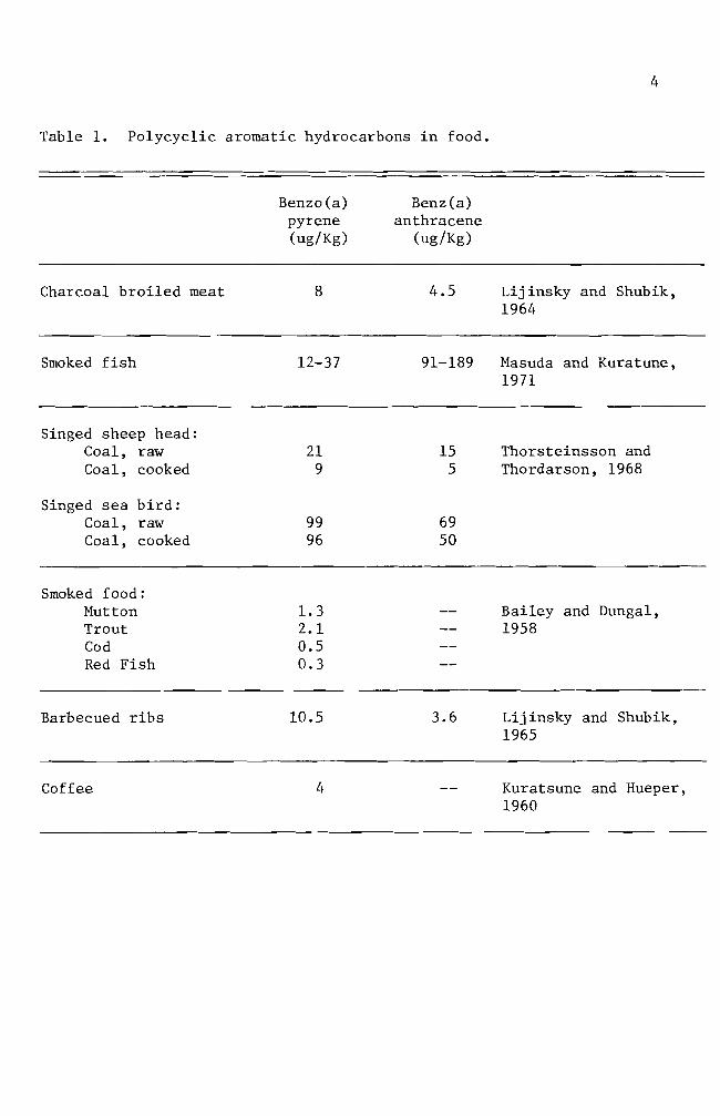
5
Researchers attention up to this point was devoted to isolating
chemical carcinogens from our environment and to characterizing the
chemical carcinogens Some researchers Allison and Mallucci (1964)
Shires et ale (1966) Brooks and Lawley (1964) Wang et ale (1972)
started to investigate the mechanisms involved in chemical carcinoshy
genesis
Allison and Mallucci (1964) utilized fluorescence microscopy
phase contrast microscopy and autoradiography techniques to locate
carcinogenic hydrocarbons in mammalian cell cultures These investishy
gators concluded that the hydrocarbon carcinogens were not concentrated
in either mitochondria or the nuclei They were however found to be
within lysosomes Allison and Paton (1965) Allison and Dingle (1966)
and Allison (1968) centered their studies on lysosomes and their
relationship to cancer
A study similar to the one mentioned above was carried out two
years later by Shires and Richter (1966) These workers exposed mammashy
lian cells in vitro to carcinogenic hydrocarbons for periods ranging
from 12 hours to 4 days The treatment was terminated at different
intervals for microscopic observations The researchers were unable to
detect any incorporation of benzo(a)pyrene into the nuclei or chromoshy
somes during a complete generation cycle
On the other hand Brooks and Lawley (1964) tried to show the
binding of carcinogenic hydrocarbons to the nucleic acids in mouse skin
They did so by applying the hydrocarbon carcinogens to shaved regions on
the backs of mice The mice were sacrificed at various times and the
treated areas of skin were removed and used for deoxyribonucleic acid
(DNA) and ribonucleic acid (RNA) isolation This study indicated that
6
DNA reacts with the hydrocarbon carcinogens tested Experimental
results did not indicate how the hydrocarbon carcinogens are bound to
the nucleic acids
Lerman (1960) investigated the way in which polynuclear hydroshy
carbons react with DNA He used mutagenic acridine dye derivatives
for his study This investigation showed that the dyes are intershy
calating into the DNA helix through extension of DNA backbone This
information enhanced the idea that polycyclic aromatic hydrocarbons
require structural planality for the carcinogenic potency A similar
mechanism of the intercalation reaction between hydrocarbon carcinoshy
gens and DNA was proposed by Liquori (1962) Liquoris findings were
based upon the increased solubility of benzo(a)pyrene in DNA aqueous
solutions This is significant because benzo(a)pyrene is virtually
insoluble in water
Boyland and Green (1964) expressed a slightly different opinion
They showed that carcinogenic hydrocarbon molecules react with DNA by
slipping into the sites where the DNA is already stretched Such
stretching of DNA can only occur during DNA replication or RNA transhy
scription An expanded idea was postulated by Arcos and Argus (1968)
They indicated that the solubilization of carcinogenic hydrocarbons by
DNA can be accomplished by either intercalation or through external
binding mechanisms (Figure 2)
Van Duuren et al (1969) suggested a reconsideration of the
intercalation mechanism This was proposed because a) solubilization
of detergent micelles of sodium lauryl sulfate and aqueous DNA solution
caused the same results which were found with hydrocarbon carcinogens
7
e oI I I I I I IIgt I I
I
A B
Figure 2 Noncova1ent interactions of polycyclic aromatic hydroshycarbons (illustrated with 34-benzpyrene) with DNA A Intercalation into DNA double helix B External binding to DNA (From Arcos and Argus 1968)
8
and aqueous DNA solution and b) potent carcinogens such as 712shy
dimethy1benz(a)anthracene and 3-methy1cho1anthrene are thicker than
the distance between adjacent base pairs in DNA
Because previous investigations had been unable to prove whether
or not particular parent carcinogenic hydrocarbon molecules react with
DNA research interest was directed to the isolation and characterization
of intermediate metabolites Benzo(a)pyrene-45-oxide (K-region
epoxide) proved to be one of the highly reactive metabolites of
benzo(a)pyrene (Wang et a1 1972 Selkirk et a1 1971) Another
metabolite 78-dihydro-78-dihydroxybenzo(a)pyrene-910-oxide has been
investigated for its metabolism and mutagenicity It was confirmed to
be carcinogenic (Neubo1d and Brooks 1976 Sims et a1 1974)
Thus far accumulated data favor the theory of hydrocarbon carcinoshy
gens binding to DNA On the other hand the significance of 1ysosomes
in regard to carcinogenesis cannot be ignored Both Allison and Ma11ucci
(1964) and Shires and Richter (1966) utilized fluorescence and phase
microscopies and concluded that 1ysosomes were somehow involved In
addition to optical systems Allison and Ma11ucci (1964) used autoshy
radiography techniques for localization of hydrocarbon carcinogens in
mammalian cell cultures They concluded that the amount present if
any in cellular organelles other than 1ysosomes is too small to detect
by these methods
It was the purpose of this investigation to challenge the credishy
bility of autoradiographic techniques as an indicator of
benzo(a)pyrene-DNA incorporation and to consider the problems that
were encountered with this technique
MATERIALS AND METHODS
Experimental animals
Heterogenic white mice were obtained from the Emporia State
University research animal facility and from the Mid-Continent
Research Company (Kansas City Kansas) They were maintained in the
animal facility at Emporia State University
Chemicals
Benzo(a)pyrene (B(a)P) obtained from Sigma Chemical Company
was first dissolved in acetone (10 mgm1) and then dispersed into complete
growth medium with a Pyrex disposable syringe to give a concentration of
100 ugm1 of medium (Berwald and Sachs 1965) This B(a)P suspension was
used as stock solution from which dilutions were made Dilutions were
01 ug 10 ug and 50 ug B(a)P per milliliter of medium The stock solushy
tion was stored at OdegC The desired concentrations of B(a)P solution
were applied to cells in culture
Tissue culture medium
Du1beccos modified Eagles medium containing 45 gl glucose was
purchased from GIBCO The growth medium was supplemented with 10 fetal
bovine serum (GIBCO) and 05 m1 combiotics This complete growth medium
was used to grow and maintain the cells in vitro GKN (calcium and
magnesium free saline) was used for cell harvest and cell transfer with
025 trypsin and trypsin-versene (0002-005) solutions respectively
Stock combiotic solutions were prepared by combing penicillin G
sodium (1650 Umg) and streptomycin sulfate (740 mggm) in 1X Hanks
BSS Stock solutions were sterilized using Mi11ipore filters One
milliliter a1iquots were prepared and stored frozen until needed Cell
10
harvesting solutions were prepared by adding 05 ml of the combiotic to
every 100 ml of GKN Likewise cell maintenance solutions were preshy
pared by adding 05 ml of combiotic stock solution to every 100 ml of
growth medium
Fetal bovine serum which was mycoplasma tested and virus screened
was obtained from GIBCO and used throughout this study
Carbon dioxide gas prepared by mixing calcium carbonate and
sulfuric acid was used to maintain the desired pH of the medium
Glassware
Glassware was cleaned by soaking in an acid bath (17 mixture of
concentrated hydrochloric acid and concentrated sulfuric acid) overshy
night followed by washing with Alconox solution and repetitive rinsing
with tap water All glassware was rinsed with deionized distilled
water after being washed
Microscope coverslips (22 X 50 mm) were used to make cell growing
covers lips (22 X 8 mm) These were immersed in an Alconox solution and
rinsed with running tap water for 10-13 hours They were rinsed individshy
ually 10 times in three consecutive deionized distilled water baths and
one 100 ethanol bath then stored in an absolute ethanol bath until
use
Cell culture
Minced whole embryos of heterogenic white mice served as the source
of normal cells Pregnant mice (15-18 days in term) were sacrificed by
cervical dislocation Embryos were then removed aseptically from the
mouse and transferred to a petri dish containing GKN-combiotics mixture
Embryos were minced and placed into a trypsinizing flask The tissue
11
chunks were subjected to gentle stirring while being trypsinized at
room temperature for 30 minutes Trypsinized tissue was filtered
through four layers of cheese cloth The filtrate was washed with one
change of GKN and one change of complete growth medium Cell popushy
lations were adjusted with complete growth medium to a concentration
6of 3 X 10 cellsmI Three milliliter aliquots were planted in 25
2 cm polystyrene culture flasks and incubated at 37degC for one to three
days (primary cell culture) Primary cell cultures were treated with
trypsin-versene solution for two minutes washed centrifuged and
resuspended to give a concentration of 6 X 10⁵ cellsmI They were
inoculated in test tubes containing covers lips (7 X 22 mm) for 24 hours
at 37degC (secondary culture) prior to usage in the studies Secondary
cell cultures were treated with either a colloidal suspension of B(a)P
(10 ugml) and 3H- B(a)P (10 uCiml) or 3H- B(a)P (10 uCiml) alone
Autoradiography
Tritium-labeled B(a)P treated cells growing on the coverslips were
removed from the test tubes rinsed with two changes of GKN solution and
fixed in an absolute methanol bath for five minutes The covers lips
were mounted on a slide with the cell side up using Pro-texx mounting
medium The slides were dried overnight on a heating plate at 37degC
Ilford emulsion (1-4 Polysciences Inc) was melted in a 40degC
water bath Ten milliliters of melted emulsion were diluted with an
equal volume of deionized distilled water and any bubbles resulting in
the mixing operation were removed through a layer of cheese cloth The
slides were dipped into the diluted emulsion and immediately removed
Residual emulsion was cleaned from the back of the slides and the
12
slides held in a vertical position for 15 minutes to allow the emulsion
to dry Slides were sealed in a light tight slide box by wrapping the
box with aluminum foil The slides were exposed for 10 days at OdegC
The exposed slides were developed in Dekto1 (D-17) for two
minutes rinsed in a deionized distilled water stop bath and fixed in
acid fixer for three minutes After washing for 15 minutes in slowly
running tap water the slides were dried and stained for four minutes
in 005 basic fuchsin solution The slides were dehydrated in a
series of ethanol baths and immersed in a mixture of cedar wood oil
and absolute ethanol (1 1) for one hour followed by infiltration of
a Pro-texx mounting medium and xylene mixture (1 1) for one hour
The slides were examined for the appearance of silver grains
(black dots) located within the cells thereby indicating B(a)P uptake
by the cell
Cell viability test
The toxic effect of B(a)P on cultured cells was examined by adding
5 m1 of B(a)P at different concentrations Concentrations used ranged
from 01 to 50 ug B(a)P per milliliter of growth medium At regular
intervals B(a)P-growth medium mixtures were collected from culture
flasks and centrifuged for five minutes at 800 rpm The supernatant
was siphoned off and discarded Pellets were resuspended with 1 m1
growth medium and 01 m1 of erythrosin (04) was added and stained for
two minutes Dead cells staining pink were counted on a hemocytometer
Numbers of dead cells were plotted against the durations of B(a)P treatshy
ment
13
Inoculation of B(a)P treated cells into animals
Cells treated with B(a)P were harvested with a trypsin-versene
solution for three minutes Cell harvest was made at preselected inshy
tervals of time The cell population was adjusted to 10 X 106 ce11sm1
with complete growth medium From this solution 02 m1 were injected
into a group of five male mice Injections were made subcutaneously on
the left thigh (modified from Evans et a1 1957) Inoculation sites
were checked every 14 days for tumor development until termination of
the experiment
Histological study
Twenty-eight days after inoculation of B(a)P treated cells mice
which developed tumors were sacrificed for histological studies The
tumor tissue was excised and fixed overnight in Bouins fixative
Several solution changes of 70 ethanol saturated with lithium carbonate
were made to eliminate any fixative in the tissue Dehydration of fixed
tissue was accomplished in a series of ethanol baths of one hour each
The tissue was then immersed for one hour in a mixture of absolute
ethanol and xylene (1 1) It was soaked in a xylene bath overnight
and then transferred to a mixture of xylene and paraffin (56 0 tissuemat)
for 15 minutes Infiltration of hot tissuemat was carried out in four
changes of tissuemat solution Tissue was then embedded in a plastic
ice cube tray with hot tissuemat and cooled to room temperature The
blocks were sectioned on the microtome at a thickness of seven microns
The sections were stained by standard hematoxylin and eosin procedures
The slides were then carefully examined for histological abnormalities
14
Chromosome preparation
Cells in culture were mitotically blocked by lyophilized colcemid
006 ugml (GIBCO) for two hours Harvesting of cells was accomplished
by trypsinization and centrifuging for five minutes at 800 rpm The
pellet was resuspended in 1 ml of complete growth medium with a Pasteur
pipette Hypotonic solution (3 ml deionized distilled water) was added
to this suspension for seven minutes at room temperature One milliliter
of freshly prepared methanol-acetic acid (3 1) fixative was introduced
to the hypotonic solution for 10 minutes The suspension was centri shy
fuged and fixed by repeated dispersion at 10 minute intervals in three
changes of 31 methanol-acetic acid fixative During these fixation
periods microscope slides were washed in an Alconox solution and stored
in deionized distilled water at 4degC After the third fixation the
pellet was resuspended in 03-1 ml (depending on size of the pellet)
fixative solution Several drops of the final cell suspension were
applied to the slides The backs of the slides were wiped dry with
tissue paper A film of water on the upper surface of the slides was
left to facilitate spreading of the cells The slides were air-dried
and stained for 15 minutes in modified Giemsa stain Modification
was made according to Gude et al (1955) For the autoradiographic
study the slides were stained for 15 minutes after the completion of
photographic processes
RESULTS
Cell viability test
The toxic effect of B(a)P was tested for nine days Results are
summarized in Figure 3 From the data linear regression curves were
prepared for each concentration The slope of the untreated cell line
is 48 The slopes of the B(a)P treated cell lines are 65 for 01 ug
B(a)P treated cells 70 for 10 ug B(a)P treated cells and 76 for 50
ug B(a)P treated cells A statistical analysis did not indicate a
significant difference between the normal cell line and the B(a)P
treated cell lines Re-examination of the data used in Figure 3 will
help in understanding effect of treatment on cell viability for specific
periods of time In Figure 4 one and three day incubation periods
showed a smaller rate of increase (slope for one day 007 and slope
for three days 016) which implies that the B(a)P concentration did
not have a significant effect in terms of cell killing ability The dead
cell numbers in five day B(a)P treated cultures presented about 28-fold
increase from that of three day B(a)P treated cultures A statistical
analysis showed a significant difference indicating that the critical
factor in cell killing ability of B(a)P is dependent upon the exposure
period
Cell morphology in culture
Mouse embryo cells in culture were observed under the light microshy
scope for their morphology Embryonic cells were round immediately after
implantation into culture flasks Approximately three hours incubation
at 37degC after initial cell plantation some cells started to establish
their natural morphological appearance (elongation of cytoplasm) The
16
elongation process was an indication of cell attachment on the surface
of culture flasks Owens et a1 (1974) reported that in earlier days
of incubation two cell types epithelial cells and fibroblast cells
can be identified Epithelial cells from mono1ayered sheets which are
attached to each other (Figures 5 and 9) Vasi1iev and Ge1hand (1977)
reported that fibroblast cells form several morphological varieties in
culture mono1ayers of mutually oriented cells which are not firmly
attached to each other (Figures 6 and 8) multilayered sheets (Figure
7) or spherical aggregates (Figure 7 lower right and upper left
darkly stained aggregates) Fibroblasts andor fibroblast-like cells
became predominant throughout the entire cell population when cells were
cultured for over 30 days (Figures 10 11 and 12) These fibroblast
cells are characterized by elongated or stellate cytoplasmic structures
(Figures 10 11 and 12)
Mouse embryo cells were treated with 10 ug B(a)P for predesignated
exposure periods to observe an effect on cell morphology Some B(a)P
treated cells showed an abnormal spindle shape while the other cells
retained their normal morphological appearance (Figures 13 15 and 16)
In Figures 15 and 16 some cells were found to have vacuoles within their
cytoplasm Many of these also showed attenuated cytoplasms (Figure 16)
Autoradiography
Cultured cells were incubated with B(a)P for several different types
of treatment One treatment consisted of incubating cultured cells with
B(a)P at 10 ugm1 and 3H- B(a)P at 10 uCim1 until the termination of
experiment (full-term treatment) Other cells were partially exposed
to B(a)P at the same concentration and radioactivity level The exposure
17
60
-l S-r-
0 0 0 -l
gtlt
40-
Ul -l -l ltl) tJ
d Cll ltl) d
~
0 ltl)
20 l z
o o 5 10
Days
Figure 3 Toxic effect of B(a)P on cultured mouse embryo cells Untreated cells 0 Cells treated with 01 ug B(a)P e Cells treated with 10 ug B(a)P X Cells treated with 50 ug B(a) P
__
--------
18
60
-~ s
I
I ~ ~7 r 0 0 0 ~
x -
Ul ~ ~
eu 40u 0 ell eu 0
4-l 0
Ul H eu ~ l Ie z
20 3
9
_ ---41---------------- 1 o
o 20 40 60
B(a)P concentration (ugml)
Figure 4 Toxicity effect of B(a)P concentration The numbers 1 3 5 7 and 9 indicate the days of B(a)P treatment
Figure 5 Two day old normal mouse embryo cells in culture Epithelial cells in the central area of fibroshyblast cells surrounding the epithelial cells are shown here May-Grunwald and Giemsa stain (100 X)
Figure 6 Four day old normal mouse embryo cells in culture Large nucleated epithelial-like cell and fibroshyblast cells are shown here May-Grunwald and Giemsa stain (400 X)
Figure 7 Six day old normal mouse embryo cells in culture Multilayered fibroblast cells and two heavily stained aggregates are shown here May-Grunwald and Giemsa stain (100 X)
Figure 8 Eight day old normal mouse embryo cells in culture Fibroblast cells formed a monolayer sheet MayshyGrunwald and Giemsa stain (100 X)
Figure 9 Thirteen day old normal mouse embryo cells in culture Epithelial cells in the central and upper left areas are surrounded by well elongated fibroblast cells May-Grunwald and Giemsa stain (100 X)
Figure 10 Thirty day old normal mouse embryo cells in culture May-Grunwald and Giemsa stain (100 X)
Figure 11 Thirty five day old normal mouse embryo cells in culture Cell orientation is obscure and vacuoles are seen May-Grunwald and Giemsa stain (100 X)
Figure 12 Forty day old normal mouse embryo cells in culture Cell orientation is obscure and vacuoles are present May-Grunwald and Giemsa stain (100 X)
9Z
Figure 13 Mouse embryo cells incubated with 10 ug B(a)P for 1 day May-Grunwald stain (100 X)
Figure 14 Mouse embryo cells incubated with 10 ug B(a)P for 2 days May-Grunwald stain (100 X)
Figure 15 Mouse embryo cells treated with 10 ug B(a)P for 6 days May-Grunwald stain (100 X)
Figure 16 Mouse embryo cells treated with 10 ug B(a)P for 9 days Basic Fuchsin stain (100 X)
31
periods were 24 hours and four days
Figures 18 through 23 represent the full-term treatment and events
for the first 17 hours are summarized in Figure 17 After
hours exposure to 3H- B(a)P B(a)P accumulation in the nuclei
low Its cytoplasmic concentration was also minimal (Figure
3H-B(a)P uptake by the cells increased rather sharply after 35
of incubation (Figures 19 20 and 21) Long term incubation (six
and 20 days) showed a concentration of B(a)P in the cytoplasm while no
concentration of B(a)P was observed (Figures 22 and 23)
In other treatments cultured cells were partially exposed to
B(a)P at the same concentration Radioactivity levels were as in the
treatment The cells were exposed to B(a)P for one and four
days At the end of the exposure period the complete growth medium
was replaced for further incubation Figures 24 through 27 indicate
some of the results of 24 hour treated cells Overall results are
summarized in Figure 24 Nuclear accumulation of 3H-B(a)P was maximum
at 24 hours and decreased drastically when fresh complete growth medium
was introduced and incubation was continued (Figures 24 27 and 28)
The nuclear accumulation of B(a)P exceeded that of cytoplasm only at
24 hours incubation (Figure 24) Cytoplasmic accumulation of 3H-B(a)P
was at its highest peak on the 6th day of incubation and followed the
same pattern of decrease as the nuclear accumulation curve Extrashy
polation of the two curves suggests disappearance of B(a)P from the
cells occurs between the 24th and 26th day Some cells as exemplified
in Figure 28 lost nuclear B(a)P and most of the cytoplasmic B(a)P at
50
100
32
00
0 1 bull
O Hours
Figure 17 Time-course of 3H- B(a)P accumulation in the cells Cells in autoradiograms were counted for nuclear accumulation () cytoplasmic accumulation (0) and no accumulation (X)
I I Ii I Ii(I I ~ I I
2 4 6 8 10 12 14 16 18
33
21 days of incubation in the growth medium
Figures 29 through 32 represent the results of four day treated
Four (no additional incubation in growth medium) and 11 days
(additional six day incubation in growth medium) cells show nuclear and
cytoplasmic accumulation of B(a)P but not as intense as seen in the one
cells (Figures 29 and 30) Fifteen day incubated cells had
some B(a)P molecules in the cytoplasm The cells incubated for 27 days
of B(a)P in any part of the cells (Figure 32)
Figures 33 through 35 are the chromosome spreads from 3H- B(a)p
treated cells Silver grains (black dots) were seen on or near the chromoshy
somes in three autoradiograms (Figures 33 34 and 35) Autoradiograms of
chromosomes prepared from one two and three day treated cells with
3H- B(a)P (10 uCiml) were counted for use in labeling analysis Chroshy
mosome spreads from one and two day treated cells produced 936 and 920
labeled chromosome spreads respectively Total chromosome spreads examshy
ined were 94 and 100 Cells treated for three days gave 304 labeled
chromosome spreads
Figures 34 and 35 showed chromosome breakages
Induction of abnormalities and its histology
B(a)P treated cells were injected into adult mice The mouse in
Figure 36 was a normal male mouse The mouse in Figure 37 exemplifies
the abnormal genital inflammation characterized by many of the mice inshy
jected with the B(a)P treated cells From the 26 injected mice 31 deshy
veloped a genital inflammation In contrast only 38 of the control
mice exhibited genital abnormalities The controls consisted of three
groups of mice One group of 32 mice received no injections Another
Autoradiogram Mouse embryo cells in culture were incubated for 15 hours with 10 ug B(a)P(lO uCi) Only a few radioactive molecules are present Hematoxylin and eosin stain (100 X)
Figure 19 Autoradiogram Mouse embryo cells in culture were incubated for 35 hours with 10 ug B(a)P(lO uCi) Nuclear and cytoplasmic accumulation of B(a)P is seen Hematoxylin and eosin stain (100 X)
Figure 20 Autoradiogram Mouse embryo cells in culture were incubated for 17 hours with 10 ug B(a)P(10 uCi) Basic Fuchsin stain (1000 X)
Figure 21 Autoradiogram Mouse embryo cells in culture were incubated for 24 hours with 10 ug B(a)P(10 uCi) Basic Fuchsin stain (1000 X)
Figure 22 Autoradiogram Mouse embryo cells in culture were incubated for six days with 10 ug B(a)P(10 uCi) Hematoxylin and eosin stain (100 X)
Figure 23 Autoradiogram Mouse embryo cells in culture were incubated for 20 days with 10 ug B(a)P(10 uCi) A heavy accumulation of B(a)P in cytoplasm is seen here Hematoxylin and eosin stain (400 X)
-
6pound
40
00
100
~ -
00 c
-l M Q)
~ 50M
~
ro - lQI
z M
10 20 Time in days
Figure 24 3ime-course of 3H- B(a)P accumulation in the cells H-B(a)P was replaced with normal growth medium
after 24 hours incubation Cells in autoradiograms were counted for nuclear accumulation (e) cytoshyplasmic accumulation (0) and no accumulation (X)
00
Figure 25 Autoradiogram Mouse embryo cells in culture were incubated for one day with 10 ug B(a)P(lO uCi) Nuclear and cytoplasmic accumulation of B(a)P is seen Basic Fuchsin stain (1000 X)
Figure 26 Autoradiogram Mouse embryo cells in culture were incubated for one day with 10 ug B(a)P(lO uCi) The B(a)P was then removed The cells were incubated an additional 3 days in complete growth medium Basic Fuchsin stain (1000 X)
Figure 27 Autoradiogram Mouse embryo cells in culture were incubated for one day with 10 ug B(a)P(lO uCi) The B(a)P was then removed The cells were incubated an additional 11 days in complete growth medium Basic Fuchsin stain (1000 X)
Figure 28 Autoradiogram Mouse embryo cells in culture were incubated for one day with 10 ug B(a)P(lO uCi) The B(a)P mixture was then removed The cells were incubated an additional 21 days in complete growth medium Basic Fuchsin stain (1000 X)
shy
Figure 29 Autoradiogram Mouse embryo cells in culture were incubated for four days with 10 ug B(a)P(lO uCi) Negri body stain (1000 X)
Figure 30 Autoradiogram Mouse embryo cells in culture were incubated for four days with 10 ug B(a)P(lO uCi) The B(a)P mixture was then removed The cells were incubated an additional six days in complete growth medium Some silver grains are seen Basic Fuchsin stain (1000 X)
97
Figure 31 Autoradiogram Mouse embryo cells in culture were incubated for four days with 10 ug B(a)P(lO uCi) The B(a)P was then removed The cells were incubated an additional 11 days in complete growth medium Some silver grains were seen in the cytoplasm Basic Fuchsin stain (1000 X)
Figure 32 Autoradiogram Mouse embryo cells in culture were incubated for four days with 10 ug B(a)P(lO uei) The B(a)P mixture was then removed The cells were incubated an additional 23 days in complete growth medium Basic Fuchsin stain (1000 X)
81]
Figure 33 Autoradiogram of chromosomes Mouse embryo cells in culture were incubated for one day with 005 ug B(a)P(lO uCi) Giemsa stain (1000 X)
Figure 34 Autoradiogram of chromosomes Mouse embryo cells in culture were incubated for two days with 005 ug B(a)P(lO uCi) Giemsa stain (1000 X)
-
os
Figure 35 Autoradiogram of chromosomes Mouse embryo cells in culture were incubated for three days with 005 ug B(a)P(10 uCi) Giemsa stain (1000 X)
Figure 36 Normal male mouse
53
group of 10 mice were injected with normal untreated cells The last
group of 10 mice was inoculated with growth medium that was used to
maintain the cells (Table 2) The effect of B(a)P treated cells in
the mice is summarized on Table 2 The mice inoculated with the cells
of four days B(a)P preincubation resulted in a 25-50 incidence of
abnormal development Of these abnormal mice two mice developed a
small nodule (ca 4-5 rom in diameter) on the peritoneum (Figure 38)
This nodule was processed for histological study Microscopic examishy
nation revealed that the nodule was the undescended testis and showed
hyperplasia of Leydig cells (Figure 43) Atrophy of seminiferous
tubules (Figure 39) is clearly recognized when they are compared with
those of normal tissue (Figures 42 and 43) Seminiferous tubules
from abnormal tissue did not show spermatogenesis (Figure 43) The
ductus epididymis (Figure 41) however showed normal morphological
appearance but clumps of spermatozoa were not seen in the ductuli
Hyperplasia of connective tissue surrounding ductus epididymis was
observed as well The mice inoculated with 10 day B(a)P treated cells
had a higher incidence (80) of abnormal development Long term
observation (146 days) did not reveal the development of any abnorshy
mality although the inoculated cells were treated for one day with
B(a)P
Tab
le
2
Eff
ect
of
B(a
)P tr
eate
d cell
s in
ad
ult
mic
e
Day
s o
f D
ays
of
Num
ber
of
cell
s D
ays
in
Day
s aft
er
incu
bati
on
w
ith
B(a
)P
incu
bat
ion
aft
er
B(a
)P
ino
cu
late
d
(x
106
) cu
ltu
re
ino
cu
lati
on
A
bn
orm
alit
ies
rem
oved
4 0
10
47
62
24
4 4a
10
43
68
14
4 6
10
41
70
14
10
0 10
43
70
4
5
1 0
1 6
146
05
1 0
10
6 14
6 0
4
Co
ntr
ols
U
ntr
eate
d M
ouse
-shy
-shy
--shy
13
2
Nor
mal
cell
s 10
1
1
146
11
0
Gro
wth
med
ium
o
nly
(f
rom
a)
4 70
0
10
In
+shy
Figure 37 Mouse with genital abnormality
Figure 38 Mouse with a nodule on peritoneum
9~
Figure 40 Transverse section of ductus epididymis from normal mouse testis Hematoxylin and eosin stain (100 X)
Figure 41 Transverse section of ductus epididymis from undescended testis Clumps of spermatozoa are not seen in the ductu1i Spaces among the ductu1i are abnormally large Hematoxylin and eosin stain (100 X)
6S
Figure 42 Transverse section of seminiferous tubules from normal mouse testis Seminiferous tubules are separated by a slight amount of interstitial connective tissue containing some Leydig cells Hematoxylin and eosin stain (100 X)
Figure 43 Transverse sections of seminiferous tubules from undescended testis Atrophy of seminiferous tubules is seen Hyperplasia of Leydig cells is recognized Hematoxylin and eosin stain (100 X)
19
DISCUSSION
Cook et a1 (1932 1933) isolated carcinogenic hydrocarbons from
coal tar pitch The melting point (mp) of one of the hydrocarbons
was determined to be 1755-1765degC This particular hydrocarbon species
was shown to be pure benzo(a)pyrene (B(a)P) by comparison with the
synthesized B(a)P B(a)P has alternative names such as 12-benzpyrene
and 34-benzpyrene It has the molecular weight of 25230 its
structure is shown in diagram on page 3
B(a)P and other closely related carcinogenic hydrocarbons (see
diagram on page 3) are being used to determine a mechanism of carshy
cinogenesis in several research systems B(a)P is known to be one of
the chemically inert carcinogens To be activated B(a)P must first be
taken up by the cells Thus invasion into the cell is the first step
in the mechanism B(a)P particles in the growth medium are taken into
the cells by endocytosis (Allison and Ma11ucci 1964) Engulfed B(a)P
particles then require enzymatic activations to acquire cytotoxic
mutagenic (Huberman et a1 1971 1976 Newbold and Brooks 1976) and
carcinogenic abilities in the cells (Wood et a1 1976 Wis10cki et a1
1976)
The greatly increased number of dead cells in cultures which had
been treated with B(a)P for five days suggested that enzymatic activations
of ingested B(a)P might have occurred during a one to three day incubation
period As a result B(a)P acquired cytotoxic potency to the cells
(Figure 4) Figures 17 and 24 indicate that the cytoplasmic accumushy
lation had maximum values between 35 hours and six days This sugshy
gested the possibility that B(a)P metabolism takes place between these
periods A reliable explanation for the cytotoxic ability of B(a)P was
63
made by several workers (Nebert and Gelboin 1968a 1968b Kasper
1971 Khandwala and Kasper 1973) There exists an enzyme aryl
hydrocarbon hydroxylase that functions in the metabolism of B(a)P
The metabolite is 3-hydroxybenzopyrene which also possesses cytotoxic
potency to the cell Benzo(a)pyrene-hydroxylase was shown to be preshy
sent at low concentrations in the nuclear membrane The existence of
inducible B(a)P-hydroxylase in a microsomal preparation was reported
as well The major difference between nuclear and microsomal memshy
branes is its specificity Nuclear membranes produce hydrocarbon
hydroxylase specifically by the stimulation of hydrocarbon carcinogens
while microsomal membranes produce hydrocarbon hydroxylase in response
to the addition of hydrocarbon carcinogens or phenobarbital
In addition to enzymatic activation in the cytoplasm B(a)P
molecules may be accumulated in one or some of the cellular organelles
To determine the ultimate B(a)P accumulation site in the cell it is
important to define the target organelle(s) for hydrocarbon carcinoshy
gens Several techniques were developed for this purpose and an autoshy
radiography technique was the one utilized in this study Autoradioshy
graphy techniques in the biomedical field are a powerful tool for
localization and visualization of desired chemicals in the cell or the
tissues Autoradiograms obtained in this study indicated that the
cytoplasmic accumulation of B(a)P occurred as early as 15 hours after
the initial application and lasted about 14 days (Figure 17 and 24)
The silver grains seen in Figures 19 20 and 27 indicate that B(a)P
molecules may be encapsulated as a phagosome perhaps independently
from lysosomes or fused to lysosomes where enzymatic alterations as
64
well as detoxification reactions take place Along with the cytoplasmic
accumulation a maximum nuclear accumulation (52 labeling) was observed
in one day (Figure 24) Although nuclear accumulation of B(a)P was
shown it does not clearly indicate the incorporation of B(a)P into
genetic material (DNA) To answer this question chromosome spreads
combined with autoradiography were prepared (Figures 33 34 and 35)
These figures indicate that an interaction of B(a)P and chromosomes
might have occurred in the nuclei This result is supported by the work
of Brooks and Lawley (1964) who isolated DNA and RNA from mouse skin to
which radioactive carcinogenic hydrocarbons were previously applied
One of their results is shown in Figure 44 which demonstrates that the
maximum binding of B(a)P to proteins occurred about 24 hours after
application of B(a)P This result suggests the possibility that B(a)P
molecules bind to DNA
Figures 34 and 35 demonstrated chromosome breakages A generally
accepted explanation for chromosome aberrations is that a direct
chemical or physical attack on DNA has occurred However Allison and
Patton (1965) proposed another possible mechanism which could cause
chromosome breakages The idea was that damage to 1ysosomes can cause
chromosomal damage It was demonstrated by De Duve and his colleagues
that 1ysosomes contain hydrolytic enzymes acid phosphatase acid DNase
acid RNase and acid protease (reviewed by Allison and Ma11ucci 1964)
Among these lysosomal enzymes DNase can break the DNA which is the
backbone of the uncoiled interphase chromatid Deoxyribonuclease must
therefore enter into the nucleus for chromosome breakages to occur
Widne11 and Tata (1964) reported a result which supports DNase migration
0
I
I
I
02
r-
U l
I
r- (]J CIl o
d shyI r- (]J
jJ o ~
01
~ o ~
jJ
r- gt r-
jJ U co U
r- ~
r- U
(]J
0shy
Cfl
o 5
0
150
25
0
Fig
ure
4
4
T
ime-
cou
rse
of
the
bin
din
g
of
(3H
) la
bell
ed
hy
dro
carb
on
s to
to
tal
pro
tein
s o
f m
ouse
sk
in
Resu
lts
are
ex
pre
ssed
as
specif
ic ra
dio
shyacti
vit
y
of
pro
tein
(u
Ci
g)
div
ided
by
th
e d
ose
(u
Ci
mo
use
) g
iven
at
zero
ti
me
plo
tted
ag
ain
st
tim
e
34
-ben
zp
yre
ne
(Part
iall
y
rep
rod
uce
d
from
B
roo
kes
an
d L
awle
y
19
64
)
V1
66
thus DNA-dependent RNA polymerase activity in isolated nuclei is
blocked by addition of DNase Newton et al (1962) also reported that
DNA breakdown occurred shortly after infection with herpes virus This
report implies that lysosomal damage by herpes virus induced a libershy
ation of DNase which in turn caused DNA breakage
The studies conducted so far do not give any indication concerning
mechanisms by which B(a)P binds to the nucleic acids Many investi shy
gators worked to clarify the steps involved as B(a)P migrates from the
cytoplasm to the nucleus B(a)P is enzymatically converted to at least
four phenols three dihydrodiols and three quinones and epoxides
(reviewed by Yung et al 1976) The active B(a)P metabolites which are
converted by microsomal hydroxylase within the cells possess a relatively
high tendency to react with the nucleophilic sites of the macromolecules
Thus the B(a)P metabolite has a rare chance of reacting with nuclear
DNA (Alexandrov and Thompson 1977) This report implies that further
metabolic activation of B(a)P metabolites by the other enzymes of microshy
somes or the nuclear envelope becomes necessary (Rogan et al 1976)
A similar interpretation of a two-step activation mechanism was reported
by several workers (Sims et al 1974 Weinstein et al 1976 Spelsberg
et al 1977) Diol-epoxide intermediates (7S-dihydro-7S-dihydroshy
xybenz(a)pyrene and 7S-dihydro-7S-dihydroxybenzo(a)pyrene-9lO-oxide)
were reported to be the metabolites responsible for DNA-hydrocarbon
binding (Brooks et al 1975 Sims et al 1974) Brooks et al (1975)
also indicated that the purine moieties of DNA react with diol-epoxide
intermediates A specific isomer of a diol-epoxide derivative of B(a)P
(plusmn)-7B Sa-dihydroxy-9a lOa-7S9lO-tetra-hydrobenzo(a)pyrene also
67
appeared to be a B(a)P metabolite in terms of covalent binding to
nucleic acids (Weinstein et a1 1976) Another isomeric form of a
dio1 epoxide derivative of B(a)P 7a 8B-dihydroxy-9B10B-epoxy-789
10-tetrahydrobenzo(a)pyrene was proved to be a highly mutagenic intershy
mediate of B(a)P metabolism (Wood et a1 1976 Huberman et a1 1976)
This metabolite is suspected to be the ultimate carcinogenic species
in B(a)P carcinogenesis The reactions mentioned above are the events
occurring as B(a)P molecules move into the nucleus of the cell It is
important to s~e the relationship between the duration of B(a)P treatshy
ment and cell transformation To determine the tumorigenicity of B(a)P
in the cells in vitro suspected cells must be injected into a host
animal to observe the behavior of the cells (Fedoroff 1967) In this
experiment several conditions for B(a)P treatments were designed and
inoculation of mice with B(a)P treated cells was carried out The
results are summarized in Table 3 Two mice developed palpable tumors
(Figure 38) while the remainder showed genital abnormality (Figure 37)
The mice inoculated with four day B(a)P treated cells developed 25-50
abnormality development Some four day B(a)P treated cells were incushy
bated additional days in a normal growth medium before inoculation
Abnormal development was not affected significantly by the additional
incubation period This is because during a four day incubation period
most of the cells may be transformed This idea can be supported by
the work of Berwald and Sachs (1965) They reported that two days of
incubation with B(a)P is necessary for cell transformation Ten day
B(a)P incubation created an 80 rate of abnormal development Higher
genital development in 10 day B(a)P treated cells was possibly due to
68
the increased probability of cell transformation Berwald and Sachs
(1965) reported that the normal mouse cells in vitro showed spontaneous
transformation sometime after 45 to 80 days in the culture As can be
seen on Table 3 the cells used for the inoculations were cultured from
41 to 47 days which is in the range of Berwalds figures Thus this
spontaneous transformation factor may be involved in the results shown
on Table 3 The mice inoculated with one day B(a)P treated cells
developed no abnormalities (Table 3) One reason for the absence of
abnormal development may be related to the age of the cells in culture
the cells used were cultured for six days while the abnorma1ityshy
producing cells were cultured from 41 to 47 days
A tumor development in host mice by the inoculation of suspected
cells must be further examined by histological means Figures 40 through
43 are transverse sections of the ductus epididymis and the seminiferous
tubules from abnormal and normal mouse testis Figures 40 and 42 from
the control mouse show a normal histological appearance The ductu1i
are well packed and contain aggregates of spermatozoa In contrast
Figures 41 and 43 show atrophy of ducts of the epididymis resulting in
the formation of large interstitial spaces Closer observations revealed
a deterioration of the steroci1ia as well Figures 40 and 42 show the
normal condition in which seminiferous tubules contain newly formed
spermatozoa primary spermatocytes and secondary spermatocytes Figures
41 and 43 show atrophy of seminiferous tubules with a few supporting
cells (Serto1i) located near the basement membrane An abnormal increase
in the number of interstitial Leydig cells was also seen Occasionally
binuc1eated Leydig cells were observed Detailed observation of the
excised nodule revealed it to be histologically abnormal It is however
69
difficult to determine if the abnormal nodule was caused by the direct
action of B(a)P treated cells
Allison and Mallucci (1964) reported that autoradiographic
techniques failed to visualize accumulation of chemical carcinogens in
the nucleus Through this study it was demonstrated that autoradioshy
graphic techniques could show the existence of B(a)P in the nucleus
Furthermore autoradiograms of chromosomes gave an indication that
interaction between B(a)P and chromosomes may have occurred
SUMMARY
1) Benzo(a)pyrene accumulation in the cell occurred after 24 hours
of incubation period
2) Autoradiograms of chromosomes prepared from benzo(a)pyrene
treated cells indicated that benzo(a)pyrene reacted with
chromosomes
3) Autoradiographic techniques were shown to have the capacity to
localize the sites of benzo(a)pyrene accumulation in the cell
4) Cytotoxicity studies of benzo(a)pyrene showed a time-dependent
effect on the cultured mouse embryo cells
5) A tumor induction study indicated that tumor inducibility in the
host animal seems to be affected by the length of cell culture
period
LITERATURE CITED
Allison A C and L Ma11ucci 1964 Uptake of hydrocarbon carcinogens by 1ysosomes Nature 2031024-1027
Allison A C and G R Paton 1965 Chromosome damage in human diploid cells following activation of lysosomal enzymes Nature 2091170-1173
Allison A C and J T Dingle 1966 Role of 1ysosomes in adrenal necrosis caused by dimethy1benzanthracene Nature 209303-304
Allison A C 1968 Lysosomes in relation to cancer induction and treatment Europ J Cancer 3481-490
Arcos C J and F M Argus 1968 Molecular geometry and carcinoshygenic activity pp 348-435 Advances in Cancer Research Vol 11 Academic Press Inc
A1exandrov K and H M Thompson 1977 Influence of inducers and inhibitors of mixed-function oxidase on benzo(a)pyrene binding to the DNA of rat nuclei Cancer Res 371443-1449
Bailey J E and N Dunga1 1958 Polycyclic hydrocarbons in icelandic smoked food Brit J Cancer 12348-350
Berwald Y and L Sachs 1965 In vitro transformation of normal cells to tumor cells by carcinogenic hydrocarbons J Nat1 Cancer Inst 35(4)641-661
Boyland E and B Green 1964 On the reported sedimentation of polycyclic hydrocarbons from aqueous solutions of DNA J Mol Bio1 9589-597
Boyland E and P Sims 1965 Induction of adrenal damage and cancer with metabolites of 712-dimethy1benz(a)anthracene Nature 207816-817
Brookes P and P D Lawley 1964 Evidence for the binding of polynuclear aromatic hydrocarbons to the nucleic acids of mouse skin Relation between carcinogenic power of hydrocarbons and their binding to deoxyribonucleic acid Nature 202781-784
Brookes P P Jones and J Amos 1975 The nature of the deoxyribonuc1eosides involved in the binding of carcinogenic hydrocarbons to the DNA of mouse embryo cells Int J Cancer 15912-917
Cook J W C Hewett and L Hieger 1932 Coal tar constituents and cancer Nature 130926
73
Cook J W 1933 The isolation of a cancer producing hydrocarbon from coal tar Chern Society J 398395-405
Dipple A 1976 Chemical Carcinogens ACS Monograph pp 245-314
Evans V J N M Hawkins B B Westfall and W R Earle 1957 Studies on culture lines derived from mouse liver parenchymatous cells grown in long term tissue culture Cancer Res 18261-273
Evans I A and J Mason 1965 Carcinogenic activity of bracken Nature 208913-914
Fa1k H L A Miller and P Kotin 1958 Elution of 34-benzpyrene and related hydrocarbons from soots by plasma proteins Science 127474-475
Fedoroff S 1967 Proposed usage of animal tissue culture terms J Nat1 Cancer Inst 38(4)607-611
Gude W D A C Upton and T T Odell 1955 Giemsa staining of autoradiograrns prepared with stripping film Stain Tech 30161-162
Hecht S S R M Ornaf and D Hoffman 1974 Chemical studies on tobacco smoke XXXIII N-nitrosonornicotine in tobacco Analysis of possible contributing factors and biologic implishycations J Nat1 Cancer Inst 54(5)1234-1244
Henderson E B J R Gordon H Menck J Soohoo P S Martin and C M Pike 1975 Original contributions Lung cancer and air pollution in South central Los Angeles county American J of Epidemiology 101(6)477-488
Hirono I C Shibuya K Fushimi and M Raga 1970 Studies on carcinogenic properties of bracken Pteridium aqui1iurn J Nat1 Cancer Inst 45(1)179-188
Hirono I C Shibuya M Shimizu and K Fushimi 1972 Carcinogenic activity of processed bracken used as human food J Nat1 Cancer Inst 48(4)1245-1249
Hirono I K Fushimi H Mori T Miwa and M Haga 1973 Comparative study of carcinogenic activity in each part of bracken J Natl Cancer Inst 50(5)1367-1371
Haberman E L Aspiras C Heidelberger P L Grover and P Sims 1971 Mutagenicity to mammalian cells of epoxides and other derivatives of polycyclic hydrocarbons Nat1 Acad Sci USA Proceeding 68(12)3195-3199
Kasper C B 1971 Biochemical distinctions between the nuclear and microsomal membranes from rat hepatocytes J Bio1 Chern 246(3)577-581
74
Khandwa1a A S and C B Kasper 1973 Preferential induction of aryl hydroxylase activity in rat liver nuclear envelope by 3-methy1cho1anthrene Biochem Biophys Res Commun 54(4)1241-1246
Kuratsune M and W C Hueper 1960 Polycyclic hydrocarbons in roasted coffee J Nat1 Cancer Inst 24(2)463-469
Kraybi11 H F 1977 Advances in Modern Toxicology Vol 3 pp 27shy29 Hemisphere Publishing Co Washington D C
Lerman L S 1960 Structural considerations in the interaction of DNA and acridines J Mol Bio1 318-30
Lijinsky W and P Shubik 1964 Benzo(a)pyrene and other polynuclear hydrocarbons in charcoal-broiled meat Science 14553-55
1965 Polynuclear hydrocarbon carcinogens in cooked meat and smoked food Indus Med Surge 34152-154
Liquori A M 1962 Interaction between DNA and polycyclic aromatic hydrocarbons J Mol Bio1 5521-526
Masuda Y and M Kuratsune 1971 Polycyclic aromatic hydrocarbons in smoked fish Katsuobushi Gann 6227-30
Nebert D W and H V Ge1boin 1968a Substrate-inducible microsomal aryl hydroxylase in mammalian cell culture I Assay and properties of induced enzyme J Bio1 Chern 243(23)6242-6249
1968b Substrate-inducible microsomal aryl hydroxylase in mammalian cell culture II Cellular responses during enzyme induction J Bio1 Chern 243(23)6250-6251
Newton A P P Dendy C L Smith and P Wi1dy 1962 A pool size problem associated with the use of tritiated thymidine Nature 194886-887
Newbold R F and P Brooks 1976 Exceptional mutagenicity of a benzo(a)pyrene dio1 epoxide in cultured mammalian cells Nature 26152-54
Owens R B H S Smith and A J Hackett 1974 Epithelial cell cultures from normal glandular tissue of mice J NatL Cancer Inst 53(1)261-269
Owens R B 1974 Brief communication Glandular epithelial cells from mice A method for selective cultivation J Nat1 Cancer Inst 52(4)1375-1378
75
Pamukcu A M and M J Price 1969 Induction of intestinal and urinary bladder cancer in rats by feeding bracken fern (Pteris aquilina) J Natl Cancer Inst 43(1)275-281
Rogan~ E G~ P Mailander and E Cavalieri 1976 Metabolic activation of aromatic hydrocarbons in purified rat liver nuclei Induction of enzyme activities and binding to DNA with and without monooxygenase-catalyzed formation of active oxygen Natl Acad Sci USA Proceeding 73(2)457-461
Selkirk J K E Huberman and C Heidelberger 1971 An epoxide is an intermediate in the microsomal metabolism of the chemical carcinogen dibenz(ah)anthracene Biochem Biophys Res Commun 43(5)1010-1016
Shires T K and K M Richter 1966 The distribution of 34shybenz(a)pyrene in replicating mammalian cells in vitro Exp Cell Res 44617-620 -------shy
Sims P P L Grover~ A Swaisland K Pal and A Hewer 1974 Metabolic activation of benzo(a)pyrene proceeds by a diol-epoxide Nature 252326-328
Spelsberg T C T H Zytkovicz and H L Moses 1977 Effects of metabolism on the binding of polycyclic hydrocarbons to nuclear subfractions of cultured AKR mouse embryo cells Cancer Res 371490-1496
Thorsteinsson T and G Thordarson 1968 Polycyclic hydrocarbons in singed food in ice land Cancer 21390-392
Van Cantfort J and J Gielen 1975 Organ specificity of aryl hydrocarbon hydroxylase induction by cigarette smoke in rats and mice Biochem Pharmacol 241253-1256
Van Duuren~ B L B M Goldschmidt and H H Seltzman 1969 The interaction of mutagenic and carcinogenic agents with nucleic acids Ann N Y Acad Sci 153744-757
Vasiliev~ J M and I M Gelhand 1977 Mechanisms of Morphogenesis in cell cultures International Review of Cytology Vol 50 pp 159-163 Academic Press~ Inc
Wang~ I Y~ R E Rasmussen and T T Crocker 1972 Isolation and characterization of an active DNA-binding of benzo(a)pyrene from hamster liver microsomal incubation system Biochem Biophys Res Commun 49(4)1142-1149
Weinstein~ B 1 A M Jeffery K W Jennette~ S H Blobstein R G Harvey~ C Harris H Autrup~ H Kasai and K Nakanishi 1976 Benzo(a)pyrene diol epoxides as intermediates in nucleic acid binding in vitro and in vivo Science 193592-595
76
Weisburger E K 1977 Cancer-causing chemicals Chemistry 50(1) 42-48
Widnell C C and J R Tata 1964 Evidence for two DNA-dependent RNA polymerase activities in isolated rat-liner nuclei Biochem J 92313-315
Wislocki P G A W Wood R L Chang W Levin H Yagi O Hernandez P M Dansette D M Jerina and A H Conney 1976 Mutagenicity and cytotoxicity of benzo(a)pyrene arene oxides phenols quinones and dihydrodiols in bacterial and mammalian cells Cancer Res 363350-3357
Wood A W P G Wislocki R L Chang W Levin Y Anthony H Lu H Yagi O Hernandez D M Jerina and A H Conney 1976 Mutagenicity and cytotoxicity of benzo(a)pyrene benzo-ring epoxides Cancer Res 36(9)3358-3366
Yamagiwa K and K Ichikawa 1918 Experimental study of the pathoshygenesis of carcinoma J Cancer Res 3(1)1-29
Yung S K D W McCourt P P Roller and H V Gelboin 1976 Enzymatic conversion of benzo(a)pyrene leading predominantly to the diol-epoxide r-7t-8-dihydroxy-t-910-oxy-789lOshytetrahydrobenzo(a)pyrene through a single enantiomer of r-7 t-8-dihydroxy-78-dihydro-benzo(a)pyrene Natl Acad Sci USA Proceeding 73(8)2594-2598
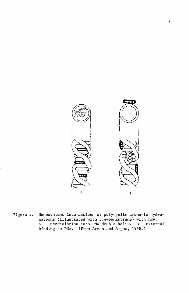
10
harvesting solutions were prepared by adding 05 ml of the combiotic to
every 100 ml of GKN Likewise cell maintenance solutions were preshy
pared by adding 05 ml of combiotic stock solution to every 100 ml of
growth medium
Fetal bovine serum which was mycoplasma tested and virus screened
was obtained from GIBCO and used throughout this study
Carbon dioxide gas prepared by mixing calcium carbonate and
sulfuric acid was used to maintain the desired pH of the medium
Glassware
Glassware was cleaned by soaking in an acid bath (17 mixture of
concentrated hydrochloric acid and concentrated sulfuric acid) overshy
night followed by washing with Alconox solution and repetitive rinsing
with tap water All glassware was rinsed with deionized distilled
water after being washed
Microscope coverslips (22 X 50 mm) were used to make cell growing
covers lips (22 X 8 mm) These were immersed in an Alconox solution and
rinsed with running tap water for 10-13 hours They were rinsed individshy
ually 10 times in three consecutive deionized distilled water baths and
one 100 ethanol bath then stored in an absolute ethanol bath until
use
Cell culture
Minced whole embryos of heterogenic white mice served as the source
of normal cells Pregnant mice (15-18 days in term) were sacrificed by
cervical dislocation Embryos were then removed aseptically from the
mouse and transferred to a petri dish containing GKN-combiotics mixture
Embryos were minced and placed into a trypsinizing flask The tissue
11
chunks were subjected to gentle stirring while being trypsinized at
room temperature for 30 minutes Trypsinized tissue was filtered
through four layers of cheese cloth The filtrate was washed with one
change of GKN and one change of complete growth medium Cell popushy
lations were adjusted with complete growth medium to a concentration
6of 3 X 10 cellsmI Three milliliter aliquots were planted in 25
2 cm polystyrene culture flasks and incubated at 37degC for one to three
days (primary cell culture) Primary cell cultures were treated with
trypsin-versene solution for two minutes washed centrifuged and
resuspended to give a concentration of 6 X 10⁵ cellsmI They were
inoculated in test tubes containing covers lips (7 X 22 mm) for 24 hours
at 37degC (secondary culture) prior to usage in the studies Secondary
cell cultures were treated with either a colloidal suspension of B(a)P
(10 ugml) and 3H- B(a)P (10 uCiml) or 3H- B(a)P (10 uCiml) alone
Autoradiography
Tritium-labeled B(a)P treated cells growing on the coverslips were
removed from the test tubes rinsed with two changes of GKN solution and
fixed in an absolute methanol bath for five minutes The covers lips
were mounted on a slide with the cell side up using Pro-texx mounting
medium The slides were dried overnight on a heating plate at 37degC
Ilford emulsion (1-4 Polysciences Inc) was melted in a 40degC
water bath Ten milliliters of melted emulsion were diluted with an
equal volume of deionized distilled water and any bubbles resulting in
the mixing operation were removed through a layer of cheese cloth The
slides were dipped into the diluted emulsion and immediately removed
Residual emulsion was cleaned from the back of the slides and the
12
slides held in a vertical position for 15 minutes to allow the emulsion
to dry Slides were sealed in a light tight slide box by wrapping the
box with aluminum foil The slides were exposed for 10 days at OdegC
The exposed slides were developed in Dekto1 (D-17) for two
minutes rinsed in a deionized distilled water stop bath and fixed in
acid fixer for three minutes After washing for 15 minutes in slowly
running tap water the slides were dried and stained for four minutes
in 005 basic fuchsin solution The slides were dehydrated in a
series of ethanol baths and immersed in a mixture of cedar wood oil
and absolute ethanol (1 1) for one hour followed by infiltration of
a Pro-texx mounting medium and xylene mixture (1 1) for one hour
The slides were examined for the appearance of silver grains
(black dots) located within the cells thereby indicating B(a)P uptake
by the cell
Cell viability test
The toxic effect of B(a)P on cultured cells was examined by adding
5 m1 of B(a)P at different concentrations Concentrations used ranged
from 01 to 50 ug B(a)P per milliliter of growth medium At regular
intervals B(a)P-growth medium mixtures were collected from culture
flasks and centrifuged for five minutes at 800 rpm The supernatant
was siphoned off and discarded Pellets were resuspended with 1 m1
growth medium and 01 m1 of erythrosin (04) was added and stained for
two minutes Dead cells staining pink were counted on a hemocytometer
Numbers of dead cells were plotted against the durations of B(a)P treatshy
ment
13
Inoculation of B(a)P treated cells into animals
Cells treated with B(a)P were harvested with a trypsin-versene
solution for three minutes Cell harvest was made at preselected inshy
tervals of time The cell population was adjusted to 10 X 106 ce11sm1
with complete growth medium From this solution 02 m1 were injected
into a group of five male mice Injections were made subcutaneously on
the left thigh (modified from Evans et a1 1957) Inoculation sites
were checked every 14 days for tumor development until termination of
the experiment
Histological study
Twenty-eight days after inoculation of B(a)P treated cells mice
which developed tumors were sacrificed for histological studies The
tumor tissue was excised and fixed overnight in Bouins fixative
Several solution changes of 70 ethanol saturated with lithium carbonate
were made to eliminate any fixative in the tissue Dehydration of fixed
tissue was accomplished in a series of ethanol baths of one hour each
The tissue was then immersed for one hour in a mixture of absolute
ethanol and xylene (1 1) It was soaked in a xylene bath overnight
and then transferred to a mixture of xylene and paraffin (56 0 tissuemat)
for 15 minutes Infiltration of hot tissuemat was carried out in four
changes of tissuemat solution Tissue was then embedded in a plastic
ice cube tray with hot tissuemat and cooled to room temperature The
blocks were sectioned on the microtome at a thickness of seven microns
The sections were stained by standard hematoxylin and eosin procedures
The slides were then carefully examined for histological abnormalities
14
Chromosome preparation
Cells in culture were mitotically blocked by lyophilized colcemid
006 ugml (GIBCO) for two hours Harvesting of cells was accomplished
by trypsinization and centrifuging for five minutes at 800 rpm The
pellet was resuspended in 1 ml of complete growth medium with a Pasteur
pipette Hypotonic solution (3 ml deionized distilled water) was added
to this suspension for seven minutes at room temperature One milliliter
of freshly prepared methanol-acetic acid (3 1) fixative was introduced
to the hypotonic solution for 10 minutes The suspension was centri shy
fuged and fixed by repeated dispersion at 10 minute intervals in three
changes of 31 methanol-acetic acid fixative During these fixation
periods microscope slides were washed in an Alconox solution and stored
in deionized distilled water at 4degC After the third fixation the
pellet was resuspended in 03-1 ml (depending on size of the pellet)
fixative solution Several drops of the final cell suspension were
applied to the slides The backs of the slides were wiped dry with
tissue paper A film of water on the upper surface of the slides was
left to facilitate spreading of the cells The slides were air-dried
and stained for 15 minutes in modified Giemsa stain Modification
was made according to Gude et al (1955) For the autoradiographic
study the slides were stained for 15 minutes after the completion of
photographic processes
RESULTS
Cell viability test
The toxic effect of B(a)P was tested for nine days Results are
summarized in Figure 3 From the data linear regression curves were
prepared for each concentration The slope of the untreated cell line
is 48 The slopes of the B(a)P treated cell lines are 65 for 01 ug
B(a)P treated cells 70 for 10 ug B(a)P treated cells and 76 for 50
ug B(a)P treated cells A statistical analysis did not indicate a
significant difference between the normal cell line and the B(a)P
treated cell lines Re-examination of the data used in Figure 3 will
help in understanding effect of treatment on cell viability for specific
periods of time In Figure 4 one and three day incubation periods
showed a smaller rate of increase (slope for one day 007 and slope
for three days 016) which implies that the B(a)P concentration did
not have a significant effect in terms of cell killing ability The dead
cell numbers in five day B(a)P treated cultures presented about 28-fold
increase from that of three day B(a)P treated cultures A statistical
analysis showed a significant difference indicating that the critical
factor in cell killing ability of B(a)P is dependent upon the exposure
period
Cell morphology in culture
Mouse embryo cells in culture were observed under the light microshy
scope for their morphology Embryonic cells were round immediately after
implantation into culture flasks Approximately three hours incubation
at 37degC after initial cell plantation some cells started to establish
their natural morphological appearance (elongation of cytoplasm) The
16
elongation process was an indication of cell attachment on the surface
of culture flasks Owens et a1 (1974) reported that in earlier days
of incubation two cell types epithelial cells and fibroblast cells
can be identified Epithelial cells from mono1ayered sheets which are
attached to each other (Figures 5 and 9) Vasi1iev and Ge1hand (1977)
reported that fibroblast cells form several morphological varieties in
culture mono1ayers of mutually oriented cells which are not firmly
attached to each other (Figures 6 and 8) multilayered sheets (Figure
7) or spherical aggregates (Figure 7 lower right and upper left
darkly stained aggregates) Fibroblasts andor fibroblast-like cells
became predominant throughout the entire cell population when cells were
cultured for over 30 days (Figures 10 11 and 12) These fibroblast
cells are characterized by elongated or stellate cytoplasmic structures
(Figures 10 11 and 12)
Mouse embryo cells were treated with 10 ug B(a)P for predesignated
exposure periods to observe an effect on cell morphology Some B(a)P
treated cells showed an abnormal spindle shape while the other cells
retained their normal morphological appearance (Figures 13 15 and 16)
In Figures 15 and 16 some cells were found to have vacuoles within their
cytoplasm Many of these also showed attenuated cytoplasms (Figure 16)
Autoradiography
Cultured cells were incubated with B(a)P for several different types
of treatment One treatment consisted of incubating cultured cells with
B(a)P at 10 ugm1 and 3H- B(a)P at 10 uCim1 until the termination of
experiment (full-term treatment) Other cells were partially exposed
to B(a)P at the same concentration and radioactivity level The exposure
17
60
-l S-r-
0 0 0 -l
gtlt
40-
Ul -l -l ltl) tJ
d Cll ltl) d
~
0 ltl)
20 l z
o o 5 10
Days
Figure 3 Toxic effect of B(a)P on cultured mouse embryo cells Untreated cells 0 Cells treated with 01 ug B(a)P e Cells treated with 10 ug B(a)P X Cells treated with 50 ug B(a) P
__
--------
18
60
-~ s
I
I ~ ~7 r 0 0 0 ~
x -
Ul ~ ~
eu 40u 0 ell eu 0
4-l 0
Ul H eu ~ l Ie z
20 3
9
_ ---41---------------- 1 o
o 20 40 60
B(a)P concentration (ugml)
Figure 4 Toxicity effect of B(a)P concentration The numbers 1 3 5 7 and 9 indicate the days of B(a)P treatment
Figure 5 Two day old normal mouse embryo cells in culture Epithelial cells in the central area of fibroshyblast cells surrounding the epithelial cells are shown here May-Grunwald and Giemsa stain (100 X)
Figure 6 Four day old normal mouse embryo cells in culture Large nucleated epithelial-like cell and fibroshyblast cells are shown here May-Grunwald and Giemsa stain (400 X)
Figure 7 Six day old normal mouse embryo cells in culture Multilayered fibroblast cells and two heavily stained aggregates are shown here May-Grunwald and Giemsa stain (100 X)
Figure 8 Eight day old normal mouse embryo cells in culture Fibroblast cells formed a monolayer sheet MayshyGrunwald and Giemsa stain (100 X)
Figure 9 Thirteen day old normal mouse embryo cells in culture Epithelial cells in the central and upper left areas are surrounded by well elongated fibroblast cells May-Grunwald and Giemsa stain (100 X)
Figure 10 Thirty day old normal mouse embryo cells in culture May-Grunwald and Giemsa stain (100 X)
Figure 11 Thirty five day old normal mouse embryo cells in culture Cell orientation is obscure and vacuoles are seen May-Grunwald and Giemsa stain (100 X)
Figure 12 Forty day old normal mouse embryo cells in culture Cell orientation is obscure and vacuoles are present May-Grunwald and Giemsa stain (100 X)
9Z
Figure 13 Mouse embryo cells incubated with 10 ug B(a)P for 1 day May-Grunwald stain (100 X)
Figure 14 Mouse embryo cells incubated with 10 ug B(a)P for 2 days May-Grunwald stain (100 X)
Figure 15 Mouse embryo cells treated with 10 ug B(a)P for 6 days May-Grunwald stain (100 X)
Figure 16 Mouse embryo cells treated with 10 ug B(a)P for 9 days Basic Fuchsin stain (100 X)
31
periods were 24 hours and four days
Figures 18 through 23 represent the full-term treatment and events
for the first 17 hours are summarized in Figure 17 After
hours exposure to 3H- B(a)P B(a)P accumulation in the nuclei
low Its cytoplasmic concentration was also minimal (Figure
3H-B(a)P uptake by the cells increased rather sharply after 35
of incubation (Figures 19 20 and 21) Long term incubation (six
and 20 days) showed a concentration of B(a)P in the cytoplasm while no
concentration of B(a)P was observed (Figures 22 and 23)
In other treatments cultured cells were partially exposed to
B(a)P at the same concentration Radioactivity levels were as in the
treatment The cells were exposed to B(a)P for one and four
days At the end of the exposure period the complete growth medium
was replaced for further incubation Figures 24 through 27 indicate
some of the results of 24 hour treated cells Overall results are
summarized in Figure 24 Nuclear accumulation of 3H-B(a)P was maximum
at 24 hours and decreased drastically when fresh complete growth medium
was introduced and incubation was continued (Figures 24 27 and 28)
The nuclear accumulation of B(a)P exceeded that of cytoplasm only at
24 hours incubation (Figure 24) Cytoplasmic accumulation of 3H-B(a)P
was at its highest peak on the 6th day of incubation and followed the
same pattern of decrease as the nuclear accumulation curve Extrashy
polation of the two curves suggests disappearance of B(a)P from the
cells occurs between the 24th and 26th day Some cells as exemplified
in Figure 28 lost nuclear B(a)P and most of the cytoplasmic B(a)P at
50
100
32
00
0 1 bull
O Hours
Figure 17 Time-course of 3H- B(a)P accumulation in the cells Cells in autoradiograms were counted for nuclear accumulation () cytoplasmic accumulation (0) and no accumulation (X)
I I Ii I Ii(I I ~ I I
2 4 6 8 10 12 14 16 18
33
21 days of incubation in the growth medium
Figures 29 through 32 represent the results of four day treated
Four (no additional incubation in growth medium) and 11 days
(additional six day incubation in growth medium) cells show nuclear and
cytoplasmic accumulation of B(a)P but not as intense as seen in the one
cells (Figures 29 and 30) Fifteen day incubated cells had
some B(a)P molecules in the cytoplasm The cells incubated for 27 days
of B(a)P in any part of the cells (Figure 32)
Figures 33 through 35 are the chromosome spreads from 3H- B(a)p
treated cells Silver grains (black dots) were seen on or near the chromoshy
somes in three autoradiograms (Figures 33 34 and 35) Autoradiograms of
chromosomes prepared from one two and three day treated cells with
3H- B(a)P (10 uCiml) were counted for use in labeling analysis Chroshy
mosome spreads from one and two day treated cells produced 936 and 920
labeled chromosome spreads respectively Total chromosome spreads examshy
ined were 94 and 100 Cells treated for three days gave 304 labeled
chromosome spreads
Figures 34 and 35 showed chromosome breakages
Induction of abnormalities and its histology
B(a)P treated cells were injected into adult mice The mouse in
Figure 36 was a normal male mouse The mouse in Figure 37 exemplifies
the abnormal genital inflammation characterized by many of the mice inshy
jected with the B(a)P treated cells From the 26 injected mice 31 deshy
veloped a genital inflammation In contrast only 38 of the control
mice exhibited genital abnormalities The controls consisted of three
groups of mice One group of 32 mice received no injections Another
Autoradiogram Mouse embryo cells in culture were incubated for 15 hours with 10 ug B(a)P(lO uCi) Only a few radioactive molecules are present Hematoxylin and eosin stain (100 X)
Figure 19 Autoradiogram Mouse embryo cells in culture were incubated for 35 hours with 10 ug B(a)P(lO uCi) Nuclear and cytoplasmic accumulation of B(a)P is seen Hematoxylin and eosin stain (100 X)
Figure 20 Autoradiogram Mouse embryo cells in culture were incubated for 17 hours with 10 ug B(a)P(10 uCi) Basic Fuchsin stain (1000 X)
Figure 21 Autoradiogram Mouse embryo cells in culture were incubated for 24 hours with 10 ug B(a)P(10 uCi) Basic Fuchsin stain (1000 X)
Figure 22 Autoradiogram Mouse embryo cells in culture were incubated for six days with 10 ug B(a)P(10 uCi) Hematoxylin and eosin stain (100 X)
Figure 23 Autoradiogram Mouse embryo cells in culture were incubated for 20 days with 10 ug B(a)P(10 uCi) A heavy accumulation of B(a)P in cytoplasm is seen here Hematoxylin and eosin stain (400 X)
-
6pound
40
00
100
~ -
00 c
-l M Q)
~ 50M
~
ro - lQI
z M
10 20 Time in days
Figure 24 3ime-course of 3H- B(a)P accumulation in the cells H-B(a)P was replaced with normal growth medium
after 24 hours incubation Cells in autoradiograms were counted for nuclear accumulation (e) cytoshyplasmic accumulation (0) and no accumulation (X)
00
Figure 25 Autoradiogram Mouse embryo cells in culture were incubated for one day with 10 ug B(a)P(lO uCi) Nuclear and cytoplasmic accumulation of B(a)P is seen Basic Fuchsin stain (1000 X)
Figure 26 Autoradiogram Mouse embryo cells in culture were incubated for one day with 10 ug B(a)P(lO uCi) The B(a)P was then removed The cells were incubated an additional 3 days in complete growth medium Basic Fuchsin stain (1000 X)
Figure 27 Autoradiogram Mouse embryo cells in culture were incubated for one day with 10 ug B(a)P(lO uCi) The B(a)P was then removed The cells were incubated an additional 11 days in complete growth medium Basic Fuchsin stain (1000 X)
Figure 28 Autoradiogram Mouse embryo cells in culture were incubated for one day with 10 ug B(a)P(lO uCi) The B(a)P mixture was then removed The cells were incubated an additional 21 days in complete growth medium Basic Fuchsin stain (1000 X)
shy
Figure 29 Autoradiogram Mouse embryo cells in culture were incubated for four days with 10 ug B(a)P(lO uCi) Negri body stain (1000 X)
Figure 30 Autoradiogram Mouse embryo cells in culture were incubated for four days with 10 ug B(a)P(lO uCi) The B(a)P mixture was then removed The cells were incubated an additional six days in complete growth medium Some silver grains are seen Basic Fuchsin stain (1000 X)
97
Figure 31 Autoradiogram Mouse embryo cells in culture were incubated for four days with 10 ug B(a)P(lO uCi) The B(a)P was then removed The cells were incubated an additional 11 days in complete growth medium Some silver grains were seen in the cytoplasm Basic Fuchsin stain (1000 X)
Figure 32 Autoradiogram Mouse embryo cells in culture were incubated for four days with 10 ug B(a)P(lO uei) The B(a)P mixture was then removed The cells were incubated an additional 23 days in complete growth medium Basic Fuchsin stain (1000 X)
81]
Figure 33 Autoradiogram of chromosomes Mouse embryo cells in culture were incubated for one day with 005 ug B(a)P(lO uCi) Giemsa stain (1000 X)
Figure 34 Autoradiogram of chromosomes Mouse embryo cells in culture were incubated for two days with 005 ug B(a)P(lO uCi) Giemsa stain (1000 X)
-
os
Figure 35 Autoradiogram of chromosomes Mouse embryo cells in culture were incubated for three days with 005 ug B(a)P(10 uCi) Giemsa stain (1000 X)
Figure 36 Normal male mouse
53
group of 10 mice were injected with normal untreated cells The last
group of 10 mice was inoculated with growth medium that was used to
maintain the cells (Table 2) The effect of B(a)P treated cells in
the mice is summarized on Table 2 The mice inoculated with the cells
of four days B(a)P preincubation resulted in a 25-50 incidence of
abnormal development Of these abnormal mice two mice developed a
small nodule (ca 4-5 rom in diameter) on the peritoneum (Figure 38)
This nodule was processed for histological study Microscopic examishy
nation revealed that the nodule was the undescended testis and showed
hyperplasia of Leydig cells (Figure 43) Atrophy of seminiferous
tubules (Figure 39) is clearly recognized when they are compared with
those of normal tissue (Figures 42 and 43) Seminiferous tubules
from abnormal tissue did not show spermatogenesis (Figure 43) The
ductus epididymis (Figure 41) however showed normal morphological
appearance but clumps of spermatozoa were not seen in the ductuli
Hyperplasia of connective tissue surrounding ductus epididymis was
observed as well The mice inoculated with 10 day B(a)P treated cells
had a higher incidence (80) of abnormal development Long term
observation (146 days) did not reveal the development of any abnorshy
mality although the inoculated cells were treated for one day with
B(a)P
Tab
le
2
Eff
ect
of
B(a
)P tr
eate
d cell
s in
ad
ult
mic
e
Day
s o
f D
ays
of
Num
ber
of
cell
s D
ays
in
Day
s aft
er
incu
bati
on
w
ith
B(a
)P
incu
bat
ion
aft
er
B(a
)P
ino
cu
late
d
(x
106
) cu
ltu
re
ino
cu
lati
on
A
bn
orm
alit
ies
rem
oved
4 0
10
47
62
24
4 4a
10
43
68
14
4 6
10
41
70
14
10
0 10
43
70
4
5
1 0
1 6
146
05
1 0
10
6 14
6 0
4
Co
ntr
ols
U
ntr
eate
d M
ouse
-shy
-shy
--shy
13
2
Nor
mal
cell
s 10
1
1
146
11
0
Gro
wth
med
ium
o
nly
(f
rom
a)
4 70
0
10
In
+shy
Figure 37 Mouse with genital abnormality
Figure 38 Mouse with a nodule on peritoneum
9~
Figure 40 Transverse section of ductus epididymis from normal mouse testis Hematoxylin and eosin stain (100 X)
Figure 41 Transverse section of ductus epididymis from undescended testis Clumps of spermatozoa are not seen in the ductu1i Spaces among the ductu1i are abnormally large Hematoxylin and eosin stain (100 X)
6S
Figure 42 Transverse section of seminiferous tubules from normal mouse testis Seminiferous tubules are separated by a slight amount of interstitial connective tissue containing some Leydig cells Hematoxylin and eosin stain (100 X)
Figure 43 Transverse sections of seminiferous tubules from undescended testis Atrophy of seminiferous tubules is seen Hyperplasia of Leydig cells is recognized Hematoxylin and eosin stain (100 X)
19
DISCUSSION
Cook et a1 (1932 1933) isolated carcinogenic hydrocarbons from
coal tar pitch The melting point (mp) of one of the hydrocarbons
was determined to be 1755-1765degC This particular hydrocarbon species
was shown to be pure benzo(a)pyrene (B(a)P) by comparison with the
synthesized B(a)P B(a)P has alternative names such as 12-benzpyrene
and 34-benzpyrene It has the molecular weight of 25230 its
structure is shown in diagram on page 3
B(a)P and other closely related carcinogenic hydrocarbons (see
diagram on page 3) are being used to determine a mechanism of carshy
cinogenesis in several research systems B(a)P is known to be one of
the chemically inert carcinogens To be activated B(a)P must first be
taken up by the cells Thus invasion into the cell is the first step
in the mechanism B(a)P particles in the growth medium are taken into
the cells by endocytosis (Allison and Ma11ucci 1964) Engulfed B(a)P
particles then require enzymatic activations to acquire cytotoxic
mutagenic (Huberman et a1 1971 1976 Newbold and Brooks 1976) and
carcinogenic abilities in the cells (Wood et a1 1976 Wis10cki et a1
1976)
The greatly increased number of dead cells in cultures which had
been treated with B(a)P for five days suggested that enzymatic activations
of ingested B(a)P might have occurred during a one to three day incubation
period As a result B(a)P acquired cytotoxic potency to the cells
(Figure 4) Figures 17 and 24 indicate that the cytoplasmic accumushy
lation had maximum values between 35 hours and six days This sugshy
gested the possibility that B(a)P metabolism takes place between these
periods A reliable explanation for the cytotoxic ability of B(a)P was
63
made by several workers (Nebert and Gelboin 1968a 1968b Kasper
1971 Khandwala and Kasper 1973) There exists an enzyme aryl
hydrocarbon hydroxylase that functions in the metabolism of B(a)P
The metabolite is 3-hydroxybenzopyrene which also possesses cytotoxic
potency to the cell Benzo(a)pyrene-hydroxylase was shown to be preshy
sent at low concentrations in the nuclear membrane The existence of
inducible B(a)P-hydroxylase in a microsomal preparation was reported
as well The major difference between nuclear and microsomal memshy
branes is its specificity Nuclear membranes produce hydrocarbon
hydroxylase specifically by the stimulation of hydrocarbon carcinogens
while microsomal membranes produce hydrocarbon hydroxylase in response
to the addition of hydrocarbon carcinogens or phenobarbital
In addition to enzymatic activation in the cytoplasm B(a)P
molecules may be accumulated in one or some of the cellular organelles
To determine the ultimate B(a)P accumulation site in the cell it is
important to define the target organelle(s) for hydrocarbon carcinoshy
gens Several techniques were developed for this purpose and an autoshy
radiography technique was the one utilized in this study Autoradioshy
graphy techniques in the biomedical field are a powerful tool for
localization and visualization of desired chemicals in the cell or the
tissues Autoradiograms obtained in this study indicated that the
cytoplasmic accumulation of B(a)P occurred as early as 15 hours after
the initial application and lasted about 14 days (Figure 17 and 24)
The silver grains seen in Figures 19 20 and 27 indicate that B(a)P
molecules may be encapsulated as a phagosome perhaps independently
from lysosomes or fused to lysosomes where enzymatic alterations as
64
well as detoxification reactions take place Along with the cytoplasmic
accumulation a maximum nuclear accumulation (52 labeling) was observed
in one day (Figure 24) Although nuclear accumulation of B(a)P was
shown it does not clearly indicate the incorporation of B(a)P into
genetic material (DNA) To answer this question chromosome spreads
combined with autoradiography were prepared (Figures 33 34 and 35)
These figures indicate that an interaction of B(a)P and chromosomes
might have occurred in the nuclei This result is supported by the work
of Brooks and Lawley (1964) who isolated DNA and RNA from mouse skin to
which radioactive carcinogenic hydrocarbons were previously applied
One of their results is shown in Figure 44 which demonstrates that the
maximum binding of B(a)P to proteins occurred about 24 hours after
application of B(a)P This result suggests the possibility that B(a)P
molecules bind to DNA
Figures 34 and 35 demonstrated chromosome breakages A generally
accepted explanation for chromosome aberrations is that a direct
chemical or physical attack on DNA has occurred However Allison and
Patton (1965) proposed another possible mechanism which could cause
chromosome breakages The idea was that damage to 1ysosomes can cause
chromosomal damage It was demonstrated by De Duve and his colleagues
that 1ysosomes contain hydrolytic enzymes acid phosphatase acid DNase
acid RNase and acid protease (reviewed by Allison and Ma11ucci 1964)
Among these lysosomal enzymes DNase can break the DNA which is the
backbone of the uncoiled interphase chromatid Deoxyribonuclease must
therefore enter into the nucleus for chromosome breakages to occur
Widne11 and Tata (1964) reported a result which supports DNase migration
0
I
I
I
02
r-
U l
I
r- (]J CIl o
d shyI r- (]J
jJ o ~
01
~ o ~
jJ
r- gt r-
jJ U co U
r- ~
r- U
(]J
0shy
Cfl
o 5
0
150
25
0
Fig
ure
4
4
T
ime-
cou
rse
of
the
bin
din
g
of
(3H
) la
bell
ed
hy
dro
carb
on
s to
to
tal
pro
tein
s o
f m
ouse
sk
in
Resu
lts
are
ex
pre
ssed
as
specif
ic ra
dio
shyacti
vit
y
of
pro
tein
(u
Ci
g)
div
ided
by
th
e d
ose
(u
Ci
mo
use
) g
iven
at
zero
ti
me
plo
tted
ag
ain
st
tim
e
34
-ben
zp
yre
ne
(Part
iall
y
rep
rod
uce
d
from
B
roo
kes
an
d L
awle
y
19
64
)
V1
66
thus DNA-dependent RNA polymerase activity in isolated nuclei is
blocked by addition of DNase Newton et al (1962) also reported that
DNA breakdown occurred shortly after infection with herpes virus This
report implies that lysosomal damage by herpes virus induced a libershy
ation of DNase which in turn caused DNA breakage
The studies conducted so far do not give any indication concerning
mechanisms by which B(a)P binds to the nucleic acids Many investi shy
gators worked to clarify the steps involved as B(a)P migrates from the
cytoplasm to the nucleus B(a)P is enzymatically converted to at least
four phenols three dihydrodiols and three quinones and epoxides
(reviewed by Yung et al 1976) The active B(a)P metabolites which are
converted by microsomal hydroxylase within the cells possess a relatively
high tendency to react with the nucleophilic sites of the macromolecules
Thus the B(a)P metabolite has a rare chance of reacting with nuclear
DNA (Alexandrov and Thompson 1977) This report implies that further
metabolic activation of B(a)P metabolites by the other enzymes of microshy
somes or the nuclear envelope becomes necessary (Rogan et al 1976)
A similar interpretation of a two-step activation mechanism was reported
by several workers (Sims et al 1974 Weinstein et al 1976 Spelsberg
et al 1977) Diol-epoxide intermediates (7S-dihydro-7S-dihydroshy
xybenz(a)pyrene and 7S-dihydro-7S-dihydroxybenzo(a)pyrene-9lO-oxide)
were reported to be the metabolites responsible for DNA-hydrocarbon
binding (Brooks et al 1975 Sims et al 1974) Brooks et al (1975)
also indicated that the purine moieties of DNA react with diol-epoxide
intermediates A specific isomer of a diol-epoxide derivative of B(a)P
(plusmn)-7B Sa-dihydroxy-9a lOa-7S9lO-tetra-hydrobenzo(a)pyrene also
67
appeared to be a B(a)P metabolite in terms of covalent binding to
nucleic acids (Weinstein et a1 1976) Another isomeric form of a
dio1 epoxide derivative of B(a)P 7a 8B-dihydroxy-9B10B-epoxy-789
10-tetrahydrobenzo(a)pyrene was proved to be a highly mutagenic intershy
mediate of B(a)P metabolism (Wood et a1 1976 Huberman et a1 1976)
This metabolite is suspected to be the ultimate carcinogenic species
in B(a)P carcinogenesis The reactions mentioned above are the events
occurring as B(a)P molecules move into the nucleus of the cell It is
important to s~e the relationship between the duration of B(a)P treatshy
ment and cell transformation To determine the tumorigenicity of B(a)P
in the cells in vitro suspected cells must be injected into a host
animal to observe the behavior of the cells (Fedoroff 1967) In this
experiment several conditions for B(a)P treatments were designed and
inoculation of mice with B(a)P treated cells was carried out The
results are summarized in Table 3 Two mice developed palpable tumors
(Figure 38) while the remainder showed genital abnormality (Figure 37)
The mice inoculated with four day B(a)P treated cells developed 25-50
abnormality development Some four day B(a)P treated cells were incushy
bated additional days in a normal growth medium before inoculation
Abnormal development was not affected significantly by the additional
incubation period This is because during a four day incubation period
most of the cells may be transformed This idea can be supported by
the work of Berwald and Sachs (1965) They reported that two days of
incubation with B(a)P is necessary for cell transformation Ten day
B(a)P incubation created an 80 rate of abnormal development Higher
genital development in 10 day B(a)P treated cells was possibly due to
68
the increased probability of cell transformation Berwald and Sachs
(1965) reported that the normal mouse cells in vitro showed spontaneous
transformation sometime after 45 to 80 days in the culture As can be
seen on Table 3 the cells used for the inoculations were cultured from
41 to 47 days which is in the range of Berwalds figures Thus this
spontaneous transformation factor may be involved in the results shown
on Table 3 The mice inoculated with one day B(a)P treated cells
developed no abnormalities (Table 3) One reason for the absence of
abnormal development may be related to the age of the cells in culture
the cells used were cultured for six days while the abnorma1ityshy
producing cells were cultured from 41 to 47 days
A tumor development in host mice by the inoculation of suspected
cells must be further examined by histological means Figures 40 through
43 are transverse sections of the ductus epididymis and the seminiferous
tubules from abnormal and normal mouse testis Figures 40 and 42 from
the control mouse show a normal histological appearance The ductu1i
are well packed and contain aggregates of spermatozoa In contrast
Figures 41 and 43 show atrophy of ducts of the epididymis resulting in
the formation of large interstitial spaces Closer observations revealed
a deterioration of the steroci1ia as well Figures 40 and 42 show the
normal condition in which seminiferous tubules contain newly formed
spermatozoa primary spermatocytes and secondary spermatocytes Figures
41 and 43 show atrophy of seminiferous tubules with a few supporting
cells (Serto1i) located near the basement membrane An abnormal increase
in the number of interstitial Leydig cells was also seen Occasionally
binuc1eated Leydig cells were observed Detailed observation of the
excised nodule revealed it to be histologically abnormal It is however
69
difficult to determine if the abnormal nodule was caused by the direct
action of B(a)P treated cells
Allison and Mallucci (1964) reported that autoradiographic
techniques failed to visualize accumulation of chemical carcinogens in
the nucleus Through this study it was demonstrated that autoradioshy
graphic techniques could show the existence of B(a)P in the nucleus
Furthermore autoradiograms of chromosomes gave an indication that
interaction between B(a)P and chromosomes may have occurred
SUMMARY
1) Benzo(a)pyrene accumulation in the cell occurred after 24 hours
of incubation period
2) Autoradiograms of chromosomes prepared from benzo(a)pyrene
treated cells indicated that benzo(a)pyrene reacted with
chromosomes
3) Autoradiographic techniques were shown to have the capacity to
localize the sites of benzo(a)pyrene accumulation in the cell
4) Cytotoxicity studies of benzo(a)pyrene showed a time-dependent
effect on the cultured mouse embryo cells
5) A tumor induction study indicated that tumor inducibility in the
host animal seems to be affected by the length of cell culture
period
LITERATURE CITED
Allison A C and L Ma11ucci 1964 Uptake of hydrocarbon carcinogens by 1ysosomes Nature 2031024-1027
Allison A C and G R Paton 1965 Chromosome damage in human diploid cells following activation of lysosomal enzymes Nature 2091170-1173
Allison A C and J T Dingle 1966 Role of 1ysosomes in adrenal necrosis caused by dimethy1benzanthracene Nature 209303-304
Allison A C 1968 Lysosomes in relation to cancer induction and treatment Europ J Cancer 3481-490
Arcos C J and F M Argus 1968 Molecular geometry and carcinoshygenic activity pp 348-435 Advances in Cancer Research Vol 11 Academic Press Inc
A1exandrov K and H M Thompson 1977 Influence of inducers and inhibitors of mixed-function oxidase on benzo(a)pyrene binding to the DNA of rat nuclei Cancer Res 371443-1449
Bailey J E and N Dunga1 1958 Polycyclic hydrocarbons in icelandic smoked food Brit J Cancer 12348-350
Berwald Y and L Sachs 1965 In vitro transformation of normal cells to tumor cells by carcinogenic hydrocarbons J Nat1 Cancer Inst 35(4)641-661
Boyland E and B Green 1964 On the reported sedimentation of polycyclic hydrocarbons from aqueous solutions of DNA J Mol Bio1 9589-597
Boyland E and P Sims 1965 Induction of adrenal damage and cancer with metabolites of 712-dimethy1benz(a)anthracene Nature 207816-817
Brookes P and P D Lawley 1964 Evidence for the binding of polynuclear aromatic hydrocarbons to the nucleic acids of mouse skin Relation between carcinogenic power of hydrocarbons and their binding to deoxyribonucleic acid Nature 202781-784
Brookes P P Jones and J Amos 1975 The nature of the deoxyribonuc1eosides involved in the binding of carcinogenic hydrocarbons to the DNA of mouse embryo cells Int J Cancer 15912-917
Cook J W C Hewett and L Hieger 1932 Coal tar constituents and cancer Nature 130926
73
Cook J W 1933 The isolation of a cancer producing hydrocarbon from coal tar Chern Society J 398395-405
Dipple A 1976 Chemical Carcinogens ACS Monograph pp 245-314
Evans V J N M Hawkins B B Westfall and W R Earle 1957 Studies on culture lines derived from mouse liver parenchymatous cells grown in long term tissue culture Cancer Res 18261-273
Evans I A and J Mason 1965 Carcinogenic activity of bracken Nature 208913-914
Fa1k H L A Miller and P Kotin 1958 Elution of 34-benzpyrene and related hydrocarbons from soots by plasma proteins Science 127474-475
Fedoroff S 1967 Proposed usage of animal tissue culture terms J Nat1 Cancer Inst 38(4)607-611
Gude W D A C Upton and T T Odell 1955 Giemsa staining of autoradiograrns prepared with stripping film Stain Tech 30161-162
Hecht S S R M Ornaf and D Hoffman 1974 Chemical studies on tobacco smoke XXXIII N-nitrosonornicotine in tobacco Analysis of possible contributing factors and biologic implishycations J Nat1 Cancer Inst 54(5)1234-1244
Henderson E B J R Gordon H Menck J Soohoo P S Martin and C M Pike 1975 Original contributions Lung cancer and air pollution in South central Los Angeles county American J of Epidemiology 101(6)477-488
Hirono I C Shibuya K Fushimi and M Raga 1970 Studies on carcinogenic properties of bracken Pteridium aqui1iurn J Nat1 Cancer Inst 45(1)179-188
Hirono I C Shibuya M Shimizu and K Fushimi 1972 Carcinogenic activity of processed bracken used as human food J Nat1 Cancer Inst 48(4)1245-1249
Hirono I K Fushimi H Mori T Miwa and M Haga 1973 Comparative study of carcinogenic activity in each part of bracken J Natl Cancer Inst 50(5)1367-1371
Haberman E L Aspiras C Heidelberger P L Grover and P Sims 1971 Mutagenicity to mammalian cells of epoxides and other derivatives of polycyclic hydrocarbons Nat1 Acad Sci USA Proceeding 68(12)3195-3199
Kasper C B 1971 Biochemical distinctions between the nuclear and microsomal membranes from rat hepatocytes J Bio1 Chern 246(3)577-581
74
Khandwa1a A S and C B Kasper 1973 Preferential induction of aryl hydroxylase activity in rat liver nuclear envelope by 3-methy1cho1anthrene Biochem Biophys Res Commun 54(4)1241-1246
Kuratsune M and W C Hueper 1960 Polycyclic hydrocarbons in roasted coffee J Nat1 Cancer Inst 24(2)463-469
Kraybi11 H F 1977 Advances in Modern Toxicology Vol 3 pp 27shy29 Hemisphere Publishing Co Washington D C
Lerman L S 1960 Structural considerations in the interaction of DNA and acridines J Mol Bio1 318-30
Lijinsky W and P Shubik 1964 Benzo(a)pyrene and other polynuclear hydrocarbons in charcoal-broiled meat Science 14553-55
1965 Polynuclear hydrocarbon carcinogens in cooked meat and smoked food Indus Med Surge 34152-154
Liquori A M 1962 Interaction between DNA and polycyclic aromatic hydrocarbons J Mol Bio1 5521-526
Masuda Y and M Kuratsune 1971 Polycyclic aromatic hydrocarbons in smoked fish Katsuobushi Gann 6227-30
Nebert D W and H V Ge1boin 1968a Substrate-inducible microsomal aryl hydroxylase in mammalian cell culture I Assay and properties of induced enzyme J Bio1 Chern 243(23)6242-6249
1968b Substrate-inducible microsomal aryl hydroxylase in mammalian cell culture II Cellular responses during enzyme induction J Bio1 Chern 243(23)6250-6251
Newton A P P Dendy C L Smith and P Wi1dy 1962 A pool size problem associated with the use of tritiated thymidine Nature 194886-887
Newbold R F and P Brooks 1976 Exceptional mutagenicity of a benzo(a)pyrene dio1 epoxide in cultured mammalian cells Nature 26152-54
Owens R B H S Smith and A J Hackett 1974 Epithelial cell cultures from normal glandular tissue of mice J NatL Cancer Inst 53(1)261-269
Owens R B 1974 Brief communication Glandular epithelial cells from mice A method for selective cultivation J Nat1 Cancer Inst 52(4)1375-1378
75
Pamukcu A M and M J Price 1969 Induction of intestinal and urinary bladder cancer in rats by feeding bracken fern (Pteris aquilina) J Natl Cancer Inst 43(1)275-281
Rogan~ E G~ P Mailander and E Cavalieri 1976 Metabolic activation of aromatic hydrocarbons in purified rat liver nuclei Induction of enzyme activities and binding to DNA with and without monooxygenase-catalyzed formation of active oxygen Natl Acad Sci USA Proceeding 73(2)457-461
Selkirk J K E Huberman and C Heidelberger 1971 An epoxide is an intermediate in the microsomal metabolism of the chemical carcinogen dibenz(ah)anthracene Biochem Biophys Res Commun 43(5)1010-1016
Shires T K and K M Richter 1966 The distribution of 34shybenz(a)pyrene in replicating mammalian cells in vitro Exp Cell Res 44617-620 -------shy
Sims P P L Grover~ A Swaisland K Pal and A Hewer 1974 Metabolic activation of benzo(a)pyrene proceeds by a diol-epoxide Nature 252326-328
Spelsberg T C T H Zytkovicz and H L Moses 1977 Effects of metabolism on the binding of polycyclic hydrocarbons to nuclear subfractions of cultured AKR mouse embryo cells Cancer Res 371490-1496
Thorsteinsson T and G Thordarson 1968 Polycyclic hydrocarbons in singed food in ice land Cancer 21390-392
Van Cantfort J and J Gielen 1975 Organ specificity of aryl hydrocarbon hydroxylase induction by cigarette smoke in rats and mice Biochem Pharmacol 241253-1256
Van Duuren~ B L B M Goldschmidt and H H Seltzman 1969 The interaction of mutagenic and carcinogenic agents with nucleic acids Ann N Y Acad Sci 153744-757
Vasiliev~ J M and I M Gelhand 1977 Mechanisms of Morphogenesis in cell cultures International Review of Cytology Vol 50 pp 159-163 Academic Press~ Inc
Wang~ I Y~ R E Rasmussen and T T Crocker 1972 Isolation and characterization of an active DNA-binding of benzo(a)pyrene from hamster liver microsomal incubation system Biochem Biophys Res Commun 49(4)1142-1149
Weinstein~ B 1 A M Jeffery K W Jennette~ S H Blobstein R G Harvey~ C Harris H Autrup~ H Kasai and K Nakanishi 1976 Benzo(a)pyrene diol epoxides as intermediates in nucleic acid binding in vitro and in vivo Science 193592-595
76
Weisburger E K 1977 Cancer-causing chemicals Chemistry 50(1) 42-48
Widnell C C and J R Tata 1964 Evidence for two DNA-dependent RNA polymerase activities in isolated rat-liner nuclei Biochem J 92313-315
Wislocki P G A W Wood R L Chang W Levin H Yagi O Hernandez P M Dansette D M Jerina and A H Conney 1976 Mutagenicity and cytotoxicity of benzo(a)pyrene arene oxides phenols quinones and dihydrodiols in bacterial and mammalian cells Cancer Res 363350-3357
Wood A W P G Wislocki R L Chang W Levin Y Anthony H Lu H Yagi O Hernandez D M Jerina and A H Conney 1976 Mutagenicity and cytotoxicity of benzo(a)pyrene benzo-ring epoxides Cancer Res 36(9)3358-3366
Yamagiwa K and K Ichikawa 1918 Experimental study of the pathoshygenesis of carcinoma J Cancer Res 3(1)1-29
Yung S K D W McCourt P P Roller and H V Gelboin 1976 Enzymatic conversion of benzo(a)pyrene leading predominantly to the diol-epoxide r-7t-8-dihydroxy-t-910-oxy-789lOshytetrahydrobenzo(a)pyrene through a single enantiomer of r-7 t-8-dihydroxy-78-dihydro-benzo(a)pyrene Natl Acad Sci USA Proceeding 73(8)2594-2598
11
chunks were subjected to gentle stirring while being trypsinized at
room temperature for 30 minutes Trypsinized tissue was filtered
through four layers of cheese cloth The filtrate was washed with one
change of GKN and one change of complete growth medium Cell popushy
lations were adjusted with complete growth medium to a concentration
6of 3 X 10 cellsmI Three milliliter aliquots were planted in 25
2 cm polystyrene culture flasks and incubated at 37degC for one to three
days (primary cell culture) Primary cell cultures were treated with
trypsin-versene solution for two minutes washed centrifuged and
resuspended to give a concentration of 6 X 10⁵ cellsmI They were
inoculated in test tubes containing covers lips (7 X 22 mm) for 24 hours
at 37degC (secondary culture) prior to usage in the studies Secondary
cell cultures were treated with either a colloidal suspension of B(a)P
(10 ugml) and 3H- B(a)P (10 uCiml) or 3H- B(a)P (10 uCiml) alone
Autoradiography
Tritium-labeled B(a)P treated cells growing on the coverslips were
removed from the test tubes rinsed with two changes of GKN solution and
fixed in an absolute methanol bath for five minutes The covers lips
were mounted on a slide with the cell side up using Pro-texx mounting
medium The slides were dried overnight on a heating plate at 37degC
Ilford emulsion (1-4 Polysciences Inc) was melted in a 40degC
water bath Ten milliliters of melted emulsion were diluted with an
equal volume of deionized distilled water and any bubbles resulting in
the mixing operation were removed through a layer of cheese cloth The
slides were dipped into the diluted emulsion and immediately removed
Residual emulsion was cleaned from the back of the slides and the
12
slides held in a vertical position for 15 minutes to allow the emulsion
to dry Slides were sealed in a light tight slide box by wrapping the
box with aluminum foil The slides were exposed for 10 days at OdegC
The exposed slides were developed in Dekto1 (D-17) for two
minutes rinsed in a deionized distilled water stop bath and fixed in
acid fixer for three minutes After washing for 15 minutes in slowly
running tap water the slides were dried and stained for four minutes
in 005 basic fuchsin solution The slides were dehydrated in a
series of ethanol baths and immersed in a mixture of cedar wood oil
and absolute ethanol (1 1) for one hour followed by infiltration of
a Pro-texx mounting medium and xylene mixture (1 1) for one hour
The slides were examined for the appearance of silver grains
(black dots) located within the cells thereby indicating B(a)P uptake
by the cell
Cell viability test
The toxic effect of B(a)P on cultured cells was examined by adding
5 m1 of B(a)P at different concentrations Concentrations used ranged
from 01 to 50 ug B(a)P per milliliter of growth medium At regular
intervals B(a)P-growth medium mixtures were collected from culture
flasks and centrifuged for five minutes at 800 rpm The supernatant
was siphoned off and discarded Pellets were resuspended with 1 m1
growth medium and 01 m1 of erythrosin (04) was added and stained for
two minutes Dead cells staining pink were counted on a hemocytometer
Numbers of dead cells were plotted against the durations of B(a)P treatshy
ment
13
Inoculation of B(a)P treated cells into animals
Cells treated with B(a)P were harvested with a trypsin-versene
solution for three minutes Cell harvest was made at preselected inshy
tervals of time The cell population was adjusted to 10 X 106 ce11sm1
with complete growth medium From this solution 02 m1 were injected
into a group of five male mice Injections were made subcutaneously on
the left thigh (modified from Evans et a1 1957) Inoculation sites
were checked every 14 days for tumor development until termination of
the experiment
Histological study
Twenty-eight days after inoculation of B(a)P treated cells mice
which developed tumors were sacrificed for histological studies The
tumor tissue was excised and fixed overnight in Bouins fixative
Several solution changes of 70 ethanol saturated with lithium carbonate
were made to eliminate any fixative in the tissue Dehydration of fixed
tissue was accomplished in a series of ethanol baths of one hour each
The tissue was then immersed for one hour in a mixture of absolute
ethanol and xylene (1 1) It was soaked in a xylene bath overnight
and then transferred to a mixture of xylene and paraffin (56 0 tissuemat)
for 15 minutes Infiltration of hot tissuemat was carried out in four
changes of tissuemat solution Tissue was then embedded in a plastic
ice cube tray with hot tissuemat and cooled to room temperature The
blocks were sectioned on the microtome at a thickness of seven microns
The sections were stained by standard hematoxylin and eosin procedures
The slides were then carefully examined for histological abnormalities
14
Chromosome preparation
Cells in culture were mitotically blocked by lyophilized colcemid
006 ugml (GIBCO) for two hours Harvesting of cells was accomplished
by trypsinization and centrifuging for five minutes at 800 rpm The
pellet was resuspended in 1 ml of complete growth medium with a Pasteur
pipette Hypotonic solution (3 ml deionized distilled water) was added
to this suspension for seven minutes at room temperature One milliliter
of freshly prepared methanol-acetic acid (3 1) fixative was introduced
to the hypotonic solution for 10 minutes The suspension was centri shy
fuged and fixed by repeated dispersion at 10 minute intervals in three
changes of 31 methanol-acetic acid fixative During these fixation
periods microscope slides were washed in an Alconox solution and stored
in deionized distilled water at 4degC After the third fixation the
pellet was resuspended in 03-1 ml (depending on size of the pellet)
fixative solution Several drops of the final cell suspension were
applied to the slides The backs of the slides were wiped dry with
tissue paper A film of water on the upper surface of the slides was
left to facilitate spreading of the cells The slides were air-dried
and stained for 15 minutes in modified Giemsa stain Modification
was made according to Gude et al (1955) For the autoradiographic
study the slides were stained for 15 minutes after the completion of
photographic processes
RESULTS
Cell viability test
The toxic effect of B(a)P was tested for nine days Results are
summarized in Figure 3 From the data linear regression curves were
prepared for each concentration The slope of the untreated cell line
is 48 The slopes of the B(a)P treated cell lines are 65 for 01 ug
B(a)P treated cells 70 for 10 ug B(a)P treated cells and 76 for 50
ug B(a)P treated cells A statistical analysis did not indicate a
significant difference between the normal cell line and the B(a)P
treated cell lines Re-examination of the data used in Figure 3 will
help in understanding effect of treatment on cell viability for specific
periods of time In Figure 4 one and three day incubation periods
showed a smaller rate of increase (slope for one day 007 and slope
for three days 016) which implies that the B(a)P concentration did
not have a significant effect in terms of cell killing ability The dead
cell numbers in five day B(a)P treated cultures presented about 28-fold
increase from that of three day B(a)P treated cultures A statistical
analysis showed a significant difference indicating that the critical
factor in cell killing ability of B(a)P is dependent upon the exposure
period
Cell morphology in culture
Mouse embryo cells in culture were observed under the light microshy
scope for their morphology Embryonic cells were round immediately after
implantation into culture flasks Approximately three hours incubation
at 37degC after initial cell plantation some cells started to establish
their natural morphological appearance (elongation of cytoplasm) The
16
elongation process was an indication of cell attachment on the surface
of culture flasks Owens et a1 (1974) reported that in earlier days
of incubation two cell types epithelial cells and fibroblast cells
can be identified Epithelial cells from mono1ayered sheets which are
attached to each other (Figures 5 and 9) Vasi1iev and Ge1hand (1977)
reported that fibroblast cells form several morphological varieties in
culture mono1ayers of mutually oriented cells which are not firmly
attached to each other (Figures 6 and 8) multilayered sheets (Figure
7) or spherical aggregates (Figure 7 lower right and upper left
darkly stained aggregates) Fibroblasts andor fibroblast-like cells
became predominant throughout the entire cell population when cells were
cultured for over 30 days (Figures 10 11 and 12) These fibroblast
cells are characterized by elongated or stellate cytoplasmic structures
(Figures 10 11 and 12)
Mouse embryo cells were treated with 10 ug B(a)P for predesignated
exposure periods to observe an effect on cell morphology Some B(a)P
treated cells showed an abnormal spindle shape while the other cells
retained their normal morphological appearance (Figures 13 15 and 16)
In Figures 15 and 16 some cells were found to have vacuoles within their
cytoplasm Many of these also showed attenuated cytoplasms (Figure 16)
Autoradiography
Cultured cells were incubated with B(a)P for several different types
of treatment One treatment consisted of incubating cultured cells with
B(a)P at 10 ugm1 and 3H- B(a)P at 10 uCim1 until the termination of
experiment (full-term treatment) Other cells were partially exposed
to B(a)P at the same concentration and radioactivity level The exposure
17
60
-l S-r-
0 0 0 -l
gtlt
40-
Ul -l -l ltl) tJ
d Cll ltl) d
~
0 ltl)
20 l z
o o 5 10
Days
Figure 3 Toxic effect of B(a)P on cultured mouse embryo cells Untreated cells 0 Cells treated with 01 ug B(a)P e Cells treated with 10 ug B(a)P X Cells treated with 50 ug B(a) P
__
--------
18
60
-~ s
I
I ~ ~7 r 0 0 0 ~
x -
Ul ~ ~
eu 40u 0 ell eu 0
4-l 0
Ul H eu ~ l Ie z
20 3
9
_ ---41---------------- 1 o
o 20 40 60
B(a)P concentration (ugml)
Figure 4 Toxicity effect of B(a)P concentration The numbers 1 3 5 7 and 9 indicate the days of B(a)P treatment
Figure 5 Two day old normal mouse embryo cells in culture Epithelial cells in the central area of fibroshyblast cells surrounding the epithelial cells are shown here May-Grunwald and Giemsa stain (100 X)
Figure 6 Four day old normal mouse embryo cells in culture Large nucleated epithelial-like cell and fibroshyblast cells are shown here May-Grunwald and Giemsa stain (400 X)
Figure 7 Six day old normal mouse embryo cells in culture Multilayered fibroblast cells and two heavily stained aggregates are shown here May-Grunwald and Giemsa stain (100 X)
Figure 8 Eight day old normal mouse embryo cells in culture Fibroblast cells formed a monolayer sheet MayshyGrunwald and Giemsa stain (100 X)
Figure 9 Thirteen day old normal mouse embryo cells in culture Epithelial cells in the central and upper left areas are surrounded by well elongated fibroblast cells May-Grunwald and Giemsa stain (100 X)
Figure 10 Thirty day old normal mouse embryo cells in culture May-Grunwald and Giemsa stain (100 X)
Figure 11 Thirty five day old normal mouse embryo cells in culture Cell orientation is obscure and vacuoles are seen May-Grunwald and Giemsa stain (100 X)
Figure 12 Forty day old normal mouse embryo cells in culture Cell orientation is obscure and vacuoles are present May-Grunwald and Giemsa stain (100 X)
9Z
Figure 13 Mouse embryo cells incubated with 10 ug B(a)P for 1 day May-Grunwald stain (100 X)
Figure 14 Mouse embryo cells incubated with 10 ug B(a)P for 2 days May-Grunwald stain (100 X)
Figure 15 Mouse embryo cells treated with 10 ug B(a)P for 6 days May-Grunwald stain (100 X)
Figure 16 Mouse embryo cells treated with 10 ug B(a)P for 9 days Basic Fuchsin stain (100 X)
31
periods were 24 hours and four days
Figures 18 through 23 represent the full-term treatment and events
for the first 17 hours are summarized in Figure 17 After
hours exposure to 3H- B(a)P B(a)P accumulation in the nuclei
low Its cytoplasmic concentration was also minimal (Figure
3H-B(a)P uptake by the cells increased rather sharply after 35
of incubation (Figures 19 20 and 21) Long term incubation (six
and 20 days) showed a concentration of B(a)P in the cytoplasm while no
concentration of B(a)P was observed (Figures 22 and 23)
In other treatments cultured cells were partially exposed to
B(a)P at the same concentration Radioactivity levels were as in the
treatment The cells were exposed to B(a)P for one and four
days At the end of the exposure period the complete growth medium
was replaced for further incubation Figures 24 through 27 indicate
some of the results of 24 hour treated cells Overall results are
summarized in Figure 24 Nuclear accumulation of 3H-B(a)P was maximum
at 24 hours and decreased drastically when fresh complete growth medium
was introduced and incubation was continued (Figures 24 27 and 28)
The nuclear accumulation of B(a)P exceeded that of cytoplasm only at
24 hours incubation (Figure 24) Cytoplasmic accumulation of 3H-B(a)P
was at its highest peak on the 6th day of incubation and followed the
same pattern of decrease as the nuclear accumulation curve Extrashy
polation of the two curves suggests disappearance of B(a)P from the
cells occurs between the 24th and 26th day Some cells as exemplified
in Figure 28 lost nuclear B(a)P and most of the cytoplasmic B(a)P at
50
100
32
00
0 1 bull
O Hours
Figure 17 Time-course of 3H- B(a)P accumulation in the cells Cells in autoradiograms were counted for nuclear accumulation () cytoplasmic accumulation (0) and no accumulation (X)
I I Ii I Ii(I I ~ I I
2 4 6 8 10 12 14 16 18
33
21 days of incubation in the growth medium
Figures 29 through 32 represent the results of four day treated
Four (no additional incubation in growth medium) and 11 days
(additional six day incubation in growth medium) cells show nuclear and
cytoplasmic accumulation of B(a)P but not as intense as seen in the one
cells (Figures 29 and 30) Fifteen day incubated cells had
some B(a)P molecules in the cytoplasm The cells incubated for 27 days
of B(a)P in any part of the cells (Figure 32)
Figures 33 through 35 are the chromosome spreads from 3H- B(a)p
treated cells Silver grains (black dots) were seen on or near the chromoshy
somes in three autoradiograms (Figures 33 34 and 35) Autoradiograms of
chromosomes prepared from one two and three day treated cells with
3H- B(a)P (10 uCiml) were counted for use in labeling analysis Chroshy
mosome spreads from one and two day treated cells produced 936 and 920
labeled chromosome spreads respectively Total chromosome spreads examshy
ined were 94 and 100 Cells treated for three days gave 304 labeled
chromosome spreads
Figures 34 and 35 showed chromosome breakages
Induction of abnormalities and its histology
B(a)P treated cells were injected into adult mice The mouse in
Figure 36 was a normal male mouse The mouse in Figure 37 exemplifies
the abnormal genital inflammation characterized by many of the mice inshy
jected with the B(a)P treated cells From the 26 injected mice 31 deshy
veloped a genital inflammation In contrast only 38 of the control
mice exhibited genital abnormalities The controls consisted of three
groups of mice One group of 32 mice received no injections Another
Autoradiogram Mouse embryo cells in culture were incubated for 15 hours with 10 ug B(a)P(lO uCi) Only a few radioactive molecules are present Hematoxylin and eosin stain (100 X)
Figure 19 Autoradiogram Mouse embryo cells in culture were incubated for 35 hours with 10 ug B(a)P(lO uCi) Nuclear and cytoplasmic accumulation of B(a)P is seen Hematoxylin and eosin stain (100 X)
Figure 20 Autoradiogram Mouse embryo cells in culture were incubated for 17 hours with 10 ug B(a)P(10 uCi) Basic Fuchsin stain (1000 X)
Figure 21 Autoradiogram Mouse embryo cells in culture were incubated for 24 hours with 10 ug B(a)P(10 uCi) Basic Fuchsin stain (1000 X)
Figure 22 Autoradiogram Mouse embryo cells in culture were incubated for six days with 10 ug B(a)P(10 uCi) Hematoxylin and eosin stain (100 X)
Figure 23 Autoradiogram Mouse embryo cells in culture were incubated for 20 days with 10 ug B(a)P(10 uCi) A heavy accumulation of B(a)P in cytoplasm is seen here Hematoxylin and eosin stain (400 X)
-
6pound
40
00
100
~ -
00 c
-l M Q)
~ 50M
~
ro - lQI
z M
10 20 Time in days
Figure 24 3ime-course of 3H- B(a)P accumulation in the cells H-B(a)P was replaced with normal growth medium
after 24 hours incubation Cells in autoradiograms were counted for nuclear accumulation (e) cytoshyplasmic accumulation (0) and no accumulation (X)
00
Figure 25 Autoradiogram Mouse embryo cells in culture were incubated for one day with 10 ug B(a)P(lO uCi) Nuclear and cytoplasmic accumulation of B(a)P is seen Basic Fuchsin stain (1000 X)
Figure 26 Autoradiogram Mouse embryo cells in culture were incubated for one day with 10 ug B(a)P(lO uCi) The B(a)P was then removed The cells were incubated an additional 3 days in complete growth medium Basic Fuchsin stain (1000 X)
Figure 27 Autoradiogram Mouse embryo cells in culture were incubated for one day with 10 ug B(a)P(lO uCi) The B(a)P was then removed The cells were incubated an additional 11 days in complete growth medium Basic Fuchsin stain (1000 X)
Figure 28 Autoradiogram Mouse embryo cells in culture were incubated for one day with 10 ug B(a)P(lO uCi) The B(a)P mixture was then removed The cells were incubated an additional 21 days in complete growth medium Basic Fuchsin stain (1000 X)
shy
Figure 29 Autoradiogram Mouse embryo cells in culture were incubated for four days with 10 ug B(a)P(lO uCi) Negri body stain (1000 X)
Figure 30 Autoradiogram Mouse embryo cells in culture were incubated for four days with 10 ug B(a)P(lO uCi) The B(a)P mixture was then removed The cells were incubated an additional six days in complete growth medium Some silver grains are seen Basic Fuchsin stain (1000 X)
97
Figure 31 Autoradiogram Mouse embryo cells in culture were incubated for four days with 10 ug B(a)P(lO uCi) The B(a)P was then removed The cells were incubated an additional 11 days in complete growth medium Some silver grains were seen in the cytoplasm Basic Fuchsin stain (1000 X)
Figure 32 Autoradiogram Mouse embryo cells in culture were incubated for four days with 10 ug B(a)P(lO uei) The B(a)P mixture was then removed The cells were incubated an additional 23 days in complete growth medium Basic Fuchsin stain (1000 X)
81]
Figure 33 Autoradiogram of chromosomes Mouse embryo cells in culture were incubated for one day with 005 ug B(a)P(lO uCi) Giemsa stain (1000 X)
Figure 34 Autoradiogram of chromosomes Mouse embryo cells in culture were incubated for two days with 005 ug B(a)P(lO uCi) Giemsa stain (1000 X)
-
os
Figure 35 Autoradiogram of chromosomes Mouse embryo cells in culture were incubated for three days with 005 ug B(a)P(10 uCi) Giemsa stain (1000 X)
Figure 36 Normal male mouse
53
group of 10 mice were injected with normal untreated cells The last
group of 10 mice was inoculated with growth medium that was used to
maintain the cells (Table 2) The effect of B(a)P treated cells in
the mice is summarized on Table 2 The mice inoculated with the cells
of four days B(a)P preincubation resulted in a 25-50 incidence of
abnormal development Of these abnormal mice two mice developed a
small nodule (ca 4-5 rom in diameter) on the peritoneum (Figure 38)
This nodule was processed for histological study Microscopic examishy
nation revealed that the nodule was the undescended testis and showed
hyperplasia of Leydig cells (Figure 43) Atrophy of seminiferous
tubules (Figure 39) is clearly recognized when they are compared with
those of normal tissue (Figures 42 and 43) Seminiferous tubules
from abnormal tissue did not show spermatogenesis (Figure 43) The
ductus epididymis (Figure 41) however showed normal morphological
appearance but clumps of spermatozoa were not seen in the ductuli
Hyperplasia of connective tissue surrounding ductus epididymis was
observed as well The mice inoculated with 10 day B(a)P treated cells
had a higher incidence (80) of abnormal development Long term
observation (146 days) did not reveal the development of any abnorshy
mality although the inoculated cells were treated for one day with
B(a)P
Tab
le
2
Eff
ect
of
B(a
)P tr
eate
d cell
s in
ad
ult
mic
e
Day
s o
f D
ays
of
Num
ber
of
cell
s D
ays
in
Day
s aft
er
incu
bati
on
w
ith
B(a
)P
incu
bat
ion
aft
er
B(a
)P
ino
cu
late
d
(x
106
) cu
ltu
re
ino
cu
lati
on
A
bn
orm
alit
ies
rem
oved
4 0
10
47
62
24
4 4a
10
43
68
14
4 6
10
41
70
14
10
0 10
43
70
4
5
1 0
1 6
146
05
1 0
10
6 14
6 0
4
Co
ntr
ols
U
ntr
eate
d M
ouse
-shy
-shy
--shy
13
2
Nor
mal
cell
s 10
1
1
146
11
0
Gro
wth
med
ium
o
nly
(f
rom
a)
4 70
0
10
In
+shy
Figure 37 Mouse with genital abnormality
Figure 38 Mouse with a nodule on peritoneum
9~
Figure 40 Transverse section of ductus epididymis from normal mouse testis Hematoxylin and eosin stain (100 X)
Figure 41 Transverse section of ductus epididymis from undescended testis Clumps of spermatozoa are not seen in the ductu1i Spaces among the ductu1i are abnormally large Hematoxylin and eosin stain (100 X)
6S
Figure 42 Transverse section of seminiferous tubules from normal mouse testis Seminiferous tubules are separated by a slight amount of interstitial connective tissue containing some Leydig cells Hematoxylin and eosin stain (100 X)
Figure 43 Transverse sections of seminiferous tubules from undescended testis Atrophy of seminiferous tubules is seen Hyperplasia of Leydig cells is recognized Hematoxylin and eosin stain (100 X)
19
DISCUSSION
Cook et a1 (1932 1933) isolated carcinogenic hydrocarbons from
coal tar pitch The melting point (mp) of one of the hydrocarbons
was determined to be 1755-1765degC This particular hydrocarbon species
was shown to be pure benzo(a)pyrene (B(a)P) by comparison with the
synthesized B(a)P B(a)P has alternative names such as 12-benzpyrene
and 34-benzpyrene It has the molecular weight of 25230 its
structure is shown in diagram on page 3
B(a)P and other closely related carcinogenic hydrocarbons (see
diagram on page 3) are being used to determine a mechanism of carshy
cinogenesis in several research systems B(a)P is known to be one of
the chemically inert carcinogens To be activated B(a)P must first be
taken up by the cells Thus invasion into the cell is the first step
in the mechanism B(a)P particles in the growth medium are taken into
the cells by endocytosis (Allison and Ma11ucci 1964) Engulfed B(a)P
particles then require enzymatic activations to acquire cytotoxic
mutagenic (Huberman et a1 1971 1976 Newbold and Brooks 1976) and
carcinogenic abilities in the cells (Wood et a1 1976 Wis10cki et a1
1976)
The greatly increased number of dead cells in cultures which had
been treated with B(a)P for five days suggested that enzymatic activations
of ingested B(a)P might have occurred during a one to three day incubation
period As a result B(a)P acquired cytotoxic potency to the cells
(Figure 4) Figures 17 and 24 indicate that the cytoplasmic accumushy
lation had maximum values between 35 hours and six days This sugshy
gested the possibility that B(a)P metabolism takes place between these
periods A reliable explanation for the cytotoxic ability of B(a)P was
63
made by several workers (Nebert and Gelboin 1968a 1968b Kasper
1971 Khandwala and Kasper 1973) There exists an enzyme aryl
hydrocarbon hydroxylase that functions in the metabolism of B(a)P
The metabolite is 3-hydroxybenzopyrene which also possesses cytotoxic
potency to the cell Benzo(a)pyrene-hydroxylase was shown to be preshy
sent at low concentrations in the nuclear membrane The existence of
inducible B(a)P-hydroxylase in a microsomal preparation was reported
as well The major difference between nuclear and microsomal memshy
branes is its specificity Nuclear membranes produce hydrocarbon
hydroxylase specifically by the stimulation of hydrocarbon carcinogens
while microsomal membranes produce hydrocarbon hydroxylase in response
to the addition of hydrocarbon carcinogens or phenobarbital
In addition to enzymatic activation in the cytoplasm B(a)P
molecules may be accumulated in one or some of the cellular organelles
To determine the ultimate B(a)P accumulation site in the cell it is
important to define the target organelle(s) for hydrocarbon carcinoshy
gens Several techniques were developed for this purpose and an autoshy
radiography technique was the one utilized in this study Autoradioshy
graphy techniques in the biomedical field are a powerful tool for
localization and visualization of desired chemicals in the cell or the
tissues Autoradiograms obtained in this study indicated that the
cytoplasmic accumulation of B(a)P occurred as early as 15 hours after
the initial application and lasted about 14 days (Figure 17 and 24)
The silver grains seen in Figures 19 20 and 27 indicate that B(a)P
molecules may be encapsulated as a phagosome perhaps independently
from lysosomes or fused to lysosomes where enzymatic alterations as
64
well as detoxification reactions take place Along with the cytoplasmic
accumulation a maximum nuclear accumulation (52 labeling) was observed
in one day (Figure 24) Although nuclear accumulation of B(a)P was
shown it does not clearly indicate the incorporation of B(a)P into
genetic material (DNA) To answer this question chromosome spreads
combined with autoradiography were prepared (Figures 33 34 and 35)
These figures indicate that an interaction of B(a)P and chromosomes
might have occurred in the nuclei This result is supported by the work
of Brooks and Lawley (1964) who isolated DNA and RNA from mouse skin to
which radioactive carcinogenic hydrocarbons were previously applied
One of their results is shown in Figure 44 which demonstrates that the
maximum binding of B(a)P to proteins occurred about 24 hours after
application of B(a)P This result suggests the possibility that B(a)P
molecules bind to DNA
Figures 34 and 35 demonstrated chromosome breakages A generally
accepted explanation for chromosome aberrations is that a direct
chemical or physical attack on DNA has occurred However Allison and
Patton (1965) proposed another possible mechanism which could cause
chromosome breakages The idea was that damage to 1ysosomes can cause
chromosomal damage It was demonstrated by De Duve and his colleagues
that 1ysosomes contain hydrolytic enzymes acid phosphatase acid DNase
acid RNase and acid protease (reviewed by Allison and Ma11ucci 1964)
Among these lysosomal enzymes DNase can break the DNA which is the
backbone of the uncoiled interphase chromatid Deoxyribonuclease must
therefore enter into the nucleus for chromosome breakages to occur
Widne11 and Tata (1964) reported a result which supports DNase migration
0
I
I
I
02
r-
U l
I
r- (]J CIl o
d shyI r- (]J
jJ o ~
01
~ o ~
jJ
r- gt r-
jJ U co U
r- ~
r- U
(]J
0shy
Cfl
o 5
0
150
25
0
Fig
ure
4
4
T
ime-
cou
rse
of
the
bin
din
g
of
(3H
) la
bell
ed
hy
dro
carb
on
s to
to
tal
pro
tein
s o
f m
ouse
sk
in
Resu
lts
are
ex
pre
ssed
as
specif
ic ra
dio
shyacti
vit
y
of
pro
tein
(u
Ci
g)
div
ided
by
th
e d
ose
(u
Ci
mo
use
) g
iven
at
zero
ti
me
plo
tted
ag
ain
st
tim
e
34
-ben
zp
yre
ne
(Part
iall
y
rep
rod
uce
d
from
B
roo
kes
an
d L
awle
y
19
64
)
V1
66
thus DNA-dependent RNA polymerase activity in isolated nuclei is
blocked by addition of DNase Newton et al (1962) also reported that
DNA breakdown occurred shortly after infection with herpes virus This
report implies that lysosomal damage by herpes virus induced a libershy
ation of DNase which in turn caused DNA breakage
The studies conducted so far do not give any indication concerning
mechanisms by which B(a)P binds to the nucleic acids Many investi shy
gators worked to clarify the steps involved as B(a)P migrates from the
cytoplasm to the nucleus B(a)P is enzymatically converted to at least
four phenols three dihydrodiols and three quinones and epoxides
(reviewed by Yung et al 1976) The active B(a)P metabolites which are
converted by microsomal hydroxylase within the cells possess a relatively
high tendency to react with the nucleophilic sites of the macromolecules
Thus the B(a)P metabolite has a rare chance of reacting with nuclear
DNA (Alexandrov and Thompson 1977) This report implies that further
metabolic activation of B(a)P metabolites by the other enzymes of microshy
somes or the nuclear envelope becomes necessary (Rogan et al 1976)
A similar interpretation of a two-step activation mechanism was reported
by several workers (Sims et al 1974 Weinstein et al 1976 Spelsberg
et al 1977) Diol-epoxide intermediates (7S-dihydro-7S-dihydroshy
xybenz(a)pyrene and 7S-dihydro-7S-dihydroxybenzo(a)pyrene-9lO-oxide)
were reported to be the metabolites responsible for DNA-hydrocarbon
binding (Brooks et al 1975 Sims et al 1974) Brooks et al (1975)
also indicated that the purine moieties of DNA react with diol-epoxide
intermediates A specific isomer of a diol-epoxide derivative of B(a)P
(plusmn)-7B Sa-dihydroxy-9a lOa-7S9lO-tetra-hydrobenzo(a)pyrene also
67
appeared to be a B(a)P metabolite in terms of covalent binding to
nucleic acids (Weinstein et a1 1976) Another isomeric form of a
dio1 epoxide derivative of B(a)P 7a 8B-dihydroxy-9B10B-epoxy-789
10-tetrahydrobenzo(a)pyrene was proved to be a highly mutagenic intershy
mediate of B(a)P metabolism (Wood et a1 1976 Huberman et a1 1976)
This metabolite is suspected to be the ultimate carcinogenic species
in B(a)P carcinogenesis The reactions mentioned above are the events
occurring as B(a)P molecules move into the nucleus of the cell It is
important to s~e the relationship between the duration of B(a)P treatshy
ment and cell transformation To determine the tumorigenicity of B(a)P
in the cells in vitro suspected cells must be injected into a host
animal to observe the behavior of the cells (Fedoroff 1967) In this
experiment several conditions for B(a)P treatments were designed and
inoculation of mice with B(a)P treated cells was carried out The
results are summarized in Table 3 Two mice developed palpable tumors
(Figure 38) while the remainder showed genital abnormality (Figure 37)
The mice inoculated with four day B(a)P treated cells developed 25-50
abnormality development Some four day B(a)P treated cells were incushy
bated additional days in a normal growth medium before inoculation
Abnormal development was not affected significantly by the additional
incubation period This is because during a four day incubation period
most of the cells may be transformed This idea can be supported by
the work of Berwald and Sachs (1965) They reported that two days of
incubation with B(a)P is necessary for cell transformation Ten day
B(a)P incubation created an 80 rate of abnormal development Higher
genital development in 10 day B(a)P treated cells was possibly due to
68
the increased probability of cell transformation Berwald and Sachs
(1965) reported that the normal mouse cells in vitro showed spontaneous
transformation sometime after 45 to 80 days in the culture As can be
seen on Table 3 the cells used for the inoculations were cultured from
41 to 47 days which is in the range of Berwalds figures Thus this
spontaneous transformation factor may be involved in the results shown
on Table 3 The mice inoculated with one day B(a)P treated cells
developed no abnormalities (Table 3) One reason for the absence of
abnormal development may be related to the age of the cells in culture
the cells used were cultured for six days while the abnorma1ityshy
producing cells were cultured from 41 to 47 days
A tumor development in host mice by the inoculation of suspected
cells must be further examined by histological means Figures 40 through
43 are transverse sections of the ductus epididymis and the seminiferous
tubules from abnormal and normal mouse testis Figures 40 and 42 from
the control mouse show a normal histological appearance The ductu1i
are well packed and contain aggregates of spermatozoa In contrast
Figures 41 and 43 show atrophy of ducts of the epididymis resulting in
the formation of large interstitial spaces Closer observations revealed
a deterioration of the steroci1ia as well Figures 40 and 42 show the
normal condition in which seminiferous tubules contain newly formed
spermatozoa primary spermatocytes and secondary spermatocytes Figures
41 and 43 show atrophy of seminiferous tubules with a few supporting
cells (Serto1i) located near the basement membrane An abnormal increase
in the number of interstitial Leydig cells was also seen Occasionally
binuc1eated Leydig cells were observed Detailed observation of the
excised nodule revealed it to be histologically abnormal It is however
69
difficult to determine if the abnormal nodule was caused by the direct
action of B(a)P treated cells
Allison and Mallucci (1964) reported that autoradiographic
techniques failed to visualize accumulation of chemical carcinogens in
the nucleus Through this study it was demonstrated that autoradioshy
graphic techniques could show the existence of B(a)P in the nucleus
Furthermore autoradiograms of chromosomes gave an indication that
interaction between B(a)P and chromosomes may have occurred
SUMMARY
1) Benzo(a)pyrene accumulation in the cell occurred after 24 hours
of incubation period
2) Autoradiograms of chromosomes prepared from benzo(a)pyrene
treated cells indicated that benzo(a)pyrene reacted with
chromosomes
3) Autoradiographic techniques were shown to have the capacity to
localize the sites of benzo(a)pyrene accumulation in the cell
4) Cytotoxicity studies of benzo(a)pyrene showed a time-dependent
effect on the cultured mouse embryo cells
5) A tumor induction study indicated that tumor inducibility in the
host animal seems to be affected by the length of cell culture
period
LITERATURE CITED
Allison A C and L Ma11ucci 1964 Uptake of hydrocarbon carcinogens by 1ysosomes Nature 2031024-1027
Allison A C and G R Paton 1965 Chromosome damage in human diploid cells following activation of lysosomal enzymes Nature 2091170-1173
Allison A C and J T Dingle 1966 Role of 1ysosomes in adrenal necrosis caused by dimethy1benzanthracene Nature 209303-304
Allison A C 1968 Lysosomes in relation to cancer induction and treatment Europ J Cancer 3481-490
Arcos C J and F M Argus 1968 Molecular geometry and carcinoshygenic activity pp 348-435 Advances in Cancer Research Vol 11 Academic Press Inc
A1exandrov K and H M Thompson 1977 Influence of inducers and inhibitors of mixed-function oxidase on benzo(a)pyrene binding to the DNA of rat nuclei Cancer Res 371443-1449
Bailey J E and N Dunga1 1958 Polycyclic hydrocarbons in icelandic smoked food Brit J Cancer 12348-350
Berwald Y and L Sachs 1965 In vitro transformation of normal cells to tumor cells by carcinogenic hydrocarbons J Nat1 Cancer Inst 35(4)641-661
Boyland E and B Green 1964 On the reported sedimentation of polycyclic hydrocarbons from aqueous solutions of DNA J Mol Bio1 9589-597
Boyland E and P Sims 1965 Induction of adrenal damage and cancer with metabolites of 712-dimethy1benz(a)anthracene Nature 207816-817
Brookes P and P D Lawley 1964 Evidence for the binding of polynuclear aromatic hydrocarbons to the nucleic acids of mouse skin Relation between carcinogenic power of hydrocarbons and their binding to deoxyribonucleic acid Nature 202781-784
Brookes P P Jones and J Amos 1975 The nature of the deoxyribonuc1eosides involved in the binding of carcinogenic hydrocarbons to the DNA of mouse embryo cells Int J Cancer 15912-917
Cook J W C Hewett and L Hieger 1932 Coal tar constituents and cancer Nature 130926
73
Cook J W 1933 The isolation of a cancer producing hydrocarbon from coal tar Chern Society J 398395-405
Dipple A 1976 Chemical Carcinogens ACS Monograph pp 245-314
Evans V J N M Hawkins B B Westfall and W R Earle 1957 Studies on culture lines derived from mouse liver parenchymatous cells grown in long term tissue culture Cancer Res 18261-273
Evans I A and J Mason 1965 Carcinogenic activity of bracken Nature 208913-914
Fa1k H L A Miller and P Kotin 1958 Elution of 34-benzpyrene and related hydrocarbons from soots by plasma proteins Science 127474-475
Fedoroff S 1967 Proposed usage of animal tissue culture terms J Nat1 Cancer Inst 38(4)607-611
Gude W D A C Upton and T T Odell 1955 Giemsa staining of autoradiograrns prepared with stripping film Stain Tech 30161-162
Hecht S S R M Ornaf and D Hoffman 1974 Chemical studies on tobacco smoke XXXIII N-nitrosonornicotine in tobacco Analysis of possible contributing factors and biologic implishycations J Nat1 Cancer Inst 54(5)1234-1244
Henderson E B J R Gordon H Menck J Soohoo P S Martin and C M Pike 1975 Original contributions Lung cancer and air pollution in South central Los Angeles county American J of Epidemiology 101(6)477-488
Hirono I C Shibuya K Fushimi and M Raga 1970 Studies on carcinogenic properties of bracken Pteridium aqui1iurn J Nat1 Cancer Inst 45(1)179-188
Hirono I C Shibuya M Shimizu and K Fushimi 1972 Carcinogenic activity of processed bracken used as human food J Nat1 Cancer Inst 48(4)1245-1249
Hirono I K Fushimi H Mori T Miwa and M Haga 1973 Comparative study of carcinogenic activity in each part of bracken J Natl Cancer Inst 50(5)1367-1371
Haberman E L Aspiras C Heidelberger P L Grover and P Sims 1971 Mutagenicity to mammalian cells of epoxides and other derivatives of polycyclic hydrocarbons Nat1 Acad Sci USA Proceeding 68(12)3195-3199
Kasper C B 1971 Biochemical distinctions between the nuclear and microsomal membranes from rat hepatocytes J Bio1 Chern 246(3)577-581
74
Khandwa1a A S and C B Kasper 1973 Preferential induction of aryl hydroxylase activity in rat liver nuclear envelope by 3-methy1cho1anthrene Biochem Biophys Res Commun 54(4)1241-1246
Kuratsune M and W C Hueper 1960 Polycyclic hydrocarbons in roasted coffee J Nat1 Cancer Inst 24(2)463-469
Kraybi11 H F 1977 Advances in Modern Toxicology Vol 3 pp 27shy29 Hemisphere Publishing Co Washington D C
Lerman L S 1960 Structural considerations in the interaction of DNA and acridines J Mol Bio1 318-30
Lijinsky W and P Shubik 1964 Benzo(a)pyrene and other polynuclear hydrocarbons in charcoal-broiled meat Science 14553-55
1965 Polynuclear hydrocarbon carcinogens in cooked meat and smoked food Indus Med Surge 34152-154
Liquori A M 1962 Interaction between DNA and polycyclic aromatic hydrocarbons J Mol Bio1 5521-526
Masuda Y and M Kuratsune 1971 Polycyclic aromatic hydrocarbons in smoked fish Katsuobushi Gann 6227-30
Nebert D W and H V Ge1boin 1968a Substrate-inducible microsomal aryl hydroxylase in mammalian cell culture I Assay and properties of induced enzyme J Bio1 Chern 243(23)6242-6249
1968b Substrate-inducible microsomal aryl hydroxylase in mammalian cell culture II Cellular responses during enzyme induction J Bio1 Chern 243(23)6250-6251
Newton A P P Dendy C L Smith and P Wi1dy 1962 A pool size problem associated with the use of tritiated thymidine Nature 194886-887
Newbold R F and P Brooks 1976 Exceptional mutagenicity of a benzo(a)pyrene dio1 epoxide in cultured mammalian cells Nature 26152-54
Owens R B H S Smith and A J Hackett 1974 Epithelial cell cultures from normal glandular tissue of mice J NatL Cancer Inst 53(1)261-269
Owens R B 1974 Brief communication Glandular epithelial cells from mice A method for selective cultivation J Nat1 Cancer Inst 52(4)1375-1378
75
Pamukcu A M and M J Price 1969 Induction of intestinal and urinary bladder cancer in rats by feeding bracken fern (Pteris aquilina) J Natl Cancer Inst 43(1)275-281
Rogan~ E G~ P Mailander and E Cavalieri 1976 Metabolic activation of aromatic hydrocarbons in purified rat liver nuclei Induction of enzyme activities and binding to DNA with and without monooxygenase-catalyzed formation of active oxygen Natl Acad Sci USA Proceeding 73(2)457-461
Selkirk J K E Huberman and C Heidelberger 1971 An epoxide is an intermediate in the microsomal metabolism of the chemical carcinogen dibenz(ah)anthracene Biochem Biophys Res Commun 43(5)1010-1016
Shires T K and K M Richter 1966 The distribution of 34shybenz(a)pyrene in replicating mammalian cells in vitro Exp Cell Res 44617-620 -------shy
Sims P P L Grover~ A Swaisland K Pal and A Hewer 1974 Metabolic activation of benzo(a)pyrene proceeds by a diol-epoxide Nature 252326-328
Spelsberg T C T H Zytkovicz and H L Moses 1977 Effects of metabolism on the binding of polycyclic hydrocarbons to nuclear subfractions of cultured AKR mouse embryo cells Cancer Res 371490-1496
Thorsteinsson T and G Thordarson 1968 Polycyclic hydrocarbons in singed food in ice land Cancer 21390-392
Van Cantfort J and J Gielen 1975 Organ specificity of aryl hydrocarbon hydroxylase induction by cigarette smoke in rats and mice Biochem Pharmacol 241253-1256
Van Duuren~ B L B M Goldschmidt and H H Seltzman 1969 The interaction of mutagenic and carcinogenic agents with nucleic acids Ann N Y Acad Sci 153744-757
Vasiliev~ J M and I M Gelhand 1977 Mechanisms of Morphogenesis in cell cultures International Review of Cytology Vol 50 pp 159-163 Academic Press~ Inc
Wang~ I Y~ R E Rasmussen and T T Crocker 1972 Isolation and characterization of an active DNA-binding of benzo(a)pyrene from hamster liver microsomal incubation system Biochem Biophys Res Commun 49(4)1142-1149
Weinstein~ B 1 A M Jeffery K W Jennette~ S H Blobstein R G Harvey~ C Harris H Autrup~ H Kasai and K Nakanishi 1976 Benzo(a)pyrene diol epoxides as intermediates in nucleic acid binding in vitro and in vivo Science 193592-595
76
Weisburger E K 1977 Cancer-causing chemicals Chemistry 50(1) 42-48
Widnell C C and J R Tata 1964 Evidence for two DNA-dependent RNA polymerase activities in isolated rat-liner nuclei Biochem J 92313-315
Wislocki P G A W Wood R L Chang W Levin H Yagi O Hernandez P M Dansette D M Jerina and A H Conney 1976 Mutagenicity and cytotoxicity of benzo(a)pyrene arene oxides phenols quinones and dihydrodiols in bacterial and mammalian cells Cancer Res 363350-3357
Wood A W P G Wislocki R L Chang W Levin Y Anthony H Lu H Yagi O Hernandez D M Jerina and A H Conney 1976 Mutagenicity and cytotoxicity of benzo(a)pyrene benzo-ring epoxides Cancer Res 36(9)3358-3366
Yamagiwa K and K Ichikawa 1918 Experimental study of the pathoshygenesis of carcinoma J Cancer Res 3(1)1-29
Yung S K D W McCourt P P Roller and H V Gelboin 1976 Enzymatic conversion of benzo(a)pyrene leading predominantly to the diol-epoxide r-7t-8-dihydroxy-t-910-oxy-789lOshytetrahydrobenzo(a)pyrene through a single enantiomer of r-7 t-8-dihydroxy-78-dihydro-benzo(a)pyrene Natl Acad Sci USA Proceeding 73(8)2594-2598
13
Inoculation of B(a)P treated cells into animals
Cells treated with B(a)P were harvested with a trypsin-versene
solution for three minutes Cell harvest was made at preselected inshy
tervals of time The cell population was adjusted to 10 X 106 ce11sm1
with complete growth medium From this solution 02 m1 were injected
into a group of five male mice Injections were made subcutaneously on
the left thigh (modified from Evans et a1 1957) Inoculation sites
were checked every 14 days for tumor development until termination of
the experiment
Histological study
Twenty-eight days after inoculation of B(a)P treated cells mice
which developed tumors were sacrificed for histological studies The
tumor tissue was excised and fixed overnight in Bouins fixative
Several solution changes of 70 ethanol saturated with lithium carbonate
were made to eliminate any fixative in the tissue Dehydration of fixed
tissue was accomplished in a series of ethanol baths of one hour each
The tissue was then immersed for one hour in a mixture of absolute
ethanol and xylene (1 1) It was soaked in a xylene bath overnight
and then transferred to a mixture of xylene and paraffin (56 0 tissuemat)
for 15 minutes Infiltration of hot tissuemat was carried out in four
changes of tissuemat solution Tissue was then embedded in a plastic
ice cube tray with hot tissuemat and cooled to room temperature The
blocks were sectioned on the microtome at a thickness of seven microns
The sections were stained by standard hematoxylin and eosin procedures
The slides were then carefully examined for histological abnormalities
14
Chromosome preparation
Cells in culture were mitotically blocked by lyophilized colcemid
006 ugml (GIBCO) for two hours Harvesting of cells was accomplished
by trypsinization and centrifuging for five minutes at 800 rpm The
pellet was resuspended in 1 ml of complete growth medium with a Pasteur
pipette Hypotonic solution (3 ml deionized distilled water) was added
to this suspension for seven minutes at room temperature One milliliter
of freshly prepared methanol-acetic acid (3 1) fixative was introduced
to the hypotonic solution for 10 minutes The suspension was centri shy
fuged and fixed by repeated dispersion at 10 minute intervals in three
changes of 31 methanol-acetic acid fixative During these fixation
periods microscope slides were washed in an Alconox solution and stored
in deionized distilled water at 4degC After the third fixation the
pellet was resuspended in 03-1 ml (depending on size of the pellet)
fixative solution Several drops of the final cell suspension were
applied to the slides The backs of the slides were wiped dry with
tissue paper A film of water on the upper surface of the slides was
left to facilitate spreading of the cells The slides were air-dried
and stained for 15 minutes in modified Giemsa stain Modification
was made according to Gude et al (1955) For the autoradiographic
study the slides were stained for 15 minutes after the completion of
photographic processes
RESULTS
Cell viability test
The toxic effect of B(a)P was tested for nine days Results are
summarized in Figure 3 From the data linear regression curves were
prepared for each concentration The slope of the untreated cell line
is 48 The slopes of the B(a)P treated cell lines are 65 for 01 ug
B(a)P treated cells 70 for 10 ug B(a)P treated cells and 76 for 50
ug B(a)P treated cells A statistical analysis did not indicate a
significant difference between the normal cell line and the B(a)P
treated cell lines Re-examination of the data used in Figure 3 will
help in understanding effect of treatment on cell viability for specific
periods of time In Figure 4 one and three day incubation periods
showed a smaller rate of increase (slope for one day 007 and slope
for three days 016) which implies that the B(a)P concentration did
not have a significant effect in terms of cell killing ability The dead
cell numbers in five day B(a)P treated cultures presented about 28-fold
increase from that of three day B(a)P treated cultures A statistical
analysis showed a significant difference indicating that the critical
factor in cell killing ability of B(a)P is dependent upon the exposure
period
Cell morphology in culture
Mouse embryo cells in culture were observed under the light microshy
scope for their morphology Embryonic cells were round immediately after
implantation into culture flasks Approximately three hours incubation
at 37degC after initial cell plantation some cells started to establish
their natural morphological appearance (elongation of cytoplasm) The
16
elongation process was an indication of cell attachment on the surface
of culture flasks Owens et a1 (1974) reported that in earlier days
of incubation two cell types epithelial cells and fibroblast cells
can be identified Epithelial cells from mono1ayered sheets which are
attached to each other (Figures 5 and 9) Vasi1iev and Ge1hand (1977)
reported that fibroblast cells form several morphological varieties in
culture mono1ayers of mutually oriented cells which are not firmly
attached to each other (Figures 6 and 8) multilayered sheets (Figure
7) or spherical aggregates (Figure 7 lower right and upper left
darkly stained aggregates) Fibroblasts andor fibroblast-like cells
became predominant throughout the entire cell population when cells were
cultured for over 30 days (Figures 10 11 and 12) These fibroblast
cells are characterized by elongated or stellate cytoplasmic structures
(Figures 10 11 and 12)
Mouse embryo cells were treated with 10 ug B(a)P for predesignated
exposure periods to observe an effect on cell morphology Some B(a)P
treated cells showed an abnormal spindle shape while the other cells
retained their normal morphological appearance (Figures 13 15 and 16)
In Figures 15 and 16 some cells were found to have vacuoles within their
cytoplasm Many of these also showed attenuated cytoplasms (Figure 16)
Autoradiography
Cultured cells were incubated with B(a)P for several different types
of treatment One treatment consisted of incubating cultured cells with
B(a)P at 10 ugm1 and 3H- B(a)P at 10 uCim1 until the termination of
experiment (full-term treatment) Other cells were partially exposed
to B(a)P at the same concentration and radioactivity level The exposure
17
60
-l S-r-
0 0 0 -l
gtlt
40-
Ul -l -l ltl) tJ
d Cll ltl) d
~
0 ltl)
20 l z
o o 5 10
Days
Figure 3 Toxic effect of B(a)P on cultured mouse embryo cells Untreated cells 0 Cells treated with 01 ug B(a)P e Cells treated with 10 ug B(a)P X Cells treated with 50 ug B(a) P
__
--------
18
60
-~ s
I
I ~ ~7 r 0 0 0 ~
x -
Ul ~ ~
eu 40u 0 ell eu 0
4-l 0
Ul H eu ~ l Ie z
20 3
9
_ ---41---------------- 1 o
o 20 40 60
B(a)P concentration (ugml)
Figure 4 Toxicity effect of B(a)P concentration The numbers 1 3 5 7 and 9 indicate the days of B(a)P treatment
Figure 5 Two day old normal mouse embryo cells in culture Epithelial cells in the central area of fibroshyblast cells surrounding the epithelial cells are shown here May-Grunwald and Giemsa stain (100 X)
Figure 6 Four day old normal mouse embryo cells in culture Large nucleated epithelial-like cell and fibroshyblast cells are shown here May-Grunwald and Giemsa stain (400 X)
Figure 7 Six day old normal mouse embryo cells in culture Multilayered fibroblast cells and two heavily stained aggregates are shown here May-Grunwald and Giemsa stain (100 X)
Figure 8 Eight day old normal mouse embryo cells in culture Fibroblast cells formed a monolayer sheet MayshyGrunwald and Giemsa stain (100 X)
Figure 9 Thirteen day old normal mouse embryo cells in culture Epithelial cells in the central and upper left areas are surrounded by well elongated fibroblast cells May-Grunwald and Giemsa stain (100 X)
Figure 10 Thirty day old normal mouse embryo cells in culture May-Grunwald and Giemsa stain (100 X)
Figure 11 Thirty five day old normal mouse embryo cells in culture Cell orientation is obscure and vacuoles are seen May-Grunwald and Giemsa stain (100 X)
Figure 12 Forty day old normal mouse embryo cells in culture Cell orientation is obscure and vacuoles are present May-Grunwald and Giemsa stain (100 X)
9Z
Figure 13 Mouse embryo cells incubated with 10 ug B(a)P for 1 day May-Grunwald stain (100 X)
Figure 14 Mouse embryo cells incubated with 10 ug B(a)P for 2 days May-Grunwald stain (100 X)
Figure 15 Mouse embryo cells treated with 10 ug B(a)P for 6 days May-Grunwald stain (100 X)
Figure 16 Mouse embryo cells treated with 10 ug B(a)P for 9 days Basic Fuchsin stain (100 X)
31
periods were 24 hours and four days
Figures 18 through 23 represent the full-term treatment and events
for the first 17 hours are summarized in Figure 17 After
hours exposure to 3H- B(a)P B(a)P accumulation in the nuclei
low Its cytoplasmic concentration was also minimal (Figure
3H-B(a)P uptake by the cells increased rather sharply after 35
of incubation (Figures 19 20 and 21) Long term incubation (six
and 20 days) showed a concentration of B(a)P in the cytoplasm while no
concentration of B(a)P was observed (Figures 22 and 23)
In other treatments cultured cells were partially exposed to
B(a)P at the same concentration Radioactivity levels were as in the
treatment The cells were exposed to B(a)P for one and four
days At the end of the exposure period the complete growth medium
was replaced for further incubation Figures 24 through 27 indicate
some of the results of 24 hour treated cells Overall results are
summarized in Figure 24 Nuclear accumulation of 3H-B(a)P was maximum
at 24 hours and decreased drastically when fresh complete growth medium
was introduced and incubation was continued (Figures 24 27 and 28)
The nuclear accumulation of B(a)P exceeded that of cytoplasm only at
24 hours incubation (Figure 24) Cytoplasmic accumulation of 3H-B(a)P
was at its highest peak on the 6th day of incubation and followed the
same pattern of decrease as the nuclear accumulation curve Extrashy
polation of the two curves suggests disappearance of B(a)P from the
cells occurs between the 24th and 26th day Some cells as exemplified
in Figure 28 lost nuclear B(a)P and most of the cytoplasmic B(a)P at
50
100
32
00
0 1 bull
O Hours
Figure 17 Time-course of 3H- B(a)P accumulation in the cells Cells in autoradiograms were counted for nuclear accumulation () cytoplasmic accumulation (0) and no accumulation (X)
I I Ii I Ii(I I ~ I I
2 4 6 8 10 12 14 16 18
33
21 days of incubation in the growth medium
Figures 29 through 32 represent the results of four day treated
Four (no additional incubation in growth medium) and 11 days
(additional six day incubation in growth medium) cells show nuclear and
cytoplasmic accumulation of B(a)P but not as intense as seen in the one
cells (Figures 29 and 30) Fifteen day incubated cells had
some B(a)P molecules in the cytoplasm The cells incubated for 27 days
of B(a)P in any part of the cells (Figure 32)
Figures 33 through 35 are the chromosome spreads from 3H- B(a)p
treated cells Silver grains (black dots) were seen on or near the chromoshy
somes in three autoradiograms (Figures 33 34 and 35) Autoradiograms of
chromosomes prepared from one two and three day treated cells with
3H- B(a)P (10 uCiml) were counted for use in labeling analysis Chroshy
mosome spreads from one and two day treated cells produced 936 and 920
labeled chromosome spreads respectively Total chromosome spreads examshy
ined were 94 and 100 Cells treated for three days gave 304 labeled
chromosome spreads
Figures 34 and 35 showed chromosome breakages
Induction of abnormalities and its histology
B(a)P treated cells were injected into adult mice The mouse in
Figure 36 was a normal male mouse The mouse in Figure 37 exemplifies
the abnormal genital inflammation characterized by many of the mice inshy
jected with the B(a)P treated cells From the 26 injected mice 31 deshy
veloped a genital inflammation In contrast only 38 of the control
mice exhibited genital abnormalities The controls consisted of three
groups of mice One group of 32 mice received no injections Another
Autoradiogram Mouse embryo cells in culture were incubated for 15 hours with 10 ug B(a)P(lO uCi) Only a few radioactive molecules are present Hematoxylin and eosin stain (100 X)
Figure 19 Autoradiogram Mouse embryo cells in culture were incubated for 35 hours with 10 ug B(a)P(lO uCi) Nuclear and cytoplasmic accumulation of B(a)P is seen Hematoxylin and eosin stain (100 X)
Figure 20 Autoradiogram Mouse embryo cells in culture were incubated for 17 hours with 10 ug B(a)P(10 uCi) Basic Fuchsin stain (1000 X)
Figure 21 Autoradiogram Mouse embryo cells in culture were incubated for 24 hours with 10 ug B(a)P(10 uCi) Basic Fuchsin stain (1000 X)
Figure 22 Autoradiogram Mouse embryo cells in culture were incubated for six days with 10 ug B(a)P(10 uCi) Hematoxylin and eosin stain (100 X)
Figure 23 Autoradiogram Mouse embryo cells in culture were incubated for 20 days with 10 ug B(a)P(10 uCi) A heavy accumulation of B(a)P in cytoplasm is seen here Hematoxylin and eosin stain (400 X)
-
6pound
40
00
100
~ -
00 c
-l M Q)
~ 50M
~
ro - lQI
z M
10 20 Time in days
Figure 24 3ime-course of 3H- B(a)P accumulation in the cells H-B(a)P was replaced with normal growth medium
after 24 hours incubation Cells in autoradiograms were counted for nuclear accumulation (e) cytoshyplasmic accumulation (0) and no accumulation (X)
00
Figure 25 Autoradiogram Mouse embryo cells in culture were incubated for one day with 10 ug B(a)P(lO uCi) Nuclear and cytoplasmic accumulation of B(a)P is seen Basic Fuchsin stain (1000 X)
Figure 26 Autoradiogram Mouse embryo cells in culture were incubated for one day with 10 ug B(a)P(lO uCi) The B(a)P was then removed The cells were incubated an additional 3 days in complete growth medium Basic Fuchsin stain (1000 X)
Figure 27 Autoradiogram Mouse embryo cells in culture were incubated for one day with 10 ug B(a)P(lO uCi) The B(a)P was then removed The cells were incubated an additional 11 days in complete growth medium Basic Fuchsin stain (1000 X)
Figure 28 Autoradiogram Mouse embryo cells in culture were incubated for one day with 10 ug B(a)P(lO uCi) The B(a)P mixture was then removed The cells were incubated an additional 21 days in complete growth medium Basic Fuchsin stain (1000 X)
shy
Figure 29 Autoradiogram Mouse embryo cells in culture were incubated for four days with 10 ug B(a)P(lO uCi) Negri body stain (1000 X)
Figure 30 Autoradiogram Mouse embryo cells in culture were incubated for four days with 10 ug B(a)P(lO uCi) The B(a)P mixture was then removed The cells were incubated an additional six days in complete growth medium Some silver grains are seen Basic Fuchsin stain (1000 X)
97
Figure 31 Autoradiogram Mouse embryo cells in culture were incubated for four days with 10 ug B(a)P(lO uCi) The B(a)P was then removed The cells were incubated an additional 11 days in complete growth medium Some silver grains were seen in the cytoplasm Basic Fuchsin stain (1000 X)
Figure 32 Autoradiogram Mouse embryo cells in culture were incubated for four days with 10 ug B(a)P(lO uei) The B(a)P mixture was then removed The cells were incubated an additional 23 days in complete growth medium Basic Fuchsin stain (1000 X)
81]
Figure 33 Autoradiogram of chromosomes Mouse embryo cells in culture were incubated for one day with 005 ug B(a)P(lO uCi) Giemsa stain (1000 X)
Figure 34 Autoradiogram of chromosomes Mouse embryo cells in culture were incubated for two days with 005 ug B(a)P(lO uCi) Giemsa stain (1000 X)
-
os
Figure 35 Autoradiogram of chromosomes Mouse embryo cells in culture were incubated for three days with 005 ug B(a)P(10 uCi) Giemsa stain (1000 X)
Figure 36 Normal male mouse
53
group of 10 mice were injected with normal untreated cells The last
group of 10 mice was inoculated with growth medium that was used to
maintain the cells (Table 2) The effect of B(a)P treated cells in
the mice is summarized on Table 2 The mice inoculated with the cells
of four days B(a)P preincubation resulted in a 25-50 incidence of
abnormal development Of these abnormal mice two mice developed a
small nodule (ca 4-5 rom in diameter) on the peritoneum (Figure 38)
This nodule was processed for histological study Microscopic examishy
nation revealed that the nodule was the undescended testis and showed
hyperplasia of Leydig cells (Figure 43) Atrophy of seminiferous
tubules (Figure 39) is clearly recognized when they are compared with
those of normal tissue (Figures 42 and 43) Seminiferous tubules
from abnormal tissue did not show spermatogenesis (Figure 43) The
ductus epididymis (Figure 41) however showed normal morphological
appearance but clumps of spermatozoa were not seen in the ductuli
Hyperplasia of connective tissue surrounding ductus epididymis was
observed as well The mice inoculated with 10 day B(a)P treated cells
had a higher incidence (80) of abnormal development Long term
observation (146 days) did not reveal the development of any abnorshy
mality although the inoculated cells were treated for one day with
B(a)P
Tab
le
2
Eff
ect
of
B(a
)P tr
eate
d cell
s in
ad
ult
mic
e
Day
s o
f D
ays
of
Num
ber
of
cell
s D
ays
in
Day
s aft
er
incu
bati
on
w
ith
B(a
)P
incu
bat
ion
aft
er
B(a
)P
ino
cu
late
d
(x
106
) cu
ltu
re
ino
cu
lati
on
A
bn
orm
alit
ies
rem
oved
4 0
10
47
62
24
4 4a
10
43
68
14
4 6
10
41
70
14
10
0 10
43
70
4
5
1 0
1 6
146
05
1 0
10
6 14
6 0
4
Co
ntr
ols
U
ntr
eate
d M
ouse
-shy
-shy
--shy
13
2
Nor
mal
cell
s 10
1
1
146
11
0
Gro
wth
med
ium
o
nly
(f
rom
a)
4 70
0
10
In
+shy
Figure 37 Mouse with genital abnormality
Figure 38 Mouse with a nodule on peritoneum
9~
Figure 40 Transverse section of ductus epididymis from normal mouse testis Hematoxylin and eosin stain (100 X)
Figure 41 Transverse section of ductus epididymis from undescended testis Clumps of spermatozoa are not seen in the ductu1i Spaces among the ductu1i are abnormally large Hematoxylin and eosin stain (100 X)
6S
Figure 42 Transverse section of seminiferous tubules from normal mouse testis Seminiferous tubules are separated by a slight amount of interstitial connective tissue containing some Leydig cells Hematoxylin and eosin stain (100 X)
Figure 43 Transverse sections of seminiferous tubules from undescended testis Atrophy of seminiferous tubules is seen Hyperplasia of Leydig cells is recognized Hematoxylin and eosin stain (100 X)
19
DISCUSSION
Cook et a1 (1932 1933) isolated carcinogenic hydrocarbons from
coal tar pitch The melting point (mp) of one of the hydrocarbons
was determined to be 1755-1765degC This particular hydrocarbon species
was shown to be pure benzo(a)pyrene (B(a)P) by comparison with the
synthesized B(a)P B(a)P has alternative names such as 12-benzpyrene
and 34-benzpyrene It has the molecular weight of 25230 its
structure is shown in diagram on page 3
B(a)P and other closely related carcinogenic hydrocarbons (see
diagram on page 3) are being used to determine a mechanism of carshy
cinogenesis in several research systems B(a)P is known to be one of
the chemically inert carcinogens To be activated B(a)P must first be
taken up by the cells Thus invasion into the cell is the first step
in the mechanism B(a)P particles in the growth medium are taken into
the cells by endocytosis (Allison and Ma11ucci 1964) Engulfed B(a)P
particles then require enzymatic activations to acquire cytotoxic
mutagenic (Huberman et a1 1971 1976 Newbold and Brooks 1976) and
carcinogenic abilities in the cells (Wood et a1 1976 Wis10cki et a1
1976)
The greatly increased number of dead cells in cultures which had
been treated with B(a)P for five days suggested that enzymatic activations
of ingested B(a)P might have occurred during a one to three day incubation
period As a result B(a)P acquired cytotoxic potency to the cells
(Figure 4) Figures 17 and 24 indicate that the cytoplasmic accumushy
lation had maximum values between 35 hours and six days This sugshy
gested the possibility that B(a)P metabolism takes place between these
periods A reliable explanation for the cytotoxic ability of B(a)P was
63
made by several workers (Nebert and Gelboin 1968a 1968b Kasper
1971 Khandwala and Kasper 1973) There exists an enzyme aryl
hydrocarbon hydroxylase that functions in the metabolism of B(a)P
The metabolite is 3-hydroxybenzopyrene which also possesses cytotoxic
potency to the cell Benzo(a)pyrene-hydroxylase was shown to be preshy
sent at low concentrations in the nuclear membrane The existence of
inducible B(a)P-hydroxylase in a microsomal preparation was reported
as well The major difference between nuclear and microsomal memshy
branes is its specificity Nuclear membranes produce hydrocarbon
hydroxylase specifically by the stimulation of hydrocarbon carcinogens
while microsomal membranes produce hydrocarbon hydroxylase in response
to the addition of hydrocarbon carcinogens or phenobarbital
In addition to enzymatic activation in the cytoplasm B(a)P
molecules may be accumulated in one or some of the cellular organelles
To determine the ultimate B(a)P accumulation site in the cell it is
important to define the target organelle(s) for hydrocarbon carcinoshy
gens Several techniques were developed for this purpose and an autoshy
radiography technique was the one utilized in this study Autoradioshy
graphy techniques in the biomedical field are a powerful tool for
localization and visualization of desired chemicals in the cell or the
tissues Autoradiograms obtained in this study indicated that the
cytoplasmic accumulation of B(a)P occurred as early as 15 hours after
the initial application and lasted about 14 days (Figure 17 and 24)
The silver grains seen in Figures 19 20 and 27 indicate that B(a)P
molecules may be encapsulated as a phagosome perhaps independently
from lysosomes or fused to lysosomes where enzymatic alterations as
64
well as detoxification reactions take place Along with the cytoplasmic
accumulation a maximum nuclear accumulation (52 labeling) was observed
in one day (Figure 24) Although nuclear accumulation of B(a)P was
shown it does not clearly indicate the incorporation of B(a)P into
genetic material (DNA) To answer this question chromosome spreads
combined with autoradiography were prepared (Figures 33 34 and 35)
These figures indicate that an interaction of B(a)P and chromosomes
might have occurred in the nuclei This result is supported by the work
of Brooks and Lawley (1964) who isolated DNA and RNA from mouse skin to
which radioactive carcinogenic hydrocarbons were previously applied
One of their results is shown in Figure 44 which demonstrates that the
maximum binding of B(a)P to proteins occurred about 24 hours after
application of B(a)P This result suggests the possibility that B(a)P
molecules bind to DNA
Figures 34 and 35 demonstrated chromosome breakages A generally
accepted explanation for chromosome aberrations is that a direct
chemical or physical attack on DNA has occurred However Allison and
Patton (1965) proposed another possible mechanism which could cause
chromosome breakages The idea was that damage to 1ysosomes can cause
chromosomal damage It was demonstrated by De Duve and his colleagues
that 1ysosomes contain hydrolytic enzymes acid phosphatase acid DNase
acid RNase and acid protease (reviewed by Allison and Ma11ucci 1964)
Among these lysosomal enzymes DNase can break the DNA which is the
backbone of the uncoiled interphase chromatid Deoxyribonuclease must
therefore enter into the nucleus for chromosome breakages to occur
Widne11 and Tata (1964) reported a result which supports DNase migration
0
I
I
I
02
r-
U l
I
r- (]J CIl o
d shyI r- (]J
jJ o ~
01
~ o ~
jJ
r- gt r-
jJ U co U
r- ~
r- U
(]J
0shy
Cfl
o 5
0
150
25
0
Fig
ure
4
4
T
ime-
cou
rse
of
the
bin
din
g
of
(3H
) la
bell
ed
hy
dro
carb
on
s to
to
tal
pro
tein
s o
f m
ouse
sk
in
Resu
lts
are
ex
pre
ssed
as
specif
ic ra
dio
shyacti
vit
y
of
pro
tein
(u
Ci
g)
div
ided
by
th
e d
ose
(u
Ci
mo
use
) g
iven
at
zero
ti
me
plo
tted
ag
ain
st
tim
e
34
-ben
zp
yre
ne
(Part
iall
y
rep
rod
uce
d
from
B
roo
kes
an
d L
awle
y
19
64
)
V1
66
thus DNA-dependent RNA polymerase activity in isolated nuclei is
blocked by addition of DNase Newton et al (1962) also reported that
DNA breakdown occurred shortly after infection with herpes virus This
report implies that lysosomal damage by herpes virus induced a libershy
ation of DNase which in turn caused DNA breakage
The studies conducted so far do not give any indication concerning
mechanisms by which B(a)P binds to the nucleic acids Many investi shy
gators worked to clarify the steps involved as B(a)P migrates from the
cytoplasm to the nucleus B(a)P is enzymatically converted to at least
four phenols three dihydrodiols and three quinones and epoxides
(reviewed by Yung et al 1976) The active B(a)P metabolites which are
converted by microsomal hydroxylase within the cells possess a relatively
high tendency to react with the nucleophilic sites of the macromolecules
Thus the B(a)P metabolite has a rare chance of reacting with nuclear
DNA (Alexandrov and Thompson 1977) This report implies that further
metabolic activation of B(a)P metabolites by the other enzymes of microshy
somes or the nuclear envelope becomes necessary (Rogan et al 1976)
A similar interpretation of a two-step activation mechanism was reported
by several workers (Sims et al 1974 Weinstein et al 1976 Spelsberg
et al 1977) Diol-epoxide intermediates (7S-dihydro-7S-dihydroshy
xybenz(a)pyrene and 7S-dihydro-7S-dihydroxybenzo(a)pyrene-9lO-oxide)
were reported to be the metabolites responsible for DNA-hydrocarbon
binding (Brooks et al 1975 Sims et al 1974) Brooks et al (1975)
also indicated that the purine moieties of DNA react with diol-epoxide
intermediates A specific isomer of a diol-epoxide derivative of B(a)P
(plusmn)-7B Sa-dihydroxy-9a lOa-7S9lO-tetra-hydrobenzo(a)pyrene also
67
appeared to be a B(a)P metabolite in terms of covalent binding to
nucleic acids (Weinstein et a1 1976) Another isomeric form of a
dio1 epoxide derivative of B(a)P 7a 8B-dihydroxy-9B10B-epoxy-789
10-tetrahydrobenzo(a)pyrene was proved to be a highly mutagenic intershy
mediate of B(a)P metabolism (Wood et a1 1976 Huberman et a1 1976)
This metabolite is suspected to be the ultimate carcinogenic species
in B(a)P carcinogenesis The reactions mentioned above are the events
occurring as B(a)P molecules move into the nucleus of the cell It is
important to s~e the relationship between the duration of B(a)P treatshy
ment and cell transformation To determine the tumorigenicity of B(a)P
in the cells in vitro suspected cells must be injected into a host
animal to observe the behavior of the cells (Fedoroff 1967) In this
experiment several conditions for B(a)P treatments were designed and
inoculation of mice with B(a)P treated cells was carried out The
results are summarized in Table 3 Two mice developed palpable tumors
(Figure 38) while the remainder showed genital abnormality (Figure 37)
The mice inoculated with four day B(a)P treated cells developed 25-50
abnormality development Some four day B(a)P treated cells were incushy
bated additional days in a normal growth medium before inoculation
Abnormal development was not affected significantly by the additional
incubation period This is because during a four day incubation period
most of the cells may be transformed This idea can be supported by
the work of Berwald and Sachs (1965) They reported that two days of
incubation with B(a)P is necessary for cell transformation Ten day
B(a)P incubation created an 80 rate of abnormal development Higher
genital development in 10 day B(a)P treated cells was possibly due to
68
the increased probability of cell transformation Berwald and Sachs
(1965) reported that the normal mouse cells in vitro showed spontaneous
transformation sometime after 45 to 80 days in the culture As can be
seen on Table 3 the cells used for the inoculations were cultured from
41 to 47 days which is in the range of Berwalds figures Thus this
spontaneous transformation factor may be involved in the results shown
on Table 3 The mice inoculated with one day B(a)P treated cells
developed no abnormalities (Table 3) One reason for the absence of
abnormal development may be related to the age of the cells in culture
the cells used were cultured for six days while the abnorma1ityshy
producing cells were cultured from 41 to 47 days
A tumor development in host mice by the inoculation of suspected
cells must be further examined by histological means Figures 40 through
43 are transverse sections of the ductus epididymis and the seminiferous
tubules from abnormal and normal mouse testis Figures 40 and 42 from
the control mouse show a normal histological appearance The ductu1i
are well packed and contain aggregates of spermatozoa In contrast
Figures 41 and 43 show atrophy of ducts of the epididymis resulting in
the formation of large interstitial spaces Closer observations revealed
a deterioration of the steroci1ia as well Figures 40 and 42 show the
normal condition in which seminiferous tubules contain newly formed
spermatozoa primary spermatocytes and secondary spermatocytes Figures
41 and 43 show atrophy of seminiferous tubules with a few supporting
cells (Serto1i) located near the basement membrane An abnormal increase
in the number of interstitial Leydig cells was also seen Occasionally
binuc1eated Leydig cells were observed Detailed observation of the
excised nodule revealed it to be histologically abnormal It is however
69
difficult to determine if the abnormal nodule was caused by the direct
action of B(a)P treated cells
Allison and Mallucci (1964) reported that autoradiographic
techniques failed to visualize accumulation of chemical carcinogens in
the nucleus Through this study it was demonstrated that autoradioshy
graphic techniques could show the existence of B(a)P in the nucleus
Furthermore autoradiograms of chromosomes gave an indication that
interaction between B(a)P and chromosomes may have occurred
SUMMARY
1) Benzo(a)pyrene accumulation in the cell occurred after 24 hours
of incubation period
2) Autoradiograms of chromosomes prepared from benzo(a)pyrene
treated cells indicated that benzo(a)pyrene reacted with
chromosomes
3) Autoradiographic techniques were shown to have the capacity to
localize the sites of benzo(a)pyrene accumulation in the cell
4) Cytotoxicity studies of benzo(a)pyrene showed a time-dependent
effect on the cultured mouse embryo cells
5) A tumor induction study indicated that tumor inducibility in the
host animal seems to be affected by the length of cell culture
period
LITERATURE CITED
Allison A C and L Ma11ucci 1964 Uptake of hydrocarbon carcinogens by 1ysosomes Nature 2031024-1027
Allison A C and G R Paton 1965 Chromosome damage in human diploid cells following activation of lysosomal enzymes Nature 2091170-1173
Allison A C and J T Dingle 1966 Role of 1ysosomes in adrenal necrosis caused by dimethy1benzanthracene Nature 209303-304
Allison A C 1968 Lysosomes in relation to cancer induction and treatment Europ J Cancer 3481-490
Arcos C J and F M Argus 1968 Molecular geometry and carcinoshygenic activity pp 348-435 Advances in Cancer Research Vol 11 Academic Press Inc
A1exandrov K and H M Thompson 1977 Influence of inducers and inhibitors of mixed-function oxidase on benzo(a)pyrene binding to the DNA of rat nuclei Cancer Res 371443-1449
Bailey J E and N Dunga1 1958 Polycyclic hydrocarbons in icelandic smoked food Brit J Cancer 12348-350
Berwald Y and L Sachs 1965 In vitro transformation of normal cells to tumor cells by carcinogenic hydrocarbons J Nat1 Cancer Inst 35(4)641-661
Boyland E and B Green 1964 On the reported sedimentation of polycyclic hydrocarbons from aqueous solutions of DNA J Mol Bio1 9589-597
Boyland E and P Sims 1965 Induction of adrenal damage and cancer with metabolites of 712-dimethy1benz(a)anthracene Nature 207816-817
Brookes P and P D Lawley 1964 Evidence for the binding of polynuclear aromatic hydrocarbons to the nucleic acids of mouse skin Relation between carcinogenic power of hydrocarbons and their binding to deoxyribonucleic acid Nature 202781-784
Brookes P P Jones and J Amos 1975 The nature of the deoxyribonuc1eosides involved in the binding of carcinogenic hydrocarbons to the DNA of mouse embryo cells Int J Cancer 15912-917
Cook J W C Hewett and L Hieger 1932 Coal tar constituents and cancer Nature 130926
73
Cook J W 1933 The isolation of a cancer producing hydrocarbon from coal tar Chern Society J 398395-405
Dipple A 1976 Chemical Carcinogens ACS Monograph pp 245-314
Evans V J N M Hawkins B B Westfall and W R Earle 1957 Studies on culture lines derived from mouse liver parenchymatous cells grown in long term tissue culture Cancer Res 18261-273
Evans I A and J Mason 1965 Carcinogenic activity of bracken Nature 208913-914
Fa1k H L A Miller and P Kotin 1958 Elution of 34-benzpyrene and related hydrocarbons from soots by plasma proteins Science 127474-475
Fedoroff S 1967 Proposed usage of animal tissue culture terms J Nat1 Cancer Inst 38(4)607-611
Gude W D A C Upton and T T Odell 1955 Giemsa staining of autoradiograrns prepared with stripping film Stain Tech 30161-162
Hecht S S R M Ornaf and D Hoffman 1974 Chemical studies on tobacco smoke XXXIII N-nitrosonornicotine in tobacco Analysis of possible contributing factors and biologic implishycations J Nat1 Cancer Inst 54(5)1234-1244
Henderson E B J R Gordon H Menck J Soohoo P S Martin and C M Pike 1975 Original contributions Lung cancer and air pollution in South central Los Angeles county American J of Epidemiology 101(6)477-488
Hirono I C Shibuya K Fushimi and M Raga 1970 Studies on carcinogenic properties of bracken Pteridium aqui1iurn J Nat1 Cancer Inst 45(1)179-188
Hirono I C Shibuya M Shimizu and K Fushimi 1972 Carcinogenic activity of processed bracken used as human food J Nat1 Cancer Inst 48(4)1245-1249
Hirono I K Fushimi H Mori T Miwa and M Haga 1973 Comparative study of carcinogenic activity in each part of bracken J Natl Cancer Inst 50(5)1367-1371
Haberman E L Aspiras C Heidelberger P L Grover and P Sims 1971 Mutagenicity to mammalian cells of epoxides and other derivatives of polycyclic hydrocarbons Nat1 Acad Sci USA Proceeding 68(12)3195-3199
Kasper C B 1971 Biochemical distinctions between the nuclear and microsomal membranes from rat hepatocytes J Bio1 Chern 246(3)577-581
74
Khandwa1a A S and C B Kasper 1973 Preferential induction of aryl hydroxylase activity in rat liver nuclear envelope by 3-methy1cho1anthrene Biochem Biophys Res Commun 54(4)1241-1246
Kuratsune M and W C Hueper 1960 Polycyclic hydrocarbons in roasted coffee J Nat1 Cancer Inst 24(2)463-469
Kraybi11 H F 1977 Advances in Modern Toxicology Vol 3 pp 27shy29 Hemisphere Publishing Co Washington D C
Lerman L S 1960 Structural considerations in the interaction of DNA and acridines J Mol Bio1 318-30
Lijinsky W and P Shubik 1964 Benzo(a)pyrene and other polynuclear hydrocarbons in charcoal-broiled meat Science 14553-55
1965 Polynuclear hydrocarbon carcinogens in cooked meat and smoked food Indus Med Surge 34152-154
Liquori A M 1962 Interaction between DNA and polycyclic aromatic hydrocarbons J Mol Bio1 5521-526
Masuda Y and M Kuratsune 1971 Polycyclic aromatic hydrocarbons in smoked fish Katsuobushi Gann 6227-30
Nebert D W and H V Ge1boin 1968a Substrate-inducible microsomal aryl hydroxylase in mammalian cell culture I Assay and properties of induced enzyme J Bio1 Chern 243(23)6242-6249
1968b Substrate-inducible microsomal aryl hydroxylase in mammalian cell culture II Cellular responses during enzyme induction J Bio1 Chern 243(23)6250-6251
Newton A P P Dendy C L Smith and P Wi1dy 1962 A pool size problem associated with the use of tritiated thymidine Nature 194886-887
Newbold R F and P Brooks 1976 Exceptional mutagenicity of a benzo(a)pyrene dio1 epoxide in cultured mammalian cells Nature 26152-54
Owens R B H S Smith and A J Hackett 1974 Epithelial cell cultures from normal glandular tissue of mice J NatL Cancer Inst 53(1)261-269
Owens R B 1974 Brief communication Glandular epithelial cells from mice A method for selective cultivation J Nat1 Cancer Inst 52(4)1375-1378
75
Pamukcu A M and M J Price 1969 Induction of intestinal and urinary bladder cancer in rats by feeding bracken fern (Pteris aquilina) J Natl Cancer Inst 43(1)275-281
Rogan~ E G~ P Mailander and E Cavalieri 1976 Metabolic activation of aromatic hydrocarbons in purified rat liver nuclei Induction of enzyme activities and binding to DNA with and without monooxygenase-catalyzed formation of active oxygen Natl Acad Sci USA Proceeding 73(2)457-461
Selkirk J K E Huberman and C Heidelberger 1971 An epoxide is an intermediate in the microsomal metabolism of the chemical carcinogen dibenz(ah)anthracene Biochem Biophys Res Commun 43(5)1010-1016
Shires T K and K M Richter 1966 The distribution of 34shybenz(a)pyrene in replicating mammalian cells in vitro Exp Cell Res 44617-620 -------shy
Sims P P L Grover~ A Swaisland K Pal and A Hewer 1974 Metabolic activation of benzo(a)pyrene proceeds by a diol-epoxide Nature 252326-328
Spelsberg T C T H Zytkovicz and H L Moses 1977 Effects of metabolism on the binding of polycyclic hydrocarbons to nuclear subfractions of cultured AKR mouse embryo cells Cancer Res 371490-1496
Thorsteinsson T and G Thordarson 1968 Polycyclic hydrocarbons in singed food in ice land Cancer 21390-392
Van Cantfort J and J Gielen 1975 Organ specificity of aryl hydrocarbon hydroxylase induction by cigarette smoke in rats and mice Biochem Pharmacol 241253-1256
Van Duuren~ B L B M Goldschmidt and H H Seltzman 1969 The interaction of mutagenic and carcinogenic agents with nucleic acids Ann N Y Acad Sci 153744-757
Vasiliev~ J M and I M Gelhand 1977 Mechanisms of Morphogenesis in cell cultures International Review of Cytology Vol 50 pp 159-163 Academic Press~ Inc
Wang~ I Y~ R E Rasmussen and T T Crocker 1972 Isolation and characterization of an active DNA-binding of benzo(a)pyrene from hamster liver microsomal incubation system Biochem Biophys Res Commun 49(4)1142-1149
Weinstein~ B 1 A M Jeffery K W Jennette~ S H Blobstein R G Harvey~ C Harris H Autrup~ H Kasai and K Nakanishi 1976 Benzo(a)pyrene diol epoxides as intermediates in nucleic acid binding in vitro and in vivo Science 193592-595
76
Weisburger E K 1977 Cancer-causing chemicals Chemistry 50(1) 42-48
Widnell C C and J R Tata 1964 Evidence for two DNA-dependent RNA polymerase activities in isolated rat-liner nuclei Biochem J 92313-315
Wislocki P G A W Wood R L Chang W Levin H Yagi O Hernandez P M Dansette D M Jerina and A H Conney 1976 Mutagenicity and cytotoxicity of benzo(a)pyrene arene oxides phenols quinones and dihydrodiols in bacterial and mammalian cells Cancer Res 363350-3357
Wood A W P G Wislocki R L Chang W Levin Y Anthony H Lu H Yagi O Hernandez D M Jerina and A H Conney 1976 Mutagenicity and cytotoxicity of benzo(a)pyrene benzo-ring epoxides Cancer Res 36(9)3358-3366
Yamagiwa K and K Ichikawa 1918 Experimental study of the pathoshygenesis of carcinoma J Cancer Res 3(1)1-29
Yung S K D W McCourt P P Roller and H V Gelboin 1976 Enzymatic conversion of benzo(a)pyrene leading predominantly to the diol-epoxide r-7t-8-dihydroxy-t-910-oxy-789lOshytetrahydrobenzo(a)pyrene through a single enantiomer of r-7 t-8-dihydroxy-78-dihydro-benzo(a)pyrene Natl Acad Sci USA Proceeding 73(8)2594-2598
14
Chromosome preparation
Cells in culture were mitotically blocked by lyophilized colcemid
006 ugml (GIBCO) for two hours Harvesting of cells was accomplished
by trypsinization and centrifuging for five minutes at 800 rpm The
pellet was resuspended in 1 ml of complete growth medium with a Pasteur
pipette Hypotonic solution (3 ml deionized distilled water) was added
to this suspension for seven minutes at room temperature One milliliter
of freshly prepared methanol-acetic acid (3 1) fixative was introduced
to the hypotonic solution for 10 minutes The suspension was centri shy
fuged and fixed by repeated dispersion at 10 minute intervals in three
changes of 31 methanol-acetic acid fixative During these fixation
periods microscope slides were washed in an Alconox solution and stored
in deionized distilled water at 4degC After the third fixation the
pellet was resuspended in 03-1 ml (depending on size of the pellet)
fixative solution Several drops of the final cell suspension were
applied to the slides The backs of the slides were wiped dry with
tissue paper A film of water on the upper surface of the slides was
left to facilitate spreading of the cells The slides were air-dried
and stained for 15 minutes in modified Giemsa stain Modification
was made according to Gude et al (1955) For the autoradiographic
study the slides were stained for 15 minutes after the completion of
photographic processes
RESULTS
Cell viability test
The toxic effect of B(a)P was tested for nine days Results are
summarized in Figure 3 From the data linear regression curves were
prepared for each concentration The slope of the untreated cell line
is 48 The slopes of the B(a)P treated cell lines are 65 for 01 ug
B(a)P treated cells 70 for 10 ug B(a)P treated cells and 76 for 50
ug B(a)P treated cells A statistical analysis did not indicate a
significant difference between the normal cell line and the B(a)P
treated cell lines Re-examination of the data used in Figure 3 will
help in understanding effect of treatment on cell viability for specific
periods of time In Figure 4 one and three day incubation periods
showed a smaller rate of increase (slope for one day 007 and slope
for three days 016) which implies that the B(a)P concentration did
not have a significant effect in terms of cell killing ability The dead
cell numbers in five day B(a)P treated cultures presented about 28-fold
increase from that of three day B(a)P treated cultures A statistical
analysis showed a significant difference indicating that the critical
factor in cell killing ability of B(a)P is dependent upon the exposure
period
Cell morphology in culture
Mouse embryo cells in culture were observed under the light microshy
scope for their morphology Embryonic cells were round immediately after
implantation into culture flasks Approximately three hours incubation
at 37degC after initial cell plantation some cells started to establish
their natural morphological appearance (elongation of cytoplasm) The
16
elongation process was an indication of cell attachment on the surface
of culture flasks Owens et a1 (1974) reported that in earlier days
of incubation two cell types epithelial cells and fibroblast cells
can be identified Epithelial cells from mono1ayered sheets which are
attached to each other (Figures 5 and 9) Vasi1iev and Ge1hand (1977)
reported that fibroblast cells form several morphological varieties in
culture mono1ayers of mutually oriented cells which are not firmly
attached to each other (Figures 6 and 8) multilayered sheets (Figure
7) or spherical aggregates (Figure 7 lower right and upper left
darkly stained aggregates) Fibroblasts andor fibroblast-like cells
became predominant throughout the entire cell population when cells were
cultured for over 30 days (Figures 10 11 and 12) These fibroblast
cells are characterized by elongated or stellate cytoplasmic structures
(Figures 10 11 and 12)
Mouse embryo cells were treated with 10 ug B(a)P for predesignated
exposure periods to observe an effect on cell morphology Some B(a)P
treated cells showed an abnormal spindle shape while the other cells
retained their normal morphological appearance (Figures 13 15 and 16)
In Figures 15 and 16 some cells were found to have vacuoles within their
cytoplasm Many of these also showed attenuated cytoplasms (Figure 16)
Autoradiography
Cultured cells were incubated with B(a)P for several different types
of treatment One treatment consisted of incubating cultured cells with
B(a)P at 10 ugm1 and 3H- B(a)P at 10 uCim1 until the termination of
experiment (full-term treatment) Other cells were partially exposed
to B(a)P at the same concentration and radioactivity level The exposure
17
60
-l S-r-
0 0 0 -l
gtlt
40-
Ul -l -l ltl) tJ
d Cll ltl) d
~
0 ltl)
20 l z
o o 5 10
Days
Figure 3 Toxic effect of B(a)P on cultured mouse embryo cells Untreated cells 0 Cells treated with 01 ug B(a)P e Cells treated with 10 ug B(a)P X Cells treated with 50 ug B(a) P
__
--------
18
60
-~ s
I
I ~ ~7 r 0 0 0 ~
x -
Ul ~ ~
eu 40u 0 ell eu 0
4-l 0
Ul H eu ~ l Ie z
20 3
9
_ ---41---------------- 1 o
o 20 40 60
B(a)P concentration (ugml)
Figure 4 Toxicity effect of B(a)P concentration The numbers 1 3 5 7 and 9 indicate the days of B(a)P treatment
Figure 5 Two day old normal mouse embryo cells in culture Epithelial cells in the central area of fibroshyblast cells surrounding the epithelial cells are shown here May-Grunwald and Giemsa stain (100 X)
Figure 6 Four day old normal mouse embryo cells in culture Large nucleated epithelial-like cell and fibroshyblast cells are shown here May-Grunwald and Giemsa stain (400 X)
Figure 7 Six day old normal mouse embryo cells in culture Multilayered fibroblast cells and two heavily stained aggregates are shown here May-Grunwald and Giemsa stain (100 X)
Figure 8 Eight day old normal mouse embryo cells in culture Fibroblast cells formed a monolayer sheet MayshyGrunwald and Giemsa stain (100 X)
Figure 9 Thirteen day old normal mouse embryo cells in culture Epithelial cells in the central and upper left areas are surrounded by well elongated fibroblast cells May-Grunwald and Giemsa stain (100 X)
Figure 10 Thirty day old normal mouse embryo cells in culture May-Grunwald and Giemsa stain (100 X)
Figure 11 Thirty five day old normal mouse embryo cells in culture Cell orientation is obscure and vacuoles are seen May-Grunwald and Giemsa stain (100 X)
Figure 12 Forty day old normal mouse embryo cells in culture Cell orientation is obscure and vacuoles are present May-Grunwald and Giemsa stain (100 X)
9Z
Figure 13 Mouse embryo cells incubated with 10 ug B(a)P for 1 day May-Grunwald stain (100 X)
Figure 14 Mouse embryo cells incubated with 10 ug B(a)P for 2 days May-Grunwald stain (100 X)
Figure 15 Mouse embryo cells treated with 10 ug B(a)P for 6 days May-Grunwald stain (100 X)
Figure 16 Mouse embryo cells treated with 10 ug B(a)P for 9 days Basic Fuchsin stain (100 X)
31
periods were 24 hours and four days
Figures 18 through 23 represent the full-term treatment and events
for the first 17 hours are summarized in Figure 17 After
hours exposure to 3H- B(a)P B(a)P accumulation in the nuclei
low Its cytoplasmic concentration was also minimal (Figure
3H-B(a)P uptake by the cells increased rather sharply after 35
of incubation (Figures 19 20 and 21) Long term incubation (six
and 20 days) showed a concentration of B(a)P in the cytoplasm while no
concentration of B(a)P was observed (Figures 22 and 23)
In other treatments cultured cells were partially exposed to
B(a)P at the same concentration Radioactivity levels were as in the
treatment The cells were exposed to B(a)P for one and four
days At the end of the exposure period the complete growth medium
was replaced for further incubation Figures 24 through 27 indicate
some of the results of 24 hour treated cells Overall results are
summarized in Figure 24 Nuclear accumulation of 3H-B(a)P was maximum
at 24 hours and decreased drastically when fresh complete growth medium
was introduced and incubation was continued (Figures 24 27 and 28)
The nuclear accumulation of B(a)P exceeded that of cytoplasm only at
24 hours incubation (Figure 24) Cytoplasmic accumulation of 3H-B(a)P
was at its highest peak on the 6th day of incubation and followed the
same pattern of decrease as the nuclear accumulation curve Extrashy
polation of the two curves suggests disappearance of B(a)P from the
cells occurs between the 24th and 26th day Some cells as exemplified
in Figure 28 lost nuclear B(a)P and most of the cytoplasmic B(a)P at
50
100
32
00
0 1 bull
O Hours
Figure 17 Time-course of 3H- B(a)P accumulation in the cells Cells in autoradiograms were counted for nuclear accumulation () cytoplasmic accumulation (0) and no accumulation (X)
I I Ii I Ii(I I ~ I I
2 4 6 8 10 12 14 16 18
33
21 days of incubation in the growth medium
Figures 29 through 32 represent the results of four day treated
Four (no additional incubation in growth medium) and 11 days
(additional six day incubation in growth medium) cells show nuclear and
cytoplasmic accumulation of B(a)P but not as intense as seen in the one
cells (Figures 29 and 30) Fifteen day incubated cells had
some B(a)P molecules in the cytoplasm The cells incubated for 27 days
of B(a)P in any part of the cells (Figure 32)
Figures 33 through 35 are the chromosome spreads from 3H- B(a)p
treated cells Silver grains (black dots) were seen on or near the chromoshy
somes in three autoradiograms (Figures 33 34 and 35) Autoradiograms of
chromosomes prepared from one two and three day treated cells with
3H- B(a)P (10 uCiml) were counted for use in labeling analysis Chroshy
mosome spreads from one and two day treated cells produced 936 and 920
labeled chromosome spreads respectively Total chromosome spreads examshy
ined were 94 and 100 Cells treated for three days gave 304 labeled
chromosome spreads
Figures 34 and 35 showed chromosome breakages
Induction of abnormalities and its histology
B(a)P treated cells were injected into adult mice The mouse in
Figure 36 was a normal male mouse The mouse in Figure 37 exemplifies
the abnormal genital inflammation characterized by many of the mice inshy
jected with the B(a)P treated cells From the 26 injected mice 31 deshy
veloped a genital inflammation In contrast only 38 of the control
mice exhibited genital abnormalities The controls consisted of three
groups of mice One group of 32 mice received no injections Another
Autoradiogram Mouse embryo cells in culture were incubated for 15 hours with 10 ug B(a)P(lO uCi) Only a few radioactive molecules are present Hematoxylin and eosin stain (100 X)
Figure 19 Autoradiogram Mouse embryo cells in culture were incubated for 35 hours with 10 ug B(a)P(lO uCi) Nuclear and cytoplasmic accumulation of B(a)P is seen Hematoxylin and eosin stain (100 X)
Figure 20 Autoradiogram Mouse embryo cells in culture were incubated for 17 hours with 10 ug B(a)P(10 uCi) Basic Fuchsin stain (1000 X)
Figure 21 Autoradiogram Mouse embryo cells in culture were incubated for 24 hours with 10 ug B(a)P(10 uCi) Basic Fuchsin stain (1000 X)
Figure 22 Autoradiogram Mouse embryo cells in culture were incubated for six days with 10 ug B(a)P(10 uCi) Hematoxylin and eosin stain (100 X)
Figure 23 Autoradiogram Mouse embryo cells in culture were incubated for 20 days with 10 ug B(a)P(10 uCi) A heavy accumulation of B(a)P in cytoplasm is seen here Hematoxylin and eosin stain (400 X)
-
6pound
40
00
100
~ -
00 c
-l M Q)
~ 50M
~
ro - lQI
z M
10 20 Time in days
Figure 24 3ime-course of 3H- B(a)P accumulation in the cells H-B(a)P was replaced with normal growth medium
after 24 hours incubation Cells in autoradiograms were counted for nuclear accumulation (e) cytoshyplasmic accumulation (0) and no accumulation (X)
00
Figure 25 Autoradiogram Mouse embryo cells in culture were incubated for one day with 10 ug B(a)P(lO uCi) Nuclear and cytoplasmic accumulation of B(a)P is seen Basic Fuchsin stain (1000 X)
Figure 26 Autoradiogram Mouse embryo cells in culture were incubated for one day with 10 ug B(a)P(lO uCi) The B(a)P was then removed The cells were incubated an additional 3 days in complete growth medium Basic Fuchsin stain (1000 X)
Figure 27 Autoradiogram Mouse embryo cells in culture were incubated for one day with 10 ug B(a)P(lO uCi) The B(a)P was then removed The cells were incubated an additional 11 days in complete growth medium Basic Fuchsin stain (1000 X)
Figure 28 Autoradiogram Mouse embryo cells in culture were incubated for one day with 10 ug B(a)P(lO uCi) The B(a)P mixture was then removed The cells were incubated an additional 21 days in complete growth medium Basic Fuchsin stain (1000 X)
shy
Figure 29 Autoradiogram Mouse embryo cells in culture were incubated for four days with 10 ug B(a)P(lO uCi) Negri body stain (1000 X)
Figure 30 Autoradiogram Mouse embryo cells in culture were incubated for four days with 10 ug B(a)P(lO uCi) The B(a)P mixture was then removed The cells were incubated an additional six days in complete growth medium Some silver grains are seen Basic Fuchsin stain (1000 X)
97
Figure 31 Autoradiogram Mouse embryo cells in culture were incubated for four days with 10 ug B(a)P(lO uCi) The B(a)P was then removed The cells were incubated an additional 11 days in complete growth medium Some silver grains were seen in the cytoplasm Basic Fuchsin stain (1000 X)
Figure 32 Autoradiogram Mouse embryo cells in culture were incubated for four days with 10 ug B(a)P(lO uei) The B(a)P mixture was then removed The cells were incubated an additional 23 days in complete growth medium Basic Fuchsin stain (1000 X)
81]
Figure 33 Autoradiogram of chromosomes Mouse embryo cells in culture were incubated for one day with 005 ug B(a)P(lO uCi) Giemsa stain (1000 X)
Figure 34 Autoradiogram of chromosomes Mouse embryo cells in culture were incubated for two days with 005 ug B(a)P(lO uCi) Giemsa stain (1000 X)
-
os
Figure 35 Autoradiogram of chromosomes Mouse embryo cells in culture were incubated for three days with 005 ug B(a)P(10 uCi) Giemsa stain (1000 X)
Figure 36 Normal male mouse
53
group of 10 mice were injected with normal untreated cells The last
group of 10 mice was inoculated with growth medium that was used to
maintain the cells (Table 2) The effect of B(a)P treated cells in
the mice is summarized on Table 2 The mice inoculated with the cells
of four days B(a)P preincubation resulted in a 25-50 incidence of
abnormal development Of these abnormal mice two mice developed a
small nodule (ca 4-5 rom in diameter) on the peritoneum (Figure 38)
This nodule was processed for histological study Microscopic examishy
nation revealed that the nodule was the undescended testis and showed
hyperplasia of Leydig cells (Figure 43) Atrophy of seminiferous
tubules (Figure 39) is clearly recognized when they are compared with
those of normal tissue (Figures 42 and 43) Seminiferous tubules
from abnormal tissue did not show spermatogenesis (Figure 43) The
ductus epididymis (Figure 41) however showed normal morphological
appearance but clumps of spermatozoa were not seen in the ductuli
Hyperplasia of connective tissue surrounding ductus epididymis was
observed as well The mice inoculated with 10 day B(a)P treated cells
had a higher incidence (80) of abnormal development Long term
observation (146 days) did not reveal the development of any abnorshy
mality although the inoculated cells were treated for one day with
B(a)P
Tab
le
2
Eff
ect
of
B(a
)P tr
eate
d cell
s in
ad
ult
mic
e
Day
s o
f D
ays
of
Num
ber
of
cell
s D
ays
in
Day
s aft
er
incu
bati
on
w
ith
B(a
)P
incu
bat
ion
aft
er
B(a
)P
ino
cu
late
d
(x
106
) cu
ltu
re
ino
cu
lati
on
A
bn
orm
alit
ies
rem
oved
4 0
10
47
62
24
4 4a
10
43
68
14
4 6
10
41
70
14
10
0 10
43
70
4
5
1 0
1 6
146
05
1 0
10
6 14
6 0
4
Co
ntr
ols
U
ntr
eate
d M
ouse
-shy
-shy
--shy
13
2
Nor
mal
cell
s 10
1
1
146
11
0
Gro
wth
med
ium
o
nly
(f
rom
a)
4 70
0
10
In
+shy
Figure 37 Mouse with genital abnormality
Figure 38 Mouse with a nodule on peritoneum
9~
Figure 40 Transverse section of ductus epididymis from normal mouse testis Hematoxylin and eosin stain (100 X)
Figure 41 Transverse section of ductus epididymis from undescended testis Clumps of spermatozoa are not seen in the ductu1i Spaces among the ductu1i are abnormally large Hematoxylin and eosin stain (100 X)
6S
Figure 42 Transverse section of seminiferous tubules from normal mouse testis Seminiferous tubules are separated by a slight amount of interstitial connective tissue containing some Leydig cells Hematoxylin and eosin stain (100 X)
Figure 43 Transverse sections of seminiferous tubules from undescended testis Atrophy of seminiferous tubules is seen Hyperplasia of Leydig cells is recognized Hematoxylin and eosin stain (100 X)
19
DISCUSSION
Cook et a1 (1932 1933) isolated carcinogenic hydrocarbons from
coal tar pitch The melting point (mp) of one of the hydrocarbons
was determined to be 1755-1765degC This particular hydrocarbon species
was shown to be pure benzo(a)pyrene (B(a)P) by comparison with the
synthesized B(a)P B(a)P has alternative names such as 12-benzpyrene
and 34-benzpyrene It has the molecular weight of 25230 its
structure is shown in diagram on page 3
B(a)P and other closely related carcinogenic hydrocarbons (see
diagram on page 3) are being used to determine a mechanism of carshy
cinogenesis in several research systems B(a)P is known to be one of
the chemically inert carcinogens To be activated B(a)P must first be
taken up by the cells Thus invasion into the cell is the first step
in the mechanism B(a)P particles in the growth medium are taken into
the cells by endocytosis (Allison and Ma11ucci 1964) Engulfed B(a)P
particles then require enzymatic activations to acquire cytotoxic
mutagenic (Huberman et a1 1971 1976 Newbold and Brooks 1976) and
carcinogenic abilities in the cells (Wood et a1 1976 Wis10cki et a1
1976)
The greatly increased number of dead cells in cultures which had
been treated with B(a)P for five days suggested that enzymatic activations
of ingested B(a)P might have occurred during a one to three day incubation
period As a result B(a)P acquired cytotoxic potency to the cells
(Figure 4) Figures 17 and 24 indicate that the cytoplasmic accumushy
lation had maximum values between 35 hours and six days This sugshy
gested the possibility that B(a)P metabolism takes place between these
periods A reliable explanation for the cytotoxic ability of B(a)P was
63
made by several workers (Nebert and Gelboin 1968a 1968b Kasper
1971 Khandwala and Kasper 1973) There exists an enzyme aryl
hydrocarbon hydroxylase that functions in the metabolism of B(a)P
The metabolite is 3-hydroxybenzopyrene which also possesses cytotoxic
potency to the cell Benzo(a)pyrene-hydroxylase was shown to be preshy
sent at low concentrations in the nuclear membrane The existence of
inducible B(a)P-hydroxylase in a microsomal preparation was reported
as well The major difference between nuclear and microsomal memshy
branes is its specificity Nuclear membranes produce hydrocarbon
hydroxylase specifically by the stimulation of hydrocarbon carcinogens
while microsomal membranes produce hydrocarbon hydroxylase in response
to the addition of hydrocarbon carcinogens or phenobarbital
In addition to enzymatic activation in the cytoplasm B(a)P
molecules may be accumulated in one or some of the cellular organelles
To determine the ultimate B(a)P accumulation site in the cell it is
important to define the target organelle(s) for hydrocarbon carcinoshy
gens Several techniques were developed for this purpose and an autoshy
radiography technique was the one utilized in this study Autoradioshy
graphy techniques in the biomedical field are a powerful tool for
localization and visualization of desired chemicals in the cell or the
tissues Autoradiograms obtained in this study indicated that the
cytoplasmic accumulation of B(a)P occurred as early as 15 hours after
the initial application and lasted about 14 days (Figure 17 and 24)
The silver grains seen in Figures 19 20 and 27 indicate that B(a)P
molecules may be encapsulated as a phagosome perhaps independently
from lysosomes or fused to lysosomes where enzymatic alterations as
64
well as detoxification reactions take place Along with the cytoplasmic
accumulation a maximum nuclear accumulation (52 labeling) was observed
in one day (Figure 24) Although nuclear accumulation of B(a)P was
shown it does not clearly indicate the incorporation of B(a)P into
genetic material (DNA) To answer this question chromosome spreads
combined with autoradiography were prepared (Figures 33 34 and 35)
These figures indicate that an interaction of B(a)P and chromosomes
might have occurred in the nuclei This result is supported by the work
of Brooks and Lawley (1964) who isolated DNA and RNA from mouse skin to
which radioactive carcinogenic hydrocarbons were previously applied
One of their results is shown in Figure 44 which demonstrates that the
maximum binding of B(a)P to proteins occurred about 24 hours after
application of B(a)P This result suggests the possibility that B(a)P
molecules bind to DNA
Figures 34 and 35 demonstrated chromosome breakages A generally
accepted explanation for chromosome aberrations is that a direct
chemical or physical attack on DNA has occurred However Allison and
Patton (1965) proposed another possible mechanism which could cause
chromosome breakages The idea was that damage to 1ysosomes can cause
chromosomal damage It was demonstrated by De Duve and his colleagues
that 1ysosomes contain hydrolytic enzymes acid phosphatase acid DNase
acid RNase and acid protease (reviewed by Allison and Ma11ucci 1964)
Among these lysosomal enzymes DNase can break the DNA which is the
backbone of the uncoiled interphase chromatid Deoxyribonuclease must
therefore enter into the nucleus for chromosome breakages to occur
Widne11 and Tata (1964) reported a result which supports DNase migration
0
I
I
I
02
r-
U l
I
r- (]J CIl o
d shyI r- (]J
jJ o ~
01
~ o ~
jJ
r- gt r-
jJ U co U
r- ~
r- U
(]J
0shy
Cfl
o 5
0
150
25
0
Fig
ure
4
4
T
ime-
cou
rse
of
the
bin
din
g
of
(3H
) la
bell
ed
hy
dro
carb
on
s to
to
tal
pro
tein
s o
f m
ouse
sk
in
Resu
lts
are
ex
pre
ssed
as
specif
ic ra
dio
shyacti
vit
y
of
pro
tein
(u
Ci
g)
div
ided
by
th
e d
ose
(u
Ci
mo
use
) g
iven
at
zero
ti
me
plo
tted
ag
ain
st
tim
e
34
-ben
zp
yre
ne
(Part
iall
y
rep
rod
uce
d
from
B
roo
kes
an
d L
awle
y
19
64
)
V1
66
thus DNA-dependent RNA polymerase activity in isolated nuclei is
blocked by addition of DNase Newton et al (1962) also reported that
DNA breakdown occurred shortly after infection with herpes virus This
report implies that lysosomal damage by herpes virus induced a libershy
ation of DNase which in turn caused DNA breakage
The studies conducted so far do not give any indication concerning
mechanisms by which B(a)P binds to the nucleic acids Many investi shy
gators worked to clarify the steps involved as B(a)P migrates from the
cytoplasm to the nucleus B(a)P is enzymatically converted to at least
four phenols three dihydrodiols and three quinones and epoxides
(reviewed by Yung et al 1976) The active B(a)P metabolites which are
converted by microsomal hydroxylase within the cells possess a relatively
high tendency to react with the nucleophilic sites of the macromolecules
Thus the B(a)P metabolite has a rare chance of reacting with nuclear
DNA (Alexandrov and Thompson 1977) This report implies that further
metabolic activation of B(a)P metabolites by the other enzymes of microshy
somes or the nuclear envelope becomes necessary (Rogan et al 1976)
A similar interpretation of a two-step activation mechanism was reported
by several workers (Sims et al 1974 Weinstein et al 1976 Spelsberg
et al 1977) Diol-epoxide intermediates (7S-dihydro-7S-dihydroshy
xybenz(a)pyrene and 7S-dihydro-7S-dihydroxybenzo(a)pyrene-9lO-oxide)
were reported to be the metabolites responsible for DNA-hydrocarbon
binding (Brooks et al 1975 Sims et al 1974) Brooks et al (1975)
also indicated that the purine moieties of DNA react with diol-epoxide
intermediates A specific isomer of a diol-epoxide derivative of B(a)P
(plusmn)-7B Sa-dihydroxy-9a lOa-7S9lO-tetra-hydrobenzo(a)pyrene also
67
appeared to be a B(a)P metabolite in terms of covalent binding to
nucleic acids (Weinstein et a1 1976) Another isomeric form of a
dio1 epoxide derivative of B(a)P 7a 8B-dihydroxy-9B10B-epoxy-789
10-tetrahydrobenzo(a)pyrene was proved to be a highly mutagenic intershy
mediate of B(a)P metabolism (Wood et a1 1976 Huberman et a1 1976)
This metabolite is suspected to be the ultimate carcinogenic species
in B(a)P carcinogenesis The reactions mentioned above are the events
occurring as B(a)P molecules move into the nucleus of the cell It is
important to s~e the relationship between the duration of B(a)P treatshy
ment and cell transformation To determine the tumorigenicity of B(a)P
in the cells in vitro suspected cells must be injected into a host
animal to observe the behavior of the cells (Fedoroff 1967) In this
experiment several conditions for B(a)P treatments were designed and
inoculation of mice with B(a)P treated cells was carried out The
results are summarized in Table 3 Two mice developed palpable tumors
(Figure 38) while the remainder showed genital abnormality (Figure 37)
The mice inoculated with four day B(a)P treated cells developed 25-50
abnormality development Some four day B(a)P treated cells were incushy
bated additional days in a normal growth medium before inoculation
Abnormal development was not affected significantly by the additional
incubation period This is because during a four day incubation period
most of the cells may be transformed This idea can be supported by
the work of Berwald and Sachs (1965) They reported that two days of
incubation with B(a)P is necessary for cell transformation Ten day
B(a)P incubation created an 80 rate of abnormal development Higher
genital development in 10 day B(a)P treated cells was possibly due to
68
the increased probability of cell transformation Berwald and Sachs
(1965) reported that the normal mouse cells in vitro showed spontaneous
transformation sometime after 45 to 80 days in the culture As can be
seen on Table 3 the cells used for the inoculations were cultured from
41 to 47 days which is in the range of Berwalds figures Thus this
spontaneous transformation factor may be involved in the results shown
on Table 3 The mice inoculated with one day B(a)P treated cells
developed no abnormalities (Table 3) One reason for the absence of
abnormal development may be related to the age of the cells in culture
the cells used were cultured for six days while the abnorma1ityshy
producing cells were cultured from 41 to 47 days
A tumor development in host mice by the inoculation of suspected
cells must be further examined by histological means Figures 40 through
43 are transverse sections of the ductus epididymis and the seminiferous
tubules from abnormal and normal mouse testis Figures 40 and 42 from
the control mouse show a normal histological appearance The ductu1i
are well packed and contain aggregates of spermatozoa In contrast
Figures 41 and 43 show atrophy of ducts of the epididymis resulting in
the formation of large interstitial spaces Closer observations revealed
a deterioration of the steroci1ia as well Figures 40 and 42 show the
normal condition in which seminiferous tubules contain newly formed
spermatozoa primary spermatocytes and secondary spermatocytes Figures
41 and 43 show atrophy of seminiferous tubules with a few supporting
cells (Serto1i) located near the basement membrane An abnormal increase
in the number of interstitial Leydig cells was also seen Occasionally
binuc1eated Leydig cells were observed Detailed observation of the
excised nodule revealed it to be histologically abnormal It is however
69
difficult to determine if the abnormal nodule was caused by the direct
action of B(a)P treated cells
Allison and Mallucci (1964) reported that autoradiographic
techniques failed to visualize accumulation of chemical carcinogens in
the nucleus Through this study it was demonstrated that autoradioshy
graphic techniques could show the existence of B(a)P in the nucleus
Furthermore autoradiograms of chromosomes gave an indication that
interaction between B(a)P and chromosomes may have occurred
SUMMARY
1) Benzo(a)pyrene accumulation in the cell occurred after 24 hours
of incubation period
2) Autoradiograms of chromosomes prepared from benzo(a)pyrene
treated cells indicated that benzo(a)pyrene reacted with
chromosomes
3) Autoradiographic techniques were shown to have the capacity to
localize the sites of benzo(a)pyrene accumulation in the cell
4) Cytotoxicity studies of benzo(a)pyrene showed a time-dependent
effect on the cultured mouse embryo cells
5) A tumor induction study indicated that tumor inducibility in the
host animal seems to be affected by the length of cell culture
period
LITERATURE CITED
Allison A C and L Ma11ucci 1964 Uptake of hydrocarbon carcinogens by 1ysosomes Nature 2031024-1027
Allison A C and G R Paton 1965 Chromosome damage in human diploid cells following activation of lysosomal enzymes Nature 2091170-1173
Allison A C and J T Dingle 1966 Role of 1ysosomes in adrenal necrosis caused by dimethy1benzanthracene Nature 209303-304
Allison A C 1968 Lysosomes in relation to cancer induction and treatment Europ J Cancer 3481-490
Arcos C J and F M Argus 1968 Molecular geometry and carcinoshygenic activity pp 348-435 Advances in Cancer Research Vol 11 Academic Press Inc
A1exandrov K and H M Thompson 1977 Influence of inducers and inhibitors of mixed-function oxidase on benzo(a)pyrene binding to the DNA of rat nuclei Cancer Res 371443-1449
Bailey J E and N Dunga1 1958 Polycyclic hydrocarbons in icelandic smoked food Brit J Cancer 12348-350
Berwald Y and L Sachs 1965 In vitro transformation of normal cells to tumor cells by carcinogenic hydrocarbons J Nat1 Cancer Inst 35(4)641-661
Boyland E and B Green 1964 On the reported sedimentation of polycyclic hydrocarbons from aqueous solutions of DNA J Mol Bio1 9589-597
Boyland E and P Sims 1965 Induction of adrenal damage and cancer with metabolites of 712-dimethy1benz(a)anthracene Nature 207816-817
Brookes P and P D Lawley 1964 Evidence for the binding of polynuclear aromatic hydrocarbons to the nucleic acids of mouse skin Relation between carcinogenic power of hydrocarbons and their binding to deoxyribonucleic acid Nature 202781-784
Brookes P P Jones and J Amos 1975 The nature of the deoxyribonuc1eosides involved in the binding of carcinogenic hydrocarbons to the DNA of mouse embryo cells Int J Cancer 15912-917
Cook J W C Hewett and L Hieger 1932 Coal tar constituents and cancer Nature 130926
73
Cook J W 1933 The isolation of a cancer producing hydrocarbon from coal tar Chern Society J 398395-405
Dipple A 1976 Chemical Carcinogens ACS Monograph pp 245-314
Evans V J N M Hawkins B B Westfall and W R Earle 1957 Studies on culture lines derived from mouse liver parenchymatous cells grown in long term tissue culture Cancer Res 18261-273
Evans I A and J Mason 1965 Carcinogenic activity of bracken Nature 208913-914
Fa1k H L A Miller and P Kotin 1958 Elution of 34-benzpyrene and related hydrocarbons from soots by plasma proteins Science 127474-475
Fedoroff S 1967 Proposed usage of animal tissue culture terms J Nat1 Cancer Inst 38(4)607-611
Gude W D A C Upton and T T Odell 1955 Giemsa staining of autoradiograrns prepared with stripping film Stain Tech 30161-162
Hecht S S R M Ornaf and D Hoffman 1974 Chemical studies on tobacco smoke XXXIII N-nitrosonornicotine in tobacco Analysis of possible contributing factors and biologic implishycations J Nat1 Cancer Inst 54(5)1234-1244
Henderson E B J R Gordon H Menck J Soohoo P S Martin and C M Pike 1975 Original contributions Lung cancer and air pollution in South central Los Angeles county American J of Epidemiology 101(6)477-488
Hirono I C Shibuya K Fushimi and M Raga 1970 Studies on carcinogenic properties of bracken Pteridium aqui1iurn J Nat1 Cancer Inst 45(1)179-188
Hirono I C Shibuya M Shimizu and K Fushimi 1972 Carcinogenic activity of processed bracken used as human food J Nat1 Cancer Inst 48(4)1245-1249
Hirono I K Fushimi H Mori T Miwa and M Haga 1973 Comparative study of carcinogenic activity in each part of bracken J Natl Cancer Inst 50(5)1367-1371
Haberman E L Aspiras C Heidelberger P L Grover and P Sims 1971 Mutagenicity to mammalian cells of epoxides and other derivatives of polycyclic hydrocarbons Nat1 Acad Sci USA Proceeding 68(12)3195-3199
Kasper C B 1971 Biochemical distinctions between the nuclear and microsomal membranes from rat hepatocytes J Bio1 Chern 246(3)577-581
74
Khandwa1a A S and C B Kasper 1973 Preferential induction of aryl hydroxylase activity in rat liver nuclear envelope by 3-methy1cho1anthrene Biochem Biophys Res Commun 54(4)1241-1246
Kuratsune M and W C Hueper 1960 Polycyclic hydrocarbons in roasted coffee J Nat1 Cancer Inst 24(2)463-469
Kraybi11 H F 1977 Advances in Modern Toxicology Vol 3 pp 27shy29 Hemisphere Publishing Co Washington D C
Lerman L S 1960 Structural considerations in the interaction of DNA and acridines J Mol Bio1 318-30
Lijinsky W and P Shubik 1964 Benzo(a)pyrene and other polynuclear hydrocarbons in charcoal-broiled meat Science 14553-55
1965 Polynuclear hydrocarbon carcinogens in cooked meat and smoked food Indus Med Surge 34152-154
Liquori A M 1962 Interaction between DNA and polycyclic aromatic hydrocarbons J Mol Bio1 5521-526
Masuda Y and M Kuratsune 1971 Polycyclic aromatic hydrocarbons in smoked fish Katsuobushi Gann 6227-30
Nebert D W and H V Ge1boin 1968a Substrate-inducible microsomal aryl hydroxylase in mammalian cell culture I Assay and properties of induced enzyme J Bio1 Chern 243(23)6242-6249
1968b Substrate-inducible microsomal aryl hydroxylase in mammalian cell culture II Cellular responses during enzyme induction J Bio1 Chern 243(23)6250-6251
Newton A P P Dendy C L Smith and P Wi1dy 1962 A pool size problem associated with the use of tritiated thymidine Nature 194886-887
Newbold R F and P Brooks 1976 Exceptional mutagenicity of a benzo(a)pyrene dio1 epoxide in cultured mammalian cells Nature 26152-54
Owens R B H S Smith and A J Hackett 1974 Epithelial cell cultures from normal glandular tissue of mice J NatL Cancer Inst 53(1)261-269
Owens R B 1974 Brief communication Glandular epithelial cells from mice A method for selective cultivation J Nat1 Cancer Inst 52(4)1375-1378
75
Pamukcu A M and M J Price 1969 Induction of intestinal and urinary bladder cancer in rats by feeding bracken fern (Pteris aquilina) J Natl Cancer Inst 43(1)275-281
Rogan~ E G~ P Mailander and E Cavalieri 1976 Metabolic activation of aromatic hydrocarbons in purified rat liver nuclei Induction of enzyme activities and binding to DNA with and without monooxygenase-catalyzed formation of active oxygen Natl Acad Sci USA Proceeding 73(2)457-461
Selkirk J K E Huberman and C Heidelberger 1971 An epoxide is an intermediate in the microsomal metabolism of the chemical carcinogen dibenz(ah)anthracene Biochem Biophys Res Commun 43(5)1010-1016
Shires T K and K M Richter 1966 The distribution of 34shybenz(a)pyrene in replicating mammalian cells in vitro Exp Cell Res 44617-620 -------shy
Sims P P L Grover~ A Swaisland K Pal and A Hewer 1974 Metabolic activation of benzo(a)pyrene proceeds by a diol-epoxide Nature 252326-328
Spelsberg T C T H Zytkovicz and H L Moses 1977 Effects of metabolism on the binding of polycyclic hydrocarbons to nuclear subfractions of cultured AKR mouse embryo cells Cancer Res 371490-1496
Thorsteinsson T and G Thordarson 1968 Polycyclic hydrocarbons in singed food in ice land Cancer 21390-392
Van Cantfort J and J Gielen 1975 Organ specificity of aryl hydrocarbon hydroxylase induction by cigarette smoke in rats and mice Biochem Pharmacol 241253-1256
Van Duuren~ B L B M Goldschmidt and H H Seltzman 1969 The interaction of mutagenic and carcinogenic agents with nucleic acids Ann N Y Acad Sci 153744-757
Vasiliev~ J M and I M Gelhand 1977 Mechanisms of Morphogenesis in cell cultures International Review of Cytology Vol 50 pp 159-163 Academic Press~ Inc
Wang~ I Y~ R E Rasmussen and T T Crocker 1972 Isolation and characterization of an active DNA-binding of benzo(a)pyrene from hamster liver microsomal incubation system Biochem Biophys Res Commun 49(4)1142-1149
Weinstein~ B 1 A M Jeffery K W Jennette~ S H Blobstein R G Harvey~ C Harris H Autrup~ H Kasai and K Nakanishi 1976 Benzo(a)pyrene diol epoxides as intermediates in nucleic acid binding in vitro and in vivo Science 193592-595
76
Weisburger E K 1977 Cancer-causing chemicals Chemistry 50(1) 42-48
Widnell C C and J R Tata 1964 Evidence for two DNA-dependent RNA polymerase activities in isolated rat-liner nuclei Biochem J 92313-315
Wislocki P G A W Wood R L Chang W Levin H Yagi O Hernandez P M Dansette D M Jerina and A H Conney 1976 Mutagenicity and cytotoxicity of benzo(a)pyrene arene oxides phenols quinones and dihydrodiols in bacterial and mammalian cells Cancer Res 363350-3357
Wood A W P G Wislocki R L Chang W Levin Y Anthony H Lu H Yagi O Hernandez D M Jerina and A H Conney 1976 Mutagenicity and cytotoxicity of benzo(a)pyrene benzo-ring epoxides Cancer Res 36(9)3358-3366
Yamagiwa K and K Ichikawa 1918 Experimental study of the pathoshygenesis of carcinoma J Cancer Res 3(1)1-29
Yung S K D W McCourt P P Roller and H V Gelboin 1976 Enzymatic conversion of benzo(a)pyrene leading predominantly to the diol-epoxide r-7t-8-dihydroxy-t-910-oxy-789lOshytetrahydrobenzo(a)pyrene through a single enantiomer of r-7 t-8-dihydroxy-78-dihydro-benzo(a)pyrene Natl Acad Sci USA Proceeding 73(8)2594-2598
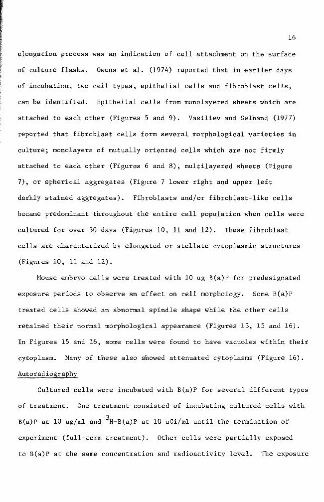
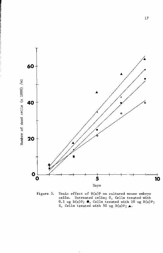
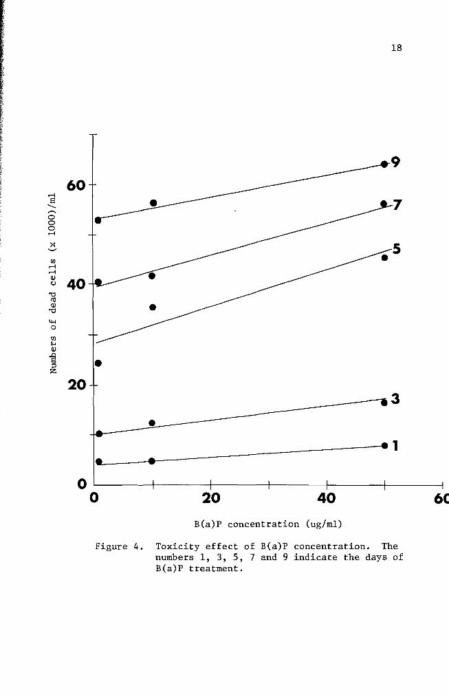
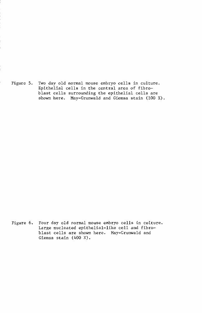
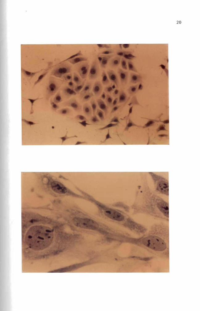
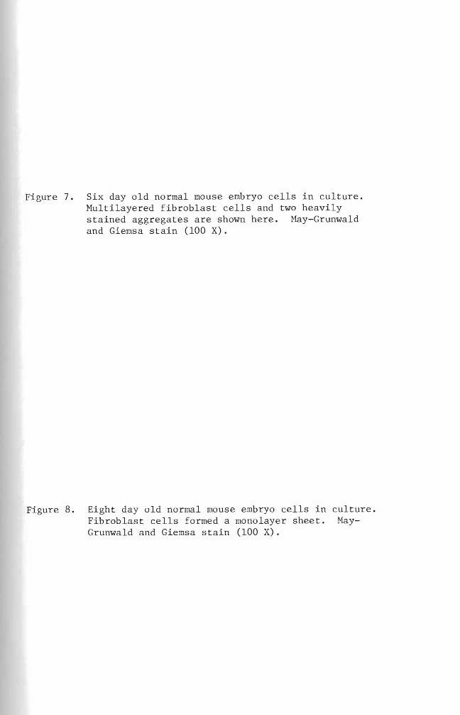
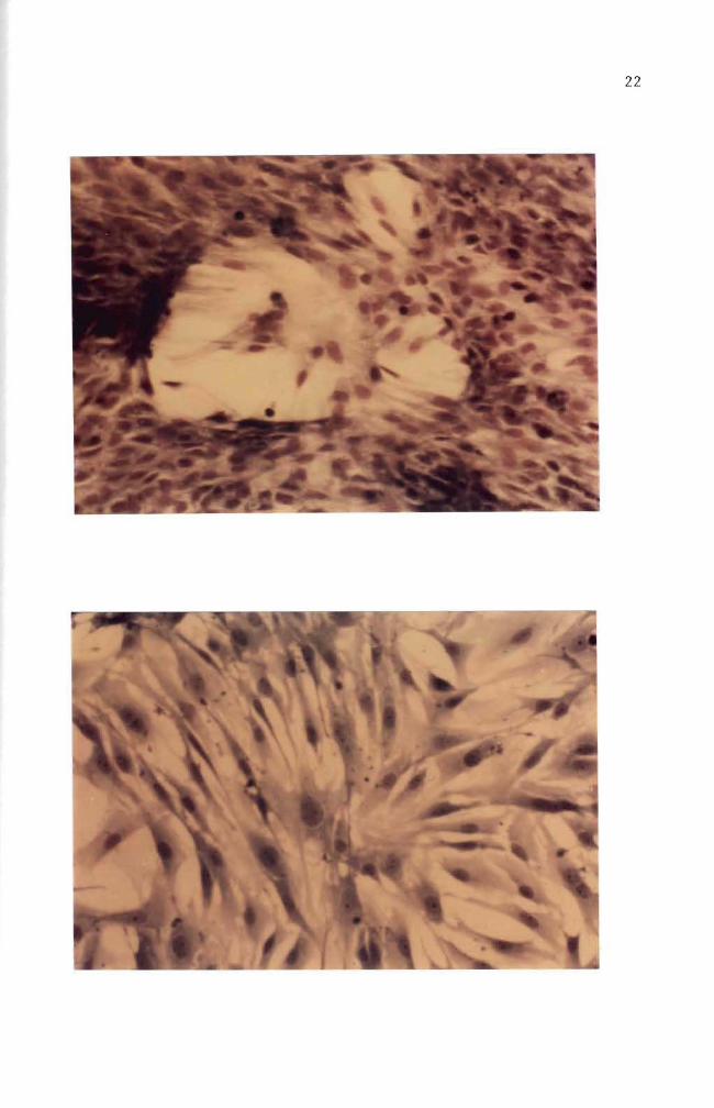
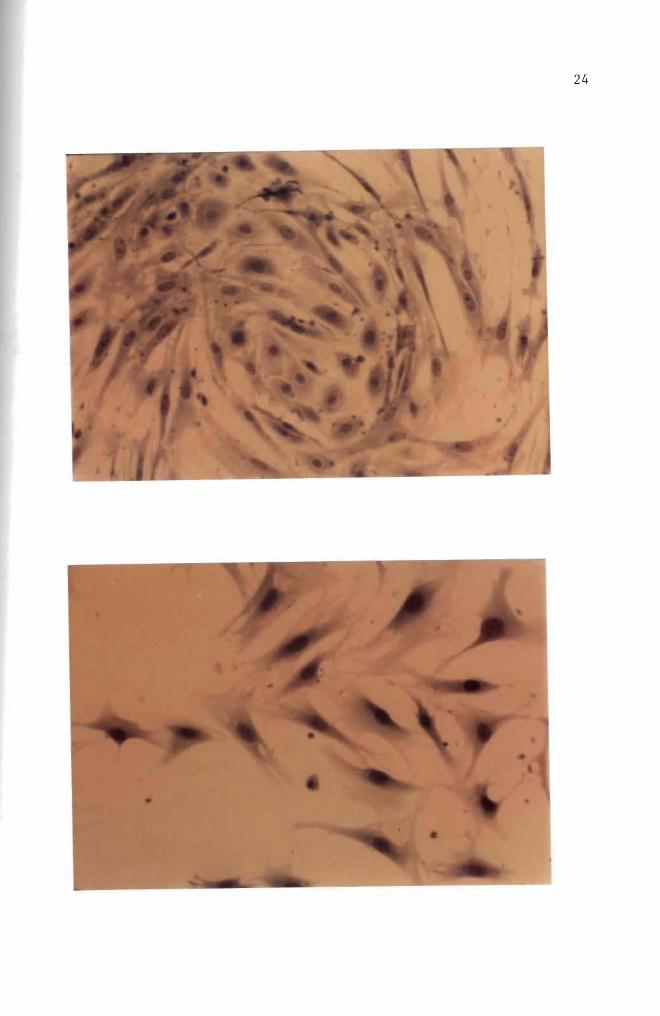
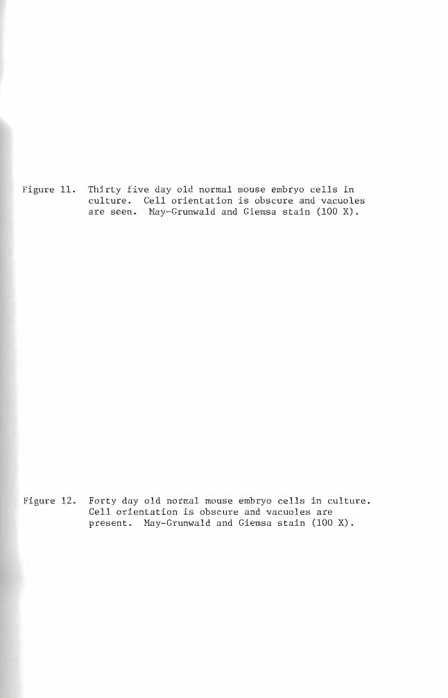
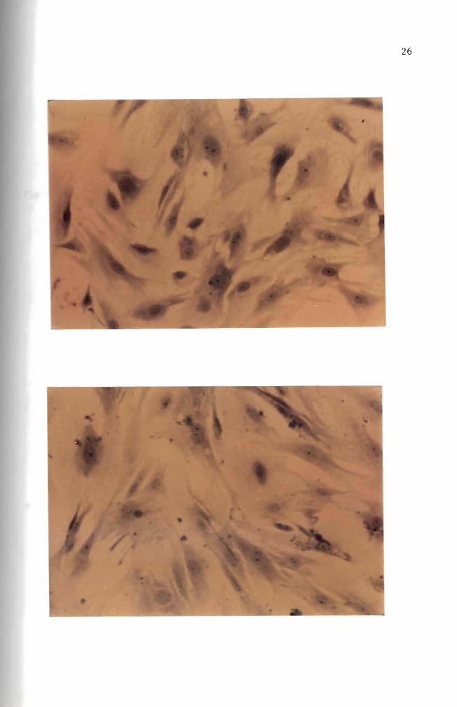
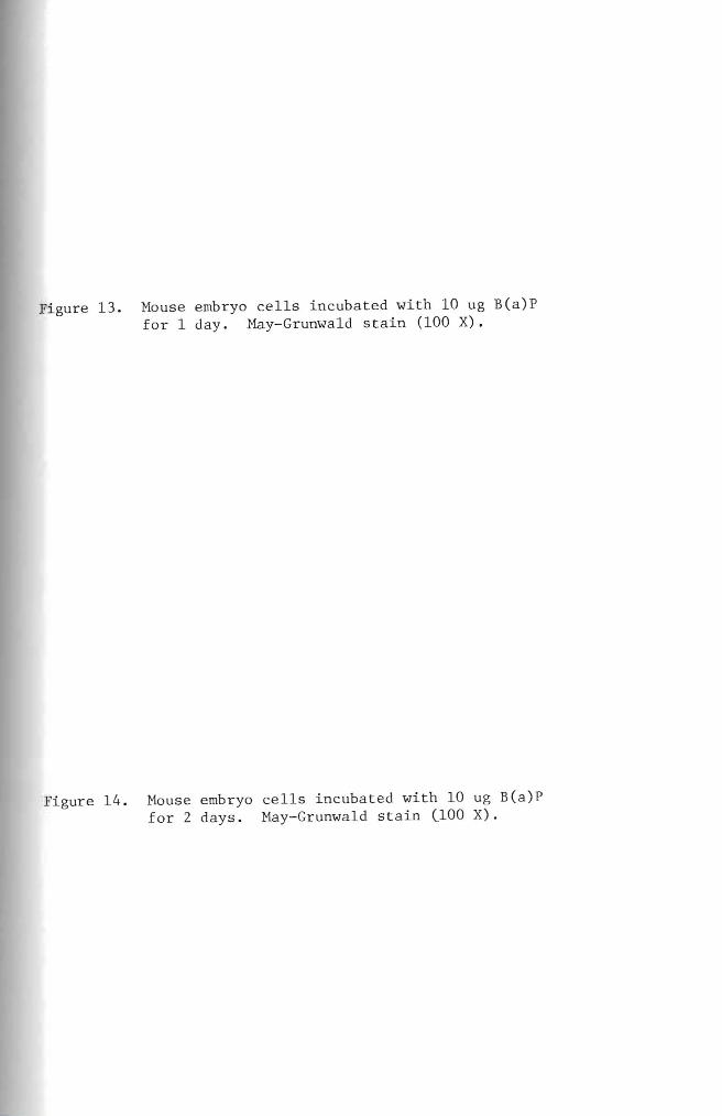
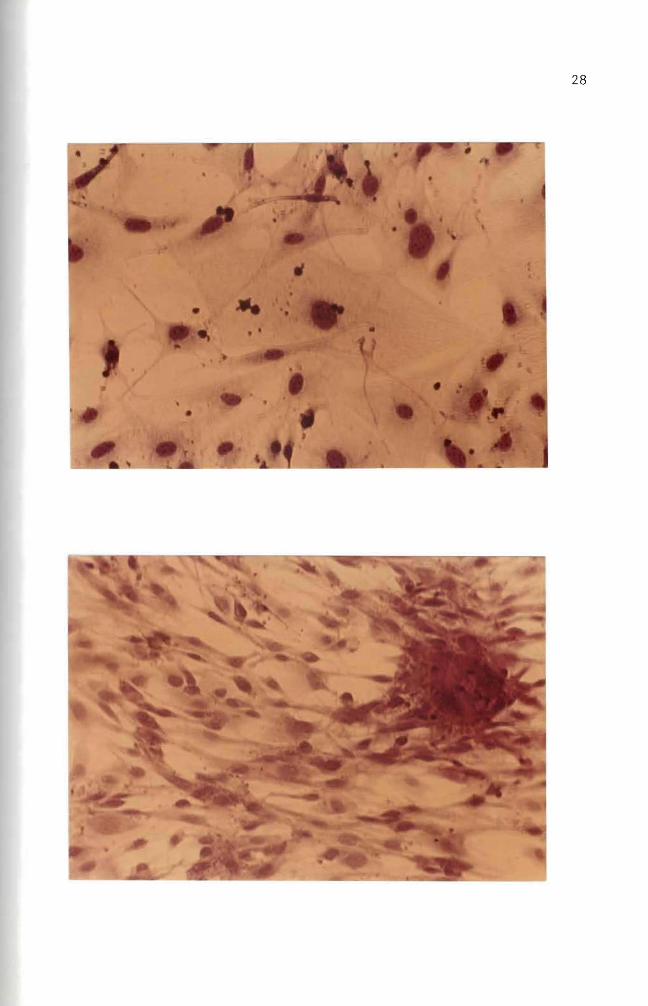
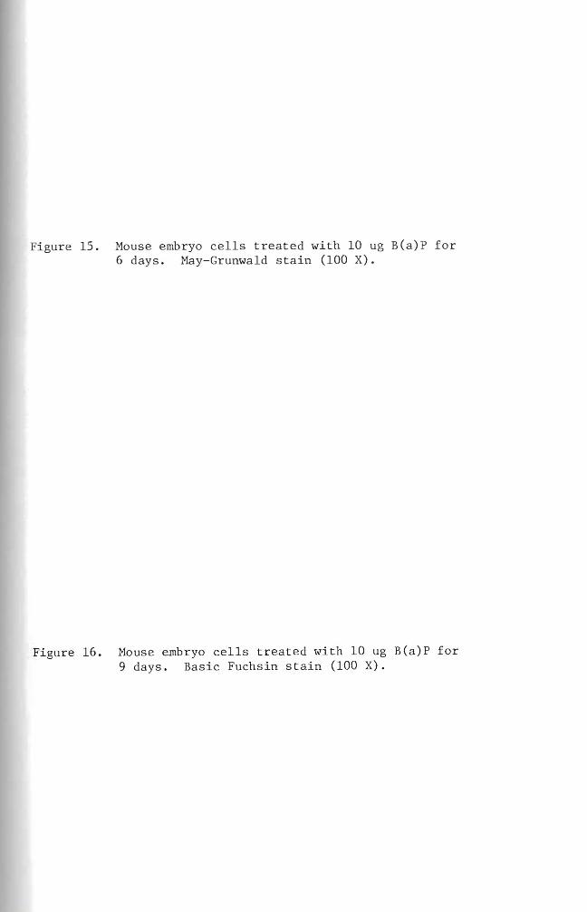
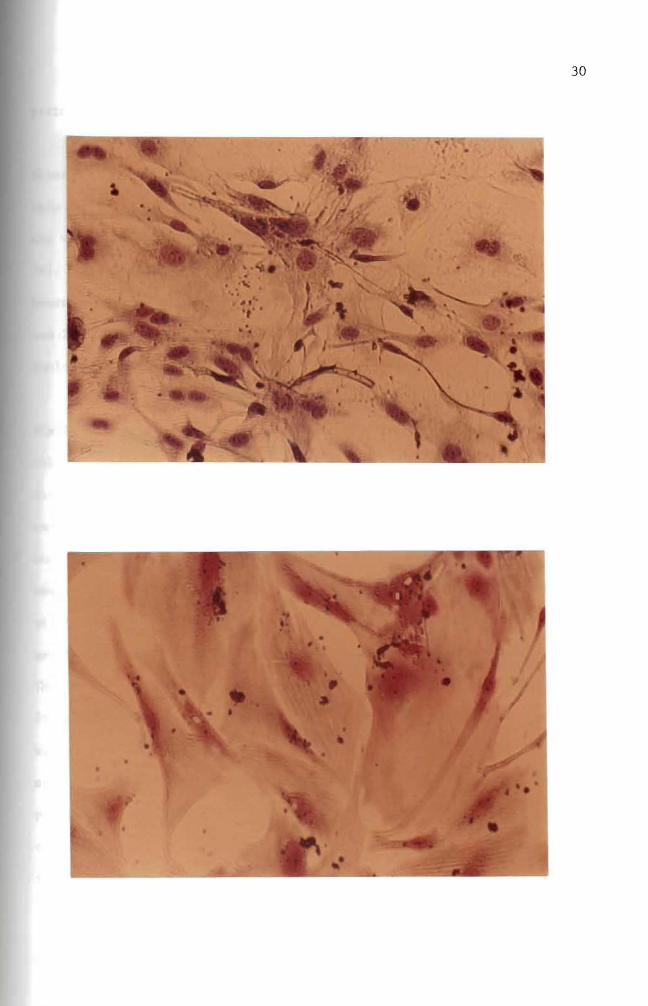
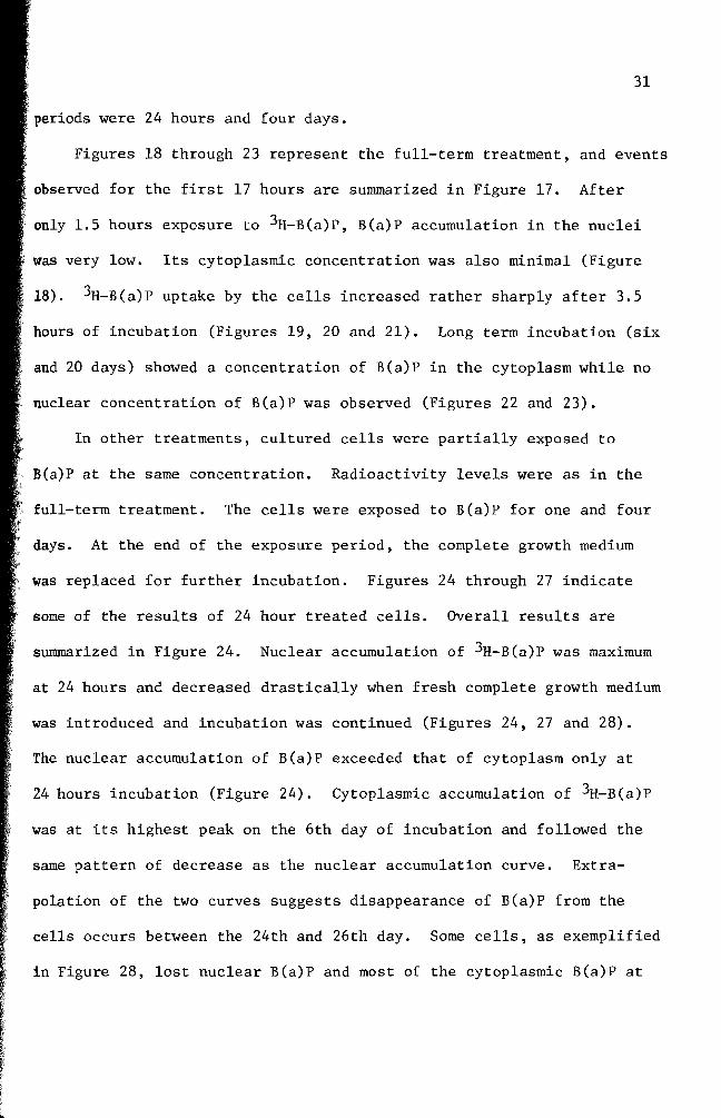
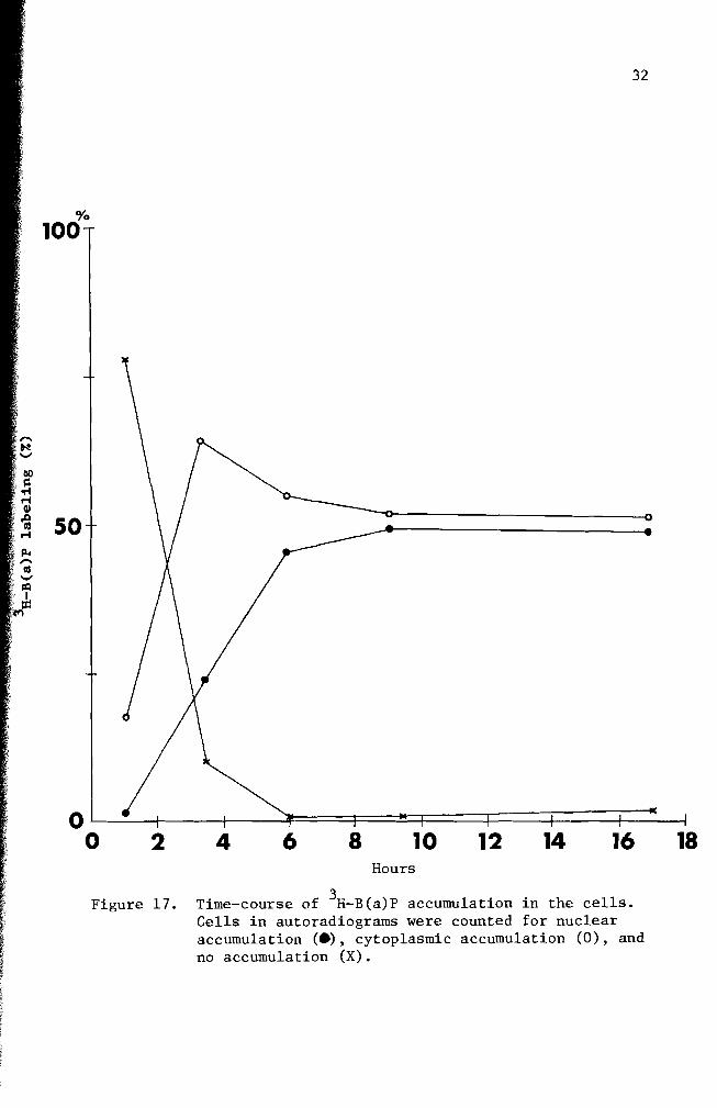