Embed Size (px)
Citation preview
Nanotoxicology, 2013; Early Online, 1–12© 2013 Informa UK, Ltd.ISSN: 1743-5390 print / 1743-5404 onlineDOI: 10.3109/17435390.2013.831500
In vitro toxicological screening of nanoparticles on primary humanendothelial cells and the role of flow in modulating cell response
Nadia Ucciferri1,2,3, Eva-Marie Collnot3, Birgit K. Gaiser4, Annalisa Tirella1, Vicki Stone4, Claudio Domenici2,Claus-Michael Lehr3, & Arti Ahluwalia1,2
1Interdepartmental Research Center “E. Piaggio”, University of Pisa, Pisa, Italy, 2Institute of Clinical Physiology, CNR, Pisa, Italy,3Helmholtz Institute for Pharmaceutical Research Saarland, HZI, Saarbrücken, Germany and 4Heriott Watt University,Edinburgh, UK
AbstractAfter passage through biological barriers, nanomaterialsinevitably end up in contact with the vascular endothelium andcan induce cardiovascular damage. In this study the toxicity andsub-lethal effects of six types of nanoparticle, including four ofindustrial and biomedical importance, on human endothelialcells were investigated using different in vitro assays. The resultsshow that all the particles investigated induce some level ofdamage to the cells and that silver particles were most toxic,followed by titanium dioxide. Furthermore, endothelial cellswere shown to be more susceptible when exposed to silvernanoparticles under flow conditions in a bioreactor. The studyunderlines that although simple in vitro tests are useful to screencompounds and to identify the type of effect induced on cells,they may not be sufficient to define safe exposure limits.Therefore, once initial toxicity screening has been conducted onnanomaterials, it is necessary to develop more physiologicallyrelevant in vitromodels to better understand how nanomaterialscan impact on human health.
Keywords: nanotoxicology, bio-engineering, biotechnology
Introduction
The unique toxic properties of nanomaterials are inextricablylinked with their attractive physico-chemical features andsize (The Royal Society 2004). We know that nanoparticles(NPs) can be very reactive or catalytic (Ying 2001) because oftheir high surface-to-volume ratio. Furthermore, they haveaccess to transport mechanisms not open to larger struc-tures, overcoming biological barriers and penetrating theinterior of cells (Isidoro et al. 2012; Hillaireau & Couvreur2009; dos Santos et al. 2011). For this reason much concernhas been expressed as the safety of many nanomaterials hasnot yet been demonstrated and there are still a large numberof unanswered questions regarding both toxicity mechan-isms and methods of assessment. Toxicity studies are
therefore essential to gain new insights into the hazards ofNPs and to define new standards for testing as well asconstructing databases which provide information for mate-rial safety data sheets and basis for potential NP risk assess-ment and risk management. The gold standard for toxicitytesting is still based on in vivo experiments. However, due tothe large variety of different NPs and the difficulty of extrap-olating animal studies to humans, it is desirable to starttoxicity assessment using in vitro assays, which are cheaperand ethically less questionable than animal testing.
No matter what the port of entry of a nanomaterial, if NPsare able to cross biological barriers (skin, intestines, lungs,etc.) and to enter the general circulation, they end up incontact with the vascular endothelium. The endothelium isthe first internal layer of blood vessels and is distributedthroughout the body. It is of primary importance for trans-port and acts as a selective barrier between the circulationand the surrounding tissue, controlling the passage of mole-cules in and out of the bloodstream. Moreover, the endo-thelium modulates a number of pathways such as lipidmetabolism and also releases signals involved in vascularinflammation and the modulation of vascular tone. Altera-tions in these pathways are correlated with the onset ofcardiovascular dysfunction leading to cardiovascular dis-ease, the number one cause of mortality in the West (Wu&Wu 2006). Considering the importance of the endotheliumin human physiology, and the very high probability that NPscome into contact with the vascular system after crossingbiological barriers, this study is focused on the toxic effects ofengineered nanomaterials on primary endothelial cells.
Endothelial cells are the only adherent cells in the humanbody exposed to direct and continuous tangential fluid flow.They are known to be highly responsive to flow, modifyingtheir morphology, cytoskeletal organisation and gene expres-sion in the presence of fluid dynamic forces (Li et al. 2005).Therefore, we initially assessed the effects of exposure of arange of engineered nanomaterials using microwell-based cultures; then the material identified as the most
Correspondence: Nadia Ucciferri, Institute of Clinical Physiology, CNR, Pisa, Italy. E-mail: [email protected]
(Received 5 October 2012; accepted 30 July 2013)
Nan
otox
icol
ogy
Dow
nloa
ded
from
info
rmah
ealth
care
.com
by
Lul
ea U
nive
rsity
Of
Tec
hnol
ogy
on 0
9/26
/13
For
pers
onal
use
onl
y.
hazardous was tested in the presence of flow using a fluidicchamber.
Although the flow dependency of NP uptake in endothe-lial cells has recently been the object of investigation(Samuel et al. 2012; Kusunose et al. 2012), there are veryfew studies on the effects of flow- and nanomaterial-induced toxicity. Only Kim et al. (2011) have studied thecytotoxic effects of silica nanomaterials in the presence offlow, reporting a flow-rate-dependent toxicity.
In this work, 6 different nanomaterials (15 nm and 80 nmAu, NM 101 TiO2 (7 nm), NM 300 Ag (20 nm) and 50 nm and200 nm fluorescent labelled (with fluorescein isothiocyanateor FITC) polystyrene (PS) NPs) were first characterised incell-culture medium. Human umbilical vein endothelial cells(HUVECs) were then exposed to the materials and ranked fortheir toxicity. The most toxic NP was then further assessedusing a fluidic system to better simulate physiological con-ditions. The materials were chosen because of their rele-vance to the nanomaterial industry and in particular the TiO2
and Ag are categorised as OECD reference manufacturednanomaterials. Some of them have not been tested onendothelial cells and others have yielded controversialresults (Peters et al. 2004; Brammer et al. 2008; Peng et al.2009; Montiel-Dávalos et al. 2012). Our contribution is thusassessment of damage induced by well-characterised metals,metal oxides and PS beads in primary endothelial cells(Peters et al. 2007; Gojova et al. 2007; Meng et al. 2012;Oesterling et al. 2008) and an evaluation of the effects of flowon nanomaterial-induced toxicity. The results of the studycan be used to establish a minimal set of sensitive markersand endpoints for evaluating NP cytotoxicity on endothelialcells and underline that flow can affect the toxicity ofpotentially hazardous nanomaterials.
Materials and methods
Experimental workflowTheaimof this paperwas to screenNPswithin a set of standardassays commonly used by nanotoxicology community, andthen determine the effect of flow on toxicity by exposingHUVECs to a significantly toxic material in a bioreactor.
To reach this goal, we first characterised each NP in theHUVEC medium, than we tested them in standard staticconditions within a large concentration range to determinethe LC50. This value was taken to classify NPs in two groups,low toxicity and high toxicity, in order to identify an appro-priate range of concentrations to be further assessed for sub-lethal effects. Finally, we selected the most toxic NP fortesting in dynamic conditions demonstrating that even the
application of gentle flow can change the susceptibility ofcells to toxic insult from nanomaterials.
ReagentsAll the reagents were purchased from Sigma-Aldrich (St.Louis, USA) unless otherwise specified.
Culture of endothelial cellsHuvecs were isolated (Baudin et al. 2007), passaged andcultured as described in SI.1. All the experiments, includingthose involving NP characterisation, were performed inEagle’s minimum essential medium (EMEM) with supple-ments (see SI.1) henceforth referred to as HUVEC medium.
For the toxicity experiments HUVECs were seeded on 1%gelatin-coated wells (static tests) or 13-mm-diameter plasticslides (NUNC, Denmark) (dynamic tests) at a concentrationof 20,000 cells/cm2 and allowed to reach confluence(typically 24 h) before exposure to NPs.
NP preparation and characterisationThe types and sources of the NPs tested are listed in Table I.
All the dispersion and dilution protocols (Jacobsen et al,2010) are reported in SI.2.
Particle hydrodynamic diameter was determined viadynamic light scattering (DLS) using Malvern Zetasizernano(Malvern Instruments, Herrenberg, Germany). Size andpolydispersity were determined at time points 0, 1, 2, 4, 6,8 and 24 h after preparation of the respective dilution.Between measurements, dilutions were kept at room tem-perature and protected from light. Diluted samples wereagain sonicated for 16 min in an ultrasonic bath. For eachtime point, two independent samples were measured threetimes each and mean and standard deviation were reported.
Due to interference with the zeta sizer measurement, AgNPs could not be evaluated using DLS in our work. Therefore,in the case of Ag NP, size distribution and stability wereinvestigated using NanoSight LM10 instrument (NanoSightLimited, Amesbury, UK) which uses single-particle trackinganalysis. Two concentrations were tested and mean particlediameter (out of three independent measurements) and widthof the distribution are reported. Changes in Ag NP agglomer-ation and Ag loss through adsorption or deposition in thebioreactor were monitored by comparing optical absorptionspectra before and after 24 h of circulation in the fluidic system.
Static set-upThe viability evaluation was performed in a 96-well platewith 200 mL of HUVEC medium containing a range of10 concentrations (from 625 to 1.2 mg/cm2) for each NP,
Table I. Nominal dimensions of NPs and their sources.
Nanoparticle Acronym in text Source
PS FITC Fluoresbrite� microparticles 0.05 mm (~50 nm) PS 50 Polysciences Inc. (Germany)
PS FITC Fluoresbrite� microparticles 0.2 mm (~200 nm) PS 200
Ag NM 300, 20 nm Ag Ras GmbH
TiO2 NM 101 anatase, thermal, 7 nm TiO2 Hombikat UV100
TPPMS-Au NP 15 nm Au 15 Kind donation from Dr W.G. Kreyling(Helmholtz Zentrum Munich)TPPMS-Au NP 80 nm Au 80
N. Ucciferri et al.
Nan
otox
icol
ogy
Dow
nloa
ded
from
info
rmah
ealth
care
.com
by
Lul
ea U
nive
rsity
Of
Tec
hnol
ogy
on 0
9/26
/13
For
pers
onal
use
onl
y.
performed as simple serial dilutions from a stock using theHUVEC medium.
All other assays were performed testing 3–5 significantconcentrations in a 24-well plate. In each case the assay wasperformed as an endpoint after 24-h exposure of the cells tothe NPs. Control samples were seeded and analysed in thesame conditions and time of treated samples. They wereuntreated HUVECs (negative control) and 0.1% Triton�
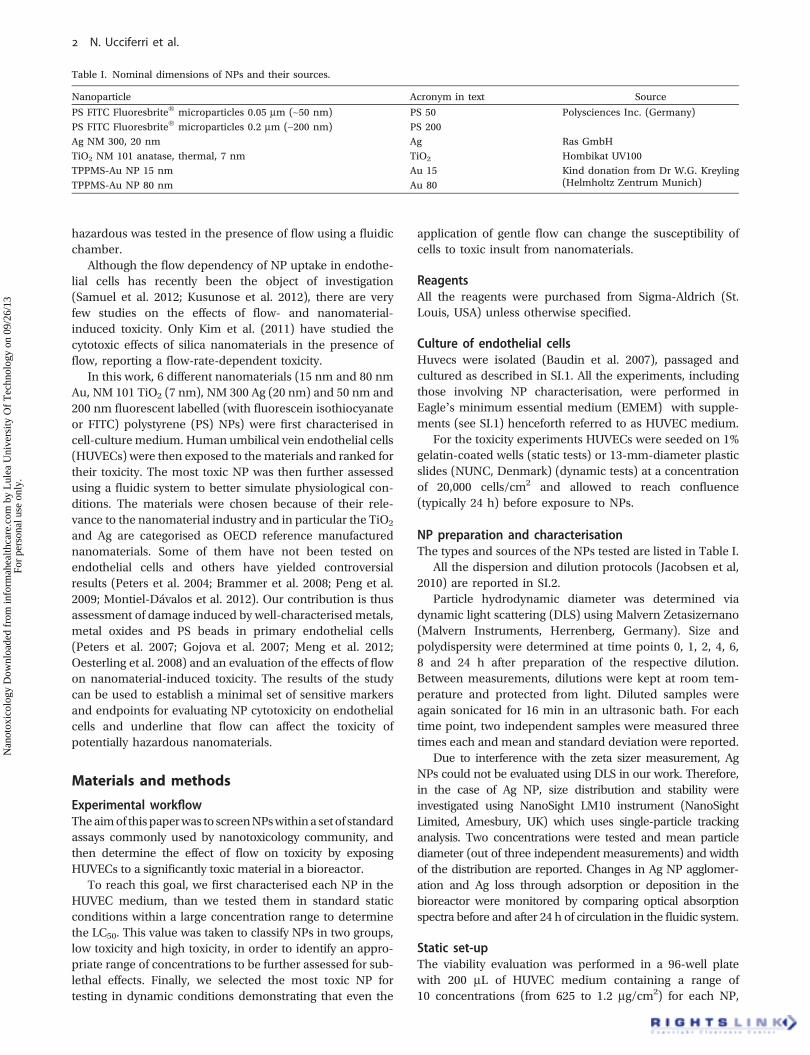
X-100 (named Triton)-treated HUVECs (positive control).Micrographs for the controls and flow experiments arereported in Figure 1A – C. Figure 1A shows the typicalcobble-stone morphology of endothelial cells in vitro. Foreach particle concentration the interaction with the assaywas also tested in the absence of cells in order to avoid false-negative or -positive results.
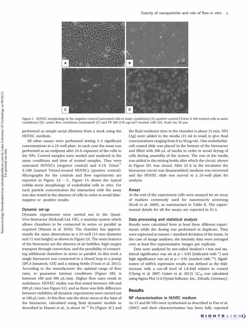
Dynamic set-upDynamic experiments were carried out in the Quasi-Vivo bioreactor (Kirkstall Ltd, UK), a modular system whichallows chambers to be connected in series or parallel asrequired (Mazzei et al. 2010). The chamber has approxi-mately the same dimensions as a 24-well (13 mm diameterand 11 mm height) as shown in Figure 2A. The main featuresof the bioreactor are the absence of air bubbles, high oxygentransport through convection, and the possibility of connect-ing additional chambers in series or parallel. In this work asingle bioreactor was connected in a closed loop to a pump(IPC4 Ismatech, CH) and a mixing bottle (Vozzi et al. 2011).According to the manufacturer the optimal range of flowrates, to guarantee laminar conditions (Figure 2B), isbetween 100 and 500 mL/min. Higher flow rates result inturbulence. HUVEC vitality was first tested between 100 and500 mL/min (see Figure S1), and as there was little differencebetween viabilities all dynamic experiments were carried outat 100 mL/min. At this flow rate the shear stress at the base ofthe bioreactor, calculated using fluid dynamic models asdescribed in Mazzei et al., is about 10�6 Pa (Figure 2C) and
the fluid residence time in the chamber is about 15 min. NPs(Ag) were added to the media (15 ml in total) to give finalconcentrations ranging from 0 to 50 mg/mL. One endothelial-cell-coated slide was placed in the bottom of the bioreactorand filled with 500 mL of media in order to avoid drying ofcells during assembly of the system. The rest of the mediawas added to the mixing bottle after which the circuit, shownin Figure 2D, was closed. After 24 h in the incubator thebioreactor circuit was disassembled; medium was recoveredand the HUVEC slide was moved to a 24-well plate foranalysis.
AssaysAt the end of the experiment cells were assayed for an arrayof markers commonly used for nanotoxicity screening(Kroll et al. 2009), as summarised in Table II. The experi-mental details for all the assays are reported in S1.4.
Data processing and statistical analysisResults were calculated from at least three different experi-ments while the dosing was performed in duplicate. Theywere expressed asmeans ± standard deviation of themean. Inthe case of image analyses, the intensity data were averagedover at least five representative images per replicate.
Data were analysed by two-tailed Student’s t-test and sta-tistical significance was set at p < 0.05 (indicated with *) andhigh significance was set at p < 0.01 (marked with **). Signif-icance of mRNA expression results was defined as the fold-increase with a cut-off level of 1.8-fold relative to control(Cheng et al. 2007; Gaiser et al. 2013). LC50 was calculatedusing Sigma Plot 11.0 (Systat Software, Inc., Erkrath, Germany).
Results
NP characterisation in HUVEC mediumAu 15 and 80 NPs were synthesised as described in Pan et al.(2007) and their characterisation has been fully reported
A B
C D
Figure 1. HUVEC morphology in the negative control (untreated cells in static conditions) (A), positive control (Triton X 100–treated cells in staticconditions) (B), under flow conditions (untreated) (C) and PS 200 (156 mg/cm2)-treated cells (D). Scale bar 50 mm.
Toxicity of nanoparticles and role of flow in vitro
Nan
otox
icol
ogy
Dow
nloa
ded
from
info
rmah
ealth
care
.com
by
Lul
ea U
nive
rsity
Of
Tec
hnol
ogy
on 0
9/26
/13
For
pers
onal
use
onl
y.
(Hirn et al. 2011; Schleh et al. 2012). Briefly the hydrody-namic diameter from DLS analysis was 21 and 85 nm whilethe polydispersity index (PDI) was 0.10 and 0.12. The surfacewas negatively charged, thanks to triphenylphosphinemo-nosulfonate (TPPMS) group showing a z potential of –22.8 ±3.1 and –22.3 ± 1.6 mV, respectively.
We found the two AuNPs and PS 50 easy to disperse in theHUVEC medium over the whole concentration range tested.For each of these NP the measured hydrodynamic diameterfits with the nominal diameter given by the manufacturer(Table III). A minor increase in size could be observed for Aulikely due to serum protein absorption on the particle surface.The PDI was found to be <0.1 even at high concentrations of1 mg/mL. In contrast, a significant increase in size could beobserved for PS 200: at 0.1 mg/mL big agglomerates of~900 nm size were formed and agglomeration increased
during 24-h storage. Agglomeration was less pronouncedfor concentrations of 0.01 and 0.001 mg/mL and showed asize of ~420 nm and PDI <0.2 together with a monomodaldistribution of the particle population.
Being a reference material, TiO2 was well characterisedboth from JRC and in Kermanizadeh et al. (2012). Brieflytransmission electronmicroscopy (TEM) analysis showed twostructures with size of 4–8 and 50–100 nm. Surface area (withBrunauer-Emmett-Teller method, BET) was 322 m2/g andsize (from DLS analysis) in the medium was 185 and 742 nm.
In our study, TiO2 dispersibility was very poor in plainwater (data not shown) but was improved in HUVECmedium likely due to the high serum/protein concentrationand steric stabilisation of the particle dispersion. Neverthe-less, agglomerates were formed varying in size between700 and 1200 nm with a high PDI (>0.4) and two to three
Shear stress, µPa
8
0
1
2
3max 3.226
min 0.175–8y, mm
x, m
m
–8
8
A
C
Pump
Endothelial cells
Mixing bottle
Ag Np
D
x
y
Z
Cell culture region–0.02 –0.01 0 0.01
0.01
0
0.02 0
B
Figure 2. The Quasi-Vivo chamber (A), velocity streamlines in the central xz plane of the chamber (100 mL/min, dimension inm), cells are seeded atthe base of the bioreactor (B), shear stress map at the base of the chamber (C), schematic diagram for the experimental set-up used in dynamicconditions (D). B and C were generated using COMSOL Multiphysics applying the Navier–Stokes equations for an incompressible fluid with thesame density and viscosity as water at 37�C as described in Mazzei et al. (2010).
Table II. Chosen assays for the screening of NP toxicity. Sometimes more than one assay was performed to evaluate the same biological endpoint inorder to exclude cross-reactions between NPs and assay (Kroll et al. 2009).
Function Assay Source References
Viability Metabolic activity:Alamar Blue
CellTiter-Blue�, Promega Kroll et al. 2009
Membrane integrity:LDH assay
Cytotoxicity Detection Kit, Roche
Inflammation IL-8sICAM-1TNF-a
BD FLEX kit bead-based ELISA, BD Biosciences Middleton et al. 1997Gearing et al. 1992; Languino et al. 1993;Springer 1995Cines et al. 1998
Apoptosis Fas ligand BD FLEX kit bead-based ELISA, BD Biosciences Solarska et al. 2012; Park et al. 2010
Endothelial specificmarker stress: vWF
mRNA expression DyNAmo� FlashSYBR�
Green qPCR kit, FinnzymesSumpio et al. 2002
Immunostaining Monoclonal mouse anti-hvWF, Dako
N. Ucciferri et al.
Nan
otox
icol
ogy
Dow
nloa
ded
from
info
rmah
ealth
care
.com
by
Lul
ea U
nive
rsity
Of
Tec
hnol
ogy
on 0
9/26
/13
For
pers
onal
use
onl
y.
subpopulations could be identified in the size distributionprofile at concentrations of 0.1 and 0.01 mg/mL, confirmingTEM data from Kermanizadeh’s group. In the most dilutedconcentration of 0.001 mg/mL, hydrodynamic diameter wasimproved varying between 200 and 300 nm, although thesample was still highly polydispersed (PDI ~ 0.25).
Ag was also characterised both from JRC (Klein et al.2011) and Kermanizadeh et al. (2012). They found an aver-age size at TEM of 17.5 nm with euhedral morphology. Sizein the medium measured with DLS was 12, 28 and 114 nm.
Using the Nanosight system, Ag presented mean hydro-dynamic diameters of 120 nm hinting at particle clusterformation together with serum proteins. Size distributionwas monomodal in single-particle tracking analysis and nofurther agglomeration was observed within 24 h of storage atroom temperature. After 24 h of circulation in the bioreactorsystem we detected a slight decrease (between 10% and 30%depending on the concentration) in Ag concentration due toadsorption onto the walls of the bioreactor chamber andtubing, but no wavelength shifts, characteristic of particleagglomeration, were recorded.
Further details about NP’s characteristics in HUVECmedia can be found in the Supplementary Information.
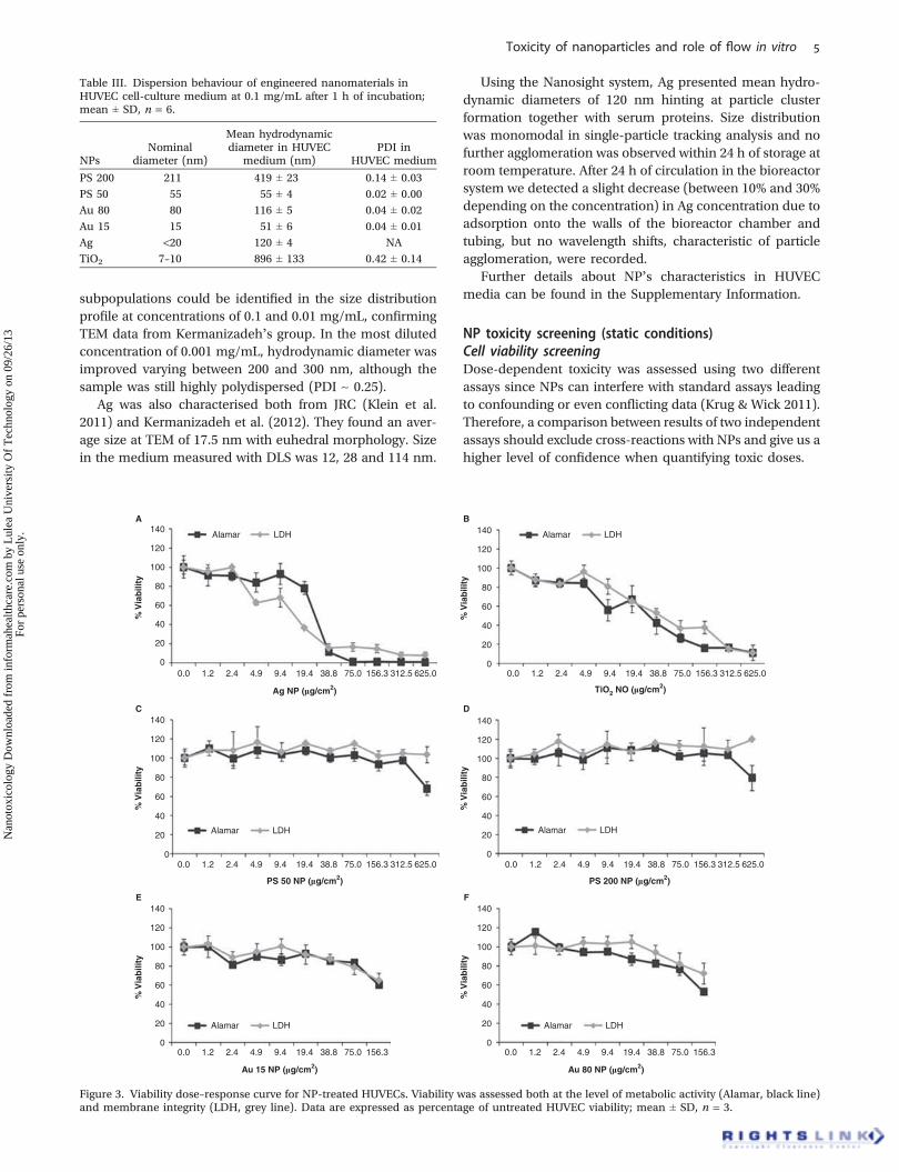
NP toxicity screening (static conditions)Cell viability screeningDose-dependent toxicity was assessed using two differentassays since NPs can interfere with standard assays leadingto confounding or even conflicting data (Krug & Wick 2011).Therefore, a comparison between results of two independentassays should exclude cross-reactions with NPs and give us ahigher level of confidence when quantifying toxic doses.
Table III. Dispersion behaviour of engineered nanomaterials inHUVEC cell-culture medium at 0.1 mg/mL after 1 h of incubation;mean ± SD, n = 6.
NPsNominal
diameter (nm)
Mean hydrodynamicdiameter in HUVEC
medium (nm)PDI in
HUVEC medium
PS 200 211 419 ± 23 0.14 ± 0.03
PS 50 55 55 ± 4 0.02 ± 0.00
Au 80 80 116 ± 5 0.04 ± 0.02
Au 15 15 51 ± 6 0.04 ± 0.01
Ag <20 120 ± 4 NA
TiO2 7–10 896 ± 133 0.42 ± 0.14
0.00
20
40
60
80
100
120
140
1.2 2.4 4.9 9.4 19.4 38.8 75.0 156.3 312.5 625.0
0.0 1.2 2.4 4.9 9.4 19.4 38.8 75.0 156.3 312.5 625.0 0.0 1.2 2.4 4.9 9.4 19.4 38.8 75.0 156.3 312.5 625.0
Ag NP (mg/cm2)
A B
C D
E F
0.0 1.2 2.4 4.9 9.4 19.4 38.8 75.0 156.3 312.5 625.0
% V
iab
ility
0
20
40
60
80
100
120
140
PS 50 NP (mg/cm2)
0.0 1.2 2.4 4.9 9.4 19.4 38.8 75.0 156.3
Au 15 NP (mg/cm2)
0.0 1.2 2.4 4.9 9.4 19.4 38.8 75.0 156.3
Au 80 NP (mg/cm2)
PS 200 NP (mg/cm2)
% V
iab
ility
0
20
40
60
80
100
120
140
% V
iab
ility
0
20
40
60
80
100
120
140
% V
iab
ility
0
20
40
60
80
100
120
140
% V
iab
ility
Alamar LDH
Alamar LDH Alamar LDH
Alamar LDH
20
0
40
60
80
100
120
140
TiO2 NO (mg/cm2)
% V
iab
ility
Alamar LDHAlamar LDH
Figure 3. Viability dose–response curve for NP-treated HUVECs. Viability was assessed both at the level of metabolic activity (Alamar, black line)and membrane integrity (LDH, grey line). Data are expressed as percentage of untreated HUVEC viability; mean ± SD, n = 3.
Toxicity of nanoparticles and role of flow in vitro
Nan
otox
icol
ogy
Dow
nloa
ded
from
info
rmah
ealth
care
.com
by
Lul
ea U
nive
rsity
Of
Tec
hnol
ogy
on 0
9/26
/13
For
pers
onal
use
onl
y.
Metabolic activity was evaluated using Alamar blue addeddirectly to treated cells while membrane integrity wasassayed measuring lactate dehydrogenase (LDH) levels inthe medium. Figure 3 shows the dose–response curves of theNPs; in both assays high toxicity was shown for Ag and TiO2
with an LC50 of 24.5 ± 7.5 mg/cm2 (Alamar) and 12.2 ±2.6 mg/cm2 (LDH) for Ag and LC50 values of 25.0 ± 7.9 mg/cm2 (Alamar) and 47.0 ± 9.5 mg/cm2 (LDH) for TiO2
(Figure 3A and B). However, preprocessing of the LDHraw data was required in order to correct values for particleinterference as a reduction in enzyme availability wasnoticed in the presence of Ag. This interference is thoughtto be due to the adsorption of the enzyme onto the particlesurface, reducing enzyme activity (Clift et al. 2008). Thepositive control value (Triton treated) was 0.74 ± 0.11% withAlamar assay and 0 ± 0.9% with LDH assay. The toxicity ofthe Ag dispersant was also investigated, and no effects in therange of dispersant concentrations used were observed(data not shown).
There was no significant toxicity with PS 50 and PS200 (Figure 3C and D), besides some SD fluctuation, which
may be due to primary cell variability, while significant toxicitywas shown for both Au particle sizes at the highest testedconcentration (Figure 3E and F); however, the LC50 was notreached with the concentrations used in this study.
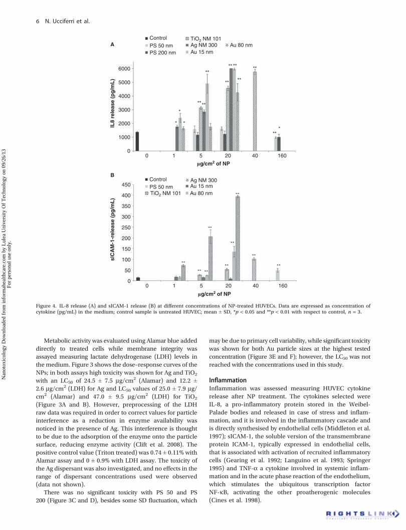
InflammationInflammation was assessed measuring HUVEC cytokinerelease after NP treatment. The cytokines selected wereIL-8, a pro-inflammatory protein stored in the Weibel-Palade bodies and released in case of stress and inflam-mation, and it is involved in the inflammatory cascade andis directly synthesised by endothelial cells (Middleton et al.1997); sICAM-1, the soluble version of the transmembraneprotein ICAM-1, typically expressed in endothelial cells,that is associated with activation of recruited inflammatorycells (Gearing et al. 1992; Languino et al. 1993; Springer1995) and TNF-a a cytokine involved in systemic inflam-mation and in the acute phase reaction of the endothelium,which stimulates the ubiquitous transcription factorNF-kB, activating the other proatherogenic molecules(Cines et al. 1998).
6000
5000
4000
3000
2000
1000
0
0
50
100
150
200
250
300
350
400
450
0 1 5 20 40 160
mg/cm2 of NP
A
B
0 1 5 20 40 160
mg/cm2 of NP
IL8
rele
ase
(pg
/mL
)sl
CA
M-1
-rel
ease
(p
g/m
L)
ControlPS 50 nmPS 200 nm
TiO2 NM 101Ag NM 300Au 15 nm
Au 80 nm
ControlPS 50 nmTiO2 NM 101
Ag NM 300Au 15 nmAu 80 nm
Figure 4. IL-8 release (A) and sICAM-1 release (B) at different concentrations of NP-treated HUVECs. Data are expressed as concentration ofcytokine (pg/mL) in the medium; control sample is untreated HUVEC; mean ± SD, *p < 0.05 and **p < 0.01 with respect to control, n = 3.
N. Ucciferri et al.
Nan
otox
icol
ogy
Dow
nloa
ded
from
info
rmah
ealth
care
.com
by
Lul
ea U
nive
rsity
Of
Tec
hnol
ogy
on 0
9/26
/13
For
pers
onal
use
onl
y.
Three concentrations for each NP type were tested, from160 to 5 mg/cm2 for low-toxicity NPs and from 20 (or 40 mg/cm2 for TiO2) to 1 mg/cm2 for high-toxicity ones.
PS NPs of both sizes induced IL-8 expression comparableto controls with a slight decrease at the highest concentration(Figure 4A). On the contrary, Ag, TiO2 and Au of both sizesshowed a significant increase with respect to the negativecontrol (untreated). The increase was found to be concen-tration dependent for all particles. For comparison, theIL-8 level for Triton-treated cells value was extremely low,68.59 ± 22.57 pg/mL.
sICAM-1 was only detected at the highest concentrationof PS 50 (Figure 4B) while in all concentrations of PS 200 itwas under the limit of detection. TiO2 and Au showed highlysignificant (p < 0.01) increases of sICAM-1. In these experi-ments the highest sICAM-1 levels (28-fold higher than incontrols) were found in the presence of 20 mg/cm2 of Au 80.Interestingly, sICAM-1 was detected neither in Ag-treatedsamples, nor in the Triton-treated cells.
TNF-a was below the limit of detection in all samples(data not shown), although investigations using other celltypes have reported measurable levels (Kaur & Tikoo 2013;Xue et al. 2012).
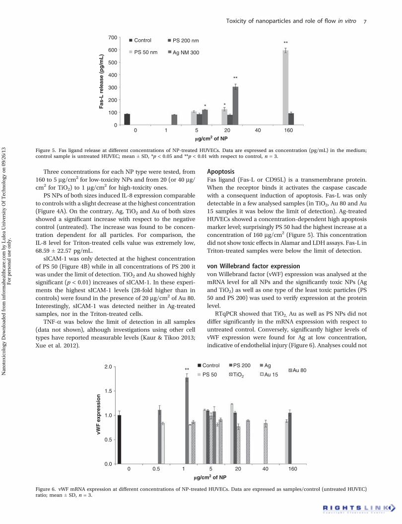
ApoptosisFas ligand (Fas-L or CD95L) is a transmembrane protein.When the receptor binds it activates the caspase cascadewith a consequent induction of apoptosis. Fas-L was onlydetectable in a few analysed samples (in TiO2, Au 80 and Au15 samples it was below the limit of detection). Ag-treatedHUVECs showed a concentration-dependent high apoptosismarker level; surprisingly PS 50 had the highest increase at aconcentration of 160 mg/cm2 (Figure 5). This concentrationdid not show toxic effects in Alamar and LDH assays. Fas-L inTriton-treated samples were below the limit of detection.
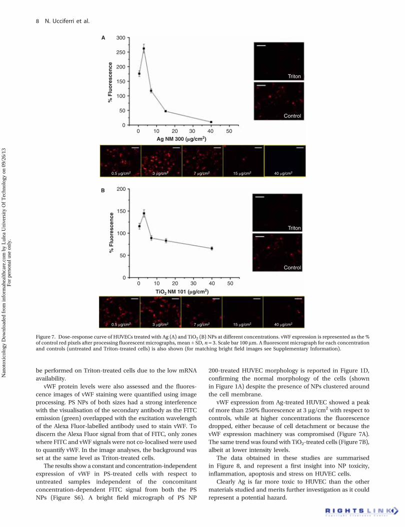
von Willebrand factor expressionvon Willebrand factor (vWF) expression was analysed at themRNA level for all NPs and the significantly toxic NPs (Agand TiO2) as well as one type of the least toxic particles (PS50 and PS 200) was used to verify expression at the proteinlevel.
RTqPCR showed that TiO2, Au as well as PS NPs did notdiffer significantly in the mRNA expression with respect tountreated control. Conversely, significantly higher levels ofvWF expression were found for Ag at low concentration,indicative of endothelial injury (Figure 6). Analyses could not
00
100
200
300
400
500
600
700
1 5 20 40 160
mg/cm2 of NP
Fas-
L r
elea
se (
pg
/mL
)
Control
PS 50 nm
PS 200 nm
Ag NM 300
Figure 5. Fas ligand release at different concentrations of NP-treated HUVECs. Data are expressed as concentration (pg/mL) in the medium;control sample is untreated HUVEC; mean ± SD, *p < 0.05 and **p < 0.01 with respect to control, n = 3.
00.0
0.5
1.0
1.5
2.0
0.5 1 5 20 40 160
mg/cm2 of NP
vWF
exp
ress
ion
Control
PS 50
PS 200
TiO2
Ag
Au 15Au 80
Figure 6. vWF mRNA expression at different concentrations of NP-treated HUVECs. Data are expressed as samples/control (untreated HUVEC)ratio; mean ± SD, n = 3.
Toxicity of nanoparticles and role of flow in vitro
Nan
otox
icol
ogy
Dow
nloa
ded
from
info
rmah
ealth
care
.com
by
Lul
ea U
nive
rsity
Of
Tec
hnol
ogy
on 0
9/26
/13
For
pers
onal
use
onl
y.
be performed on Triton-treated cells due to the low mRNAavailability.
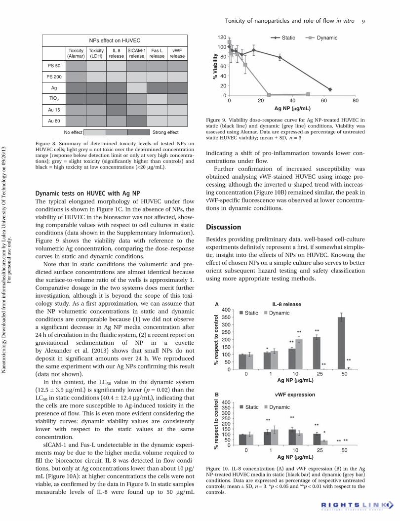
vWF protein levels were also assessed and the fluores-cence images of vWF staining were quantified using imageprocessing. PS NPs of both sizes had a strong interferencewith the visualisation of the secondary antibody as the FITCemission (green) overlapped with the excitation wavelengthof the Alexa Fluor-labelled antibody used to stain vWF. Todiscern the Alexa Fluor signal from that of FITC, only zoneswhere FITC and vWF signals were not co-localised were usedto quantify vWF. In the image analyses, the background wasset at the same level as Triton-treated cells.
The results show a constant and concentration-independentexpression of vWF in PS-treated cells with respect tountreated samples independent of the concomitantconcentration-dependent FITC signal from both the PSNPs (Figure S6). A bright field micrograph of PS NP
200-treated HUVEC morphology is reported in Figure 1D,confirming the normal morphology of the cells (shownin Figure 1A) despite the presence of NPs clustered aroundthe cell membrane.
vWF expression from Ag-treated HUVEC showed a peakof more than 250% fluorescence at 3 mg/cm2 with respect tocontrols, while at higher concentrations the fluorescencedropped, either because of cell detachment or because thevWF expression machinery was compromised (Figure 7A).The same trend was found with TiO2-treated cells (Figure 7B),albeit at lower intensity levels.
The data obtained in these studies are summarisedin Figure 8, and represent a first insight into NP toxicity,inflammation, apoptosis and stress on HUVEC cells.
Clearly Ag is far more toxic to HUVEC than the othermaterials studied and merits further investigation as it couldrepresent a potential hazard.
00
50
100
150
200
250
300
10 20
Ag NM 300 (mg/cm2)
% F
luo
resc
ence
0
50
100
150
200
% F
luo
resc
ence
30 40 50
0 10 20
TiO2 NM 101 (mg/cm2)
30 40 50
Triton
Control
Triton
Control
0.5 µg/cm2 3 µg/cm2 7 µg/cm2 15 µg/cm2 40 µg/cm2
0.5 µg/cm2 3 µg/cm2 7 µg/cm2 15 µg/cm2 40 µg/cm2
A
B
Figure 7. Dose–response curve of HUVECs treated with Ag (A) and TiO2 (B) NPs at different concentrations. vWF expression is represented as the %of control red pixels after processing fluorescent micrographs, mean ± SD, n = 3. Scale bar 100 mm. A fluorescent micrograph for each concentrationand controls (untreated and Triton-treated cells) is also shown (for matching bright field images see Supplementary Information).
N. Ucciferri et al.
Nan
otox
icol
ogy
Dow
nloa
ded
from
info
rmah
ealth
care
.com
by
Lul
ea U
nive
rsity
Of
Tec
hnol
ogy
on 0
9/26
/13
For
pers
onal
use
onl
y.
Dynamic tests on HUVEC with Ag NPThe typical elongated morphology of HUVEC under flowconditions is shown in Figure 1C. In the absence of NPs, theviability of HUVEC in the bioreactor was not affected, show-ing comparable values with respect to cell cultures in staticconditions (data shown in the Supplementary Information).Figure 9 shows the viability data with reference to thevolumetric Ag concentration, comparing the dose–responsecurves in static and dynamic conditions.
Note that in static conditions the volumetric and pre-dicted surface concentrations are almost identical becausethe surface-to-volume ratio of the wells is approximately 1.Comparative dosage in the two systems does merit furtherinvestigation, although it is beyond the scope of this toxi-cology study. As a first approximation, we can assume thatthe NP volumetric concentrations in static and dynamicconditions are comparable because (1) we did not observea significant decrease in Ag NP media concentration after24 h of circulation in the fluidic system, (2) a recent report ongravitational sedimentation of NP in a cuvetteby Alexander et al. (2013) shows that small NPs do notdeposit in significant amounts over 24 h. We reproducedthe same experiment with our Ag NPs confirming this result(data not shown).
In this context, the LC50 value in the dynamic system(12.5 ± 3.9 mg/mL) is significantly lower (p = 0.02) than theLC50 in static conditions (40.4 ± 12.4 mg/mL), indicating thatthe cells are more susceptible to Ag-induced toxicity in thepresence of flow. This is even more evident considering theviability curves: dynamic viability values are consistentlylower with respect to the static values at the sameconcentration.
sICAM-1 and Fas-L undetectable in the dynamic experi-ments may be due to the higher media volume required tofill the bioreactor circuit. IL-8 was detected in flow condi-tions, but only at Ag concentrations lower than about 10 mg/mL (Figure 10A): at higher concentrations the cells were notviable, as confirmed by the data in Figure 9. In static samplesmeasurable levels of IL-8 were found up to 50 mg/mL
indicating a shift of pro-inflammation towards lower con-centrations under flow.
Further confirmation of increased susceptibility wasobtained analysing vWF-stained HUVEC using image pro-cessing; although the inverted u-shaped trend with increas-ing concentration (Figure 10B) remained similar, the peak invWF-specific fluorescence was observed at lower concentra-tions in dynamic conditions.
Discussion
Besides providing preliminary data, well-based cell-cultureexperiments definitely represent a first, if somewhat simplis-tic, insight into the effects of NPs on HUVEC. Knowing theeffect of chosen NPs on a simple culture also serves to betterorient subsequent hazard testing and safety classificationusing more appropriate testing methods.
NPs effect on HUVEC
Toxicity(Alamar)
Toxicity(LDH)
IL 8release
SlCAM-1release
Fas Lrelease
vWFrelease
PS 50
PS 200
Ag
Au 15
Au 80
No effect Strong effect
TiO2
Figure 8. Summary of determined toxicity levels of tested NPs onHUVEC cells; light grey = not toxic over the determined concentrationrange (response below detection limit or only at very high concentra-tions); grey = slight toxicity (significantly higher than controls) andblack = high toxicity at low concentrations (<20 mg/mL).
00
20
40
60
80
100
120
20 40
Ag NP (mg/mL)
% V
iab
ility
60
Static Dynamic
80
Figure 9. Viability dose–response curve for Ag NP-treated HUVEC instatic (black line) and dynamic (grey line) conditions. Viability wasassessed using Alamar. Data are expressed as percentage of untreatedstatic HUVEC viability; mean ± SD, n = 3.
0 10
50100150200250300350400
10 25Ag NP (mg/mL)
% r
esp
ect
to c
on
tro
l%
res
pec
t to
co
ntr
ol
50
00
50100150200250300350400
1 10 25Ag NP (mg/mL)
50
A
B
Static Dynamic
IL-8 release
vWF expression
Static Dynamic
Figure 10. IL-8 concentration (A) and vWF expression (B) in the AgNP-treated HUVEC media in static (black bar) and dynamic (grey bar)conditions. Data are expressed as percentage of respective untreatedcontrols; mean ± SD, n = 3. *p < 0.05 and **p < 0.01 with respect to thecontrols.
Toxicity of nanoparticles and role of flow in vitro
Nan
otox
icol
ogy
Dow
nloa
ded
from
info
rmah
ealth
care
.com
by
Lul
ea U
nive
rsity
Of
Tec
hnol
ogy
on 0
9/26
/13
For
pers
onal
use
onl
y.

The cardiovascular system is considered an important siteof nanomaterial-induced toxicity and it has been suggestedthat NPs can alter the endothelium integrity impairingvascular function (Mann et al. 2012). As a consequence,several investigations on toxicity of different NPs on HUVEChave been conducted. Solarska et al. (2012) showed thatnanodiamond particles lead to cell death in a time- andconcentration-dependent manner. Carbon black NPs werealso found to increase expression of endothelial intracellularadhesion molecules and reactive oxygen species(Vesterdal et al. 2012). Magnetic particles were found toinhibit HUVEC proliferation through an increase of eNOSactivity and NO production (Su et al. 2012).
As different endpoints and results have been described inthe literature for a range of NPs, in this study the toxicity andeffects of sub-lethal exposure of a range of well-characterised industrial NPs to HUVEC were assessed atdifferent concentrations with different assays. Both Alamarand LDH assays confirmed that Ag and TiO2 NPs were highlytoxic for these cells (Figure 3A and B). Although the LC50
values were very low, they were not identical as already foundin Gaiser et al. (2013), indicating that the two assays haveslightly different sensitivities for the two NPs or that themechanism of cell death induction differs. In fact cytotoxicityis first manifested by an increase in membrane permeability inthe case of Ag, while TiO2 appears to compromise the met-abolic machinery before inducing membrane damage mirror-ing observed for carbon black NPs (Vesterdal et al. 2012). Thetoxicity of TiO2 on HUVEC is particularly interesting since thesame NPs (NM 101) were tested on a hepatocyte cell line andno toxic effects were observed (Gaiser et al. 2013).
PS 50 and PS 200 were not toxic across the concentrationrange tested while slight toxicity was observed with AuNPs ofboth sizes.
However, NPs have many other effects besides cell death(Foldbjerg et al. 2009). For instance, carbon black, TiO2 andsilver NPs are known to induce oxidative stress or inflamma-tion (Hussain et al. 2010; Lee et al. 2011; Park et al. 2010). Infact, inflammation is one of the most common sub-lethal reactions to occur in cells after exposure to NPs(Oberdörster et al. 2007; Peters et al. 2007). Therefore, tobetter understand the effects of lower and more physiologi-cally relevant NP concentrations, we also measured IL-8,sICAM-1 and TNF-a levels in the media. We underline thatnone of these markers were expressed at high levels in theTriton-treated cells, which were set as our positive control. Infact while Triton is a good positive control for viability, it is notsuitable for stress markers since cells probably died before theonset of inflammatory signalling. TNF-a was undetectablewhile high IL-8 levels were found in all samples except forPS NP-treated cells, which showed a small decrease at thehighest concentration (Figure 4A). There was a slight increasein sICAM-1 levels at the highest PS 50 concentration(Figure 4B), indicating that only high amounts of the smallerPS 50 initiate a low-level pro-inflammatory signal. The mosttoxic NPs, Ag and TiO2, released large amounts of IL-8, butinterestingly, only in TiO2-treated samples were sICAM-1levels also increased. In fact, sICAM-1 may be involved in adifferent pathway as it is known to be stimulated by IL-1 and
TNF-a in hepatocytes (Satoh et al. 1994), endothelial cells(McHale et al. 1999) and vascular smooth muscular cells(Couffinhal et al. 1993). This result suggests that inflammationcan actually be triggered through different pathways andinvolve different markers depending on the type of NPs.
Increased cytokine expression after exposure to TiO2 NPswas also observed byMontiel-Dávalos et al. (2012) in HUVECcells, confirming our result; they also showed that cell deathwas mainly through necrosis, which may explain the absenceof Fas-L in our experiments.
Au NPs also induced high levels of IL-8 and sICAM-1release (Figure 4A and B), with higher sICAM-1 for Au80 than Au 15 and higher levels of IL-8 for the smallerAu. Therefore, gold NPs seem to activate both pathways,but in a size-dependent manner.
In summary, Ag trigged inflammation activating anIL-8-dependent pathway and did not have any effect onsICAM induction. Both TiO2 and Au 15 triggered inflamma-tion by IL-8- and sICAM-related pathways, but principallythat involving IL-8, while Au 80 evidently activates the sICAMto a greater extent. Whatever the pathway, the data show thatalmost every NP induces inflammation in cells, even at lowconcentrations as described in the literature for manyNPs (Peters et al. 2007; Montiel-Dávalos et al. 2012;Kermanizadeh et al. 2012; Gaiser et al. 2013).
Although we did not observe any toxicity and only slightpro-inflammatory effects on cells (Figure 4B) in the presenceof PS, the apoptosis marker Fas-L increased on exposure toPS 50. These low-toxicity NPs induce low inflammation atvery high, un-physiological doses that appear to lead to theinitiation of apoptotic signalling already described for endo-thelial and lung epithelial cells by other groups(Solarska et al. 2012; Park et al. 2010). Finally, the vWFmRNA and protein expression data correlate quite well inAg-exposed HUVECs; after an increase in vWF expression atlow concentrations of Ag, as already described for other NPs(Bauer et al. 2011), the expression dropped. The decreasecan be ascribed to a decrease in cell number due to Agtoxicity which results in fewer viable cells being stained(Figure 7A). On the contrary there was no correlationbetween mRNA and protein expression in TiO2-exposedcells. Although the vWF immunohistochemical staining forthe TiO2-treated HUVECs (Figure 7B) first increased andthen decreased at higher concentrations (albeit to a lesserextent than with Ag), mRNA expression did not change withincreasing TiO2 concentration. In the case of Au NPs, mRNAexpression data showed that both Au NPs were not able toaffect HUVEC vWF production (Figure 6). No relevant vWFexpression from PS NPs was observed, both at mRNA andprotein level, confirming that these NPs do not triggerinflammation, despite the increase in the FITC signal fromuptake/binding to the membrane with increasing concen-tration (Figure S6).
As summarised in Figure 8, of the different particles used,the most toxic was Ag. Although there was little loss inviability, at very low concentrations there was significantinflammation (Figure 4) as well as pro-apoptotic signalling(Figure 5) probably leading to cell death (Figure 3) at higherconcentrations. One hypothesis about Ag NP toxicity is that it
N. Ucciferri et al.
Nan
otox
icol
ogy
Dow
nloa
ded
from
info
rmah
ealth
care
.com
by
Lul
ea U
nive
rsity
Of
Tec
hnol
ogy
on 0
9/26
/13
For
pers
onal
use
onl
y.
is partially due to toxic Ag ions released from the NP(Klein et al. 2011). However, any Ag ions will tend toprecipitate due to the presence of chloride ions in themedium. Thus, a more complex mechanism must be theroot of Ag NP toxicity.
As the endothelium is exposed to tangential flow in vivoand the presence of a fluidic environment has been reportedto increase endothelial susceptibility to toxicity from silicaNPs (Kim et al. 2011), we also probed the effects of Agexposure in dynamic conditions. The bioreactor used applieslow shear stress to cells, while ensuring high flow rates andoxygen transport. Although endothelial cells in vivo aregenerally subject to much higher shear (1–10�3 Pa(Caro et al. 2011)) than in our experiments, the studiesconfirm that Ag toxicity and Ag-induced inflammatory stressincrease even in the presence of very low shear stressconditions, highlighting the sensitivity of HUVECs to shear(Vozzi et al. 2011). The LC50 as well as peaks from IL-8 andvWF release shifted to lower concentrations, while main-taining the general trend of the cell responses in staticconditions. This indicates that the organism is likely to bemore susceptible to NP-induced injury than standard in vitrotests would suggest and that even NPs with only slight toxicityin static tests (Figure 8) should be assessed under flow. A fewinvestigations have shown that cells such as hepatocytes andmyoblasts are more sensitive to drug-induced injury indynamic conditions (Vinci et al. 2011; Tirella et al. 2008),albeit the mechanism by which this occurs has yet to beelucidated. Although endothelial cells are known to undergosignificant changes, such as increased membrane permeabil-ity and ruffling, elongation (as observed in our experiments),stiffening and expression of adhesion and vasodilative factorsunder flow (Samuel et al. 2012; Kusunose et al. 2012), it is stilla question of debate as to whether fluid mechanical forcesdirectly influence cell response to flow, or if the increasedmass transport brought about by convection is actuallyresponsible for most of the flow-dependent phenomenaobserved in endothelial and other cells (Vandrangi et al.2012). Certainly these studies show that simple in vitro testsmay not be sufficient to screen compounds and define safeexposure limits and that once initial toxicity screening hasbeen conducted on nanomaterials, it is necessary to developmore physiologically relevant in vitromodels to better under-stand how nanomaterials can impact on human health.
Conclusion
In this paper we studied the toxicity and sub-lethal effects ofa panel of industrial NPs on primary human endothelialcells, finding the highest toxicity with Ag treatment. Thegeneral trend in the inflammation and apoptosis cells induc-tion to our NPs well reflects literature data on HUVEC andother cell types, confirming that particles can be toxic atdifferent levels. The NP toxicity screening as performed inthis paper and several other studies in the literature areuseful but purely a qualitative analysis. We demonstrate thatmoving a small step closer to in vivo environment, just byadding tangential flow to cells, results in the same qualitativeresponse, but the sensitivity of cells is markedly different.
Therefore, to better quantify nanomaterial hazard, especiallyfor the estimation of toxic thresholds, without resorting toanimal tests, physiologically relevant cell-culture methodsmore representative of the in vivo milieu are necessary.
Acknowledgements
The authors would like to acknowledge funding from theEuropean Union FP7 project InLiveTox (NMP4-SL-2009-228625) and HIPS and HZI for partial funding ofNU’s HIPS-Euro-PhD Fellowship.
Declaration of interest
The authors report no conflicts of interest. The authors aloneare responsible for the content and writing of the paper.
ReferencesAlexander CM, Dabrowiak JC, Goodisman J. 2013. Gravitational sed-
imentation of gold nanoparticles. J Colloid Interface Sci 396:53–62.Baudin B, Bruneel A, Bosselut N, Vaubourdolle M. 2007. A protocol for
isolation and culture of human umbilical vein endothelial cells. NatProtocols 2(3):481–485.
Bauer AT, Strozyk EA, Gorzelanny C, Westerhausen C, Desch A,Schneider MF, et al. 2011. Cytotoxicity of silica nanoparticlesthrough exocytosis of von Willebrand factor and necrotic cell deathin primary human endothelial cells. Biomaterials 32(33):8385–8393.
Brammer KS, Oh S, Gallagher JO, Jin S. 2008. Enhanced cellularmobility guided by TiO2 nanotube surfaces. Nano Lett 8(3):786–793.
Caro C, Pedley TJ, Schroter RC, Seed WA. 2011. The mechanics of thecirculation. Cambridge: University Press.
Cheng C, Yun KY, Ressom HW, Mohanty B, Bajic VB, Jia Y, et al. 2007.An early response regulatory cluster induced by low temperatureand hydrogen peroxide in seedlings of chilling-tolerant japonicarice. BMC Genomics 8:175.
Cines DB, Pollak ES, Buck CA, Loscalzo J, Zimmerman GA,McEver RP, et al. 1998. Endothelial cells in physiology and in thepathophysiology of vascular disorders. Blood 91(10):3527–3561.
Clift MJD, Rothen-Rutishauser B, Brown DM, Duffin R, Donaldson K,Proudfoot L, et al. 2008. The impact of different nanoparticle surfacechemistry and size on uptake and toxicity in a murine macrophagecell line. Toxicol Appl Pharmacol 232(3):418–427.
Couffinhal T, Duplàa C, Labat L, Lamaziere JM, Moreau C,Printseva O, et al. 1993. Tumor necrosis factor-alpha stimulatesICAM-1 expression in human vascular smooth muscle cells. Arter-ioscler Thromb 13(3):407–414.
dos Santos T, Varela J, Lynch I, Salvati A, Dawson KA. 2011. Quan-titative assessment of the comparative nanoparticle-uptake effi-ciency of a range of cell lines. Small 7(23):3341–3349.
Foldbjerg R, Olesen P, Hougaard M, Dang DA, Hoffmann HJ,Autrup H. 2009. PVP-coated silver nanoparticles and silver ionsinduce reactive oxygen species, apoptosis and necrosis inTHP-1 monocytes. Toxicol Lett 190(2):156–162.
Gaiser BK, Hirn S, Kermanizadeh A, Kanase N, Fytianos K,Wenk A, et al. 2013. Effects of silver nanoparticles on the liverand hepatocytes in vitro. Toxicol Sci 131(2):537–547.
Gearing AJ, Hemingway I, Pigott R, Hughes J, Rees AJ,Cashman SJ. 1992. Soluble forms of vascular adhesion molecules,E-selectin, ICAM-1, and VCAM-1: pathological significance. Ann NYAcad Sci 667:324–331.
Gojova A, Guo B, Kota RS, Rutledge JC, Kennedy IM, Barakat AI. 2007.Induction of inflammation in vascular endothelial cells by metaloxide nanoparticles: effect of particle composition. Environ HealthPerspect 115(3):403–409.
Hillaireau H, Couvreur P. 2009. Nanocarriers’ entry into the cell:relevance to drug delivery. Cell Mol Life Sci 66(17):2873–2896.
Hirn S, Semmler-Behnke M, Schleh C, Wenk A, Lipka J,Schäffler M, et al. 2011. Particle size-dependent and surface char-ge-dependent biodistribution of gold nanoparticles after intrave-nous administration. Eur J Pharm Biopharm 77(3):407–416.
Hussain S, Thomassen LCJ, Ferecatu I, Borot M-C, Andreau K,Martens JA, et al. 2010. Carbon black and titanium dioxide
Toxicity of nanoparticles and role of flow in vitro
Nan
otox
icol
ogy
Dow
nloa
ded
from
info
rmah
ealth
care
.com
by
Lul
ea U
nive
rsity
Of
Tec
hnol
ogy
on 0
9/26
/13
For
pers
onal
use
onl
y.
nanoparticles elicit distinct apoptotic pathways in bronchial epi-thelial cells. Part Fibre Toxicol 7:10.
Isidoro C, Maneerat E, Giovia A, Caputo G. 2012. Biocompatibility,endocytosis, and intracellular trafficking of mesoporous silica andpolystyrene nanoparticles in ovarian cancer cells: effects of size andsurface charge groups. Int J Nanomed 7:4147.
Jacobsen NR, Pojano G, Wallin H, Jensen KA. 2010. Nanomaterialdispersion protocol for toxicological studies in ENPRA. InternalENPRA Project Report. The National Research Centre for the Work-ing Environment 2010, available on request from the NRCWE.
Kaur J, Tikoo K. 2013. Evaluating cell specific cytotoxicity of differen-tially charged silver nanoparticles. Food Chem Toxicol 51:1–14.
Kermanizadeh A, Gaiser BK, Hutchison GR, Stone V. 2012. An in vitroliver model - assessing oxidative stress and genotoxicity followingexposure of hepatocytes to a panel of engineered nanomaterials.Part Fibre Toxicol 9:28.
Kim D, Lin Y-S, Haynes CL. 2011. On-chip evaluation of shear stresseffect on cytotoxicity of mesoporous silica nanoparticles. Anal Chem83(22):8377–8382.
Klein CL, Comero S, Locoro G, Gawlik BM, Linsinger T,Stahlmecke B, et al. 2011. NM-Series of Representative Manufac-tured Nanomaterials NM-300 Silver Characterisation, Stability,Homogeneity, JRC 60709; EUR 24693 EN, 86 pp., European Union.
Kroll A, Pillukat MH, Hahn D, Schnekenburger J. 2009. Current in vitromethods in nanoparticle risk assessment: limitations and chal-lenges. Eur J Pharm Biopharm 72(2):370–377.
Krug HF, Wick P. 2011. Nanotoxicology: an interdisciplinary challenge.Angew Chem Int Ed Engl 50(6):1260–1278.
Kusunose J, Zhang H, GagnonMKJ, Pan T, Simon SI, Ferrara KW. 2012.Microfluidic system for facilitated quantification of nanoparticleaccumulation to cells under laminar flow. Ann Biomed Eng in press.
Languino LR, Plescia J, Duperray A, Brian AA, Plow EF, Geltosky JE, et al.1993. Fibrinogen mediates leukocyte adhesion to vascular endothe-lium through an ICAM-1-dependent pathway. Cell 73:1423.
Lee YS, Kim DW, Lee YH, Oh JH, Yoon S, Choi MS, et al. 2011. Silvernanoparticles induce apoptosis and G2/M arrest via PKCz-depen-dent signaling in A549 lung cells. Arch Toxicol 85(12):1529–1540.
Li Y-SJ, Haga JH, Chien S. 2005. Molecular basis of the effects of shearstress on vascular endothelial cells. J Biomech 38(10):1949–1971.
Mann EE, Thompson LC, Shannahan JH, Wingard CJ. 2012. Changes incardiopulmonary function induced by nanoparticles. Wiley inter-disciplinary reviews. Nanomed Nanobiotechnol 4(6):691–702.
Mazzei D, Guzzardi MA, Giusti S, Ahluwalia A. 2010. A low shear stressmodular bioreactor for connected cell culture under high flow rates.Biotechnol Bioeng 106(1):127–137.
McHale JF, Harari OA, Marshall D, Haskard DO. 1999. TNF-alpha andIL-1 sequentially induce endothelial ICAM-1 and VCAM-1 expressionin MRL/lpr lupus-prone mice. J Immunol 163(7):3993–4000.
Meng J, Yang X, Jia L, Liang X-J, Wang C. 2012. Impacts of nanopar-ticles on cardiovascular diseases: modulating metabolism and func-tion of endothelial cells. Curr Drug Metab 13(8):1123–1129.
Middleton J, Neil S, Wintle J, Clark-Lewis I, Moore H, Lam C, et al.1997. Transcytosis and surface presentation of IL-8 by venularendothelial cells. Cell 91:385.
Montiel-Dávalos A, Ventura-Gallegos JL, Alfaro-Moreno E,Soria-Castro E, García-Latorre E, Cabañas-Moreno JG, et al. 2012.TiO2 nanoparticles induce dysfunction and activation of humanendothelial cells. Chem Res Toxicol 25(4):920–930.
Oberdörster G, Stone V, Donaldson K. 2007. Toxicology of nanopar-ticles: a historical perspective. Nanotoxicology 1(1):2–25.
Oesterling E, Chopra N, Gavalas V, Arzuaga X, Lim EJ, Sultana R, et al.2008. Alumina nanoparticles induce expression of endothelial celladhesion molecules. Toxicol Lett 178(3):160–166.
Pan Y, Neuss S, Leifert A, Fischler M, Wen F, Simon U, et al. 2007.Size-dependent cytotoxicity of gold nanoparticles. Small 3(11):1941–1949.
Park HS, Jang S, Cha HR, Park JW, Shin JY, Kim SY, et al. 2010. Silvernanoparticles induce apoptosis by regulation of cyclic AMPresponse element-binding protein in lung cancer cells. CancerPrev Res 3(12 Suppl 2):A18.
Peng L, Eltgroth ML, LaTempa TJ, Grimes CA, Desai TA. 2009. Theeffect of TiO2 nanotubes on endothelial function and smoothmuscleproliferation. Biomaterials 30(7):1268–1272.
Peters K, Unger RE, Gatti AM, Sabbioni E, Tsaryk R,Kirkpatrick CJ. 2007. Metallic nanoparticles exhibit paradoxicaleffects on oxidative stress and pro-inflammatory response in endo-thelial cells in vitro. Int J Immunopathol Pharmacol 20(4):685–695.
Peters K, Unger RE, Kirkpatrick CJ, Gatti AM, Monari E. 2004. Effects ofnano-scaled particles on endothelial cell function in vitro: studieson viability, proliferation and inflammation. J Mater Sci Mater Med15(4):321–325.
Samuel SP, Jain N, O’Dowd F, Paul T, Kashanin D, Gerard VA, et al.2012. Multifactorial determinants that govern nanoparticle uptakeby human endothelial cells under flow. Int J Nanomedicine7:2943–2956.
Satoh S, Nüssler AK, Liu ZZ, Thomson AW. 1994. Proinflammatorycytokines and endotoxin stimulate ICAM-1 gene expression andsecretion by normal human hepatocytes. Immunology 82(4):571–576.
Schleh C, Semmler-Behnke M, Lipka J, Wenk A, Hirn S,Schäffler M, et al. 2012. Size and surface charge of gold nanoparticlesdetermine absorption across intestinal barriers and accumulation insecondary target organs after oral administration. Nanotoxicology 6(1):36–46.
Solarska K, Gajewska A, Bartosz G, Mitura K. 2012. Induction ofapoptosis in human endothelial cells by nanodiamond particles.J Nanosci Nanotechnol 12(6):5117–5121.
Springer TA. 1995. Traffic signals on endothelium for lymphocyterecirculation and leukocyte emigration. Annu Rev Physiol 57:827.
Su L, Han L, Ge F, Zhang SL, Zhang Y, Zhao BX, et al. 2012. The effect ofnovel magnetic nanoparticles on vascular endothelial cell functionin vitro and in vivo. J Hazard Mater 235-236:316–325.
Sumpio BE, Riley JT, Dardik A. 2002. Cells in focus: endothelial cell. IntJ Biochem Cell Biol 34(12):1508–1512.
The Royal Society. Nanoscience and nanotechnologies: opportunitiesand uncertainties. http://www.nanotec.org.uk/finalReport.htm.
Tirella A, Marano M, Vozzi F, Ahluwalia A. 2008. A microfluidicgradient maker for toxicity testing of bupivacaine and lidocaine.Toxicol In Vitro 22(8):1957–1964.
Vandrangi P, Sosa M, Shyy JY-J, Rodgers VGJ. 2012. Flow-dependentmass transfer may trigger endothelial signaling cascades. PloS One 7(4):e35260.
Vesterdal LK, Mikkelsen L, Folkmann JK, Sheykhzade M, Cao Y,Roursgaard M, et al. 2012. Carbon black nanoparticles and vasculardysfunction in cultured endothelial cells and artery segments.Toxicol Lett 214(1):19–26.
Vinci B, Duret C, Klieber S, Gerbal-Chaloin S, Sa-Cunha A,Laporte S, et al. 2011. Modular bioreactor for primary humanhepatocyte culture: medium flow stimulates expression and activityof detoxification genes. Biotechnol J 6(5):554–564.
Vozzi F, Mazzei D, Vinci B, Vozzi G, Sbrana T, Ricotti L, et al. 2011.A flexible bioreactor system for constructing in vitro tissue and organmodels. Biotechnol Bioeng 108(9):2129–2140.
Wu JT, Wu LL. 2006. Linking inflammation and atherogenesis: solublemarkers identified for the detection of risk factors and for early riskassessment. Clin Chim Acta 366(1-2):74–80.
Xue Y, Wu J, Sun J. 2012. Four types of inorganic nanoparticlesstimulate the inflammatory reaction in brain microglia and damageneurons in vitro. Toxicol Lett 214(2):91–98.
Ying J. 2001. Nanostructured materials (Advances in Chemical Engi-neering). Academic Press, 222. ISBN:9780127444512.
Supplementary material available online
Supplementary Figures S1–S8.
N. Ucciferri et al.
Nan
otox
icol
ogy
Dow
nloa
ded
from
info
rmah
ealth
care
.com
by
Lul
ea U
nive
rsity
Of
Tec
hnol
ogy
on 0
9/26
/13
For
pers
onal
use
onl
y.