Embed Size (px)
Citation preview
Brief Original Article
In vitro antimicrobial susceptibility patterns of Propionibacterium acnes isolated from patients with acne vulgaris Indu Biswal1, Rajni Gaind1, Neeraj Kumar2, Srujana Mohanty1, Vikas Manchanda3, Niti Khunger2, Ramesh V2, Manorama Deb1
1 Department of Microbiology, Vardhman Mahavir Medical College and Safdarjang Hospital, New Delhi, India 2 Department of Skin and Venereal Diseases, Vardhman Mahavir Medical College and Safdarjang Hospital, New Delhi, India 3 Department of Microbiology, Maulana Azad Medical College, India Abstract Introduction: Propionibacterium acnes has been implicated in the development of acne vulgaris. Rampant use of topical and systemic
antibiotics for acne vulgaris has led to resistance due to selective pressure. This study aimed to determine antibiotic resistance of P. acnes.
Methodology: A total of 102 samples were collected from acne lesions and cultured onto sheep’s blood agar and brain-heart infusion agar
supplemented with 5 g/L glucose and 2 mg/L furazolidone) (BHIg) under aerobic and anaerobic conditions. Species identification was done
by conventional methods and the VITEK2 Compact system. The isolates were tested for penicillin, erythromycin, clindamycin, ciprofloxacin,
nadifloxacin, and tetracycline by E-test, and minimum inhibitory concentration (MIC) of minocycline was determined by agar dilution on
BHIg. MIC results were interpreted as per EUCAST (European Committee on Antimicrobial Susceptibility Testing) and CLSI (Clinical
Laboratory Standards Institute) guidelines.
Results: P. acnes was the most common anaerobe (66%) isolated. Resistance rates using EUCAST and CLSI breakpoints were 10.6% and
6.1%, 7.6% and 0%, 7.8% and 0% for erythromycin, clindamycin, and minocycline, respectively. Tetracycline resistance was observed in 9.2%
isolates irrespective of the interpretative criteria used. MIC50 and MIC90 values for nadifloxacin (0.25 and 1 µg/mL) were found to be twofold
lower than those for ciprofloxacin (0.5 and 1 µg/mL). Similarly, MIC50 and MIC90 values for minocycline (0.125 and 0.5 µg/mL) were also
two- to threefold lower than those for tetracycline (0.38 and 1 µg/mL).
Conclusions: To the best of our knowledge, this is the first study focusing on P. acnes resistance from India.
Key words: Propionibacterium acnes; acne vulgaris; EUCAST; CLSI. J Infect Dev Ctries 2016; 10(10):1140-1145. doi:10.3855/jidc.6862
(Received 12 March 2015 – Accepted 13 June 2015)
Copyright © 2016 Biswal et al. This is an open-access article distributed under the Creative Commons Attribution License, which permits unrestricted use,
distribution, and reproduction in any medium, provided the original work is properly cited.
Introduction Acne vulgaris is a chronic inflammatory disorder of
pilosebaceous follicles that affects more than 85% of
adolescents and young adults [1]. It is characterized by
a pleomorphic eruption of comedones, erythematous
papules, pustules, and sometimes nodules, frequently
followed by scarring [2]. Acne is not an infectious
disease, but organisms residing on the surface of skin
and pilosebaceous ducts may trigger infection. These
organisms include Propionibacterium acnes,
Staphylococcus epidermidis, and Staphylococcus
aureus. Pathogenesis of acne is a complex interplay of
inflammation, hyperkeratinisation of the sebaceous
duct, high sensitivity to circulating androgens, and
bacterial colonization [2]. Topical antibiotics such as
erythromycin, clindamycin, and tetracycline are
routinely used for long-term treatment of acne vulgaris,
which exerts considerable selective pressure for the
development of drug resistance [3]. This study was
undertaken to examine the bacteriological profile of
acne vulgaris and to ascertain its antimicrobial
resistance patterns.
Methodology A cross-sectional study was undertaken in the
outpatient department of dermatology, Safdarjang
Hospital, over a period of two years between 2010 and
2012. A total of 102 patients with acne vulgaris were
included after informed consent was obtained. The
study was approved by ethical committee of Safdarjang
hospital (reference 52-11-EC [17/17]). Detailed history
and clinical examination was carried out with reference
to Pillsbury grading [4].
Biswal et al. – Drug resistance in Propionibacterium acnes J Infect Dev Ctries 2016; 10(10):1140-1145.
1141
Skin sampling
Samples were collected from acne lesions (38
comedones, 18 papules, and 44 pustules) by aseptic
techniques using a comedone extractor. In the case of
closed comedones (whiteheads), papules, and pustules,
the lesion was punctured with a sterile hypodermic
needle (25 × 35 mm) using aseptic precautions. All
specimens were subjected to aerobic and anaerobic
culture.
Bacteriological study
The specimens were inoculated onto 5% sheep’s
blood agar, MacConkey agar, and brain-heart infusion
agar (HiMedia, Mumbai, India) supplemented with 5
g/L glucose and 2 mg/L furazolidone. Plates were
incubated at 37°C under both aerobic and anaerobic
conditions for 2–7 days and examined for growth.
Anaerobic culture was performed using the Gaspak
system (HiMedia Labs., Mumbai, India).
Identification
Aerobic and anaerobic bacteria were identified by
Gram stain, colony morphology, and standard
biochemical tests [5]. P. acnes strains were
presumptively identified as Gram-positive bacilli
grown anaerobically with positive indole, catalase, and
nitrate reduction tests. Final identification was
confirmed by the automated VITEK2 Compact
(Biomerieux, Marcy l’Etoile, France) system.
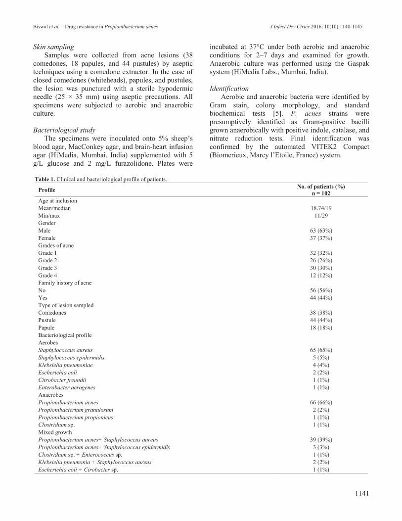
Table 1. Clinical and bacteriological profile of patients.
Profile No. of patients (%)
n = 102
Age at inclusion
Mean/median 18.74/19
Min/max 11/29
Gender
Male 63 (63%)
Female 37 (37%)
Grades of acne
Grade 1 32 (32%)
Grade 2 26 (26%)
Grade 3 30 (30%)
Grade 4 12 (12%)
Family history of acne
No 56 (56%)
Yes 44 (44%)
Type of lesion sampled
Comedones 38 (38%)
Pustule 44 (44%)
Papule 18 (18%)
Bacteriological profile
Aerobes
Staphylococcus aureus 65 (65%)
Staphylococcus epidermidis 5 (5%)
Klebsiella pneumoniae 4 (4%)
Escherichia coli 2 (2%)
Citrobacter freundii 1 (1%)
Enterobacter aerogenes 1 (1%)
Anaerobes
Propionibacterium acnes 66 (66%)
Propionibacterium granulosum 2 (2%)
Propionibacterium propionicus 1 (1%)
Clostridium sp. 1 (1%)
Mixed growth
Propionibacterium acnes+ Staphylococcus aureus 39 (39%)
Propionibacterium acnes+ Staphylococcus epidermidis 3 (3%)
Clostridium sp. + Enterococcus sp. 1 (1%)
Klebsiella pneumonia + Staphylococcus aureus 2 (2%)
Escherichia coli + Cirobacter sp. 1 (1%)
Biswal et al. – Drug resistance in Propionibacterium acnes J Infect Dev Ctries 2016; 10(10):1140-1145.
1142
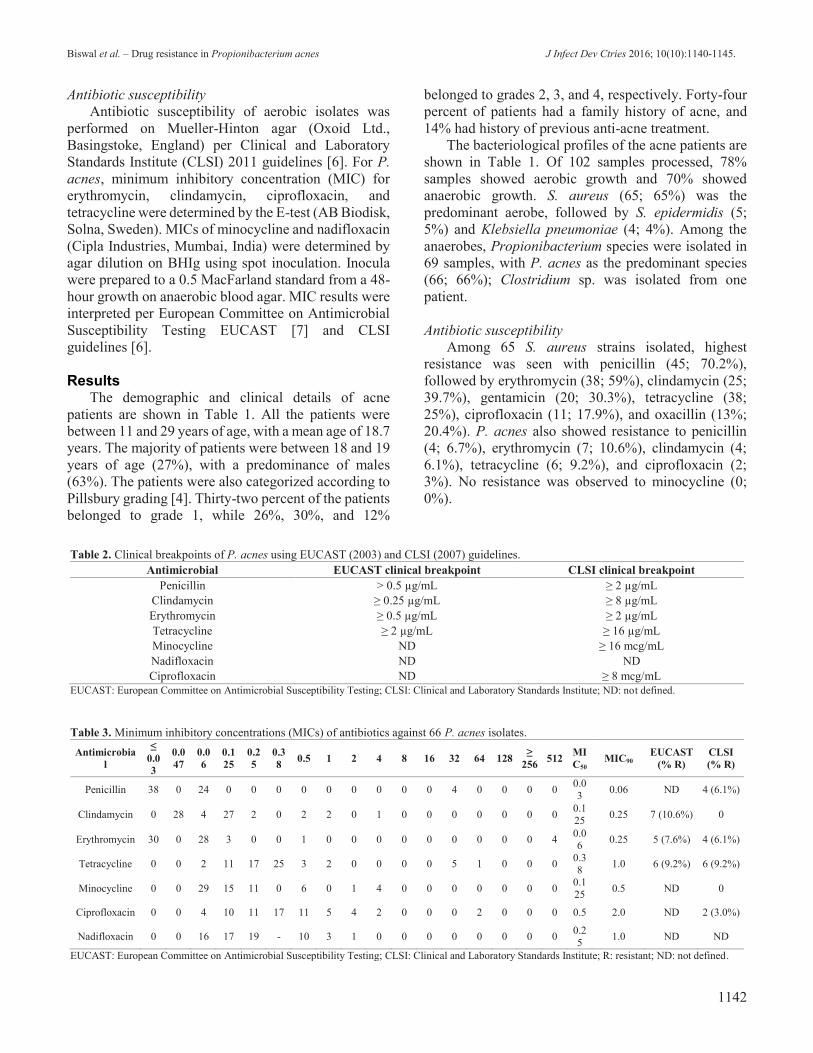
Antibiotic susceptibility
Antibiotic susceptibility of aerobic isolates was
performed on Mueller-Hinton agar (Oxoid Ltd.,
Basingstoke, England) per Clinical and Laboratory
Standards Institute (CLSI) 2011 guidelines [6]. For P.
acnes, minimum inhibitory concentration (MIC) for
erythromycin, clindamycin, ciprofloxacin, and
tetracycline were determined by the E-test (AB Biodisk,
Solna, Sweden). MICs of minocycline and nadifloxacin
(Cipla Industries, Mumbai, India) were determined by
agar dilution on BHIg using spot inoculation. Inocula
were prepared to a 0.5 MacFarland standard from a 48-
hour growth on anaerobic blood agar. MIC results were
interpreted per European Committee on Antimicrobial
Susceptibility Testing EUCAST [7] and CLSI
guidelines [6].
Results The demographic and clinical details of acne
patients are shown in Table 1. All the patients were
between 11 and 29 years of age, with a mean age of 18.7
years. The majority of patients were between 18 and 19
years of age (27%), with a predominance of males
(63%). The patients were also categorized according to
Pillsbury grading [4]. Thirty-two percent of the patients
belonged to grade 1, while 26%, 30%, and 12%
belonged to grades 2, 3, and 4, respectively. Forty-four
percent of patients had a family history of acne, and
14% had history of previous anti-acne treatment.
The bacteriological profiles of the acne patients are
shown in Table 1. Of 102 samples processed, 78%
samples showed aerobic growth and 70% showed
anaerobic growth. S. aureus (65; 65%) was the
predominant aerobe, followed by S. epidermidis (5;
5%) and Klebsiella pneumoniae (4; 4%). Among the
anaerobes, Propionibacterium species were isolated in
69 samples, with P. acnes as the predominant species
(66; 66%); Clostridium sp. was isolated from one
patient.
Antibiotic susceptibility
Among 65 S. aureus strains isolated, highest
resistance was seen with penicillin (45; 70.2%),
followed by erythromycin (38; 59%), clindamycin (25;
39.7%), gentamicin (20; 30.3%), tetracycline (38;
25%), ciprofloxacin (11; 17.9%), and oxacillin (13%;
20.4%). P. acnes also showed resistance to penicillin
(4; 6.7%), erythromycin (7; 10.6%), clindamycin (4;
6.1%), tetracycline (6; 9.2%), and ciprofloxacin (2;
3%). No resistance was observed to minocycline (0;
0%).
Table 2. Clinical breakpoints of P. acnes using EUCAST (2003) and CLSI (2007) guidelines.
Antimicrobial EUCAST clinical breakpoint CLSI clinical breakpoint
Penicillin > 0.5 µg/mL ≥ 2 µg/mL
Clindamycin ≥ 0.25 µg/mL ≥ 8 µg/mL
Erythromycin ≥ 0.5 µg/mL ≥ 2 µg/mL
Tetracycline ≥ 2 µg/mL ≥ 16 µg/mL
Minocycline ND ≥ 16 mcg/mL
Nadifloxacin ND ND
Ciprofloxacin ND ≥ 8 mcg/mL
EUCAST: European Committee on Antimicrobial Susceptibility Testing; CLSI: Clinical and Laboratory Standards Institute; ND: not defined.
Table 3. Minimum inhibitory concentrations (MICs) of antibiotics against 66 P. acnes isolates.
Antimicrobia
l
≤
0.0
3
0.0
47
0.0
6
0.1
25
0.2
5
0.3
8 0.5 1 2 4 8 16 32 64 128
≥
256 512
MI
C50 MIC90
EUCAST
(% R)
CLSI
(% R)
Penicillin 38 0 24 0 0 0 0 0 0 0 0 0 4 0 0 0 0 0.0
3 0.06 ND 4 (6.1%)
Clindamycin 0 28 4 27 2 0 2 2 0 1 0 0 0 0 0 0 0 0.1
25 0.25 7 (10.6%) 0
Erythromycin 30 0 28 3 0 0 1 0 0 0 0 0 0 0 0 0 4 0.06
0.25 5 (7.6%) 4 (6.1%)
Tetracycline 0 0 2 11 17 25 3 2 0 0 0 0 5 1 0 0 0 0.3
8 1.0 6 (9.2%) 6 (9.2%)
Minocycline 0 0 29 15 11 0 6 0 1 4 0 0 0 0 0 0 0 0.1
25 0.5 ND 0
Ciprofloxacin 0 0 4 10 11 17 11 5 4 2 0 0 0 2 0 0 0 0.5 2.0 ND 2 (3.0%)
Nadifloxacin 0 0 16 17 19 - 10 3 1 0 0 0 0 0 0 0 0 0.2
5 1.0 ND ND
EUCAST: European Committee on Antimicrobial Susceptibility Testing; CLSI: Clinical and Laboratory Standards Institute; R: resistant; ND: not defined.
Biswal et al. – Drug resistance in Propionibacterium acnes J Infect Dev Ctries 2016; 10(10):1140-1145.
1143
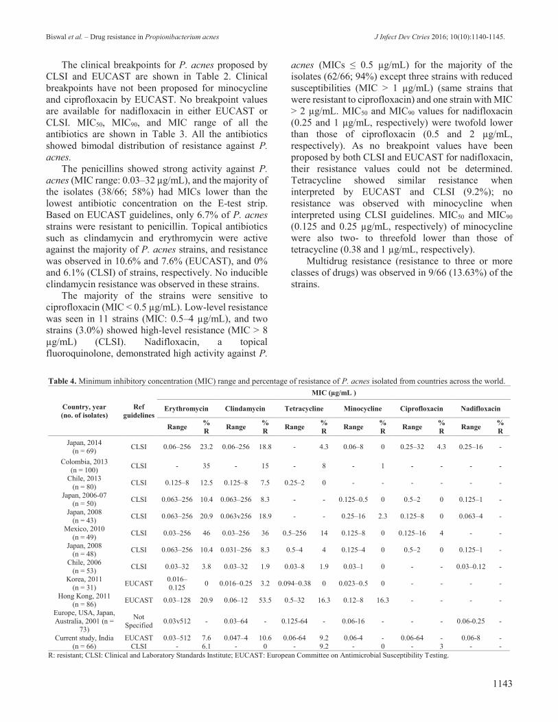
The clinical breakpoints for P. acnes proposed by
CLSI and EUCAST are shown in Table 2. Clinical
breakpoints have not been proposed for minocycline
and ciprofloxacin by EUCAST. No breakpoint values
are available for nadifloxacin in either EUCAST or
CLSI. MIC50, MIC90, and MIC range of all the
antibiotics are shown in Table 3. All the antibiotics
showed bimodal distribution of resistance against P.
acnes.
The penicillins showed strong activity against P.
acnes (MIC range: 0.03–32 µg/mL), and the majority of
the isolates (38/66; 58%) had MICs lower than the
lowest antibiotic concentration on the E-test strip.
Based on EUCAST guidelines, only 6.7% of P. acnes
strains were resistant to penicillin. Topical antibiotics
such as clindamycin and erythromycin were active
against the majority of P. acnes strains, and resistance
was observed in 10.6% and 7.6% (EUCAST), and 0%
and 6.1% (CLSI) of strains, respectively. No inducible
clindamycin resistance was observed in these strains.
The majority of the strains were sensitive to
ciprofloxacin (MIC < 0.5 µg/mL). Low-level resistance
was seen in 11 strains (MIC: 0.5–4 µg/mL), and two
strains (3.0%) showed high-level resistance (MIC > 8
µg/mL) (CLSI). Nadifloxacin, a topical
fluoroquinolone, demonstrated high activity against P.
acnes (MICs ≤ 0.5 µg/mL) for the majority of the
isolates (62/66; 94%) except three strains with reduced
susceptibilities (MIC > 1 µg/mL) (same strains that
were resistant to ciprofloxacin) and one strain with MIC
> 2 µg/mL. MIC50 and MIC90 values for nadifloxacin
(0.25 and 1 µg/mL, respectively) were twofold lower
than those of ciprofloxacin (0.5 and 2 µg/mL,
respectively). As no breakpoint values have been
proposed by both CLSI and EUCAST for nadifloxacin,
their resistance values could not be determined.
Tetracycline showed similar resistance when
interpreted by EUCAST and CLSI (9.2%); no
resistance was observed with minocycline when
interpreted using CLSI guidelines. MIC50 and MIC90
(0.125 and 0.25 µg/mL, respectively) of minocycline
were also two- to threefold lower than those of
tetracycline (0.38 and 1 µg/mL, respectively).
Multidrug resistance (resistance to three or more
classes of drugs) was observed in 9/66 (13.63%) of the
strains.
Table 4. Minimum inhibitory concentration (MIC) range and percentage of resistance of P. acnes isolated from countries across the world.
Country, year
(no. of isolates)
Ref
guidelines
MIC (µg/mL )
Erythromycin Clindamycin Tetracycline Minocycline Ciprofloxacin Nadifloxacin
Range %
R Range
%
R Range
%
R Range
%
R Range
%
R Range
%
R
Japan, 2014
(n = 69) CLSI 0.06–256 23.2 0.06–256 18.8 - 4.3 0.06–8 0 0.25–32 4.3 0.25–16 -
Colombia, 2013
(n = 100) CLSI - 35 - 15 - 8 - 1 - - - -
Chile, 2013
(n = 80) CLSI 0.125–8 12.5 0.125–8 7.5 0.25–2 0 - - - - - -
Japan, 2006-07 (n = 50)
CLSI 0.063–256 10.4 0.063–256 8.3 - - 0.125–0.5 0 0.5–2 0 0.125–1 -
Japan, 2008
(n = 43) CLSI 0.063–256 20.9 0.063v256 18.9 - - 0.25–16 2.3 0.125–8 0 0.063–4 -
Mexico, 2010
(n = 49) CLSI 0.03–256 46 0.03–256 36 0.5–256 14 0.125–8 0 0.125–16 4 - -
Japan, 2008 (n = 48)
CLSI 0.063–256 10.4 0.031–256 8.3 0.5–4 4 0.125–4 0 0.5–2 0 0.125–1 -
Chile, 2006
(n = 53) CLSI 0.03–32 3.8 0.03–32 1.9 0.03–8 1.9 0.03–1 0 - - 0.03–0.12 -
Korea, 2011
(n = 31) EUCAST
0.016–
0.125 0 0.016–0.25 3.2 0.094–0.38 0 0.023–0.5 0 - - - -
Hong Kong, 2011 (n = 86)
EUCAST 0.03–128 20.9 0.06–12 53.5 0.5–32 16.3 0.12–8 16.3 - - - -
Europe, USA, Japan,
Australia, 2001 (n = 73)
Not
Specified 0.03v512 - 0.03–64 - 0.125-64 - 0.06-16 - - - 0.06-0.25 -
Current study, India
(n = 66)
EUCAST 0.03–512 7.6 0.047–4 10.6 0.06-64 9.2 0.06-4 - 0.06-64 - 0.06-8 -
CLSI - 6.1 - 0 - 9.2 - 0 - 3 - -
R: resistant; CLSI: Clinical and Laboratory Standards Institute; EUCAST: European Committee on Antimicrobial Susceptibility Testing.
Biswal et al. – Drug resistance in Propionibacterium acnes J Infect Dev Ctries 2016; 10(10):1140-1145.
1144
Discussion Cutaneous Propionibacterium has been implicated
in acne, although their role in inflammation is still
poorly understood. There is widespread resistance in P.
acnes due to overuse of topical and systemic antibiotics
for treatment of acne vulgaris [3,8-16], as shown in
Table 4. Various studies have used different
interpretative criteria to estimate the resistance among
P. acne strains to different anti-acne drugs. There is a
paucity of data on antibiotic resistance among these
isolates in the literature; also, no standard interpretative
criterion is available for estimating the resistance
among anti-acne drugs in P. acnes. Normally, drug
susceptibility is not requisitioned, owing to the slow
growth of the bacteria and the cost and complexity of
testing methods. However, it is important to get
resistance information so that correct therapeutic
decisions can be made, particularly in resistant cases
not responding to routine therapy. At the same time, if
facilities that are equipped to perform anaerobic culture
are available, the samples should be sent there to get the
exact sensitivity pattern for the particular patient,
because based on the findings of the study, a fair
amount of resistance was observed in the causative
organisms.
Our results confirm that P. acnes (66%) and S.
aureus (65%) predominated the bacterial flora in acne
vulgaris patients. This is in contrast to a study done in
France by Dreno et al., where among 16 different
organisms isolated from acne patients, S. epidermidis
(95%) and P. acnes (90%) were predominant, followed
by S. capitis (47.5%), Micrococcus (47.5%), and P.
granulosum (32.5%) [17]. Another study done in Iran
by Zandi et al. showed a predominance of P. acnes
(57%), followed by S. epidermidis (32%) and S. aureus
(5%) [18]. This difference in microbial profile in our
study could be explained by the variations in
geographical location, host factors, and antibiotic
usage, as has been described previously.
In the present study, it was observed that resistance
among P. acnes to anti-acne drugs was lower than that
among S. aureus. The resistance among S. aureus was
two- to sevenfold higher than P. acnes, with S. aureus
versus P. acnes for: penicillin (70.2% versus 6.1%),
ciprofloxacin (17.9% versus 3%), erythromycin (59%
versus 7.6%), clindamycin (39.7% versus 10.6%), and
tetracycline (25% versus 9.2%). These findings stress
that anti-acne antibiotics continue to maintain activity
against P. acnes. Lower resistance may be due to the
fact that the majority of patients do not undergo
treatment of acne in India.
Previously, researchers have used either EUCAST
or CLSI guidelines to analyze the resistance profile of
P. acnes. In present study, resistance rates of
erythromycin and clindamycin were higher when
analyzed according to EUCAST guidelines (7.6% and
10.6%, respectively), as compared to CLSI (6% and
0%, respectively). Tetracycline resistance was observed
in 9.2% of isolates, irrespective of the interpretative
criteria used. MIC breakpoints for interpretation are
lower in EUCAST guidelines than in CLSI guidelines.
From a clinical and epidemiological point of view,
EUCAST guidelines are better for analyzing the
resistance data than are CLSI guidelines. CLSI
guidelines have been used by researchers for drugs
where EUCAST data is not available.
In the present study, it was observed that the highest
number of isolates (38) showed minimum MIC values
of 0.03 µg/mL to penicillin, followed by erythromycin
and clindamycin. The MIC range of penicillin was
0.03–0.06 µg/mL, although only four strains had MIC
> 32 µg/mL. Penicillin is not a very good anti-acne
antibiotic; however, as it is not available in topical
preparations, and has various adverse effects, namely
penicillin allergy and anaphylaxis. Among topical
antibiotics, erythromycin (0.03–512 µg/mL) and
clindamycin (0.047–4 µg/mL) showed very low
resistance.
MIC50 and MIC90 values of nadifloxacin (0.25 and
1 µg/mL) were found to be twofold lower than those of
ciprofloxacin (0.5 and 2 µg/mL; thus, nadifloxacin
emerged as a better drug for management of acne
patients. MIC50 and MIC90 of minocycline (0.125 and
0.5 µg/mL) was also found to be two- to threefold lower
than those of tetracycline (0.38 and 1 µg/mL). Our
findings are in sync with studies across the world
showing no resistance to P. acnes against minocycline.
We searched for relevant studies indexed in the
PubMed, Medline, and Google databases for articles
with words "Propionibacterium" and
"Propionibacterium acnes India". Based on available
literature and to the best of our knowledge, this is the
first study from India focusing on the bacteriology of
acne vulgaris and P. acnes resistance.
Conclusions Antibiotic resistance in acne vulgaris has gradually
risen over the years, making it difficult to treat these
patients. This is corroborated by evidence of reduced
clinical response to antibiotic therapy, potential
increase in pathogenicity of P. acnes, and transfer of
resistance to more pathogenic organisms. Effective
strategies to combat antibiotic resistance in acne are
Biswal et al. – Drug resistance in Propionibacterium acnes J Infect Dev Ctries 2016; 10(10):1140-1145.
1145
required and include judicious and limited duration of
antibiotic usage and the use of topical retinoids in lieu
of antibiotics.
References 1. Hanna S, Sharma J, Klotz J (2003) Acne vulgaris: More than
skin deep. Dermatol Online J 9: 8.
2. Leyden JJ (1995) New understandings of the pathogenesis of
acne. J Am Acad Dermatol 32: 15-25.
3. Ross JI, Snelling AM, Eady EA, Cove JH, Cunliffe WJ,
Leyden JJ, Collignon P, Dréno B, Reynaud A, Fluhr J, Oshima
S (2001) Phenotypic and genotypic characterization of
antibiotic-resistant Propionibacterium acnes isolated from
acne patients attending dermatology clinics in Europe, the
USA, Japan and Australia. Br J Dermatol 144: 339-346.
4. Witkowski JA, Parish LC (2004) The assessment of acne: An
evaluation of grading and lesion counting in the measurement
of acne. Clin Dermatol 22: 394-397.
5. Miles RS, Amyes SGB (1996) Laboratory control of
antimicrobial therapy. In Collee JG, Fraser AG, Marmion BP,
Simmons A, editors. Mackie and McCartney Practical Medical
Microbiology, 14th edition. Edinburgh: Churchill Livingstone,
Elsevier. 131-150.
6. Clinical and Laboratory Standards Institute (CLSI) (2011)
Performance standards for antimicrobial susceptibility testing;
twenty first informational supplement. CLSI document M100-
S2. Wayne, PA: CLSI.
7. European Committee on Antimicrobial Susceptibility Testing
(2011) Breakpoint tables for interpretation of MICs and zone
diameters, version 1.3, January 5, 2011. Basel: EUCAST.
8. Nakase K, Nakaminami H, Takenaka Y, Hayashi N,
Kawashima M, Noguchi N (2014) Relationship between the
severity of acne vulgaris and antimicrobial resistance of
bacteria isolated from acne lesions in a hospital in Japan. J Med
Microbiol 63: 721-728.
9. Mendoza N, Hernandez PO, Tyring SK, Haitz KA, Motta A
(2013) Antimicrobial susceptibility of Propionibacterium
acnes isolates from acne patients in Colombia. Int J Dermatol
52: 688-692.
10. Schafer F, Fich F, Lam M, Gárate C, Wozniak A, Garcia P
(2013) Antimicrobial susceptibility and genetic characteristics
of Propionibacterium acnes isolated from patients with acne.
Int J Dermatol 53: 418-425.
11. Nakase K, Nakaminami H, Noguchi N, Nishijima S, Sasatsu M
(2012) First report of high levels of clindamycin- resistant
Propionibacterium acnes carrying erm (X) in Japanese patients
with acne vulgaris. J Dermatol: 794-796.
12. Gonzalez R, Welsh O, Ocampo J, Robles RMH, Cabrera LV,
Delaney ML, Gomez M (2010) In vitro antimicrobial
susceptibility of Propionibacterium acnes isolated from acne
patients in northern Mexico. Int J Dermatol 49: 1003-1007.
13. Ishida N, Nakaminami H, Noguchi N, Kurokawa I, Nishijima
S, Sasatsu M (2008) Antimicrobial susceptibilities of
Propionibacterium acnes isolated from patients with acne
vulgaris. Microbiol Immunol 52: 621-624.
14. Gubelin W, Martinez MA, Molina MT, Zapate S, Valenzuela
ME (2006) Antimicrobial susceptibility of strains of
Propionibacterium acnes isolated from inflammatory acne.
Rev Latinoam Microbio l48: 14-16.
15. Song M, Seo SH, Ko HC, Oh CK, Kwon KS, Chang CL, Kim
MB (2011) Antibiotic susceptibility of Propionibacterium
acnes isolated from acne vulgaris in Korea. J Dermatol 38:
667-673.
16. Luk NM, Hui M, Lee HC, Fu LH, Liu ZH, Lam LY, Eastel M,
Chan YK, Tang LS, Cheng TS, Siu FY, Ng SC, Lai YK, Ho
KM (2013) Antibiotic resistant Propionibacterium acnes
among acne patients in a regional skin centre in Hong Kong. J
Eur Acad Dermatol Venereol 27: 31-36.
17. Dreno B, Reynaud A, Moyse D, Habert H, Richet H (2001)
Erythromycin- resistance of cutaneous bacterial flora in acne.
Eur J Dermatol 11: 549-553.
18. Zandi S, Vares B, Abdollahi H (2011) Determination of
microbial agents of acne vulgaris and Propionibacterium
acnesantibiotic resistance in patients referred to dermatology
clinics in Kerman, Iran. Jundishapur J Microbiol 4: 17-22.
Corresponding author Dr. Rajni Gaind
Department of Microbiology, VMMC & Safdarjang Hospital
Room no- 508, New Delhi
110029, India
Phone: 09810528344
Email: [email protected]
Conflict of interests: No conflict of interests is declared.