Embed Size (px)
Citation preview
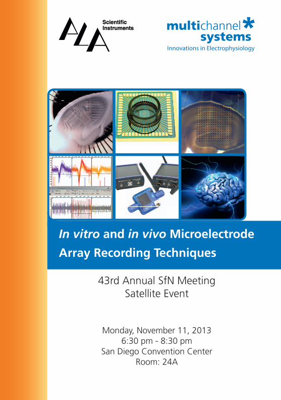
In vitro and in vivo Microelectrode
Array Recording Techniques
43rd Annual SfN MeetingSatellite Event
Monday, November 11, 20136:30 pm - 8:30 pm
San Diego Convention CenterRoom: 24A
Program
Use of Multi-Electrode Arrays for Large-Scale Genetic Studies of Synaptic Transmission Phenotypes in Hippocampal Slices
Maksym Kopanitsa, PhD
Synome Ltd, Babraham Research Campus, Cambridge, UK
Optogenetic Stimulation: From Single Neurons to Populations on MEAs
Thomas DeMarse, PhD
Department of Biomedical Engineering, University of Florida, USA
Simultaneous Stimulation and Detection of Axonal Action Potential Propagation Using High-Density CMOS-Based
Microelectrode Arrays (‘Neurochip’)
Günther Zeck, PhD
Natural and Medical Sciences Institute, Reutlingen, Germany
Investigating the Physiology and Pathophysiology of the Ventral Tegmental Area in Behaving Rodents
Stanislav Koulchitsky, PhD University of Liège, Laboratory of Pharmacology, Liège, Belgium
Use of Multi-Electrode Arrays for Large-Scale Genetic Studies of Synaptic Transmission
Phenotypes in Hippocampal Slices Maksym Kopanitsa, PhD
Synome Ltd, Babraham Research Campus, Cambridge, UK
The importance of the genetic regulation of the nervous system has been widely recognised. Due to evolutionary closeness to humans, mice are one of the most favoured model organisms in genetics. Nonetheless, despite several decades of electrophysiological experiments on hundreds of spontaneous and engineered mouse mutants, the literature does not permit comparison of synaptic phenotypes and thus no general principles describing genetic regulation of vertebrate synaptic transmission have been formulated. Among reasons for this shortcoming are the variability between laboratories in their electrophysiological methods and the absence of large-scale studies comparing many mutations in the identical setting.
We conducted a large-scale study of synaptic transmission utilising CA3-CA1 fEPSPs recorded from hippocampal slices of genetically altered mice and we have accumulated a standardised set of such measurements in over 50 such mutants. In this presentation, I will show how problems of scale and variability can be tackled by the use of multi-electrode array technology. Using examples of phenotypes caused by genetic mutations, I will also consider how traditional electrophysiological measurements, such as fEPSP size, paired-pulse facilitation and long-term potentiation can be utilised for uniform quantitative comparisons of multiple experimental conditions (e.g., comparing different mutations or drug treatments).
Supported by the European Union Seventh Framework Programme under grant agreement numbers HEALTH-F2-2009-241498 (“EUROSPIN” project), HEALTH-F2–2009-242167 (“SynSys project”), and HEALTH-F2-2009-241995 (“GENCODYS project”).
References:
Kopanitsa MV, Afinowi NO, Grant SG. 2006, Recording long-term potentiation of synaptic transmission
by three-dimensional multi-electrode arrays. BMC Neurosci., 7:61.
Ryan TJ, Kopanitsa MV, Indersmitten T et al., 2013, Evolution of GluN2A/B cytoplasmic domains
diversified vertebrate synaptic plasticity and behavior. Nat Neurosci.16(1):25-32.
Optogenetic Stimulation: From Single Neurons to Populations on MEAs
Thomas DeMarse, PhD Department of Biomedical Engineering, University of Florida, USA
Optogenetic tools are light-sensitive proteins derived from single-cell organisms. Upon expression in mammalian neurons, they can be used to effectively control neuronal excitability down to the millisecond timescale. There are two main classes of optogenetic tools that are used to control depolarization and hyperpolarization, and respectively generate or inhibit action potentials in selected populations of neurons. By applying these tools within the MEA platform, it is now possible to gain precise control over the excitation and inhibition of neural activity across large neural populations down to nearly simultaneous and independent control over individual neurons within that population.
This presentation will describe ongoing research in my laboratory using optogenetic stimulation in dissociated culture and cortical/hippocampal slices for modulating excitability using ChR2 and eNpHr. I will also cover specific strategies an MEA researcher might use to facilitate the application of optogenetic tools within their own research including transfection in vitro and in vivo, hardware for stimulation, and various tips on implementing this new stimulation paradigm.
References:
Tye, K. M., & Deisseroth, K. (2012). Optogenetic investigation of neural circuits underlying brain
disease in animal models. Nature Reviews. Neuroscience, 13(4), 251-66. doi:10.1038/nrn3171

Simultaneous Stimulation and Detection of Axonal Action Potential Propagation Using High-
Density CMOS-Based MEAs (‘Neurochip’) Günther Zeck, PhD
Natural and Medical Sciences Institute, Reutlingen, Germany
CMOS based high density microelectrode arrays (MEAs) provide the opportunity to electrically stimulate neurons (Eickenscheidt et al. 2012) and to record the occurrence and propagation of action potentials in the stimulated neurons (Zeck et al. 2011). However, simultaneous electrical stimulation and recording of many neurons has not been possible so far.
Here we present stimulation and recording results obtained with a newly developed electrode array which comprises 4225 recording sites (pitch 16 µm) interlaced with 1024 capacitive stimulation sites. The entire array is insulated by a thin, inert and biocompatible oxide layer. Continuous recording of all sensors over several minutes at a sampling rate of 25 kHz is demonstrated. Electrical stimulation can be performed using arbitrary stimulus shapes presented at different positions. The electrode area can be varied during the experiment.
We selected the ex vivo guinea pig retina as an appropriate nervous tissue. Light-stimuli applied to the retina elicit reliable action potentials in the ganglion cells, which are detected by the multiple sensors on the array. These stimuli allow to precisely locating the position of the ganglion cells. The axonal path is revealed in electrical images calculated from spike triggered averages.
Based on the cell and axon position we demonstrate how stimuli of different polarity, of different shape and position evoke action potentials in nearby or in distant neurons. Action potential detection is achieved within a few hundred microseconds after stimulus onset.
The presented arrays enable bidirectional ‘closed-loop’- type interfacing with other neuronal preparations to target specific sets of neurons.
References:
Zeck G, Lambacher A, Fromherz P. 2011, Axonal Transmission in the Retina Introduces a Small Dispersion of
Relative Timing in the Ganglion Cell Population Response. PLoS ONE 6(6): e20810.
Eickenscheidt M, Jenkner M, Thewes R, Fromherz P, Zeck G. 2012, Electrical stimulation of retinal neurons in
epiretinal and subretinal configuration using a multicapacitor array. J Neurophysiol 107: 2747-2755, 2012.
Investigating the Physiology and Pathophysiology of the Ventral Tegmental Area
in Behaving RodentsStanislav Koulchitsky, PhD
University of Liège, Laboratory of Pharmacology, Liège, Belgium
The mesocorticolimbic reward system is known to mediate the reinforcement of some behaviors which are directly related to the survival (search for food, foraging, sexual activity, etc.). It was also shown to mediate an appreciation of some abstract items lacking natural reward value, but allowing us to engage in the world, such as music and other arts, self-development, etc. This system originates in the ventral tegmental area (VTA) and comprises both dopaminergic (DA) and GABAergic neurons.Cocaine as well as the other addictive drugs „hijack“ this system, totally modifying the behavior of the addicted person. Previous studies have shown that all drugs of abuse increase the concentration of dopamine in the nucleus accumbens, a major target area of the VTA, but the precise dynamics of VTA neurons and of the “VTA network” during drug self-administration is unknown.We are currently studying both neuronal activity in the VTA and some behavioral parameters (i) during natural conditions in an open field and (ii) during acute and chronic exposure to cocaine in awake, freely moving rats using the Multichannel Wireless system. Both extracellular single unit activity and local field potentials are recorded. The latter mostly consist of theta (~8 Hz) rhythms, interspersed with gamma (~50 Hz) activity. In particular, coupling between the phase of the theta rhythm and spiking of individual neurons is investigated. We have also started to study the activity of the VTA during cocaine self-administration. During the talk, some results on these various topics will be presented.One of our next aims is to simultaneously record neuronal activity from the VTA and major afferent brain areas, constituting the reward system. This will hopefully allow us to decipher the spatio-temporal dynamics of this system.The expected outcomes of our work are to: (i) reveal some mechanisms of cocaine addiction and cocaine-induced behavior; (ii) better understand the functional wiring of the reward system.

Notes
Maksym Kopanitsa, PhDMaksym Kopanitsa was born and educated in Ukraine. He received his PhD in Biophysics at the Bogomoletz Institute of Physiology (Kyiv, Ukraine) in the laboratory of Prof. Oleg Krishtal where he carried out research on pharmacology of voltage- and ligand-gated ion channels and synaptic transmission. During his post-doctoral appointments in the labs of Prof. Jeremy Lambert (Univer-sity of Dundee, 2003-2004) and Prof. Seth Grant (The Wellcome Trust Sanger Institute, 2004-2010) Maksym worked with mutant mice to study genetic regulation of the synaptic transmission in brain slices. Now Maksym Kopanitsa works at Synome Ltd in Cambridge, UK. He leads a mul-tidisciplinary team of scientists deciphering phenotypic effects of mutations and pharmacological treatments using electrophysiological recordings, fluorescent imaging and behavioural tests.
Günther Zeck, PhDDr. Zeck completed his PhD in Biophysics at Max Planck Insitute of Biochemsitry in the laboratory of P. Fromherz, where he stimulated and recorded signals from a synaptically connected neuronal network on a semiconductor chip. During his post-doc at the Massachusetts General Hospital and Harvard Medical School in Boston he started to investigate the coding of vertebrate retinas.After his return to Germany he now interfaced retina to semiconductor chips (Neurochips) achiev-ing detection and stimulation at high spatial resolution.Since 2010, Dr. Zeck leads a research group at the NMI at the University Tübingen, where he extends Neurochip applications to other in vitro preparations, with the long term goal of improv-ing neural implants.
Thomas DeMarse, PhDDr. Thomas DeMarse received his MS and PhD in Learning and Memory at Purdue University with Dr Peter Urcuioli. Dr DeMarse has worked as research fellow in the Biology Department at the California Institute of Technology and the Biomedical Engineering Department at Georgia Tech interfacing cortical neurons to computer systems using MEAs in Dr Steve Potter’s Laboratory. He is currently research scientist in Neural Engineering in the Department of Biomedical Engineering at the University of Florida. Dr DeMarse’s research interests include basic research into neural computation including information processing and transmission in combination with MEMs tun-nel devices, learning and memory in living neural systems including adapting plasticity for cortical/hippocampal remodeling (antiepileptogenesis) in epilepsy.
Stanislav Koulchitsky, PhDDr. Koulchitsky completed his PhD in biology at the Institute of Physiology (National Academy of Sciences, Minsk, Belarus), where he studied the role of dorsomedial and ventrolateral medulla in the regulation of pain sensitivity during endotoxemia. Later, he investigated mechanisms regulat-ing the neuronal excitability in the trigeminal ganglion and the spinal trigeminal nucleus (Insti-tute of Physiology and Experimental Pathophysiology, Erlangen, Germany). In 2008 he joined the Laboratory of Pharmacology (GIGA Neurosciences, University of Liège). He was involved in the in-stallation of the telemetric system for the registration of neuronal activity from freely moving rats.His current research fields are the functional wiring of the reward system, the effect of narcotic drugs on the neuronal populations constituting the reward system, and the basic mechanisms of addiction.
www.multichannelsystems.com www.alascience.com
Speakers