Embed Size (px)
Citation preview
For Peer Review
In vitro and in vivo evaluation of positively charged liposaccharide derivatives as oral absorption enhancers for
the delivery of anionic drugs
Journal: Journal of Pharmaceutical Sciences
Manuscript ID: 09-326
Wiley - Manuscript type: Research Article
Date Submitted by the Author:
22-May-2009
Complete List of Authors: Bergeon, Julie; The University of Queensland, School of Chemistry
and Molecular Biosciences Ziora, Zita; The University of Queensland, Centre for Integrated Preclinical Drug Development (CIPDD) Abdelrahim, Adel; The University of Queensland, School of Chemistry and Molecular Biosciences Pernevi, Niklas; The University of Queensland, School of Chemistry and Molecular Biosciences Moss, Anne; The University of Queensland, School of Chemistry and Molecular Biosciences Toth, Istvan; The University of Queensland, Centre for Integrated Preclinical Drug Development (CIPDD); The University of
Queensland, School of Chemistry and Molecular Biosciences
Keywords: Oral drug delivery, Absorption enhancer, Bioavailability, Caco-2 cells, Calorimetry (ITC), Pharmacokinetics/pharmacodynamics, Cationic lipids
John Wiley & Sons, Inc.
Journal of Pharmaceutical Sciences
For Peer Review
In vitro and in vivo evaluation of positively charged liposaccharide derivatives as oral absorption
enhancers for the delivery of anionic drugs
Julie A. Bergeon1, Zyta M. Ziora
2, Adel S. Abdelrahim
1, Niklas U. Pernevi, Anne R. Moss
1, Istvan Toth
1,2
1 The University of Queensland, School of Chemistry and Molecular Biosciences, Brisbane, Qld 4072,
Australia 2 The University of Queensland, Centre for Integrated Preclinical Drug Development (CIPDD),
Brisbane, Qld 4072, Australia
Keywords: charged liposaccharide, ion-pairing, Caco-2 cells, calorimetry (ITC), absorption enhancer,
piperacillin, oral drug delivery, bioavailability
ABSTRACT
Oral delivery of hydrophilic, ionisable drugs remains a major challenge in drug development and a
number of active pharmaceuticals fail to reach the market of oral drugs because of a lack of
absorption and/or stability issues. One possible approach to improving the bioavailability of such
drug candidates is to increase their lipophilicity, which is a key parameter in the permeation across
cell membranes. However, modifying the chemical structure of an active compound by adding lipid
residues often results in changes in activity. With ionised molecules, ion-pairing can be considered to
associate charged lipid moieties with the parent drug without altering its structure and therefore
activity. This study presents the results of in vitro and in vivo evaluation of a series of positively
charged liposaccharide derivatives combined with an anionic model drug, piperacillin. The
antimicrobial activity, plasma stability, permeability in Caco-2 cell monolayers and oral absorption of
the synthesised conjugates were assessed.
INTRODUCTION
Recent advances in drug development have shown the crucial need for developing new active drugs
to treat and/or prevent diseases which currently resist conventional therapies.
However, the promising therapeutical profiles of a large number of potential drug candidates
(including ionic/ionisable molecules, peptides and protein-like pharmaceuticals with a high
hydrophilic character) is often dampened by issues such as low oral bioavailability and poor in situ
stability.1,2
To overcome these difficulties, different strategies such as structural modifications by
adjunction of various protecting groups to improve stability, or use of surfactants and absorption
enhancers to increase membrane permeability, essentially by passive diffusion, have been
investigated. 3,4
Considering the lipidic nature of the bilayered cell membrane, the derivatisation of
drugs by lipidation has been extensively investigated and improvements in enzymatic stability and
membrane permeability have been reported. 5,6
Although increased lipophilicity can facilitate
absorption across membranes, it is also frequently associated with solubility difficulties in aqueous
media.
Another approach to overcome absorption problems revolves around the ionizability of polar
molecules. Hydrophilic drugs are commonly commercialised as salts (e.g. sodium, acetate, chloride
salts), more soluble and often more stable7 than the parent molecule, which means that the drug
itself can potentially exhibit charges depending on pH conditions (both in solution and in situ). This
offers ample possibilities to associate lipophilic counter-ionic moieties by ion-pairing in order to
enhance the overall lipophilicity of these hydrophilic drugs while preserving their biologically active
chemical structure. 8
Page 1 of 20
John Wiley & Sons, Inc.
Journal of Pharmaceutical Sciences
123456789101112131415161718192021222324252627282930313233343536373839404142434445464748495051525354555657585960
For Peer Review
In an earlier study, we reported that the in vitro permeability of piperacillin across Caco-2
monolayers could be notably improved by associating this anionic β-lactam antibiotic to a positively
charged liposaccharide containing a D-glucose moiety coupled to a twelve-carbon long lipoamino
acid (α-amino acid with lipophilic alkyl side-chain). 9 Positive results were also observed when
varying the lipid chain length. Lipoamino acids (LAAs) are versatile molecules with the unique ability
to be linked through either their amino or carboxyl extremities. They can be readily obtained from
alkyl bromide precursors in a few, short and easy synthetic steps. Lipoamino acids combined with
carbohydrates offer several advantages versus lipids alone. Firstly, it allows for a modulation of the
degree of lipophilicity brought by the lipid chain via the hydroxyl residues present on the
carbohydrate moiety, hence preserving the water solubility characteristics of the modified drug
while still increasing its lipophilicity. Secondly, sugars can be selected to interact with specific
membrane receptors where available, offering the possibility of a mediated transport across
membranes as an alternative to passive diffusion.
The amine function of the lipoamino acids is subject to acido-basic equilibrium and therefore is not
constantly protonated, which can be problematic as this residual charge is the key element of the
interaction between the drugs of interest and counter ionic liposaccharides. Quaternization of the
terminal amine was therefore considered to create a constantly, positively charged residue. Also, as
the sugar moiety introduced a significant degree of hydrophilicity, additional lipophilic components
were considered in the design of new charged liposaccharides for a better, adjustable balance of
lipophilicity versus hydrophilicity.
MATERIALS AND METHODS
1. Chemistry and analytical characterisation
All solvents and reagents were obtained at the highest available purity from Sigma–Aldrich (Castle
Hill, NSW, Australia) and used directly without further purification. The liposaccharide derivatives
presented in this publication were prepared according to the methodology described by Abdelrahim
et al. 10
. Commercially available piperacillin sodium was converted into piperacillin acid using an IR-
120-[H+] cationic exchange resin (Rohm Haas, Philadelphia, PA, USA). The salt was dissolved in water
and vigorously stirred with the resin for 30 mins, until pH stabilised at 3 and piperacillin acid
precipitated. The precipitate was then dissolved in acetonitrile and the resin was filtered off, washed
with acetonitrile, and the filtrate was then lyophilised on an Alpha 2-4/LSC (Martin Chris, Osterode
am Harz, Germany) at -80°C, < 150 psi. Two methodologies for lyophilisation were used; (i) with
piperacillin sodium, the liposaccharide derivatives were mixed with the commercial salt and
dissolved in pure glacial acetic acid; the excess acid was removed under vacuo and the mixture was
then lyophilised; (ii) with piperacillin acid, the liposaccharide derivatives and the drug were dissolved
in water/acetonitrile (1:1) and lyophilised.
ESI-MS and MS/MS analyses were carried out on a PE Sciex API3000 triple quadrupole mass
spectrometer, using a mixture of solvent A (1% formic acid in water) and B (1% formic acid in 9:1
acetonitrile/water) at 0.1 mL/min. For LC–MS/MS experiments, a Phenomenex luna C18 column (5
µm, 50 mm × 2.0 mm) equipped with a C18 guard column (4 × 3.0 mm) was attached to the mass
spectrometer and a 30-100% gradient of B in A over 4 minutes was used for elution at a flow rate of
0.5 mL/min with a 1:10 splitter upstream from the ionisation source (Shimadzu LC-10AT system). 9
Data were acquired with Analyst 1.4 software (Applied Biosystems/ MDS Sciex, Toronto, Canada).
2. Isothermal Titration Microcalorimetry (ITC)
ITC measurements were performed on a MicroCal VP-ITC microcalorimeter (Northampton, MA,
USA), with Origin 5.0 and VP viewer 2000 software. Experiments were performed at 37°C in purified
water (MiiliQ Gradient A10, Millipore, North Ryde, NSW, Australia) with degassed, sonicated
Page 2 of 20
John Wiley & Sons, Inc.
Journal of Pharmaceutical Sciences
123456789101112131415161718192021222324252627282930313233343536373839404142434445464748495051525354555657585960
For Peer Review
samples. The reference cell was loaded with water while the sample cell (1.4395 mL) was charged
with piperacillin acid (0.05 – 0.5 mM) and rotated at 300 rpm. The liposaccharide derivatives (4 mM)
were injected into the sample cell using a microsyringe at a rate of 3-10 µL every 6 minutes (total
injection volume 300 µL).
3. Antimicrobial assays (MIC)
MIC experiments were carried using a broth dilution method. Two strains of bacteria were used,
Escherichia coli (MC4100) and Pseudomonas aeruginosa (A01). Bacteria were cultured in Luria-
Bertani (LB) broth medium (1% w/v NaCl, 1% w/v tryptone, 0.5% w/v yeast extract, adjusted to pH 7
and autoclaved at 121°C for 20 minutes before use; reagents purchased from Sigma-Aldrich, Castle
Hill, NSW, Australia). Bacterial growth was monitored by optical density (OD) measurements,
performed on an Ultrospec 2000 (Pharmacia Biotech, Uppsala, Sweden). Bacteria were grown to an
OD ≥ 1 then diluted with LB broth and sub-cultured again to reach an OD of 0.3-0.8. They were then
diluted to a count of 1 × 10⁶ cfu / 100 µL (OD 0.1) and plated in 96-well round bottom plates (TPP,
Zurich, Switzerland).
Lyophilised preparations of piperacillin and liposaccharides were dissolved in 5% dimethylsulfoxide
(DMSO)/water and serially diluted (1:3) with LB broth to reach a final piperacillin concentration of 0-
25 mM for E. coli and 0-52 mM for P. aeruginosa. The bacteria (100 µL) were then mixed with the
drug solutions (100 µL) and incubated for 16-20 hrs at 37°C, after which bacterial growth was
assessed by OD measurements at 600 nm on a Spectramax 250 plate reader (Molecular Devices,
Sunnyvale, CA, USA). The MICs for each formulation was then determined by plotting the OD values
vs piperacillin concentrations.
4. Haemolytic assays
Haemolytic assays were undertaken with approval from the University of Queensland Ethics
Committee (approval # 2006000950). Full venous blood samples were taken from healthy adult
volunteers. Red blood cells (hRBCs) were isolated from the whole blood by centrifugation at 2576 G
for 15 min (Sigma 2-5 centrifuge, Sigma Laborzentrifugen GmbH, Osterode am Harz, Germany).
Plasma was removed and the cells were washed several times with PBS before being resuspended in
PBS (to original blood volume) and aliquoted (100 µL) in 96-well flat bottom plates (TPP, Zurich,
Switzerland).
Solutions of the tested compounds were prepared in PBS (0.2 – 20 µM) and dispensed in the wells
containing the hRBCs preparations. The plates were then incubated at 37°C for 60 mins on a
Heidolph ShakingIncubator & Titramax 1000 (Schwabach, Germany) at 400 rpm, after which they
were centrifuged at 2129 G for 15 min; 75 µL of the supernatant was then removed and placed into
new plates before absorbance was measured at 540 nm using a Spectramax 250 microplate reader.
PBS was used as a negative control; water and SDS were used as positive controls. The percentage of
haemolysis caused by each of the tested formulation was calculated by the following equation:
where min A540 represents the absorbance at 540 nm of PBS alone, and max A540 represents the
absorbance at 540 nm of SDS.
5. Caco-2 permeability assay
Cell culture reagents were purchased from Gibco-BRL (Grand Island, NY, USA) except of Hank’s
balanced saline solution (HBSS) and 14
C-labelled mannitol which were supplied by Sigma–Aldrich
(Castle Hill, NSW, Australia) and Amersham Biosciences (Piscataway, NJ, USA), respectively. Tissue
culture flasks (75 cm2) were obtained from BD Bioscience (Franklin Lakes, NJ, USA).
Page 3 of 20
John Wiley & Sons, Inc.
Journal of Pharmaceutical Sciences
123456789101112131415161718192021222324252627282930313233343536373839404142434445464748495051525354555657585960
For Peer Review
Caco-2 cells from the American type culture collection (Rockville, MD, USA) were maintained in
Dulbecco’s modified Eagle’s medium (DMEM) supplemented with 10% foetal bovine serum (FBS),
and 1% nonessential amino acids at 95% humidity and 37°C in an atmosphere of 5% CO2. The
medium was changed every second day. After reaching 80% confluence, the cells were subcultured
using 0.2% ethylenediaminetetraacetic acid (EDTA) and 0.25% trypsin. 100 µL of a solution
containing approx. 106 cells/mL (passage number 31-40) were seeded onto polycarbonate
Transwell® inserts supplied by Coastar (Cambridge, MA, USA; mean pore size = 0.45 µm, 6.5 mm
diameter) and cultivated in DMEM supplemented with 10% FBS, 1% non-essential amino acids, 100
U/mL penicillin and 100 mg/mL streptomycin. The cells were allowed to grow for 21–28 days and the
medium was changed every other day.
The tested compounds were dissolved in HBSS–HEPES (HBSS buffered with 25 mM 4-(2-
hydroxyethyl)piperazine-1-ethanesulfonic acid (HEPES) at pH 7.4) to a final concentration of 200 µM.
Prior to the transport studies, the Caco-2 monolayers were washed three times with pre-warmed
HBSS–HEPES and the integrity of the monolayers was assessed by measuring the TEER values using a
Millicell-ERS system (Millipore Corp., Bedford, MA, USA); TEER were also measured after each
experiment. Typical values were in the range 0.8-1.6 kΩ.cm2. The drug solutions (100 µL) were added
to the donor side of the monolayers and the Transwell® plates were placed in a shaking incubator
set to 400 rpm and 37°C for the duration of the experiment. At pre-determined time points (30, 90,
120 and 150 min), samples (400 µL) were taken from the receiver chamber and replaced with the
same amount of HBSS–HEPES. All compounds were tested in two to three independent assays, using
four wells each time. Concentrations in piperacillin were determined by LC–MS/MS. The transport of 14
C-mannitol (200 µM), a marker of paracellular transport, was also measured in parallel. After
dilution of samples (400 µL) with 4 mL of Wallac OptiPhase HiSafe 3 liquid scintillation cocktail
(Montreal, Canada), radioactivity was measured using a Beckman Coulter LS650 Multipurpose liquid
scintillation spectrometer (Beckman Instruments, Fullerton, CA) and permeability values determined
by the following equation:
where dC/dt is the steady-state rate of change in the chemical in the receiver chamber (mM, or dpm
mL-1
), Vr is the volume of the receiver chamber (mL), A is the surface area of the cell monolayers
(cm2) and C0 is the initial concentration in the donor chamber (mM, or dpm.mL
-1).
6. Oral absorption studies
Animal studies were undertaken with approval from the University of Queensland Ethics Committee
(approval #SMMS/002/08/ARC). 8-week old male Sprague Dawley rats (300-350g, 4 rats/compound)
were administered formulations of the tested compounds in 5% DMSO/water by oral gavage, to a
level of 50 mg/kg of piperacillin for each rat. At regular time points (0, 5, 10, 15, 30, 60, 90, 120
minutes), blood samples (200 µL) were collected in heparinised tubes by tail vein bleeding. The rats
were euthanized immediately after the experiment by inhalation of a 1:1 O2/CO2 mixture; death was
further asserted by cervical dislocation.
Blood samples were centrifuged for 20 min at 1717 g and the plasma was collected, treated with
acetonitrile (1:1 v/v) to precipitate the proteins and centrifuged. The supernatant was then collected
for analyses by LC-MS/MS (or kept at -30°C if analyses could not be run immediately).
7. Statistical analysis
Results were expressed as mean values ± standard error mean (SEM). Where applicable, statistical
analysis of values was performed by one-way analysis of variance (ANOVA) of repeated measures to
a significance level of 0.05 (p < 0.05), followed by Tukey’s post hoc test (multiple pairwise
Page 4 of 20
John Wiley & Sons, Inc.
Journal of Pharmaceutical Sciences
123456789101112131415161718192021222324252627282930313233343536373839404142434445464748495051525354555657585960
For Peer Review
comparison of means). Computations were realised using GraphPad Prism v5.01 (GraphPad
software, La Jolla, CA, USA).
RESULTS AND DISCUSSION
Design and preparation of a new series of charged liposaccharide derivative carriers
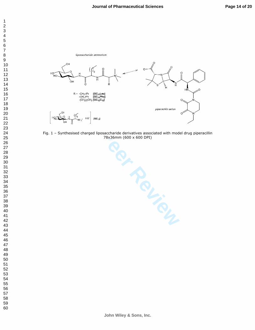
A novel series of liposaccharide derivatives was synthesised by modifying the amino extremity of the
liposaccharide. The resulting compounds comprised a D-glucose (G) moiety and a twelve carbon long
LAA (C12) scaffold, onto which additional amino residues, namely leucine, phenylalanine (two of the
most hydrophobic amino acids) 11
and another C12 were attached. The N-terminus amino group was
then converted into a quaternary ammonium salt using Amberlite 400 [HO-] as a base
10 and the
obtained liposaccharide derivatives were lyophilised together with the model drug piperacillin (Fig.
1).
Fig. 1 – Synthesised charged liposaccharide derivatives associated with model drug piperacillin
Initially, lyophilisation was attempted directly with the commercially available piperacillin sodium
salt under acidic conditions (glacial acetic acid); however, preliminary observations (binding studies
and nuclear magnetic resonance analyses, results not shown) suggested that limited interactions
occurred between the anionic drug and its cationic liposaccharide counterparts. It is believed that
this could be due to strong ionic binding between the piperacillin anion and the sodium cation,
which is smaller and has a greater charge density compared to the charged liposaccharide
derivatives (calculated Hückel charge of 0.788 (GC12C12)); as a consequence, the exchange between
the two cations was likely to be thermodynamically unfavourable. To facilitate this substitution,
piperacillin sodium was dissolved in water and stirred with a cation-exchange resin under controlled
pH to obtain piperacillin acid, which was then immediately lyophilised with different liposaccharide
derivatives from a 1:1 acetonitrile-water solution. The prepared conjugates were stored at -20°C and
their integrity was monitored by analytical examination by 1H-NMR.
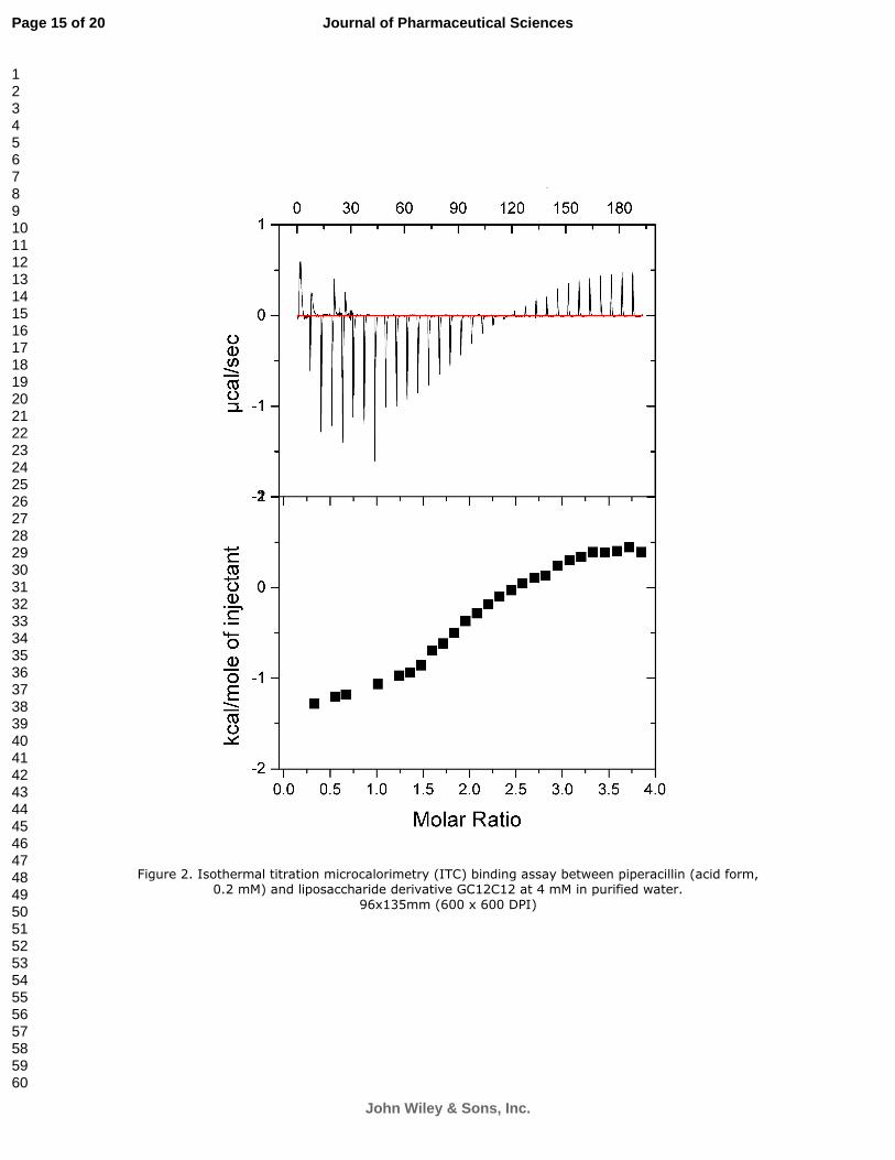
Different ratios of drug/liposaccharide were considered in this study. Initial measurements of
binding by isothermal titration microcalorimetry (ITC) revealed that two concurrent processes were
taking place, binding (ion-pairing) and micellization. A 1:1 ratio did not seem to be sufficient to
observe optimum ionic interactions, possibly due to the presence of multiple interaction sites on the
model drug. Indeed, when examining interactions between piperacillin and GC12C12, a ratio close to
1:2 drug/liposaccharide was found to give optimal ion interactions (Fig. 2). The negative enthalpy
observed initially below a 1:2 ratio is indicative of favourable ion-pairing, while above 1:2, the
process becomes endothermic as a result of micellization occurring. As the ratio of
drug/liposaccharide was increased, the micellization of the lipidic cations was predominantly
observed over the binding process (results not shown) and therefore ion-pairing, if present, could
not be significantly assessed.
Figure 2. Isothermal titration microcalorimetry (ITC) binding assay between piperacillin (acid form,
0.2 mM) and liposaccharide derivative GC12C12 at 4 mM in purified water.
All liposaccharide derivatives were therefore tested in association with piperacillin in a 1:2
drug/counter-ion ratio. A 1:1 ratio was also examined to see if even a lower quantity of
Page 5 of 20
John Wiley & Sons, Inc.
Journal of Pharmaceutical Sciences
123456789101112131415161718192021222324252627282930313233343536373839404142434445464748495051525354555657585960

For Peer Review
liposaccharide derivatives was sufficient to generate an increase in oral absorption (penetration
enhancing effect).
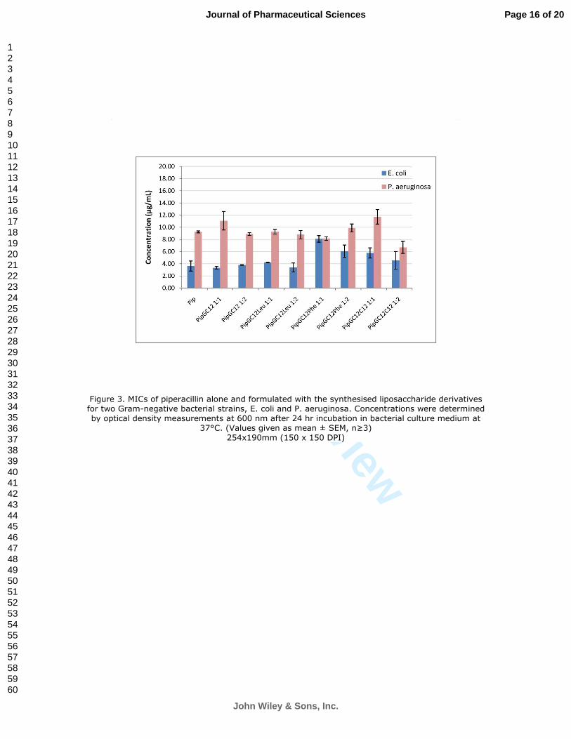
Antimicrobial activity
The antimicrobial activity of the drug conjugates was assessed in vitro in two Gram-negative
bacterial strains, Escherichia coli and Pseudomonas aeruginosa. Minimum inhibitory concentrations
(MICs) were determined by a broth dilution method 12
and compared to the MIC of piperacillin
alone. Previously synthesised 2-amino-N-(2’-α,D-gluco)dodecanamide (GC12) 9 was also tested in the
same 1:1 and 1:2 molar ratios.
After 16-20 hours incubation with bacterial cultures at 37 °C, resistance to piperacillin alone was
found to be in agreement with reference values of 2-4 µg/mL for E. Coli 13
and 3-6 µg/mL for most
strains of P. aeruginosa. 14,15
. When the drug was tested in association with the synthesised charged
liposaccharide derivatives, no significant change was observed in the MIC values for P. aeruginosa,
all averaging 7-12 µg/mL (Fig. 3). For E. coli, slight, however not significant, increases in the MICs
were noticed when piperacillin was formulated with GC12Phe (1:1 and 1:2) and GC12C12 (1:1).
The pairing of the liposaccharide derivatives to piperacillin therefore does not appear to have
negatively impacted the original antimicrobial activity against either bacterial strain. The absence of
significant variation in MICs (verified using a one-way Anova statistical comparison followed by
Tukey’s post-hoc test) when using different ratios of liposaccharides would indicate that the
modified liposaccharides do not have antimicrobial or antagonist properties of their own.
Figure 3. MICs of piperacillin alone and formulated with the synthesised liposaccharide derivatives
for two Gram-negative bacterial strains, E. coli and P. aeruginosa. Concentrations were
determined by optical density measurements at 600 nm after 24 hr incubation in bacterial culture
medium at 37°C. (Values given as mean ± SEM, n≥3)
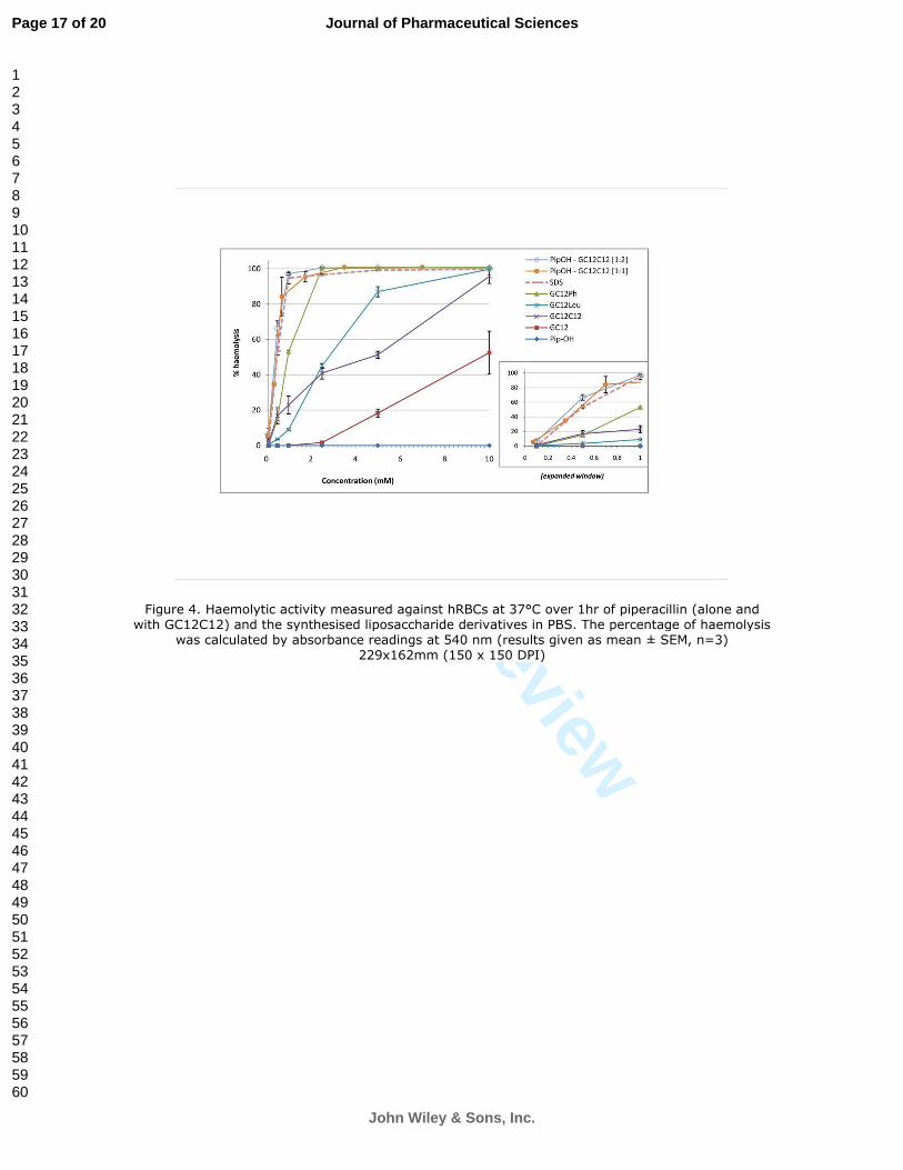
Haemolytic activity
Although destined to oral drug delivery, the liposaccharide derivatives and their ionic complexes
with piperacillin were nonetheless tested for signs of haemolytic activity, which could cause
disruptions to the biological membranes and yield to toxicity.16,17
Haemolytic activity was assessed in
triplicate against human red blood cells (hRBCs) over 60 minutes at 6 different concentrations,
ranging from 0.1 µM to 10 mM; the tested solutions were formulated in phosphate buffered saline
(PBS) and compared to piperacillin alone. Sodium dodecylsulphate (SDS), a surfactant known to be
toxic to hRBCs at the concentrations used in this assay, was also included as a positive control.
Measurements were done by absorbance readings at 540 nm and the percentage of haemolysis was
determined by the following equation:
Initially, only the synthesised liposaccharide derivatives were considered; one of the complexes, Pip-
GC12C12, was additionally evaluated to examine any possible synergistic effect.
Figure 4. Haemolytic activity measured against hRBCs at 37°C over 1hr of piperacillin (alone and
with GC12C12) and the synthesised liposaccharide derivatives in PBS. The percentage of haemolysis
was calculated by absorbance readings at 540 nm (results given as mean ± SEM, n=3)
Page 6 of 20
John Wiley & Sons, Inc.
Journal of Pharmaceutical Sciences
123456789101112131415161718192021222324252627282930313233343536373839404142434445464748495051525354555657585960
For Peer Review
As shown in Figure 4, the synthetic liposaccharide derivatives expectedly induced haemolysis of
hRBCs to some extent, yet the values remain significantly less than those of SDS in the low
concentration range. Interestingly, the comparison between GC12 and GC12C12 revealed a two-fold
increase on average in haemolytic activity, suggesting a direct correlation between the greater
lipophilicity brought by the second C12 moiety and the haemolytic properties of the liposaccharide.
At commonly used therapeutic concentrations however (i.e. < 1 mM when considering the
standardised intravenous administration of piperacillin sodium), most liposaccharide derivatives
tested showed less than 25 % haemolysis, which is considered to be the threshold for tolerable
toxicity to hRBCs. 18
However, piperacillin in association with GC12C12 in 1:1 and 1:2 ratios yielded
synergistic effects resulting in increased haemolysis, to levels comparable to SDS.
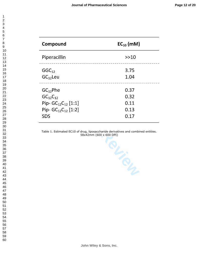
When examining the EC10 (drug concentration causing 10% haemolysis of hRBCs; see Table 1 – the
values were estimated from the graphical data presented in Fig. 4), GC12 and GC12Leu were found to
be moderately haemolytic (0.5 mM < EC10 < 5 mM) while the other two liposaccharide derivatives
GC12Phe and GC12C12 as well as the combined Pip-GC12C12 were classified as strongly haemolytic (0.5
mM < EC10), along with SDS. 17
Table 1. Estimated EC10 of drug, liposaccharide derivatives and combined entities.
These findings are of great importance when considering LAAs as penetration enhancer candidates
for general drug delivery. Although the concentrations used in this assay are far greater than those
used in vivo, the LAA-based molecules tested here showed noticeable signs of toxicity against hRBCs.
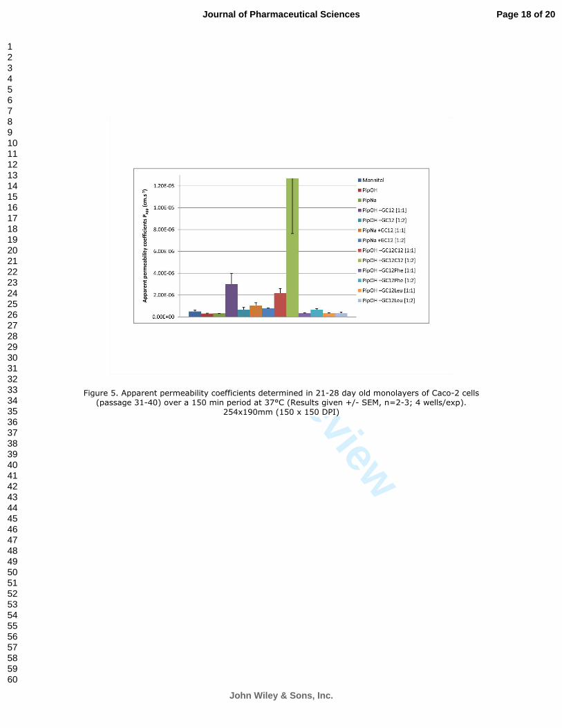
In vitro permeability assessment in Caco-2 cells
Prior to conducting oral absorption studies, the in vitro permeability of the synthesised compounds
was assessed in Caco-2 cells. This cell line originates from a human colorectal adenocarcinoma and is
commonly used as a model for predicting the transport of drugs across the intestinal epithelium.
When cultured, Caco-2 cells spontaneously form differentiated, polarised monolayers which express
most structural and functional characteristics of the small intestine, including enzymes and efflux
proteins. Artursson et al. demonstrated that the apparent permeability coefficients determined with
this model can be correlated to human oral drug absorption values with good respect. 19
.
200 µM solutions of piperacillin associated with the different liposaccharide derivatives (1:1 and 1:2
molar ratios) in Hank’s balanced salt solution (HBSS) were added to the apical side of the Caco-2
monolayers and permeability to the basolateral side of the monolayer was studied over 150 min,
using the paracellular marker 14
C-mannitol as a negative control (typical permeability values of 14
C-
mannitol are around 1-5 × 10⁻⁷
cm.s-1
19
). Apparent permeability coefficients (Fig. 5) were calculated
from concentrations of piperacillin determined by liquid chromatography coupled to mass
spectrometry (LC-MS/MS) against an 8 point standard curve while 14
C-mannitol was quantified by
liquid scintillation counting (β-emission) 9.
Piperacillin alone expectedly showed very low permeability across Caco-2 monolayers; the observed
values were higher than reported previously, 3.07(OH)-3.22(Na) × 10-7
cm.s-1
versus 3.24(Na) × 10-8
cm.s-1
9. A similar 10-fold variation was noticed for Pip-GC12 [1:1] and
14C-mannitol, this variability
could be explained by the difference in cell passage numbers, since the cells used in the present
study were much younger than in the previous examination. Reports in literature have shown that
the differentiation of the cells was affected by early passage numbers, mainly a lower cohesion of
Page 7 of 20
John Wiley & Sons, Inc.
Journal of Pharmaceutical Sciences
123456789101112131415161718192021222324252627282930313233343536373839404142434445464748495051525354555657585960
For Peer Review
the monolayer and reduced tightness of the paracellular junctions. 20
This was also evidenced by
measurement of transepithelial electrical resistances (TEER), 21
which averaged 0.9-1.1 kΩ in this
study, slightly less than in previous experiments.
As shown in Figure 5, piperacillin demonstrated a marked increase in membrane permeability when
associated in a 1:1 ratio with GC12, which is in agreement with previously reported data. 9
Interestingly, when testing GC12C12 in that same ratio, the increase was slightly less. This could be
correlated to the ITC data which suggest that a 1:1 ratio was not sufficient in the case of GC12C12 to
obtain optimal binding between the parent drug and the charged liposaccharide derivatives. Indeed,
when assessing the drug in a 1:2 ratio with the liposaccharide derivative, the permeability was
greatly enhanced to reach a mean value of 1.27 × 10⁻⁵
cm.s-1
, nearly forty times higher than
piperacillin alone. Drugs with apparent permeability coefficients across Caco-2 monolayers of 10-6
cm.s-1
or greater usually present excellent oral bioavailability. 19
; the results obtained in this in vitro
assay therefore suggest that Pip-GC12C12 [1:2] might be a good candidate for oral absorption
evaluation.
Figure 5. Apparent permeability coefficients determined in 21-28 day old monolayers of Caco-2
cells (passage 31-40) over a 150 min period at 37°C (Results given +/- SEM, n=2-3; 4 wells/exp).
The major increase in permeability observed with Pip-GC12C12 [1:2] can also, to a certain extent, be
linked to its toxicity, as evidenced earlier in the haemolytic assay. The measurement of the TEER
values prior to, and after each experiment revealed a drop in resistance across the monolayer, sign
of loss of integrity of the monolayer. It is however unknown whether the loss was permanent or if
the monolayer would recover given time and optimum culture conditions. The other conjugates
tested did not yield to such marked variation in TEER. Surprisingly, the penetration of piperacillin
was not significantly improved when combined with GC12 in a 1:2 molar ratio. No satisfactory ITC
data could be obtained for GC12 so the ratio for optimal binding could not be assessed accurately. It
is also possible that alternative phenomena occurred that could decrease the permeability
enhancing effects of GC12 (e.g. efflux, aggregation, adhesion to membranes). The other
liposaccharide derivatives tested in formulation with piperacillin did not yield significant changes in
permeability, independent of the ratio used.
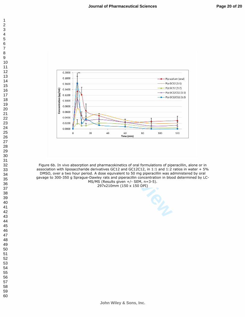
Oral absorption in rats
Based on the in vitro results and particularly the permeability assays, four of the tested formulations
were evaluated in vivo, namely Pip-GC12 [1:1], Pip-GC12 [1:2], Pip-GC12C12 [1:1] and Pip-GC12C12 [1:2].
Male Sprague-Dawley rats (300-350 g) were administered the formulations by oral gavage; blood
samples were taken from the tail vein over a two hour period and analysed by LC-MS/MS.
Piperacillin given both orally and intravenously (i.v.) was used as a control. The i.v. curve (Fig. 6a)
demonstrates a rapid distribution of piperacillin after injection with mean peak concentration of 70
µg.mL-1
at around 5 minutes. The antibiotic is then rapidly cleared from the circulatory system with
virtually no drug detected after 1h.
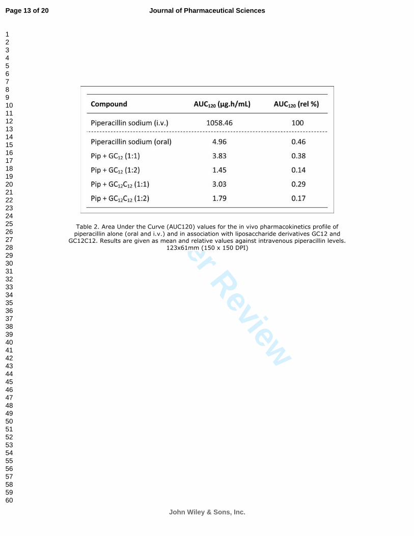
The oral uptake of piperacillin was calculated from the AUC120 (mean area under the curve, see Table
2) and was expectedly very low (0.5 %). When examining the absorption of the piperacillin
conjugates, the absorption curves were very similar to that of piperacillin alone, administered orally.
Surprisingly, the improvements in permeability noticed in vitro with the Caco-2 monolayer model
were not observed in vivo, either with GC12 (1:1) or GC12C12 (1:1 and 1:2). The determination of the
AUCs showed 0.14 % to 0.38 % absorption, which statistically is not significantly different from the
0.5 % absorption found for commercial piperacillin sodium (Fig. 6b).
Page 8 of 20
John Wiley & Sons, Inc.
Journal of Pharmaceutical Sciences
123456789101112131415161718192021222324252627282930313233343536373839404142434445464748495051525354555657585960
For Peer Review
Figure 6a. In vivo pharmacokinetics of piperacillin sodium administered intravenously at 50 mg/kg
in saline (0.9 % NaCl) over a 2h period; piperacillin concentration in blood was determined by LC-
MS/MS (Results given +/- SEM, n=3-5).
Figure 6b. In vivo absorption and pharmacokinetics of oral formulations of piperacillin, alone or in
association with liposaccharide derivatives GC12 and GC12C12, in 1:1 and 1:2 ratios in water + 5%
DMSO, over a two hour period. A dose equivalent to 50 mg piperacillin was administered by oral
gavage to 300-350 g Sprague-Dawley rats and piperacillin concentration in blood determined by
LC-MS/MS (Results given +/- SEM, n=3-5).
Table 2. Area Under the Curve (AUC120) values for the in vivo pharmacokinetics profile of
piperacillin alone (oral and i.v.) and in association with liposaccharide derivatives GC12 and GC12C12.
Results are given as mean and relative values against intravenous piperacillin levels.
The literature abounds in reports correlating in vitro predictions of permeability obtained with the
Caco-2 model of intestinal epithelium and in vivo oral absorption. The variations observed in these
experiments are therefore more likely to be inherent to the nature of the compounds tested, as it
appears that piperacillin was most probably released from the liposaccharide derivatives post-
administration.
CONCLUSION
We report here the biological evaluation of a novel series of ionic liposaccharide derivatives
designed as potential oral penetration enhancers. The synthetic constructs were engineered to
combine in a single molecular entity a carbohydrate-based hydrophilic moiety coupled to lipophilic
residues, such as hydrophobic amino acids and twelve carbon long LAAs. Piperacillin, a potent β-
lactam antibiotic with known low oral bioavailability, was used as a model drug and associated in
various molar ratios with the liposaccharide derivatives by lyophilisation. ITC measurements were
performed to determine the critical micelle concentration and binding affinity of the amphiphilic
conjugates and led to two ratios being considered for further examinations, respectively 1:1 and 1:2
drug/liposaccharide.
Antimicrobial assays revealed that the minimum inhibitory concentration of drug was practically
unchanged when piperacillin was combined with the liposaccharide derivatives. When examining the
effect of the tested conjugates against human red blood cells, an increase of haemolytic activity to
levels similar to SDS was observed in the case of Pip-GC12C12, yet the levels of toxicity remained low
when piperacillin and the liposaccharide derivatives were tested alone; the values observed denoted
a direct correlation between the lipophilicity of the conjugates and their haemolytic activity. The in
vitro evaluation of the synthesised associates in Caco-2 monolayers showed that apparent
membrane permeability values were significantly improved with three formulations, Pip-GC12 (1:1),
Pip-GC12C12 (1:1) and Pip-GC12C12 (1:2). The latter displayed in vitro permeability coefficients nearly
forty times higher than the parent piperacillin drug alone. Unfortunately, these promising results
were not confirmed in vivo. Current research is now being focused on investigating and
characterising the nature of the interactions between the drug and its counter-ionic liposaccharide
derivative, as well as their stability at various pH values.
ACKNOWLEDGEMENTS
Page 9 of 20
John Wiley & Sons, Inc.
Journal of Pharmaceutical Sciences
123456789101112131415161718192021222324252627282930313233343536373839404142434445464748495051525354555657585960
For Peer Review
This work was supported by an ARC project grant (DP0558334). The authors wish to thank Prof
Michael F. Jennings for his kind assistance in providing facilities and equipment to conduct the
microbiology experiments, and Mr Michael Moore for his help with handling animals.
REFERENCES
1. Trapani A, Garcia-Fuentes M, Alonso MJ. 2008. Novel drug nanocarriers combining hydrophilic
cyclodextrins and chitosan. Nanotechnology 19(18):185101/185101-185101/185110.
2. Cano-Cebrian MJ, Zornoza T, Granero L, Polache A. 2005. Intestinal absorption enhancement
via the paracellular route by fatty acids, chitosans and others: A target for drug delivery. Curr
Drug Deliv 2(1):9-22.
3. Shaji J, Patole V. 2008. Protein and Peptide Drug Delivery: Oral Approaches. Indian J Pharm Sci
70(3):269-277.
4. Touitou E, Barry BW. 2006. Enhancement in Drug Delivery. ed.: CRC Press. p 633.
5. Pouton CW, Porter CJH. 2008. Formulation of lipid-based delivery systems for oral
administration: Materials, methods and strategies. Adv Drug Deliv Rev 60(6):625-637.
6. Wong A, Toth I. 2001. Lipid, Sugar and Liposaccharide Based Delivery Systems. Curr Med Chem
8:1123-1136.
7. Bastin RJ, Bowker MJ, Slater BJ. 2000. Salt Selection and Optimisation Procedures for
Pharmaceutical New Chemical Entities. Organic Process Research & Development 4(5):427-
435.
8. Neubert R. 1989. Ion-pair transport across membranes. Pharm Res 6(9):743-747.
9. Violette A, Cortes DAF, Bergeon JA, Falconer RA, Toth I. 2008. Optimized LC-MS/MS
quantification method for the detection of piperacillin and application to the development of
charged liposaccharides as oral penetration enhancers. Int J Pharm 351(1-2):152-157.
10. Abdelrahim AS, Ziora ZM, Bergeon JA, Moss AR, Toth I. 2009. Synthesis and calorimetric
titration of new glucolipopeptides as potential cationic penetration enhancers. Tetrahedron
((submitted)).
11. Kyte J, Doolittle RF. 1982. A simple method for displaying the hydropathic character of a
protein. J Mol Biol 157(1):105-132.
12. Andrews JM. 2001. Determination of minimum inhibitory concentrations. J Antimicrob
Chemoth 48:5-16.
13. Nolting A, DallaCosta T, Rand KH, Derendorf H. 1996. Pharmacokinetic pharmacodynamic
modeling of the antibiotic effect of piperacillin in vitro. Pharm Res 13(1):91-96.
14. Hoogkampkorstanje JAA, Westerdaal NAC. 1979. Activity and synergy of piperacillin and
aminoglycosides against Pseudomonas-Aeruginiosa. A Van Leeuw J Microb 45(4):617-618.
15. Korvick JA, Yu VL. 1991. Antimicrobial agent therapy for Pseudomonas aeruginosa. Antimicrob
Agents Chemother 35(11):2167-2172.
16. Pignatello R, Noce C, Campisi A, Acquaviva R, Bucolo C, Puglisi G, Toth I. 2007. Evaluation of
Cell Tolerability of a Series of Lipoamino Acids Using Biological Membranes and a
Biomembrane Model. Curr Drug Deliv 4:109-121.
17. Ross BP, Braddy AC, McGeary RP, Blanchfield JT, Prokai L, Toth I. 2004. Micellar aggregation
and membrane partitioning of bile salts, fatty acids, sodium dodecyl sulfate, and sugar-
conjugated fatty acids: Correlation with hemolytic potency and implications for drug delivery.
Molecular Pharmaceutics 1(3):233-245.
18. Amin K, Dannenfelser RM. 2006. In vitro hemolysis: Guidance for the pharmaceutical scientist.
J Pharm Sci 95(6):1173-1176.
19. Artursson P, Palm K, Luthman K. 2001. Caco-2 monolayers in experimental and theoretical
predictions of drug transport. Adv Drug Deliv Rev 46(1-3):27-43.
Page 10 of 20
John Wiley & Sons, Inc.
Journal of Pharmaceutical Sciences
123456789101112131415161718192021222324252627282930313233343536373839404142434445464748495051525354555657585960
For Peer Review
20. BriskeAnderson MJ, Finley JW, Newman SM. 1997. The influence of culture time and passage
number on the morphological and physiological development of Caco-2 cells. Proceedings of
the Society for Experimental Biology and Medicine 214(3):248-257.
21. Donna AV. 2008. Variability in Caco-2 and MDCK cell-based intestinal permeability assays. J
Pharm Sci 97(2):712-725.
Page 11 of 20
John Wiley & Sons, Inc.
Journal of Pharmaceutical Sciences
123456789101112131415161718192021222324252627282930313233343536373839404142434445464748495051525354555657585960
For Peer Review
Table 1. Estimated EC10 of drug, liposaccharide derivatives and combined entities. 58x42mm (600 x 600 DPI)
Page 12 of 20
John Wiley & Sons, Inc.
Journal of Pharmaceutical Sciences
123456789101112131415161718192021222324252627282930313233343536373839404142434445464748495051525354555657585960
For Peer Review
Table 2. Area Under the Curve (AUC120) values for the in vivo pharmacokinetics profile of
piperacillin alone (oral and i.v.) and in association with liposaccharide derivatives GC12 and GC12C12. Results are given as mean and relative values against intravenous piperacillin levels.
123x61mm (150 x 150 DPI)
Page 13 of 20
John Wiley & Sons, Inc.
Journal of Pharmaceutical Sciences
123456789101112131415161718192021222324252627282930313233343536373839404142434445464748495051525354555657585960
For Peer Review
Fig. 1 – Synthesised charged liposaccharide derivatives associated with model drug piperacillin 78x36mm (600 x 600 DPI)
Page 14 of 20
John Wiley & Sons, Inc.
Journal of Pharmaceutical Sciences
123456789101112131415161718192021222324252627282930313233343536373839404142434445464748495051525354555657585960
For Peer Review
Figure 2. Isothermal titration microcalorimetry (ITC) binding assay between piperacillin (acid form, 0.2 mM) and liposaccharide derivative GC12C12 at 4 mM in purified water.
96x135mm (600 x 600 DPI)
Page 15 of 20
John Wiley & Sons, Inc.
Journal of Pharmaceutical Sciences
123456789101112131415161718192021222324252627282930313233343536373839404142434445464748495051525354555657585960
For Peer Review
Figure 3. MICs of piperacillin alone and formulated with the synthesised liposaccharide derivatives for two Gram-negative bacterial strains, E. coli and P. aeruginosa. Concentrations were determined by optical density measurements at 600 nm after 24 hr incubation in bacterial culture medium at
37°C. (Values given as mean ± SEM, n≥3) 254x190mm (150 x 150 DPI)
Page 16 of 20
John Wiley & Sons, Inc.
Journal of Pharmaceutical Sciences
123456789101112131415161718192021222324252627282930313233343536373839404142434445464748495051525354555657585960
For Peer Review
Figure 4. Haemolytic activity measured against hRBCs at 37°C over 1hr of piperacillin (alone and with GC12C12) and the synthesised liposaccharide derivatives in PBS. The percentage of haemolysis
was calculated by absorbance readings at 540 nm (results given as mean ± SEM, n=3) 229x162mm (150 x 150 DPI)
Page 17 of 20
John Wiley & Sons, Inc.
Journal of Pharmaceutical Sciences
123456789101112131415161718192021222324252627282930313233343536373839404142434445464748495051525354555657585960
For Peer Review
Figure 5. Apparent permeability coefficients determined in 21-28 day old monolayers of Caco-2 cells (passage 31-40) over a 150 min period at 37°C (Results given +/- SEM, n=2-3; 4 wells/exp).
254x190mm (150 x 150 DPI)
Page 18 of 20
John Wiley & Sons, Inc.
Journal of Pharmaceutical Sciences
123456789101112131415161718192021222324252627282930313233343536373839404142434445464748495051525354555657585960

For Peer Review
Figure 6a. In vivo pharmacokinetics of piperacillin sodium administered intravenously at 50 mg/kg in saline (0.9 % NaCl) over a 2h period; piperacillin concentration in blood was determined by LC-
MS/MS (Results given +/- SEM, n=3-5). 297x210mm (150 x 150 DPI)
Page 19 of 20
John Wiley & Sons, Inc.
Journal of Pharmaceutical Sciences
123456789101112131415161718192021222324252627282930313233343536373839404142434445464748495051525354555657585960
For Peer Review
Figure 6b. In vivo absorption and pharmacokinetics of oral formulations of piperacillin, alone or in association with liposaccharide derivatives GC12 and GC12C12, in 1:1 and 1:2 ratios in water + 5%
DMSO, over a two hour period. A dose equivalent to 50 mg piperacillin was administered by oral gavage to 300-350 g Sprague-Dawley rats and piperacillin concentration in blood determined by LC-
MS/MS (Results given +/- SEM, n=3-5). 297x210mm (150 x 150 DPI)
Page 20 of 20
John Wiley & Sons, Inc.
Journal of Pharmaceutical Sciences
123456789101112131415161718192021222324252627282930313233343536373839404142434445464748495051525354555657585960