Embed Size (px)
Citation preview
1
In-vitro activity of sodium-hypochlorite gel on
bacteria associated with periodontitis
Karolina Jurczyk • Sandor Nietzsche • Claudia Ender • Anton Sculean • Sigrun Eick
Karolina Jurczyk • Anton Sculean • Sigrun Eick ()
Department of Periodontology, School of Dental Medicine, University of Bern, Freiburgstrasse
7, CH-3010 Bern, Switzerland
Karolina Jurczyk
Department of Conservative Dentistry and Periodontology, Poznan University of Medical
Sciences, Poznan, Poland
Sandor Nietzsche • Claudia Ender
Center of Electron Microscopy, University Hospital Jena, Jena, Germany
Corresponding author
Sigrun Eick
University of Bern
School of Dental Medicine
Department of Periodontology; Laboratory of Oral Microbiology
Freiburgstrasse 7, 3010
Bern, Switzerland
Tel: + 41 31 632 25 42
Fax: + 41 31 632 49 15
Email: [email protected]
2
Objectives: The aim of the present study was to assess the antimicrobial activity of a sodium
hypochlorite formulation incl. its components against bacteria associated with periodontal
disease.
Materials and Methods: Sodium hypochlorite formulation (NaOCl gel), its components sodium
hypochlorite (NaOCl) and the activating vehicle were compared with 0.1% chlorhexidine
digluconate (CHX) solution. The antimicrobial activity was proven by determination of
minimal inhibitory concentrations (MIC), minimal bactericidal concentrations, and killing
assays. Furthermore, the influence on formation as well as on a 4-days old 6-species biofilm
was tested.
Results: Except for one strain (Parvimonas micra ATCC 33270 in case of NaOCl gel) the MICs
both of the CHX solution and NaOCl gel did not exceed 10% of the formulations’
concentration. In general MICs of the NaOCl gel were equal as of the CHX solution against
Gram-negatives but higher against Gram-positive bacteria. CHX but not NaOCl gel clearly
inhibited biofilm formation, however the activity of NaOCl gel was more remarkable on a 4-d
old biofilm. NaOCl killed bacteria in the biofilm and interfered with the matrix.
Conclusions: The NaOCl gel acts antimicrobial in particular against Gram-negative species
associated with periodontitis. Moreover, its component NaOCl hypochlorite is able to alter
biofilm matrices.
Clinical relevance: The NaOCl gel may represent a potential alternative for adjunctive topical
antimicrobial treatment in periodontitis
Key words: sodium hypochlorite, periodontal infection, biofilm
3
1. Introduction
Periodontitis is a chronic inflammatory disease of the tooth supporting tissues associated with
high counts of certain bacterial species interacting with the host’ immune system [1]. Oral
microbial-plaque communities are biofilms composed of numerous bacteria on host surfaces.
[2]. Bacteria more present in patients with chronic periodontitis than in periodontally healthy
subjects are Treponema denticola, Porphyromonas gingivalis, Tannerella forsythia, and several
others [3]. P. gingivalis a Gram-negative anaerobe bacterium, is considered a key-stone
pathogen in developing periodontal disease [4].
Non-surgical mechanical removal of the hard and soft microbial deposits from the root surfaces
(i.e. scaling and root planing (SRP)) is the standard in any cause-related periodontal therapy
[5]. Substantial evidence indicates that during supportive periodontal therapy (SPT)
periodontitis can be successfully treated and controlled by thorough mechanical plaque removal
by the patient coupled with supra -and subgingival debridement by the therapist with or without
the use of local antimicrobials [6]. During the last decade, various antimicrobials such as
chlorhexidine, azithromycin, metronidazole, doxycycline, minocycline and tetracycline used
subgingivally in conjunction with SRP have been tested [7]. Among the supragingivally used
antiseptics, chlorhexidine based dentifrices/gels are still being the gold standard although tooth
surface discoloration is observed as side effect [8].
An alternative approach to improve the outcomes of subgingival SRP might be the application
of sodium hypochlorite. Its broad antimicrobial activity, fast bactericidal action and non-
toxicity at application concentration, has been known since many years [9]. Already in 1918 it
was published that sodium hypochlorite has a higher dissolving effect on necrotic tissue than
on vital one [10]. However its use on everyday basis in dentistry is known basically in
endodontics as one of the main canal irrigants [11]. Activity of sodium hypochlorite is
depending on pH. It is most active when applied at neutral or slightly acidic pH [12].
Comparison of the activity of different antiseptics on experimental biofilm with various
endodontic/periodontal pathogens, showed that the highest bactericidal activity was obtained
with 2.25% sodium hypochlorite and 10% povidone-iodine and last by 0.2% chlorhexidine [13].
Already in the early 80ies of the last century , the use of sodium hypochlorite combined with
curretage was histologically shown to be effective in reducing soft tissue inflammation in SPT
[14]. The results showed that sodium hypochlorite achieves predictable chemolysis of the soft
tissue wall of the periodontal pocket with minimal effect on the adjacent tissues while the
4
antiseptic did not impede the healing phase [14]. Moreover, it was suggested by Perova [15],
that the use of 0.1% sodium hypochlorite during periodontal surgeries might improve the
healing, through a markedly better regeneration of the connective tissue at the gingival base of
the sites. Despite its promising properties, sodium hypochlorite did not stay in the light of
interest for long, until it has been rediscovered just recently. An oral mouthrinse with 0.05%
sodium hypochlorite resulted in significant reduction in supragingival biofilm accumulation
and gingival inflammation [16]. In subsequent studies twice-weekly rinsing with 0.25% sodium
hypochlorite solution decreased dental plaque level and reduced bleeding on probing in
periodontal pockets [17, 18].
A formulation of a sodium hypochlorite gel to be used in periodontal therapy has been recently
introduced to the market. It is composed of two components, sodium hypochlorite solution and
a mixture of amino acids. After combining the two substrates different chloramines are
synthesized from free sodium hypochlorite, which may additionally enhance the antimicrobial
activity.
The purpose of this in vitro study was to determine the antimicrobial activity of the sodium
hypochlorite gel and its components in comparison to chlorhexidine digluconate on microbial
species associated with periodontitis including P. gingivalis, T. forsythia, Aggregatibacter
actinomycetemcomitans, Parvimonas micra and others.
The hypothesis was that there is a minimal inhibitory concentration of sodium hypochlorite gel,
which inhibits predictably the growth and biofilm formation of bacteria associated with
periodontal disease.
2. Material and methods
2.1. Substances
Test substances were component 1 (NaOCl: sodium hypochlorite solution 0.95%), component
2 (activating vehicle: glutamic acid, leucine, lysine, carboxymethyl cellulose, and ultrapure
water) and sodium hypochlorite gel (NaOCl gel (Perisolv, Regedent AG, Zurich, Switzerland)),
composed of the previous two components mixed together. Chlorhexidine digluconate solution
5
(CHX) in the concentration of 0.1% was used as a positive control, whereas 0.9% sodium
chloride (NaCl) as a negative control.
2.2. Microorganisms
The following bacterial strains were tested as single bacterial species: P. gingivalis ATCC
33277, T. forsythia ATCC 43037, Fusobacterium nucleatum ATCC 25586, Streptococcus
gordonii ATCC 10558, Actinomyces naeslundii ATCC 12104, Parvimonas micra ATCC
33270, Prevotella intermedia ATCC 25611, A. actinomycetemcomitans ATCC 33384,
Campylobacter rectus ATCC 33238, Eikenella corrodens ATCC 23834, Filifactor alocis
ATCC 33099, Capnocytophaga gingivalis ATCC 33624, Eubacterium nodatum ATCC 33270
and three clinical isolates of P. gingivalis and T. forsythia. The mixed microbiota consisted of
the following bacterial strains: P. gingivalis ATCC 33277, T. forsythia ATCC 43037, F.
nucleatum ATCC 25586, S. gordonii ATCC 10558, A. naeslundii ATCC 12104 and P. micra
ATCC 33270. Before an experiment, all strains were precultivated on Schaedler agar plates
(Oxoid, Basingstoke, UK) with 5% sheep blood and vitamin K addition, in an anaerobic
atmosphere or with 5% CO2 (A. actinomycetemcomitans ATCC 33384 and S. gordonii ATCC
10558).
2.3. Susceptibility tests: Determination of the minimal inhibitory
concentrations and minimal bactericidal concentrations
Determination of MICs was performed by the micro-broth dilution technique using the 96-well-
microtiter plates. The MICs of component 1, component 2, Perisolv and 0.1% chlorhexidine
solution for single microorganisms and mixed species were checked.
After subcultivation of bacterial strains, a defined inoculum, with an adjusted turbidity of
McFarland 4 (0.5 for S. gordonii) was added to Wilkins Chalgren broth (Oxoid) supplemented
with nicotinamide adenine dinucleotide and N-acetyl muramic acid, in a 1:17 ratio. Defined
concentrations of NaOCl gel, its components and chlorhexidine as positive control were added.
6
After 42 h of incubation time (18 h for S. gordonii), the growth of microorganisms was analyzed
visually by checking the turbidity. MIC represents the lowest concentration without visible
turbidity of the broth.
For determination of the minimal bactericidal concentration, non-turbid cultures were
subcultivated on agar plates without the addition of any antimicrobial agent. After incubating,
the MBC was the lowest concentration without any growth of the colonies on the agar plates
(equivalent to a reduction by 99.9% of the initial inoculum).
Tests were performed in independent replicates.
2.4. Killing
A defined inoculum of microorganisms (about 106 /ml), prepared in doubled concentrated
nutrient media (Wilkins Chalgren broth), was added to NaOCl gel in final concentrations of
20%, 10% and 5%, as well as 0.01% chlorhexidine. In this experiment the following bacterial
species were used: P. gingivalis ATCC 3327, T. forsythia ATCC 43037, P. micra ATCC 33270
and the mixed species. After 1 h, 2 h, 6 h as well as 24 h of incubation, the numbers of viable
bacteria were determined by enumeration of colony forming units (cfu). The test was
performed in independent replicates.
2.5. Activity against bacteria in biofilms
In these experiments a multispecies biofilm consisting of S. gordonii ATCC 10558, P.
gingivalis ATCC 33277, T. forsythia ATCC 43037, F. nucleatum ATCC 25586, A. naeslundii
ATCC 12104 and P. micra ATCC 33270 was used. The influence of the test substances on the
developing biofilm was examined. First the wells of 96-well-plates were covered with 20 μl of
test substance. After 1 h of incubation, 10 µl of protein solution consisting of 25% serum and
5% albumin was added to the surface, followed by 170 µl of bacterial suspension. Bacterial
suspension was prepared by adding a defined inoculum (1 part S. gordonii, 2 parts A. naeslundii
and each 4 parts of the other species; each McFarland 4) to Wilkins Chalgren broth (Oxoid)
supplemented with nicotinamide adenine dinucleotide and N-acetyl muramic acid, in a 1:17
7
ratio. The 96-well-plates were incubated anaerobically at 37° under static conditions. The cfu
were counted after 6 h, 24 h and 48 h development of biofilm.
Additionally, the influence of the test substances was evaluated on a formed biofilm.
Multispecies biofilm composed of six bacterial species was developed for four days. First the
wells of 96-well-plates were covered with 10 μl of 25% v/v inactivated human serum/well for
1 h. Then bacterial suspension was prepared and mixed with the Wilkins Chalgren broth as
described above. The plates were incubated in the anaerobic atmosphere at 37°C. After 48 h the
medium was carefully exchanged with a new bacterial suspension of P. gingivalis ATCC 33277
and T. forsythia ATCC 43037 added to the nutrient medium before application to the wells. The
renewed addition of selected bacterial strains guaranteed a sufficient number of these species
within the biofilms.
After an additional incubation for 24 h, the medium was removed carefully and 20 μl of the
tested substances were added to the biofilm. After 1 min, 180 µl of Wilkins Chalgren broth was
added and the plates were incubated for 18h. Finally, after short washing the biofilm was
carefully scraped, mixed by pipetting and cfu were enumerated after serial dilutions, spreading
of each 25 μl on agar plates and incubation for 7 d.
Half of the 96-well plate was used for staining of the biofilm with the Kwasny and Opperman
method [19]. After a manual plate washing, the biofilm was fixed by incubating the washed
plate at 60oC for 60 min. Finally the staining was performed with 0.06% (w/v) solution of
crystal violet dissolved in dH2O. 50 μl of the above stain was used per well and left for 5
minutes. After the staining procedure, the excess was removed by repeated washing. The
amount of crystal violet bounded in each well was directly measured spectrophotometrically by
measuring OD600 using microplate reader.
These experiments were made in two independent experiments in independent sextuplicates.
In addition, 4-d old biofilm samples on glass slides, treated with the test substances and
processed as described above were stained with 0.1% acridine orange solution (Merck,
Darmstadt, Germany) as a general nucleic acid stain. Samples were examined by using
fluorescent microscope (Olympus BX51, Tokyo, Japan).
Furthermore, scanning electron microscope photographs were taken to visualize the results.
Exemplarily each test substance was chosen. Samples were fixed in 2% glutaraldehyde in

8
cacodylate buffer for 30 min, washed twice with cacodylate buffer and dehydrated using a
graded ethanol series (15 min each concentration). Following critical point drying, samples
were sputter-coated with gold and examined with a ZEISS LEO-1530 Gemini (Carl Zeiss NTS
GmbH) equipped with a field emission electron gun at 8 keV.
2.6. Statistical analysis
All data are presented as mean and standard deviation (SD). Data were compared using a one-
way analysis of variance (ANOVA) with post-hoc comparisons of groups using LSD
corrections. A p-value of 0.05 was considered to be statistically significant. However, in case
of log10 cfu values the cfu log10 reductions are of importance, following only log10 cfu values
are presented. SPSS software (version 22.0) was used for statistical analysis.
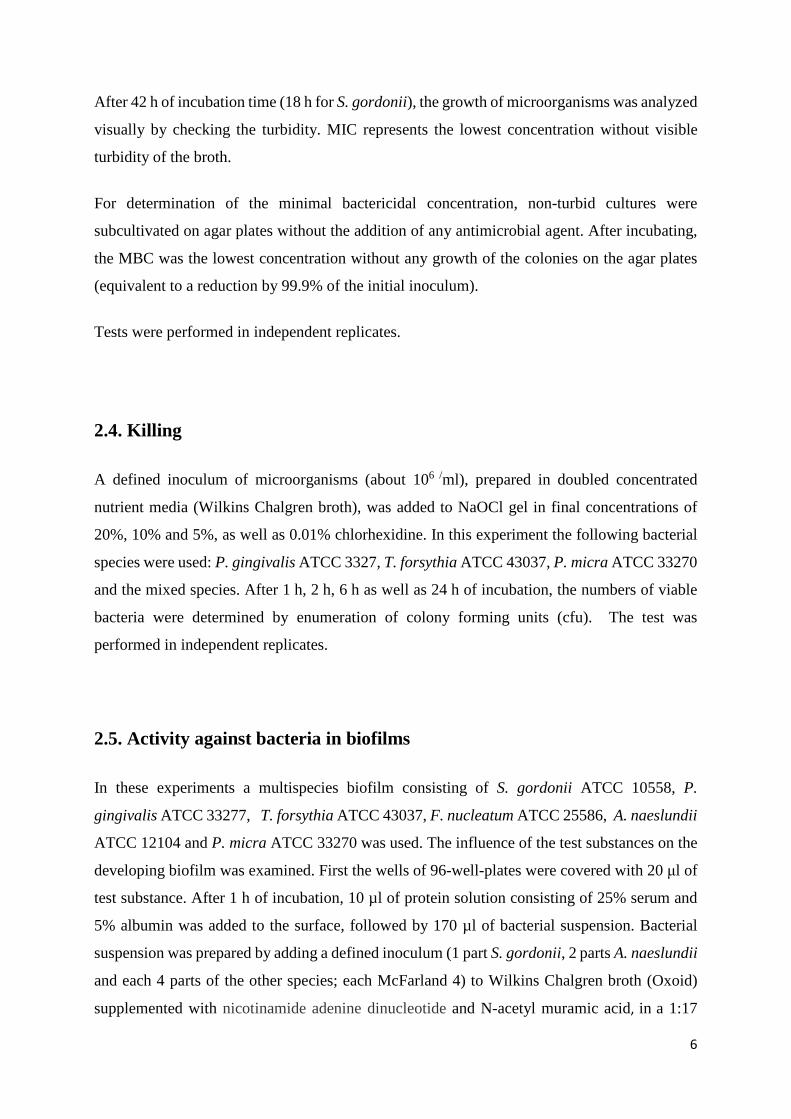
3. Results All results were related in % to the working (commercially available) concentration (NaOCl
gel and 0.1% CHX respectively).
3.1. Minimal inhibitory concentrations and minimal bactericidal
concentrations
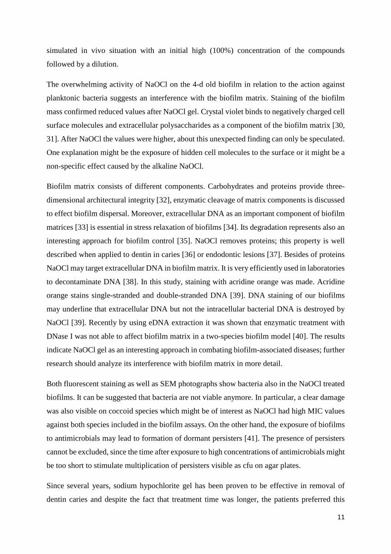
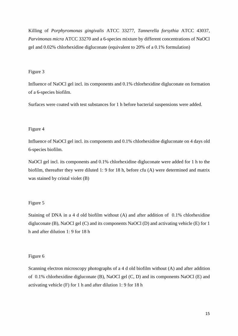
Except for one strain (Parvimonas micra ATCC 33270 in case of NaOCl gel) the MICs both of
the CHX solution and NaOCl gel did not exceed 10% of the formulations’ concentration. In
general MICs of CHX were lower than those of the NaOCl gel. However, when differentiating
between Gram-positives and Gram-negatives the difference was mainly due to the Gram-
positive bacteria. NaOCl gel acted more growth inhibitory on Gram-negatives than on Gram-
positives. Mainly NaOCl was the active compound of NaOCl gel. But the activating vehicle
exerted also some activity on Gram-negatives; here a synergistic effect can be stated when
comparing NaOCl gel with its compounds (Figure 1).
The MBC values were in general equal or one step higher than the corresponding MIC values.
The difference was more or equal 2 steps for CHX against two microorganisms (incl. the
9
mixture) and for NaOCl gel against seven microorganisms (incl. the mixture). MBC values and
individual MIC data are presented in Suppl. table 1.
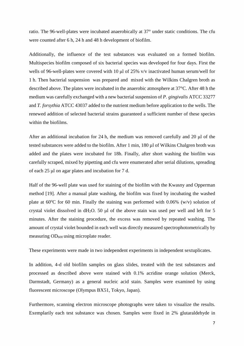
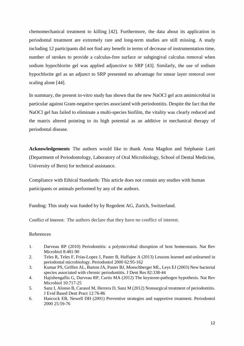
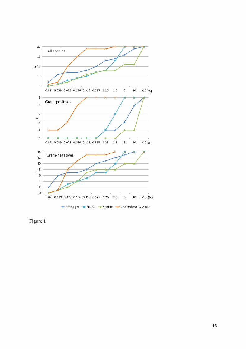
3.2. Killing curves
Killing curves show the fast and total killing activity of 20% of the CHX solution and of the
NaOCl gel. Only a few cells of T. forsythia were able to survive the exposure to the compounds.
NaOCl gel was also tested in the lower concentrations of 5% and 10%. In part, a concentration
dependent activity was visible (Figure 2).
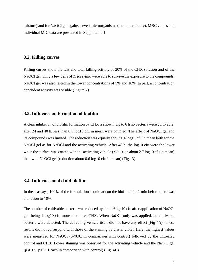
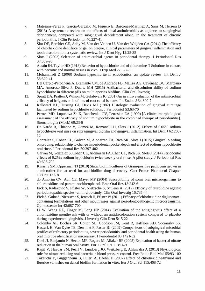
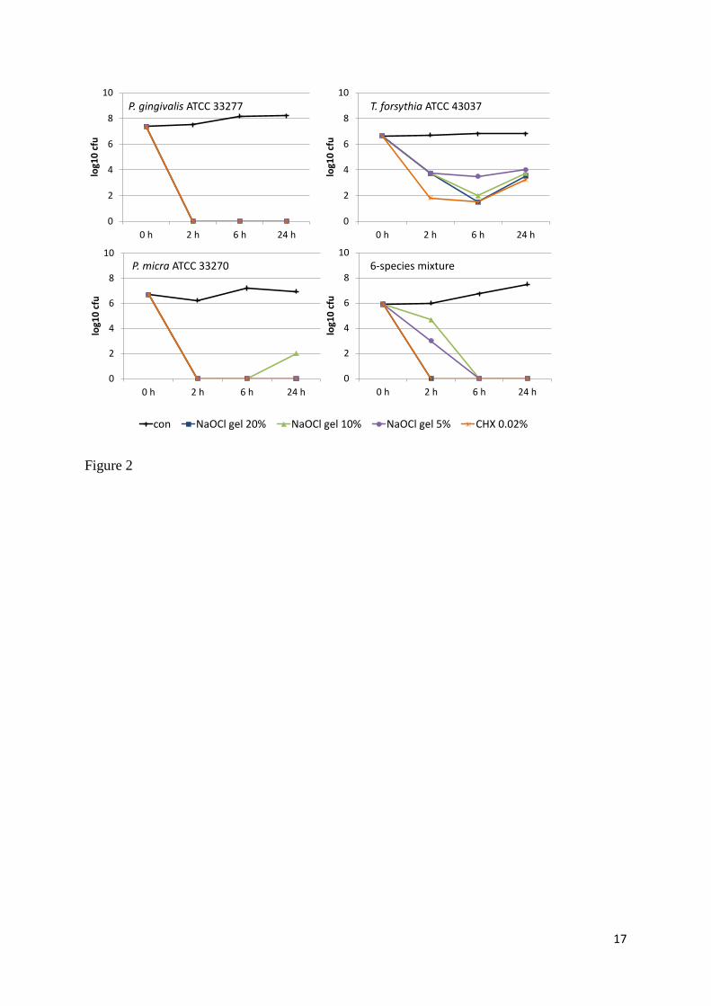
3.3. Influence on formation of biofilm
A clear inhibition of biofilm formation by CHX is shown. Up to 6 h no bacteria were cultivable;
after 24 and 48 h, less than 0.5 log10 cfu in mean were counted. The effect of NaOCl gel and
its compounds was limited. The reduction was equally about 1.4 log10 cfu in mean both for the
NaOCl gel as for NaOCl and the activating vehicle. After 48 h, the log10 cfu were the lower
when the surface was coated with the activating vehicle (reduction about 2.7 log10 cfu in mean)
than with NaOCl gel (reduction about 0.6 log10 cfu in mean) (Fig. 3).
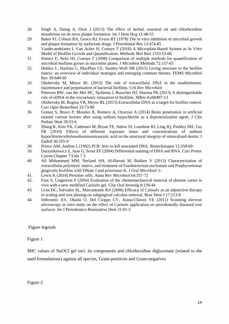
3.4. Influence on 4 d old biofilm
In these assays, 100% of the formulations could act on the biofilms for 1 min before there was
a dilution to 10%.
The number of cultivable bacteria was reduced by about 6 log10 cfu after application of NaOCl
gel, being 1 log10 cfu more than after CHX. When NaOCl only was applied, no cultivable
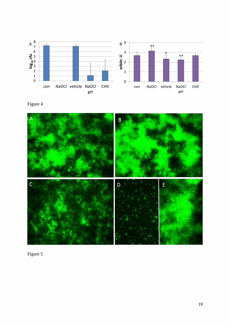
bacteria were detected. The activating vehicle itself did not have any effect (Fig 4A). These
results did not correspond with those of the staining by cristal violet. Here, the highest values
were measured for NaOCl (p<0.01 in comparison with control) followed by the untreated
control and CHX. Lower staining was observed for the activating vehicle and the NaOCl gel
(p<0.05, p<0.01 each in comparison with control) (Fig. 4B).
10
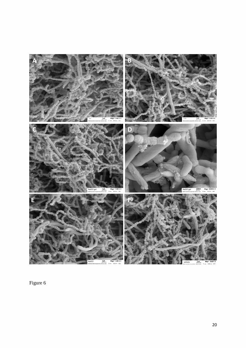
Staining with acridine orange showed a diffuse staining of the biofilm control, after application
of CHX and the activating vehicle. The staining is less diffuse after NaOCl gel, whereas after
NaOCl, only clear defined structures (bacteria) are stained (Fig. 5).
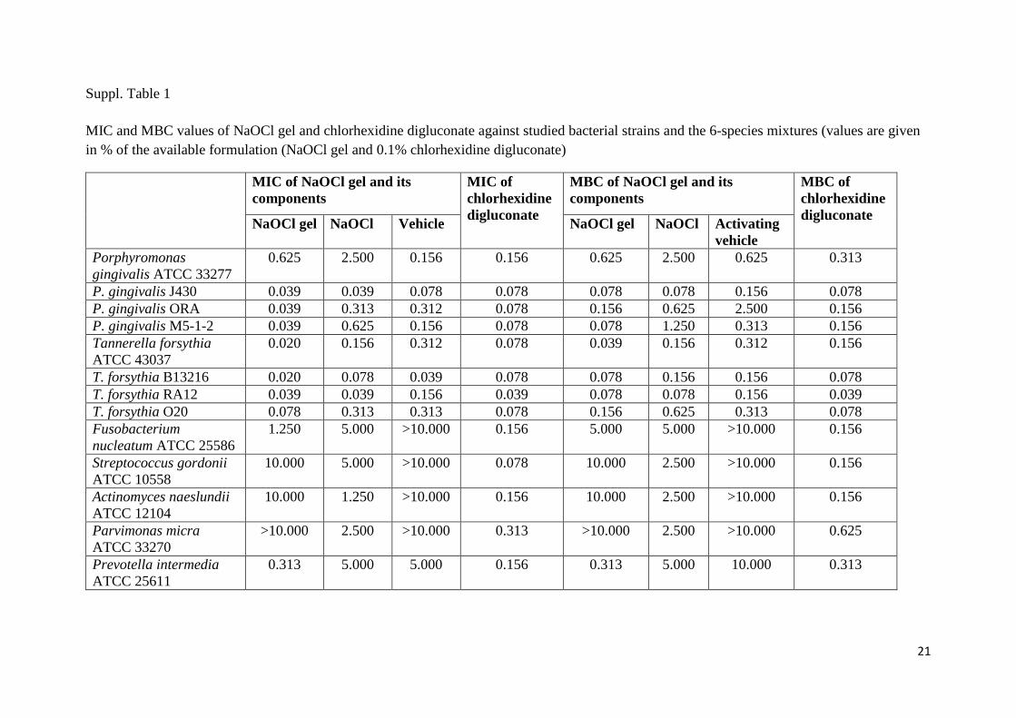
In all SEM photographs, many bacteria were visible. In part, damaged bacterial surfaces were
detected after application of NaOCl and NaOCl gel. The matrix seemed to be less after NaOCl
in comparison with the activating vehicle (Fig. 6).
4. Discussion
In this in vitro-study a sodium-hypochlorite gel was compared with a CHX solution which is
still the gold standard in periodontal therapy. Growth inhibition and killing as well as the
activity on a 6-species biofilm were evaluated.
MIC and MBC were determined by using standard procedures. The obtained values related to
the available formulations revealed extremely low MICs of CHX and confirms findings from
other studies [20, 21]. However, it should be mentioned that in the present study CHX was
tested without any additives. It is well known that additives may influence disadvantageously
the antimicrobial activity of commercial CHX formulations [22, 23]. The activity of NaOCl gel
differed between Gram-positive and Gram-negative bacteria, growth of Gram-negatives is
inhibited by lower concentrations. Interestingly, in the present study also the activating vehicle
itself exerted certain antimicrobial activity. This selective inhibition may favor a more Gram-
positive microbiota with Actinomyces spp., oral streptococci being in general more associated
with periodontal health [24]. Moreover, it should be noted, that Actinomyces spp. are able to
reduce nitrate to nitrite [25]. Killing of those bacteria by broad-spectrum antiseptics disturbs
the physiological role of these oral bacteria in blood pressure control [26].
One important question to be answered was whether the application of NaOCl gel on a surface
after mechanical plaque removal may prevent biofilm formation. This property is well known
for chlorhexidine [27, 28] which is in part linked to its high substantivity [29]. Our in vitro
study confirms the inhibition of biofilm formation for CHX, but for NaOCl gel no clear activity
was seen. A slight inhibition (>1 log10 cfu) lasted only up to 24 h. However, NaOCl gel and in
particular its component NaOCl clearly reduced vitality of a 4-d old biofilm. The used mode
11
simulated in vivo situation with an initial high (100%) concentration of the compounds
followed by a dilution.
The overwhelming activity of NaOCl on the 4-d old biofilm in relation to the action against
planktonic bacteria suggests an interference with the biofilm matrix. Staining of the biofilm
mass confirmed reduced values after NaOCl gel. Crystal violet binds to negatively charged cell
surface molecules and extracellular polysaccharides as a component of the biofilm matrix [30,
31]. After NaOCl the values were higher, about this unexpected finding can only be speculated.
One explanation might be the exposure of hidden cell molecules to the surface or it might be a
non-specific effect caused by the alkaline NaOCl.
Biofilm matrix consists of different components. Carbohydrates and proteins provide three-
dimensional architectural integrity [32], enzymatic cleavage of matrix components is discussed
to effect biofilm dispersal. Moreover, extracellular DNA as an important component of biofilm
matrices [33] is essential in stress relaxation of biofilms [34]. Its degradation represents also an
interesting approach for biofilm control [35]. NaOCl removes proteins; this property is well
described when applied to dentin in caries [36] or endodontic lesions [37]. Besides of proteins
NaOCl may target extracellular DNA in biofilm matrix. It is very efficiently used in laboratories
to decontaminate DNA [38]. In this study, staining with acridine orange was made. Acridine
orange stains single-stranded and double-stranded DNA [39]. DNA staining of our biofilms
may underline that extracellular DNA but not the intracellular bacterial DNA is destroyed by
NaOCl [39]. Recently by using eDNA extraction it was shown that enzymatic treatment with
DNase I was not able to affect biofilm matrix in a two-species biofilm model [40]. The results
indicate NaOCl gel as an interesting approach in combating biofilm-associated diseases; further
research should analyze its interference with biofilm matrix in more detail.
Both fluorescent staining as well as SEM photographs show bacteria also in the NaOCl treated
biofilms. It can be suggested that bacteria are not viable anymore. In particular, a clear damage
was also visible on coccoid species which might be of interest as NaOCl had high MIC values
against both species included in the biofilm assays. On the other hand, the exposure of biofilms
to antimicrobials may lead to formation of dormant persisters [41]. The presence of persisters
cannot be excluded, since the time after exposure to high concentrations of antimicrobials might
be too short to stimulate multiplication of persisters visible as cfu on agar plates.
Since several years, sodium hypochlorite gel has been proven to be effective in removal of
dentin caries and despite the fact that treatment time was longer, the patients preferred this
12
chemomechanical treatment to killing [42]. Furthermore, the data about its application in
periodontal treatment are extremely rare and long-term studies are still missing. A study
including 12 participants did not find any benefit in terms of decrease of instrumentation time,
number of strokes to provide a calculus-free surface or subgingival calculus removal when
sodium hypochlorite gel was applied adjunctive to SRP [43]. Similarly, the use of sodium
hypochlorite gel as an adjunct to SRP presented no advantage for smear layer removal over
scaling alone [44].
In summary, the present in-vitro study has shown that the new NaOCl gel acts antimicrobial in
particular against Gram-negative species associated with periodontitis. Despite the fact that the
NaOCl gel has failed to eliminate a multi-species biofilm, the vitality was clearly reduced and
the matrix altered pointing to its high potential as an additive in mechanical therapy of
periodontal disease.
Acknowledgements The authors would like to thank Anna Magdon and Stéphanie Larti
(Department of Periodontology, Laboratory of Oral Microbiology, School of Dental Medicine,
University of Bern) for technical assistance.
Compliance with Ethical Standards: This article does not contain any studies with human
participants or animals performed by any of the authors.
Funding: This study was funded by by Regedent AG, Zurich, Switzerland.
Conflict of Interest: The authors declare that they have no conflict of interest.
References
1. Darveau RP (2010) Periodontitis: a polymicrobial disruption of host homeostasis. Nat Rev Microbiol 8:481-90
2. Teles R, Teles F, Frias-Lopez J, Paster B, Haffajee A (2013) Lessons learned and unlearned in periodontal microbiology. Periodontol 2000 62:95-162
3. Kumar PS, Griffen AL, Barton JA, Paster BJ, Moeschberger ML, Leys EJ (2003) New bacterial species associated with chronic periodontitis. J Dent Res 82:338-44
4. Hajishengallis G, Darveau RP, Curtis MA (2012) The keystone-pathogen hypothesis. Nat Rev Microbiol 10:717-25
5. Sanz I, Alonso B, Carasol M, Herrera D, Sanz M (2012) Nonsurgical treatment of periodontitis. J Evid Based Dent Pract 12:76-86
6. Hancock EB, Newell DH (2001) Preventive strategies and supportive treatment. Periodontol 2000 25:59-76
13
7. Matesanz-Perez P, Garcia-Gargallo M, Figuero E, Bascones-Martinez A, Sanz M, Herrera D (2013) A systematic review on the effects of local antimicrobials as adjuncts to subgingival debridement, compared with subgingival debridement alone, in the treatment of chronic periodontitis. J Clin Periodontol 40:227-41
8. Slot DE, Berchier CE, Addy M, Van der Velden U, Van der Weijden GA (2014) The efficacy of chlorhexidine dentifrice or gel on plaque, clinical parameters of gingival inflammation and tooth discoloration: a systematic review. Int J Dent Hyg 12:25-35
9. Slots J (2002) Selection of antimicrobial agents in periodontal therapy. J Periodontal Res 37:389-98
10. Austin JH, Taylor HD (1918) Behavior of hypochlorite and of chloramine-T Solutions in contact with necrotic and normal tissues in vivo. J Exp Med 27:627-33
11. Mohammadi Z (2008) Sodium hypochlorite in endodontics: an update review. Int Dent J 58:329-41
12. Del Carpio-Perochena A, Bramante CM, de Andrade FB, Maliza AG, Cavenago BC, Marciano MA, Amoroso-Silva P, Duarte MH (2015) Antibacterial and dissolution ability of sodium hypochlorite in different pHs on multi-species biofilms. Clin Oral Investig
13. Spratt DA, Pratten J, Wilson M, Gulabivala K (2001) An in vitro evaluation of the antimicrobial efficacy of irrigants on biofilms of root canal isolates. Int Endod J 34:300-7
14. Kalkwarf KL, Tussing GJ, Davis MJ (1982) Histologic evaluation of gingival curettage facilitated by sodium hypochlorite solution. J Periodontol 53:63-70
15. Perova MD, Lopunova Zh K, Banchenko GV, Petrosian EA (1990) [A clinico-morphological assessment of the efficacy of sodium hypochlorite in the combined therapy of periodontitis]. Stomatologiia (Mosk) 69:23-6
16. De Nardo R, Chiappe V, Gomez M, Romanelli H, Slots J (2012) Effects of 0.05% sodium hypochlorite oral rinse on supragingival biofilm and gingival inflammation. Int Dent J 62:208-12
17. Gonzalez S, Cohen CL, Galvan M, Alonaizan FA, Rich SK, Slots J (2015) Gingival bleeding on probing: relationship to change in periodontal pocket depth and effect of sodium hypochlorite oral rinse. J Periodontal Res 50:397-402
18. Galvan M, Gonzalez S, Cohen CL, Alonaizan FA, Chen CT, Rich SK, Slots J (2014) Periodontal effects of 0.25% sodium hypochlorite twice-weekly oral rinse. A pilot study. J Periodontal Res 49:696-702
19. Kwasny SM, Opperman TJ (2010) Static biofilm cultures of Gram-positive pathogens grown in a microtiter format used for anti-biofilm drug discovery. Curr Protoc Pharmacol Chapter 13:Unit 13A 8
20. do Amorim CV, Aun CE, Mayer MP (2004) Susceptibility of some oral microorganisms to chlorhexidine and paramonochlorophenol. Braz Oral Res 18:242-6
21. Eick S, Radakovic S, Pfister W, Nietzsche S, Sculean A (2012) Efficacy of taurolidine against periodontopathic species--an in vitro study. Clin Oral Investig 16:735-44
22. Eick S, Goltz S, Nietzsche S, Jentsch H, Pfister W (2011) Efficacy of chlorhexidine digluconate-containing formulations and other mouthrinses against periodontopathogenic microorganisms. Quintessence Int 42:687-700
23. Li W, Wang RE, Finger M, Lang NP (2014) Evaluation of the antigingivitis effect of a chlorhexidine mouthwash with or without an antidiscoloration system compared to placebo during experimental gingivitis. J Investig Clin Dent 5:15-22
24. Colombo AP, Boches SK, Cotton SL, Goodson JM, Kent R, Haffajee AD, Socransky SS, Hasturk H, Van Dyke TE, Dewhirst F, Paster BJ (2009) Comparisons of subgingival microbial profiles of refractory periodontitis, severe periodontitis, and periodontal health using the human oral microbe identification microarray. J Periodontol 80:1421-32
25. Doel JJ, Benjamin N, Hector MP, Rogers M, Allaker RP (2005) Evaluation of bacterial nitrate reduction in the human oral cavity. Eur J Oral Sci 113:14-9
26. Kapil V, Haydar SM, Pearl V, Lundberg JO, Weitzberg E, Ahluwalia A (2013) Physiological role for nitrate-reducing oral bacteria in blood pressure control. Free Radic Biol Med 55:93-100
27. Takeuchi Y, Guggenheim B, Filieri A, Baehni P (2007) Effect of chlorhexidine/thymol and fluoride varnishes on dental biofilm formation in vitro. Eur J Oral Sci 115:468-72
14
28. Singh A, Daing A, Dixit J (2013) The effect of herbal, essential oil and chlorhexidine mouthrinse on de novo plaque formation. Int J Dent Hyg 11:48-52
29. Baker PJ, Coburn RA, Genco RJ, Evans RT (1978) The in vitro inhibition of microbial growth and plaque formation by surfactant drugs. J Periodontal Res 13:474-85
30. Vandecandelaere I, Van Acker H, Coenye T (2016) A Microplate-Based System as In Vitro Model of Biofilm Growth and Quantification. Methods Mol Biol 1333:53-66
31. Peeters E, Nelis HJ, Coenye T (2008) Comparison of multiple methods for quantification of microbial biofilms grown in microtiter plates. J Microbiol Methods 72:157-65
32. Hobley L, Harkins C, MacPhee CE, Stanley-Wall NR (2015) Giving structure to the biofilm matrix: an overview of individual strategies and emerging common themes. FEMS Microbiol Rev 39:649-69
33. Okshevsky M, Meyer RL (2013) The role of extracellular DNA in the establishment, maintenance and perpetuation of bacterial biofilms. Crit Rev Microbiol
34. Peterson BW, van der Mei HC, Sjollema J, Busscher HJ, Sharma PK (2013) A distinguishable role of eDNA in the viscoelastic relaxation of biofilms. MBio 4:e00497-13
35. Okshevsky M, Regina VR, Meyer RL (2015) Extracellular DNA as a target for biofilm control. Curr Opin Biotechnol 33:73-80
36. Gomez S, Bravo P, Morales R, Romero A, Oyarzun A (2014) Resin penetration in artificial enamel carious lesions after using sodium hypochlorite as a deproteinization agent. J Clin Pediatr Dent 39:51-6
37. Zhang K, Kim YK, Cadenaro M, Bryan TE, Sidow SJ, Loushine RJ, Ling JQ, Pashley DH, Tay FR (2010) Effects of different exposure times and concentrations of sodium hypochlorite/ethylenediaminetetraacetic acid on the structural integrity of mineralized dentin. J Endod 36:105-9
38. Prince AM, Andrus L (1992) PCR: how to kill unwanted DNA. Biotechniques 12:358-60 39. Darzynkiewicz Z, Juan G, Srour EF (2004) Differential staining of DNA and RNA. Curr Protoc
Cytom Chapter 7:Unit 7 3 40. Ali Mohammed MM, Nerland AH, Al-Haroni M, Bakken V (2013) Characterization of
extracellular polymeric matrix, and treatment of Fusobacterium nucleatum and Porphyromonas gingivalis biofilms with DNase I and proteinase K. J Oral Microbiol 5:
41. Lewis K (2010) Persister cells. Annu Rev Microbiol 64:357-72 42. Fure S, Lingstrom P (2004) Evaluation of the chemomechanical removal of dentine caries in
vivo with a new modified Carisolv gel. Clin Oral Investig 8:139-44 43. Grisi DC, Salvador SL, Marcantonio RA (2006) Efficacy of Carisolv as an adjunctive therapy
to scaling and root planing on subgingival calculus removal. Braz Dent J 17:213-8 44. Imbronito AV, Okuda O, Del Cioppo CV, Arana-Chavez VE (2011) Scanning electron
microscopy in vitro study on the effect of Carisolv application on periodontally diseased root surfaces. Int J Periodontics Restorative Dent 31:91-5
Figure legends
Figure 1
MIC values of NaOCl gel incl. its components and chlorhexidine digluconate (related to the
used formulations) against all species, Gram-positives and Gram-negatives
Figure 2
15
Killing of Porphyromonas gingivalis ATCC 33277, Tannerella forsythia ATCC 43037,
Parvimonas micra ATCC 33270 and a 6-species mixture by different concentrations of NaOCl
gel and 0.02% chlorhexidine digluconate (equivalent to 20% of a 0.1% formulation)
Figure 3
Influence of NaOCl gel incl. its components and 0.1% chlorhexidine digluconate on formation
of a 6-species biofilm.
Surfaces were coated with test substances for 1 h before bacterial suspensions were added.
Figure 4
Influence of NaOCl gel incl. its components and 0.1% chlorhexidine digluconate on 4 days old
6-species biofilm.
NaOCl gel incl. its components and 0.1% chlorhexidine digluconate were added for 1 h to the
biofilm, thereafter they were diluted 1: 9 for 18 h, before cfu (A) were determined and matrix
was stained by cristal violet (B)
Figure 5
Staining of DNA in a 4 d old biofilm without (A) and after addition of 0.1% chlorhexidine
digluconate (B), NaOCl gel (C) and its components NaOCl (D) and activating vehicle (E) for 1
h and after dilution 1: 9 for 18 h
Figure 6
Scanning electron microscopy photographs of a 4 d old biofilm without (A) and after addition
of 0.1% chlorhexidine digluconate (B), NaOCl gel (C, D) and its components NaOCl (E) and
activating vehicle (F) for 1 h and after dilution 1: 9 for 18 h
16
Figure 1
0
5
10
15
20
0.02 0.039 0.078 0.156 0.313 0.625 1.25 2.5 5 10 >10
n
Perio+ NaOCl vehicle CHX
0
1
2
3
4
5
0.02 0.039 0.078 0.156 0.313 0.625 1.25 2.5 5 10 >10
n
Perio+ NaOCl vehicle CHX(%)
0
2
4
6
8
10
12
14
0.02 0.039 0.078 0.156 0.313 0.625 1.25 2.5 5 10 >10
n
NaOCl gel NaOCl vehicle CHX
(%)
all species
Gram-positives
Gram-negatives
(related to 0.1%)
(%)
17
Figure 2
0
1
2
3
4
5
6
7
8
0 h 2 h 6 h 24 h
log1
0 cf
u
con NaOCl gel 20% NaOCl gel 10% NaOCl gel 5% CHX 0.02%
0
2
4
6
8
10
0 h 2 h 6 h 24 h
log1
0 cf
u
0
2
4
6
8
10
0 h 2 h 6 h 24 h
log1
0 cf
u
0
2
4
6
8
10
0 h 2 h 6 h 24 h
log1
0 cf
u
0
2
4
6
8
10
0 h 2 h 6 h 24 h
log1
0 cf
u
P. gingivalis ATCC 33277
P. micra ATCC 33270
T. forsythia ATCC 43037
6-species mixture
18
Figure 3
0
1
2
3
4
5
6
7
8
9
10
6 h 24 h 48 h
log10
cfu con
NaOClvehicleNaOCl gelCHX
19
Figure 4
Figure 5
012345678
con NaOCl vehicle NaOClgel
CHX
log 10
cfu
0
1
2
3
4
con NaOCl vehicle NaOClgel
CHX
arbi
tr. U
**
***
A B
A
C D E
B
20
Figure 6
NaOCl gel NaOCl gel
NaOCl vehicle
A B
C D
E F
21
Suppl. Table 1
MIC and MBC values of NaOCl gel and chlorhexidine digluconate against studied bacterial strains and the 6-species mixtures (values are given in % of the available formulation (NaOCl gel and 0.1% chlorhexidine digluconate)
MIC of NaOCl gel and its components
MIC of chlorhexidine digluconate
MBC of NaOCl gel and its components
MBC of chlorhexidine digluconate NaOCl gel NaOCl Vehicle NaOCl gel NaOCl Activating
vehicle Porphyromonas gingivalis ATCC 33277
0.625 2.500 0.156 0.156 0.625 2.500 0.625 0.313
P. gingivalis J430 0.039 0.039 0.078 0.078 0.078 0.078 0.156 0.078 P. gingivalis ORA 0.039 0.313 0.312 0.078 0.156 0.625 2.500 0.156 P. gingivalis M5-1-2 0.039 0.625 0.156 0.078 0.078 1.250 0.313 0.156 Tannerella forsythia ATCC 43037
0.020 0.156 0.312 0.078 0.039 0.156 0.312 0.156
T. forsythia B13216 0.020 0.078 0.039 0.078 0.078 0.156 0.156 0.078 T. forsythia RA12 0.039 0.039 0.156 0.039 0.078 0.078 0.156 0.039 T. forsythia O20 0.078 0.313 0.313 0.078 0.156 0.625 0.313 0.078 Fusobacterium nucleatum ATCC 25586
1.250 5.000 >10.000 0.156 5.000 5.000 >10.000 0.156
Streptococcus gordonii ATCC 10558
10.000 5.000 >10.000 0.078 10.000 2.500 >10.000 0.156
Actinomyces naeslundii ATCC 12104
10.000 1.250 >10.000 0.156 10.000 2.500 >10.000 0.156
Parvimonas micra ATCC 33270
>10.000 2.500 >10.000 0.313 >10.000 2.500 >10.000 0.625
Prevotella intermedia ATCC 25611
0.313 5.000 5.000 0.156 0.313 5.000 10.000 0.313
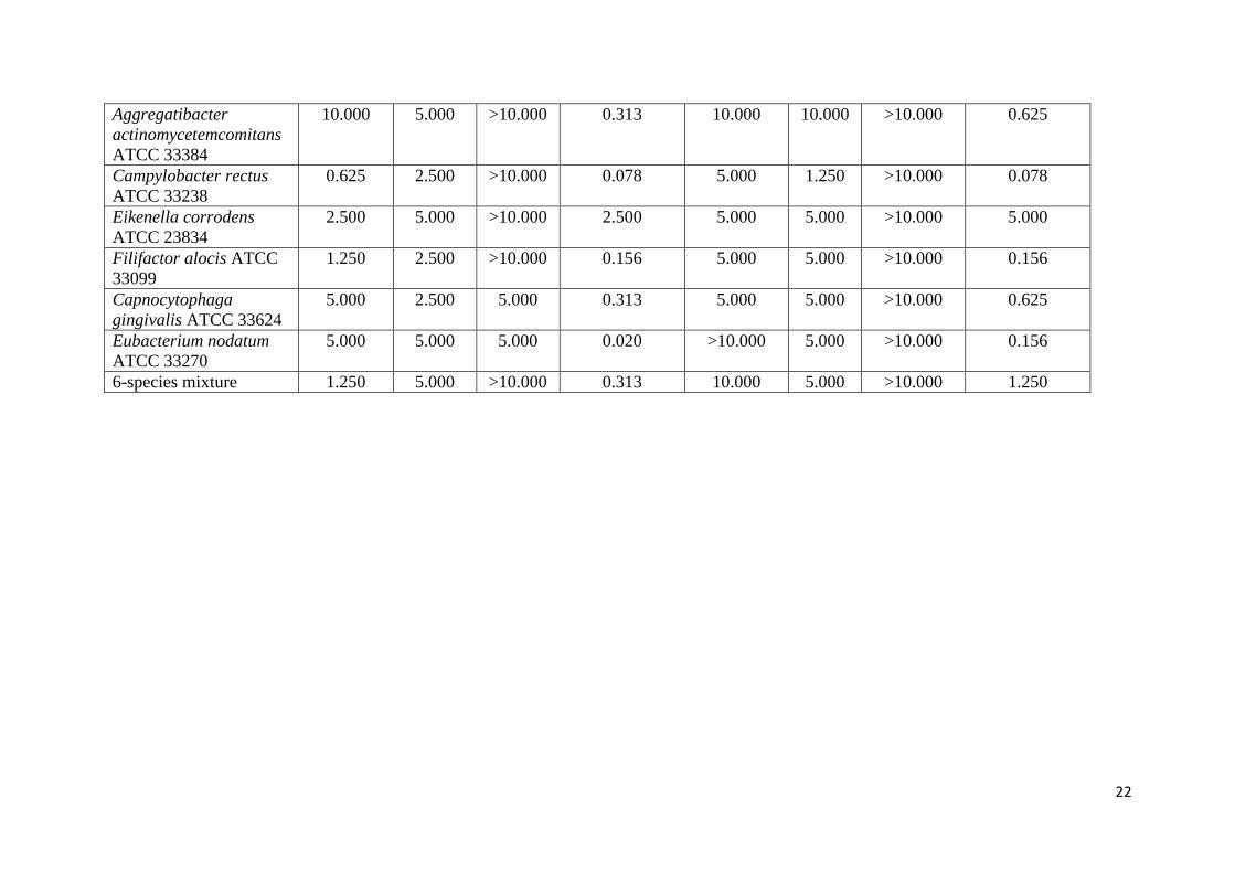
22
Aggregatibacter actinomycetemcomitans ATCC 33384
10.000 5.000 >10.000 0.313 10.000 10.000 >10.000 0.625
Campylobacter rectus ATCC 33238
0.625 2.500 >10.000 0.078 5.000 1.250 >10.000 0.078
Eikenella corrodens ATCC 23834
2.500 5.000 >10.000 2.500 5.000 5.000 >10.000 5.000
Filifactor alocis ATCC 33099
1.250 2.500 >10.000 0.156 5.000 5.000 >10.000 0.156
Capnocytophaga gingivalis ATCC 33624
5.000 2.500 5.000 0.313 5.000 5.000 >10.000 0.625
Eubacterium nodatum ATCC 33270
5.000 5.000 5.000 0.020 >10.000 5.000 >10.000 0.156
6-species mixture 1.250 5.000 >10.000 0.313 10.000 5.000 >10.000 1.250