Embed Size (px)
Citation preview

In the name of God
Congenital Laryngeal Anomalies
M. H. Baradaranfar M.D professor of otolaryngology
Head and Neck surgery Rhinologist
Introduction Wide range of problems Anatomy Embryology Diagnosis Types: supraglottis, glottis,
subglottis Presentation/diagnosis Management
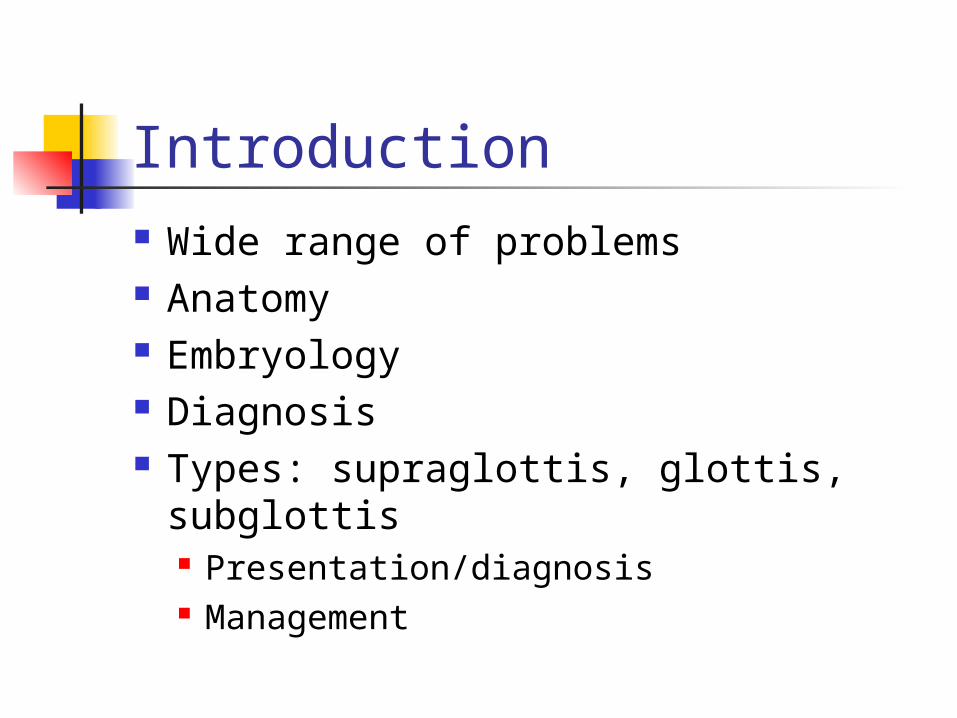
Normal Anatomy Larynx
Ventilates and protects lungs Clears secretions Voice
Differences in adults and infants 1/3 size at birth Narrow dimensions (subglottis vs.
glottis) Higher in neck and more pliable Epiglottis narrower
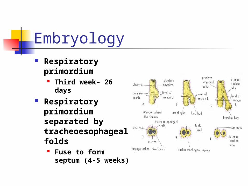
Embryology Respiratory
primordium Third week– 26
days Respiratory
primordium separated by tracheoesophageal folds
Fuse to form septum (4-5 weeks)
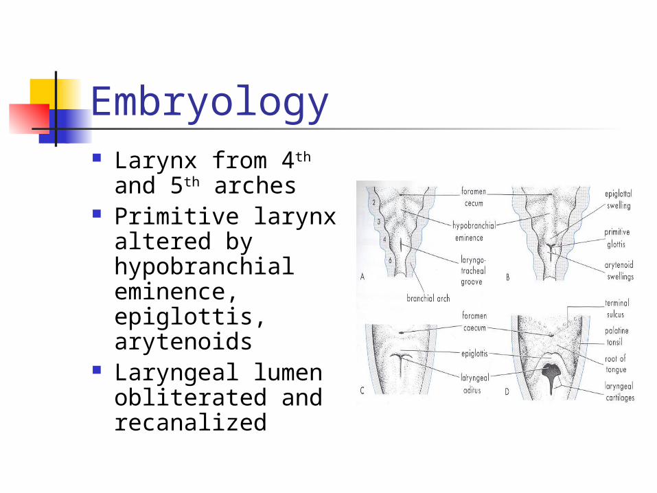
Embryology Larynx from 4th and
5th arches Primitive larynx
altered by hypobranchial eminence, epiglottis, arytenoids
Laryngeal lumen obliterated and recanalized
Clinical Manifestations Respiratory obstruction Stridor Weak cry Dyspnea Tachypnea Aspiration Cyanosis Sudden death
Clinical Diagnosis History
Premature, medical problems Birth records, intubation history Symptom frequency, feeding
Physical exam Observation Voice Flexible exam
Clinical Diagnosis Radiography
Neck films, chest films Barium swallow CT/MRI
Endoscopy in OR Gold standard
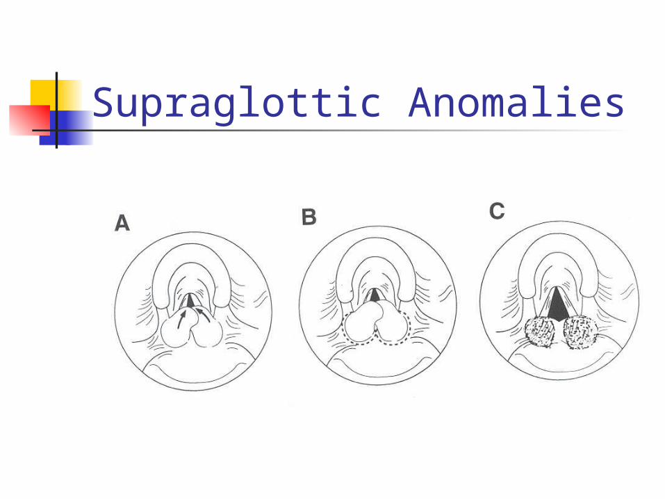
Supraglottic Anomalies Laryngomalacia
Most common (60%) Boys>girls Inspiratory stridor: *not always at
birth Benign, self-limiting May be severe Immature larynx
Supraglottic Anomalies Laryngomalacia
Diagnosis: flexible laryngoscopy Occasional endoscopy Treatment= expectant, reassurance
Position changes Close follow up
Severe cases= surgery
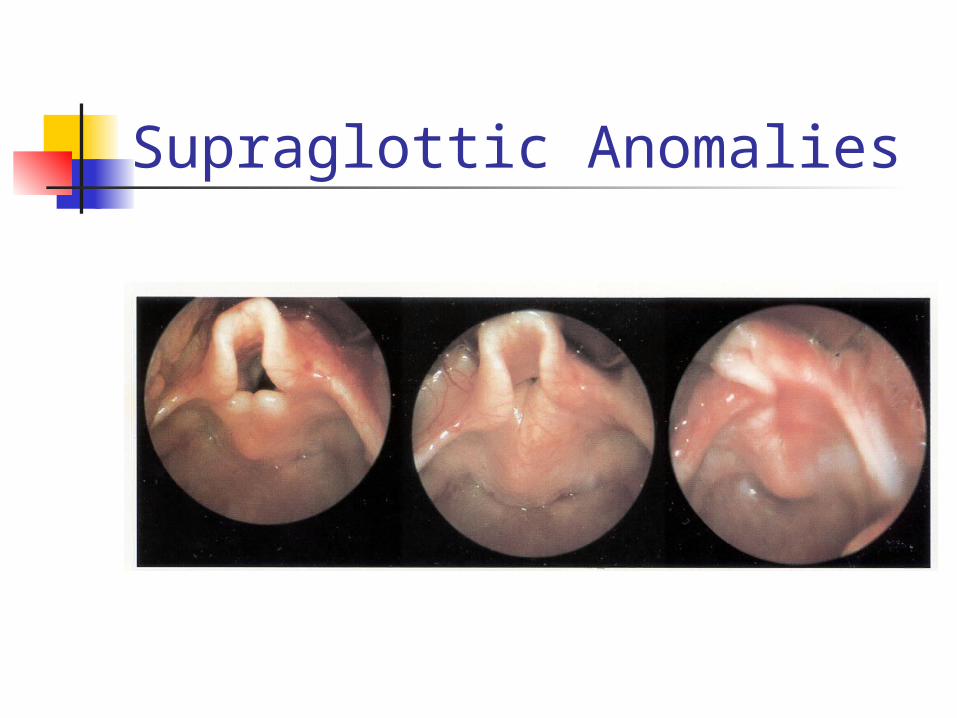
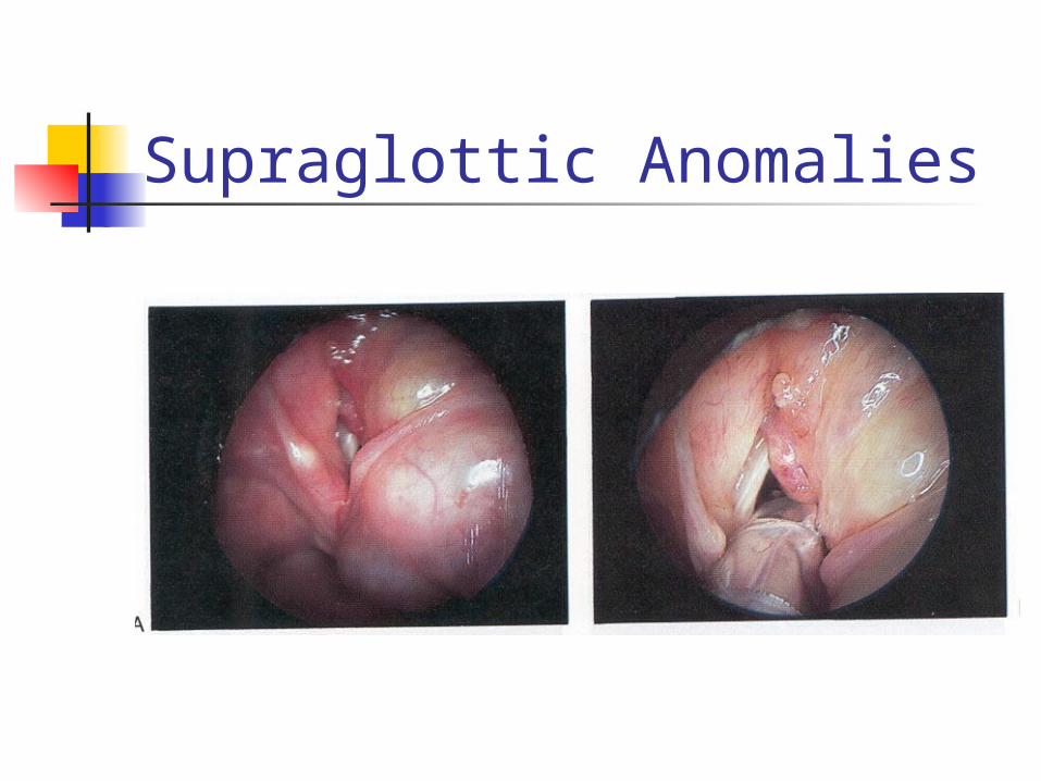
Supraglottic Anomalies
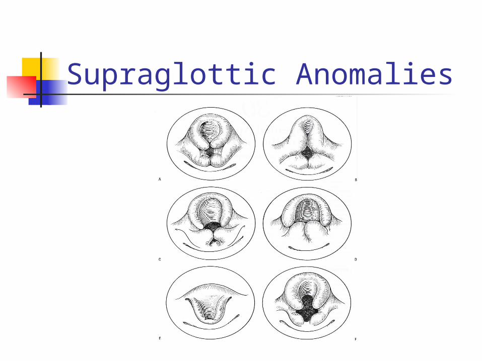
Supraglottic Anomalies
Supraglottic Anomalies
Supraglottic Anomalies
Supraglottic Anomalies
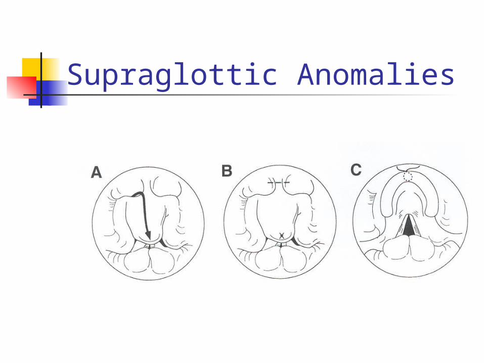
Supraglottic Anomalies
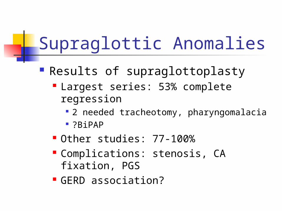
Supraglottic Anomalies Results of supraglottoplasty
Largest series: 53% complete regression
2 needed tracheotomy, pharyngomalacia ?BiPAP
Other studies: 77-100% Complications: stenosis, CA fixation,
PGS GERD association?
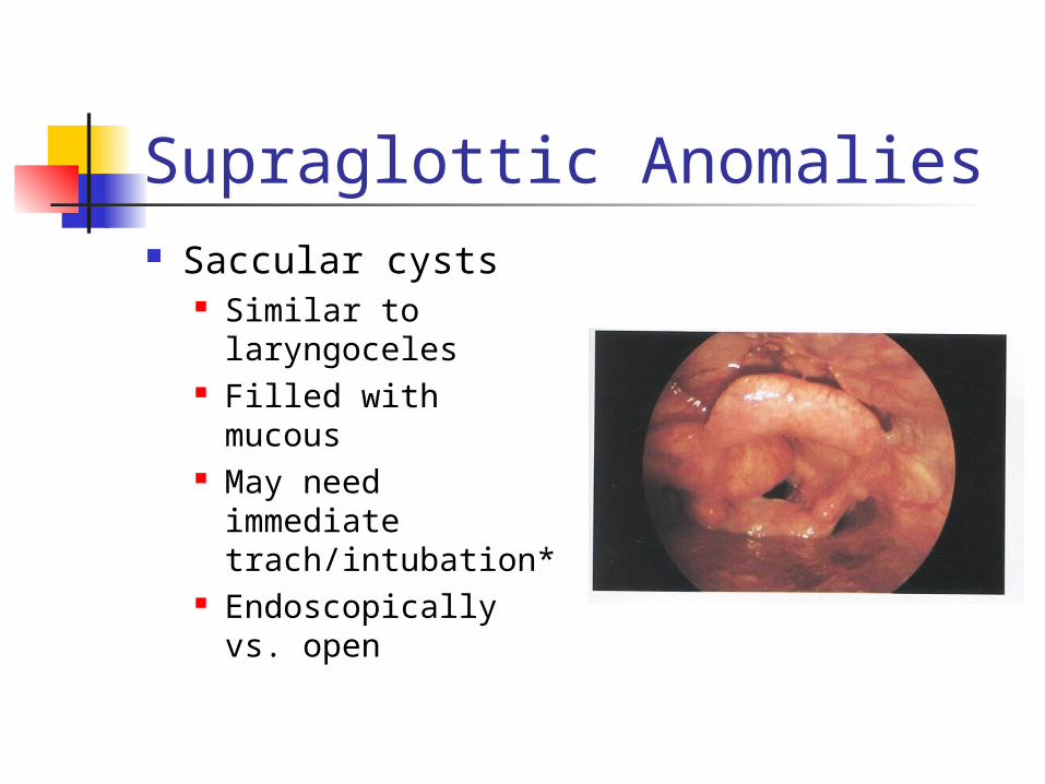
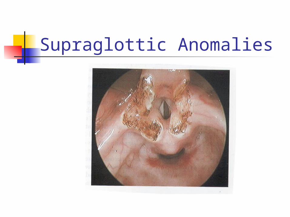
Supraglottic Anomalies Saccular cysts
Similar to laryngoceles
Filled with mucous May need
immediate trach/intubation*
Endoscopically vs. open
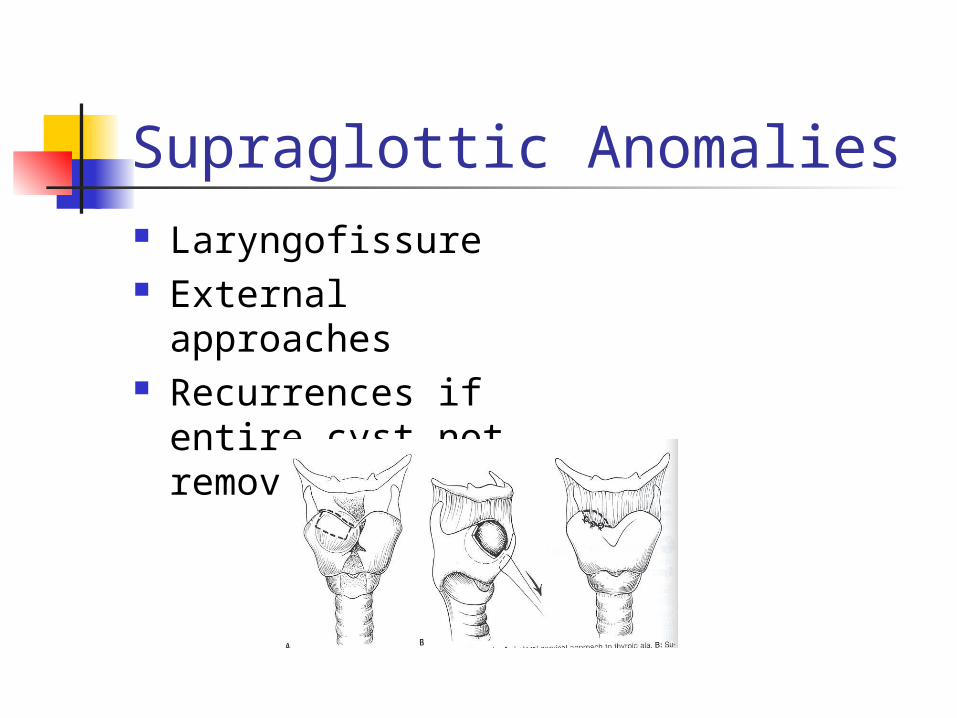
Supraglottic Anomalies Laryngofissure External
approaches Recurrences if
entire cyst not removed
Supraglottic Anomalies Laryngocele
Dilated sac filled with air (ventricle) Internal vs. external May present at birth– stridor* Difficult to diagnose– CT? Endoscopic or open procedures Recurrences low
Supraglottic Anomalies Vascular and lymphatic malformations
Hemangiomas 30% birth– grow in first 6-18 months Dyspnea, stridor, feeding problems later* Endoscopic evaluation Multiple treatment options
Lymphangiomas Compress epiglottis– airway distress at birth* Symptoms varied Endoscopic evaluation: CO2 laser
Supraglottic Anomalies
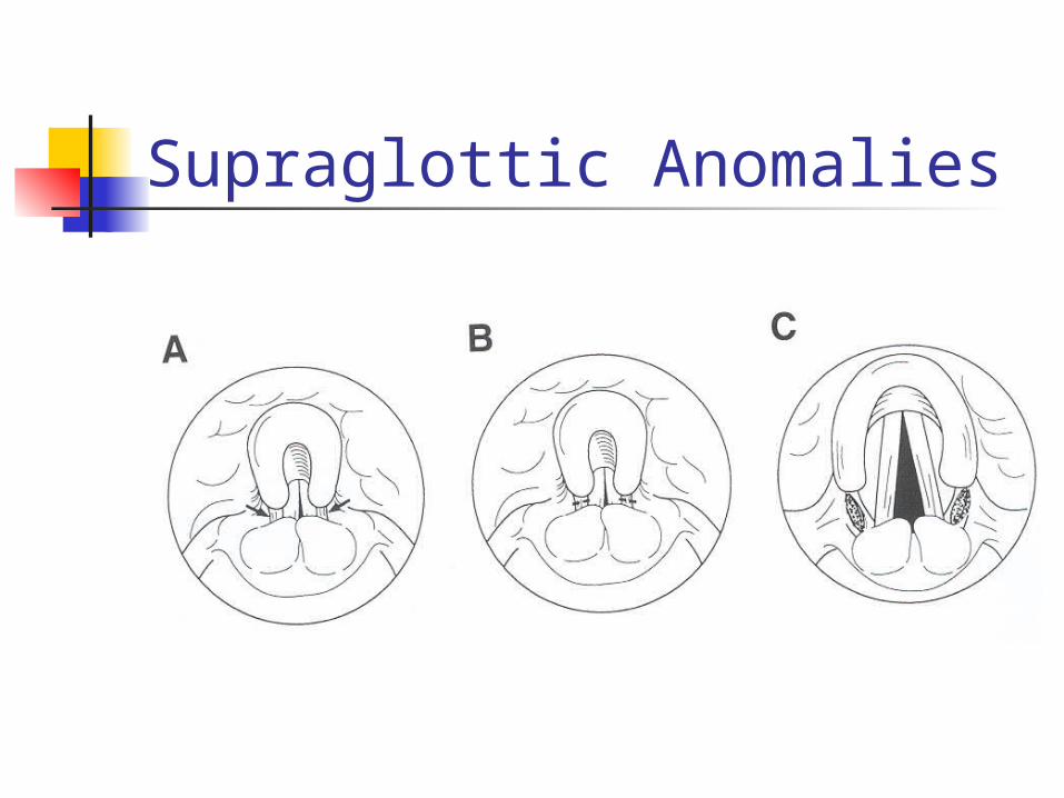
Supraglottic Anomalies Supraglottic webs– rare Anomalous cuneiform cartilage Bifid epiglottis
Pallister-Hall syndrome (hypothalmus, polydactaly, laryngeal)
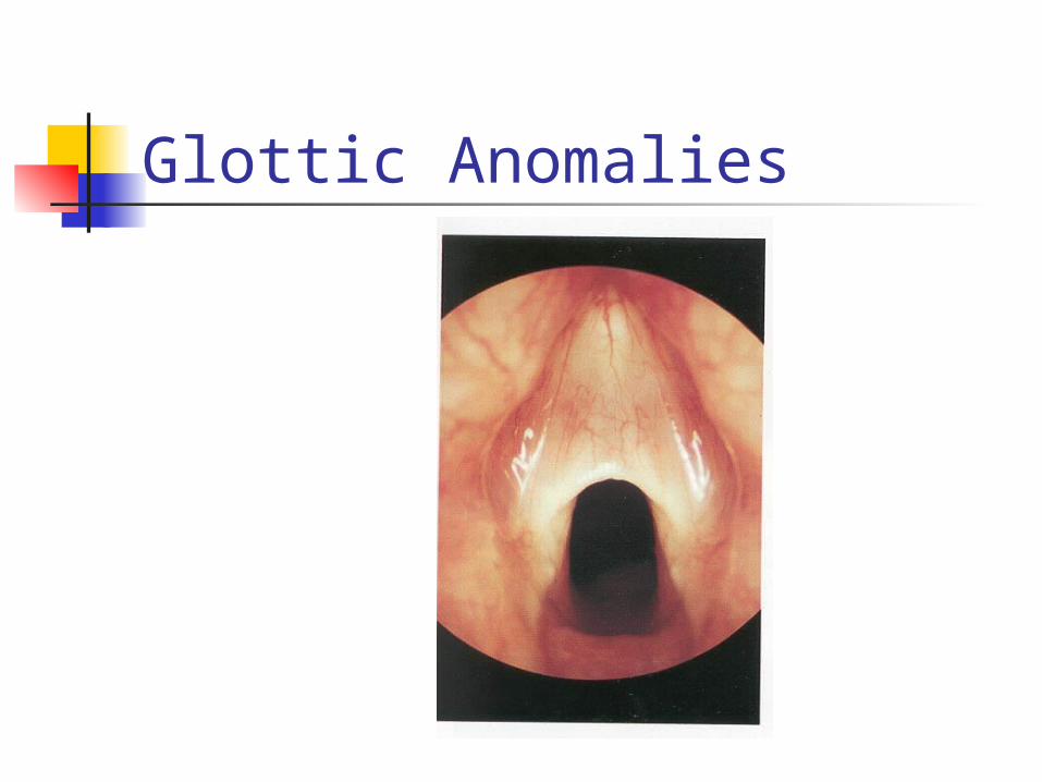
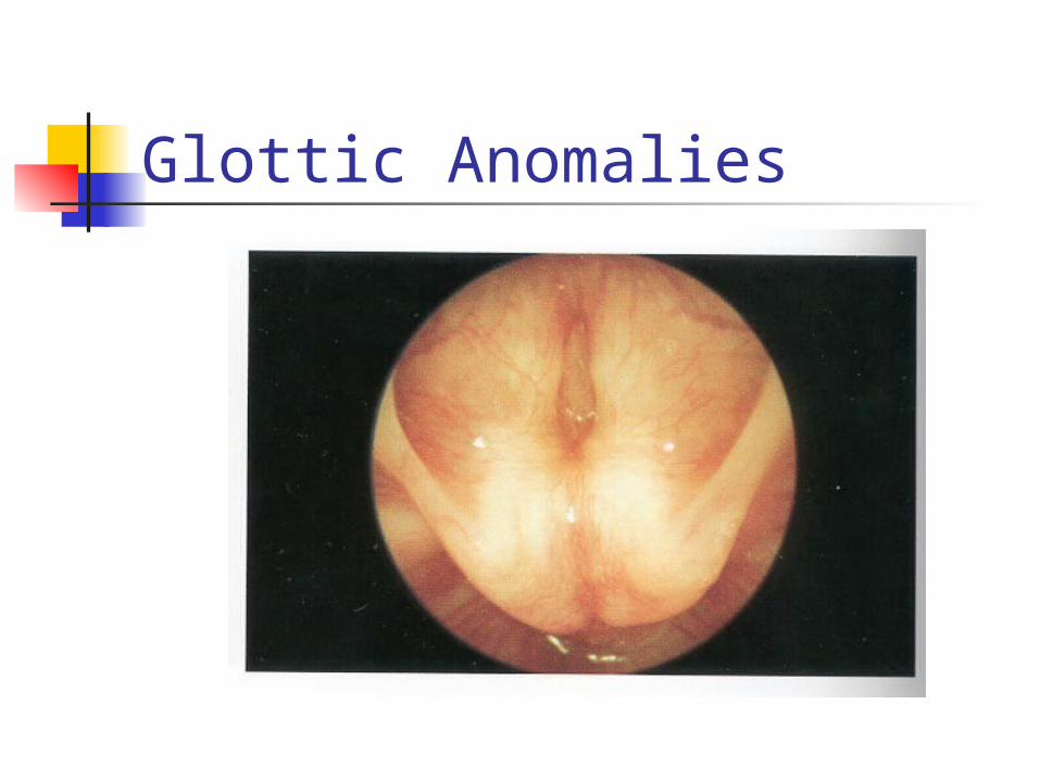
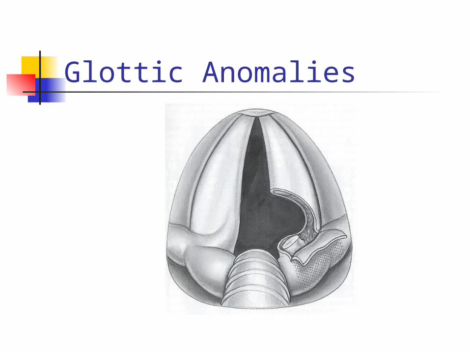
Glottic Anomalies Laryngeal webs
Failure of recanalization of larynx 75% at glottic level Most anterior with subglottic
involvement Four types– increasing severity May present at birth* Diagnosis: flexible laryngoscopy
Airway films helpful with subglottis
Glottic Anomalies
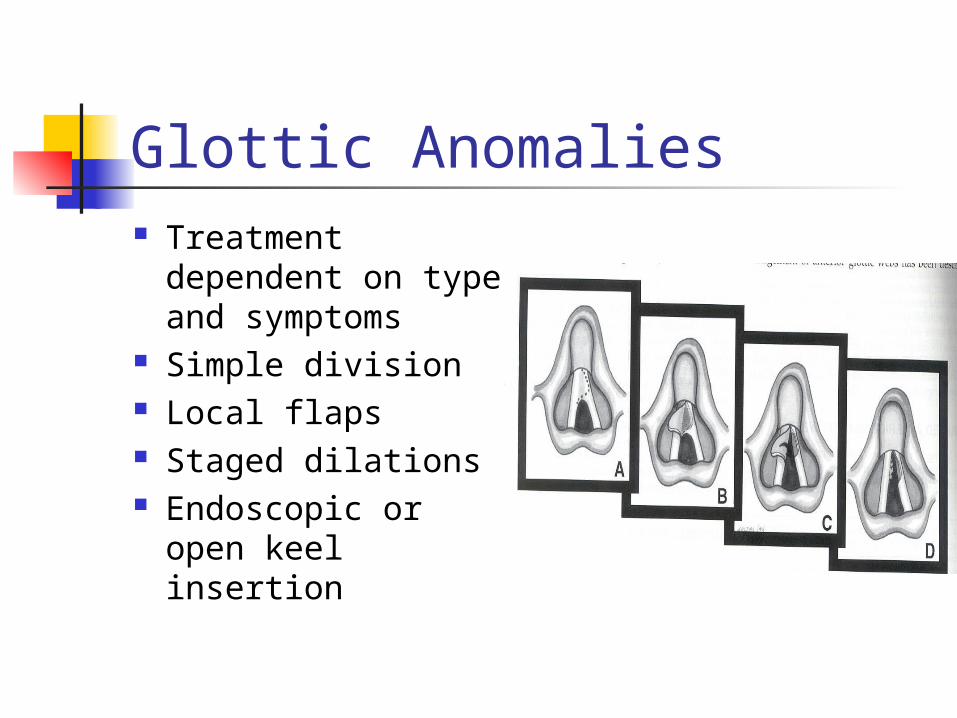
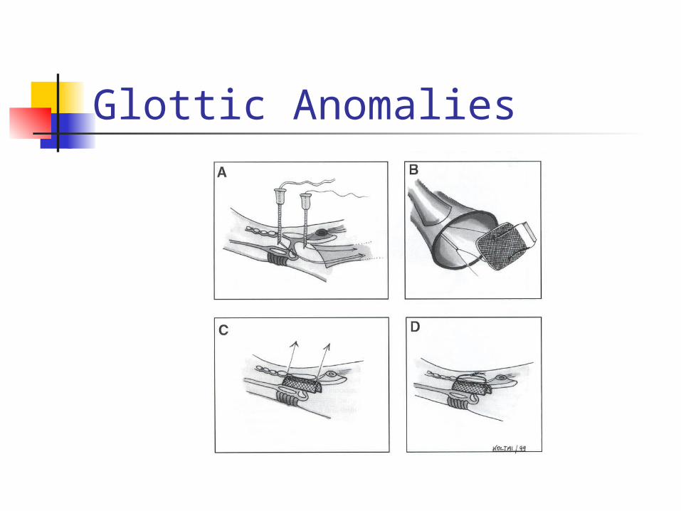
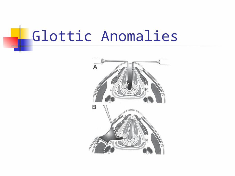
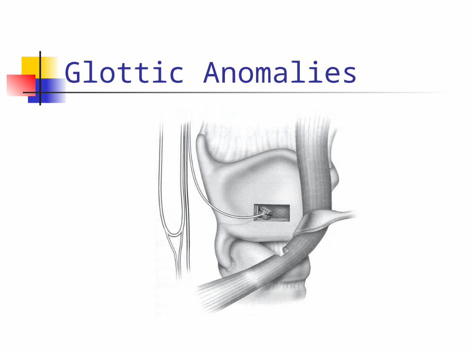
Glottic Anomalies Treatment
dependent on type and symptoms
Simple division Local flaps Staged dilations Endoscopic or open
keel insertion
Glottic Anomalies
Glottic Anomalies Laryngeal Atresia
Most severe process from failed recanalization
Always present at birth* Only survive if TEF or immediate
trach Later LTR Other anomalies
Glottic Anomalies
Glottic Anomalies Congenital High Upper Airway
Obstruction (CHAOS) 1994– ultrasound with large lungs, flat
diaphragms, dilated airways, fetal ascites
EXIT procedure (ex utero intrapartum treatment)
Multidisciplinary team C-section, maintain placental blood flow, quick
tracheotomy
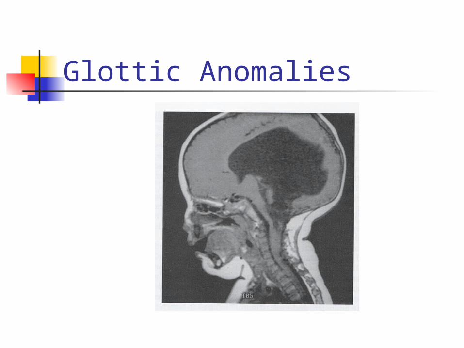
Glottic Anomalies Vocal cord paralysis
Second most common cause of stridor 10-15% of laryngeal pathology Unilateral vs. bilateral Vagus nerve damage Idiopathic (47%) ACM, hydrocephalus, trauma, cardiac
problems
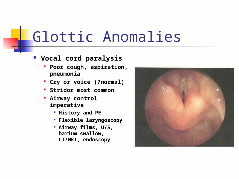
Glottic Anomalies Vocal cord paralysis
Poor cough, aspiration, pneumonia
Cry or voice (?normal) Stridor most common Airway control
imperative History and PE Flexible laryngoscopy Airway films, U/S,
barium swallow, CT/MRI, endoscopy
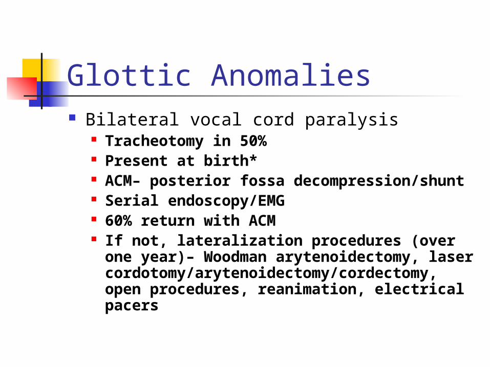
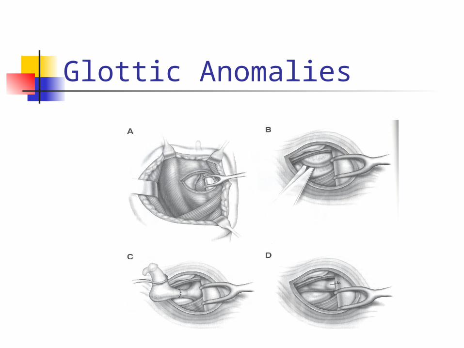
Glottic Anomalies Bilateral vocal cord paralysis
Tracheotomy in 50% Present at birth* ACM– posterior fossa decompression/shunt Serial endoscopy/EMG 60% return with ACM If not, lateralization procedures (over one
year)– Woodman arytenoidectomy, laser cordotomy/arytenoidectomy/cordectomy, open procedures, reanimation, electrical pacers
Glottic Anomalies
Glottic Anomalies
Glottic Anomalies
Glottic Anomalies
Glottic Anomalies
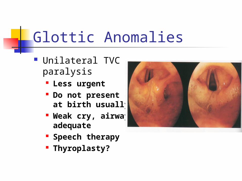
Glottic Anomalies Unilateral TVC
paralysis Less urgent Do not present
at birth usually Weak cry,
airway adequate Speech therapy Thyroplasty?
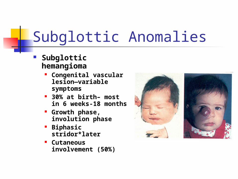
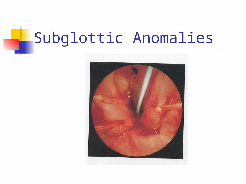
Subglottic Anomalies Subglottic
hemangioma Congenital vascular
lesion—variable symptoms
30% at birth– most in 6 weeks-18 months
Growth phase, involution phase
Biphasic stridor*later
Cutaneous involvement (50%)
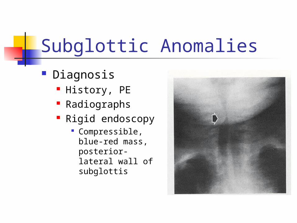
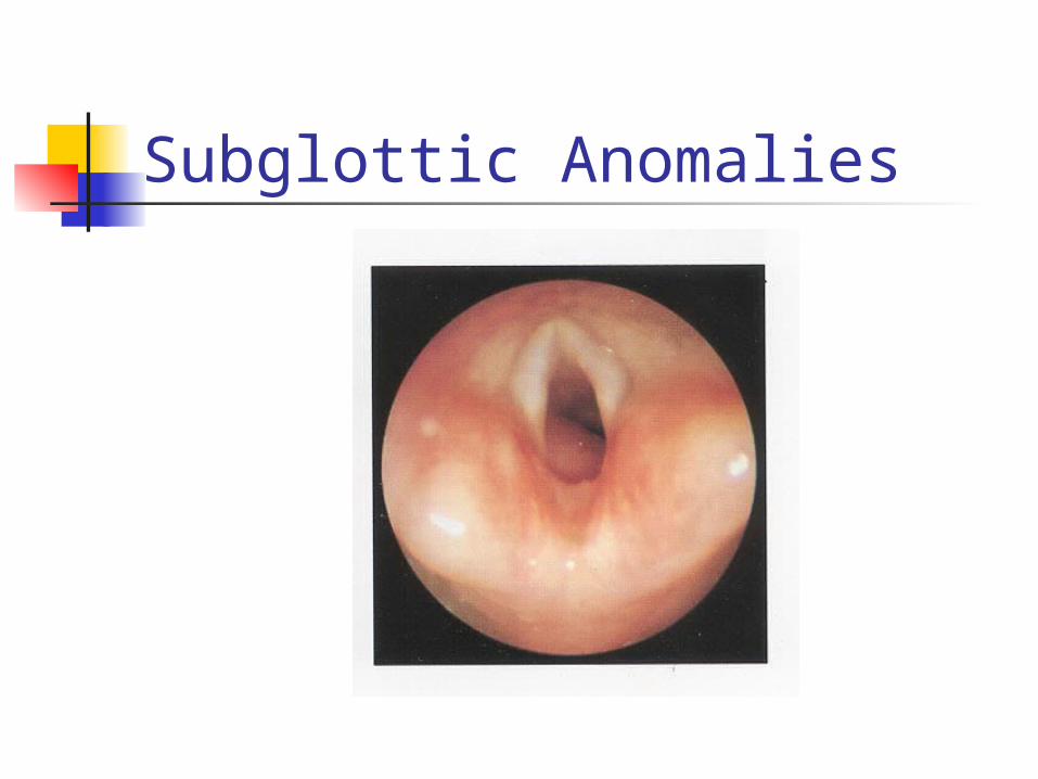
Subglottic Anomalies Diagnosis
History, PE Radiographs Rigid endoscopy
Compressible, blue-red mass, posterior-lateral wall of subglottis
Subglottic Anomalies
Subglottic Anomalies Subglottic hemangioma
Tracheotomy Laser ablation– CO2 vs. KTP EBR, cryotherapy, sclerosing agents Corticosteroids Open excision
Subglottic Anomalies Posterior laryngeal cleft
Failure of tracheoesophageal septum development (rostral portion)
6% with TEF have PLC Pallister-Hall syndrome May present at birth* Respiratory distress with feeds, cyanosis Aspiration, pneumonia, death
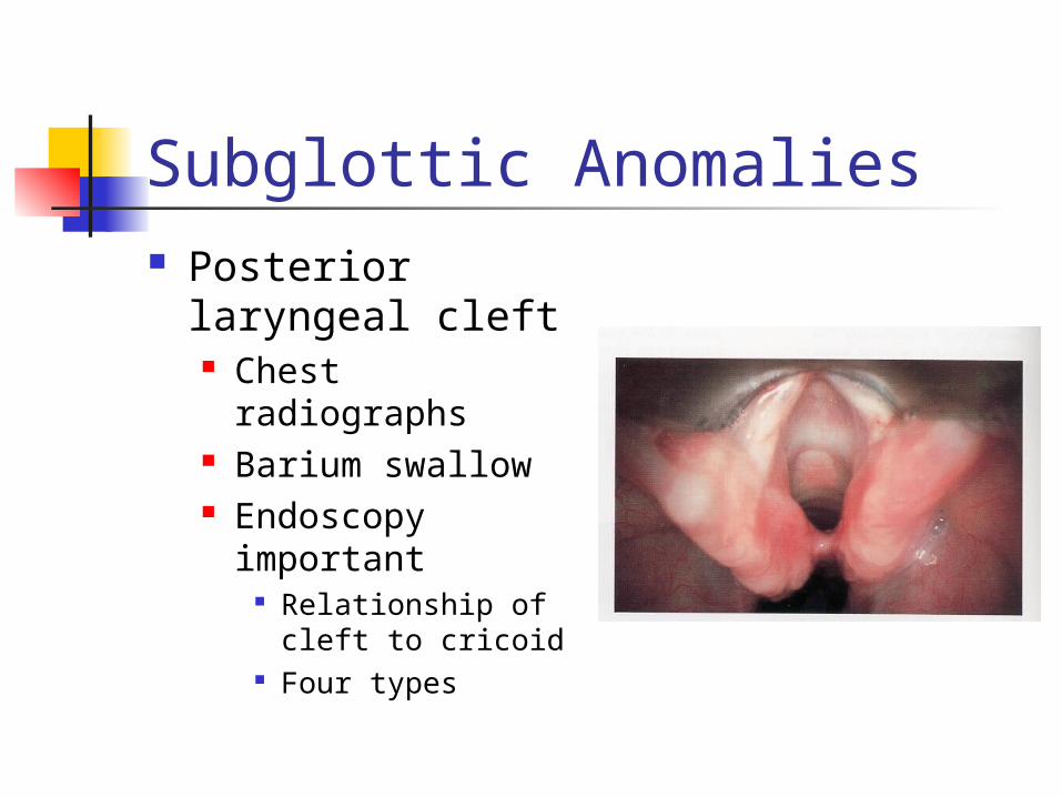
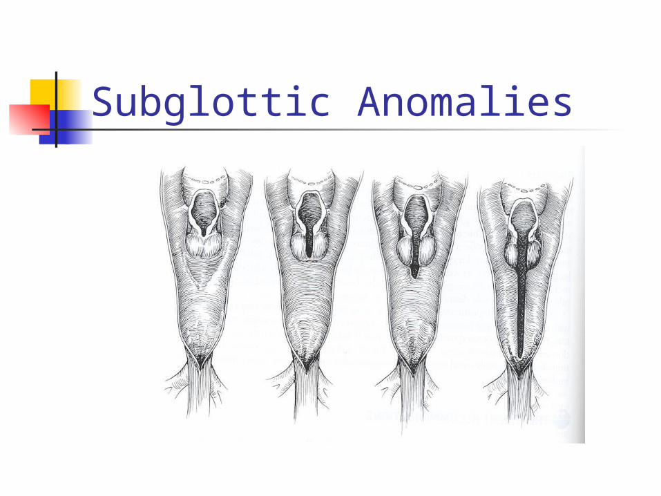
Subglottic Anomalies Posterior
laryngeal cleft Chest radiographs Barium swallow Endoscopy
important Relationship of
cleft to cricoid Four types
Subglottic Anomalies
Subglottic Anomalies
Subglottic Anomalies
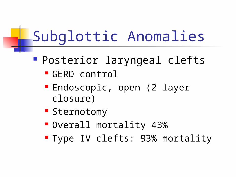
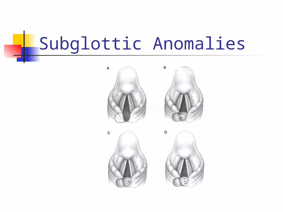
Subglottic Anomalies Posterior laryngeal clefts
GERD control Endoscopic, open (2 layer closure) Sternotomy Overall mortality 43% Type IV clefts: 93% mortality
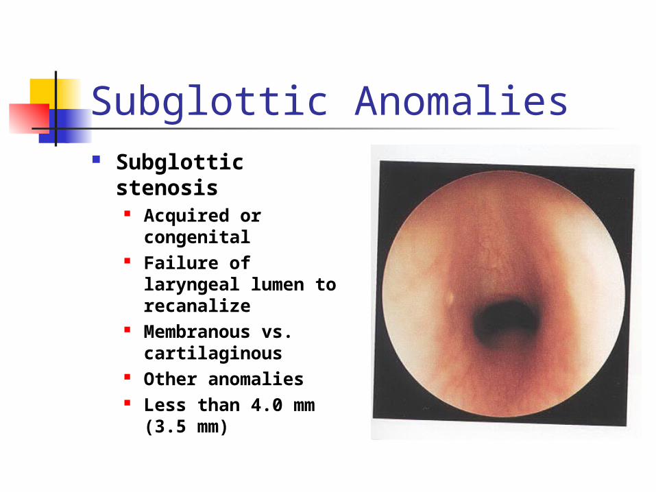
Subglottic Anomalies Subglottic
stenosis Acquired or
congenital Failure of laryngeal
lumen to recanalize Membranous vs.
cartilaginous Other anomalies Less than 4.0 mm
(3.5 mm)
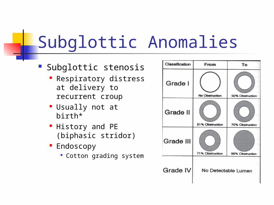
Subglottic Anomalies Subglottic stenosis
Respiratory distress at delivery to recurrent croup
Usually not at birth*
History and PE (biphasic stridor)
Endoscopy Cotton grading
system
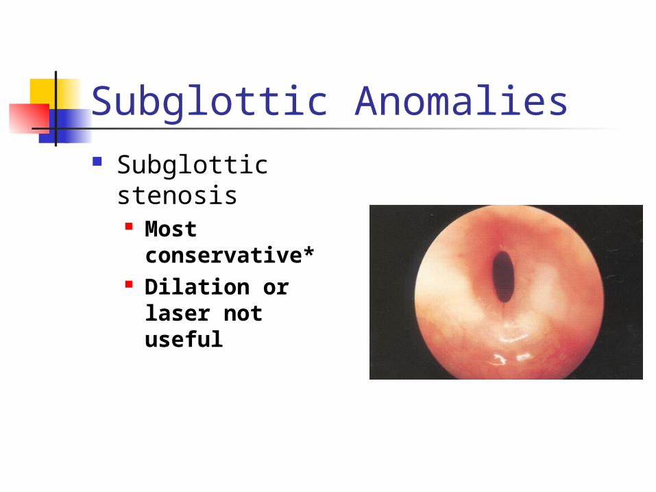
Subglottic Anomalies Subglottic
stenosis Most
conservative* Dilation or laser
not useful
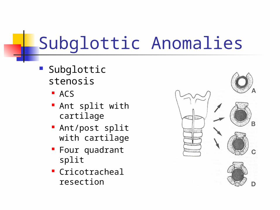
Subglottic Anomalies Subglottic stenosis
ACS Ant split with
cartilage Ant/post split with
cartilage Four quadrant split Cricotracheal
resection