Embed Size (px)
Citation preview

In the name of In the name of GodGod
ByBy: Dr. : Dr. S. S. Khoramrooz, Ph.D.S. S. Khoramrooz, Ph.D.
Department of Microbiology, Faculty of Medicine, Department of Microbiology, Faculty of Medicine,
Yasuj University of Medical Sciences, Yasuj, IranYasuj University of Medical Sciences, Yasuj, Iran
Enterobacteriacae identification
Yasouj University of Medical ScienceYasouj University of Medical Science
Department Department OfOf
MicrobiologyMicrobiology
11
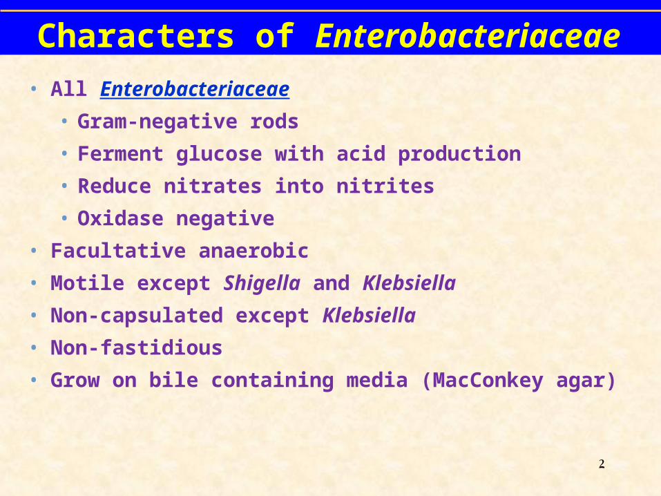
Characters of Enterobacteriaceae
• All Enterobacteriaceae
• Gram-negative rods
• Ferment glucose with acid production
• Reduce nitrates into nitrites
• Oxidase negative
• Facultative anaerobic
• Motile except Shigella and Klebsiella
• Non-capsulated except Klebsiella
• Non-fastidious
• Grow on bile containing media (MacConkey agar)
2

Classification of EnterobacteriaceaeClassification of Enterobacteriaceae
There are several selective and differential media used to isolate distinguishes between LF & LNFThe most important media are:
MacConkey agarEosin Methylene Blue (EMB) agarSalmonella Shigella (SS) agarIn addition to Kiligler Iron agar (KIA)
3

Tests To Know• Case Study Tests
• Indole
• Methyl Red/Voges Proskauer
• Citrate
• H2S production in SIM
• Urea hydrolysis
• Motility
• Lactose fermentation
• Glucose fermentation & gas production
• Decarboxylation of amino acis
• Fermentation of sugars
• Reaction on selective media
4

Growth of Enterobacteriaceae on MacConkey agar
Uninoculated plateLactose non feremtersSalmonella, Shigella,
Proteus
Lactose feremtersE. coli, Citrobacter
Klebsiella, Enterobacter
Colorless colonies Pink colonies
5

Kligler Iron AgarLactose Fermentation
Glucose fermentation
Gas Production (H2 & CO2 )
H2S Production
6

glucoselactoseFerrous sulfatepH indicator: phenol red
Kligler Iron Agar (KIA)
• Proteins
7

8

Red/Red Alkaline /Alkaline K/K Lactose -/Glucose –
Yellow/Yellow Acid/Acid A/A Lactose +/Glucose +
Red/Yellow Alkaline/Acid K/A Lactose -/Glucose +
Gas - H2S -
Red/Yellow Alkaline/Acid K/A Lactose -/Glucose +
Gas + H2S -
Red/Yellow Alkaline/Acid K/A Lactose -/Glucose +
Gas - H2S +
Red/Black Alkaline/Acid K/A Lactose -/Glucose +
Gas + H2S +
9

Reaction on KIA
Result
Example
Butt color
Slant color
H2S
Red Red NegativeAlk/Alk/-
(No action on sugars)
Non fermenter e.g.
Pseudomonas
Yellow Red
Negative A/Alk/- (Glucose fermented
without H2S)
LNF e.g. Shigella
Yellow Red
Positiveblack in
butt
A/Alk/+ (Glucose fermented
with H2S)
LNF e.g. Salmonella &
Proteus
Yellow YellowNegative
A/A/- (three sugars are
fermented)
LF e.g. E. coli, Klebsiella,
Enterobacter
Result
10

11

IMViC Test
• Indole, Methyl Red, Voges-Prosakaur, Citrate (IMViC) Tests:
• The following four tests comprise a series of important determinations that are collectively called the IMViC series of reactions
• The IMViC series of reactions allows for the differentiation of the various members of Enterobacteriaceae.
12

IMViC: Indole test Principle
Certain microorganisms can metabolize tryptophan by tryptophanase
The enzymatic degradation leads to the formation of pyruvic acid, indole and ammonia
The presence of indole is detected by addition of Kovac's reagent.
Tryptophaneamino acids
Tryptophanase Indole + Pyurvic acid + NH3
Kovac’s Reagent
Red color in upper organic layer` 13

IMViC: Indole test
Result:
A bright pink color in the top layer indicates the presence of indole
The absence of color means that indole was not produced i.e. indole is negative
Special Features:
Used in the differentiation of genera and species. e.g. E. coli (+) from Klebsiella (-).
Positive teste.g. E. coli
Negative teste.g. Klebsiella
14

IMViC testMethyl Red-Voges Proskauer (MR-VP) Tests
Glucose
Acidic pathway
Mixed acids pH less than 4.4
Methyl Redindicator
Red color
Principle
MR positiveE. coli
Or Neutral pathway
Acety methyl carbinol(ACETOIN)
solution Asolution B
Pink colorVP positiveKlebsiella
15

Butylene Glycol Pathway of Glucose Fermentation
• In the butylene glycol pathway
• pyruvic acid to acetoin and butylene glycol.
• Acetoin and butylene glycol are detected by oxidation to diacteyl at an alkaline pH.
• and the addition of -naphthol which forms a red-colored complex with diacetyl.
• Important biochemical property used for the identification of Klebsiella, Enterobacter, and Serratia.
16

Voges-Proskauer Reaction
• Acetoin and butylene glycol are detected by oxidation to diacteyl at an alkaline pH, and the addition of -naphthol which forms a red-colored complex with diacetyl.
• The production of acetoin and butylene glycol by glucose fermentation is an important biochemical property used for the identification of Klebsiella, Enterobacter, and Serratia.
17

IMViC test: MRVP test
Inoculate the tested organism into MRVP broth
Incubate the tubes at 37°C for 24 hours
• For methyl red: Add 6-8 drops of methyl
red reagent.
• For Voges-Proskauer: Add 12 drops of
solution A (-naphthol), mix, 4 drops of
Solution B (40% KOH), mix
Method
18

IMViC test: MR/VP testResults
Methyl Red test Voges-Proskauer test
Red: Positive MR (E. coli)
Yellow or orange: Negative MR (Klebsiella)
Pink: Positive VP (Klebsiella)
No pink: Negative VP (E. coli) 19

Citrate Utilization TestCitrate Utilization TestPrinciple:Citrate Na2CO3
Alkaline,↑pH
Blue colour
Bromothymol blue
Simmone’s Citrate media
Positive test: Klebsiella, Enterobacter, Citrobacter
CO2 + Na + H2OPyruvate
Positive test
Negative test: E. coli
Contains Citrate as a sole of C source
20

Citrate Utilization Test
Incubate at 37°C for 24 hours.
MethodMethod
Streak a Streak a Simmon's Citrate agar slant with slant with
the organism the organism
21

Citrate Utilization Test
Examine for growth (+)
Growth on the medium is accompanied by a rise in pH to
change the medium from its initial green color to deep blue
ResultResult
PositiveKlebsiella, Enterobacter
NegativeE. coli 22

Urease Test
Urea agar contains urea and phenol red
Urease is an enzyme that catalyzes the conversion of urea to CO2 and NH3
Ammonia combines with water to produce ammonium hydroxide, a strong base which ↑ pH of the medium.
↑ in the pH causes phenol red r to turn a deep pink. This is indicative of a positive reaction for urease
UreaUrease
CO2 + NH3H2O
NH4 OH ↑ in pH
Phenol Red
PinkPositive test
Streak a urea agar tube with the organism
incubate at 37°C for 24 h
Method
PrincipPrincipllee
23

Urease Test
• If color of medium turns from yellow to pink indicates positive test.
• Proteus give positive reaction after 4 h while Kelebsiella and Enterobacter gave positive results after 24 h
Result
Positive test Negative test
24

MotilityFrom left to right:+ – +
25

SIM Sulfide, Indole, Motility
04/18/23 S. S. Khoramrooz 26

Phenylalanine Deaminase Reaction
• Enterobacteriaceae utilize amino acids in a variety of ways including deamination.
• Phenylalanine is an amino acid that forms the keto acid phenylpyruvic acid when deaminated.
• Phenylpyruvic acid is detected by addition of ferric chloride that forms an intensely dark olive-green colored complex when binding to phenylpyruvic acid.
• The deamination of phenylalanine is an important biochemical property of Proteus, Morganella, and Providencia.
28

Amino Acid Decarboxylation
• Enterobacteriaceae contain decarboxylases with substrate specificity for amino acids, and are detected using Moeller decarboxylase broth overlayed with mineral oil for anaerobiosis.
• Moeller broth contains glucose for fermentation, peptone and beef extract, an amino acid, pyridoxal, and the pH indicator bromcresol purple.
29

Amino Acid Decarboxylation• If an Enterobacteriaceae contains amino acid
decarboxylase, amines produced by decarboxylase action cause an alkaline pH, and bromcresol purple turns purple.
• Lysine, ornithine, and arginine are utilized.
• A base broth without amino acid is included in which glucose fermentation acidifies the broth, turning the bromcresol purple yellow.
30

Amino Acid Decarboxylation1
Lysine → Cadaverine
Ornithine → Putrescine
Arginine → Citrulline → Ornithine → Putrescine1Conversion of arginine to citrulline is a dihydrolase reaction
31

Amino Acid Decarboxylation
• Decarboxylation patterns are essential for the genus identification of Klebsiella, Enterobacter, Escherichia, and Salmonella.
• Decarboxylation patterns are also essential for the species identification of Enterobacter aerogenes, Enterobacter cloacae, Proteus mirabilis, and Shigella sonnei.
32

Amino Acid Decarboxylation
Lys Orn Arg
Klebsiella + – –
Enterobacter +/– + +/–
Escherichia + +/– –/+
Salmonella + + +
33

Amino Acid Decarboxylation
Lys Orn Arg
E. aerogenes + + –
E. cloacae – + +
P. mirabilis – + –
P. vulgaris – – –
Shigella D – + –
Shigella A-C – – –
34

IPViC Reactions for Initial Grouping of the Enterobacteriaceae
• Indole
• Phenylalanine deaminase
• Voges-Proskauer
• Citrate
35

Initial Grouping of the Enterobacteriaceae (VP=Voges Proskauer, PDA=Phenylalanine
Deaminase)
GENERA VP PDA
Klebsiella POSITIVE NEGATIVE
Enterobacter POSITIVE NEGATIVE
Serratia POSITIVE NEGATIVE
Hafnia POSITIVE NEGATIVE
Pantoea POSITIVE NEGATIVE
36

Initial Grouping of the Enterobacteriaceae
GENERA VP PDA
Proteus1 NEGATIVE POSITIVE
Morganella NEGATIVE POSITIVE
Providencia NEGATIVE POSITIVE
1Proteus mirabilis: 50% of strains VP positive37

Initial Grouping of the Enterobacteriaceae
GENERA VP PDAEscherichia NEGATIVE NEGATIVEShigella NEGATIVE NEGATIVEEdwardsiella NEGATIVE NEGATIVESalmonella NEGATIVE NEGATIVECitrobacter NEGATIVE NEGATIVEYersinia NEGATIVE NEGATIVE
38

Initial Grouping of the Enterobacteriaceae1
GENERA INDOLE CITRATE
Escherichia POSITIVE NEGATIVE
Shigella Yersinia
POSITIVE2
POSITIVE3 NEGATIVE NEGATIVE
Edwardsiella POSTIVE NEGATIVE
1VP negative, PDA negative 2Shigella groups A, B, and C variably positive for indole production (25-50%), group D Shigella negative. 3Yersinia enterocolitica 50% positive
39

Initial Grouping of the Enterobacteriaceae1
GENERA INDOLE CITRATE Salmonella NEGATIVE POSITIVE2
Citrobacter NEGATIVE POSITIVE
1VP negative, PDA negative 2Salmonella serotype Paratyphi A and Typhi negative
40
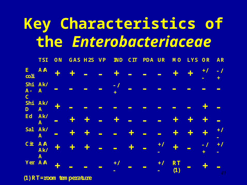
Key Characteristics of the Enterobacteriaceae
TSI ON GAS H2S VP IND CIT PDA UR MO LYS OR AR
E coli
A/A + + + + + +/
/ +
Shi A-C
Ak/A /
+ Shi D
Ak/A + +
Ed Ak/A + + + + + +
Sal Ak/A + + + + + + +/
Cit A/A
Ak/A
+ + + + +/ + /
+ +/
Yer A/A + +/
+/
RT (1) +
(1) RT=room temperature 41

Key Characteristics of the Enterobacteriaceae
TSI ON GAS H2S VP IND CIT PDA UR MO LYS OR AR
Kle pne
A/A + + + + + + Kleoxy
A/A + + + + + + + En aer
A/A + + + + + + + En cloa
A/A + + + + +/ + + + Serr (1)
A/A + + + + + + + Haf Ak/
A + + + + + + Pan A/A
Alk/A
+ /+ +/ /+ +/ /+ /+
(1) Produces DNase, lipase, and gelatinase 42

Key Characteristics of the Enterobacteriaceae
TSI ON GAS H2S VP IND CIT PDA UR MO LYS OR AR
Prot mira
Ak/A + + +/ +/ + + +s +
Prot vulg
A/A +/ + + /+ + + +s Mor Ak/
A + + + + + + Prov
Ak/A + + + + +
s = swarming motility
43

Biochemical Characteristics of Escherichia coli and Shiglla
E. coli E. coli O157:H7 ShigellaTSI A/Ag A/Ag Alk/ALactose + + –ONPG + + –/+1
Sorbitol + – +/–Indole + + +/–Methyl red + + +VP – – –Citrate – – –Lysine + + –Motility + + –
1Shigella sonnei (group D) ONPG +
44

Biochemical Characteristics of Salmonella Most Serotypes Typhi Paratyphi
ATSI Alk/A Alk/A Alk/A
H2S (TSI) + + (weak) –Citrate + – –Lysine + + –Ornithine + – +Dulcitol + – +Rhamnose + – +Indole – – –Methyl red + + +VP – – –
45

Xylose Lysine Deoxycholate (XLD) Agar: Composition
• Xylose 0.35%• Lysine 0.5%• Lactose 0.75%• Sucrose 0.75%• Sodium chloride 0.5%• Yeast extract 0.3%• Sodium deoxycholate 0.25%• Sodium thiosulfate• Ferric ammonium citrate• Agar 1.35%• Phenol red• pH = 7.4
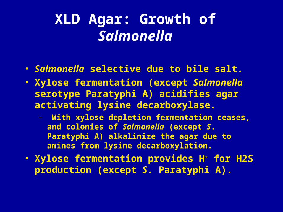
XLD Agar: Growth of Salmonella
• Salmonella selective due to bile salt.
• Xylose fermentation (except Salmonella serotype Paratyphi A) acidifies agar activating lysine decarboxylase. – With xylose depletion fermentation ceases, and colonies of
Salmonella (except S. Paratyphi A) alkalinize the agar due to amines from lysine decarboxylation.
• Xylose fermentation provides H+ for H2S production (except S. Paratyphi A).

XLD Agar: Appearance of Salmonella
• Ferric ammonium citrate present in XLD agar reacts with H2S gas and forms black precipitates within colonies of Salmonella.
• Agar becomes red-purple due to alkaline pH produced by amines.
• Back colonies growing on red-purple agar-presumptive for Salmonella.


XLD Agar: Growth of Escherichia coli and Klebsiella pneumoniae
Escherichia coli and Klebsiella pneumoniae are lysine-positive coliforms that are also lactose and sucrose fermenters.
The high lactose and sucrose concentrations result in strong acid production, which quenches amines roduced by lysine decarboxylation.
Colonies and agar appear bright yellow. Neither Escherichia coli nor Klebsiella pneumoniae produce H2S.

XLD Agar: Growth of Shigella and Proteus
Shigella species do not ferment xylose, lactose, and sucrose, do not decarboxylate lysine, and do not produce H2S. Colonies appear colorless.
Proteus mirabilis ferments xylose, and thereby provides H+ for H2S production. Colonies appear black on an agar unchanged in color (Proteus deaminates rather than decarboxylates amino acids).
Proteus vulgaris ferments sucrose, and colonies appear black on a yellow agar.


Hektoen Enteric (HE) Agar: Composition
• Peptone 1.2%• Yeast extract 0.3%• Bile salts 0.9%• Lactose 1.2%• Sucrose 1.2%• Salicin 0.2%• Sodium chloride 0.5%• Ferric ammonium citrate• Acid fuchsin• Thymol blue• Agar 1.4%• pH = 7.6

HE Agar: Growth of Enteric Pathogens and Commensals
• High bile salt concentration inhibits growth of gram-positive and gram-negative intestinal commensals, and thereby selects for pathogenic Salmonella (bile-resistant growth) present in fecal specimens.
• Salmonella species as non-lactose and non-sucrose fermenters that produce H2S form colorless colonies with black centers.
• Shigella species (non-lactose and non-sucrose fermenters, no H2S production) form colorless colonies.
• Lactose and sucrose fermenters (E. coli, K. pneumoniae) form orange to yellow colonies due to acid production.


58

59
Some strains appear mucoid
Particularly common in patients with cystic fibrosis
Some strains produce diffusible pigments
Pyocyanin [blue]
Fluorescein [yellow]
Pyorubin [redbrown]
Pseudomonas aeruginosa

60
Laboratory Diagnosis
CultureGrow easily on common isolation media such as blood agar and MacConkey37-42C
Identification
The colonial morphology (e.g., colony size, hemolytic activity, pigmentation, odor)
Biochemical tests (e.g., positive oxidase reaction)
+

61

62

The End
63