Embed Size (px)
Citation preview
Muscarinic M2 acetylcholine receptor distribution
in the guinea pig gastrointestinal tract
Satoshi Iino and Yoshiaki Nojyo
Department of Anatomy, University of Fukui Faculty of Medical Sciences
Matsuoka, Fukui 910-1193 JAPAN
Corresponding author: Satoshi Iino
Address: Department of Anatomy, University of Fukui Faculty of Medical Sciences
Matsuoka, Fukui 910-1193 JAPAN
Tel.: 81-776-61-8301
FAX: 81-776-61-8132
E-mail: [email protected]
Number of: Text pages: 36
Figures: 6
1
Section Editor: Dr. C. R. Gerfen
NIMH, Lab of Systems Neuroscience, Bldg 36, Room 2D-30, MSC 4075, 36 Convent Drive,
Bethesda, MD 20892-4075, USA
2
Abstract
In the enteric nervous system, acetylcholine is the most common neurotransmitter to
induce gastrointestinal smooth muscle contractions. Cholinergic signaling is mediated by
muscarinic acetylcholine receptors on the surface of smooth muscle cells. Five different
muscarinic receptor subtypes (M1-M5) have been identified and characterized, all of which
belong to the superfamily of the G-protein-coupled receptor. The muscarinic M2
acetylcholine receptor is the major muscarinic receptor subtype expressed by smooth muscle
tissues in the gastrointestinal tract, where it is coexpressed with a smaller population of M3
receptor. In this study, we examined the immunohistochemical distribution of the M2
receptor using a specific antibody. M2 receptor-like immunoreactivity (M2R-LI) was mainly
observed as associated with smooth muscle cells in the gastrointestinal tract. M2R-LI in
smooth muscle cells was distributed throughout the cell membrane associated with caveolae.
In the proximal colon, M2R-LI in the smooth muscle cells was weak. In the small intestine,
interstitial cells of Cajal that possessed neurokinin 1 receptor-LI had intense M2R-LI. In the
proximal colon, intramuscular and myenteric interstitial cells of Cajal exhibited M2R-LI.
These findings indicate that, in the gastrointestinal musculature, M2 receptors are distributed
both in the smooth muscle cells and interstitial cells of Cajal, suggesting that the M2 receptor
3
elicits smooth muscle cell contraction and the interstitial cells of Cajal are the sites of
innervation by enteric cholinergic neurons.
Key words: enteric nervous system, smooth muscle, interstitial cells of Cajal, caveolae,
acetylcholine, neurokinin 1 receptor
4
Introduction
Acetylcholine is distributed widely and acts as a neurotransmitter in the central
nervous system and peripheral nervous system. In the enteric nervous system, acetylcholine
is the most common neurotransmitter to induce gastrointestinal tract contractions. In the
gastrointestinal tract, acetylcholine is released from excitatory enteric motor neurons that have
choline acetyltransferase and vesicular acetylcholine transporter, and mediates immediate
smooth muscle contraction (Bornstein et al., 2004; Furness, 2000). Acetylcholine is
believed to be functionally predominant in inducing smooth muscle contractions.
Acetylcholine signaling is mediated by nicotinic acetylcholine receptors and
muscarinic acetylcholine receptors on the cell surface. Muscarinic acetylcholine receptors
are known to control of many central and peripheral cholinergic responses (Wess, 1996; Wess,
2004). To date, five different muscarinic receptor subtypes (M1-M5) have been identified
and characterized (Caulfield and Birdsall, 1998; Wess, 1996), all of which belong to the
superfamily of the G-protein-coupled receptor. M1, M3, and M5 receptors act preferentially
to activate the phospholipase C pathway through selective coupling to G protein Gq/G11,
whereas M2 and M4 receptors mainly mediate the inhibition of adenylate cyclase by coupling
to G protein Gi/Go (Caulfield and Birdsall, 1998; Wess, 1996). Many researches, including
5
immunohistochemistry and in situ hybridization studies, have shown that muscarinic
receptors are present in many organs and tissues (Dorje et al., 1991; Eglen et al., 1996; Ehlert
et al., 1997; Levey, 1993; Wess, 2004). The M1 receptor is mainly distributed in the brain,
M2 receptor in the brain, heart and smooth muscle organs, M3 receptor in the brain, smooth
muscle organs, exocrine glands and eyes, M4 receptor in the brain and lung, and M5 receptor
is expressed at rather low levels in both neuronal and nonneuronal cells. Because of the lack
of ligands endowed with a high degree of receptor subtype selectivity and as most tissues
express two or more muscarinic receptor subtypes, identification of the physiological and
pathophysiological roles of the individual muscarinic receptor subtypes has proved difficult.
In smooth muscle tissues such as the gastrointestinal tract, urogenital tract and
respiratory tract, the M2 receptor is the major muscarinic receptor subtype, where it is
coexpressed with a smaller population of M3 receptors (Eglen et al., 1996; Ehlert et al., 1997;
Levey, 1993). In the gastrointestinal tract, more than 70% muscarinic receptors are
composed from the M2 receptor and about 20% of muscarinic receptors are from the M3
receptor. From a molecular study using the RT-PCR technique, both smooth muscle cells
and interstitial cells of Cajal expressed both M2 and M3 receptors (Epperson et al., 2000).
Interstitial cells of Cajal are the third cell types different from smooth muscle cells and enteric
6
neurons in the gastrointestinal musculature and are involved in muscle contraction and
neuronal modulation of contraction (Rumessen and Vanderwinden, 2003; Sanders, 1998).
To help decipher the biological function of muscarinic M2 acetylcholine receptors in vivo, the
histological and cytological distribution of M2 receptor must be elucidated. In this study, we
examined the immunohistochemical distribution of the M2 receptor using a specific antibody,
and the intracellular distribution of the M2 receptor on the electron microscopic level.
7
Experimental procedures
Adult male Hartley guinea pigs aged 4-6 weeks and weighing 250-400 g (Japan SLC,
Japan) were used in this study. The use and treatment of animals followed the Guidelines
for Animal Experiments, University of Fukui Faculty of Medical Sciences. All guinea pigs
were anesthetized with an intraperitoneal injection of sodium pentobarbital (50 mg/kg) before
fixation.
Guinea pigs were transcardially perfused with Zamboni's fixative (Iino, 2000). The
gastrointestinal tract was dissected out and further immersed in Zamboni's fixative for 4 h at
room temperature. After washing with 0.01 M phosphate-buffered saline (PBS, pH 7.2), the
specimens were soaked overnight in 30% sucrose in 0.1 M phosphate buffer (PB, pH 7.4) at
4°C, embedded in OCT compound (Miles, USA) and frozen quickly. Twelve-μm thick
sections were cut using a cryostat and thaw-mounted onto poly-L-lysine-coated glass slides.
The sections were treated with methanol containing 0.3% H2O2. After washing with PBS,
sections were incubated for 1 h in 10% normal goat serum (diluted with PBS), and then
reacted with rat anti-M2 receptor (1:500 with PBS, MAB367, Chemicon, USA) (Levey et al.,
1995a) for 12 h at room temperature. Sections were washed three times with PBS, and then
reacted with biotinylated goat anti-rat IgG (1:200 with PBS, Vector, USA) for 1 h. After
8
washing with PBS, they were reacted with avidin-biotin-peroxidase complex (ABC Kit,
Vector) for 1 h. The sections were incubated for several minutes with a solution containing
0.03% diaminobenzidine, 0.005% H2O2 in 0.1 M Tris-HCl, pH 7.6. All sections were
dehydrated with ethanol and mounted in Entellan (Merck, Germany).
For fluorescence immunohistochemistry, sections were incubated in a mixture of rat
anti-M2 receptor (1:500 with PBS) and mouse anti-smooth muscle actin (1:600 with PBS,
clone 1A4, Sigma, USA), mouse anti-actin (1:500 with PBS, clone B4, ICN, USA), rabbit
anti-neurokinin 1 receptor (1:500 with PBS, #94168) (Grady et al., 1996; Southwell and
Furness, 2001), or mouse anti-caveolin 1 (1:100 with PBS, clone 2297, Transduction
Laboratories, USA). Then sections were incubated for 1 h with the biotinylated anti-rat IgG
(1:200 with PBS, Vector), then for 1 h with FITC (fluorescein isothiocyanate)-conjugated
streptoavidin (1:100 with PBS, Vector, USA) and Cy3-conjugated anti-mouse IgG (1:200
with PBS, Jackson, USA), Texas Red-conjugated anti-rabbit IgG (1:100 with PBS, Vector).
These sections were examined with a confocal laser-scanning microscope GB200 (Olympus,
Japan) with excitation wavelength of 488 nm and 543 nm. Immunohistochemical study
using whole mount preparations was carried out by the method described previously (Iino et
al., 2004).
9
For immunoelectron microscopy, three guinea pigs were transcardially perfused with
Zamboni's fixative plus 0.1% glutaraldehyde. The intestine was dissected out and further
immersed in Zamboni's fixative for 4 h at room temperature. Tissues treated as above were
cut by cryostat at 12μm and mounted on poly-L-lysine coated slides. Specimens were
immunostained as were those for light microscopic observations. After coloration in
diaminobenzidine solution, specimens were post-fixed in 1% OsO4 in PB for 1 h and
block-stained with uranyl acetate, dehydrated in ethanol and embedded in Epok 812 (Oken,
Japan). Ultrathin sections were examined without electron staining using an electron
microscope H-7000 (Hitachi, Japan).
Anti-M2 receptor rat monoclonal antibody was produced as previously reported and
the specificity was checked by Levey et al. (1995ab). M2 receptor knockout mice showed no
specific immunoreactivity for this antibody (Takeuchi et al., 2005). Specificity of this
immunohistochemical study was checked by unimmunized rat serum instead of the anti-M2
receptor antibody, and no specific immunoreactivity was observed.
10
Results
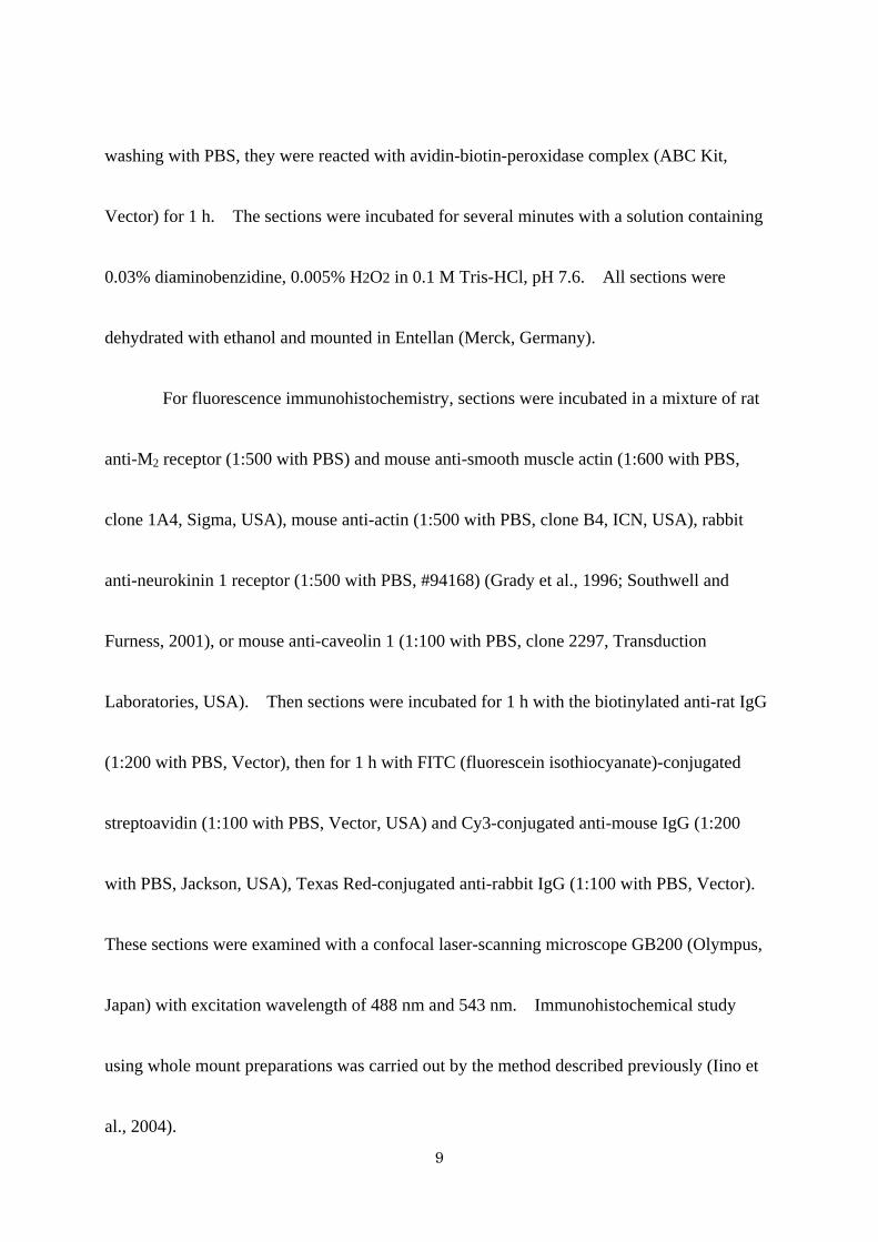
M2 receptor-like immunoreactivity (M2R-LI) was observed associated with smooth
muscle cells in the gastrointestinal tract (Fig. 1, Table 1). The external muscle layers, except
the esophagus, were immunoreactive for M2 receptor. The intensity of immunoreactivity
was higher in the stomach, small intestine, cecum, distal colon and rectum than in the
proximal colon. Distinct immunoreaction was also observed in the muscularis mucosae in
the pylorus, colon and rectum.
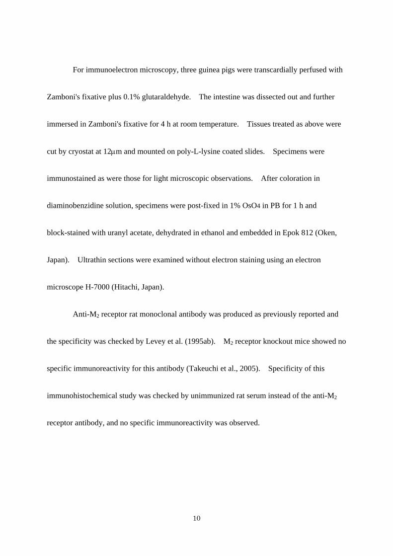
1, Esophagus
In the esophagus, both external muscle layer and muscularis mucosae showed no
M2R-LI (Fig. 1). In the lower esophageal sphincter (Fig. 2A), M2R-LI was observed only in
the smooth muscle cells. The enteric ganglia and interstitial cells of Cajal in the esophagus
were free from reaction.
2, Stomach
In the gastric fundus and corpus (Figs. 1B, 2B), M2R-LI was clearly observed in the
musculature. In the circular muscle, most smooth muscle cells, except a few cells associated
with submucosa, showed distinct immunoreactivity at their surface. In the submucosal
border, M2R-LI in the smooth muscle cells was low or absent. In the longitudinal muscle,
11
all smooth muscle cells showed M2R-LI as in circular layer cells. The enteric ganglia and
interstitial cells of Cajal were free from reaction. In the pylorus (Fig. 1C), although the
circular muscle had distinct immunoreactivity as observed in the fundus and corpus, the
longitudinal muscle showed weak M2R-LI. The muscularis mucosae in the fundus and
corpus had no immunoreactivity (Fig. 1B), whereas that in the pylorus had intense M2R-LI
(Fig. 1C).
3, Small intestine
In the duodenum (Fig. 1D), jejunum (Fig. 1E) and ileum, almost all smooth muscle
cells in the musculature had M2R-LI on their surface. Using double fluorescence
immunohistochemistry with anti-M2 receptor and anti-actin antibodies (Figs. 3A-F), all
smooth muscle cells in the outer circular muscle and longitudinal muscle had distinct M2R-LI.
The inner circular muscle, localized between the outer circular muscle and the submucosa,
had no M2R-LI.
In the deep muscular plexus layer (DMP) between the inner and outer circular muscle
(Figs. 3D-F), there were a small number of M2R-LI cells that were not stained by actin
antibodies. Different from the smooth muscle cells, these cells had M2R-LI in the periphery
and cytoplasm. These cells showed neurokinin 1 receptor-LI (Figs. 3G-I), which is a marker
12
of the interstitial cells of Cajal in the DMP (Iino et al., 2004; Lavin et al., 1998; Southwell and
Furness, 2001). Though, interstitial cells of Cajal in the small intestine are also distributed
in the myenteric layer and interstitial cells of Cajal in the duodenum and proximal jejunum
show neurokinin 1 receptor-LI (Lavin et al., 1998; Rumessen and Vanderwinden, 2003), there
were no specific cells having M2R-LI in the myenteric layer.
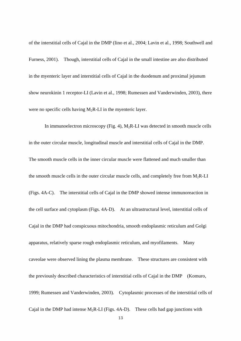
In immunoelectron microscopy (Fig. 4), M2R-LI was detected in smooth muscle cells
in the outer circular muscle, longitudinal muscle and interstitial cells of Cajal in the DMP.
The smooth muscle cells in the inner circular muscle were flattened and much smaller than
the smooth muscle cells in the outer circular muscle cells, and completely free from M2R-LI
(Figs. 4A-C). The interstitial cells of Cajal in the DMP showed intense immunoreaction in
the cell surface and cytoplasm (Figs. 4A-D). At an ultrastructural level, interstitial cells of
Cajal in the DMP had conspicuous mitochondria, smooth endoplasmic reticulum and Golgi
apparatus, relatively sparse rough endoplasmic reticulum, and myofilaments. Many
caveolae were observed lining the plasma membrane. These structures are consistent with
the previously described characteristics of interstitial cells of Cajal in the DMP (Komuro,
1999; Rumessen and Vanderwinden, 2003). Cytoplasmic processes of the interstitial cells of
Cajal in the DMP had intense M2R-LI (Figs. 4A-D). These cells had gap junctions with
13
themselves (Fig. 4C) or with the smooth muscle cells in the outer circular layer (Fig. 4D).
M2R-LI in the smooth muscle cells was distributed throughout the cell membrane at
the cytoplasmic surface in the electron microscopic level. These immunoreactivities were
detected as a patchy pattern and most reactions were around or associated with caveolae (Fig.
4F). Using double fluorescence immunohistochemistry with anti-M2 receptor (Fig. 3J) and
anti-caveolin 1 antibodies (Fig. 3B), M2R-LI was colocalized with caveolin 1-LI.
4, Large intestine
In the cecum (Fig. 1F), both circular and longitudinal muscle cells showed distinct
M2R-LI. The muscularis mucosae of the cecum had no M2R-LI.
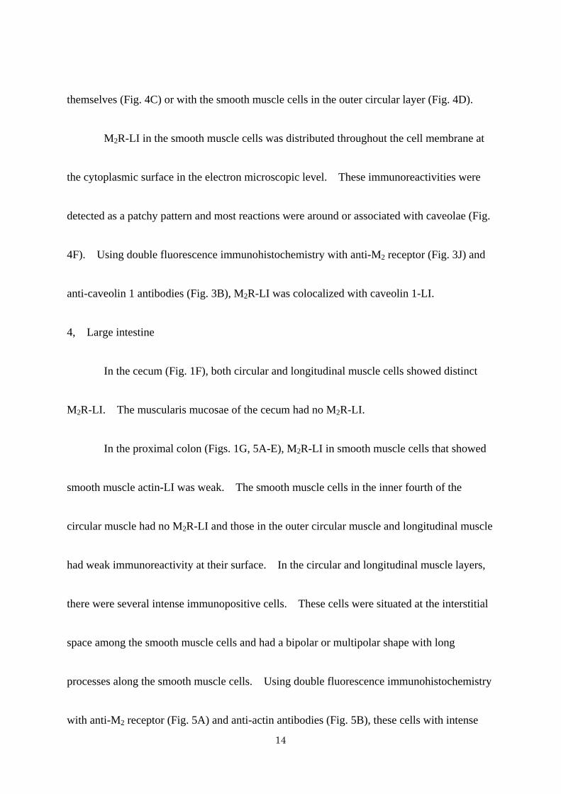
In the proximal colon (Figs. 1G, 5A-E), M2R-LI in smooth muscle cells that showed
smooth muscle actin-LI was weak. The smooth muscle cells in the inner fourth of the
circular muscle had no M2R-LI and those in the outer circular muscle and longitudinal muscle
had weak immunoreactivity at their surface. In the circular and longitudinal muscle layers,
there were several intense immunopositive cells. These cells were situated at the interstitial
space among the smooth muscle cells and had a bipolar or multipolar shape with long
processes along the smooth muscle cells. Using double fluorescence immunohistochemistry
with anti-M2 receptor (Fig. 5A) and anti-actin antibodies (Fig. 5B), these cells with intense
14
M2R-LI showed no actin-LI. Using whole mount preparations, M2R-LI cells in the circular
and longitudinal muscle (Figs. 5CD) showed bipolar or multipolar shape with long slender
processes. In the myenteric layer, there was another cell type with M2R-LI (Figs. 5ABE).
These cells had no actin-LI, were situated around the myenteric ganglia and showed
multipolar shape.
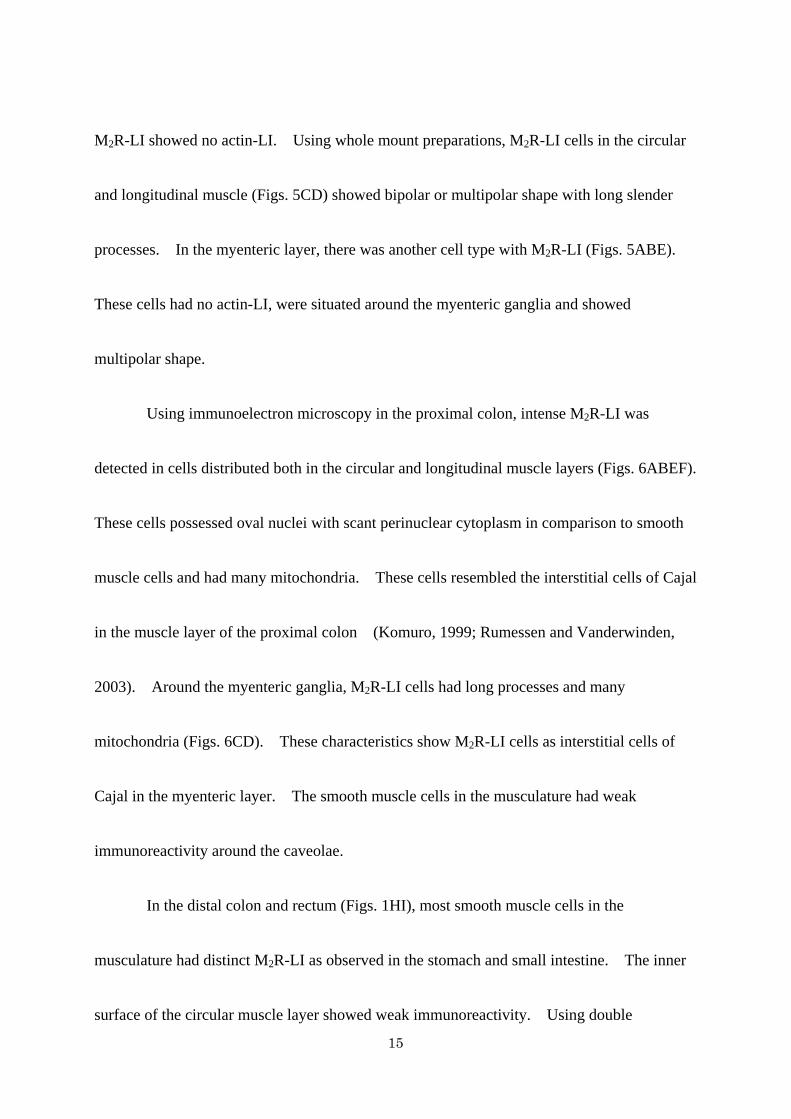
Using immunoelectron microscopy in the proximal colon, intense M2R-LI was
detected in cells distributed both in the circular and longitudinal muscle layers (Figs. 6ABEF).
These cells possessed oval nuclei with scant perinuclear cytoplasm in comparison to smooth
muscle cells and had many mitochondria. These cells resembled the interstitial cells of Cajal
in the muscle layer of the proximal colon (Komuro, 1999; Rumessen and Vanderwinden,
2003). Around the myenteric ganglia, M2R-LI cells had long processes and many
mitochondria (Figs. 6CD). These characteristics show M2R-LI cells as interstitial cells of
Cajal in the myenteric layer. The smooth muscle cells in the musculature had weak
immunoreactivity around the caveolae.
In the distal colon and rectum (Figs. 1HI), most smooth muscle cells in the
musculature had distinct M2R-LI as observed in the stomach and small intestine. The inner
surface of the circular muscle layer showed weak immunoreactivity. Using double
15
immunohistochemistry with anti-M2 receptor (Fig. 5F) and anti-actin antibodies (Fig. 5G), we
could clearly observe that most smooth muscle cells had M2R-LI at their surfaces. In the
interstitial space of the muscle layer and around the myenteric ganglia, there were no M2R-LI
interstitial cells of Cajal as observed in the proximal colon. In the muscularis mucosae of
the colon and rectum, only longitudinal arranged smooth muscle cells had M2R-LI at their
surface. The circular smooth muscle cells in the muscularis mucosae were free from
reaction.
16
Discussion
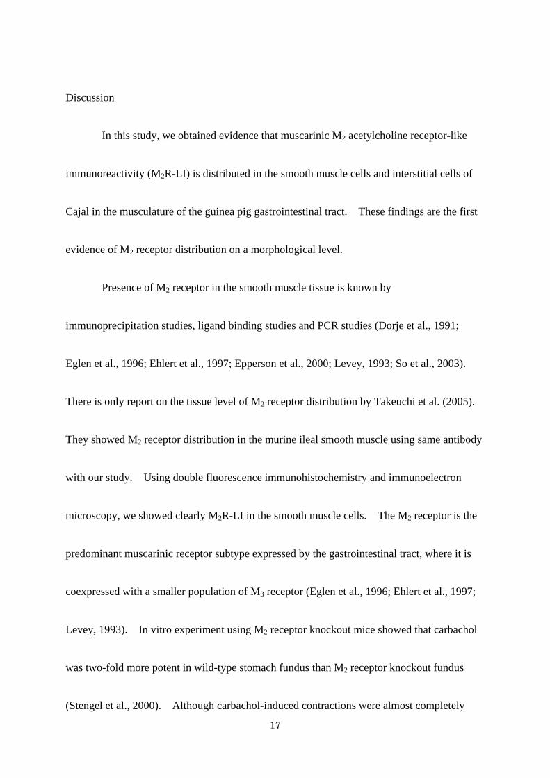
In this study, we obtained evidence that muscarinic M2 acetylcholine receptor-like
immunoreactivity (M2R-LI) is distributed in the smooth muscle cells and interstitial cells of
Cajal in the musculature of the guinea pig gastrointestinal tract. These findings are the first
evidence of M2 receptor distribution on a morphological level.
Presence of M2 receptor in the smooth muscle tissue is known by
immunoprecipitation studies, ligand binding studies and PCR studies (Dorje et al., 1991;
Eglen et al., 1996; Ehlert et al., 1997; Epperson et al., 2000; Levey, 1993; So et al., 2003).
There is only report on the tissue level of M2 receptor distribution by Takeuchi et al. (2005).
They showed M2 receptor distribution in the murine ileal smooth muscle using same antibody
with our study. Using double fluorescence immunohistochemistry and immunoelectron
microscopy, we showed clearly M2R-LI in the smooth muscle cells. The M2 receptor is the
predominant muscarinic receptor subtype expressed by the gastrointestinal tract, where it is
coexpressed with a smaller population of M3 receptor (Eglen et al., 1996; Ehlert et al., 1997;
Levey, 1993). In vitro experiment using M2 receptor knockout mice showed that carbachol
was two-fold more potent in wild-type stomach fundus than M2 receptor knockout fundus
(Stengel et al., 2000). Although carbachol-induced contractions were almost completely
17
abolished in the ileal muscle from mice deficient in both M2 and M3 receptors (Matsui et al.,
2002), M3 receptor knockout mice studies showed that the carbachol-induced contraction of
ileal and fundus smooth muscle tissues was reduced by 50 - 70% (Matsui et al., 2000, Stengel
et al., 2002). These studies suggest that M2 receptor contributes to the efficiency of
muscarinic agonist-induced smooth muscle contraction. From our observation, almost all
gastrointestinal smooth muscle cells showed M2R-LI, except weakly immunopositive smooth
muscle cells in the proximal colon. These morphological findings support the abundant and
functional expression of M2 receptor in the gastrointestinal tract. Our observation also
revealed that smooth muscle cells in the gastrointestinal tract expressed different levels of M2
receptors on the immunohistochemical level. For example, smooth muscle cells in the inner
parts of the stomach, small intestine and colon showed no or weak immunoreactivity and
smooth muscle cells of the muscularis mucosae in the stomach and small intestine showed no
M2R-LI. These results suggest that there are different sensitivities for muscarinic
stimulation in the gastrointestinal smooth muscle cells. We need to examine other types of
muscarinic receptors such as M3 and M4 receptors.
In the colon, the muscarinic receptor subtype expressions are similar to those seen in
other gastrointestinal tracts, however, the expression populations of M2 receptors and M3
18
receptors are different between humans and rats (Eglen et al., 1996). In the rat colon, lower
proportions of M2 receptor, ranging from 39% to 55%, have been reported (Gomez et al.,
1992; Zhang, 1996). Our findings show that in the proximal colon, although the smooth
muscle cells contain less M2 receptor, the interstitial cells of Cajal contain significant M2
receptor. These specific distributions suggest that the proximal colon showed different
muscarinic receptor subtype populations to those seen in other gastrointestinal tracts.
Caveolae are small flask-like-shaped invaginations of the plasma membrane and are
observed on muscle cells, endothelial cells and so on. A number of G-protein-coupled
receptors including the B2 bradykinin receptor, β-adrenergic receptor, cholecystokinin
receptor, endothelin receptor, and angiotensin II receptor have been shown to be located
within caveolae in native conditions or upon agonist stimulation (Ostrom and Insel, 2004;
Razani et al., 2002). There are many molecules crucial for transducting the signals initiated
by receptors, e.g., G proteins, adenylate cyclase, protein kinase C and components of the
mitogen-activated protein (MAP) kinase cascade (Ostrom, 2002; Ostrom and Insel, 2004).
Therefore, caveolae appear to act as centers that concentrate certain signaling molecules. In
this study, we observed M2R-LI around the smooth muscle caveolae in native conditions.
Taken together, these findings suggest that smooth muscle caveolae are involved in M2
19

receptor signaling via G protein Gi/Go.
Regulated intracellular trafficking after stimulation controls the activity and cell
surface expression of G-protein coupled receptors such as muscarinic receptors. In cardiac
myocytes, a large fraction of sarcolemmal M2 receptor is translated into caveolae upon
agonist carbachol binding (Feron et al., 1997). The internalization of G-protein-coupled
receptors from the cell surface is a commonly observed phenomenon following
agonist-stimulation (Ferguson, 2001). The activated M2 receptors were sequestered into
specialized intracellular compartments after agonist-stimulation in the expression systems of
cultured cells (Delaney et al., 2002; Roseberry and Hosey, 1999). On the smooth muscle
surface, neurokinin 1 receptors (substance P receptors), one of the G-protein-coupled
receptors, are internalized after substance P stimulation (Southwell and Furness, 2001). We
need to study internalization experiments to confirm the functional expression of M2 receptor
on smooth muscle cells.
Interstitial cells of Cajal are involved in muscle contraction and neuronal modulation
of muscle contraction (Rumessen and Vanderwinden, 2003; Sanders, 1998; Ward and Sanders,
2001). Interstitial cells of Cajal express the receptor tyrosine kinase Kit and are identified by
the Kit antibody (Rumessen and Vanderwinden, 2003). Although many researchers use rat
20

monoclonal antibody ACK2 to identify interstitial cells of Cajal (Beckett et al., 2002; Iino,
2000; Iino et al., 2004; Ward et al., 2000), we could not perform double fluorescence
immunohistochemistry with Kit ACK2 and M2 receptor because both antibodies were raised
from a single species, rat. Therefore, we examined and identified interstitial cells of Cajal
using other marker and experiments, such as electron microscope and whole mount
preparations. In the small intestine, interstitial cells of Cajal in the DMP have neurokinin 1
receptor (Iino et al., 2004; Lavin, 1998; Rumessen and Vanderwinden, 2003), and we clearly
observed that neurokinin 1 receptor-LI cells also had M2R-LI. In an electron microscopic
study, M2 receptor-expressing cells exhibited many mitochondria, smooth endoplasmic
reticulum and Golgi apparatus, caveolae and gap junctions. These features showed that
M2R-LI cells were interstitial cells of Cajal in the DMP (Komuro, 1999). In the proximal
colon, we observed M2R-LI cells having multipolar shape in the musculature using whole
mount preparations. These cells showed oval nuclei with scant perinuclear cytoplasm in
comparison to smooth muscle cells and had many mitochondria. Therefore, these cells are
interstitial cells of Cajal in the muscle layer and myenteric layer in the proximal colon
(Komuro, 1999).
Interstitial cells of Cajal are closely associated with varicose nerve terminals of
21
cholinergic neurons revealed by vesicular acetylcholine transporter (Beckett et al., 2002;
Wang et al., 2000; Ward et al., 2000). Recent studies have suggested that interstitial cells of
Cajal play an important role in neurotransmission (Ward and Sanders, 2001). In the murine
small intestine, muscarinic stimulation using electrical field stimulation or acetylcholine
administration caused protein kinase C-ε translocation in interstitial cells of Cajal in DMP
(Wang et al., 2003). The translocation in interstitial cells of Cajal was blocked by
tetrodotoxin or atropine, suggesting that these responses were due to acetylcholine release
from nerve terminals and the activation of muscarinic receptors on interstitial cells of Cajal.
There are several mice strains such as W/Wv and Sl/Sld in which interstitial cells of Cajal are
deficient in specific parts of the gastrointestinal tract (Rumessen and Vanderwinden, 2003;
Ward and Sanders, 2001). These mice lack interstitial cells of Cajal in the stomach fundus
and showed significant reduction of neural responses despite the existence of normal
cholinergic nerves (Beckett et al., 2002; Ward et al., 2000). These studies suggested that
interstitial cells of Cajal are the primary sites of innervation by enteric cholinergic neurons.
Taken together with our findings, interstitial cells of Cajal are involved in muscarinic
neurotransmission via at least the M2 receptor in the gastrointestinal musculature.
In summary, we examined the immunohistochemical distribution of the muscarinic
22
M2 acetylcholine receptor, which is the major muscarinic receptor subtype expressed by
smooth muscle tissues in the guinea pig gastrointestinal tract. M2R-LI was mainly observed
as associated with smooth muscle cells in the gastrointestinal tract. M2R-LI in smooth
muscle cells was distributed throughout the cell membrane associated with caveolae.
Interstitial cells of Cajal in the small intestine deep muscular plexus and in the proximal colon
had M2R-LI. These findings indicate that M2 receptors are distributed both in the smooth
muscle cells and interstitial cells of Cajal, and suggest M2 receptor mediated smooth muscle
contraction and enteric cholinergic innervation to the interstitial cells of Cajal.
23

Acknowledgement
We thank Prof. NW Bunnett (University of California, San Francisco) for the gift of the
anti-neurokinin 1 receptor (#94168). This work was supported by Grant-in-Aid for
Scientific Research from Japan Society for Promotion of Science.
24

References
Beckett EA, Horiguchi K, Khoyi M, Sanders KM, Ward SM (2002) Loss of enteric motor
neurotransmission in the gastric fundus of Sl/Sld mice. J Physiol (Lond) 543:871-887
Bornstein JC, Costa M, Grider JR (2004) Enteric motor and interneuronal circuits controlling
motility. Neurogastroenterol Motil 16 Suppl 1:34-38
Caulfield MP, Birdsall NJ (1998) International Union of Pharmacology. XVII. Classification
of muscarinic acetylcholine receptors. Pharmacol Rev 50:279-290
Delaney KA, Murph MM, Brown LM, Radhakrishna H (2002) Transfer of M2 muscarinic
acetylcholine receptors to clathrin-derived early endosomes following
clathrin-independent endocytosis. J Biol Chem 277:33439-33446
Dorje F, Levey AI, Brann MR (1991) Immunological detection of muscarinic receptor
subtype proteins (m1-m5) in rabbit peripheral tissues. Mol Pharmacol 40:459-462
Eglen RM, Hegde SS, Watson N (1996) Muscarinic receptor subtypes and smooth muscle
function. Pharmacol Rev 48:531-565
Ehlert FJ, Ostrom RS, Sawyer GW (1997) Subtypes of the muscarinic receptor in smooth
muscle. Life Sciences 61:1729-1740
25
Epperson A, Hatton WJ, Callaghan B, Doherty P, Walker RL, Sanders KM, Ward SM,
Horowitz B (2000) Molecular markers expressed in cultured and freshly isolated
interstitial cells of Cajal. Am J Physiol Cell Physiol 279:C529-C539
Ferguson SS (2001) Evolving concepts in G protein-coupled receptor endocytosis: the role in
receptor desensitization and signaling. Pharmacol Rev 53:1-24
Feron O, Smith TW, Michel T, Kelly RA (1997) Dynamic targeting of the agonist-stimulated
m2 muscarinic acetylcholine receptor to caveolae in cardiac myocytes. J Biol Chem
272:17744-17748
Furness JB (2000) Types of neurons in the enteric nervous system. J Auton Nerv Syst
81:87-96
Gomez A, Martos F, Bellido I, Marquez E, Garcia AJ, Pavia J, Sanchez de la Cuesta F (1992)
Muscarinic receptor subtypes in human and rat colon smooth muscle. Biochem
Pharmacol 43:2413-2419
Grady EF, Baluk P, Bohm S, Gamp PD, Wong H, Payan DG, Ansel J, Portbury AL, Furness
JB, McDonald DM, Bunnett NW (1996) Characterization of antisera specific to NK1,
NK2, and NK3 neurokinin receptors and their utilization to localize receptors in the
rat gastrointestinal tract. J Neurosci 16:6975-6986
26
Iino S (2000) Muscular innervation of the proximal duodenum of the guinea pig. Arch Histol
Cytol 63:327-343
Iino S, Ward SM, Sanders KM (2004) Interstitial cells of Cajal are functionally innervated by
excitatory motor neurones in the murine intestine. J Physiol (Lond) 556:521-530
Komuro T (1999) Comparative morphology of interstitial cells of Cajal: ultrastructural
characterization. Microsc Res Tech 47:267-285
Lavin ST, Southwell BR, Murphy R, Jenkinson KM, Furness JB (1998) Activation of
neurokinin 1 receptors on interstitial cells of Cajal of the guinea-pig small intestine
by substance P. Histochem Cell Biol 110:263-271.
Levey AI (1993) Immunological localization of m1-m5 muscarinic acetylcholine receptors in
peripheral tissues and brain. Life Sciences 52:441-448
Levey AI, Edmunds SM, Hersch SM, Wiley RG, Heilman CJ (1995a) Light and electron
microscopic study of m2 muscarinic acetylcholine receptor in the basal forebrain of
the rat. J Comp Neurol 351:339-356
Levey AI, Edmunds SM, Koliatsos V, Wiley RG, Heilman CJ (1995b) Expression of m1-m4
muscarinic acetylcholine receptor proteins in rat hippocampus and regulation by
cholinergic innervation. J Neurosci 15:4077-4092.
27
Matsui, M. Motomura, D. Fujikawa, T. Jiang, J. Takahashi, S. Manabe, T. Taketo, M M.
(2002) Mice lacking M2 and M3 muscarinic acetylcholine receptors are devoid of
cholinergic smooth muscle contractions but still viable. J Neurosci 22:10627-10632
Matsui M, Motomura D, Karasawa H, Fujikawa T, Jiang J, Komiya Y, Takahashi S, Taketo
MM (2000) Multiple functional defects in peripheral autonomic organs in mice
lacking muscarinic acetylcholine receptor gene for the M3 subtype. Proc Natl Acad
Sci U S A 97:9579-9584
Ostrom RS (2002) New determinants of receptor-effector coupling: trafficking and
compartmentation in membrane microdomains. Mol Pharmacol 61:473-476
Ostrom RS, Insel PA (2004) The evolving role of lipid rafts and caveolae in G
protein-coupled receptor signaling: implications for molecular pharmacology. Br J
Pharmacol 143:235-245
Razani B, Woodman SE, Lisanti MP (2002) Caveolae: from cell biology to animal
physiology. Pharmacol Rev 54:431-467
Roseberry AG, Hosey MM (1999) Trafficking of M2 muscarinic acetylcholine receptors. J
Biol Chem 274:33671-33676
Rumessen JJ, Vanderwinden JM (2003) Interstitial cells in the musculature of the
28
gastrointestinal tract: Cajal and beyond. Int Rev Cytol 229:115-208
Sanders KM, (1998) G protein-coupled receptors in gastrointestinal physiology IV. Neural
regulation of gastrointestinal smooth muscle. Am J Physiol Gastrointest Liver
Physiol 275:G1-G7
So I, Yang DK, Kim HJ, Min KW, Kang TM, Kim SJ, Kim KW, Park KH, Jeon JH, Choi KH,
Kim IG (2003) Five subtypes of muscarinic receptors are expressed in gastric smooth
muscles of guinea pig. Exp Molecular Medicine 35:46-52
Southwell BR, Furness JB (2001) Immunohistochemical demonstration of the NK1
tachykinin receptor on muscle and epithelia in guinea pig intestine. Gastroenterology
120:1140-1151
Stengel, PW Gomeza J Wess J Cohen ML (2000) M2 and M4 receptor knockout mice:
muscarinic receptor function in cardiac and smooth muscle in vitro. J Pharmacol Exp
Ther 292:877-885
Stengel PW, Yamada M, Wess J, Cohen ML (2002) M3-receptor knockout mice: muscarinic
receptor function in atria, stomach fundus, urinary bladder, and trachea. Am J
Physiol Regulatory Integrative Comp Physiol 282:R1443-R1449
Takeuchi T, Fujinami K, Goto H, Fujita A, Taketo MM, Manabe T, Matsui M, Hata F (2005)
29

Roles of M2 and M4 muscarinic receptors in regulating acetylcholine release from
myenteric neurons of mouse ileum. J Neurophysiol 93:2841-2848
Wang XY, Sanders KM, Ward SM (2000) Relationship between interstitial cells of Cajal and
enteric motor neurons in the murine proximal colon. Cell Tissue Res 302:331-342
Wang XY, Ward SM, Gerthoffer WT, Sanders KM (2003) PKC-epsilon translocation in
enteric neurons and interstitial cells of Cajal in response to muscarinic stimulation.
Am J Physiol Gastrointest Liver Physiol 285:G593-G601
Ward SM, Beckett EA, Wang XY, Baker F, Khoyi M, Sanders KM (2000) Interstitial cells of
Cajal mediate cholinergic neurotransmission from enteric motor neurons. J Neurosci
20:1393-1403
Ward SM, Sanders KM (2001) Interstitial cells of Cajal: primary targets of enteric motor
innervation. Anat Rec 262:125-135
Wess J (1996) Molecular biology of muscarinic acetylcholine receptors. Crit Rev Neurobiol
10:69-99
Wess J (2004) Muscarinic acetylcholine receptor knockout mice: novel phenotypes and
clinical implications. Annu Rev Pharmacol Toxicol 44:423-450
Zhang L (1996) Muscarinic receptors in developing rat colon. Eur J Pharmacol 304:211-219
30
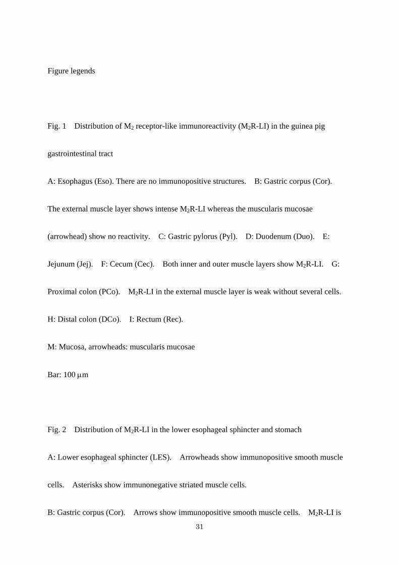
Figure legends
Fig. 1 Distribution of M2 receptor-like immunoreactivity (M2R-LI) in the guinea pig
gastrointestinal tract
A: Esophagus (Eso). There are no immunopositive structures. B: Gastric corpus (Cor).
The external muscle layer shows intense M2R-LI whereas the muscularis mucosae
(arrowhead) show no reactivity. C: Gastric pylorus (Pyl). D: Duodenum (Duo). E:
Jejunum (Jej). F: Cecum (Cec). Both inner and outer muscle layers show M2R-LI. G:
Proximal colon (PCo). M2R-LI in the external muscle layer is weak without several cells.
H: Distal colon (DCo). I: Rectum (Rec).
M: Mucosa, arrowheads: muscularis mucosae
Bar: 100 μm
Fig. 2 Distribution of M2R-LI in the lower esophageal sphincter and stomach
A: Lower esophageal sphincter (LES). Arrowheads show immunopositive smooth muscle
cells. Asterisks show immunonegative striated muscle cells.
B: Gastric corpus (Cor). Arrows show immunopositive smooth muscle cells. M2R-LI is
31
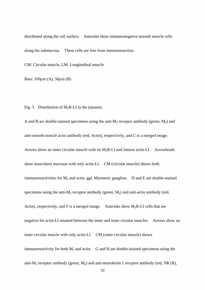
distributed along the cell surface. Asterisks show immunonegative smooth muscle cells
along the submucosa. These cells are free from immunoreaction.
CM: Circular muscle, LM: Longitudinal muscle
Bars: 100μm (A), 50μm (B)
Fig. 3 Distribution of M2R-LI in the jejunum.
A and B are double-stained specimens using the anti-M2 receptor antibody (green, M2) and
anti-smooth muscle actin antibody (red, Actin), respectively, and C is a merged image.
Arrows show an inner circular muscle with no M2R-LI and intense actin-LI. Arrowheads
show muscularis mucosae with only actin-LI. CM (circular muscle) shows both
immunoreactivities for M2 and actin. ggl: Myenteric ganglion. D and E are double-stained
specimens using the anti-M2 receptor antibody (green, M2) and anti-actin antibody (red,
Actin), respectively, and F is a merged image. Asterisks show M2R-LI cells that are
negative for actin-LI situated between the inner and outer circular muscles. Arrows show an
inner circular muscle with only actin-LI. CM (outer circular muscle) shows
immunoreactivity for both M2 and actin. G and H are double-stained specimens using the
anti-M2 receptor antibody (green, M2) and anti-neurokinin 1 receptor antibody (red, NK1R),
32
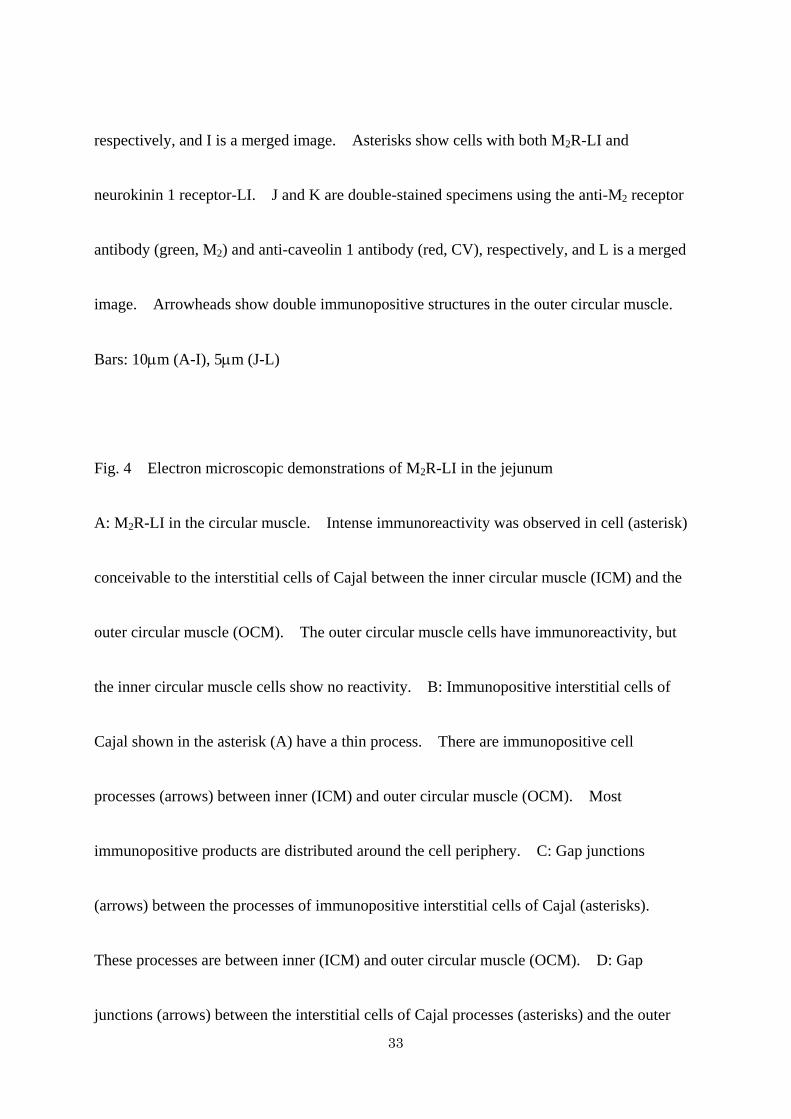
respectively, and I is a merged image. Asterisks show cells with both M2R-LI and
neurokinin 1 receptor-LI. J and K are double-stained specimens using the anti-M2 receptor
antibody (green, M2) and anti-caveolin 1 antibody (red, CV), respectively, and L is a merged
image. Arrowheads show double immunopositive structures in the outer circular muscle.
Bars: 10μm (A-I), 5μm (J-L)
Fig. 4 Electron microscopic demonstrations of M2R-LI in the jejunum
A: M2R-LI in the circular muscle. Intense immunoreactivity was observed in cell (asterisk)
conceivable to the interstitial cells of Cajal between the inner circular muscle (ICM) and the
outer circular muscle (OCM). The outer circular muscle cells have immunoreactivity, but
the inner circular muscle cells show no reactivity. B: Immunopositive interstitial cells of
Cajal shown in the asterisk (A) have a thin process. There are immunopositive cell
processes (arrows) between inner (ICM) and outer circular muscle (OCM). Most
immunopositive products are distributed around the cell periphery. C: Gap junctions
(arrows) between the processes of immunopositive interstitial cells of Cajal (asterisks).
These processes are between inner (ICM) and outer circular muscle (OCM). D: Gap
junctions (arrows) between the interstitial cells of Cajal processes (asterisks) and the outer
33
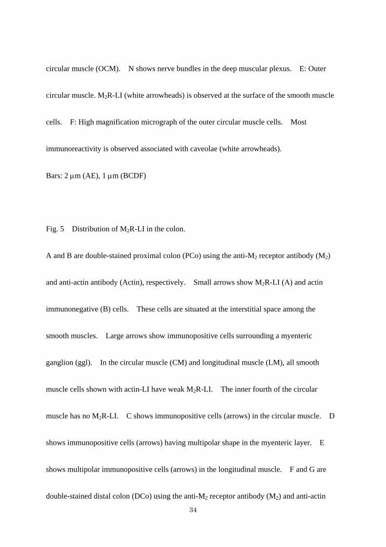
circular muscle (OCM). N shows nerve bundles in the deep muscular plexus. E: Outer
circular muscle. M2R-LI (white arrowheads) is observed at the surface of the smooth muscle
cells. F: High magnification micrograph of the outer circular muscle cells. Most
immunoreactivity is observed associated with caveolae (white arrowheads).
Bars: 2 μm (AE), 1 μm (BCDF)
Fig. 5 Distribution of M2R-LI in the colon.
A and B are double-stained proximal colon (PCo) using the anti-M2 receptor antibody (M2)
and anti-actin antibody (Actin), respectively. Small arrows show M2R-LI (A) and actin
immunonegative (B) cells. These cells are situated at the interstitial space among the
smooth muscles. Large arrows show immunopositive cells surrounding a myenteric
ganglion (ggl). In the circular muscle (CM) and longitudinal muscle (LM), all smooth
muscle cells shown with actin-LI have weak M2R-LI. The inner fourth of the circular
muscle has no M2R-LI. C shows immunopositive cells (arrows) in the circular muscle. D
shows immunopositive cells (arrows) having multipolar shape in the myenteric layer. E
shows multipolar immunopositive cells (arrows) in the longitudinal muscle. F and G are
double-stained distal colon (DCo) using the anti-M2 receptor antibody (M2) and anti-actin
34
antibody (Actin), respectively. All smooth muscle cells in the circular (CM) and
longitudinal (LM) musculature show intense M2R-LI. In the muscularis mucosae, though
longitudinal smooth muscle cells (arrowheads) have intense M2R-LI, circular smooth muscle
cells have no M2R-LI.
Bars: 10μm
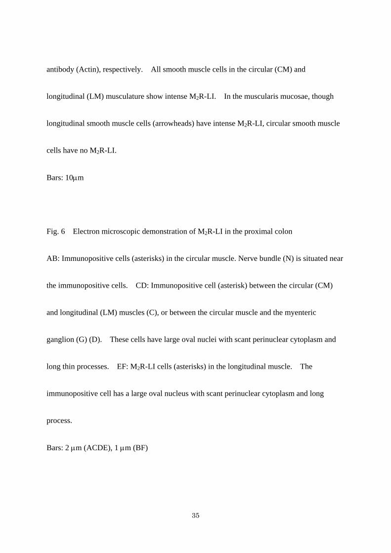
Fig. 6 Electron microscopic demonstration of M2R-LI in the proximal colon
AB: Immunopositive cells (asterisks) in the circular muscle. Nerve bundle (N) is situated near
the immunopositive cells. CD: Immunopositive cell (asterisk) between the circular (CM)
and longitudinal (LM) muscles (C), or between the circular muscle and the myenteric
ganglion (G) (D). These cells have large oval nuclei with scant perinuclear cytoplasm and
long thin processes. EF: M2R-LI cells (asterisks) in the longitudinal muscle. The
immunopositive cell has a large oval nucleus with scant perinuclear cytoplasm and long
process.
Bars: 2 μm (ACDE), 1 μm (BF)
35
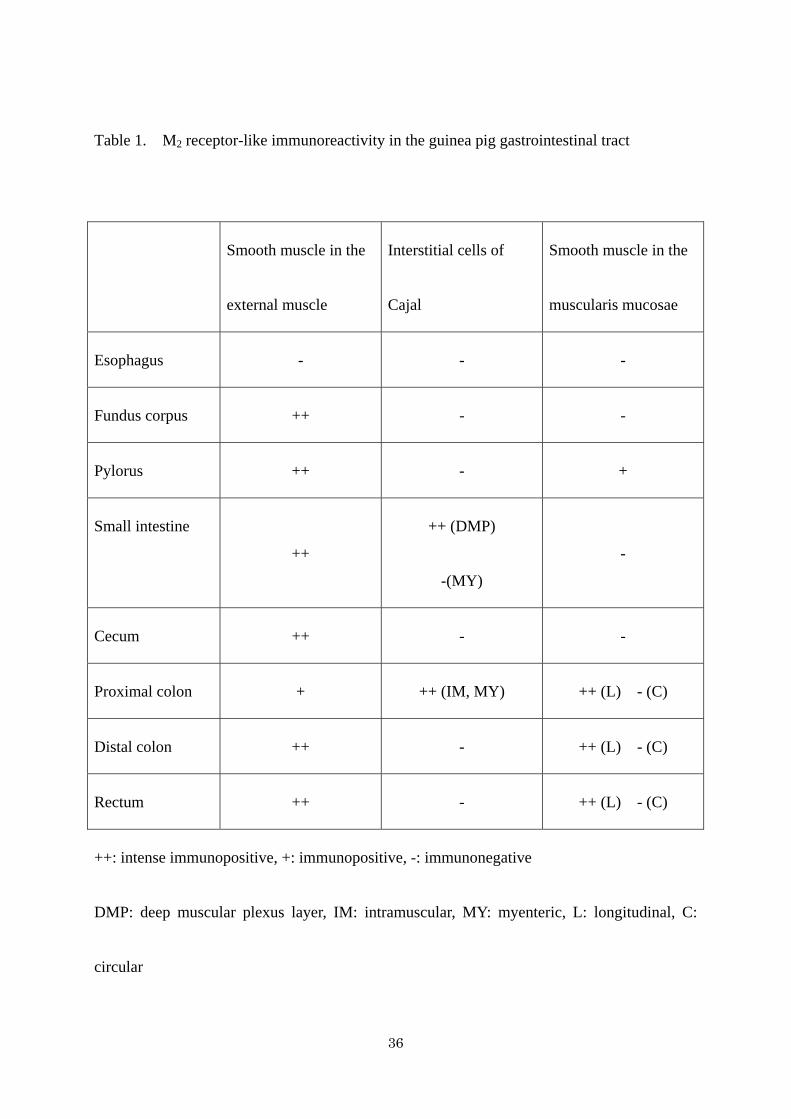
Table 1. M2 receptor-like immunoreactivity in the guinea pig gastrointestinal tract
Smooth muscle in the
external muscle
Interstitial cells of
Cajal
Smooth muscle in the
muscularis mucosae
Esophagus - - -
Fundus corpus ++ - -
Pylorus ++ - +
Small intestine
++
++ (DMP)
-(MY)
-
Cecum ++ - -
Proximal colon + ++ (IM, MY) ++ (L) - (C)
Distal colon ++ - ++ (L) - (C)
Rectum ++ - ++ (L) - (C)
++: intense immunopositive, +: immunopositive, -: immunonegative
DMP: deep muscular plexus layer, IM: intramuscular, MY: myenteric, L: longitudinal, C:
circular
36
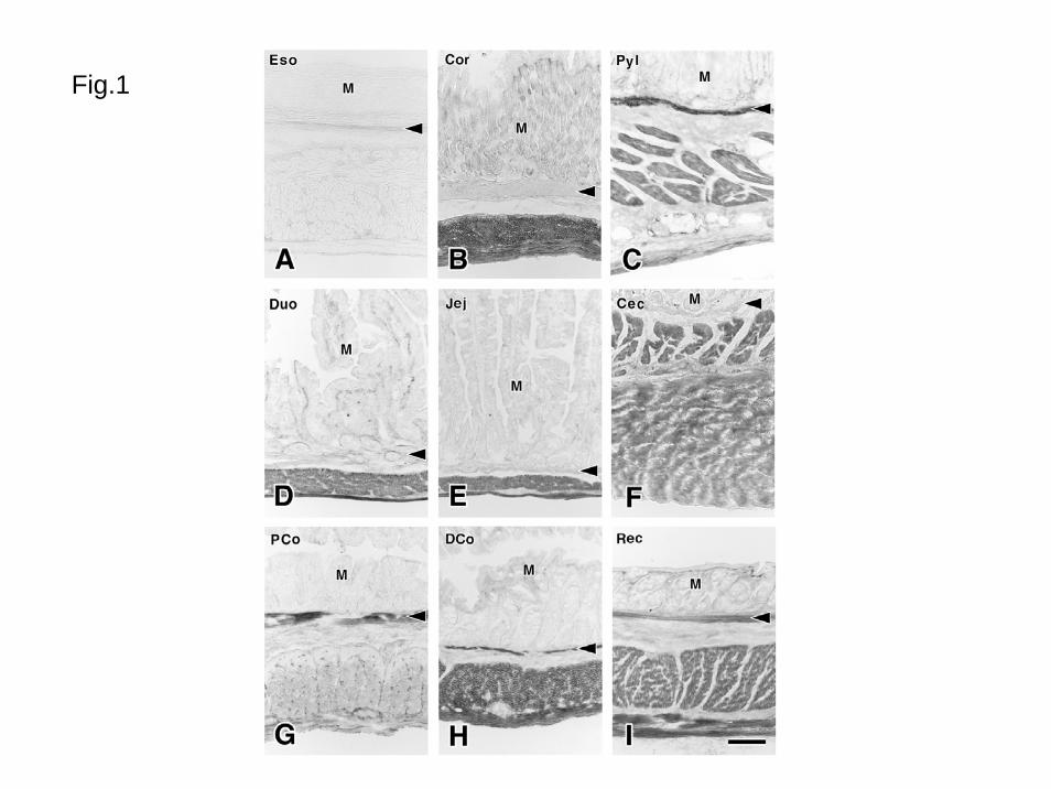
Fig.1
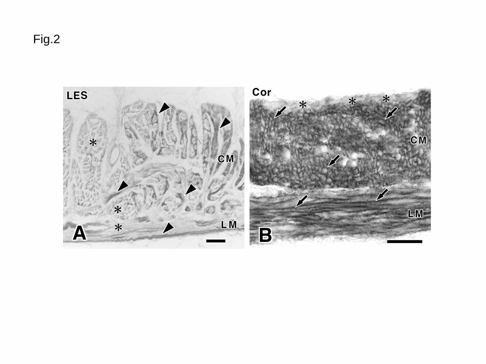
Fig.2
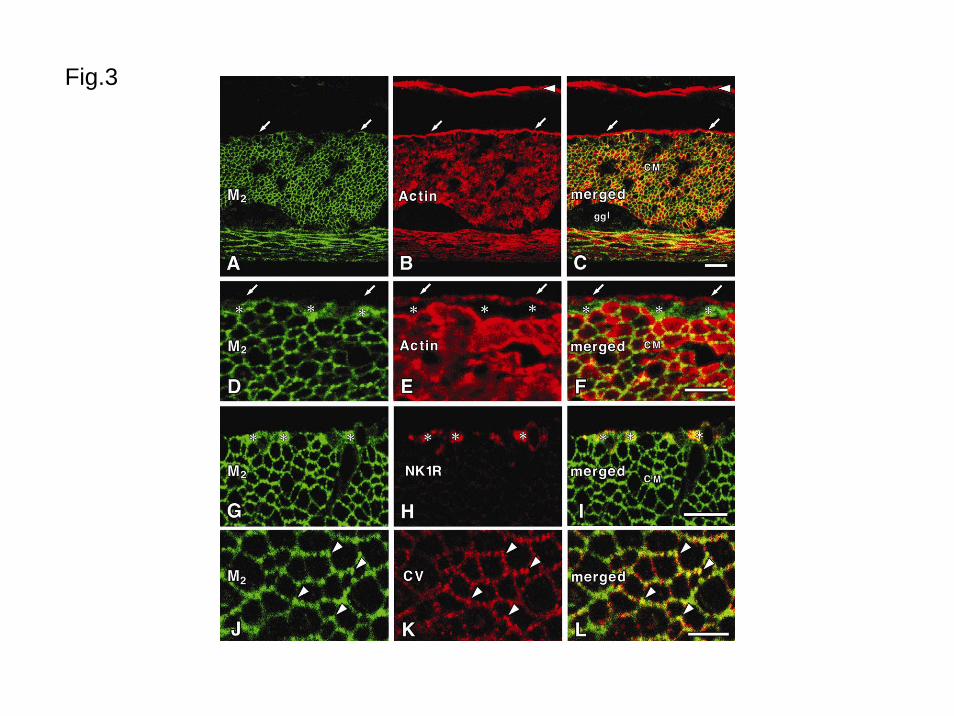
Fig.3

Fig.4
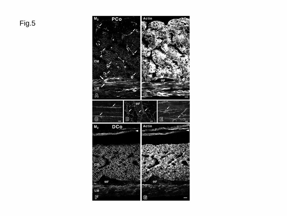
Fig.5
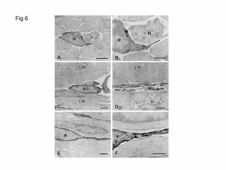
Fig.6