Embed Size (px)
Citation preview

2/9/2015
1
IN THE BEGINNINGIN THE BEGINNING
Mammography technology has come a long way since the first
machine specifically designed for producing
mammograms was introduced in 1966.
The first mammography system was
essentially a tripod supporting a special
X-ray camera
EARLY MILESTONES IN
MAMMOGRAPHY
EARLY MILESTONES IN
MAMMOGRAPHY1913 - Albert Solomon, a surgeon in Berlin, uses a conventional
X-ray machine to visualize breast cancers in 3,000 mastectomy
specimens.
1949 - Uruguayan Raul Leborgne emphasizes the need for
breast compression to identify calcifications.
1956 - Robert Egan, a radiologist in Houston, introduces
dedicated film for mammography to produce simple and
reproducible images with improved detail.
1966 – The first dedicated mammography system is introduced.
1971 – Commercial introduction of xeromammography
1980 – Introduction of single emulsion film; 2x faster at
significantly lower dose
REMEMBER THIS?REMEMBER THIS?
1953 Uruguay
If you do, you’ve been working
way too hard>and too long!
2/9/2015
2
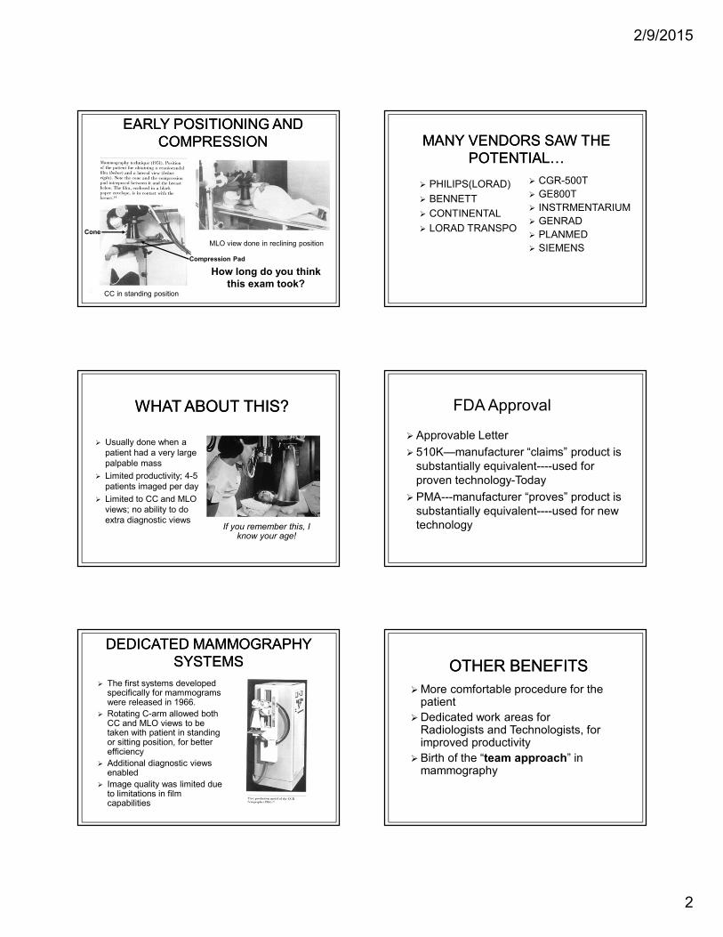
EARLY POSITIONING AND
COMPRESSION
EARLY POSITIONING AND
COMPRESSION
MLO view done in reclining position
Cone
Compression Pad
How long do you think
this exam took?CC in standing position
WHAT ABOUT THIS?WHAT ABOUT THIS?
� Usually done when a
patient had a very large
palpable mass
� Limited productivity; 4-5
patients imaged per day
� Limited to CC and MLO
views; no ability to do
extra diagnostic viewsIf you remember this, I
know your age!
DEDICATED MAMMOGRAPHY
SYSTEMS
DEDICATED MAMMOGRAPHY
SYSTEMS
� The first systems developed specifically for mammograms were released in 1966.
� Rotating C-arm allowed both CC and MLO views to be taken with patient in standing or sitting position, for better efficiency
� Additional diagnostic views enabled
� Image quality was limited due to limitations in film capabilities
� PHILIPS(LORAD)
� BENNETT
� CONTINENTAL
� LORAD TRANSPO
� CGR-500T
� GE800T
� INSTRMENTARIUM
� GENRAD
� PLANMED
� SIEMENS
MANY VENDORS SAW THE
POTENTIAL>
MANY VENDORS SAW THE
POTENTIAL>
FDA Approval
� Approvable Letter
� 510K—manufacturer “claims” product is
substantially equivalent----used for
proven technology-Today
� PMA---manufacturer “proves” product is
substantially equivalent----used for new
technology
OTHER BENEFITSOTHER BENEFITS
� More comfortable procedure for the patient
� Dedicated work areas for Radiologists and Technologists, for improved productivity
� Birth of the “team approach” in mammography

2/9/2015
3
Is This Your Past
Is This Your Past
THE NEXT ADVANCE
XEROMAMMOGRAPHY
THE NEXT ADVANCE
XEROMAMMOGRAPHY� First introduced in 1971
� Provided better image quality than systems using industrial film packs
� Allowed excellent visualization of chest wall
� The Grandpappy of Selenium digital technology
� Key Inventor – Lothar Jeromin (“Mr. Xerox”)� Holds 23 patents
Xeroradiographs (blue and white prints), as
shown above, gave adequate image quality;
however, the radiation dose to the breast was
much higher than current imaging methods.
XEROMAMMOGRAPHY DIFFERENT PERSPECTIVES
XEROMAMMOGRAPHY DIFFERENT PERSPECTIVES� LOVE
� Radiologists and Surgeons loved the image quality and ability to see chest wall and ribs
� DREAD
� Technologists were uncomfortable with length of exam and lack of training in positioning (all on the job, at that time)
� FEAR
� Patients were very uneasy, as the exam was not expected to bring good
results (done primarily for
symptomatic patients at that time)
AND IT APPEALED TO THE
PUBLIC>
AND IT APPEALED TO THE
PUBLIC>
� Advertisement in Life
and Glamour
magazine promoting
the new technology,
xeromammography
� Your Mother’s
Mammogram
Model demonstrates the latest in
mammography technology and fashion.
Check out the hosiery

2/9/2015
4
BUT WHAT A MESS!BUT WHAT A MESS!
� Technologists were
the original Smurfs!
� Lots of blue, on your
shoes, uniforms,
hands,hair etc.
� In 1985, xeromammography changed from blue
powder toner to black liquid toner
� This resulted in substantial dose reductions (and
cure of Smurf-syndrome in technologists)
IMPROVEMENTS IN
XEROMAMMOGRAPHY
IMPROVEMENTS IN
XEROMAMMOGRAPHY
1983 prototype of low-dose
xeromammography system
Lothar Jeromin with low-dose
xeromammography system at
RSNA 1985
AT THE SAME TIME>.AT THE SAME TIME>.� Single emulsion film for use in mammography was
being introduced, with the promise of providing
faster processing, improved image quality, and
significantly decreased dose
� By 1986, screen-film mammography was being
used by more than half of all radiologists
� Production of xeromammography was halted in
1989, due to declining sales
� Screen-film mammography became the gold
standard in the late 1980’s – early 1990’s
A GLIMPSE OF THE FUTURE
LOTHAR’S STORY
A GLIMPSE OF THE FUTURE
LOTHAR’S STORY
� As a testimonial of faith in their
xeromammography system, Xerox offered
free mammograms to female employees in
the breast cancer at-risk age group.
� ONE non-symptomatic woman was indeed
diagnosed with breast cancer
And so the seeds of a screening
program were sown ))
80s AND 90s PROFICIENCY
DEVELOPMENT
80s AND 90s PROFICIENCY
DEVELOPMENT� Worldwide acceptance of mammography as the first
line of defense
� Radiologists became MAMMOGRAPHERS and in
many facilities multitasked
� Technologists began to seek out specialized
training courses
� Administrators & Technologists worked together on
creating “The Breast Center”
� MQSA enacted to ensure consistently high quality
in all mammography facilities
MILESTONES IN MODERN
MAMMOGRAPHY
MILESTONES IN MODERN
MAMMOGRAPHY
1988 – Congress passes legislation to provide annual screening
mammography benefit for Medicare recipients
1990 – Breast and Cervical Cancer Mortality Prevention Act
implemented to provide free or low cost mammograms and pap
smears to low-income women
1998 – First Computer Aided Detection (CAD) system for
mammography approved by FDA
2000 – First Full Field Digital Mammography system approved
by FDA
1999 – National Mammography Quality Standards Act
implemented
1985 – Declaration of Breast Cancer Awareness Week, which
led to Nation Breast Cancer Awareness Month

2/9/2015
5
THE 80’S and 90’s
DECADES OF AWARENESS
THE 80’S and 90’s
DECADES OF AWARENESS� Significant increase in incidence of breast cancer
diagnoses between 1980 - 1987 attributed to
implementation of screening programs, enabling
earlier detection of smaller breast cancers
� Earlier detection has led to decreases in mortality
� Breast cancer mortality rates have decreased
approximately 2% annually since 1990!
� The “WAR” on Breast Cancer has been the biggest
American accelerator for development of better
detection technologies and improved treatment
options in the last two decades
FIND “WALDO”
The Image Interpreter’s Challenge:
Find
Waldo:
The Image Interpreter’s Challenge: Find Waldo
Where?
Are you
sure?
Your
time’s
up!
Are there
MANY?
Are there
ANY?
Typically, the
previous 4
images are
compared with
the latest 4
images, = 8
images viewed
If radiologist
interprets 40
patient
studies/hour,
= 1 study
every 90
seconds
Reviewing 8
images every
90 seconds =
11 seconds
per image!
(In USA),
screening
Mammograph
y exam is two
views per
breast, = 4
images/study

2/9/2015
6
The “Star Wars” fantasy of beaming digital mammograms via satellite to doctors in remote locations around the world has become a reality.
Hologic, Dimensions, Selenia, and other associated logos are trademarks and/or registered trademarks of Hologic, Inc. and/or its subsidiaries in the United States and /or other countries.
Breast Tomosynthesis
A 3D screening modality that preserves the
very high resolution of 2D FFDM
Multiple images of the breast are acquired
at different angles during a sweep of
the x-ray tube
Allows radiologists to see around
overlapping structures
In 2D FFDM:
Tissue superimposition hides
pathologies in 2D
Tissue superimposition mimics
pathologies in 2D
l
Tomosynthesis is a three-dimensional mammographic examination that can minimize the effects of structure overlap within the breast
PRE-00023

2/9/2015
7
Tube moves in a 15o arc
15 low dose images are acquired
1 image at each degree
Four second sweep
Images are reconstructed into 1 mm
slices
In combo-mode imaging, the 2D and
3D are taken in the same compression,
with no additional positioning for the
patient.
Compressed Breast
Detector Housing
LCC
2D
Hologic – Proprietary and Confidential
A 2D Mammography
Image
LCC
2D
Hologic – Proprietary and Confidential
A suspicious area in
a 2D Mammography
Image
2D 3D
The 2D Mammography Image next to one slice of a 3D Image Set
RMLO
2D
l
A 2D Mammography Image with
a suspicious area identified and
blown up
3D2D
The 2D Mammography Image next to one slice of a 3D Image Set

2/9/2015
8
Recall Reduction
Superimposed Tissue Examples
RCCRCC
2D 3D
Hologic – Proprietary and Confidential
A 2D Mammography Image with a
suspicious area identified next to a
3D image set
Slice 14 18 22
26 30
Hologic – Proprietary and Confidential
As you go thru the image set, you see that the suspicious area is
nothing more than normal breast structures overlapping
• 2D reconstruction algorithm
• Eliminates the need for a 2D mammogram
• Creates synthesized images from single tomosynthesis scan
• Reduces dose
• Additional cost

2/9/2015
9
• Routine for GE tomosynthesis-FDA Approval-Screening
• 2 D CC
• 3 D MLO
• Diagnostic Exam
• 3 D CC
• 3D MLO
• Step and shoot mode preserves microcalcification sharpness and avoids image blur, since the tube makes a complete stop for each of nine exposures
• Anti scatter solution designed for tomosynthesis, the SenoClairegrid in 3 D reduces scattered radiation while preserving dose and performance
• SenoClaire uses ASIR ®-A calcification correction reconstruction algorithim
2 D MLO 3 D MLO
A 2 D image generated from the raw DBT projection data that helps the user get a overview of the entire stack of images before reading individual tomosynthesis images
V-View
ABUS-Automated Breast Ultrasound
Breast Density
Elastography

2/9/2015
10
• Began in the 1950’s
• Incorporated the use of A mode technology
• Immersion, compression, and contact transducers for sonographic breast imaging continued through the early 1960’s
• In the late 1960’s and early 1970’s, B-mode transducers with frequencies ranging between 1.5 MHz to 10.0 MHz produced images that could differentiate types of breast tissues
• Up until the early 2000’s, breast sonography was used to differentiate between solid and cystic masses.
• Today, breast sonography uses high-frequency, high resolution, real time systems.
• Transducers should be 10 MHz or higher
• ABUS-Automated Breast Ultrasound System
• An important adjunct to mammography
• Screening tool for dense breasts?
• 21 sites
• 2809 women (at least heterogeneously dense in one quadrant)
• Mammography and physician performed handheld full breast ultrasound
• Diagnostic yield of mammo vs. mammo plus ultrasound
• Physicians underwent consistent training
• 20 minutes avg. scan time plus interpretation
• Mammography alone 7.6/1000 cancers detected
• Mammography plus US 11.8/1000
• 12 cancers detected by US alone
July 2004July 2003
Lt CC

2/9/2015
11
Technology
Precision
Confidence
• Creates a uniform compression across the entire breast
• Enables a greater penetration due to the convergent scan line geometry
• Improves detail resolution at depth
• Anatomically correct
• Automatically scans a woman’s breast capturing multiple images
• Displays them in 3-D for a radiologist review
• Ideal for women with dense breasts where effectiveness of mammography may be limited
Use existing Ultrasound
System
SonoCiné – System

2/9/2015
12
Transverse
Sagittal
Coronal
The resulting 3D data is sent to the somo.v display console for review and rendering.
• The AWBUSTM
articulating arm, which uses the transducer of your ultrasound system, provides whole breast coverage while still allowing the operator to adjust the angle and pressure of the transducer
ACUSON S2000 ABVS-Automated Breast Volume
Can be used for elasticity studies
2/9/2015
13
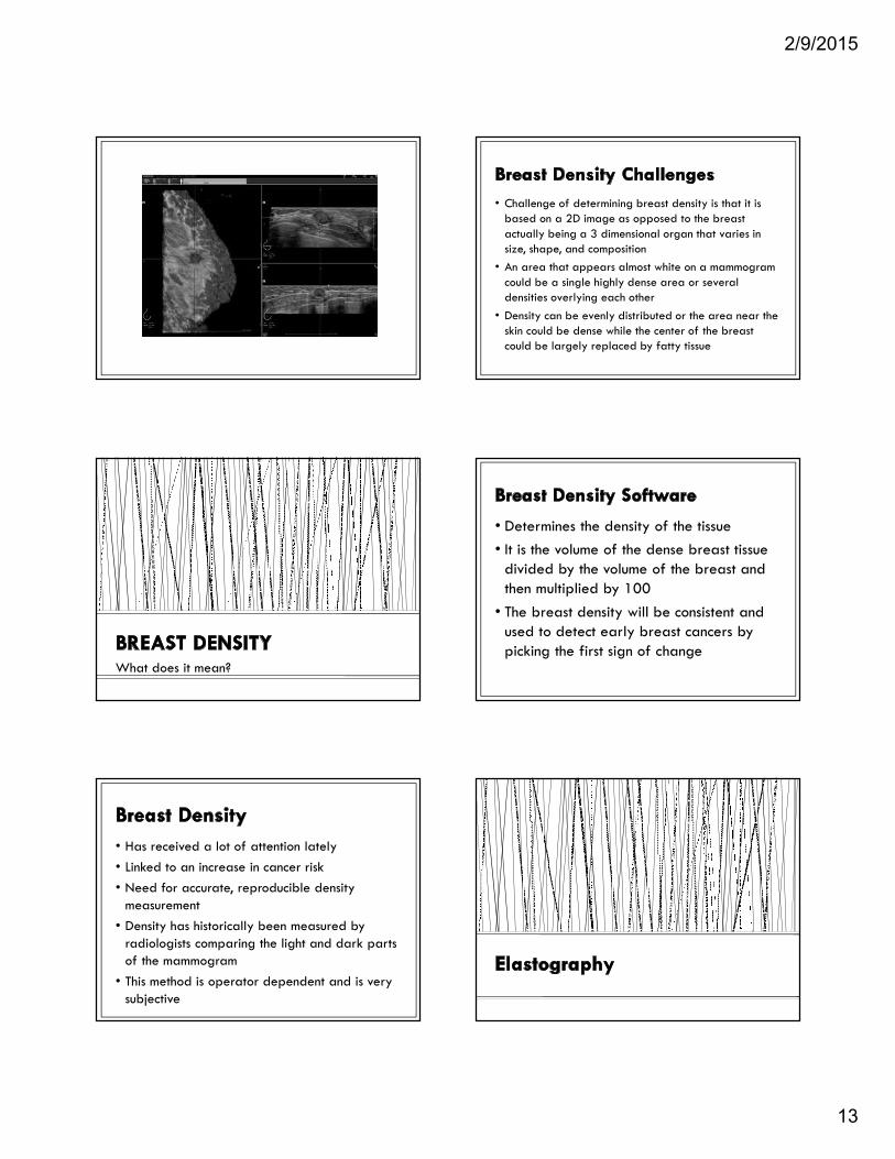
What does it mean?
• Has received a lot of attention lately
• Linked to an increase in cancer risk
• Need for accurate, reproducible density measurement
• Density has historically been measured by radiologists comparing the light and dark parts of the mammogram
• This method is operator dependent and is very subjective
• Challenge of determining breast density is that it is based on a 2D image as opposed to the breast actually being a 3 dimensional organ that varies in size, shape, and composition
• An area that appears almost white on a mammogram could be a single highly dense area or several densities overlying each other
• Density can be evenly distributed or the area near the skin could be dense while the center of the breast could be largely replaced by fatty tissue
• Determines the density of the tissue
• It is the volume of the dense breast tissue divided by the volume of the breast and then multiplied by 100
• The breast density will be consistent and used to detect early breast cancers by picking the first sign of change

2/9/2015
14
Non-invasive technique that adds value to
the sonographic examination by
evaluating the stiffness of tissue. It detects the stiffness or elasticity
compared to normal tissue
• A small amount of pressure is applied to the breast, just enough to move it slightly.
• With the pressure applied, the ultrasound system takes another image
• A computer than takes the two images and compares how elastic the different regions are
• A hard, inflexible lump is almost always a carcinoma. A flexible lump is usually benign.
• Physiologic image to detect cancer or cancer therapy results
• Indicates if cells are active and growing or inactive and shrinking
• A molecule is tagged with a positron emitting isotope (Radioactive substances combined with a natural body compound)
• 18-F fluorodeoxyglucose (a probe)
• Radioactive 18-F tagged with a glucose
• Positrons annihilate nearby electrons, emitting photons
• Photons are detected by the scanner
• Cancer accumulates glucose more than normal tissue (glucose metabolism)
• Patient prep is required (fasting, limited exercise)
• 45-90 minutes rest post injection
• 30-45 minutes scan time

2/9/2015
15
• High FP (esp. lobular and well differentiated Ca)
• High Cost: Cyclotron is needed near PET, isotopes are short lived (hours)
• Combine functional imaging of PET with anatomical imaging of CT
• Nuclear medicine exam that uses intravenous injectable FDG (fluoro-deoxyglucose)
• A glucose analog that accumulates in glucose avid cells
• Accumulates in both inflammatory and cancerous states
• Following a 4 hour fast, serum blood is drawn to determine blood glucose level
• 10mCi FDG is injected intravenously and imaging is acquired one hour later
• Imaging is obtained in a similar manner as mammography
• Breast is imaged with slight compression
2mm Resolution

2/9/2015
16
Index Lesion
Lymph node
Secondary lesion
• Dose 1.4-7mSv
• Approx $800K purchase price
• Reimbursement is controversial
• $1000 - $2000
• Need to improve the markers (FDG) to improve sensitivity
Reimbursed for:
• Pre-surgical planning and staging
• Monitoring for recurrence
• Neo-adjuvant therapy
• Equivocal exams following diagnostic workup
• Great value in preoperative identification of non-invasive breast cancer (DCIS)
• DCIS accounts for 30% of newly diagnosed patients
• DCIS is often difficult to quantitate with mammography and MRI
• Used for preoperative surgical staging
• Scintimammography FDA approved 1997
• Traditional multi-purpose gamma camera
• Indicated for planar imaging to assist mammography
• No significant difference in detection fatty vs. dense breasts
• Limitation with lesions less than 1cm
• Positioning an issue to image the entire breast
• Uses a radioactive tracer that “lights” up any areas of cancer inside the breast-
• Tracer is injected into the body through a vein in the arm
• Breast cancer cells take up the radioactive substance much more than normal cells do
• A nuclear medicine scanner that scans the breast looking for areas where the radioactive substance is concentrated
2/9/2015
17
• Imaging performed 5 minutes post injection
• Breast is lightly compressed between 2 detectors
• Images are obtained in cranial caudel and mediolateral oblique projections
• Easily compared to mammography
• MBI has an overall sensitivity of about 90% with a sensitivity of 82% for lesions less than 10 mm in size
• Sensitivity is lowest for lesions less than 5 mm in size
• Tumor detection does not appear to be dependent on tumor type, but rather on tumor size
• Holds special promise in determining cancerous lesions in dense breast
• Use of scintimammography after abnormal mammogram offers higher level of diagnostic specificity
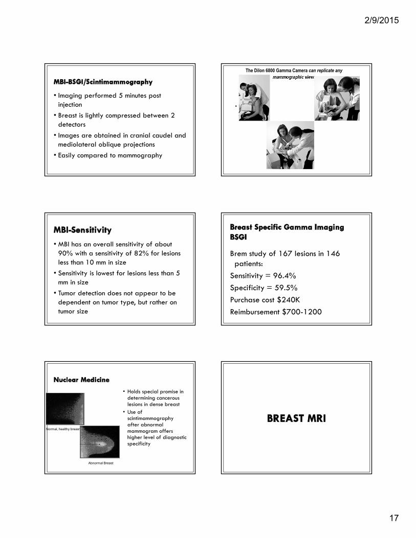
Normal, healthy breast
Abnormal Breast
•
The Dilon 6800 Gamma Camera can replicate any
mammographic view.
Brem study of 167 lesions in 146 patients:
Sensitivity = 96.4%
Specificity = 59.5%
Purchase cost $240K
Reimbursement $700-1200

2/9/2015
18
• Breast MRI the most rapidly growing segment of MR Imaging
• Improvements in MRI resolution, contrast, biopsy devices, CAD software
• American Cancer Society (ACS) recommendations published
• Clinical trials proved its worth (ACRIN 6667)
• 7 studies identified missed cancers
• Improved the cancer detection rate 10-40 per 1000 of the high risk women screened
• Average 22/1000
• Specific populations benifit
• BRCA mutation
• First degree relative with breast cancer
• Lifetime risk 20-30% as defined by BRCAPRO or other models that are family history based
• ? Radiation to the chest age 10 – 30 yrs
• NOT dense breasts
2/9/2015
19
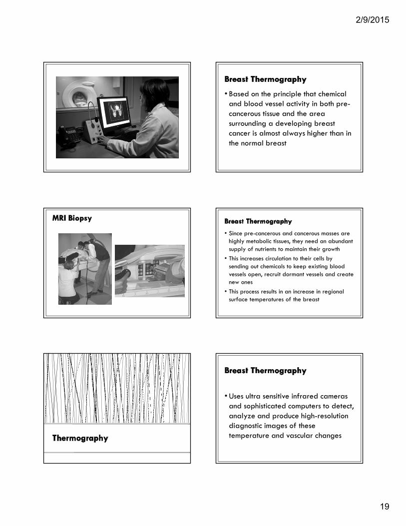
• Based on the principle that chemical and blood vessel activity in both pre-cancerous tissue and the area surrounding a developing breast cancer is almost always higher than in the normal breast
• Since pre-cancerous and cancerous masses are highly metabolic tissues, they need an abundant supply of nutrients to maintain their growth
• This increases circulation to their cells by sending out chemicals to keep existing blood vessels open, recruit dormant vessels and create new ones
• This process results in an increase in regional surface temperatures of the breast
• Uses ultra sensitive infrared cameras and sophisticated computers to detect, analyze and produce high-resolution diagnostic images of these temperature and vascular changes

2/9/2015
20
• American Society of Breast Surgeons (ASBrS) conducted a study with results presented at their annual meeting in Phoenix in May
• The conclusion is—Thermography is not a reliable breast cancer screening tool
• Too many benign biopsies are performed based on suspicious imaging abnormalities
• Women are seeking an alternative to radiation based imaging techniques