Embed Size (px)
Citation preview
ARTICLE
Received 28 Oct 2016 | Accepted 30 Mar 2017 | Published 24 May 2017
In situ dynamic tracking of heterogeneousnanocatalytic processes by shell-isolatednanoparticle-enhanced Raman spectroscopyHua Zhang1, Chen Wang1, Han-Lei Sun1, Gang Fu1, Shu Chen2, Yue-Jiao Zhang1, Bing-Hui Chen1,
Jason R. Anema1, Zhi-Lin Yang2, Jian-Feng Li1,2 & Zhong-Qun Tian1
Surface molecular information acquired in situ from a catalytic process can greatly promote
the rational design of highly efficient catalysts by revealing structure-activity relationships
and reaction mechanisms. Raman spectroscopy can provide this rich structural information,
but normal Raman is not sensitive enough to detect trace active species adsorbed on
the surface of catalysts. Here we develop a general method for in situ monitoring of
heterogeneous catalytic processes through shell-isolated nanoparticle-enhanced Raman
spectroscopy (SHINERS) satellite nanocomposites (Au-core silica-shell nanocatalyst-satellite
structures), which are stable and have extremely high surface Raman sensitivity. By
combining operando SHINERS with density functional theory calculations, we identify the
working mechanisms for CO oxidation over PtFe and Pd nanocatalysts, which are typical
low- and high-temperature catalysts, respectively. Active species, such as surface oxides,
superoxide/peroxide species and Pd–C/Pt–C bonds are directly observed during the
reactions. We demonstrate that in situ SHINERS can provide a deep understanding of
the fundamental concepts of catalysis.
DOI: 10.1038/ncomms15447 OPEN
1 MOE Key Laboratory of Spectrochemical Analysis and Instrumentation, State Key Laboratory of Physical Chemistry of Solid Surfaces, iChEM, College ofChemistry and Chemical Engineering, Xiamen University, Xiamen 361005, China. 2 Department of Physics, Research Institute for Biomimetics and SoftMatter, Xiamen University, Xiamen 361005, China. Correspondence and requests for materials should be addressed to J.-F.L. (email: [email protected]) or toB.H.C. (email: [email protected]) or to G.F. (email: [email protected]).
NATURE COMMUNICATIONS | 8:15447 | DOI: 10.1038/ncomms15447 | www.nature.com/naturecommunications 1
Understanding structure-activity relationships and reactionmechanisms is of significant importance in the rationaldesign of highly efficient catalysts1–5. After some
pioneering work on model systems by both surface andtheoretical scientists6–16, much progress has been made in thisarea. However, challenges associated with the expansion of modelsystems to industrial catalysts remain8. Thus, the development ofin situ surface analysis techniques that can be used to studyreaction processes occurring over practical catalysts is highlydesirable. For example, some in situ techniques includingenvironmental transmission electron microscopy, high-pressurescanning tunneling microscopy (STM), ambient pressure X-rayphotoelectron spectroscopy (XPS) and X-ray absorptionspectroscopy have been applied to study nanocatalysts andreveal molecular information about the catalysts under workingconditions, such as surface compositions, chemical states,morphologies and coordination environments17,18. These workshave greatly improved our understanding of catalysis.
One of the most widely used surface analysis techniques isin situ infrared (IR) spectroscopy, which can provide richinformation about fine surface structure19, active sites20 andreaction mechanisms21. However, it is very difficult for in situIR to detect intermediates with signals in the low wavenumberregion of the spectrum. In contrast, the low wavenumber region isaccessible by Raman spectroscopy22. This technique has beenused to identify intermediate species such as metal-C bonds,active oxygen species, hydroxyl groups and surface oxides23–25,but in general, normal Raman spectroscopy is not sensitiveenough to monitor trace amounts of surface species on metalcatalysts. Surface-enhanced Raman scattering (SERS) has beenemployed to circumvent this limitation, as it offers a tremendousimprovement in sensitivity (107–1010 times) and can even be usedto detect single molecules26–29. Nevertheless, only a few metals
(primarily Au, Ag and Cu) with nanostructured surfaces providea large SERS effect30. Thus, monitoring catalytic processesoccurring over other metals remains a great challenge.
Recently, our group invented a novel technique known as‘shell-isolated nanoparticle-enhanced Raman spectroscopy’ or‘SHINERS’31, which has been regarded as a ‘next generation ofadvanced spectroscopy’32. In SHINERS, plasmonic Aunanoparticles (Raman signal amplifiers) are coated withpinhole-free silica shells that prevent them from interactingwith analytical targets or the chemical environment. The silicashells are thin enough (just a couple of nanometers) that theenhanced electromagnetic field generated at the surface of thecore extends beyond the surface of the shell. SHINERS hasbroken the long-standing surface material and morphologyrestrictions that exist in traditional SERS. In principle,the SHINERS technique can be applied to a surface of anymaterial and any morphology. It has been widely used inelectrochemistry31,33–35, life science36, the materials andsemiconductor industry31,37, solar energy storage andconversion38,39, and our daily life40.
In this work, we develop a simple strategy to construct Au-coresilica-shell nanocatalyst-satellite nanocomposites, which we callSHINERS-satellite structures. Three-dimensional finite-differencetime-domain (3D-FDTD) simulations demonstrate that Ramansignals from chemical species on the satellite structures are greatlyamplified by the plasmonic activity of the core, making this anideal architecture for monitoring heterogeneous catalytic reac-tions. With this strategy we have obtained direct spectroscopicevidence of the formation of metal-O and metal-C bonds, as wellas active oxygen species, during CO oxidation. With the aid ofdensity functional theory (DFT) calculations, the SHINERS-satellite method has revealed new molecular information aboutCO oxidation.
Log(
E/E
o)4
12345678
Laser
Raman signals
SiO2 shell
Au core
Nanocatalysts
Ram
an e
nhan
cem
ent
Weak
Strong
LaserRaman signlas
e–
Au
SiO2
Pt Fe C O
b
a
c
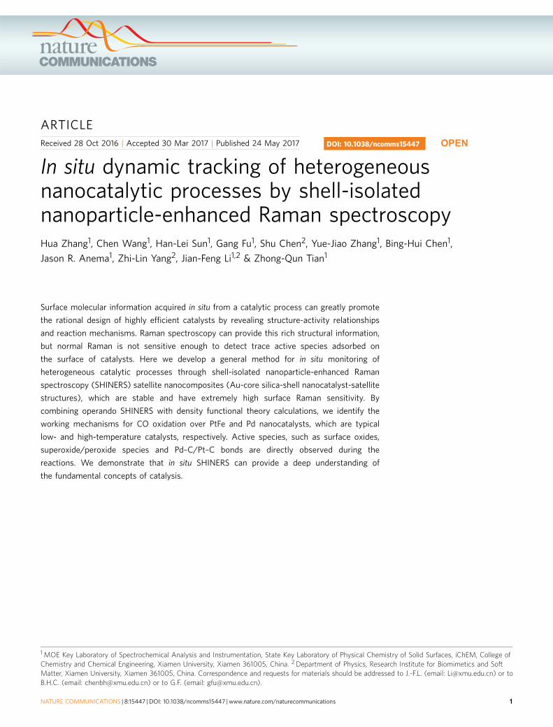
Figure 1 | Schematic illustration of a SHINERS-satellite study of a nanocatalytic process. (a) The Au-core silica-shell nanocatalyst-satellite architecture
of SHIN-enhanced Raman spectroscopy (SHINERS)-satellite structures, and the mechanism for CO oxidation over PtFe bimetallic nanocatalysts revealed by
our SHINERS-satellite method. The pinhole-free silica shell prevents the Raman signal amplifier from interfering with the system under study. (b) Schematic
illustration of CO oxidation on PtFe. The blue, orange, grey and red spheres represent Pt, Fe, C and O atoms, respectively. (c) A 3D-FDTD simulation for a
pair of Pt-on-shell isolated nanoparticle (SHIN) nanocomposite structures.
ARTICLE NATURE COMMUNICATIONS | DOI: 10.1038/ncomms15447
2 NATURE COMMUNICATIONS | 8:15447 | DOI: 10.1038/ncomms15447 | www.nature.com/naturecommunications
ResultsRaman enhancement of SHINERS-satellite nanocomposites. InSHINERS, light-induced localized surface plasmons yield areas ofenhanced electromagnetic field strength at the surface of the Aucore, and the electromagnetic field strength decays exponentiallywith distance from the surface of the Au core depending on near-field characteristics31,41. Thus, we designed a core–shell-satellitenanocomposite structure with the nanocatalysts on the silicashell, just a couple of nanometers away from the core. In this way,Raman signals from reaction intermediates on the surface of thecatalysts can be amplified effectively.
The Au-core silica-shell nanocatalyst-satellite architecture isshown in Fig. 1a. The SHINERS-satellite structure is prepared bycoating a Au nanoparticle with an ultrathin yet pinhole-free silicashell, then adding the nanocatalysts to the surface of the shell.The pinhole-free silica shell prevents adsorption of moleculesfrom the chemical environment on the Au core, and it alsoprevents any interaction between the nanocatalysts and the Aucore (Fig. 1b). Thus, information about heterogeneous catalyticreaction processes can be obtained without risk of interference bythe Raman signal amplifier. To confirm that this structure willeffectively enhance Raman signals from species on the nanoca-talysts, we modelled electromagnetic field strength using a3D-FDTD method in which the perfectly matched layer boundaryconditions were adapted to avoid non-physical reflections42. Thestructure selected for modelling was a 120 nm Au core with a2 nm silica shell supporting 2 nm Pt nanocatalysts. The dielectricfunctions of Au and Pt were taken from a multi-coefficient fittingmodel offered by Lumerical Solutions. Figure 1c shows that aregion of extraordinarily high electromagnetic field strength(a so-called ‘hotspot’) occurs in the nanoparticle-nanoparticlejunction when a pair of SHINERS-satellite structures areilluminated with a 633 nm laser (the total-field scattered-fieldsource acted as a linearly polarized light normal to the pair). The3D-FDTD simulations show that Raman scattering frommolecules on the nanocatalysts located in the junction betweenthe two shell-isolated nanoparticles (SHINs) would be enhancedby up to 8 orders of magnitude, which is strong enough forultrasensitive detection of surface species33.
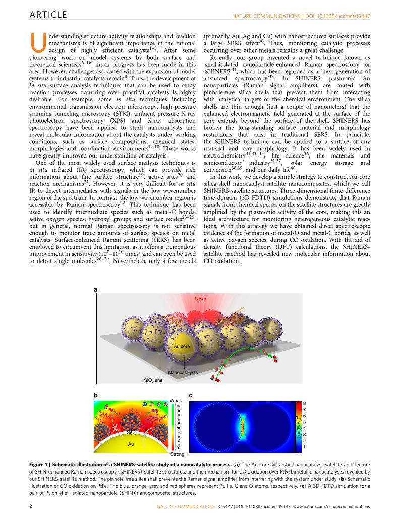
Synthesis of SHINERS-satellite nanocomposites. The nanoca-talyst satellites are added to the surface of the SHINs by a self-assembly process. The synthesis of our SHINs is described indetail elsewhere43. After synthesis, the SHIN surface is negativelycharged due to the presence of citrate (Supplementary Fig. 1). ThePtFe bimetallic nanocatalyst, which is widely used for oxygenreduction reactions in fuel cells44 and catalytic oxidation45, wasselected as a representative catalyst with which to explore thesynthesis of SHINERS-satellite nanostructures. The synthesisand characterization of the nanocatalysts is detailed in theSupplementary Methods. The PtFe nanocatalysts were modifiedto create a positively charged surface (Supplementary Figs 1–3),and mixed with the negatively charged SHINs (SupplementaryFig. 1) to achieve the self-assembly process. Figure 2a clearlyshows that the resulting nanoparticles have a core–shell-satellitetiered architecture. This structure is further demonstrated by theelement maps in Fig. 2b and the high-resolution transmissionelectron microscopy (HR-TEM) images in Supplementary Fig. 4.In a control experiment, unmodified PtFe nanocatalysts wereadded to a solution of SHINs. They did not adhere to the silicasurface, demonstrating that modification of the PtFenanocatalysts to create a positively charged surface is critical forSHINERS-satellite synthesis (Supplementary Fig. 5).
Through a systematic study, we found that the strategyemployed here for PtFe nanocatalysts can be used to assemble
many other types of nanocatalyst-on-SHIN structures(Supplementary Fig. 6). Figure 2c shows HR-TEM images ofmonometallic Pt and Pd nanocatalysts, bimetallic PtPd and PtFenanocatalysts, Au@PtFe core–shell structures, PdFeCu nano-cubes, and CeO2 and Fe2O3 metal oxides on SHINs. The size andmorphology of these nanocatalysts were unchanged by the self-assembly process, and all of them were evenly distributed on thesurface of the SHINs. Thus, it is feasible to study structure-activity relationships and reaction mechanisms of heterogeneouscatalytic processes by SHINERS after carefully manipulating thecomposition, structure, morphology and size of the nanocatalystsatellites.
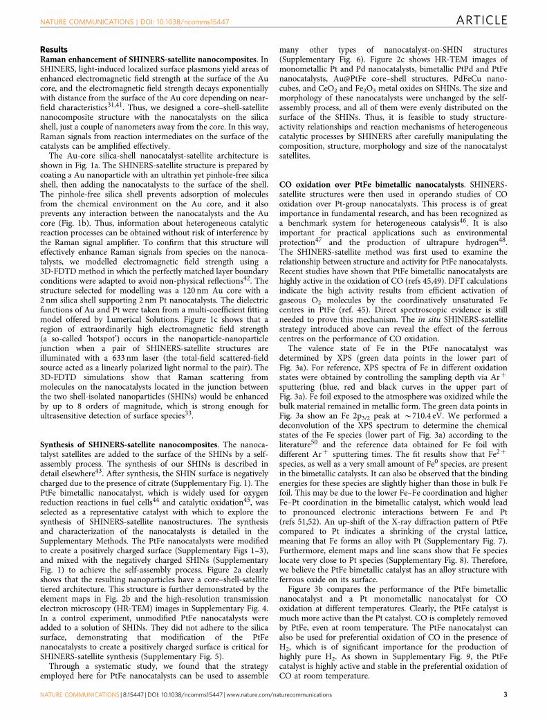
CO oxidation over PtFe bimetallic nanocatalysts. SHINERS-satellite structures were then used in operando studies of COoxidation over Pt-group nanocatalysts. This process is of greatimportance in fundamental research, and has been recognized asa benchmark system for heterogeneous catalysis46. It is alsoimportant for practical applications such as environmentalprotection47 and the production of ultrapure hydrogen48.The SHINERS-satellite method was first used to examine therelationship between structure and activity for PtFe nanocatalysts.Recent studies have shown that PtFe bimetallic nanocatalysts arehighly active in the oxidation of CO (refs 45,49). DFT calculationsindicate the high activity results from efficient activation ofgaseous O2 molecules by the coordinatively unsaturated Fecentres in PtFe (ref. 45). Direct spectroscopic evidence is stillneeded to prove this mechanism. The in situ SHINERS-satellitestrategy introduced above can reveal the effect of the ferrouscentres on the performance of CO oxidation.
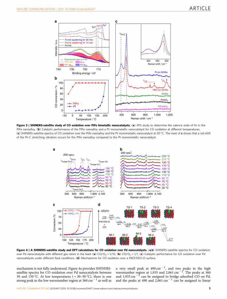
The valence state of Fe in the PtFe nanocatalyst wasdetermined by XPS (green data points in the lower part ofFig. 3a). For reference, XPS spectra of Fe in different oxidationstates were obtained by controlling the sampling depth via Arþ
sputtering (blue, red and black curves in the upper part ofFig. 3a). Fe foil exposed to the atmosphere was oxidized while thebulk material remained in metallic form. The green data points inFig. 3a show an Fe 2p3/2 peak at B710.4 eV. We performed adeconvolution of the XPS spectrum to determine the chemicalstates of the Fe species (lower part of Fig. 3a) according to theliterature50 and the reference data obtained for Fe foil withdifferent Arþ sputtering times. The fit results show that Fe2þ
species, as well as a very small amount of Fe0 species, are presentin the bimetallic catalysts. It can also be observed that the bindingenergies for these species are slightly higher than those in bulk Fefoil. This may be due to the lower Fe–Fe coordination and higherFe–Pt coordination in the bimetallic catalyst, which would leadto pronounced electronic interactions between Fe and Pt(refs 51,52). An up-shift of the X-ray diffraction pattern of PtFecompared to Pt indicates a shrinking of the crystal lattice,meaning that Fe forms an alloy with Pt (Supplementary Fig. 7).Furthermore, element maps and line scans show that Fe specieslocate very close to Pt species (Supplementary Fig. 8). Therefore,we believe the PtFe bimetallic catalyst has an alloy structure withferrous oxide on its surface.
Figure 3b compares the performance of the PtFe bimetallicnanocatalyst and a Pt monometallic nanocatalyst for COoxidation at different temperatures. Clearly, the PtFe catalyst ismuch more active than the Pt catalyst. CO is completely removedby PtFe, even at room temperature. The PtFe nanocatalyst canalso be used for preferential oxidation of CO in the presence ofH2, which is of significant importance for the production ofhighly pure H2. As shown in Supplementary Fig. 9, the PtFecatalyst is highly active and stable in the preferential oxidation ofCO at room temperature.
NATURE COMMUNICATIONS | DOI: 10.1038/ncomms15447 ARTICLE
NATURE COMMUNICATIONS | 8:15447 | DOI: 10.1038/ncomms15447 | www.nature.com/naturecommunications 3
To reveal the effect of the ferrous centres, PtFe-on-SHIN andPt-on-SHIN nanocomposites were prepared for in situ SHINERS-satellite studies. Activity tests (Supplementary Fig. 9) and XPSdata (Supplementary Fig. 10) show that the silica shell isolatesthe Au core so that it has no influence on the properties ofthe nanocatalysts. Furthermore, the capping agents on thecatalysts (such as oleylamine) were removed during fabricationof the SHINERS-satellite nanocomposites and the preparationsteps carried out before in situ SHINERS-satellite studies(Supplementary Fig. 11), so that catalytic sites were accessibleto reactants. The black and pink curves in Fig. 3c show that noRaman signals are observed for Pt or PtFe nanocatalysts on silicadue to the low sensitivity of normal Raman spectroscopy. Thegreen curve shows that there are also no Raman signals for SHINswithout nanocatalyst satellites, and therefore, the signals obtainedin the following experiments must be from species adsorbed onthe catalyst surface. The blue and red curves in Fig. 3c show thatstrong Raman peaks can be obtained from the same catalystswhen they are incorporated into the SHINERS-satellite structures.These results demonstrate the role that SHINs play in Ramansignal amplification. We note that the SHIN-satellite nanostruc-tures are also much more stable than Au-satellite nanostructuresat high temperatures (Supplementary Fig. 12). The two peaks at397 and 485 cm� 1 in the spectrum obtained from the Pt-on-SHIN structures (blue curve) can be assigned to the Pt–Cstretching vibrational mode of bridge and linear adsorbed COrespectively53. This spectrum (the blue curve) indicates that only
CO was adsorbed on the surface of the Pt, and the activation ofO2 was inhibited by the strongly competitive adsorption of CO.This can also be demonstrated by in situ SHINERS-satellitestudies of Pt under pure O2 and under CO oxidation conditionsat higher temperatures (Supplementary Fig. 13).
The spectrum obtained from the PtFe-on-SHIN structures(Fig. 3c red curve) has peaks for O2
2� at 870 and 951 cm� 1, anda peak for O2
� at 1,158 cm� 1 (refs 23–25), in addition to the COadsorption peaks at 389 and 480 cm� 1. The presence of activeoxygen species on the PtFe nanoalloy surface was confirmedby electron paramagnetic resonance (EPR) studies after COoxidation (Supplementary Fig. 14). It can be seen that there is nopeak in the 500–700 cm� 1 range, suggesting that no Pt oxide isformed at 30 �C. Furthermore, a red shift of the Pt–C stretchingvibration occurs for the PtFe nanoalloy compared to the Ptmonometallic nanocatalyst (Fig. 3c inset). This implies that Fespecies weaken the adsorption of CO, and this may be anotherreason for the high activity of the PtFe nanoalloy. These resultsindicate that CO oxidation on the PtFe nanoalloy could proceedthrough a Langmuir–Hinshelwood mechanism, even at very lowtemperature, in which the adsorbed CO and surface oxygenspecies are involved.
CO oxidation over Pd nanocatalysts. Pd based nanoparticles incatalytic converters are effective CO oxidation catalysts andreduce automotive emissions, however, the underlying
Pt on SHIN Pd on SHIN PtPd on SHIN
Pt Fe
Au+Si+Pt+FeAu+Si
SiAu
PtFe on SHIN
Au@PtFe on SHIN PdFeCu nanocubeon SHIN
Fe2O3 on SHINCeO2 on SHIN
2 nm55
nm
a
c
b
Figure 2 | The structure of various SHINERS-satellite nanocomposites. (a) TEM (inset) and scanning electron microscope images of PtFe-on-shell
isolated nanoparticle (SHIN) core–shell-satellite nanocomposites. Scale bar, 100 nm. Scale bar for the inset, 20 nm. (b) Element maps of the single particle
in the inset of (a). (c) TEM images of various nanocatalyst-on-SHIN structures. The insets in c are the zoomed-in image of the edge of the same particle as
in main image. Scale bars, 20 nm. Scale bars for the insets, 5 nm.
ARTICLE NATURE COMMUNICATIONS | DOI: 10.1038/ncomms15447
4 NATURE COMMUNICATIONS | 8:15447 | DOI: 10.1038/ncomms15447 | www.nature.com/naturecommunications
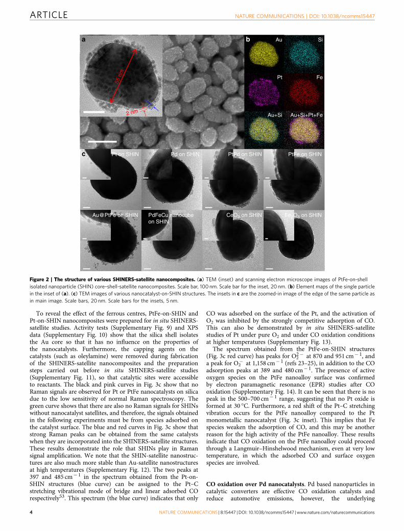
mechanism is not fully understood. Figure 4a provides SHINERS-satellite spectra for CO oxidation over Pd nanocatalysts between30 and 150 �C. At low temperatures (B30–50 �C), there is onestrong peak in the low wavenumber region at 360 cm� 1 as well as
a very small peak at 490 cm� 1, and two peaks in the highwavenumber region at 1,935 and 2,061 cm� 1. The peaks at 360and 1,935 cm� 1 can be assigned to bridge adsorbed CO on Pd,and the peaks at 490 and 2,061 cm� 1 can be assigned to linear
Fe3+
Fe-foil sputtering for 10 minFe-foil
740 730 720
400 450 500Raman shift / cm–1
710
Binding energy / eV
100
CO
con
vers
ion
/ % 80
60
40
20 PtFePt
0
–50 0 50 100Temperature / °C Raman shift / cm–1
400 600 800 1,000 1,200
PtFe/SiO2
Pt/SiO2
SHINs
PtFe-on-SHINs
Pt-on-SHINs
�O–2
� O22
–
�Pt-C
150 200
Inte
nsity
/ a.
u.
Fe2+ 2p3/2 Fe0 2p3/2
Pt 4sPeak sumExperimental data for PtFe
Fe-foil sputtering for 20 min
Fe2+ Fe0
b
a c
Figure 3 | SHINERS-satellite study of CO oxidation over PtFe bimetallic nanocatalysts. (a) XPS study to determine the valence state of Fe in the
PtFe nanoalloy. (b) Catalytic performance of the PtFe nanoalloy and a Pt monometallic nanocatalyst for CO oxidation at different temperatures.
(c) SHINERS-satellite spectra of CO oxidation over the PtFe nanoalloy and the Pt monometallic nanocatalyst at 30 �C. The inset of c shows that a red shift
of the Pt–C stretching vibration occurs for the PtFe nanoalloy compared to the Pt monometallic nanocatalyst.
ΔE(eV)
0.00
Pd(100)-O+
CO
0.45
–1.85–1.60
+O2+CO
–CO2
–CO2
–CO2
–3.51
–6.77
–3.64
IM-1IM-2 IM-3
IM-4
TS-2–2.47–2.37
–1.53TS-4
TS-3
TS-1
TS-1 TS-2 TS-4TS-3
200 cps200 cps
�bridge Pd-C
�linear Pd-C
�bridge-CO
�linear-CO
150 °C130 °C110 °C90 °C70 °C50 °C30 °C
2,1001,800
CO/O2 = 1/10
CO/O2 = 1/1100
75
55
25
0
100 125 150 175 200Temperature / °C
Con
vers
ion
/%
Raman shift/cm–1Raman shift/cm–1900600300 2,1001,800900600300
150 °C170 °C190 °C210 °C230 °C
130 °C110 °C90 °C70 °C50 °C30 °C
�Pd-O �
O22– �
O–2
IM-1 IM-2 IM-4IM-3
c d
ba
Figure 4 | A SHINERS-satellite study and DFT calculations for CO oxidation over Pd nanocatalysts. (a,b) SHINERS-satellite spectra for CO oxidation
over Pd nanocatalysts with different gas ratios in the feed: (a) CO/O2¼ 1/10, (b) CO/O2¼ 1/1. (c) Catalytic performance for CO oxidation over Pd
nanocatalysts under different feed conditions. (d) Mechanisms for CO oxidation over a PdO(100)–O surface.
NATURE COMMUNICATIONS | DOI: 10.1038/ncomms15447 ARTICLE
NATURE COMMUNICATIONS | 8:15447 | DOI: 10.1038/ncomms15447 | www.nature.com/naturecommunications 5
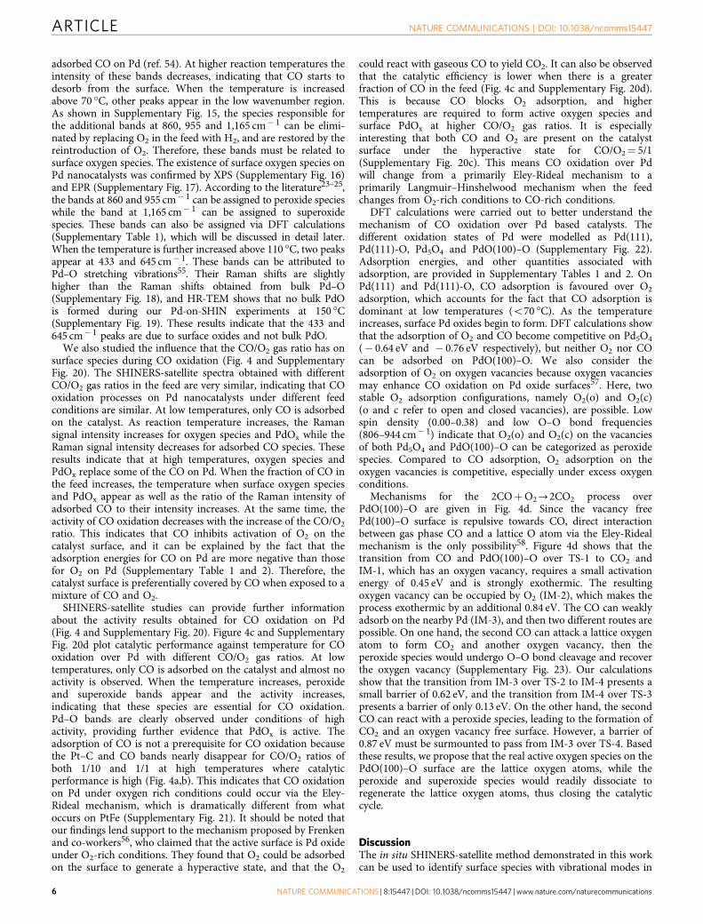
adsorbed CO on Pd (ref. 54). At higher reaction temperatures theintensity of these bands decreases, indicating that CO starts todesorb from the surface. When the temperature is increasedabove 70 �C, other peaks appear in the low wavenumber region.As shown in Supplementary Fig. 15, the species responsible forthe additional bands at 860, 955 and 1,165 cm� 1 can be elimi-nated by replacing O2 in the feed with H2, and are restored by thereintroduction of O2. Therefore, these bands must be related tosurface oxygen species. The existence of surface oxygen species onPd nanocatalysts was confirmed by XPS (Supplementary Fig. 16)and EPR (Supplementary Fig. 17). According to the literature23–25,the bands at 860 and 955 cm� 1 can be assigned to peroxide specieswhile the band at 1,165 cm� 1 can be assigned to superoxidespecies. These bands can also be assigned via DFT calculations(Supplementary Table 1), which will be discussed in detail later.When the temperature is further increased above 110 �C, two peaksappear at 433 and 645 cm� 1. These bands can be attributed toPd–O stretching vibrations55. Their Raman shifts are slightlyhigher than the Raman shifts obtained from bulk Pd–O(Supplementary Fig. 18), and HR-TEM shows that no bulk PdOis formed during our Pd-on-SHIN experiments at 150 �C(Supplementary Fig. 19). These results indicate that the 433 and645 cm� 1 peaks are due to surface oxides and not bulk PdO.
We also studied the influence that the CO/O2 gas ratio has onsurface species during CO oxidation (Fig. 4 and SupplementaryFig. 20). The SHINERS-satellite spectra obtained with differentCO/O2 gas ratios in the feed are very similar, indicating that COoxidation processes on Pd nanocatalysts under different feedconditions are similar. At low temperatures, only CO is adsorbedon the catalyst. As reaction temperature increases, the Ramansignal intensity increases for oxygen species and PdOx while theRaman signal intensity decreases for adsorbed CO species. Theseresults indicate that at high temperatures, oxygen species andPdOx replace some of the CO on Pd. When the fraction of CO inthe feed increases, the temperature when surface oxygen speciesand PdOx appear as well as the ratio of the Raman intensity ofadsorbed CO to their intensity increases. At the same time, theactivity of CO oxidation decreases with the increase of the CO/O2
ratio. This indicates that CO inhibits activation of O2 on thecatalyst surface, and it can be explained by the fact that theadsorption energies for CO on Pd are more negative than thosefor O2 on Pd (Supplementary Table 1 and 2). Therefore, thecatalyst surface is preferentially covered by CO when exposed to amixture of CO and O2.
SHINERS-satellite studies can provide further informationabout the activity results obtained for CO oxidation on Pd(Fig. 4 and Supplementary Fig. 20). Figure 4c and SupplementaryFig. 20d plot catalytic performance against temperature for COoxidation over Pd with different CO/O2 gas ratios. At lowtemperatures, only CO is adsorbed on the catalyst and almost noactivity is observed. When the temperature increases, peroxideand superoxide bands appear and the activity increases,indicating that these species are essential for CO oxidation.Pd–O bands are clearly observed under conditions of highactivity, providing further evidence that PdOx is active. Theadsorption of CO is not a prerequisite for CO oxidation becausethe Pt–C and CO bands nearly disappear for CO/O2 ratios ofboth 1/10 and 1/1 at high temperatures where catalyticperformance is high (Fig. 4a,b). This indicates that CO oxidationon Pd under oxygen rich conditions could occur via the Eley-Rideal mechanism, which is dramatically different from whatoccurs on PtFe (Supplementary Fig. 21). It should be noted thatour findings lend support to the mechanism proposed by Frenkenand co-workers56, who claimed that the active surface is Pd oxideunder O2-rich conditions. They found that O2 could be adsorbedon the surface to generate a hyperactive state, and that the O2
could react with gaseous CO to yield CO2. It can also be observedthat the catalytic efficiency is lower when there is a greaterfraction of CO in the feed (Fig. 4c and Supplementary Fig. 20d).This is because CO blocks O2 adsorption, and highertemperatures are required to form active oxygen species andsurface PdOx at higher CO/O2 gas ratios. It is especiallyinteresting that both CO and O2 are present on the catalystsurface under the hyperactive state for CO/O2¼ 5/1(Supplementary Fig. 20c). This means CO oxidation over Pdwill change from a primarily Eley-Rideal mechanism to aprimarily Langmuir–Hinshelwood mechanism when the feedchanges from O2-rich conditions to CO-rich conditions.
DFT calculations were carried out to better understand themechanism of CO oxidation over Pd based catalysts. Thedifferent oxidation states of Pd were modelled as Pd(111),Pd(111)-O, Pd5O4 and PdO(100)–O (Supplementary Fig. 22).Adsorption energies, and other quantities associated withadsorption, are provided in Supplementary Tables 1 and 2. OnPd(111) and Pd(111)-O, CO adsorption is favoured over O2
adsorption, which accounts for the fact that CO adsorption isdominant at low temperatures (o70 �C). As the temperatureincreases, surface Pd oxides begin to form. DFT calculations showthat the adsorption of O2 and CO become competitive on Pd5O4
(� 0.64 eV and � 0.76 eV respectively), but neither O2 nor COcan be adsorbed on PdO(100)–O. We also consider theadsorption of O2 on oxygen vacancies because oxygen vacanciesmay enhance CO oxidation on Pd oxide surfaces57. Here, twostable O2 adsorption configurations, namely O2(o) and O2(c)(o and c refer to open and closed vacancies), are possible. Lowspin density (0.00–0.38) and low O–O bond frequencies(806–944 cm� 1) indicate that O2(o) and O2(c) on the vacanciesof both Pd5O4 and PdO(100)–O can be categorized as peroxidespecies. Compared to CO adsorption, O2 adsorption on theoxygen vacancies is competitive, especially under excess oxygenconditions.
Mechanisms for the 2COþO2-2CO2 process overPdO(100)–O are given in Fig. 4d. Since the vacancy freePd(100)–O surface is repulsive towards CO, direct interactionbetween gas phase CO and a lattice O atom via the Eley-Ridealmechanism is the only possibility58. Figure 4d shows that thetransition from CO and PdO(100)–O over TS-1 to CO2 andIM-1, which has an oxygen vacancy, requires a small activationenergy of 0.45 eV and is strongly exothermic. The resultingoxygen vacancy can be occupied by O2 (IM-2), which makes theprocess exothermic by an additional 0.84 eV. The CO can weaklyadsorb on the nearby Pd (IM-3), and then two different routes arepossible. On one hand, the second CO can attack a lattice oxygenatom to form CO2 and another oxygen vacancy, then theperoxide species would undergo O–O bond cleavage and recoverthe oxygen vacancy (Supplementary Fig. 23). Our calculationsshow that the transition from IM-3 over TS-2 to IM-4 presents asmall barrier of 0.62 eV, and the transition from IM-4 over TS-3presents a barrier of only 0.13 eV. On the other hand, the secondCO can react with a peroxide species, leading to the formation ofCO2 and an oxygen vacancy free surface. However, a barrier of0.87 eV must be surmounted to pass from IM-3 over TS-4. Basedthese results, we propose that the real active oxygen species on thePdO(100)–O surface are the lattice oxygen atoms, while theperoxide and superoxide species would readily dissociate toregenerate the lattice oxygen atoms, thus closing the catalyticcycle.
DiscussionThe in situ SHINERS-satellite method demonstrated in this workcan be used to identify surface species with vibrational modes in
ARTICLE NATURE COMMUNICATIONS | DOI: 10.1038/ncomms15447
6 NATURE COMMUNICATIONS | 8:15447 | DOI: 10.1038/ncomms15447 | www.nature.com/naturecommunications
the low wavenumber region. When combined with moretraditional characterization techniques such as TEM, XPS,X-ray absorption spectroscopy and IR, as well as DFT calcula-tions, the SHINERS-satellite strategy can provide a deep under-standing of structure-activity relationships and reactionmechanisms.
This strategy can be used to track surface species andintermediates of other industrially important reactions, besidesthe model CO oxidation reaction. For example, adsorbed ethylenespecies are easily detected by the SHINERS-satellite method(Supplementary Fig. 24), so that epoxidation of ethylene can bestudied in situ. Another important aspect of the SHINERS-satellite strategy is its suitability for liquid phase reactions. OnlyRaman signals from species located a few nanometres from theAu core are enhanced, thus Raman signals from solventmolecules and other species in the bulk solution which do notparticipate in the reaction are not enhanced. The SHINERS-satellite method can also be used to characterize the compositionand electronic properties of a catalyst surface if probe moleculesare present (Supplementary Fig. 25). Furthermore, the Ramansignal enhancement can be improved as necessary by optimizingthe amplifier (for example, by increasing the Au core size orchanging the Au core to a Ag one as shown in SupplementaryFig. 26)40,59 to detect species with smaller Raman scatteringcross-sections. These unique properties of the SHINERS-satellitestrategy make it a universal and sensitive approach for in situstudy of various reactions occurring on different nanocatalysts.
To summarize, a simple, general and stable nanocompositestrategy has been developed to monitor heterogeneous catalyticprocesses in situ with extremely high surface sensitivity usingAu-core silica-shell nanocatalyst-satellite structures. 3D-FDTDsimulations show that Raman signals from species on the surfaceof the nanocatalysts can be amplified by 8 orders of magnitudebecause of electromagnetic field enhancement by the Au cores.The silica shells isolate the Au cores and prevent them frominteracting with the nanocatalysts and the chemical environmentwhile improving their thermal stability. Using this strategy, westudied the oxidation of CO on PtFe nanoalloys and Pdnanocatalysts. For the PtFe system, we obtained direct spectro-scopic evidence showing that the ferrous centre can weaken thePt–C bond and activate O2 at room temperature. This leads toCO oxidation by the Langmuir–Hinshelwood mechanism. Forthe Pd system, we combined the SHINERS-satellite strategy withDFT calculations and found that active O2 species are not formeduntil CO begins to desorb, causing the reaction to follow the Eley-Rideal mechanism. We have demonstrated that SHINs may becombined with a variety of other nanocatalysts as well, meaningthat our strategy can be developed as a standard characterizationmethod for in situ monitoring of reaction intermediates andelucidating reaction mechanisms.
MethodsSynthesis of SHINs and nanocatalysts. SHINs with 55 nm Au cores were syn-thesized according to our previously reported method31. In brief, 55 nm Aunanoparticles were synthesized according to Frens’ method60 and the Aunanoparticles were then coated with an ultrathin silica shell at 90 �C using3-aminopropyltrimethoxysilane as a coupling agent and sodium silicate as a siliconsource. SHINs with 120 nm Au cores were synthesized by a similar procedure, but120 nm Au nanoparticles were prepared by a seed-mediated growth method anddetails of that synthesis can be found in the Supplementary Information.Procedures for the preparation of nanocatalysts such as Pt, Pd, PtPd, PtFe,Au@PtFe, PdFeCu nanocubes, CeO2 and Fe2O3 are also described in theSupplementary Methods. Pt, Pd and PtFe nanoparticles were supported on SiO2 forcatalytic tests, and the loading of metal was controlled to B2.5 wt% as measured byinductively coupled plasma-atomic emission spectrometry (ICP-AES).
Self-assembly of nanocatalyst-on-SHIN structures. In general, 1 ml of 0.1 MNOBF4 in acetonitrile was added to each 0.5 ml dispersion of as-prepared
nanocatalysts in toluene. Each mixture was shaken vigorously for 5–10 min.Hexane and more toluene were added to precipitate the nanocatalysts, and theseparation was completed by centrifuging. The supernatant was discarded, andacetonitrile was added to form a clear dispersion of the nanocatalysts. By thisprocedure, the nanocatalysts were transferred from toluene to acetonitrile and theirsurfaces were modified to be positively charged as indicated by zeta potentialmeasurements (Supplementary Figs 1 and 6). SHINs dispersed in water were thenadded, and each mixture was shaken overnight to complete the self-assemblyprocess. Each mixture was centrifuged one last time, and the supernatant wasdiscarded to remove excess nanocatalyst. The resulting precipitate was the desiredAu-core silica-shell nanocatalyst-satellite nanocomposite, and was dispersed inacetonitrile for further use.
Characterization. Imaging and elemental analysis of these nanomaterials wereaccomplished by HR-TEM coupled with energy dispersive X-ray spectrometryusing an FEI Tecnai F30 microscope. The bulk composition of the samples wasdetermined by inductively coupled plasma-atomic emission spectrometry using anIRIS Intrepid II XSP spectrometer. XPS measurements were made using a VGMultiLab 2000 spectrometer with an Omicron Sphera II hemispherical electronenergy analyser. X-ray diffraction patterns were recorded on a Rigaku Ultima IVwith Cu Ka radiation operating at 40 kV and 30 mA. EPR spectra were acquiredwith a Bruker EMX 10/12 X-band spectrometer.
In situ SHINERS-satellite studies. About 5 ml of Au-core silica-shell nanocatalyst-satellite structures in acetonitrile were deposited on a Si substrate and dried atroom temperature. The substrate was then placed in a Raman cell with reactiontemperature and gas flow control (the cell was made in-house). The samples wereheld at 30 �C under H2 for 30 min to remove pre-adsorbed oxygen species andother contaminants. For in situ SHINERS-satellite studies of CO oxidation, thereaction temperature varied from 30 to 150 �C and the reaction gas consisted of1% CO, 10% O2 and 89% N2. The SHINERS-satellite experiments were carried outusing a Jobin-Yvon Horiba Xplora confocal Raman system. The � 50 microscopeobjective had a numerical aperture of 0.55 and a power density of B1.5 mW um� 2.The performance of the catalysts was measured in a fixed-bed lab reactor at atmo-spheric pressure with a weight hourly space velocity of 40,000 ml g� 1 � h� 1.
Data availability. The authors declare that all the data supporting the findings ofthis study are available within the paper and its Supplementary Information orfrom the corresponding author upon reasonable request.
References1. Herzing, A. A., Kiely, C. J., Carley, A. F., Landon, P. & Hutchings, G. J.
Identification of active gold nanoclusters on iron oxide supports for COoxidation. Science 321, 1331–1335 (2008).
2. Tsung, C. K. et al. Sub-10 nm platinum nanocrystals with size and shapecontrol: catalytic study for ethylene and pyrrole hydrogenation. J. Am. Chem.Soc. 131, 5816–5822 (2009).
3. Jambrina, P. G. et al. Quantum interference between H þ D2 quasiclassicalreaction mechanisms. Nat. Chem. 7, 661–667 (2015).
4. Tian, N., Zhou, Z. Y., Sun, S. G., Ding, Y. & Wang, Z. L. Synthesis oftetrahexahedral platinum nanocrystals with high-index facets and high electro-oxidation activity. Science 316, 732–735 (2007).
5. Deng, Y. et al. Multifunctional mesoporous composite microspheres with well-designed nanostructure: a highly integrated catalyst system. J. Am. Chem. Soc.132, 8466–8473 (2010).
6. Hammer, B. & Nørskov, J. K. Theoretical surface science and catalysis—calculations and concepts. Adv. Catal. 45, 71–129 (2000).
7. Somorjai, G. A., Beaumont, S. K. & Alayoglu, S. Determination of molecularsurface structure, composition, and dynamics under reaction conditions at highpressures and at the solid-liquid interface. Angew. Chem. Int. Ed. 50,10116–10129 (2011).
8. Somorjai, G. A., Frei, H. & Park, J. Y. Advancing the frontiers in nanocatalysis,biointerfaces, and renewable energy conversion by innovations of surfacetechniques. J. Am. Chem. Soc. 131, 16589–16605 (2009).
9. Imbihl, R. & Ertl, G. Oscillatory kinetics in heterogeneous catalysis. Chem. Rev.95, 697–733 (1995).
10. Goodman, D. W. Model studies in catalysis using surface science probes. Chem.Rev. 95, 523–536 (1995).
11. Arakawa, H. et al. Catalysis research of relevance to carbon management:progress, challenges, and opportunities. Chem. Rev. 101, 953–996 (2001).
12. Chen, M. & Goodman, D. W. Catalytically active gold: from nanoparticles toultrathin films. Acc. Chem. Res. 39, 739–746 (2006).
13. Liu, X., Madix, R. J. & Friend, C. M. Unraveling molecular transformations onsurfaces: a critical comparison of oxidation reactions on coinage metals. Chem.Soc. Rev. 37, 2243–2261 (2008).
14. Freund, H. J. The surface science of catalysis and more, using ultrathin oxidefilms as templates: a perspective. J. Am. Chem. Soc. 138, 8985–8996 (2016).
NATURE COMMUNICATIONS | DOI: 10.1038/ncomms15447 ARTICLE
NATURE COMMUNICATIONS | 8:15447 | DOI: 10.1038/ncomms15447 | www.nature.com/naturecommunications 7
15. Kuhlenbeck, H., Shaikhutdinov, S. & Freund, H. J. Well-ordered transitionmetal oxide layers in model catalysis – a series of case studies. Chem. Rev. 113,3986–4034 (2013).
16. Liu, W., Tkatchenko, A. & Scheffler, M. Modeling adsorption and reactions oforganic molecules at metal surfaces. Acc. Chem. Res. 47, 3369–3377 (2014).
17. Tao, F. & Crozier, P. A. Atomic-scale observations of catalyst structures underreaction conditions and during catalysis. Chem. Rev. 116, 3487–3539 (2016).
18. Tao, F. et al. Reaction-driven restructuring of Rh-Pd and Pt-Pd core-shellnanoparticles. Science 322, 932–934 (2008).
19. Qiao, B. T. et al. Single-atom catalysis of CO oxidation using Pt1/FeOx.Nat. Chem. 3, 634–641 (2011).
20. Ding, K. et al. Identification of active sites in CO oxidation and water-gas shiftover supported Pt catalysts. Science 350, 189–192 (2015).
21. Green, I. X., Tang, W., Neurock, M. & Yates, J. T. Spectroscopic observation ofdual catalytic sites during oxidation of CO on a Au/TiO2 Catalyst. Science 333,736–739 (2011).
22. Li, C. et al. UV resonance Raman spectroscopic identification of titanium atomsin the framework of TS-1 zeolite. Angew. Chem. Int. Ed. 38, 2220–2222 (1999).
23. Guzman, J., Carrettin, S. & Corma, A. Spectroscopic evidence for the supply ofreactive oxygen during CO oxidation catalyzed by gold supported onnanocrystalline CeO2. J. Am. Chem. Soc. 127, 3286–3287 (2005).
24. Guzman, J. et al. CO oxidation catalyzed by supported gold: cooperationbetween gold and nanocrystalline rare-earth supports forms reactive surfacesuperoxide and peroxide species. Angew. Chem. Int. Ed. 44, 4778–4781 (2005).
25. Weng, W. Z., Wan, H. L., Li, J. M. & Cao, Z. X. Laser-induced formation ofmetal–peroxide linkages on the surface of lanthanum sesquioxide underoxygen. Angew. Chem. Int. Ed. 43, 975–977 (2004).
26. Jeanmaire, D. L. & Van Duyne, R. P. Surface Raman spectroelectrochemistry:part I. heterocyclic, aromatic, and aliphatic amines adsorbed on the anodizedsilver electrode. J. Electroanal. Chem. Interfacial Electrochem. 84, 1–20 (1977).
27. Moskovits, M. Surface roughness and the enhanced intensity of Ramanscattering by molecules adsorbed on metals. J. Chem. Phys. 69, 4159–4161(1978).
28. Kneipp, K. et al. Single molecule detection using surface-enhanced Ramanscattering (SERS). Phys. Rev. Lett. 78, 1667–1670 (1997).
29. Nie, S. & Emory, S. R. Probing single molecules and single nanoparticles bysurface-enhanced Raman scattering. Science 275, 1102–1106 (1997).
30. Banholzer, M. J., Millstone, J. E., Qin, L. & Mirkin, C. A. Rationally designednanostructures for surface-enhanced Raman spectroscopy. Chem. Soc. Rev. 37,885–897 (2008).
31. Li, J. F. et al. Shell-isolated nanoparticle-enhanced Raman spectroscopy. Nature464, 392–395 (2010).
32. Graham, D. The next generation of advanced spectroscopy: surface enhancedRaman scattering from metal nanoparticles. Angew. Chem. Int. Ed. 49,9325–9327 (2010).
33. Li, J. F. et al. Extraordinary enhancement of Raman scattering from pyridine onsingle crystal Au and Pt electrodes by shell-isolated Au nanoparticles. J. Am.Chem. Soc. 133, 15922–15925 (2011).
34. Li, C. Y. et al. In situ monitoring of electrooxidation processes at gold singlecrystal surfaces using shell-isolated nanoparticle-enhanced raman spectroscopy.J. Am. Chem. Soc. 137, 7648–7651 (2015).
35. Hartman, T., Wondergem, C. S., Kumar, N., van den Berg, A. & Weckhuysen, B. M.Surface- and tip-enhanced Raman spectroscopy in catalysis. J. Phys. Chem. Lett. 7,1570–1584 (2016).
36. Bian, X. et al. Fabrication of graphene-isolated-Au-nanocrystal nanostructuresfor multimodal cell imaging and photothermal-enhanced chemotherapy.Sci. Rep. 4, 6093 (2014).
37. Honesty, N. R. & Gewirth, A. A. Shell-isolated nanoparticle enhanced Ramanspectroscopy (SHINERS) investigation of benzotriazole film formation onCu(100), Cu(111), and Cu(poly). J. Raman Spectrosc. 43, 46–50 (2012).
38. Xie, W., Walkenfort, B. & Schlucker, S. Label-free SERS monitoring of chemicalreactions catalyzed by small gold nanoparticles using 3D plasmonicsuperstructures. J. Am. Chem. Soc. 135, 1657–1660 (2013).
39. Xie, W. & Schlucker, S. Hot electron-induced reduction of small molecules onphotorecycling metal surfaces. Nat. Commun. 6, 7570–7575 (2015).
40. Li, J. F., Anema, J. R., Wandlowski, T. & Tian, Z. Q. Dielectric shell isolated andgraphene shell isolated nanoparticle enhanced Raman spectroscopies and theirapplications. Chem. Soc. Rev. 44, 8399–8409 (2015).
41. Maier, S. A. Plasmonics: fundamentals and applications (Springer Science& Business Media, 2007).
42. Taflove, A. & Hagness, S. C. Computational Electrodynamics: the Finite-Difference Time-Domain Method (Artech House Press, 2005).
43. Li, J. F. et al. Surface analysis using shell-isolated nanoparticle-enhanced Ramanspectroscopy. Nat. Protoc. 8, 52–65 (2013).
44. Wang, Y. J. et al. Carbon-supported Pt-based alloy electrocatalysts for theoxygen reduction reaction in polymer electrolyte membrane fuel cells: particlesize, shape, and composition manipulation and their impact to activity. Chem.Rev. 115, 3433–3467 (2015).
45. Fu, Q. et al. Interface-confined ferrous centers for catalytic oxidation. Science328, 1141–1144 (2010).
46. Freund, H. J., Meijer, G., Scheffler, M., Schlogl, R. & Wolf, M. CO oxidation as aprototypical reaction for heterogeneous processes. Angew. Chem. Int. Ed. 50,10064–10094 (2011).
47. Nishihata, Y. et al. Self-regeneration of a Pd-perovskite catalyst for automotiveemissions control. Nature 418, 164–167 (2002).
48. Alayoglu, S., Nilekar, A. U., Mavrikakis, M. & Eichhorn, B. Ru-Pt core-shellnanoparticles for preferential oxidation of carbon monoxide in hydrogen.Nat. Mater. 7, 333–338 (2008).
49. Chen, G. et al. Interfacial effects in iron-nickel hydroxide-platinumnanoparticles enhance catalytic oxidation. Science 344, 495–499 (2014).
50. Stassi, J. P., Zgolicz, P. D., de Miguel, S. R. & Scelza, O. A. Formation of differentpromoted metallic phases in PtFe and PtSn catalysts supported on carbonaceousmaterials used for selective hydrogenation. J. Catal. 306, 11–29 (2013).
51. Xu, H., Fu, Q., Yao, Y. & Bao, X. Highly active Pt-Fe bicomponent catalysts forCO oxidation in the presence and absence of H2. Energy Environ. Sci. 5,6313–6320 (2012).
52. Ma, T. et al. Reversible structural modulation of Fe–Pt bimetallic surfaces andits effect on reactivity. Chemphyschem 10, 1013–1016 (2009).
53. Fang, P. P. et al. Tailoring Au-core Pd-shell Pt-cluster nanoparticles forenhanced electrocatalytic activity. Chem. Sci. 2, 531–539 (2011).
54. Gao, F., Wang, Y. & Goodman, D. W. CO oxidation over AuPd(100) fromultrahigh vacuum to near-atmospheric pressures: the critical role of contiguousPd atoms. J. Am. Chem. Soc. 131, 5734–5735 (2009).
55. McBride, J. R., Hass, K. C. & Weber, W. H. Resonance-Raman and lattice-dynamics studies of single-crystal PdO. Phys. Rev. B 44, 5016–5028 (1991).
56. Hendriksen, B. L. M., Bobaru, S. C. & Frenken, J. W. M. Oscillatory COoxidation on Pd(100) studied with in situ scanning tunneling microscopy. Surf.Sci. 552, 229–242 (2004).
57. Weaver, J. F., Zhang, F., Pan, L., Li, T. & Asthagiri, A. Vacancy-mediated processesin the oxidation of CO on PdO(101). Acc. Chem. Res. 48, 1515–1523 (2015).
58. Hirvi, J. T., Kinnunen, T. J. J., Suvanto, M., Pakkanen, T. A. & Nørskov, J. K.CO oxidation on PdO surfaces. J. Chem. Phys. 133, 084704 (2010).
59. Ding, S. Y. et al. Nanostructure-based plasmon-enhanced Raman spectroscopyfor surface analysis of materials. Nat. Rev. Mater. 1, 16021 (2016).
60. Frens, G. Controlled nucleation for the regulation of the particle size inmonodisperse gold suspensions. Nature 241, 20–22 (1973).
AcknowledgementsThis work was supported by the NSFC (21522508, 21427813, 21373167, 21521004,21573178 and 21673187), Natural Science Foundation of Guangdong Province(2016A030308012), the Fundamental Research Funds for the Central Universities(20720150039 and 20720160046), ‘111’Project (B16029), and the Thousand YouthTalents Plan of China. We thank Dr C. Liu and M. Meng for experimental support.
Author contributionsH.Z. and J.-F.L. designed the experiments. H.Z., H.-L.S., C.W. and Y.-J.Z. carried out theexperiments. G.F. conducted the DFT calculations, and S.C. and Z.-L.Y. conducted the3D-FDTD simulations. H.Z., J.R.A., B.-H.C., J.-F.L. and Z.-Q.T. analysed the data. Allauthors contributed to the preparation of the manuscript.
Additional informationSupplementary Information accompanies this paper at http://www.nature.com/naturecommunications
Competing interests: The authors declare no competing financial interests.
Reprints and permission information is available online at http://npg.nature.com/reprintsandpermissions/
How to cite this article: Zhang, H. et al. In-situ dynamic tracking of heterogeneousnanocatalytic processes by shell-isolated nanoparticle-enhanced Raman spectroscopy.Nat. Commun. 8, 15447 doi: 10.1038/ncomms15447 (2017).
Publisher’s note: Springer Nature remains neutral with regard to jurisdictional claims inpublished maps and institutional affiliations.
This work is licensed under a Creative Commons Attribution 4.0International License. The images or other third party material in this
article are included in the article’s Creative Commons license, unless indicated otherwisein the credit line; if the material is not included under the Creative Commons license,users will need to obtain permission from the license holder to reproduce the material.To view a copy of this license, visit http://creativecommons.org/licenses/by/4.0/
r The Author(s) 2017
ARTICLE NATURE COMMUNICATIONS | DOI: 10.1038/ncomms15447
8 NATURE COMMUNICATIONS | 8:15447 | DOI: 10.1038/ncomms15447 | www.nature.com/naturecommunications