Embed Size (px)
Citation preview
For life science research only. Not for use in diagnostic procedures.
sigma-aldrich.com
In Situ Cell Death Detection Kit, TMR red
Kit for detection and quantification of apoptosis (programmed cell death) at single cell level, based on labeling of DNA strand breaks (TUNEL technology): Analysis by fluorescence microscopy or flow cytometry.
Cat. No. 12 156 792 910 1 Kit (50 tests)
Store at �15 to �25°C
y Version 12Content version: March 2016
sigma-aldrich.com 2
1. Preface
1.1 Table of Contents
1. Preface ............................................................................................................ 21.1 Table of Contents 21.2 Kit Contents 3
2. Introduction ................................................................................................... 52.1 Product Overview 52.2 Background Information 8
3. Procedures and Required Materials ...................................................... 103.1 Flow Chart 103.2 Preparation of Sample Material 113.2.1 Cell Suspension 113.2.2 Adherent Cells, Cell Smears, and Cytospin Preparations 123.2.3 Tissue Sections 133.2.3.1 Treatment of Paraffin-Embedded Tissue 133.2.3.2 Treatment of Cryopreserved Tissue 153.3 Labeling Protocol 163.3.1 Before you Begin 163.3.2 Labeling Protocol for Cell Suspensions 173.3.3 Labeling Protocol for Adherent Cells, Cell Smears, Cytospin Preparations,
and Tissues 183.3.4 Labeling Protocol for Difficult Tissue 19
4. Typical results ............................................................................................. 20
5. Appendix ....................................................................................................... 215.1 Troubleshooting 215.2 References 245.3 Ordering Information 25
Changes to previous version 26Regulatory Disclaimer 26Disclaimer of License 26
In Situ Cell Death Detection Kit, TMR red y Version 12
sigma-aldrich.com 3
1.2 Kit Contents
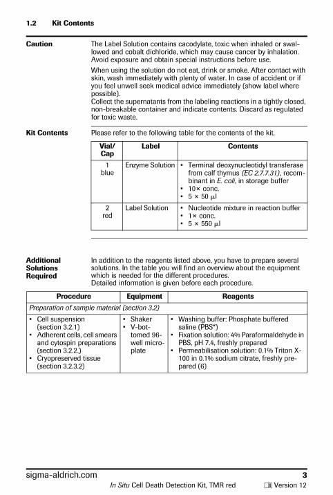
Caution The Label Solution contains cacodylate, toxic when inhaled or swal-lowed and cobalt dichloride, which may cause cancer by inhalation. Avoid exposure and obtain special instructions before use.When using the solution do not eat, drink or smoke. After contact with skin, wash immediately with plenty of water. In case of accident or if you feel unwell seek medical advice immediately (show label where possible). Collect the supernatants from the labeling reactions in a tightly closed, non-breakable container and indicate contents. Discard as regulated for toxic waste.
Kit Contents Please refer to the following table for the contents of the kit.
Vial/Cap
Label Contents
1blue
Enzyme Solution • Terminal deoxynucleotidyl transferase from calf thymus (EC 2.7.7.31), recom-binant in E. coli, in storage buffer
• 10× conc.• 5 × 50 �l
2red
Label Solution • Nucleotide mixture in reaction buffer• 1× conc.• 5 × 550 �l
Additional Solutions Required
In addition to the reagents listed above, you have to prepare several solutions. In the table you will find an overview about the equipment which is needed for the different procedures.Detailed information is given before each procedure.
Procedure Equipment Reagents
Preparation of sample material (section 3.2)
• Cell suspension (section 3.2.1)
• Adherent cells, cell smears and cytospin preparations (section 3.2.2.)
• Cryopreserved tissue (section 3.2.3.2)
• Shaker• V-bot-
tomed 96-well micro-plate
• Washing buffer: Phosphate buffered saline (PBS*)
• Fixation solution: 4% Paraformaldehyde in PBS, pH 7.4, freshly prepared
• Permeabilisation solution: 0.1% Triton X-100 in 0.1% sodium citrate, freshly pre-pared (6)
In Situ Cell Death Detection Kit, TMR red y Version 12
1.2 Kit Contents, continued
sigma-aldrich.com 4In Situ Cell Death Detection Kit, TMR red y Version 12
Paraffin-embedded tissue (section 3.2.3.1)
• Xylene and ethanol (absolute, 95%, 90%, 80%, 70%, diluted in double-distilled water)
• Washing buffer: PBS*• Proteinase K*, PCR Grade, working solu-
tion: [10 –20 �g/ml in 10 mM Tris/HCl, pH 7.4–8]
Alternative treatments• Permeabilisation solution: (0.1% Triton1)
X–100, 0.1% sodium citrate), freshly pre-pared
• Pepsin* (0.25%–0.5% in HCl, pH 2) or trypsin*, 0.01 N HCl
• 0.1 M Citrate buffer, pH 6 for microwave irradiation
Labeling protocol (section 3.3)
Positive control(section 3.3.1)
• Micrococcal nuclease or • DNase I recombinant*
• Cell suspensions (section 3.3.2)
• Adherent cells (section 3.3.3)
• Parafilm or coverslips
• Humidified chamber
Washing buffer: PBS*
Difficult tissue(section 3.3.4)
• Plastic jar• Microwave• Humidified
chamber
• Citrate buffer, 0.1 M, pH 6.0.• Washing buffer: PBS*• Tris-HCl, 0.1 M pH 7.5, containing 3%
BSA* and 20% normal bovine serum
sigma-aldrich.com 5
2. Introduction
2.1 Product Overview
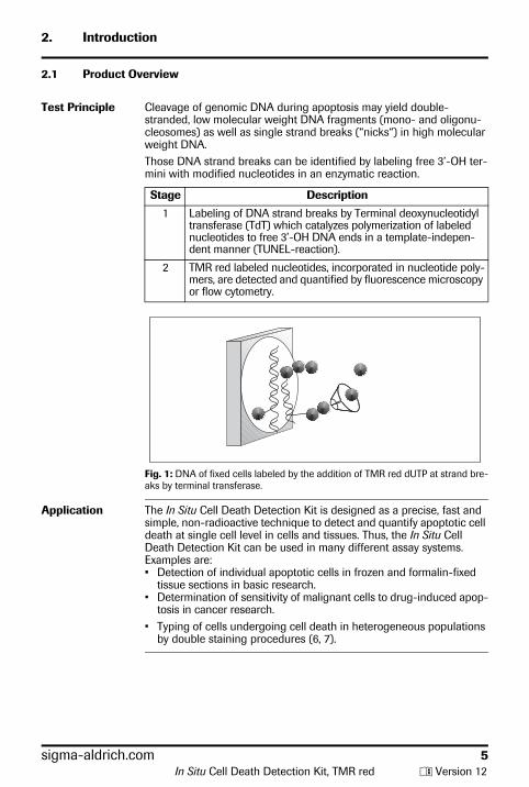
Test Principle Cleavage of genomic DNA during apoptosis may yield double-stranded, low molecular weight DNA fragments (mono- and oligonu-cleosomes) as well as single strand breaks (“nicks“) in high molecular weight DNA.Those DNA strand breaks can be identified by labeling free 3’-OH ter-mini with modified nucleotides in an enzymatic reaction.
Stage Description
1 Labeling of DNA strand breaks by Terminal deoxynucleotidyl transferase (TdT) which catalyzes polymerization of labeled nucleotides to free 3’-OH DNA ends in a template-indepen-dent manner (TUNEL-reaction).
2 TMR red labeled nucleotides, incorporated in nucleotide poly-mers, are detected and quantified by fluorescence microscopy or flow cytometry.
Fig. 1: DNA of fixed cells labeled by the addition of TMR red dUTP at strand bre-aks by terminal transferase.
Application The In Situ Cell Death Detection Kit is designed as a precise, fast and simple, non-radioactive technique to detect and quantify apoptotic cell death at single cell level in cells and tissues. Thus, the In Situ Cell Death Detection Kit can be used in many different assay systems. Examples are:• Detection of individual apoptotic cells in frozen and formalin-fixed
tissue sections in basic research.• Determination of sensitivity of malignant cells to drug-induced apop-
tosis in cancer research.• Typing of cells undergoing cell death in heterogeneous populations
by double staining procedures (6, 7).
In Situ Cell Death Detection Kit, TMR red y Version 12
2.1 Product Overview, continued
sigma-aldrich.com 6
Specificity The TUNEL reaction preferentially labels DNA strand breaks generated during apoptosis. This allows discrimination of apoptosis from necrosis and from primary DNA strand breaks induced by cytostatic drugs or irradiation (3, 4).
Test Interference False negative results: DNA cleavage can be absent or incomplete in some forms of apoptotic cell death (37). Sterical hindrance such as extracellular matrix components can prevent access of TdT to DNA strand breaks. In either case false negative results may be obtained.False positive results: Extensive DNA fragmentation may occur in cer-tain forms of necrosis (38).DNA strand breaks may also be prominent in cell populations with high proliferative or metabolic activity. In either case false positive results may be obtained.To confirm the apoptotic mode of cell death, the morphology of respective cells should be examined very carefully. Morphological changes during apoptosis have a characteristic pattern. Therefore evaluation of cell morphology is an important parameter in situations where there is any ambiguity regarding interpretation of results.
Sample Material • Cell suspensions from• permanent cell lines (2, 27, 35),• lymphocytes and leukemic cells from peripheral blood (4),• thymocytes (1, 6),• bone marrow cells• fine needle biopsies (5)
• Cytospins and cell smear preparations • Adherent cells cultured on chamber slides (31)• Frozen or formalin-fixed, paraffin-embedded tissue sections (1, 25,
26, 29, 30, 32–34, 36, 39)
Assay Time 1-2 hours, excluding culture, fixation and permeabilisation of cells and preparation of tissue sections.
Number of Tests The kit is designed for 50 tests.
Kit Storage/ Stability
The unopened kit is stable at �15 to �25°C until the expiration date printed on the label.Note: The TUNEL reaction mixture should be prepared immediately before use and should not be stored. Keep the TUNEL reaction mixture on ice until use.
In Situ Cell Death Detection Kit, TMR red y Version 12
2.1 Product Overview, continued
Advantage Please refer to the following table.
Benefit Feature
Sensitive Detection of apoptotic cell death at single cell level via fluorescence microscopy and at cell pop-ulations via FACS analysis at very early stages (1, 2, 6).
Specific Preferential labeling of apoptosis versus necrosis (3, 4).
Fast Short assay time (1– 2 h).
Convenient • No secondary detection system required.• One incubation and one washing step only.• Reagents are provided in stable, optimized
form.• No dilution steps required.• Application in combination with fluorescein
label possible
Flexible • Suitable for fixed cells and tissue. This allows accumulation, storage and transport of sam-ples (2, 5).
• Double staining enables identification of type and differentiation state of cells undergoing apoptosis (6).
Function-tested Every lot is function-tested on apoptotic cells in comparison to a master lot.
sigma-aldrich.com 7In Situ Cell Death Detection Kit, TMR red y Version 12
sigma-aldrich.com 8
2.2 Background Information
Cell Death Two distinct modes of cell death, apoptosis and necrosis, can be dis-tinguished based on differences in morphological, biochemical and molecular changes of dying cells.Programmed cell death or apoptosis is the most common form of eukaryotic cell death. It is a physiological suicide mechanism that pre-serves homeostasis, in which cell death naturally occurs during normal tissue turnover (8, 9). In general, cells undergoing apoptosis display a characteristic pattern of structural changes in nucleus and cytoplasm, including rapid blebbing of plasma membrane and nuclear disintegra-tion. The nuclear collapse is associated with extensive damage to chromatin and DNA-cleavage into oligonucleosomal length DNA frag-ments after activation of a calcium-dependent endogenous endonu-clease (10, 11). However, very rare exceptions have been described where morphological features of apoptosis are not accompanied with oligonucleosomal DNA cleavage (37).
Apoptosis Apoptosis is essential in many physiological processes, including maturation and effector mechanisms of the immune system (12, 13), embryonic development of tissue, organs and limbs (14), development of the nervous system (15, 16) and hormone-dependent tissue remodeling (17).
In Situ Cell Death Detection Kit, TMR red y Version 12
2.2 Background Information, continued
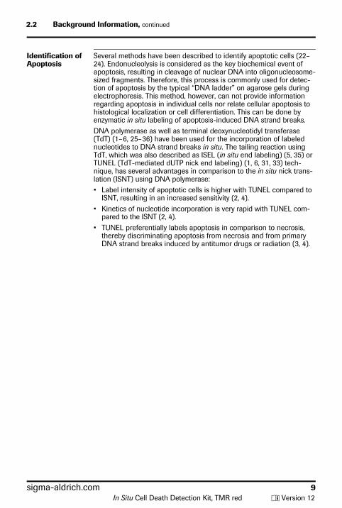
Identification of Apoptosis
Several methods have been described to identify apoptotic cells (22– 24). Endonucleolysis is considered as the key biochemical event of apoptosis, resulting in cleavage of nuclear DNA into oligonucleosome-sized fragments. Therefore, this process is commonly used for detec-tion of apoptosis by the typical “DNA ladder” on agarose gels during electrophoresis. This method, however, can not provide information regarding apoptosis in individual cells nor relate cellular apoptosis to histological localization or cell differentiation. This can be done by enzymatic in situ labeling of apoptosis-induced DNA strand breaks.DNA polymerase as well as terminal deoxynucleotidyl transferase (TdT) (1– 6, 25– 36) have been used for the incorporation of labeled nucleotides to DNA strand breaks in situ. The tailing reaction using TdT, which was also described as ISEL (in situ end labeling) (5, 35) or TUNEL (TdT-mediated dUTP nick end labeling) (1, 6, 31, 33) tech-nique, has several advantages in comparison to the in situ nick trans-lation (ISNT) using DNA polymerase:• Label intensity of apoptotic cells is higher with TUNEL compared to
ISNT, resulting in an increased sensitivity (2, 4).• Kinetics of nucleotide incorporation is very rapid with TUNEL com-
pared to the ISNT (2, 4).• TUNEL preferentially labels apoptosis in comparison to necrosis,
thereby discriminating apoptosis from necrosis and from primary DNA strand breaks induced by antitumor drugs or radiation (3, 4).
sigma-aldrich.com 9In Situ Cell Death Detection Kit, TMR red y Version 12
sigma-aldrich.com 10
3. Procedures and Required Materials
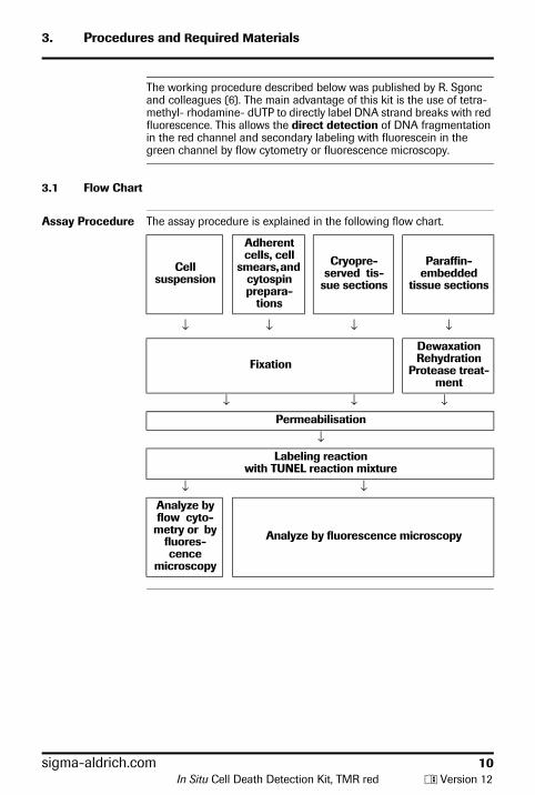
The working procedure described below was published by R. Sgonc and colleagues (6). The main advantage of this kit is the use of tetra-methyl- rhodamine- dUTP to directly label DNA strand breaks with red fluorescence. This allows the direct detection of DNA fragmentation in the red channel and secondary labeling with fluorescein in the green channel by flow cytometry or fluorescence microscopy.
3.1 Flow Chart
Assay Procedure The assay procedure is explained in the following flow chart.
Cell suspension
Adherent cells, cell
smears, and cytospin prepara-
tions
Cryopre-served tis-
sue sections
Paraffin-embedded
tissue sections
↓ ↓ ↓ ↓
Fixation
Dewaxation Rehydration
Protease treat-ment
↓ ↓ ↓Permeabilisation
↓Labeling reaction
with TUNEL reaction mixture
↓ ↓Analyze by flow cyto-
metry or by fluores-cence
microscopy
Analyze by fluorescence microscopy
In Situ Cell Death Detection Kit, TMR red y Version 12
sigma-aldrich.com 11
3.2 Preparation of Sample Material
3.2.1 Cell Suspension
Prelabeling For dual parameter flow cytometry with fluorescein-conjugated anti-bodies, incubate the cells prior to fixation with the cell surface marker.
Additional Buf-fers and Equip-ment Required
• Washing buffer: Phosphate buffered saline (PBS)• Fixation solution: Paraformaldehyde (4% in PBS, pH 7.4), freshly
prepared• Permeabilisation solution: 0.1% Triton X–100 in 0.1% sodium citrate,
freshly prepared• Shaker• V-bottomed 96-well microplateNote: Use of a V-bottomed 96-well microplate minimize cell loss during fixation, permeabilisation and labeling and allows simultaneous preparation of multiple samples.
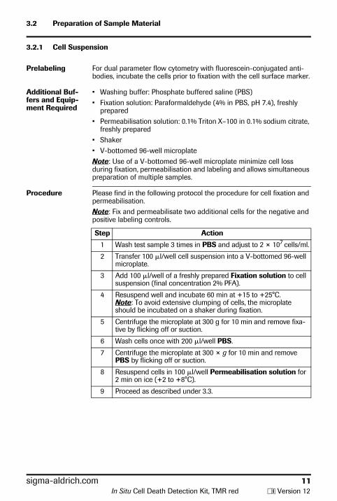
Procedure Please find in the following protocol the procedure for cell fixation and permeabilisation.Note: Fix and permeabilisate two additional cells for the negative and positive labeling controls.
Step Action
1 Wash test sample 3 times in PBS and adjust to 2 × 10⁷ cells/ml.
2 Transfer 100 �l/well cell suspension into a V-bottomed 96-well microplate.
3 Add 100 �l/well of a freshly prepared Fixation solution to cell suspension (final concentration 2% PFA).
4 Resuspend well and incubate 60 min at +15 to +25°C.Note: To avoid extensive clumping of cells, the microplate should be incubated on a shaker during fixation.
5 Centrifuge the microplate at 300 g for 10 min and remove fixa-tive by flicking off or suction.
6 Wash cells once with 200 �l/well PBS.
7 Centrifuge the microplate at 300 × g for 10 min and remove PBS by flicking off or suction.
8 Resuspend cells in 100 �l/well Permeabilisation solution for 2 min on ice (+2 to +8°C).
9 Proceed as described under 3.3.
In Situ Cell Death Detection Kit, TMR red y Version 12
sigma-aldrich.com 12
3.2.2 Adherent Cells, Cell Smears, and Cytospin Preparations
Additional Solutions Required
• Washing buffer: Phosphate buffered saline (PBS)• Fixation solution: 4% Paraformaldehyde in PBS, pH 7.4, freshly pre-
pared• Permeabilisation solution: 0.1% Triton X-100 in 0.1% sodium citrate,
freshly prepared (6)
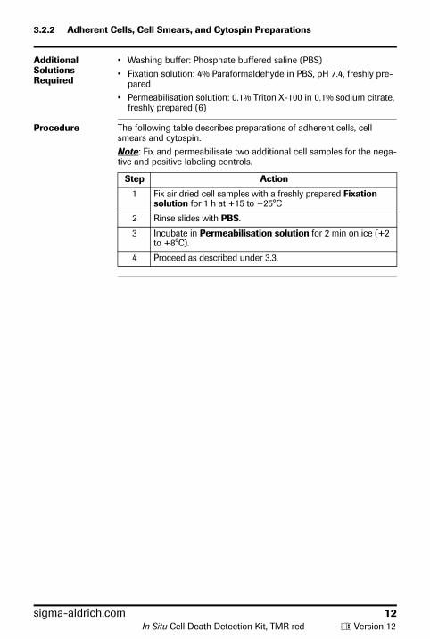
Procedure The following table describes preparations of adherent cells, cell smears and cytospin.Note: Fix and permeabilisate two additional cell samples for the nega-tive and positive labeling controls.
Step Action
1 Fix air dried cell samples with a freshly prepared Fixation solution for 1 h at +15 to +25°C
2 Rinse slides with PBS.
3 Incubate in Permeabilisation solution for 2 min on ice (+2 to +8°C).
4 Proceed as described under 3.3.
In Situ Cell Death Detection Kit, TMR red y Version 12
sigma-aldrich.com 13
3.2.3 Tissue Sections
3.2.3.1 Treatment of Paraffin-Embedded Tissue
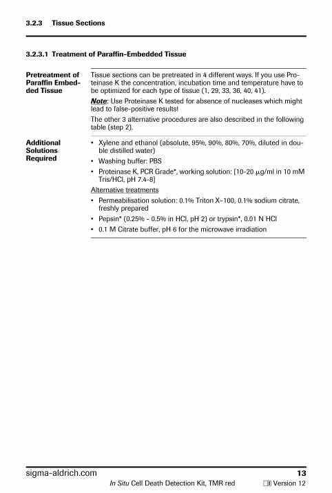
Pretreatment of Paraffin Embed-ded Tissue
Tissue sections can be pretreated in 4 different ways. If you use Pro-teinase K the concentration, incubation time and temperature have to be optimized for each type of tissue (1, 29, 33, 36, 40, 41).Note: Use Proteinase K tested for absence of nucleases which might lead to false-positive results!The other 3 alternative procedures are also described in the following table (step 2).
Additional Solutions Required
• Xylene and ethanol (absolute, 95%, 90%, 80%, 70%, diluted in dou-ble distilled water)
• Washing buffer: PBS• Proteinase K, PCR Grade*, working solution: [10-20 �g/ml in 10 mM
Tris/HCl, pH 7.4-8]Alternative treatments• Permeabilisation solution: 0.1% Triton X–100, 0.1% sodium citrate,
freshly prepared• Pepsin* (0.25% - 0.5% in HCl, pH 2) or trypsin*, 0.01 N HCl• 0.1 M Citrate buffer, pH 6 for the microwave irradiation
In Situ Cell Death Detection Kit, TMR red y Version 12
3.2.3.1 Treatment of Paraffin-Embedded Tissue, continued
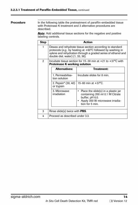
Procedure In the following table the pretreatment of paraffin-embedded tissue with Proteinase K treatment and 3 alternative procedures are described.Note: Add additional tissue sections for the negative and positive labeling controls.
Step Action
1 Dewax and rehydrate tissue section according to standard protocols (e.g., by heating at +60°C followed by washing in xylene and rehydration through a graded series of ethanol and double dist. water) (1, 33, 36).
2 Incubate tissue section for 15 –30 min at +21 to +37°C with Proteinase K working solution.
Alternatives: Treatment:
1. Permeabilisa-tion solution
Incubate slides for 8 min.
2. Pepsin* (30, 40) or trypsin
15–60 min at +37°C.
3. Microwave irradiation
• Place the slide(s) in a plastic jar containing 200 ml 0.1 M Citrate buffer, pH 6.0.
• Apply 350 W microwave irradia-tion for 5 min.
3 Rinse slide(s) twice with PBS.
4 Proceed as described under 3.3.
sigma-aldrich.com 14In Situ Cell Death Detection Kit, TMR red y Version 12
sigma-aldrich.com 15
3.2.3.2 Treatment of Cryopreserved Tissue
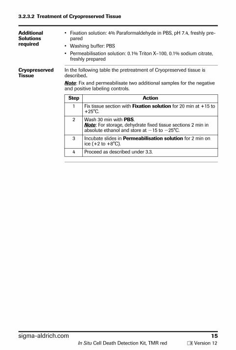
Additional Solutions required
• Fixation solution: 4% Paraformaldehyde in PBS, pH 7.4, freshly pre-pared
• Washing buffer: PBS• Permeabilisation solution: 0.1% Triton X–100, 0.1% sodium citrate,
freshly prepared
Cryopreserved Tissue
In the following table the pretreatment of Cryopreserved tissue is described.Note: Fix and permeabilisate two additional samples for the negative and positive labeling controls.
Step Action
1 Fix tissue section with Fixation solution for 20 min at +15 to +25°C.
2 Wash 30 min with PBS.Note: For storage, dehydrate fixed tissue sections 2 min in absolute ethanol and store at �15 to �25°C.
3 Incubate slides in Permeabilisation solution for 2 min on ice (+2 to +8°C).
4 Proceed as described under 3.3.
In Situ Cell Death Detection Kit, TMR red y Version 12
sigma-aldrich.com 16
3.3 Labeling Protocol
3.3.1 Before You Begin
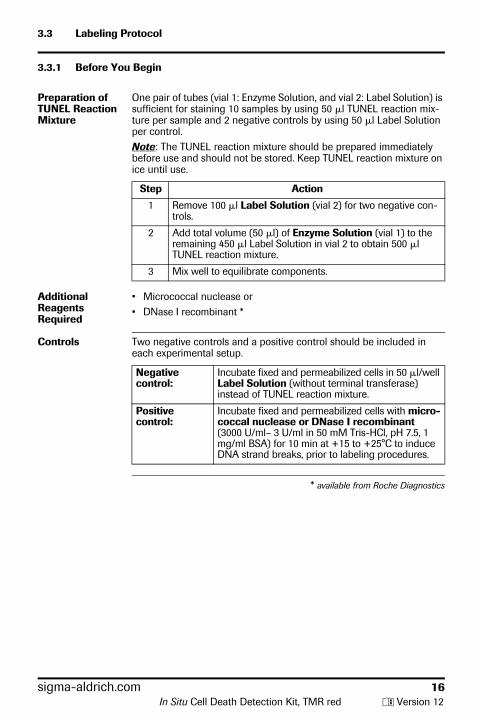
Preparation of TUNEL Reaction Mixture
One pair of tubes (vial 1: Enzyme Solution, and vial 2: Label Solution) is sufficient for staining 10 samples by using 50 �l TUNEL reaction mix-ture per sample and 2 negative controls by using 50 �l Label Solution per control.Note: The TUNEL reaction mixture should be prepared immediately before use and should not be stored. Keep TUNEL reaction mixture on ice until use.
Step Action
1 Remove 100 �l Label Solution (vial 2) for two negative con-trols.
2 Add total volume (50 �l) of Enzyme Solution (vial 1) to the remaining 450 �l Label Solution in vial 2 to obtain 500 �l TUNEL reaction mixture.
3 Mix well to equilibrate components.
Additional Reagents Required
• Micrococcal nuclease or • DNase I recombinant *
Controls Two negative controls and a positive control should be included in each experimental setup.
Negative control:
Incubate fixed and permeabilized cells in 50 �l/well Label Solution (without terminal transferase) instead of TUNEL reaction mixture.
Positive control:
Incubate fixed and permeabilized cells with micro-coccal nuclease or DNase I recombinant (3000 U/ml– 3 U/ml in 50 mM Tris-HCl, pH 7.5, 1 mg/ml BSA) for 10 min at +15 to +25°C to induce DNA strand breaks, prior to labeling procedures.
* available from Roche Diagnostics
In Situ Cell Death Detection Kit, TMR red y Version 12
sigma-aldrich.com 17
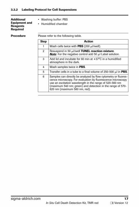
3.3.2 Labeling Protocol for Cell Suspensions
Additional Equipment and Reagents Required
• Washing buffer: PBS• Humidified chamber
Procedure Please refer to the following table.
Step Action
1 Wash cells twice with PBS (200 �l/well).
2 Resuspend in 50 �l/well TUNEL reaction mixture.Note: For the negative control add 50 �l Label solution.
3 Add lid and incubate for 60 min at +37°C in a humidified atmosphere in the dark.
4 Wash samples twice in PBS.
5 Transfer cells in a tube to a final volume of 250-500 �l in PBS.
6 Samples can directly be analyzed by flow cytometry or fluores-cence microscopy. For evaluation by fluorescence microscopy use an excitation wavelength in the range of 520-560 nm (maximum 540 nm; green) and detection in the range of 570-620 nm (maximum 580 nm, red).
In Situ Cell Death Detection Kit, TMR red y Version 12
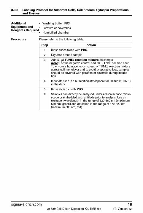
3.3.3 Labeling Protocol for Adherent Cells, Cell Smears, Cytospin Preparations, and Tissues
Additional Equipment and Reagents Required
• Washing buffer: PBS• Parafilm or coverslips• Humidified chamber
Procedure Please refer to the following table.
Step Action
1 Rinse slides twice with PBS.
2 Dry area around sample.
3 Add 50 �l TUNEL reaction mixture on sample.Note: For the negative control add 50 �l Label solution each. To ensure a homogeneous spread of TUNEL reaction mixture across cell monolayer and to avoid evaporative loss, samples should be covered with parafilm or coverslip during incuba-tion.
4 Incubate slide in a humidified atmosphere for 60 min at +37°C in the dark.
5 Rinse slide 3× with PBS.
6 Samples can directly be analysed under a fluorescence micro-scope or embedded with antifade prior to analysis. Use an excitation wavelength in the range of 520-560 nm (maximum 540 nm; green) and detection in the range of 570-620 nm (maximum 580 nm, red).
sigma-aldrich.com 18In Situ Cell Death Detection Kit, TMR red y Version 12
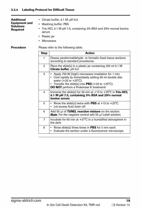
3.3.4 Labeling Protocol for Difficult Tissue
Additional Equipment and Solutions Required
• Citrate buffer, 0.1 M, pH 6.0.• Washing buffer: PBS• Tris-HCl, 0.1 M pH 7.5, containing 3% BSA and 20% normal bovine
serum• Plastic jar• Microwave
Procedure Please refer to the following table.
Step Action
1 Dewax paraformaldehyde- or formalin-fixed tissue sections according to standard procedures.
2 Place the slide(s) in a plastic jar containing 200 ml 0.1 M Citrate buffer, pH 6.0.
3 • Apply 750 W (high) microwave irradiation for 1 min.• Cool rapidly by immediately adding 80 ml double dist.
water (+20 to +25°C).• Transfer the slide(s) into PBS (+20 to +25°C).DO NOT perform a Proteinase K treatment!
4 Immerse the slide(s) for 30 min at +15 to +25°C in Tris-HCl, 0.1 M pH 7.5, containing 3% BSA and 20% normal bovine serum.
5 • Rinse the slide(s) twice with PBS at +15 to +25°C.• Let excess fluid drain off.
6 Add 50 �l of TUNEL reaction mixture on the section.Note: For the negative control add 50 �l Label solution.
7 Incubate for 60 min at +37°C in a humidified atmosphere in the dark.
8 • Rinse slide(s) three times in PBS for 5 min each.• Evaluate the section under a fluorescence microscope.
sigma-aldrich.com 19In Situ Cell Death Detection Kit, TMR red y Version 12
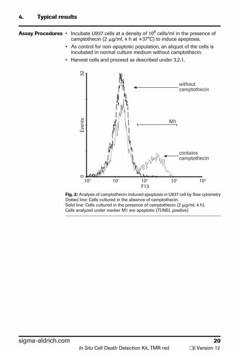
4. Typical results
Assay Procedures • Incubate U937 cells at a density of 106 cells/ml in the presence of camptothecin (2 �g/ml, 4 h at +37°C) to induce apoptosis.
• As control for non-apoptotic population, an aliquot of the cells is incubated in normal culture medium without camptothecin.
• Harvest cells and proceed as described under 3.2.1.
Fig. 2: Analysis of camptothecin induced apoptosis in U937 cell by flow cytometryDotted line: Cells cultured in the absence of camptothecin.Solid line: Cells cultured in the presence of camptothecin (2 �g/ml, 4 h).Cells analyzed under marker M1 are apoptotic (TUNEL positive)
sigma-aldrich.com 20In Situ Cell Death Detection Kit, TMR red y Version 12
5. Appendix
5.1 Troubleshooting
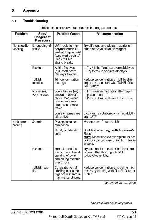
This table describes various troubleshooting parameters.
Problem Step/Reagent of Procedure
Possible Cause Recommendation
Nonspecificlabeling
Embedding of tissue
UV-irradiation forpolymerization of embedding material (e.g., methacrylate) leads to DNA strand breaks
Try different embedding material or different polymerization reagent.
Fixation Acidic fixatives (e.g., methacarn, Carnoy’s fixative)
• Try 4% buffered paraformaldehyde.• Try formalin or glutaraldehyde.
TUNEL reaction
TdT concentration too high
Reduce concentration of TdT by dilu-ting it 1:2 up to 1:10 with TUNEL Dilu-tion Buffer*.
Nucleases, Polymerases
Some tissues (e.g., smooth muscles) show DNA strand breaks very soon after tissue prepa-ration.
• Fix tissue immediately after organ preparation.
• Perfuse fixative through liver vein.
Some enzymes are still active.
Block with a solution containing ddUTP and dATP.
High back-ground
Sample Mycoplasma con-tamination
Mycoplasma Detection Kit*
Highly proliferating cells
Double staining, e.g., with Annexin-V-Fluos*.Note: Measuring via microplate reader not possible because of too high back-ground.
Fixation Formalin fixation leads to a yellowish staining of cells containing melanin precursors.
Try methanol for fixation but take into account that this might lead to reduced sensitivity.
TUNEL reac-tion
Concentration of labeling mix is too high for research in mamma carcinoma.
Reduce concentration of labeling mix to 50% by diluting with TUNEL Dilution Buffer.
continued on next page
* available from Roche Diagnostics
sigma-aldrich.com 21In Situ Cell Death Detection Kit, TMR red y Version 12
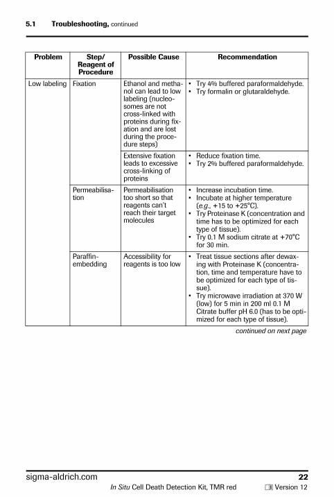
5.1 Troubleshooting, continued
Low labeling Fixation Ethanol and metha-nol can lead to low labeling (nucleo-somes are not cross-linked with proteins during fix-ation and are lost during the proce-dure steps)
• Try 4% buffered paraformaldehyde.• Try formalin or glutaraldehyde.
Extensive fixation leads to excessive cross-linking of proteins
• Reduce fixation time.• Try 2% buffered paraformaldehyde.
Permeabilisa-tion
Permeabilisation too short so that reagents can’t reach their target molecules
• Increase incubation time.• Incubate at higher temperature
(e.g., +15 to +25°C).• Try Proteinase K (concentration and
time has to be optimized for each type of tissue).
• Try 0.1 M sodium citrate at +70°C for 30 min.
Paraffin-embedding
Accessibility for reagents is too low
• Treat tissue sections after dewax-ing with Proteinase K (concentra-tion, time and temperature have to be optimized for each type of tis-sue).
• Try microwave irradiation at 370 W (low) for 5 min in 200 ml 0.1 M Citrate buffer pH 6.0 (has to be opti-mized for each type of tissue).
continued on next page
Problem Step/Reagent of Procedure
Possible Cause Recommendation
sigma-aldrich.com 22In Situ Cell Death Detection Kit, TMR red y Version 12
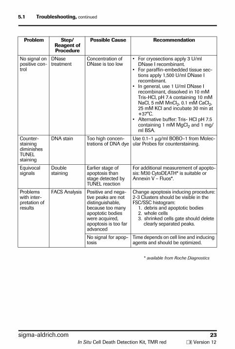
5.1 Troubleshooting, continued
No signal on positive con-trol
DNase treatment
Concentration of DNase is too low
• For cryosections apply 3 U/ml DNase I recombinant.
• For paraffin-embedded tissue sec-tions apply 1,500 U/ml DNase I recombinant.
• In general, use 1 U/ml DNase I recombinant, dissolved in 10 mM Tris-HCl, pH 7.4 containing 10 mM NaCl, 5 mM MnCl2, 0.1 mM CaCl2, 25 mM KCl and incubate 30 min at +37°C.
• Alternative buffer: Tris- HCl pH 7.5 containing 1 mM MgCl2 and 1 mg/ml BSA.
Counter-staining diminishes TUNEL staining
DNA stain Too high concen-trations of DNA dye
Use 0.1–1 �g/ml BOBO–1 from Molec-ular Probes for counterstaining.
Equivocal signals
Double staining
Earlier stage of apoptosis than stage detected by TUNEL reaction
For additional measurement of apopto-sis: M30 CytoDEATH* is suitable or Annexin V – Fluos*.
Problems with inter-pretation of results
FACS Analysis Positive and nega-tive peaks are not distinguishable, because too many apoptotic bodies were acquired, apoptosis is too far advanced
Change apoptosis inducing procedure:2-3 Clusters should be visible in the FSC/SSC histogram:
1. debris and apoptotic bodies2. whole cells3. shrinked cells gate should delete
clearly separated peaks.
No signal for apop-tosis
Time depends on cell line and inducing agents and should be optimized.
Problem Step/Reagent of Procedure
Possible Cause Recommendation
* available from Roche Diagnostics
sigma-aldrich.com 23In Situ Cell Death Detection Kit, TMR red y Version 12
5.2 References
1 Gavrieli, Y., Sherman, Y. & Ben-Sasson, S. A. (1992) J. Cell Biol. 119, 493-501.
2 Gorczyca, W., Gong, J. & Darzynkiewicz, Z. (1993) Cancer Res. 53, 1945-1951.
3 Gorczyca, W. et al. (1993) Leukemia 7, 659-670.4 Gold, R. et al. (1994) Lab. Invest. 71, 2195 Gorczyca, W. et al. (1994) Cytometry 15, 169-175.6 Sgonc, R. et al. (1994) Ternds Genetics 10, 41-42.7 Schmied, M. et al. (1993) Am. J. Pathol. 143, 446-452.8 Wyllie, A. H. et al. (1980) Int. Rev. Cytol. 68, 251.9 Kerr, J. F. R. et al. (1972) Br. J. Cancer 26, 239-257.10 Duvall, E. & Wyllie, A. H. (1986) Immunol. Today 7, 115.11 Compton, M. M. (1992) Canc. Metastasis Rev.11, 105-119.12 Allen, P. D., Bustin, S. A. & Newland, A. C. (1993) Blood Reviews 7,
63-73.13 Cohen, J. J. & Duke, R. C. (1992) Annu. Rev. Immunol. 10, 267-293.14 Clarke, P. G. H. (1990) Anat. Embryol. 181, 195-213.15 Johnson, E. M. & Deckwerth, T. L. (1993) Annu. Rev. Neurosci. 16,
31-46.16 Batistatou, A. & Greene, L. A. (1993) J. Cell Biol. 122, 523-532.17 Strange, R. et al. (1992) Development 115, 49-58.18 Carson, D. A. & Ribeiro, J. M. (1993) Lancet 341, 1251-1254.19 Edgington, S. M. (1993) Biotechnology 11, 787-792.20 Gougeon. M.-L. & Montagnier, L. (1993) Science 260, 1269-1270.21 Hickman, J. A. (1992) Cancer Metastasis Rev. 11, 121-139.22 Afanasyev, V. N. et al. (1993) Cytometry 14, 603-609.23 Bryson, G. J., Harmon, B. V. & Collins, R. J. (1994) Immunol. Cell
Biology 72, 35-41.24 Darzynkiewicz, Z. et al. (1992) Cytometry 13, 795-808.25 Ando, K. et al. (1994) J. Immunol. 152, 3245-3253.26 Berges, R. R. et al. (1993) Proc. Natl. Acad. Sci. USA 90, 8910-
8914.27 Gorczyca, W. et al. (1992) Int. J. Oncol. 1, 639-648.28 Gorczyca, W. et al. (1993) Exp. Cell Res. 207, 202-205.29 Billig, H., Furuta, I. & Hsueh, A. J. W. (1994) Endocrinology 134,
245-252.30 MacManus, J. P. et al. (1993) Neurosci. Lett. 164, 89-92.31 Mochizuki, H. et al. (1994) Neurosci. Lett. 170, 191-194.32 Oberhammer, F. et al. (1993) Hepatology 18, 1238-1246.33 Portera-Cailliau, C. (1994) Proc. Natl. Acad. Sci. USA 91, 974-978.34 Preston, G. A. et al. (1994) Cancer Res. 54, 4214-4223.35 Weller, M. et al. (1994) Eur. J. Immunol. 24, 1293-1300.36 Zager, R.A. et al. (1994) J. Am. Soc. Nephrol. 4, 1588-1597.37 Cohen, G. M. et al. (1992) Biochem. J. 286, 331-334.38 Collins, R. J. et al. (1992) Int. J. Rad. Biol. 61, 451-453.39 Sei, Y. et al. (1994) Neurosci. Lett. 171, 179-182.40 Ansari, B. et al. (1993) J. Pathol. 170, 1-8.41 Negoescu, A. et.al. (1998) Biochemica 3, 34-41
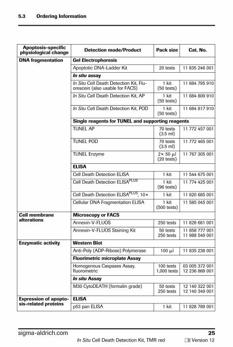
5.3 Ordering Information
Apoptosis-specific physiological change Detection mode/Product Pack size Cat. No.
DNA fragmentation Gel Electrophoresis
Apoptotic DNA-Ladder Kit 20 tests 11 835 246 001
In situ assay
In Situ Cell Death Detection Kit, Flu-orescein (also usable for FACS)
1 kit (50 tests)
11 684 795 910
In Situ Cell Death Detection Kit, AP 1 kit (50 tests)
11 684 809 910
In Situ Cell Death Detection Kit, POD 1 kit (50 tests)
11 684 817 910
Single reagents for TUNEL and supporting reagents
TUNEL AP 70 tests (3.5 ml)
11 772 457 001
TUNEL POD 70 tests (3.5 ml)
11 772 465 001
TUNEL Enzyme 2× 50 �l (20 tests)
11 767 305 001
ELISA
Cell Death Detection ELISA 1 kit 11 544 675 001
Cell Death Detection ELISAPLUS 1 kit (96 tests)
11 774 425 001
Cell Death Detection ELISAPLUS, 10× 1 kit 11 920 685 001
Cellular DNA Fragmentation ELISA 1 kit (500 tests)
11 585 045 001
Cell membrane alterations
Microscopy or FACS
Annexin-V-FLUOS 250 tests 11 828 681 001
Annexin-V-FLUOS Staining Kit 50 tests250 tests
11 858 777 00111 988 549 001
Enzymatic activity Western Blot
Anti-Poly (ADP-Ribose) Polymerase 100 �l 11 835 238 001
Fluorimetric microplate Assay
Homogenous Caspases Assay, fluorometric
100 tests1,000 tests
03 005 372 00112 236 869 001
In situ Assay
M30 CytoDEATH (formalin grade) 50 tests250 tests
12 140 322 00112 140 349 001
Expression of apopto-sis-related proteins
ELISA
p53 pan ELISA 1 kit 11 828 789 001
sigma-aldrich.com 25In Situ Cell Death Detection Kit, TMR red y Version 12
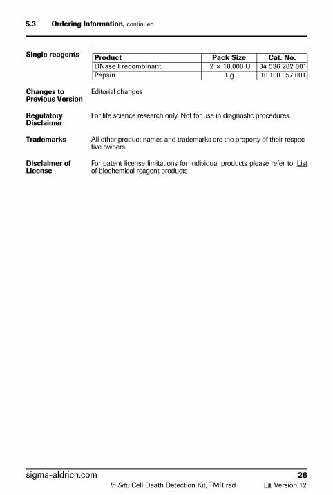
5.3 Ordering Information, continued
Single reagents Product Pack Size Cat. No.DNase I recombinant 2 × 10,000 U 04 536 282 001Pepsin 1 g 10 108 057 001
Changes to Previous Version
Editorial changes
Regulatory Disclaimer
For life science research only. Not for use in diagnostic procedures.
Trademarks All other product names and trademarks are the property of their respec-tive owners.
Disclaimer of License
For patent license limitations for individual products please refer to: List of biochemical reagent products
sigma-aldrich.com 26In Situ Cell Death Detection Kit, TMR red y Version 12

sigma-aldrich.com 27
In Situ Cell Death Detection Kit, TMR red y Version 12
Contact and Support
If you have questions or experience problems with this or any Roche product for Life Science, please contact our Technical Support staff. Our scientists are committed to providing rapid and effective help.Please also contact us if you have suggestions for enhancing Roche product performance or using our products in new or specialized ways. Such customer information has repeatedly proven invaluable to the research community worldwide.
To ask questions, solve problems, suggest enhancements or report new applications, please visit our Online Technical Support Site.
Visit sigma-aldrich.com, to download or request copies of the following materials.
• Instructions for Use• Safety Data Sheets• Certificates of Analysis• Information Material
To call, write, fax, or email us, visit sigma-aldrich.com, and select your home country to display country-specific contact information.
Roche Diagnostics GmbH Sandhofer Strasse 116 68305 Mannheim Germany
0316
.120
3802
10012


![Six Sigma (6 Sigma)[1]](https://img.pdfslide.us/doc/110x75/577d35cc1a28ab3a6b91711a/six-sigma-6-sigma1.jpg)