Embed Size (px)
Citation preview
Examination of sex differences in quadriceps fatigability and Hsp70 content
in response to intense intermittent isometric exercise
by
Andrew C. Hopf
A thesis
presented to the University of Waterloo
in fulfilment of the
thesis requirement for the degree of
Master of Science
in
Kinesiology
Waterloo, Ontario, Canada, 2009
© Andrew C. Hopf 2009

Authors Declaration
I hereby declare that I am the sole author of this thesis. This is a true copy of the thesis, including any required final revisions, as accepted by my examiners. I understand that my thesis may be made electronically available to the public.
Andrew C. Hopf
ii
Abstract
The purpose of this study was to determine if there are sex differences in induced heat
shock protein 70 (Hsp70) expression in human skeletal muscle under basal conditions
and in response to intense intermittent isometric exercise. Furthermore, this study
examined potential sex differences in muscle fatigability and sarcoplasmic reticulum (SR)
function for up to 9 days following the bout of exercise. In total, 6 male (20 ± 0.5 years
of age, 70.88 ± 10.25 kg, mean ± SE) and 6 female participants (19 ± 0.25 years of age,
58.02 ± 5.82 kg, mean ± SE) were recruited for this study to do one legged intermittent
isometric exercise with a 50% duty cycle (5 sec contraction: 5 sec relaxation) at 60% of
their maximal voluntary contraction (MVC) for 30 minutes. Muscle biopsies, blood
samples and muscle stimulation measurements were taken prior to starting exercise for
assessment of baseline values. These same measures were taken immediately POST
exercise and at 24(R1), 72(R3), 144(R6) and 216(R9) hours following the exercise.
Muscle samples were analyzed for exercise and recovery response of Hsp70,
sarco(endo)plasmic reticulum Ca2+ATPase (SERCA)1 and SERCA2 protein content, as
well as measurements of maximal Ca2+ ATPase activity and Ca2+ uptake. Blood
samples were also analyzed for serum estrogen and creatine kinase concentrations. The
results from this study show that there are no differences in basal Hsp70 protein content
between males and females, and that females have a blunted (no increase up to 9 days
post exercise) Hsp70 response following a bout of intense exercise in comparison to
males who had a robust response. Immediately following exercise females had smaller
decrements in MVC and electrically stimulated force (10 and 100Hz). It was also found
iii
that at low frequencies of stimulation (10Hz), females were able to recover at a quicker
rate than males. There was no evidence that the decrements in force or the differences in
recovery time between males and females were due to alterations in SERCA protein
content or function. This thesis is the first study in humans to show that there is sexual
dimorphism in the exercise induced Hsp70 response to exercise.
iv

Acknowledgements
First and foremorst I must thank my family. They have always been behind me
100%. Without their help there is no way I would be here right now.
Secondly, I have to thank Dr. Russ Tupling and his team of researchers, who have
taught me the skills and knowledge needed to make this thesis possible.
I would also like to thank my supervising committee (Dr. Ken Stark and Dr.
Marnia Mourtzakis) who “raised the bar” for my academic writing and planning.
v

Dedication
I would like to dedicate this thesis to the entire Hopf family, whose love and support
helped me through my academic and professional development.
vi
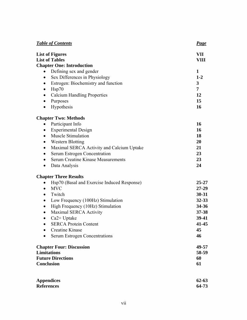
Table of Contents Page
List of Figures VII List of Tables VIII Chapter One: Introduction
• Defining sex and gender 1 • Sex Differences in Physiology 1-2 • Estrogen: Biochemistry and function 3 • Hsp70 7 • Calcium Handling Properties 12 • Purposes 15 • Hypothesis 16
Chapter Two: Methods • Participant Info 16 • Experimental Design 16 • Muscle Stimulation 18 • Western Blotting 20 • Maximal SERCA Activity and Calcium Uptake 21 • Serum Estrogen Concentration 23 • Serum Creatine Kinase Measurements 23 • Data Analysis 24
Chapter Three Results • Hsp70 (Basal and Exercise Induced Response) 25-27 • MVC 27-29 • Twitch 30-31 • Low Frequency (100Hz) Stimulation 32-33 • High Frequency (10Hz) Stimulation 34-36 • Maximal SERCA Activity 37-38 • Ca2+ Uptake 39-41 • SERCA Protein Content 41-45 • Creatine Kinase 45 • Serum Estrogen Concentrations 46
Chapter Four: Discussion 49-57 Limitations 58-59 Future Directions 60 Conclusion 61 Appendices 62-63 References 64-73
vii
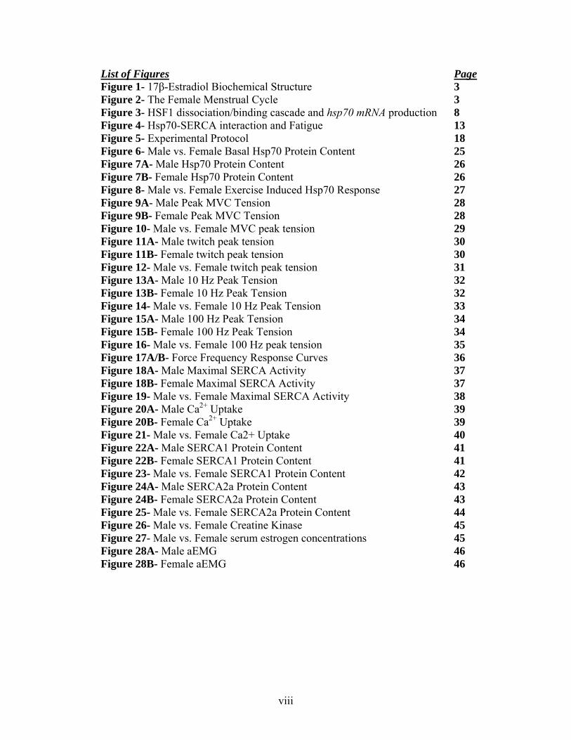
List of Figures PageFigure 1- 17β-Estradiol Biochemical Structure 3 Figure 2- The Female Menstrual Cycle 3 Figure 3- HSF1 dissociation/binding cascade and hsp70 mRNA production 8 Figure 4- Hsp70-SERCA interaction and Fatigue 13 Figure 5- Experimental Protocol 18 Figure 6- Male vs. Female Basal Hsp70 Protein Content 25 Figure 7A- Male Hsp70 Protein Content 26 Figure 7B- Female Hsp70 Protein Content 26 Figure 8- Male vs. Female Exercise Induced Hsp70 Response 27 Figure 9A- Male Peak MVC Tension 28 Figure 9B- Female Peak MVC Tension 28 Figure 10- Male vs. Female MVC peak tension 29 Figure 11A- Male twitch peak tension 30 Figure 11B- Female twitch peak tension 30 Figure 12- Male vs. Female twitch peak tension 31 Figure 13A- Male 10 Hz Peak Tension 32 Figure 13B- Female 10 Hz Peak Tension 32 Figure 14- Male vs. Female 10 Hz Peak Tension 33 Figure 15A- Male 100 Hz Peak Tension 34 Figure 15B- Female 100 Hz Peak Tension 34 Figure 16- Male vs. Female 100 Hz peak tension 35 Figure 17A/B- Force Frequency Response Curves 36 Figure 18A- Male Maximal SERCA Activity 37 Figure 18B- Female Maximal SERCA Activity 37 Figure 19- Male vs. Female Maximal SERCA Activity 38 Figure 20A- Male Ca2+ Uptake 39 Figure 20B- Female Ca2+ Uptake 39 Figure 21- Male vs. Female Ca2+ Uptake 40 Figure 22A- Male SERCA1 Protein Content 41 Figure 22B- Female SERCA1 Protein Content 41 Figure 23- Male vs. Female SERCA1 Protein Content 42 Figure 24A- Male SERCA2a Protein Content 43 Figure 24B- Female SERCA2a Protein Content 43 Figure 25- Male vs. Female SERCA2a Protein Content 44 Figure 26- Male vs. Female Creatine Kinase 45 Figure 27- Male vs. Female serum estrogen concentrations 45 Figure 28A- Male aEMG 46 Figure 28B- Female aEMG 46
viii

List of Tables Page
• Table 1 – Twitch Kinetics (+dF/dt) 31
• Table 2 – Twitch Kinetics (-dF/dt) 31
ix
Chapter 1: Introduction
The adult human being is comprised of a series of complex physiological systems.
As humans progress through life, it is well documented that males and females have
numerous differences in structural, functional and psychological physiology (Denton et al,
2007, Wheeler et al., 1991, Tarnopolsky et al, 2007). The term sexual dimorphism has
commonly been used to define a scenario where genetic or biological differences exist
between sexes (Nickerson et al, 2006). In comparison, the term gender refers to the
sociological status of male and females and their place in society and should not be used
to describe biological differences between males and females. Examples of physiological
sexual dimorphism that are well established include differences in muscle mass and
strength (Perez-Gomez et al, 2008), fibre type distribution (Komi et al, 1978), and
contractile properties (Clark et al, 2003). Overall it appears that adult females have 40%
less overall strength and significantly less muscle cross sectional area, when compared to
males (Komi et al., 1978). Females may also be more susceptible to decrements in
muscle mass and strength with aging compared to males (Mazaretti et al., 2009). This
process is referred to as sarcopenia and it may have functional consequences as older
adults lose their ability to be independent (Mazaretti et al, 2009, Lee et al., 2007 and
Doherty et al., 2003). Another example is the difference between males and females in
skeletal muscle fatigability, as females may be more fatigue resistant than males although
this finding is inconsistent (Clarke et al., 2005; Tiidus et al., 2007). The differences in
development and physiology are not just limited to the skeletal muscle. The risk of
developing pathologies such as osteoporosis and coronary artery disease also appear to be
sex dependent (Reviewed by Polk et al., 2005 and Balasch et al, 2003).
1
Role of Estrogen in Skeletal Muscle Physiology
The distinct differences between sexes have led researchers to examine potential
biological mechanisms underlying the sexual dimorphism commonly observed in disease
and function. The ovarian sex hormone estrogen, (17β-estradiol), and its effects on
various physiological pathways in the human body have been proposed as one of the
primary mechanisms behind the differences between sexes in muscle metabolism
(Tarnopolsky et al, 2008, Paroo et al, 2002 and Tiidus et al., 2009). Estrogen (or 17β-
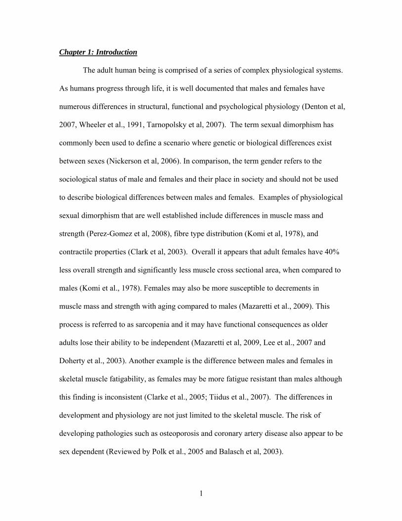
estradiol) is a four ringed steroidal hormone (Figure 1) produced by developing granulosa
cells in the ovaries of females. Estrogen is a molecule that contains an extra hydroxyl that
may serve as a natural reducing agent in the muscle cell (Knowlton et al 2005). As a
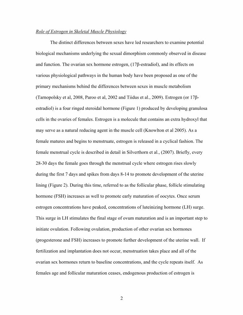
female matures and begins to menstruate, estrogen is released in a cyclical fashion. The
female menstrual cycle is described in detail in Silverthorn et al., (2007). Briefly, every
28-30 days the female goes through the menstrual cycle where estrogen rises slowly
during the first 7 days and spikes from days 8-14 to promote development of the uterine
lining (Figure 2). During this time, referred to as the follicular phase, follicle stimulating
hormone (FSH) increases as well to promote early maturation of oocytes. Once serum
estrogen concentrations have peaked, concentrations of luteinizing hormone (LH) surge.
This surge in LH stimulates the final stage of ovum maturation and is an important step to
initiate ovulation. Following ovulation, production of other ovarian sex hormones
(progesterone and FSH) increases to promote further development of the uterine wall. If
fertilization and implantation does not occur, menstruation takes place and all of the
ovarian sex hormones return to baseline concentrations, and the cycle repeats itself. As
females age and follicular maturation ceases, endogenous production of estrogen is
2
greatly reduced. This transition is called menopause and is associated with many changes
to the female body (Copeland et al, 2004).
Figure 1: 17B-Estradiol (Estrogen) Biochemical Structure: http://healthpsych.psy.vanderbilt.edu/SoyBreastCancer_files/image002.gif
Figure 2: The female menstrual cycle- This figure illustrates the typical patterns of ovarian sex hormone release into the blood stream throughout the 28 day cycle. http://en.wikipedia.org/wiki/Menstrual_cycle
3
As a result, females may supplement estrogen via oral or transdermal methods to
maintain their circulating estrogen levels to help offset some of the complications known
to be caused by a lack of the hormone. Estrogen is also responsible for the development
of secondary sex characteristics in females, as well as numerous other physiological
processes such as bone and muscle metabolism (Ropero et al., 2007 and Tarnopolsky et
al, 2007). Estrogen is also known to influence the function of organs throughout the body
including bone, brain, heart and skeletal muscle (Leung et al, 2007, Polk et al., 2005 and
Balasch et al, 2003). LH, progesterone, and FSH may also contributed to sexual
dimorphism in neural tissue (Mitsushima et al, 2003 and Gonzalez-Hernadez et al, 2000),
but very little research has been done on the role of these hormones in skeletal muscle
physiology. The remainder of this thesis will focus on the role that estrogen plays in
sexual dimorphism involving skeletal muscle physiology.
Recently it has been shown that skeletal muscle expresses estrogen receptors (ERs)
(Stice et al., 2008). Skeletal muscle has two types of ERs; ERα and ERβ. These ERs are
responsible for transducing an extracellular signal to modulate various intracellular
signalling pathways. One of the primary effects of estrogen in skeletal muscle, which
appears to be acting primarily through the NFκβ pathway, is to upregulate various stress
proteins and possibly attenuate the inflammatory processes caused as a result of various
physiological stressors such as exercise (Paroo et al, 2002, Voss et al, 2003 and
Knowlton et al., 2001), ischemia reperfusion, acidosis and hyperthermia (Knowlton et al.,
2001 and Stice et al., 2008). Furthermore, estrogen has been described as a membrane
stabilizing molecule (Tiidus et al., 2003). Due to its lipophilic properties, estrogen has the
ability to interact with the phospholipid bilayer of cells and possibly protect them from
4
damaging stressors. Cumulatively, these properties of estrogen suggest that females may
be less susceptible to stress and better able to retain function or be more resistant to
fatigue compared to males. One of the primary proteins associated in the stress response
in Heat Shock Protein 70 (Hsp70). Therefore, it stands to reason that the Hsp70 response
may display sexual dimorphism following periods of stress.
Heat Shock Proteins and the cellular stress response
Heat Shock Proteins (HSPs) are a family of highly conserved proteins that are
upregulated in response to various physiological insults to a cell and are ubiquitous
among all mammalian species (as reviewed by Noble et al., 2007). The first HSP was
discovered in 1962 when Drosophila Melanogaster larvae were exposed to an intense
heat stress. The resulting upregulation of these proteins in response to heat is what gave
them the name “Heat Shock Proteins”. Much research has been conducted in the over
forty plus years following the first discovery and there is now evidence to suggest that
HSPs function to protect cells against a variety of biological and environmental stresses.
There are many different isoforms of the HSPs including Hsp27, Hsp32, Hsp70 and
Hsp90 to name just a few, which are named based upon their molecular weights.
There are only a limited number of studies investigating the role of these proteins
in human skeletal muscle physiology and the majority of studies have focused on Hsp70.
Hsp70 is highly inducible and rapidly upregulated when cells are exposed to various
forms of stress such as exercise (Noble et al., 2007 and Locke et al., 1991), hyperthermia
(Morton et al., 2007), hypoxia (Iwaki et al, 1993) and reactive oxygen species
(Madamanchi et al, 2001). Hsp70 has been shown to be responsible for folding
un/misfolded proteins, chaperoning newly synthesized proteins to their target location
5
and preventing damaged proteins from aggregating which would render them
unfunctional (Lui et al., 2006). There are two types of Hsp70 in skeletal muscle, a non-
inducible isoform present under basal conditions, termed heat shock cognate 70 (Hsc70)
and an inducible form (Hsp70) which is up regulated in response to myocellular stress
(Reviewed in Noble et al., 2007). This isoform is also commonly referred to as Hsp72 in
the literature based on its molecular weight and difficulty distinguishing it from Hsp70
during western blotting procedures (Noble et al, 2007). The remainder of this thesis will
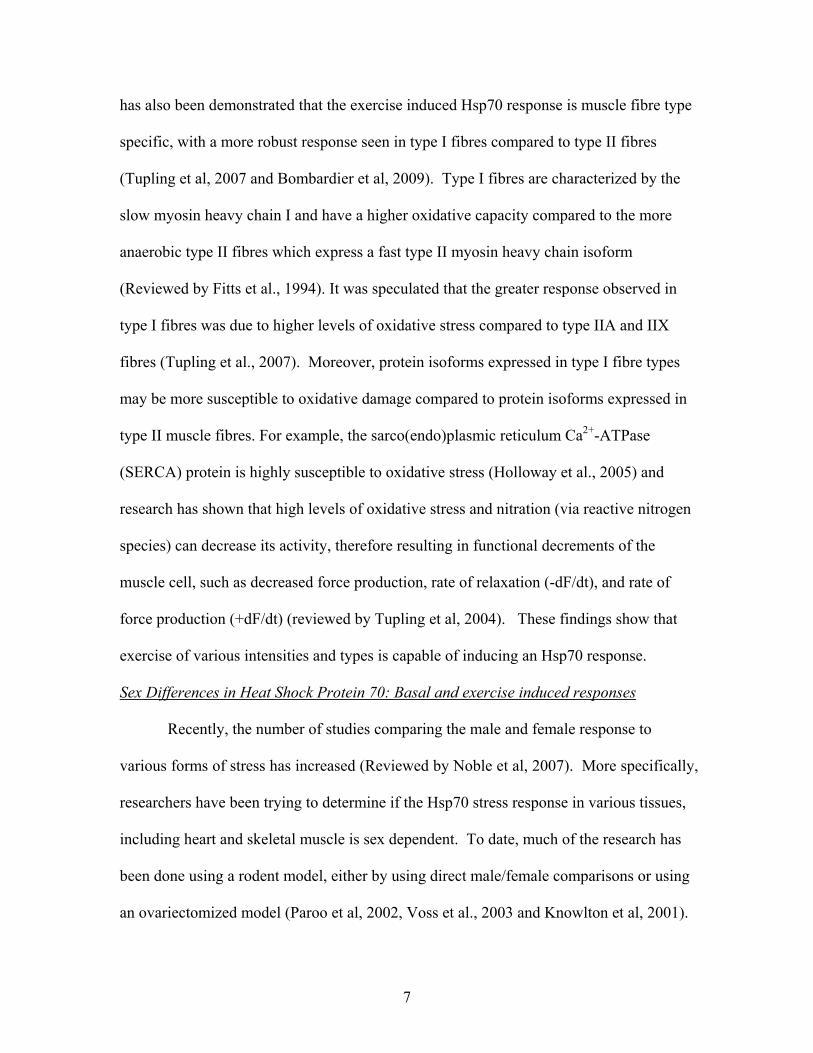
focus on the inducible form, Hsp70. Under basal, unstressed conditions, Hsp70 molecules
are bound to a transcription factor named heat shock factor 1 (HSF1) in the cytosol of a
cell. When a physiologic stress is imposed upon the muscle and oxidative damage occurs,
causing protein denaturation, Hsp70 identifies hydrophobic residues of denatured
proteins, dissociates from HSF1 and binds to denatured proteins (Noble et al., 2007 and
Knowlton et al., 2001). Unbound HSF1 then trimerizes, becomes phosphorylated and
translocates to the nucleus. A “transcriptional complex” then forms and gains the ability
to bind with the transcriptional factor Heat Shock Element (HSE), causing the
upregulation of hsp70 messenger ribonucleic acid (mRNA) (Figure 3). This cycle will
continue to occur until the stimulus to induce the Hsp70 response has ended (Noble et al.,
2007).
Numerous studies have shown that exercise can directly increase the Hsp70
content in the muscle cell (Noble et al., 2006, Tupling et al., 2007, Paroo et al, 2002 and
Punschart et al, 1996). The extent of the Hsp70 response may be accentuated with
exercise of longer duration (Febbraio et al., 2000), higher intensity (Lui et al., 1999) and
using a more eccentric type of exercise compared to concentric (Paulsen et al., 2007). It
6
has also been demonstrated that the exercise induced Hsp70 response is muscle fibre type
specific, with a more robust response seen in type I fibres compared to type II fibres
(Tupling et al, 2007 and Bombardier et al, 2009). Type I fibres are characterized by the
slow myosin heavy chain I and have a higher oxidative capacity compared to the more
anaerobic type II fibres which express a fast type II myosin heavy chain isoform
(Reviewed by Fitts et al., 1994). It was speculated that the greater response observed in
type I fibres was due to higher levels of oxidative stress compared to type IIA and IIX
fibres (Tupling et al., 2007). Moreover, protein isoforms expressed in type I fibre types
may be more susceptible to oxidative damage compared to protein isoforms expressed in
type II muscle fibres. For example, the sarco(endo)plasmic reticulum Ca2+-ATPase
(SERCA) protein is highly susceptible to oxidative stress (Holloway et al., 2005) and
research has shown that high levels of oxidative stress and nitration (via reactive nitrogen
species) can decrease its activity, therefore resulting in functional decrements of the
muscle cell, such as decreased force production, rate of relaxation (-dF/dt), and rate of
force production (+dF/dt) (reviewed by Tupling et al, 2004). These findings show that
exercise of various intensities and types is capable of inducing an Hsp70 response.
Sex Differences in Heat Shock Protein 70: Basal and exercise induced responses
Recently, the number of studies comparing the male and female response to
various forms of stress has increased (Reviewed by Noble et al, 2007). More specifically,
researchers have been trying to determine if the Hsp70 stress response in various tissues,
including heart and skeletal muscle is sex dependent. To date, much of the research has
been done using a rodent model, either by using direct male/female comparisons or using
an ovariectomized model (Paroo et al, 2002, Voss et al., 2003 and Knowlton et al, 2001).
7
Figure 3: HSF1 dissociation/binding cascade and hsp70 mRNA production. Stice et al. (2008) Mol. Med. 14(7-8). This diagram illustrates the events following stress that lead to Hsp70 binding to proteins and the resulting upregulation of hsp70 mRNA. HSF-1-Heat Shock Factor 1; HSE- Heat Shock Elemement.
Ovariectomized rats do not have an endogenous source of estrogen and
researchers use this model to replicate the male response. The ovariectomized rat can
also be supplemented with other ovarian sex hormones via time release pellet or injection,
which will bring the hormone of interest back to physiologic levels. This allows for
assessment of the independent effects of a single ovarian sex hormone (i.e. estrogen) (For
full review see Paroo et al, 2002 or Bombardier et al, 2009). Another less commonly
used method utilizes pre/post menopausal women to examine the extent of the changes
which occur once a female loses her ability to produce endogenous sources of estrogen.
Finally, males can be supplemented with an exogenous source of estrogen to test the
effect of an acute estrogen exposure on various physiological pathways. Considering sex
differences in cardiovascular disease is a very popular area of research, a number of
8
studies have examined sexual dimorphism in Hsp70 protein and hsp70 mRNA expression
in cardiac muscle. In contrast, only a few studies have investigated if sexual dimorphism
exists in the Hsp70 stress response in skeletal muscle and these have been limited to
rodent models.
There is evidence suggesting that Hsp70 can be upregulated in heart using various
stresses such as ischemia reperfusion, heat (Mestril et al, 1994), pharmacologic
treatments, or a pre conditioning exercise bout in cardiac muscle (Paroo et al., 2002).
However, following a bout of treadmill exercise, Hsp70 protein content in cardiac muscle
is increased twice as much in males than it is in females. It has also been shown in
rodents that basal Hsp70 levels are higher in females hearts compared to male hearts
(Voss et al., 2003 and Paroo et al., 2002).
Consistent with cardiac muscle, under resting conditions in skeletal muscle there
is some Hsp70 available to cope with low levels of stress. The only study to date that
directly compares male and female rodents has shown that there is no significant
difference in basal Hsp70 protein content (Voss et al., 2003). In comparison, a separate
study using an ovariectomized rodent model (no endogenous estrogen) that is
supplemented with a time released estrogen pellet, showed that rodents with estrogen had
a significantly higher level of basal Hsp70 compared to sham rodents (no estrogen)
(Bombardier et al., 2009). These two studies are the only studies that have examined the
differences in basal Hsp70 content in rodent skeletal muscle and to date no study has
examined this in human skeletal muscle.
The exercise induced Hsp70 response in skeletal muscle is similar to that of
cardiac muscle. To date, research that has been done in rodent models suggests that
9
females have a blunted Hsp70 response following a bout of intense exercise (Paroo et al.,
2002 and Nickerson et al., 2008). Conversely, males display a significant elevation in
Hsp70 following a similar bout of exercise. The blunted Hsp70 response in females also
appears to be mediated by estrogen. Bombardier et al., (2009) showed that soleus muscle
of ovariectomized female rats displayed an exercise induced Hsp70 response similar to
that observed in males of other studies. When these ovariectomized females were
supplemented with estrogen, the Hsp70 response was blunted. A separate study by Paroo
et al., (2002) showed that administering an estrogen receptor blocker (Tamoxifan) to
gonadally intact females, resulted in an Hsp70 response similar to males.
The data showing that females have a significantly higher level of basal Hsp70
and a blunted Hsp70 response to exercise have all been accumulated from rodent studies.
To date, no controlled study using human participants has examined sex differences in
the Hsp70 response to an intense bout of exercise. Since Hsp70 is a cytoprotective
protein capable of binding to and protecting muscle proteins, it stands to reason that
female skeletal muscle function may be better protected than males when exposed to
exercise.
Sexual Dimorphism in Hsp70 and muscle fatigability: Is there a link?
When a contraction cycle is initiated, an action potential is transmitted from the
post synaptic neuron, down the sarcolemma of the muscle. Through activation of the
dihydropyridine receptor and the ryanodine receptor a resulting influx of calcium into the
cytosol of the fibre ensues, causing activation of the contractile apparatus. Once the
stimulus from the neuron has ceased, the SERCA pump acts to sequester Ca2+ ions back
into the sarcoplasmic reticulum, allowing relaxation to occur. The result of numerous
10
sequential contractions is an increase of various metabolic by-products such as hydrogen
ions (Bangsbo et al., 1993), reactive oxygen species (Andrade et al.,2001), lactate, and
inorganic phosphate (Pi) (Murphy et al., 1999). These metabolic by-products and other
structural/functional changes to either the contractile proteins or the proteins involved in
excitation-contraction coupling have been proposed as some of the main contributors to
the development of fatigue.
Fatigue is defined as the inability to produce a desired level of force (Allen et al.,
2007). The mechanistic basis of fatigue is complex and is dependent upon numerous
factors including exercise intensity, contraction type, rest intervals (Sale et al., 1987) and
individual muscle fibre type profile (Essen et al., 1975). Decrements in force are
commonly observed after periods of strenuous activity. In most cases, the ability to
restore its maximal force producing capacity of skeletal muscle is restored within 30
minutes following exercise, depending on the exercise intensity (Baker et al., 1993). In
some cases when the exercise intensity is extreme, although maximal force producing
capacity may recover relatively quick, other indices of fatigue may be present for longer
periods of time (Edwards et al., 1977).
Along with maximal voluntary contraction (MVC) and supramaximal twitch
force, low frequency fatigue (LFF) and high frequency fatigue (HFF) are two
measurements commonly used when assessing fatigue. HFF is a term used to describe
force decrements which occur when a muscle is electrically stimulated at high
frequencies (50 and 100 Hz). It has been suggested that HFF is likely a result of an
impairment of the Na+/K+ ATPase, which is located within the sarcolemma of skeletal
muscle (Fowles et al., 2002). Impairment of the Na+/K+ ATPase would result in an
11
inability to maintain the proper ion gradient needed to repolarize the cell and allow action
potentials to be transmitted down the T-tubules. Low frequency fatigue is a term used to
describe force decrements which occur when a muscle is electrically stimulated at low
frequencies (10 and 20 Hz). It has been suggested that LFF is caused in part by
impairments of calcium release from the SR and decrements in the ability of the SERCA
pumps to sequester Ca2+ back into the lumen of the SR (Tupling et al., 2000, Enns et al.,
1999, Tupling et al., 2004). The inability to sequester Ca2+ back into the SR can result in
prolonged calcium transients within the cytosol of the cell and a loss of Ca2+ homeostasis,
which may activate calpains/apoptotic pathways (Murphy et al., 2006) and decrease the
ability to transmit signals from the t-tubules to the SR to initiate calcium release (as
reviewed by Tupling et al, 2004).. The mechanistic basis of fatigue is complex but clearly
SERCA pumps are extremely important in the performance of skeletal muscle and
decrements in their ability to sequester Ca2+ may result in fatigue.
As previously mentioned, although controversial, there is evidence to suggest that
sex differences exist in skeletal muscle fatigability following intense exercise. More
specifically, females may be more resistant to fatigue when compared to males, in that
they have longer endurance times and less force reduction following a bout of intense
exercise. (Clark et al, 2005, Russ et al, 2003 and Hunter et al, 2001). Most of these
studies utilized a low intensity sub-maximal contraction protocol and measured
endurance time as an indicator of fatigue. In contrast, some evidence suggests that no sex
difference exists in human skeletal muscle fatigability following exercise (Phillips et al.,
2003), which was also replicated in rodent EDL muscles (Tiidus et al, 1999).
12
Hsp70 is known to be a cytoprotective protein that can bind to and protect
functional proteins within the skeletal muscle. Recently it has been shown that Hsp70 has
the ability to bind to SERCA1 (Tupling et al., 2004) and SERCA2a (Fu et al., 2007)
isoforms and prevent thermal deactivation of both isoforms. More specifically, it was
shown that human embryonic kidney cells (HEKs) which were transfected with their
respective SERCA isoform cDNAs and then exposed to a heat stress, had a significant
decrease in maximal SERCA activity; however when these same cells were cultured with
Hsp70, they found that the heat induced decrements in SERCA activity were attenuated.
Therefore, the findings that LFF is, at least in part, caused by decrements in
SERCA pump activity and the notion that Hsp70 is a cytoprotective protein capable of
protecting SERCA pumps against damaging stress, suggests that the sexual dimorphism
observed in skeletal muscle fatigue could be a result of the higher basal Hsp70 levels
observed in females compared to males (Figure 4). This would theoretically allow
females to be more protected and maintain skeletal muscle performance better than males.
Basal Hsp70 SERCA
Fatigue
Figure 4: SERCA:Hsp70 Interaction and Fatigue- illustrates potential interaction between higher basal Hsp70 concentrations, SERCA and fatigue, and suggests Hsp70 may protect SERCA function in females which may help attenuate fatigue. Previous work by Tupling et al (2000 and 2007) using a single legged isometric
knee extension exercise model, where participants contract at 60% of their MVC, has
13
proven to be effective in inducing a robust Hsp70 response in males (Tupling et al, 2007),
as well as post contractile depression (PCD) in both males and females. PCD is defined
as prolonged fatigue at both low and high frequencies of stimulation following numerous
tetanic contractions (Tupling et al, 2000). This model has shown that females display
PCD for up to at least 1 hour following exercise (Tupling et al, 2000 and Fowles et al.,
2002) and that males can display PCD for up to 6 days following the same exercise bout.
Since these studies did not assess the time course of recovery beyond 1 hour in females
(Tupling et al, 2000), it remains to be determined if sex differences exist in the time
course of PCD and when full recovery of force occurs. Moreover, this study did not
assess the Hsp70 response to this type of exercise in females. Therefore the current thesis
will utilize the same exercise protocol to determine if sex differences exist in the time
course of PCD and if differences exist, are they associated with basal and exercise
induced Hsp70 content within human skeletal muscle.
Purposes:
1. To determine if there are sex differences in basal and exercise induced Hsp70
levels in human skeletal muscle.
2. To determine if there are sex differences in skeletal muscle fatigability in
response to an acute bout of intense intermittent isometric exercise in human
quadriceps.
3. To determine if there are sex differences in the effects of exercise on SERCA
pump function in human skeletal muscle.
14
4. To determine if there are sex differences in the time course of recovery for all
variables following intense intermittent isometric exercise.
Hypotheses:
1. Basal Hsp70 levels will be higher in females than males and females will have a
blunted Hsp70 response in comparison to the more robust response displayed by
males.
2. Females will demonstrate less fatigue and recovery muscle contractile function
more quickly than males in response to the intense isometric exercise.
3. Females will have smaller decrements in maximal SERCA activity and Ca2+
uptake following the exercise bout compared with males due expected higher
basal Hsp70 expression in females.
4. Females will recover markers of PCD and SERCA function earlier in the time
course of the study compared to males.
5. Recovery of all parameters will be complete within 9 days following the exercise
in both males and females.
15
Chapter 2: Methods
Participant Info
Twelve untrained but healthy participants (6 male and 6 female) were recruited
for this study and all completed the study. The height, weight and peak VO2 of the male
participants were 180.3 ± 2.36 cm, 70.88 ± 1.71 kg, and 41.95 ± 1.54 ml/kg/min,
respectively (Mean ± S.E.). For the females, height, weight and VO2 peak were 164 ±
0.97cm, 58.02 ± 0.97 kg and 37.9 ± 2.77 ml/kg/min (Mean ± S.E.), respectively. All
participants were asked to refrain from exercise, caffeine and alcohol for one week prior
to the exercise day and throughout the remainder of the entire study. Participants were
informed of all risks associated with the study and were asked to sign an information
consent form before partaking in the experiment. All females were asked to come in
between days 1 and 3 of their menstrual cycles (See results for serum estrogen
concentrations).
Experimental Design
On the Thursday, 5 days prior to the exercise test, participants took park in an
introductory session in the laboratory. During this session participants performed single
legged isometric knee extensions each both legs and maximal voluntary contraction
(MVC) was assessed (see Muscle Stimulation and Force Analysis Section for procedure
details). A stimulation voltage that would elicit a peak force during 100 Hz stimulation
that was comparable to ~ 60% MVC was also determined. On the following Tuesday,
participants came back to the laboratory to complete the exercise session. Legs were
randomly assigned to either the exercise (E) condition or the non-exercising control (C)
condition. Prior to the exercise, MVC and force frequency measurements were made on
16
both E and C legs (see Muscle Stimulation and Force analysis), muscle biopsies were
taken from the C leg only and venous blood samples were taken to serve as baseline
(PRE) levels. The participants then underwent a 30 minute intermittent exercise protocol,
where they performed a single legged isometric knee extension exercise at 60% of their
MVC. The study utilized a 50% duty cycle, in which participants contracted for 5
seconds and relaxed for 5 seconds. The control leg remained unstrapped and was
relatively inactive for the exercise protocol. Using an oscilloscope to monitor force
output, the participant was encouraged to maintain the preset force for as long as possible
throughout the exercise protocol. On average, participants (males and females) could
maintain the target force for ~ 20-25 minutes; however, all participants completed the full
30 minutes of exercise. Immediately following the exercise (Post), MVC and force
frequency measurements were made on both legs, muscle biopsies were taken from the
exercise leg only and venous blood samples were taken. The participants were then asked
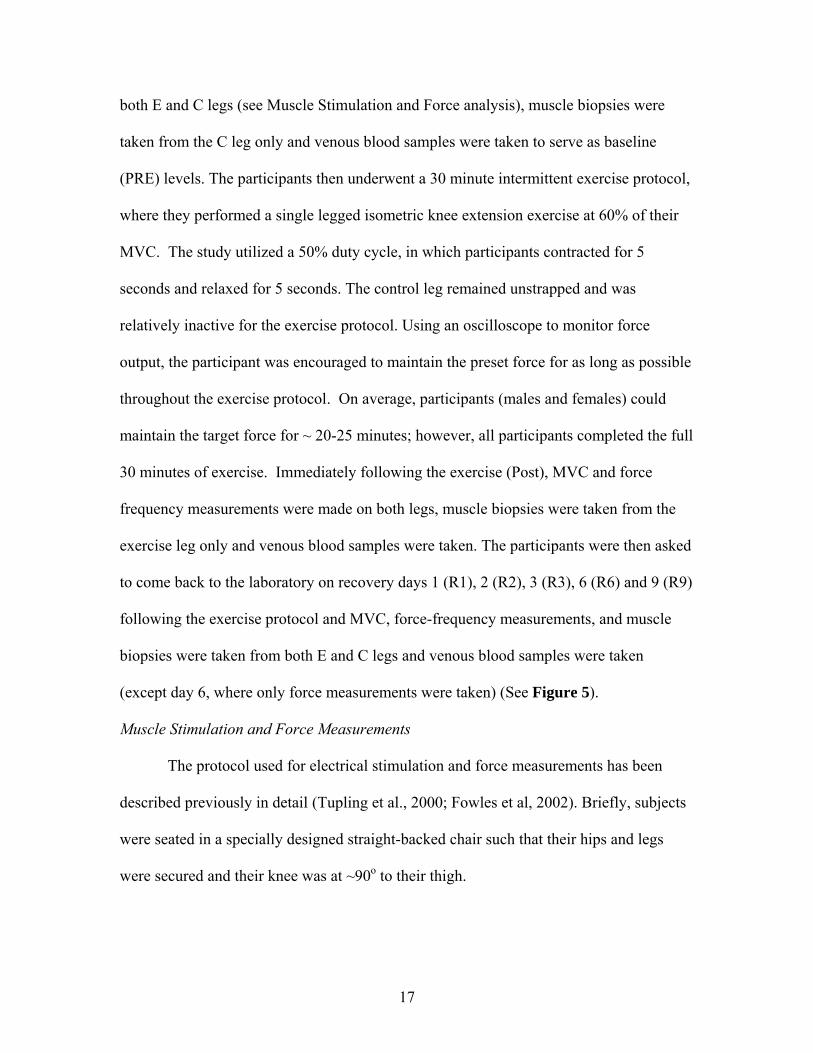
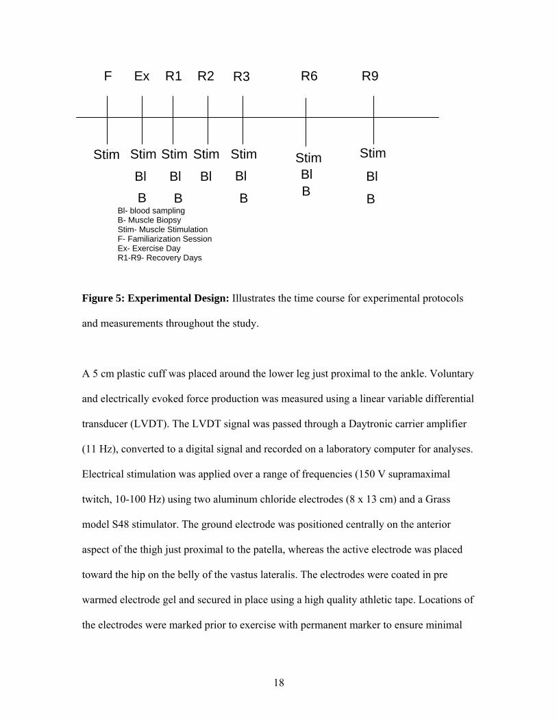
to come back to the laboratory on recovery days 1 (R1), 2 (R2), 3 (R3), 6 (R6) and 9 (R9)
following the exercise protocol and MVC, force-frequency measurements, and muscle
biopsies were taken from both E and C legs and venous blood samples were taken
(except day 6, where only force measurements were taken) (See Figure 5).
Muscle Stimulation and Force Measurements
The protocol used for electrical stimulation and force measurements has been
described previously in detail (Tupling et al., 2000; Fowles et al, 2002). Briefly, subjects
were seated in a specially designed straight-backed chair such that their hips and legs
were secured and their knee was at ~90o to their thigh.
17
F Ex R1 R2 R3 R9
Stim Stim Stim Stim Stim Stim Stim
Bl Bl Bl Bl Bl BlB B B B
Bl- blood sampling B- Muscle Biopsy Stim- Muscle Stimulation F- Familiarization Session Ex- Exercise Day R1-R9- Recovery Days
B
R6
Figure 5: Experimental Design: Illustrates the time course for experimental protocols
and measurements throughout the study.
A 5 cm plastic cuff was placed around the lower leg just proximal to the ankle. Voluntary
and electrically evoked force production was measured using a linear variable differential
transducer (LVDT). The LVDT signal was passed through a Daytronic carrier amplifier
(11 Hz), converted to a digital signal and recorded on a laboratory computer for analyses.
Electrical stimulation was applied over a range of frequencies (150 V supramaximal
twitch, 10-100 Hz) using two aluminum chloride electrodes (8 x 13 cm) and a Grass
model S48 stimulator. The ground electrode was positioned centrally on the anterior
aspect of the thigh just proximal to the patella, whereas the active electrode was placed
toward the hip on the belly of the vastus lateralis. The electrodes were coated in pre
warmed electrode gel and secured in place using a high quality athletic tape. Locations of
the electrodes were marked prior to exercise with permanent marker to ensure minimal
18
variability in placement on recovery days. At the beginning of each experimental day, all
equipment was calibrated.
Measures of muscle activation were taken using electromyography (EMG). Two
EMG electrodes (Ambu A/S, Denmark) were placed across the belly of the vastus
medialis of both the control and exercise legs and a reference electrode was placed on the
head of the fibula. All EMG electrode locations were shaved, abraded and cleaned with
alcohol to maximize the signal. The locations of the biopsy sample sites and the muscle
stimulation pads prevents measuring EMG from vastus lateralis, so the medialis muscle
was chosen for measurement of muscle activation. To maintain signal reliability the
location of the electrodes was marked with a black marker to minimize day-to-day
variation.
Muscle Biopsies and Sample Preparation
Tissue samples (~50mg) from the vastus lateralis of the C leg were obtained via
needle biopsy technique under suction immediately prior to the 30 minute exercise
protocol (will serve as PRE exercise baseline values), and from the E leg immediately
upon stopping exercise. On recovery days 1, 3, 6 and 9 muscle biopsies were obtained
from both the E and C legs. Each biopsy was taken from a separate sampling site, under
local anaesthetic (1% xylocaine). The muscle samples were diluted in a pre made sample
buffer (PMSF) (250 mM Sucrose, 5mM HEPES, 0.2mM PMSF and 0.2% NaN3; pH 7.5)
and homogenized in a crucible immersed in an ice bath. The diluted muscle homogenate
was aliquoted into microtubes and immediately frozen in liquid nitrogen, then stored at -
80◦C for later analysis of protein and Hsp70 content and calcium handling properties.
19
Western Blotting and Protein Content Determination
Western blot analysis was performed to measure the muscle protein content of
Hsp70, SERCA2a and SERCA1 in whole muscle homogenate. Band linearity and
loading concentrations were determined prior to running homogenate samples. The
diluted samples were run on a 7.5% polyacrylamide gel and the proteins were separated
using standard SDS-page protocols (Laemmli et al., 1970) and then transferred to
polyvinylidene difluoride membranes (Roche Diagnostics, Mannheim, Germany). After
blocking with 10% skim milk suspension, the membranes containing the high molecular
weight proteins (above 60 kDa) were incubated for either 16 hours with anti-Hsp70
monoclonal antibody SPA-810 (Stressgen Biotechnologies) or 1 h with either anti-
SERCA1a monoclonal antibody A52 (Zubrzcka-Gaarn, E, 1984) or anti-SERCA2a
antibody 2A7-A1 (Affinity Bioreagents) and the membranes containing lower molecular
weight proteins (below 60 kDa) were incubated for 1 hour with anti-α-sarcomeric actin
antibody 5C5 (Sigma) to control for protein loading. Membranes were then washed for
30 minutes using Tris-HCl, pH 7.5 and Tween (TBS-T). The membranes were treated
with horseradish peroxidase-conjugated goat anti-mouse secondary antibody (Hsp70-
1:2000, SERCA1-1:10000, SERCA2a-1:2000), washed again with TBS-T and then the
protein bands were detected with an enhanced chemiluminescence kit (Amersham
Pharmacia Biotech) using a bio-imaging system. Densitometric analysis was performed
using a software program (GeneSnap) to measure the band density, allowing for
quantification of the specific protein relative to total whole muscle homogenate protein.
All specific protein measurements were expressed relative to the values obtained in the
pre-exercise muscle biopsy and normalized to the concentration of α-actin and standard.
20
The standard used was a known amount of each protein of interest that was loaded on
every gel. The standard was used so direct comparisons could be made between male
and female protein contents.
Measurement of SERCA Activity
The protocol for measuring SERCA activity in homogenates prepared from
human vastus lateralis muscle is summarized by Duhamel et al., 2007. Briefly, maximal
SERCA activity was measured using a spectrophotometric assay technique developed by
Simonides and van Hardeveld (1990). Homogenate (25 μL) was added to a calcium
ATPase reaction buffer containing 100mM KCl, 20 mM HEPES, 15 mM MgCl2, 1 mM
EGTA, 10 mM NaN3 and 10 mM of phosphenolpyruvate (PEP) (pH 7.0) at 37οC.
Immediately prior to initiating the reaction, 18 µL lactate dehydrogenase (LDH), 18 µL
pyruvate kinase (PK), 10.5 µL calcium ionophore, (Sigma-A23187) 100 µL ATP and
25µL of whole muscle homogenate were added to 5 mL of the pre-made calcium
ATPase reaction buffer, vortexed vigorously and kept on ice. A range (27-30.6µL) of
calcium chloride (CaCl2) concentrations was added to microtubes. This pre-determined
range of CaCl2 additions was intended to achieve a plateau and subsequent decline in
Ca2+ ATPase activity were observed (Vmax). Also 2 µL of cyclopiazonic acid (CPA) a
specific inhibitor of SERCA activity was added to the two highest CaCl2 additions to
represent a baseline ATPase value, based on Duhamel et al (2007). A total volume of 300
µL of the reaction cocktail (calcium ATPase buffer plus other additions), was added to
the varying calcium concentrations. After vortexing vigorously, 100 µL of this cocktail
was pipetted in duplicate into a 96 well plate, 1 µL of 0.3mM NADH (previously made
that day and kept in a dark environment) was then pipetted into each well, and quickly
21
inserted into the plate reader and read at a wavelength of 340 nm to determine the
fluorescence of NADH. Ca2+ ATPase activity was calculated as the difference between
the total ATPase activity measured without CPA and the basal ATPase activity with CPA.
All measurements of Ca2+ ATPase activity were expressed relative to whole muscle
homogenate protein concentrations determined via the Lowry Protein Assay (Schacterle
and Pollock, 1973).
SERCA Calcium Uptake Analysis
Calcium uptake measurements were made using a Photon Technology
International (PTI) dual photon flourometer, as described in detail by Duhamel et al
(2007). Briefly, fluorescence measurements were collected on a dual-emission wave-
length spectrofluorometer. The excitation wavelength was set at 355 nm and 405 and
485 nm correspond to the emission wavelengths for bound (F) and free (G) indo-1,
respectively. Photon counts were collected simultaneously for each wavelength. Before
each trial session, the background fluorescence was determined in the absence of INDO-1
and subtracted prior to starting of each analysis. In brief, a reaction buffer consisting of
200mM KCl, 20mM HEPES, 10mM NaN3, 7uM TPEN, 5mM Oxalate, 15mM MgCl2;
(pH 7.0 at 37°C), was made prior to the measurements and stored at -20°C. Whole
muscle homogenate (30μL), ~2.5µL of 10mM CaCl2 and 1µL of INDO-1, was added to a
2mL four sided cuvette containing 1.9mL of the reaction buffer. The cocktail was
warmed to 37°C before starting the reaction by adding 40µL of 5mM ATP. As Ca2+
decreased because of active SR Ca2+ transport, F decreases and G increases. Using the
PTI software, the ratio of F/G (R) is used to calculate the decrease in free calcium. Free
calcium was determined by the software using the following equation:
22
(Ca2+)f=Kd x (Gmax/Gmin)(R-Rmin)(Rmax-R)
Where, Rmin and Rmax represent the min and max F/G ratios respectively, Kd represents
the equilibrium constant for the interaction between Ca2+ and indo-1 (Set at 250) and Gmin
and Gmax represent the min and max values for free indo 1
Measurements of calcium uptake were taken at 2000, 1500, 1000 and 500 nM free
Ca2+ concentrations and rates were determined as reviewed by Tupling et al (2007). All
uptake measurements were done in duplicate. All measurements of calcium uptake are
expressed relative to whole muscle homogenate protein concentrations.
Serum Creatine Kinase Measurements
To detect the severity of sarcolemmal damage caused by the intermittent exercise
protocol, measures of serum creatine kinase concentrations were taken. Venous blood
samples were taken during each testing session, in order to track the changes in serum
creatine kinase concentrations which were measured using a fluorometric assay according
to the methods of Szasz et al. (1976).
Measures of serum estrogen
In order to assess the consistency of the menstrual cycle stage between females,
circulating estrogen levels were determined. Venous blood samples were taken from the
anti-cubital vein to measure serum estrogen concentrations. Using a commercially
available radioimmunoassay kit (Coat-a-Count, Inter Medico, Markham, ON), estrogen
levels were analyzed prior to exercise and on subsequent recovery days, to confirm there
was no large variability within the study period.
23
Data Analysis
Three two-way repeated ANOVAs were performed for each of the Hsp70
measures (except basal Hsp70, where a t-test was used). More specifically, within the
male and female groups, exercise and control legs were compared across the entire time
course, to determine the effect of exercise and time. Males and females were then
compared across the time course of the study, by just analyzing the exercise leg with each
value being normalized to female PRE measurements. For all enzyme activities (CK and
SERCA activity), estrogen concentrations and force measurements two way repeated
measure ANOVAs were used. When comparing within group force response for males
and females, force was expressed as absolute values. When comparing males and female
force responses, all values were expressed relative to within group PRE values.
Neumann-Kewls post-hoc comparisons were then done to compare specific means for
each ANOVA. The level of significance was established at p values less than 0.05.
24
Chapter 3: Results
Heat Shock Protein 70 Sexual Dimorphism
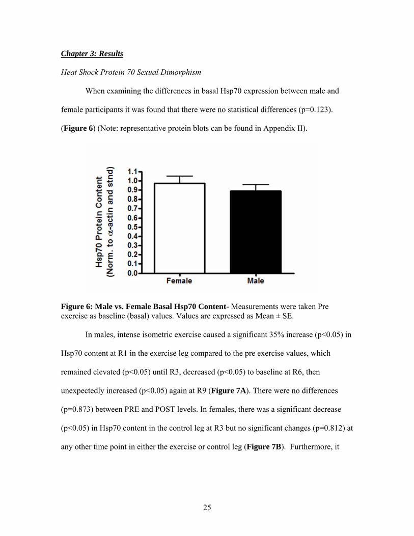
When examining the differences in basal Hsp70 expression between male and
female participants it was found that there were no statistical differences (p=0.123).
(Figure 6) (Note: representative protein blots can be found in Appendix II).
Figure 6: Male vs. Female Basal Hsp70 Content- Measurements were taken Pre exercise as baseline (basal) values. Values are expressed as Mean ± SE.
In males, intense isometric exercise caused a significant 35% increase (p<0.05) in
Hsp70 content at R1 in the exercise leg compared to the pre exercise values, which
remained elevated (p<0.05) until R3, decreased (p<0.05) to baseline at R6, then
unexpectedly increased (p<0.05) again at R9 (Figure 7A). There were no differences
(p=0.873) between PRE and POST levels. In females, there was a significant decrease
(p<0.05) in Hsp70 content in the control leg at R3 but no significant changes (p=0.812) at
any other time point in either the exercise or control leg (Figure 7B). Furthermore, it
25
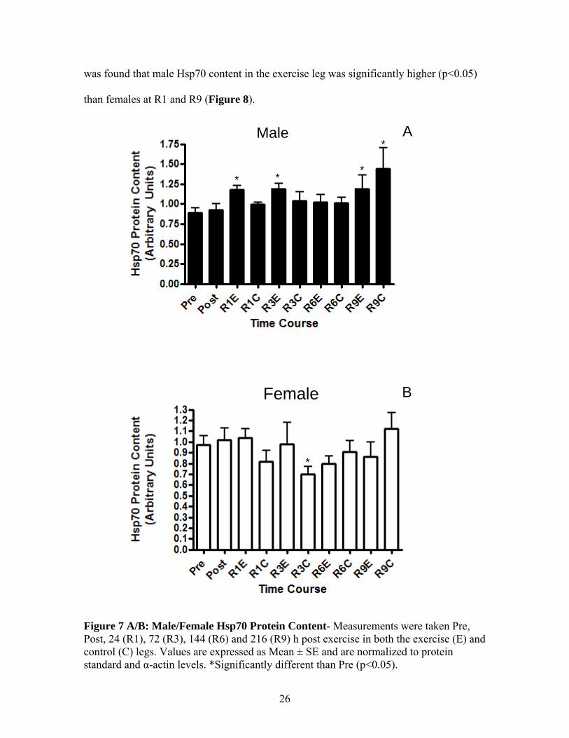
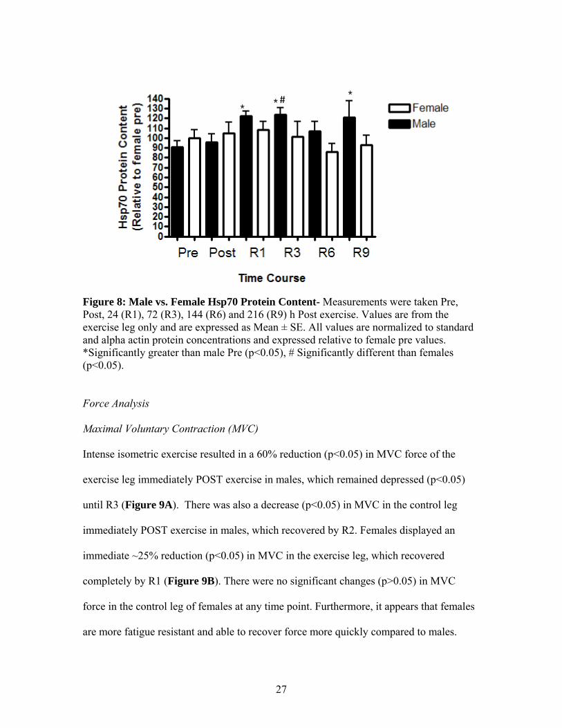
was found that male Hsp70 content in the exercise leg was significantly higher (p<0.05)
than females at R1 and R9 (Figure 8).
* **
*AMale
*
BFemale
Figure 7 A/B: Male/Female Hsp70 Protein Content- Measurements were taken Pre, Post, 24 (R1), 72 (R3), 144 (R6) and 216 (R9) h post exercise in both the exercise (E) and control (C) legs. Values are expressed as Mean ± SE and are normalized to protein standard and α-actin levels. *Significantly different than Pre (p<0.05).
26
* **#
Figure 8: Male vs. Female Hsp70 Protein Content- Measurements were taken Pre, Post, 24 (R1), 72 (R3), 144 (R6) and 216 (R9) h Post exercise. Values are from the exercise leg only and are expressed as Mean ± SE. All values are normalized to standard and alpha actin protein concentrations and expressed relative to female pre values. *Significantly greater than male Pre (p<0.05), # Significantly different than females (p<0.05). Force Analysis
Maximal Voluntary Contraction (MVC)
Intense isometric exercise resulted in a 60% reduction (p<0.05) in MVC force of the
exercise leg immediately POST exercise in males, which remained depressed (p<0.05)
until R3 (Figure 9A). There was also a decrease (p<0.05) in MVC in the control leg
immediately POST exercise in males, which recovered by R2. Females displayed an
immediate ~25% reduction (p<0.05) in MVC in the exercise leg, which recovered
completely by R1 (Figure 9B). There were no significant changes (p>0.05) in MVC
force in the control leg of females at any time point. Furthermore, it appears that females
are more fatigue resistant and able to recover force more quickly compared to males.
27
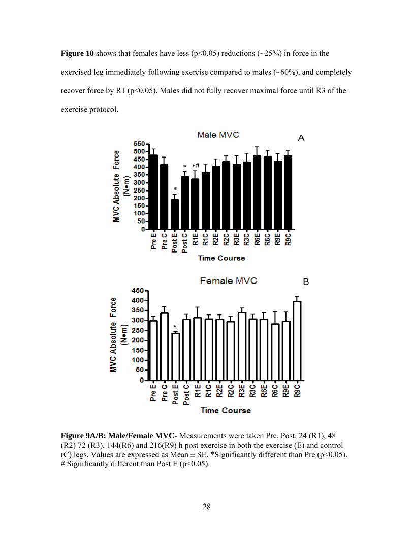
Figure 10 shows that females have less (p<0.05) reductions (~25%) in force in the
exercised leg immediately following exercise compared to males (~60%), and completely
recover force by R1 (p<0.05). Males did not fully recover maximal force until R3 of the
exercise protocol.
*
* *#
A
*
B
Figure 9A/B: Male/Female MVC- Measurements were taken Pre, Post, 24 (R1), 48 (R2) 72 (R3), 144(R6) and 216(R9) h post exercise in both the exercise (E) and control (C) legs. Values are expressed as Mean ± SE. *Significantly different than Pre (p<0.05). # Significantly different than Post E (p<0.05).
28
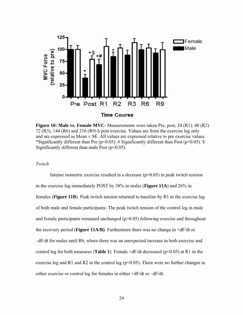
Figure 10: Male vs. Female MVC- Measurements were taken Pre, post, 24 (R1), 48 (R2) 72 (R3), 144 (R6) and 216 (R9) h post exercise. Values are from the exercise leg only and are expressed as Mean ± SE. All values are expressed relative to pre exercise values. *Significantly different than Pre (p<0.05). # Significantly different than Post (p<0.05). $ Significantly different than male Post (p<0.05).
Twitch
Intense isometric exercise resulted in a decrease (p<0.05) in peak twitch tension
in the exercise leg immediately POST by 38% in males (Figure 11A) and 26% in
females (Figure 11B). Peak twitch tension returned to baseline by R1 in the exercise leg
of both male and female participants. The peak twitch tension of the control leg in male
and female participants remained unchanged (p>0.05) following exercise and throughout
the recovery period (Figure 11A/B). Furthermore there was no change in +dF/dt or
–dF/dt for males until R6, where there was an unexpected increase in both exercise and
control leg for both measures (Table 1). Female +dF/dt decreased (p<0.05) at R1 in the
exercise leg and R1 and R2 in the control leg (p<0.05). There were no further changes in
either exercise or control leg for females in either +dF/dt or –dF/dt.
29

*
A
*
B
Figure 11A/B: Male/Female peak twitch tension (Po) - Measurements were taken Pre, Post, 24 (R1), 48 (R2) 72 (R3), 144(R6) and 216 h post exercise in both the exercise (E) and control (C) legs.. Values are expressed as Mean ± SE. * Significantly different than Pre (p<0.05).
30
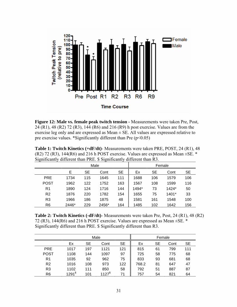
Figure 12: Male vs. female peak twitch tension - Measurements were taken Pre, Post, 24 (R1), 48 (R2) 72 (R3), 144 (R6) and 216 (R9) h post exercise. Values are from the exercise leg only and are expressed as Mean ± SE. All values are expressed relative to pre exercise values. *Significantly different than Pre (p<0.05) Table 1: Twitch Kinetics (+dF/dt)- Measurements were taken PRE, POST, 24 (R1), 48 (R2) 72 (R3), 144(R6) and 216 h POST exercise. Values are expressed as Mean ±SE. * Significantly different than PRE. $ Significantly different than R3.
Male Female
E SE Cont SE Ex SE Cont SE PRE 1734 115 1645 111 1688 106 1579 106
POST 1962 122 1752 163 1567 108 1599 116 R1 1890 124 1716 144 1494* 73 1424* 50 R2 1876 220 1782 154 1655 75 1401* 33 R3 1966 186 1875 48 1581 161 1548 100 R6 2446* 229 2456* 164 1485 102 1642 156
Table 2: Twitch Kinetics (-dF/dt)- Measurements were taken Pre, Post, 24 (R1), 48 (R2) 72 (R3), 144(R6) and 216 h POST exercise. Values are expressed as Mean ±SE. * Significantly different than PRE. $ Significantly different than R3.
Male Female Ex SE Cont SE Ex SE Cont SE
PRE 1017 197 1121 121 815 61 799 111 POST 1108 144 1097 97 725 58 775 68
R1 1035 92 962 75 833 93 681 68 R2 1016 108 973 122 768.2 81 647 47 R3 1102 111 850 58 792 51 887 87 R6 1291$ 101 1127$ 71 757 54 821 64
31
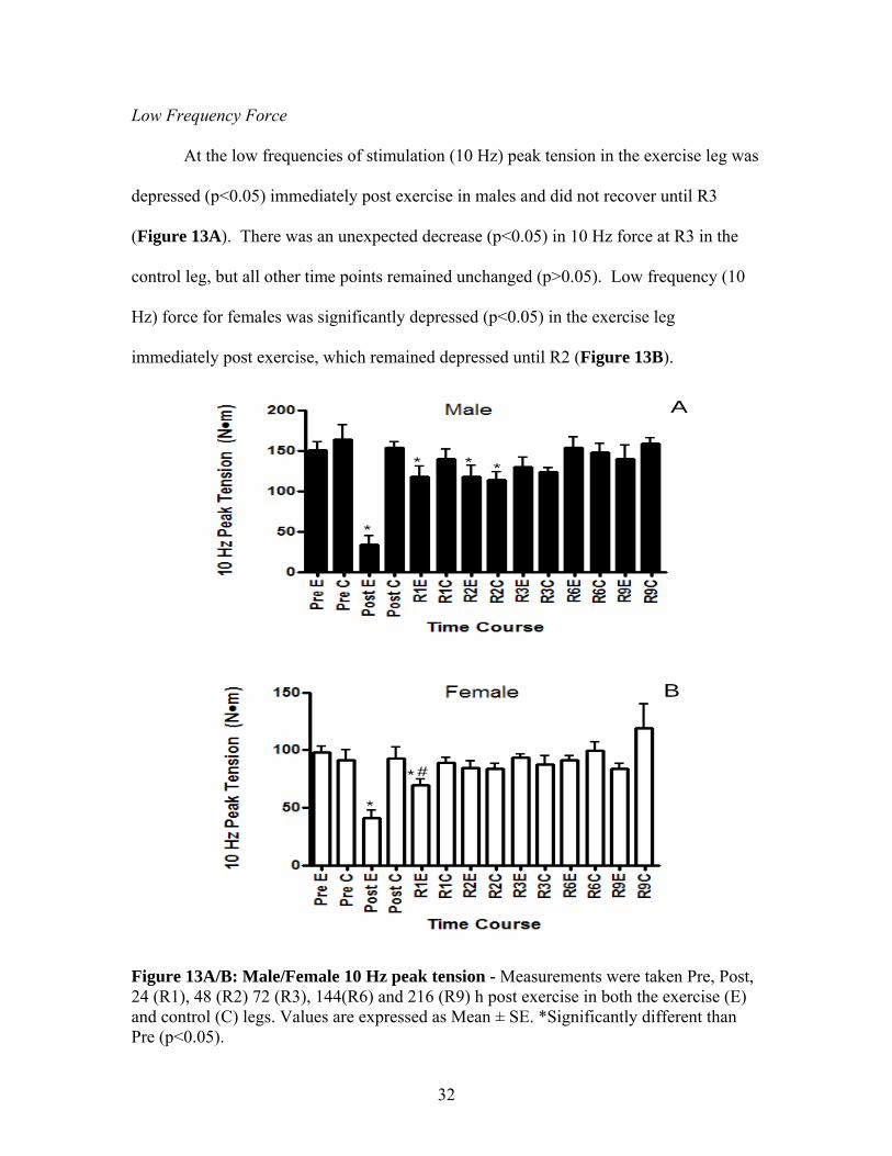
Low Frequency Force
At the low frequencies of stimulation (10 Hz) peak tension in the exercise leg was
depressed (p<0.05) immediately post exercise in males and did not recover until R3
(Figure 13A). There was an unexpected decrease (p<0.05) in 10 Hz force at R3 in the
control leg, but all other time points remained unchanged (p>0.05). Low frequency (10
Hz) force for females was significantly depressed (p<0.05) in the exercise leg
immediately post exercise, which remained depressed until R2 (Figure 13B).
*
* * *
A
*
* #
B
Figure 13A/B: Male/Female 10 Hz peak tension - Measurements were taken Pre, Post, 24 (R1), 48 (R2) 72 (R3), 144(R6) and 216 (R9) h post exercise in both the exercise (E) and control (C) legs. Values are expressed as Mean ± SE. *Significantly different than Pre (p<0.05).
32

There were no significant changes (p>0.05) in 10 Hz force in the control leg at
any time point during the protocol for females (Figure 14). Males also displayed a
greater relative force reduction (p<0.05) immediately POST in the exercise leg compared
to females (Figure 14).
Figure 14: Male vs. Female 10 Hz peak tension - Measurements were taken Pre, Post, 24 (R1), 48 (R2) 72 (R3), 144(R6) and 216 (R9) h post exercise. Values are from the exercise leg only and are expressed as Mean ± SE. All values are expressed relative to pre exercise values. *Significantly different than Pre (p<0.05). #Significantly different than males (p<0.05).$ Significantly different than Post (p<0.05)
High Frequency Force
At the high frequencies (100 Hz) of stimulation, males had a significant 33%
reduction (p<0.05) in force immediately POST exercise in the exercise leg, which
33
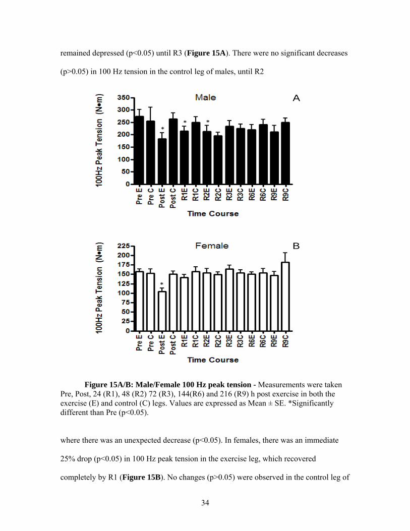
remained depressed (p<0.05) until R3 (Figure 15A). There were no significant decreases
(p>0.05) in 100 Hz tension in the control leg of males, until R2
** *
A
*
B
Figure 15A/B: Male/Female 100 Hz peak tension - Measurements were taken Pre, Post, 24 (R1), 48 (R2) 72 (R3), 144(R6) and 216 (R9) h post exercise in both the exercise (E) and control (C) legs. Values are expressed as Mean ± SE. *Significantly different than Pre (p<0.05).
where there was an unexpected decrease (p<0.05). In females, there was an immediate
25% drop (p<0.05) in 100 Hz peak tension in the exercise leg, which recovered
completely by R1 (Figure 15B). No changes (p>0.05) were observed in the control leg of
34
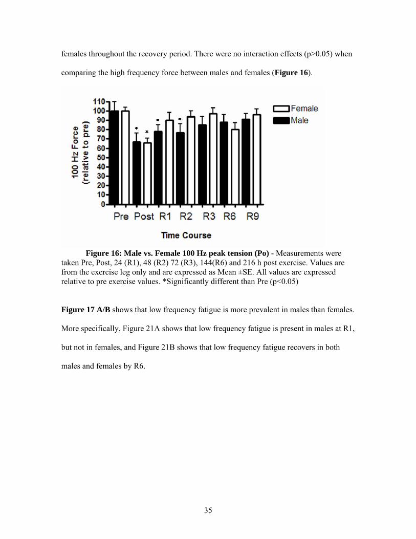
females throughout the recovery period. There were no interaction effects (p>0.05) when
comparing the high frequency force between males and females (Figure 16).
Figure 16: Male vs. Female 100 Hz peak tension (Po) - Measurements were
taken Pre, Post, 24 (R1), 48 (R2) 72 (R3), 144(R6) and 216 h post exercise. Values are from the exercise leg only and are expressed as Mean ±SE. All values are expressed relative to pre exercise values. *Significantly different than Pre (p<0.05)
Figure 17 A/B shows that low frequency fatigue is more prevalent in males than females.
More specifically, Figure 21A shows that low frequency fatigue is present in males at R1,
but not in females, and Figure 21B shows that low frequency fatigue recovers in both
males and females by R6.
35

Figure 17: Male and Female Force Frequency Response: A, compares Pre to Post and R1 for males and females. B, compares Pre to R2 and R6. All force measurements (10, 20, 50 and 100 Hz) are expressed relative to pre 100 Hz levels. All male values are dashed lines and all female values are solid red lines. Maximal SERCA Activity
Following exercise there were no changes (p>0.05) in maximal SERCA activity
(Vmax) at any time point throughout the experimental protocol in the exercise or control
leg in either males or females (Figure 18A-male and B-female). Furthermore, there were
36
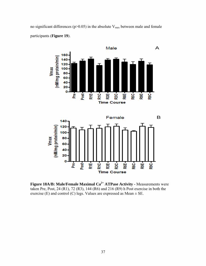
no significant differences (p>0.05) in the absolute Vmax between male and female
participants (Figure 19).
A
B
Figure 18A/B: Male/Female Maximal Ca2+ ATPase Activity - Measurements were taken Pre, Post, 24 (R1), 72 (R3), 144 (R6) and 216 (R9) h Post exercise in both the exercise (E) and control (C) legs. Values are expressed as Mean ± SE.
37
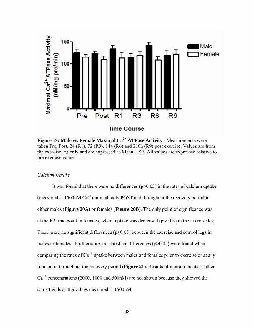
Figure 19: Male vs. Female Maximal Ca2+ ATPase Activity - Measurements were taken Pre, Post, 24 (R1), 72 (R3), 144 (R6) and 216h (R9) post exercise. Values are from the exercise leg only and are expressed as Mean ± SE. All values are expressed relative to pre exercise values. Calcium Uptake
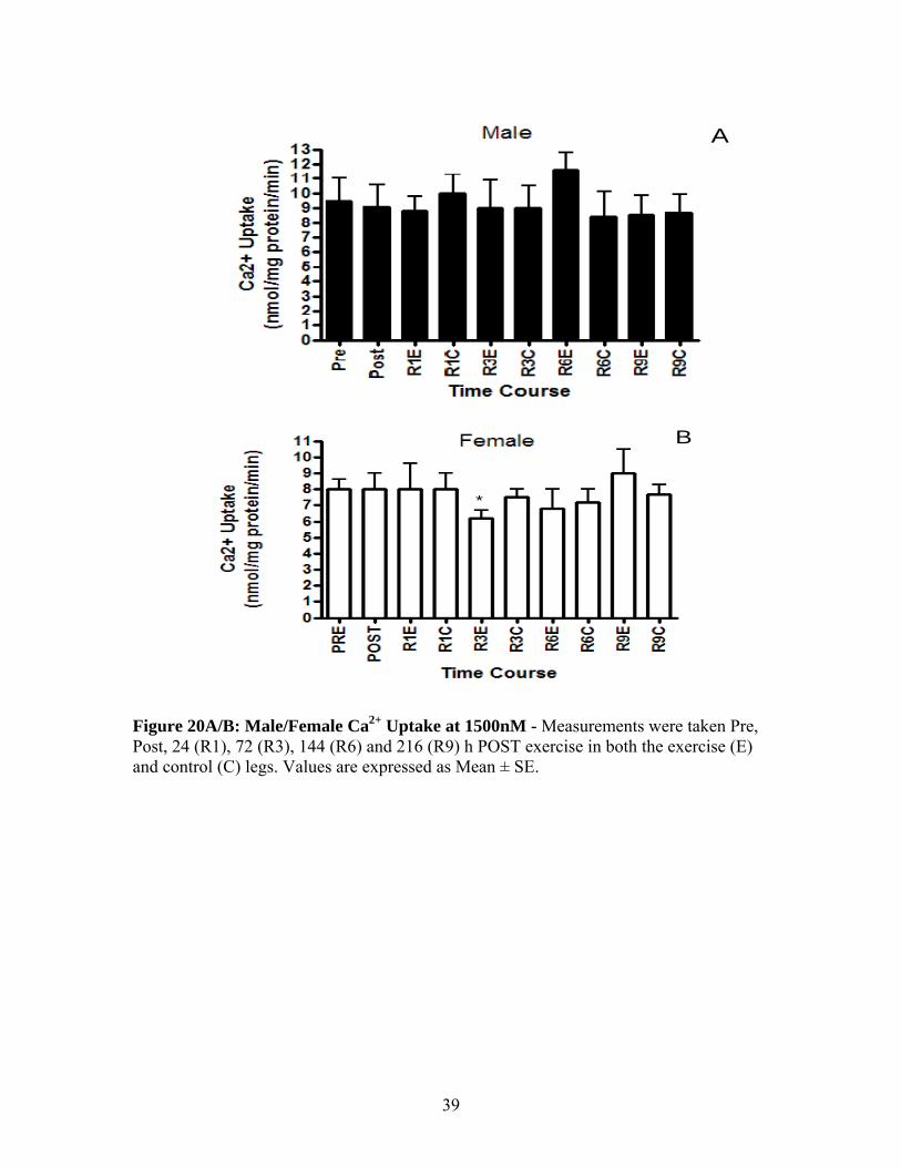
It was found that there were no differences (p>0.05) in the rates of calcium uptake
(measured at 1500nM Ca2+) immediately POST and throughout the recovery period in
either males (Figure 20A) or females (Figure 20B). The only point of significance was
at the R3 time point in females, where uptake was decreased (p<0.05) in the exercise leg.
There were no significant differences (p>0.05) between the exercise and control legs in
males or females. Furthermore, no statistical differences (p>0.05) were found when
comparing the rates of Ca2+ uptake between males and females prior to exercise or at any
time point throughout the recovery period (Figure 21). Results of measurements at other
Ca2+ concentrations (2000, 1000 and 500nM) are not shown because they showed the
same trends as the values measured at 1500nM.
38
A
*
B
Figure 20A/B: Male/Female Ca2+ Uptake at 1500nM - Measurements were taken Pre, Post, 24 (R1), 72 (R3), 144 (R6) and 216 (R9) h POST exercise in both the exercise (E) and control (C) legs. Values are expressed as Mean ± SE.
39
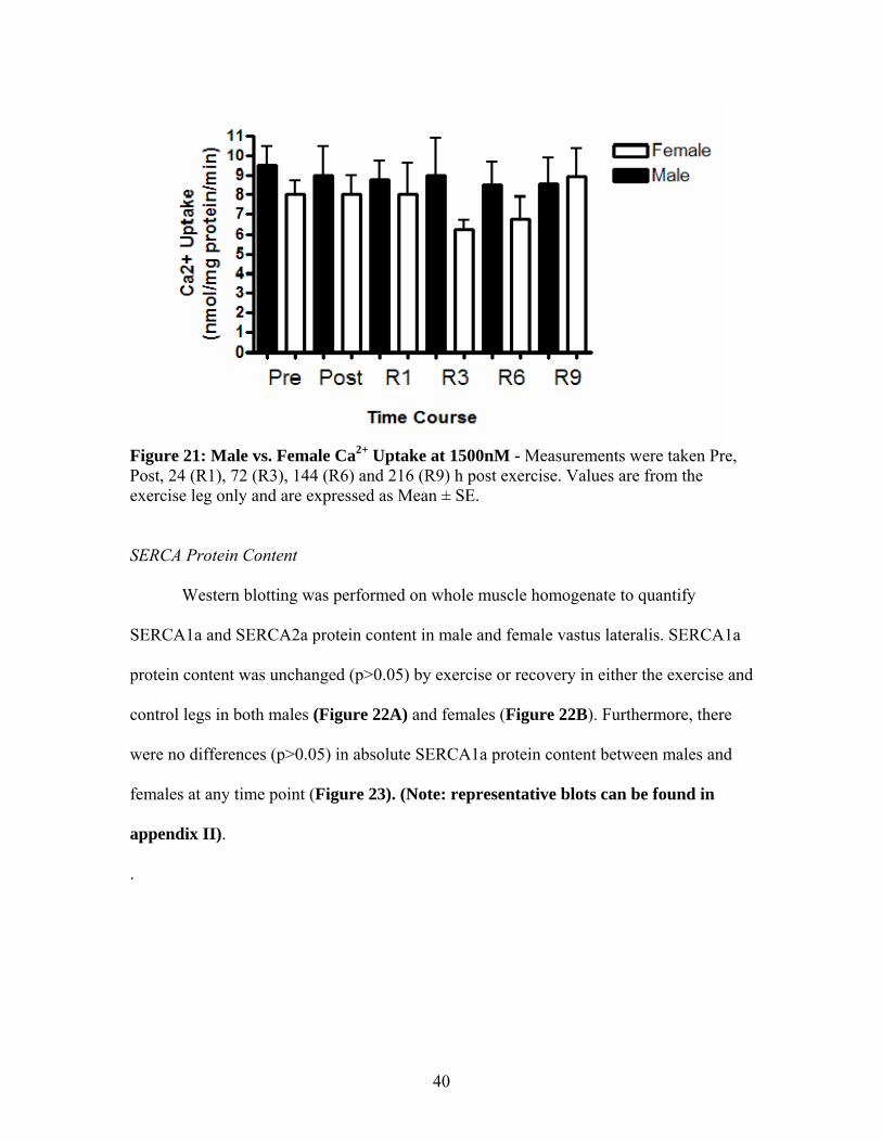
Figure 21: Male vs. Female Ca2+ Uptake at 1500nM - Measurements were taken Pre, Post, 24 (R1), 72 (R3), 144 (R6) and 216 (R9) h post exercise. Values are from the exercise leg only and are expressed as Mean ± SE. SERCA Protein Content
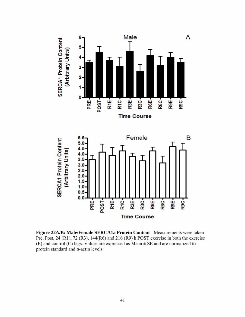
Western blotting was performed on whole muscle homogenate to quantify
SERCA1a and SERCA2a protein content in male and female vastus lateralis. SERCA1a
protein content was unchanged (p>0.05) by exercise or recovery in either the exercise and
control legs in both males (Figure 22A) and females (Figure 22B). Furthermore, there
were no differences (p>0.05) in absolute SERCA1a protein content between males and
females at any time point (Figure 23). (Note: representative blots can be found in
appendix II).
.
40
A
B
Figure 22A/B: Male/Female SERCA1a Protein Content - Measurements were taken Pre, Post, 24 (R1), 72 (R3), 144(R6) and 216 (R9) h POST exercise in both the exercise (E) and control (C) legs. Values are expressed as Mean ± SE and are normalized to protein standard and α-actin levels.
41
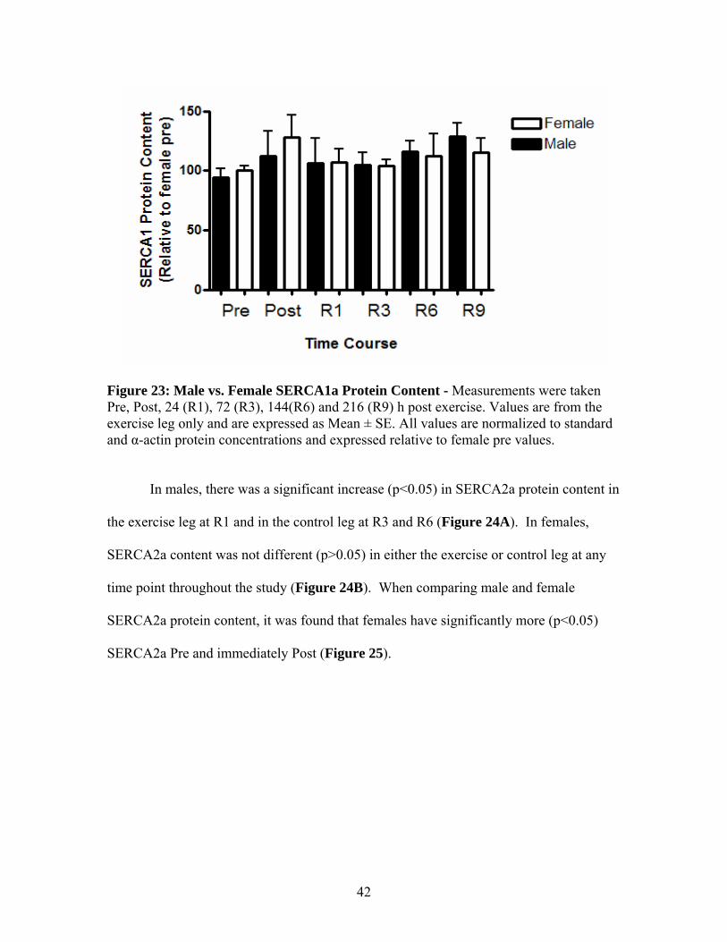
Figure 23: Male vs. Female SERCA1a Protein Content - Measurements were taken Pre, Post, 24 (R1), 72 (R3), 144(R6) and 216 (R9) h post exercise. Values are from the exercise leg only and are expressed as Mean ± SE. All values are normalized to standard and α-actin protein concentrations and expressed relative to female pre values.
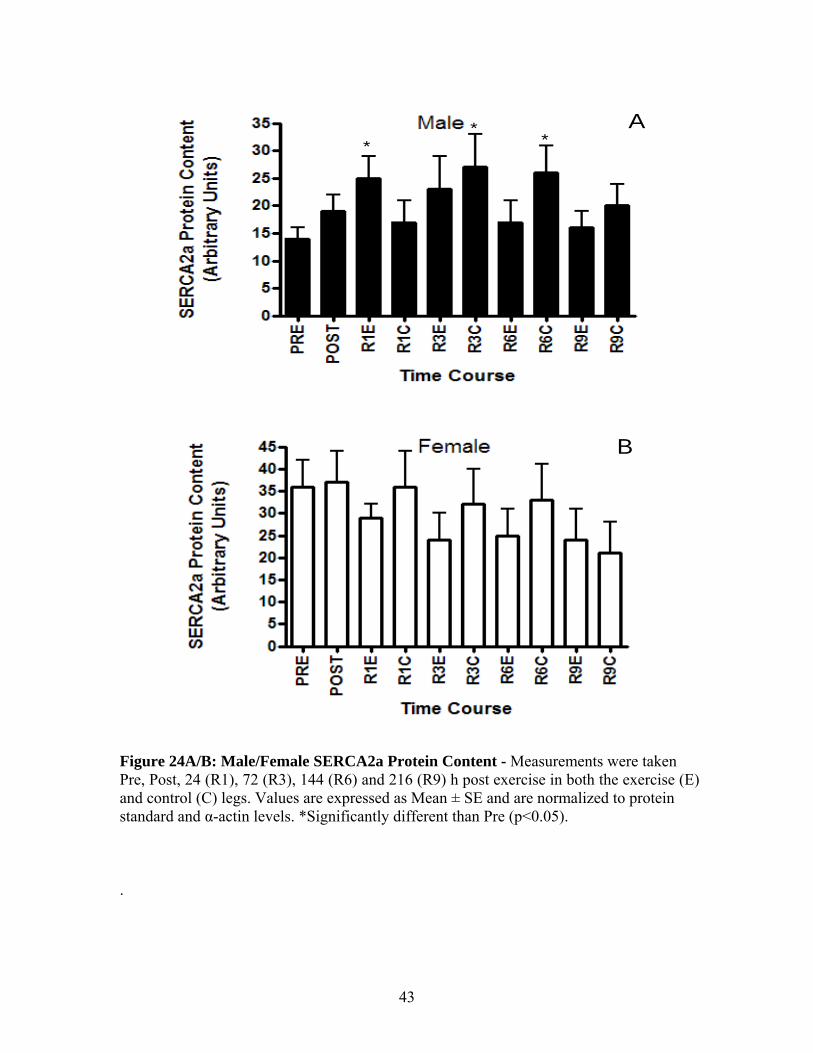
In males, there was a significant increase (p<0.05) in SERCA2a protein content in
the exercise leg at R1 and in the control leg at R3 and R6 (Figure 24A). In females,
SERCA2a content was not different (p>0.05) in either the exercise or control leg at any
time point throughout the study (Figure 24B). When comparing male and female
SERCA2a protein content, it was found that females have significantly more (p<0.05)
SERCA2a Pre and immediately Post (Figure 25).
42
**
*A
B
Figure 24A/B: Male/Female SERCA2a Protein Content - Measurements were taken Pre, Post, 24 (R1), 72 (R3), 144 (R6) and 216 (R9) h post exercise in both the exercise (E) and control (C) legs. Values are expressed as Mean ± SE and are normalized to protein standard and α-actin levels. *Significantly different than Pre (p<0.05).
.
43
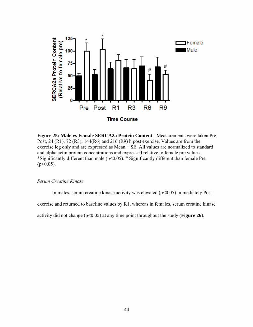
**
##
Figure 25: Male vs Female SERCA2a Protein Content - Measurements were taken Pre, Post, 24 (R1), 72 (R3), 144(R6) and 216 (R9) h post exercise. Values are from the exercise leg only and are expressed as Mean ± SE. All values are normalized to standard and alpha actin protein concentrations and expressed relative to female pre values. *Significantly different than male (p<0.05). # Significantly different than female Pre (p<0.05).
Serum Creatine Kinase
In males, serum creatine kinase activity was elevated (p<0.05) immediately Post
exercise and returned to baseline values by R1, whereas in females, serum creatine kinase
activity did not change (p<0.05) at any time point throughout the study (Figure 26).
44
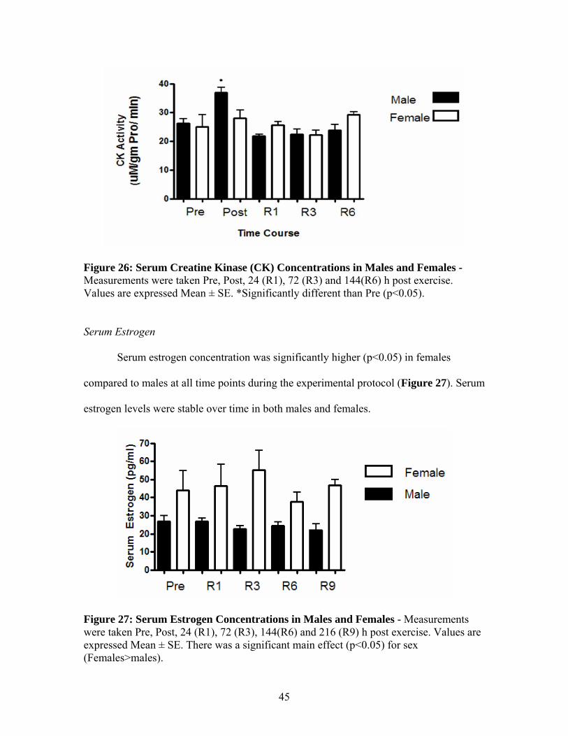
Figure 26: Serum Creatine Kinase (CK) Concentrations in Males and Females - Measurements were taken Pre, Post, 24 (R1), 72 (R3) and 144(R6) h post exercise. Values are expressed Mean ± SE. *Significantly different than Pre (p<0.05).
Serum Estrogen
Serum estrogen concentration was significantly higher (p<0.05) in females
compared to males at all time points during the experimental protocol (Figure 27). Serum
estrogen levels were stable over time in both males and females.
Figure 27: Serum Estrogen Concentrations in Males and Females - Measurements were taken Pre, Post, 24 (R1), 72 (R3), 144(R6) and 216 (R9) h post exercise. Values are expressed Mean ± SE. There was a significant main effect (p<0.05) for sex (Females>males).
45
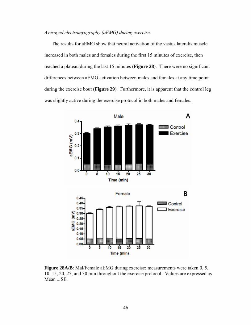
Averaged electromyography (aEMG) during exercise
The results for aEMG show that neural activation of the vastus lateralis muscle
increased in both males and females during the first 15 minutes of exercise, then
reached a plateau during the last 15 minutes (Figure 28). There were no significant
differences between aEMG activation between males and females at any time point
during the exercise bout (Figure 29). Furthermore, it is apparent that the control leg
was slightly active during the exercise protocol in both males and females.
Figure 28A/B: Mal/Female aEMG during exercise: measurements were taken 0, 5, 10, 15, 20, 25, and 30 min throughout the exercise protocol. Values are expressed as Mean ± SE.
46
Chapter 4: Discussion The major purpose of this study was to determine if sexual dimorphism exists in
the basal and exercise induced Hsp70 protein content in human skeletal muscle.
Furthermore, a major goal was to determine if any differences in Hsp70 content between
males and females might be associated with skeletal muscle fatigability and mechanical
function and changes in SERCA pump function in response to intense, intermittent,
isometric exercise. The final purpose was to determine if the time course of recovery in
all measurements differed between males and females.
Sexual dimorphism in basal Hsp70 protein content
It was found that there were no significant differences in basal Hsp70 content
between males and females. In contrast with the initial hypothesis, pre-exercise muscle
biopsy samples of the vastus lateralis showed that with no previous exposure to stress,
there are no differences between males and females in Hsp70 expression. This finding is
in agreement with one rodent study (Voss et al., 2003) which also showed no significant
differences in the male/female basal Hsp70 content of skeletal muscle in rats. However,
these results are in contrast to the findings by Bombardier et al (2009) who showed that
ovariectomized rats that were supplemented with estrogen had a significantly greater
amount of basal Hsp70 in soleus, compared to ovariectomized sham-supplemented
females. The findings from this thesis also conflict with data from cardiac muscle, which
showed that female rat heart had twice as much basal Hsp70 compared to male heart
(Voss et al, 2003 and Paroo et al, 2002). Our hypothesis was also based on the finding
that estrogen can activate the HSF1 pathway, which has been shown to increase basal
levels of Hsp70 (Knowlton et al., 2003)
47
It was hypothesized that females would have higher basal Hsp70 levels due to the
influence of estrogen on skeletal muscle physiology. As expected, compared with male
participants, serum estrogen levels were two times higher in female participants; however,
it’s possible that basal Hsp70 expression in female skeletal muscle fluctuates over the
course of the estrus cycle, and since female participants were tested between days 1-3 of
their menstrual cycle, Hsp70 expression may not have been at its peak. It is unlikely that
other sex hormones (progesterone and testosterone) have an influence on basal Hsp70
content in skeletal muscle. Milne et al. (2005) showed that castration of male rats
(removing endogenous testosterone) had no effect on basal Hsp70 content in skeletal
muscle and Bombardier et al. (2009) showed that administering exogenous progesterone
to ovariectomized females did not increase basal Hsp70 content in soleus muscle.
Bombardier et al. (2009) only measured Hsp70 content in rat soleus muscle, which is
primarily composed of Type I muscle fibres. Since human vastus lateralis is composed of
a mixture of fibre types (Green et al, 1981), it is possible that having more glycolytic
fibre types dilutes the higher levels of Hsp70 in oxidative cells. Finally, it is possible that
the observed lack of sexual dimorphism in basal Hsp70 expression in human skeletal
muscle is due to the lack of statistical power and small sample size. In order to determine
a significant difference of 25% in basal Hsp70, with a power of 0.90, it was calculated
that a sample size of 15 male and 15 female participants would be needed. Therefore,
this serves as a limitation of this study. Nevertheless, based on the results from this thesis,
it is concluded that sexual dimorphism does not exist in humans with respect to basal
Hsp70 content in skeletal muscle.
48
Sexual dimorphism in the exercise induced Hsp70 response
Previous studies have shown that exercise has the ability to upregulate Hsp70
protein content in order to protect the muscle against further damaging/lethal stresses in
male human and rodent models (Paroo et al., 2002, Locke et al, 1991, Morton et al, 2006
and Tupling et al., 2007). It was hypothesized that following 30 minutes of intense
intermittent single legged isometric knee extension exercise, females would have a
blunted Hsp70 response compared with males. This study found that the exercise
protocol employed has the ability to stimulate a robust Hsp70 response in male skeletal
muscle, similar to findings from previous work which used the exact same exercise
protocol (Tupling et al, 2007). Furthermore, as was hypothesized, this was the first study
to show in humans that females have a blunted Hsp70 response following exercise.
Several mechanisms may be responsible for the blunted Hsp70 response in
females. First, increased serum creatine kinase (CK) following intense exercise, which is
indicative of sarcolemmal damage (Tiidus et al., 1996), was significantly increased one
day after exercise in males, but not in females. This finding is consistent with previous
work which shows that females have less overall damage compared to males following
exercise (Tiidus et al., 1996, Bombardier et al, 2009 and Enns et al, 1999). This would
suggest that since females have less muscle damage and potentially less protein
denaturation, the stimulus to upregulate Hsp70 expression is lower in females compared
with males. Secondly, estrogen has been described as an antioxidant molecule similar to
vitamin E, capable of reducing the effects of increased levels of oxidative stress in
skeletal muscle (Persky et al, 1999). Estrogen has these antioxidant properties because it
has an extra hydroxyl group, capable of accepting free electrons (free radicals) which
49
accumulate in the cell during high periods of stress. If estrogen has the ability to
minimize the accumulation of ROS/RNS which normally increase with exercise and can
cause protein denaturation (Stice et al, 2008), this would suggest that less Hsp70 would
be needed to counter the increase in ROS/RNS. Although ROS/RNS accumulation was
not measured in this thesis, this may provide another explanation for the attenuated
Hsp70 response in females.
The exercise induced increase in Hsp70 of male skeletal muscle probably
occurred primarily in Type I fibre types. Using immunohistochemistry, Tupling et al
(2007) showed that Type I muscle fibres had significantly more Hsp70 following exercise
compared to type II fibres. This thesis did not confirm if fibre type differences occurred
in either males or females so further work should be done to determine if the fibre type
specific Hsp70 response displays sexual dimorphism. This study also found that there
was a trend showing male Hsp70 would have completely recovered by R9 but instead
there was an unexpected increase in Hsp70 at R9 in both E and C legs. This finding
suggests that most likely participants did not follow pre-experimental instructions to not
exercise during the experiment. Previous work by Tupling et al (2007) showed that
Hsp70 protein content in male skeletal muscle was still higher than basal levels at R6, but
was beginning to show signs of recovery.
Muscle Fatigability and Mechanical Function
Alterations to mechanical function in skeletal muscle are common following a
bout of intense activity and can be associated with decreased force and slowing of muscle
contraction (i.e. muscle fatigue) (Allen et al., 2007 and Tupling et al, 2004). It was
hypothesized that female skeletal muscle function would be more protected compared to
50

males following intense exercise, which would be associated with less fatigue
immediately post exercise, less decrements in low frequency fatigue (LFF) and a more
accelerated force recovery compared to males. It was found that following the bout of
exercise maximal voluntary contraction (MVC) was decreased by ~25% in females
compared to the much larger ~60% decrease observed in males. It was also discovered
that MVC of males took longer to return to baseline levels (recovered by R2), than
females (recovered by R1). These findings support the hypotheses that females are more
resistant to fatigue and recover mechanical function following fatiguing exercise more
quickly than males.
Similar results were observed for both low and high frequency force. At low
frequencies of stimulation (10 Hz), there was a significant 75% decrease in peak tension
for males with females showing only a 55% decrease immediately post exercise. Females
also had full recovery of low frequency force by R2, while males did not fully recover
until R3. This finding is similar to previous work from our lab which has shown peak
tension elicited at 10 Hz to be attenuated for up to 4-6 days following the bout of exercise
(Fowles et al, 2001). Intense isometric exercise also resulted in a significant decrease of
peak tension of the vastus lateralis muscle stimulated at high frequencies (100 Hz) in both
males and females. More specifically, males had a significant ~30% decrease in 100 Hz
peak tension immediately post exercise, which remained depressed until R3 of the
experimental protocol. Females displayed a similar ~30% decrease in 100 Hz peak
tension immediately post exercise; however, force was fully recovered by R1. These
findings are consistent with the results from Fowles et al (2001), which showed HFF to
be present for up to 4 days following the bout of exercise.
51
Force depressions at both high and low frequencies that persist past one day of
recovery have been termed post contractile force depressions (PCDs) (Fowles et al, 2002
and Tupling et al, 2000). Decrements in force stimulated at high frequencies have been
associated with alterations to the Na+/K+ ATPase. The Na+/K+ ATPase is responsible for
maintaining a proper ion gradient across the sarcolemma to maintain excitability of the t-
tubules. Fowles et al (2002) suggested that decrements in Na+/K+ ATPase are at least in
part due to accumulation of ROS. This may provide one mechanism by which females
experience less fatigue and recover quicker than males.
Low frequency fatigue (LFF) is a prolonged reduction in force when stimulated at
low frequencies (Tupling et al, 2004 and Allen et al, 2007). LFF is a common
observation following a bout of intense exercise. At low frequencies of stimulation, force
development relies upon the release of Ca2+ from the SR. In situations where LFF is
present, Ca2+ release from the SR is impaired and the rise in intracellular Ca2+ needed to
cause contraction is less. The result in an increase of intracellular calcium concentrations,
which has been associated with the activation of various proteolytic calpains capable of
further catabolic reactions (Murphy et al, 2006) The results of this thesis suggest that LFF
is present in both males and females, but females recovery more quickly. Figure 16A/B
uses values at various frequencies (10, 20, 50 Hz) expressed relative to force elicited at
100 Hz. Figure 16 A shows that male Post and R1 are not only lower than male Pre, but
also lower than female Pre, Post and R1. Furthermore, in Figure 16B it is apparent that
LFF does not exist in either males or females any further than R2 since force is recovered
by R3. It is likely that since females did not have as much damage as males, as illustrated
by less CK activity post exercise, the total amount of damage to membranous and
52
intracellular structures in females was less. This would potentially mean that there is less
impairment of calcium release in females and less activation of calpains, which may help
explain why females are more fatigue resistant than males.
Previous research has examined whether sexual dimorphism exists in skeletal
muscle fatigability. The evidence that currently exists is equivocal, in that some studies
show females to be more fatigue resistant than males (Clark et al, 2005, Russ et al, 2003
and Hunter et al, 2001), while others show no differences (Buckley-Bleiler et al., 1989,
Savage et al, 2002 and Thompson et al, 1997). Most previous work supporting fatigue
resistance in females used a much lower intensity of contraction (~25% MVC) and
measured endurance time (time to failure) rather than directly measuring skeletal muscle
contractile properties. These studies found that females were able to maintain the
intensity of exercise longer than males, which they attributed to differences in muscular
blood flow (Clark et al., 2005 and Russ et al, 2003). Conversely, studies which
contended the notion that females are more fatigue resistant than males utilized higher
exercise intensities and showed there were no differences in fatigue resistance between
males and females during exercise. Furthermore, these same studies suggest that there is
no difference in recovery of force in the days following exercise. In this thesis, a high
intensity exercise protocol was employed but unlike other studies that employed higher
intensity exercise protocols, the findings support the notion that females are more fatigue
resistant immediately following exercise and that females recover force more quickly
than males. Differences between this study and others that contend fatigue resistance in
females is associated with the protective effects of estrogen may be due to a couple of
reasons. First, previous work showing females were not fatigue resistant which used
53
isometric exercise used a longer work:rest ratio. Buckly-Bleiler et al (1989) used a 10:20
sec (work:rest ratio) compared to our 5:5 sec ratio. This longer work to rest ratio may
have resulted in more recovery time in between contractions, which may be the reason no
differences were detected. This same study contending fatigue resistance in females only
used a total of 40 contractions, whereas this thesis had the participants doing 180
contractions. These two differences may result in less damage to skeletal muscle and
more recovery during the exercise bout, which may help explain the discrepancy in
results compared to this thesis. Finally, previous work suggesting there is no differences
in fatigue between males and females following exercise, did not actually use a direct
male to female comparison. These studies utilized pre-menopausal comparisons
(Buckley-Bleiler et al., 1989, Savage et al, 2002 and Thompson et al, 1997) in
comparison to the male-female comparison used in this thesis.
Another debate that currently exists in the literature is whether the examination of
fatigue between males and females should be studied with respect to absolute or relative
force production. This debate stems from the finding that under low intensity
contractions, which is expressed relative to the individual’s MVC, females are more
fatigue resistant compared to males (Hunter et al., 2001, Clark et al., 2005, Russ et al.,
2003). When the intensity is increased there appear to be no differences in fatigability
between males and females (Maughan et al., 1986, Phillips et al., 1993). This argument is
based on the notion that at low intensities, although both male and female subjects
contract at a certain percentage of their MVC, males have higher absolute force
production, which results in more occlusion of peripheral vasculature and therefore puts
them under more ischemic conditions (Clark et al., 2005, Wust et al., 2008 and Maughan
54
et al., 1986). When intensity is increased, both males and females are shown to have the
same amount of occlusion, which explains the lack of sexual dimorphism observed in
fatigability at higher intensities (Maughan et al., 1986 and Russ et al., 2003). The results
of this thesis cannot rule out the fact that males may have been under more ischemic
conditions compared to females since male participants would have been producing
higher absolute forces than females due to the relative force production of the exercise
protocol. Although using a force expressed relative to an individual’s MVC is generally
accepted as a valid method of analysing fatigue between people, future studies may aim
to recruit male and female participants that have similar MVCs. This type of design
would rule out the fact that blood flow restrictions and absolute force production in males
is what is causing the sexual dimorphism observed in skeletal muscle fatigability since
both groups would be contracting with the same absolute force.
The observed sexual dimorphism in muscle fatigability and rate of fatigue
recovery is not associated with Hsp70 as hypothesized, given that no differences in basal
Hsp70 expression were observed between males and females. It is possible that the
increased fatigue resistance observed in females can be explained by estrogen having a
stabilizing effect on the cell membranes or acting as an antioxidant and preventing
oxidative damage to key cellular proteins involved in excitation-contraction coupling.
Sex Differences and the Effects of Exercise on Calcium Handling Properties in Muscle
The decrements observed in force generated by low frequency stimulations have
been associated with impairment to SERCAs ability to sequester Ca2+ back into the
lumen of the SR (Allen et al, 2007, Duhamel et al., 2007, Tupling et al, 2000). More
55
specifically, it could be a result of impaired calcium release, which associated with a
disabled excitation-contraction coupling mechanism. Since Hsp70 can bind to and protect
SERCA function, and females may have higher basal Hsp70 compared males, it was
hypothesized that SERCA function in females would be better protected during exercise
when compared to males (Tupling et al., 2004 and Fu et al., 2007).
Contrary to the hypothesis that exercise would result in a decrease in maximal
SERCA activity and Ca2+ uptake, there were actually no changes in either measure in
both males and females at any time point throughout the study protocol. These findings
do not agree with those found from other studies which showed that exercise causes
impairment of SR pump function (decrease in activity and uptake) in human skeletal
muscle (Tupling et al, 2000 and Duhamel et al, 2007). It is unlikely that the discrepancies
observed in this study are due to analytical error since both maximal SERCA activity and
calcium uptake showed similar patterns throughout the recovery period for both males
and females. Another suggestion may be that Pre values of maximal SERCA activity and
calcium uptake are lower compared to previous studies. Duhamel et al (2007) found that
maximal SERCA activity (Vmax) in males was ~150-170 µM/g protein/min prior to
exercise and decreased to ~120 µM/g protein/min immediately Post exercise, a value
which is closer to the Pre values of this thesis. Discrepancies between this thesis and
previous work may be a result of participants not following pre experimental procedures
or analytical error, in that Vmax was not actually reached in the Pre samples. It was
assumed that the CaCl2 additions which were used to determine calcium dependent
SERCA activity would be the same for each participant but it is possible that some
samples had different CaCl2 requirements than others.
56
There were no differences between males and females in SERCA1a expression
but SERCA2a content was higher in females than males at Pre. There were no changes
with exercise or recovery in protein content of either SERCA isoform in males whereas
in females SERCA2a content was decreased 6 days following the exercise and remained
lower at R9. The significance of these results is unknown but further work investigating
sexual dimorphism in the regulation of SERCA isoform expression is warranted.
Given the uncertainty in the SERCA activity results in this thesis, it is difficult to
make any conclusions regarding the role of SERCA in sexual dimorphism in muscle
fatigability. The mechanisms underlying sexual dimorphism in muscle fatigability may
related to the ability of estrogen as an antioxidant to protect Ca2+ release from the SR or
the Ca2+ sensitivity of the contractile apparatus. Unfortunately, due to limitations of this
study, these proposed mechanisms cannot be confirmed.
57
Limitations
Due to time and cost restraints, this thesis had a relatively small sample size
which may have precluded detection of potentially significant differences between males
and females, especially in basal Hsp70 expression which tended to be higher in females.
This thesis was also unable to determine fibre type differences in Hsp70 protein content
through immunohistochemistry which is a more sensitive technique than Western blotting
Therefore, it is possible that sexual dimorphism in basal Hsp70 expression does exist in
different fibre types. The fact that Hsp70 increased at R9 suggests that participants did
not fully follow pre experimental instructions. This unfortunate occurrence does not
allow conclusions to be drawn regarding the time course of complete Hsp70 recovery
following exercise in male skeletal muscle.
The finding that SERCA function (maximal SERCA activity and Ca2+ uptake)
was unaltered by exercise disagrees with previous studies that employed the exact same
exercise protocol (Tupling et al, 2000 and Duhamel et al., 2007). The results from this
study on the effects of exercise on SERCA function are therefore questionable due to the
reasons stated above. This thesis did not examine the role of calcium release as a
mechanism for low frequency fatigue. Since other studies have found that LFF is
associated with impaired calcium release, it would have been useful to determine if
fatigue in this study was related to impairments in SR Ca2+ release.
This thesis was also unable to elucidate the effects of individual sex hormones
(estrogen, progesterone, testosterone) on human skeletal muscle physiology. Future
studies should aim to use different populations (pre/post menopausal women for example)
58

to determine effects of endogenous estrogen/progesterone depletion on the Hsp70 stress
response and resulting effects on human skeletal muscle physiology.
59
Future Directions Considering the most important finding from this study was that there is sexual
dimorphism in the exercise induced Hsp70 protein content, future studies should focus on
populations where muscle mass and function are important for independence and
function. We were unable to detect any significant differences in basal Hsp70, therefore
future studies should aim to increase sample size to determine if basal differences do
exist between males and females. Furthermore, our laboratory has found that Hsp70
expression following exercise is fibre type specific in males. Therefore, future work may
try to determine if the response in females is similar. Future studies may also try to look
at different phases of the menstrual cycle to determine if varying serum estrogen
concentrations in the blood effects the basal and exercise induced Hsp70 response in
females.
Future studies should aim to work with the elderly individuals and comparing pre
and post menopausal females to determine the significance of Hsp70 at that point in the
life cycle. Future studies should also aim to elucidate the mechanism by which sexual
dimorphism in muscle fatigability exists and the interaction of Hsp70 and SERCA
between males and females.
60
Conclusion
The results of this thesis suggest that basal Hsp70 expression in skeletal muscle
does not display sexual dimorphism in humans. However, there is sexual dimorphism in
the exercise-induced Hsp70 response in skeletal muscle in humans as it was found that
males have a more robust Hsp70 response in the recovery days following exercise
compared with females. The findings of this thesis are most likely attributable to the
differences in circulating estrogen between males and females.
The results of this thesis were also able to confirm sexual dimorphism in muscle
fatigability and function. It was found that females were more fatigue resistant than
males and were able to recover force at an accelerated rate. The causes of fatigue, which
have commonly been associated with alterations in SERCA function and activity could
not be confirmed in this thesis as exercise had no effects on maximal SERCA activity,
Ca2+ uptake or SERCA protein content.
61
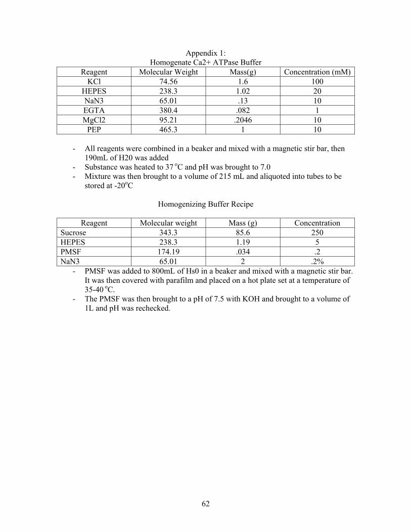
Appendix 1: Homogenate Ca2+ ATPase Buffer
Reagent Molecular Weight Mass(g) Concentration (mM)KCl 74.56 1.6 100
HEPES 238.3 1.02 20 NaN3 65.01 .13 10 EGTA 380.4 .082 1 MgCl2 95.21 .2046 10
PEP 465.3 1 10
- All reagents were combined in a beaker and mixed with a magnetic stir bar, then 190mL of H20 was added
- Substance was heated to 37 oC and pH was brought to 7.0 - Mixture was then brought to a volume of 215 mL and aliquoted into tubes to be
stored at -20oC
Homogenizing Buffer Recipe
Reagent Molecular weight Mass (g) Concentration Sucrose 343.3 85.6 250 HEPES 238.3 1.19 5 PMSF 174.19 .034 .2 NaN3 65.01 2 .2%
- PMSF was added to 800mL of Hs0 in a beaker and mixed with a magnetic stir bar. It was then covered with parafilm and placed on a hot plate set at a temperature of 35-40 oC.
- The PMSF was then brought to a pH of 7.5 with KOH and brought to a volume of 1L and pH was rechecked.
62
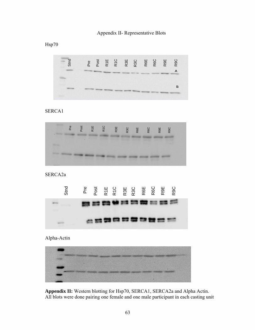
Appendix II- Representative Blots
Hsp70
Pre
Pos
t
R1E
R1C
R3E
R3C R6E
R6C
R9E
R9C
Stn
d
SERCA1
Pre
Pos
t
R1E
R1C
R3E
R3C
R6E
R6C
R9E
R9C
SERCA2a
Pre
Pos
t
R1E
R1C
R3E
R3C R6E
R6C
R9E
R9C
Stn
d
Alpha-Actin
Appendix II: Western blotting for Hsp70, SERCA1, SERCA2a and Alpha Actin. All blots were done pairing one female and one male participant in each casting unit
63
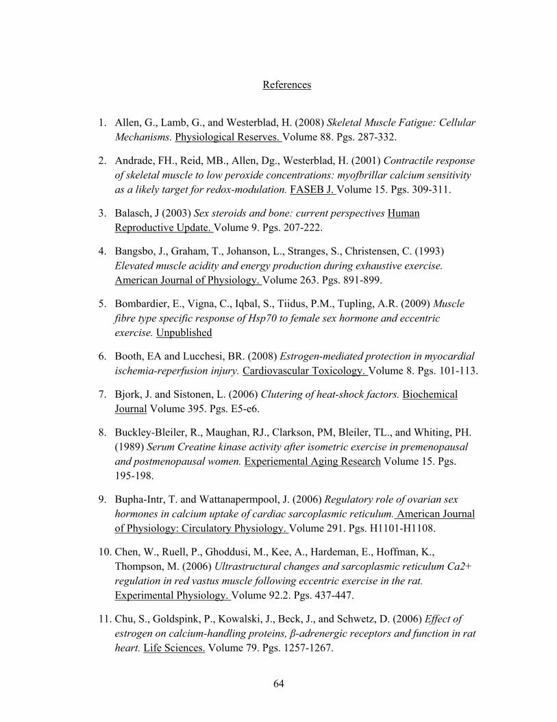
References
1. Allen, G., Lamb, G., and Westerblad, H. (2008) Skeletal Muscle Fatigue: Cellular
Mechanisms. Physiological Reserves. Volume 88. Pgs. 287-332.
2. Andrade, FH., Reid, MB., Allen, Dg., Westerblad, H. (2001) Contractile response of skeletal muscle to low peroxide concentrations: myofbrillar calcium sensitivity as a likely target for redox-modulation. FASEB J. Volume 15. Pgs. 309-311.
3. Balasch, J (2003) Sex steroids and bone: current perspectives Human Reproductive Update. Volume 9. Pgs. 207-222.
4. Bangsbo, J., Graham, T., Johanson, L., Stranges, S., Christensen, C. (1993) Elevated muscle acidity and energy production during exhaustive exercise. American Journal of Physiology. Volume 263. Pgs. 891-899.
5. Bombardier, E., Vigna, C., Iqbal, S., Tiidus, P.M., Tupling, A.R. (2009) Muscle fibre type specific response of Hsp70 to female sex hormone and eccentric exercise. Unpublished
6. Booth, EA and Lucchesi, BR. (2008) Estrogen-mediated protection in myocardial ischemia-reperfusion injury. Cardiovascular Toxicology. Volume 8. Pgs. 101-113.
7. Bjork, J. and Sistonen, L. (2006) Clutering of heat-shock factors. Biochemical Journal Volume 395. Pgs. E5-e6.
8. Buckley-Bleiler, R., Maughan, RJ., Clarkson, PM, Bleiler, TL., and Whiting, PH. (1989) Serum Creatine kinase activity after isometric exercise in premenopausal and postmenopausal women. Experiemental Aging Research Volume 15. Pgs. 195-198.
9. Bupha-Intr, T. and Wattanapermpool, J. (2006) Regulatory role of ovarian sex hormones in calcium uptake of cardiac sarcoplasmic reticulum. American Journal of Physiology: Circulatory Physiology. Volume 291. Pgs. H1101-H1108.
10. Chen, W., Ruell, P., Ghoddusi, M., Kee, A., Hardeman, E., Hoffman, K., Thompson, M. (2006) Ultrastructural changes and sarcoplasmic reticulum Ca2+ regulation in red vastus muscle following eccentric exercise in the rat. Experimental Physiology. Volume 92.2. Pgs. 437-447.
11. Chu, S., Goldspink, P., Kowalski, J., Beck, J., and Schwetz, D. (2006) Effect of estrogen on calcium-handling proteins, β-adrenergic receptors and function in rat heart. Life Sciences. Volume 79. Pgs. 1257-1267.
64
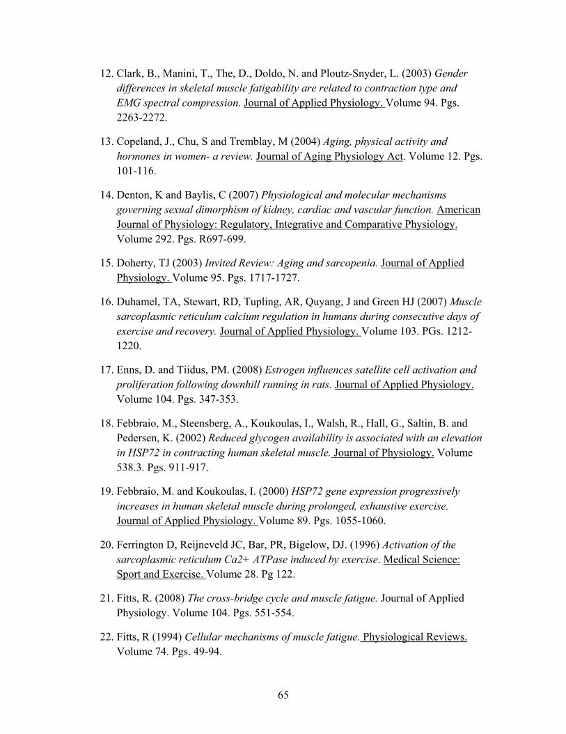
12. Clark, B., Manini, T., The, D., Doldo, N. and Ploutz-Snyder, L. (2003) Gender differences in skeletal muscle fatigability are related to contraction type and EMG spectral compression. Journal of Applied Physiology. Volume 94. Pgs. 2263-2272.
13. Copeland, J., Chu, S and Tremblay, M (2004) Aging, physical activity and hormones in women- a review. Journal of Aging Physiology Act. Volume 12. Pgs. 101-116.
14. Denton, K and Baylis, C (2007) Physiological and molecular mechanisms governing sexual dimorphism of kidney, cardiac and vascular function. American Journal of Physiology: Regulatory, Integrative and Comparative Physiology. Volume 292. Pgs. R697-699.
15. Doherty, TJ (2003) Invited Review: Aging and sarcopenia. Journal of Applied Physiology. Volume 95. Pgs. 1717-1727.
16. Duhamel, TA, Stewart, RD, Tupling, AR, Quyang, J and Green HJ (2007) Muscle sarcoplasmic reticulum calcium regulation in humans during consecutive days of exercise and recovery. Journal of Applied Physiology. Volume 103. PGs. 1212-1220.
17. Enns, D. and Tiidus, PM. (2008) Estrogen influences satellite cell activation and proliferation following downhill running in rats. Journal of Applied Physiology. Volume 104. Pgs. 347-353.
18. Febbraio, M., Steensberg, A., Koukoulas, I., Walsh, R., Hall, G., Saltin, B. and Pedersen, K. (2002) Reduced glycogen availability is associated with an elevation in HSP72 in contracting human skeletal muscle. Journal of Physiology. Volume 538.3. Pgs. 911-917.
19. Febbraio, M. and Koukoulas, I. (2000) HSP72 gene expression progressively increases in human skeletal muscle during prolonged, exhaustive exercise. Journal of Applied Physiology. Volume 89. Pgs. 1055-1060.
20. Ferrington D, Reijneveld JC, Bar, PR, Bigelow, DJ. (1996) Activation of the sarcoplasmic reticulum Ca2+ ATPase induced by exercise. Medical Science: Sport and Exercise. Volume 28. Pg 122.
21. Fitts, R. (2008) The cross-bridge cycle and muscle fatigue. Journal of Applied Physiology. Volume 104. Pgs. 551-554.
22. Fitts, R (1994) Cellular mechanisms of muscle fatigue. Physiological Reviews. Volume 74. Pgs. 49-94.
65
23. Fischer, CP., Hiscock, N., Basu, S., Vessby, B., Kallner, A., Sjogaard, L, Febbraio, MA., Pedersen, B. (2006) Vitamin E isoform-specific inhibition of the exercise-induced heat shock protein 72 response in humans Journal of Applied Physiology. Volume 100. Pgs. 1679-1687.
24. Fowles, J., Green, HJ., Tupling, R., O’Brien, S., and Roy, B. (2002) Human neuromuscular fatigue is associated with altered Na+/K+ ATPase activity following isometric exercise. Journal of Applied Physiology. Vol. 92. Pgs. 1585-1593.
25. Fu, MH and Tupling, AR (2009) Protective effects of Hsp70 on the structure and function of SERCA2a expressed in HEK-293 cells during heat stress. American Journal of Physiology: Heart and Circulatory Physiology. Volume 296. Pgs. H1175-1183.
26. Gonzalez-Hernadez, T and Gonzalez, M (2000) Gender differences and the effect of difference endocrine situations on the NOS expression pattern in the anterior pituitary gland. Journal of Histochemistry and Cytochemistry. Volume 48. Pgs. 1639-1648.
27. Green, H., McKee, NH., Carvalho, A., Phillips, SM. (1997) Reductions in sarcoplasmic reticulum Ca2+ ATPase activity in rat skeletal muscles of different fibre composition with ischemia and reperfusion. Canadian Journal of Physiology and Pharmocology. Volume 75. Pgs. 78-82.
28. Green, GJ, Daub, B., Houston, ME., Thomson, JA., Fraser, I., Raney, D. (1981) Human vastus lateralis and gastrocnemius muscles. A comparative histochemical and biochemical analysis. Journal of Neurology Science Volume 52. Pgs. 201-210.
29. Holloway, G., Green, H.J., and Tupling A.R. (2005) Differential effects of repetitive activity on sarcoplasmic reticulum responses in rat muscle of different oxidative potential. American Journal of Physiology: Regulatory, Integrative and Comparative Physiology. Volume 290. Pgs. R393-R404.
30. Hunter, S., and Enoka, R (2001) Sex differences in the fatigability of arm muscle depends on absolute force during isometric contractions. Journal of Applied Physiology. Volume 21. Pgs. 2686-2694.
31. Iwaki, K., Chi, SH, Dillmann, WH and Mestril R. (1993) Induction of Hsp70 in neonatal cardiomyocytes by hypoxia and metabolic stress. Circulation Research. Volume 87. Pgs. 2023-2032.
66
32. Kelly, DA., Tiidus, P., Houston, M., and Noble, EG (1996) Effect of vitamin E deprivation and exercise training on induction of Hsp70. Journal of Applied Physiology. Volume 81. Pgs. 2379-2385.
33. Khassaf, M., Child, R., McArdle, A. Broodie, A., Esanu, E., and Jackson MJ. (2001) Time course of responses of human skeletal muscle to oxidative stress induced by non-damaging exercise. Journal of Applied Physiology. Volume 90. Pgs. 1031-1035.
34. Knowlton, A., and Sun, L. (2001) Gender differences in the expression of heat shock proteins: the effect of estrogen. American Journal of Physiology: Heart and Circulatory Physiology. Volume 280. Pgs. H455-464.
35. Komi, P and Karlsson J. (1978) Skeletal muscle fibre types, enzyme activities and physical performance in young males and females. Acta Physiology Scandinavian. Volume 103. Pgs. 210-218.
36. Laemmli, UK (1970) Cleavage of structural proteins during the assembly of the head of bacteriophage T4. Nature. Volume 227. Pgs. 680-685.
37. Lee CE, McArdle, A and Griffiths R (2007) The role of hormones, cytokines and heat shock proteins during age related muscle loss. Clinical Nutrition. Volume 26. Pgs. 524-534.
38. Leung S., Teoh, H, Kneung, W and Man, R. (2007) Non-genomic vascular actions of female se hormones: physiological implications and signalling pathways. Clinical Experimental Pharmacological Physiology Volume 34. Pgs. 822-826.
39. Lui, Y., Lornes, W., Wang, L., Reissnecker, S. and Steinacker A. (1999) Different Skeletal Muscle Hsp70 Response to High-Intensity Strength Training and Low-Intensity Endurance Training. European Journal of Applied Physiology. Volume 91. Pgs. 330-335.
40. Liu, Y., Gampert, L., Nething, K., Steinacker, JM. (2006) Response and function
of skeletal muscle heat shock protein 70. Front Biosci. Volume 11. Pgs. 2802-2827.
41. Liu, Y., Mayr, S., Optiz-Gress, A., Zeller, C., Lormes, W., Baur, S., Lehmann, M. and Steinacker, J. (1999) Human skeletal muscle Hsp70 response to training in highly trained rowers. Journal of Applied Physiology. Volume 86. Pgs. 101-104.
67
42. Locke, M. and Noble, E. (1991). Inducible isoform of HSP70 is constitutively expressed in a muscle fiber type specific pattern. American Journal of Physiology: Cell Physiology. Volume 261. Pgs. C774-C779.
43. MacClennan, D. (2000) Ca2+ Signalling and muscle disease. European Journal of Biochemistry. Volume 267. Pgs. 5291-5297.
44. Madamanchi, NR, Li, S, Patternson, C., Runge, MS (2001) Reactive oxygen species regulate heat shock protein 70 via the JAK/STAT pathway. Atheriosclerosis Thrombosis and Vascular Research Volume 21. Pgs. 321-326.
45. Marzetti, E., Lees, A., Wohlgemuth, S, Leeuwenburgh, C. (2009) Sarcopenia of aging: underlying cellular mechanisms and protection by calorie restriction. Biofactors. Volume 35. Pgs. 28-25
46. Maughan, R., Harmon, M., Leiper, J., Sale, D. and Delman A. (1986) Endurance capacity of untrained males and females in isometric and dynamic muscular contractions. European Journal of Physiology. Volume 55. Pgs. 395-400.
47. McArdle, A., Pattwell, D., Vasilaki, A., Griffiths, RD., Jackson, MJ. (2001) Contractile activity-induced oxidative stress: cellular origin and adaptive responses. American Journal of Physiology: Cell Physiology. Volume 280. Pgs. C621-C627.
48. McClung, J., Davis, J. and Carson, J (2006) Ovarian hormone status and skeletal muscle inflammation during recovery from disuse in rats. Experimental Physiology. Volume 92. Pgs. 219-232.
49. Mestril R., Chi, S., Sayen, M., Dillmann, W. (1994) Isolation of a novel inducible rat heat shock protein (HSP70) gene and its expression during ischemia/hypoxia and heat shock. Biochemistry Journal Volume 15. Pgs. 561-569.
50. Milne, KJ, Thorp, DB., Melling, WJ and Noble, EG (2005) Castration inhibits exercise induced accumulation of Hsp70 in male rodent hearts. American Journal of Physiology. Heart and Circulatory Physiology. Volume 290. Pgs. H1610-H1616.
51. Mitsushima, D, Tin-Tin-Win-Shwe, Kimura, F. (2003) Sexual dimorphism in the GABAergic control of gonadotropin release in intact rats. Neuroscience Reserve Volume 46. Pgs. 399-405.
52. Moran, A., Nelson, S., Landisch, R., Warren, G., and Lowe, D. (2007) Estradiol replacement reverses ovariectomy-induced muscle contractile and myosin
68
dysfunction in mature female mice. Journal of Applied Physiology. Volume 102. Pgs. 1387-1393.
53. Morton, J., MacLaren, P., Cable, N., Campbell, N., Evans, L., Bongers, T., Griffiths, R., Kayani, A., McArdle, A., Drust, B. (2007) Elevated core and muscle temperature to levels comparable to exercise do not increase heat shock protein content of skeletal muscle of physically active men. Acta Physiology. Volume 190. Pgs. 319-327.
54. Morton, J., MacLaren, P., Cable, N., Campbell, N., Evans, L., Bongers, T., Griffiths, R., Kayani, A., McArdle, A., Drust, B. (2006) Time Course and differential response of the major heat shock proteins families in human skeletal muscle following acute nondamaging treadmill exercise. Journal of Applied Physiology. Volume 101. Pgs. 176-182.
55. Murphy, RM, Verburg, E., and Lamb, G. (2006) Ca2+ activation of dissusible and bound pools of mu calpain in rat skeletal muscle. Journal of Physiology. Volume 576. Pgs. 595-612.
56. Murphy, RM., Stephensen, D., and Lamb G. (1999) Effect of Creatine on contraction force and sensitivity in mechanical skinned rat skeletal muscle. American Journal of Physiology. Volume 287. Pgs. 1589-1595.
57. Neufer, P., Ordway, G., Hand, G., Shelton, J., Richardson, J., Benjamin, J., Williams, R. (1996) Continuous contractile activity induces fiber type specific expression of HSP70 in skeletal muscle. American Journal of Physiology. Volume 271. Pgs. C1828-C1837.
58. Nickerson, M., Kennedy, S., Johnson, J., and Fleshner, M. (2006) Sexual Dimorphism of the intracellular heat shock protein 72 response. Journal of Applied Physiology. Volume 101. Pgs. 566-575.
59. Noble, E. and Dzialoszynski, T. (2006) Exercise is the primary factor associated with Hsp70 induction in muscle of treadmill running rats. Scandinavian Physiological Society Volume 187. Pgs. 495-501.
60. Noble, E., Milne, K., and Melling, J. (2007) Heat Shock Proteins and Exercise: A Primer.
61. O’Neil, D. and Noble, E (2004) Constitutive expression of inducible Hsp70 is linked to natural shifts in skeletal muscle phenotype. Scandinavian Physiological Society Volume 181. Pgs. 35-41.
69
62. Paroo, Z., Dipchand, E., Noble, E. (2002) Estrogen attenuates post-exercise HSP70 expression in skeletal muscle. American Journal of Physiology: Cell Physiology. Volume 282. Pgs. C245-C251.
63. Paroo, Z., Haist, J., Karmazyn, M. and Noble, E. (2002) Exercise Improves Postischemic Cardiac Function in Males but not females. Circulation Research Volume 90. Pgs. 911-917.
64. Paulsen, G., Vissing, K., Kalhovde, J., Ugelstad, I., Bayer, M., Kadi, F., Schjerling, P, Hallen, J. and Raastad, T. (2007) Maximal eccentric exercise induces a rapid accumulation of small heat shock proteins on myofibrils and a delayed HSP70 response in humans. American Journal of Physiology: Regulatory, Integrative and Comparative Physiology. Volume 293. Pgs. R844-R853.
65. Perez-Gomez, J, Rodriquez, G., Ara, I., Olmediallas, H., Chavarren, J., Gonzalex-Henriquez, JJ, Dorado, C., Calbet, J. (2009) Roles of muscle mass on sprint performance: gender differences? European Journal of Applied Physiology. Volume 102. Pgs. 685-695.
66. Persky, A., Green, P., Stubley, L., Howell, C., Zaulyanov, L., Brazeau, G., Simpkins, J. (1999) Protective effect of estrogens against oxidative damage to heart and skeletal muscle In Vivo and In Vitro. Experimental Biology and Medicine. Volume 223. Pgs. 59-66.
67. Phillips, SK, Rook, K., Siddle, N., Bruce, S., and Woledge, R. (1993) Muscle weakness in women occurs at an earlier age than men, but strength is preserved by hormone replacement therapy. Clinical Science. Volume 84. Pgs. 95-98.
68. Polk, DM and Naqvi, T. (2005) Cardiovascular disease in women: sex differences in presentation, risk factors and evaluation. Current Cardiology Reports. Volume 7. Pgs. 166-172.
69. Puntschart, A., Vogt, M., Widmer, H., Hoppeler, H., Billeter, R. (1996) Hsp70 expression in human skeletal muscle after exercise. Scandinavian Physiological Society. Volume 157. Pgs. 411-417.
70. Rossi, A., and Dirksen, R. (2006) Sarcoplasmic Reticulum: The Dynamic Calcium Governor of Muscle. Muscle and Nerve. Volume 33. Pgs. 715-731.
71. Ropero, A., Alonso-Magdalena, P., Quesada, I., and Nadal, A. (2007) The role of estrogen receptors in the control of energy and glucose homeostasis. Steroids. Volume 73. Pgs. 874-879.
70
72. Russ, D., Kent-Braun, J. (2003) Sex differences in human skeletal muscle fatigue are eliminated under ischemic conditions. Journal of Applied Physiology. Volume 93. Pgs. 2414-2422.
73. Sale, D (1987) Influence of exercise and training on motor unit activation. Exercise and Sport Sciences Reviews. Volume 15. Pgs. 95-151
74. Savage, KJ and Clarkson, PM. (2002) Oral contraceptive use and exercise induced muscle damage and recovery. Contraception. Volume 66. Pgs. 67-71.
75. Schacterle, G and Pollock, R. (1973) A simplified method for the quantitiatvie assay of small amounts of protein in biologic material. Analytical Biochemistry. Volume 51. Pgs. 654-655.
76. Sewright, K., Hubal, M., Kearns, A., Holbrooks, M. and Clarkson, P. (2007) Sex Differences in Response to Maximal Eccentric Exercise. Medical Science in Sports Exercise. Volume 40. Pgs. 242-251.
77. Shumate, J., Brooke, M., Carroll, J and Davis, J. (1979) Increased serum Creatine Kinase after exercise: a sex-linked phenomenon. Neurology Volume 29. Pgs. 902-904.
78. Sitnick, M., Foley, AM., Brown, MB, Spangenburg, EE. (2006) Ovariectomy prevents the recovery of atrophied gastrocnemius skeletal muscle mass. Journal of Applied Physiology. Volume 100. Pgs. 286-293.
79. Srikakulam, R. and Winkelmann, D. (2006) Chaperone-mediated folding and assembly of myosin in striated muscle. Journal of Cell Science. Volume 117. Pgs. 641-652.
80. Sotiriadou, S., Kyparos, A., Albani, M., Arsos, G., Clarke, M., Sidiras, G., Angelopoulou, N and Matziari, C. (2006). Soleus muscle force following downhill running in ovariectomized rats treated with estrogen. Applied Physiology: Nutrition and Metabolism. Volume 31. Pgs. 449-459.
81. Stewart RD, Duhamel TA, Foley, KP, Quyang, J., Smith, IC, Green, HJ (2007) Protection of muscle membrane excitability during prolonged cycle exercise with glucose supplementation. Journal of Applied Physiology. Volume 103. Pgs. 331-339.
82. Stice, J. and Knowlton, A. (2008) Estrogen, NFκB and the Heat Shock Response. Molecular Medicine. Volume 14. Pgs. 517-527.
71
83. Stupka, N., Lowther, S., Chorneyko, K., Bourgeois, J., Hogben, C., and Tarnopolsky, M. (2000) Gender differences in muscle inflammation after eccentric exercise. Journal of Applied Physiology. Volume 89. Pgs. 2325-2332.
84. Szasz, G., Gruber, W., Bernt, E. (1976) Creatine kinase in serum: 1. Determination of optimum reaction conditions. Clinical Chemistry. Volume 22. Pgs. 650-656
85. Tarnopolsky, MA (2008) Sex differences in exercise metabolism and the role of 17-best estradiol. Medical Science Sports and Exercise. Volume 40. Pgs. 648-654
86. Thompson, H.S., Maynard E., Morales, E., Scordilis, S. (2003) Exercise-induced Hsp27, Hsp70 and MAPK responses in human skeletal muscle. Scandinavian Physiological Society. Volume 178. Pgs. 61-72.
87. Thompson, HS., Hyatt, JP, De Souza, MJ and Clarkson, PM (1997) The effects of oral contraceptives on delayed onset muscle soreness following exercise. Contraception Volume 56. Pgs. 59-65.
88. Tiidus, P., Holden, D., Bombardier, E., Zajchowski, S., Enns, D., and Belcastro, A. (2001) Estrogen effect on post-exercise skeletal muscle neutrophil infiltration and calpain activity. Canadian Journal of Physiology and Pharmacology. Volume 79, Pgs. 400-406.
89. Tiidus, P. (2003) Influence of Estrogen on Skeletal Muscle Damage, Inflammation and Repair. Exercise and Sport Science Reviews. Volume 31. Pgs. 40-44.
90. Tiidus, P. (1996) Can estrogens diminish exercise induced muscle damage. Canadian Journal of Applied Physiology. Volume 20. Pgs. 26-38.
91. Tiidus P. (2009) Point: Counterpoint: Estrogen and sex do/do not influence post exercise indexes of muscle damage, inflammation and repair. Journal of Applied Physiology. Volume 106. Pgs. 1010-1015.
92. Timmons, B., Hamadeh, M. and Tarnopolsky, M. (2006) No effect of short-term 17β-estradiol supplementation in healthy men on systemic inflammatory responses to exercise. American Journal of Physiology: Regulatory, Integrative and Comparative Physiology. Vol 291. Pgs. R285-R290.
93. Tupling, AR (2004) Sarcoplasmic reticulum in muscle fatigue and disease: role of the sarcoplasmic reticulum Ca2+ATPase. Canadian Journal of Applied Physiology. Volume 29. Pgs. 308-329.
72
94. Tupling, R., Green, H, Grant, S. Burnett, S. and Ranny, D. (2000) Postcontractile force depression in humans is associated with an impairment in SR Ca2+ pump function. American Journal of Physiology: Regulatory, Integrative and Comparative Physiology. Vol. 278. Pgs. R87-R94,
95. Tupling, AR., Gramolini, A., Duhamel, T., Kondo, H., Ashai, M., Tsuchiya, S., Borrelli, M., Lepock, J., Otsu, K., Hori, M., MacLennan, D., Green, H. (2004) Hsp70 Binds to the fast twitch skeletal muscle sarco(endo)plasmic reticulum Ca2+ ATPase (SERCA1a) and prevents thermal inactivation. Journal of Biological Chemistry. Volume 279. Pgs. 52382-52389.
96. Tupling, AR., Bombardier, E., Stewart, R., Vigna, C., and Aqui, A. (2007) Muscle fibre type specific response of Hsp70 expression in human quadriceps following acute isometric exercise. Journal of Applied Physiology. Volume 103. Pgs. 2105-2111.
97. Tupling, AR., Vigna, C., Ford, R.J., Tsuchiya, SC., Graham, D.A., Denniss, S.G., and Rush, J.W.E. (2007) Effects of buthionine sulfoximine treatment on diaphragm contractility and SR Ca2+ pump function in rats. Journal of Applied Physiology Volume 103. Pgs. 1921-1929.
98. Welch, WJ (1992) Mammalian stress response: cell physiology, structure/function of stress proteins and implications for medicine and disease. Physiology Reserves Volume 72. Pgs. 1063-1081.
99. Wheeler, M (1991) Physical changes of puberty. Endocrinology Metabolism Clinical North American. Volume 20. Pgs. 1-14.
100. Viner, RI., Ferrington DA, Huhmer, A., Bigelow, DJ., Schoneich, C. (1996) Accumulation of nitrotyrosine on the SERCA2a isoform of SR Ca-ATPase of rat skeletal muscle during aging: a peroxynitrite-mediated process? FEBS Lett. Volume 379. Pgs. 286-290.
101.Voss, M., Stallone, J., Li, M., Cornelussen, R., Knuefermann, P., and Knowlton, A. (2003) Gender differences in the expression of heat shock proteins: the effect of estrogen. American Journal of Physiology: Heart and Circulatory Physiology. Volume 285. Pgs. H687-H692.
102. Zubrzycka-Gaarn, E. (1984) Prog Clinical Biological Research. Volume 168. Pgs. 19-23
73