Embed Size (px)
Citation preview
Special Articles
In Defense of the Stethoscope
Raymond LH Murphy Jr MD MPH
IntroductionCase Presentation
Problems With AuscultationImportance of SquawksArguments That Support the Clinical Use of the Stethoscope
Respiratory Acoustic AnalysisGuidelinesCorrelation of Adventitious Sounds and Disease States
Types of SoundsPatterns of Distribution of SoundsComputerized Detection and Quantification
Utility of Auscultation With an Acoustic StethoscopeImprovements in EducationObserver Variability in Medical TestsComputerized Analysis
Single-Channel DevicesMulti-Channel Computerized Analysis
Summary and Recommendations
The stethoscope is widely considered to be an unreliable instrument. Many studies document thesignificant observer variability in its use. Numerous other diagnostic tools are available that aregenerally regarded to provide more reliable diagnostic information. Some even argue that teachingof the ancient art should be de-emphasized in medical schools. Yet auscultation with an acousticstethoscope can provide important, even life-saving, information. The purpose of this article is topresent evidence that supports the use of the stethoscope in clinical medicine. The argument for thestethoscope will be made by presenting relevant investigations, including clinical studies acknowl-edged to meet the criteria of evidence-based medicine. It will focus on studies that have employedcomputerized acoustic technology to correlate lung sounds with disease states. This technology hasadvanced in recent years, which has stimulated a resurgence of interest in auscultation. Numerousstudies have been done that utilized objective methods that circumvented the problem of observervariability. There is now a good deal of scientific evidence to support the hypothesis that lungsounds contain information that is clinically useful. This technology also allows this information tobe collected more efficiently than previously possible. Advances in educational technology havemade it possible to impart information on auscultation much more easily than was possible in thepast. Contrary to predictions, the stethoscope is not likely to be relegated to the museum shelf in thenear future. Computer technology is making it an even more useful clinical instrument. Key words:auscultation, lung sounds, heart sounds, computerized acoustic analysis. [Respir Care 2008;53(3):355–369. © 2008 Daedalus Enterprises]
RESPIRATORY CARE • MARCH 2008 VOL 53 NO 3 355
Introduction
This article was in part stimulated by 2 recent editori-als.1,2 One reviewed the history of the stethoscope andstated that it is safe to assume that the stethoscope wouldbe someday relegated to a museum shelf. The second dis-cussed the demise of the physical examination and pre-sented much information on problems with auscultation.The author stated that physicians are uncomfortable withuncertainty. They want more objective information thanthat obtained by physical examination. It was a balanceddiscussion in that the author also pointed out that even thetests commonly believed to be more accurate have theirsubjectivity and that physical examination could some-times provide “the vital clue” to aid in diagnosis. Themajor thrust of the article, however, was to point out thedeficiencies of physical examination and highlight the dis-dain some physicians have for it. For example, the authordescribed an oncology fellow who, “Even as he wentthrough the motions of teaching physical diagnosis, heappeared to be dismissing it.” The author stated that he,himself, is a bit of an agnostic but dutifully applies hisstethoscope to his patients’ chests “often simply out ofhabit.” The purpose of this article is to present a contraryview by reviewing evidence that supports the use of thestethoscope in clinical medicine.
Though the stethoscope is useful in several clinical ar-eas, including cardiac auscultation and detection of vas-cular abnormalities, this article will focus on auscultationof the lung, as that is the area of my main experience. Toillustrate some of the problems and clinical benefits ofauscultation I will begin with a discussion of a patient witha respiratory problem.
Case Presentation
A 69-year-old white female presented to the emergencydepartment with acute shortness of breath. She had gen-erally been in good health and regularly participated invigorous physical activity. She had a 20-pack-year historyof cigarette smoking but stopped 18 years ago. Severalmonths prior to admission she was seen by her primary
care physician because of a nonproductive cough associ-ated with intermittent dyspnea. Allergy testing was nega-tive. She was started on a bronchodilator for a presumptivediagnosis of new-onset asthma. Review of systems wasunremarkable except for complaints consistent with esoph-ageal reflux. She was intolerant of histamine-2 blockers.The night prior to admission she had a large meal togetherwith several alcoholic beverages. She awoke the next morn-ing markedly short of breath and coughing, and presentedherself to the emergency department. On physical exami-nation she was noted to be hypoxemic, and had a respira-tory rate of 22 breaths/min, temperature of 37.7°C, andpulse of 107 beats/min. The emergency department phy-sician noted mild wheezing and faint inspiratory crackles.The patient’s white-blood-cell count was 13.5 cells/�L. Achest radiograph was interpreted as within normal limits.Three hours after arrival in the emergency room, anotherphysician examined the patient and heard crackles andsquawks, but no wheezing. These findings were confirmedby automated lung-sound analysis. What is the importanceof the squawks, and how should the patient be treated?
Problems With Auscultation
This case illustrates several problems with the currentstate of auscultation. I will comment briefly on the causesof the problems and the developments in technology thatare improving the situation. The house officers and emer-gency physician missed an important clinical finding. Whenit was called to their attention, they thought it unimportant.Fortunately, the attending physician agreed to add an an-tibiotic to the regimen and the patient did well. It is notsurprising that they did not know about the importance ofsquawks. A review of many textbooks on physical diag-nosis failed to find mention of the association of squawkswith pneumonia, even though Laennec described this phe-nomenon nearly 200 years ago.3
It is also not surprising that they paid little attention tothe physical findings. A high percentage of medical pro-fessionals have little regard for chest auscultation. Indeed,Forgacs, a distinguished student of lung sounds, wrote thatit “had been reduced to a perfunctory ritual.”4 There areseveral obvious reasons for these opinions. The reliabilityof auscultation can be legitimately questioned, becauseobserver variability is high, as has been well document-ed.5–7
Furthermore, there are many tools available for diagno-sis and management of patients with cardiopulmonary dis-orders, such as chest radiographs, computed tomography(CT) scans, and magnetic resonance imaging, and theseare widely considered to be more reliable than the stetho-scope. Traditionally, auscultation has been taught at thebedside, and skill in the art is obtained through experience.This has been time-consuming. With many pressures on
Raymond LH Murphy Jr MD MPH is affiliated with Stethographics,Westborough, Massachusetts, and with Faulkner Hospital, Boston, Mas-sachusetts.
Dr Murphy is the founder, chairman, and chief medical officer of Stetho-graphics, Westborough, Massachusetts, which manufactures devices forlung-sound detection, recording, and analysis. He reports no other con-flicts of interest related to the content of this paper.
Correspondence: Raymond LH Murphy Jr MD MPH, Pulmonary De-partment, Faulkner Hospital, 1153 Centre Street, Suite 40, Boston MA02130. E-mail: [email protected].
IN DEFENSE OF THE STETHOSCOPE
356 RESPIRATORY CARE • MARCH 2008 VOL 53 NO 3
their time, physicians in training are likely to focus moreattention on the other tools for diagnosis. In addition, theproblems with auscultation have been compounded by dif-ficulties with nomenclature. This had become so severethat it had been said that “almost every physician had hisown terminology.”8 Fortunately this problem has beengreatly reduced by the publication of guidelines, as will bediscussed in more detail below.9,10 More importantly, therehas been a resurgence of interest in lung sounds, and nu-merous studies have been done that have improved ourknowledge of this ancient art. This has been stimulated bythe development of computerized acoustic technology.Comprehensive reviews of these studies have been pre-sented.7,11–13 I will present the evidence that shows thatthere is objective information that supports the conceptthat auscultation of the chest, when done by a knowledge-able clinician, remains clinically useful and is indeed im-portant. The recommendation to treat this patient with an-tibiotics was the result of a clinician having knowledge ofstudies done with computer-assisted auscultation. I willalso present other examples where auscultation remainsuseful.
Importance of Squawks
Squawks are short, inspiratory, musical sounds that havea rather distinctive sound that is easily recognizable, oncelearned. Squawks have been described in patients withhypersensitivity pneumonitis and fibrotic lung disorders.14
They also have been reported to be present in 15% ofpatients with pneumonia.15 When squawks are heard andthere is no evidence of hypersensitivity or other restrictivedisorders and the patient has an acute syndrome consistentwith respiratory infection, their presence provides rela-
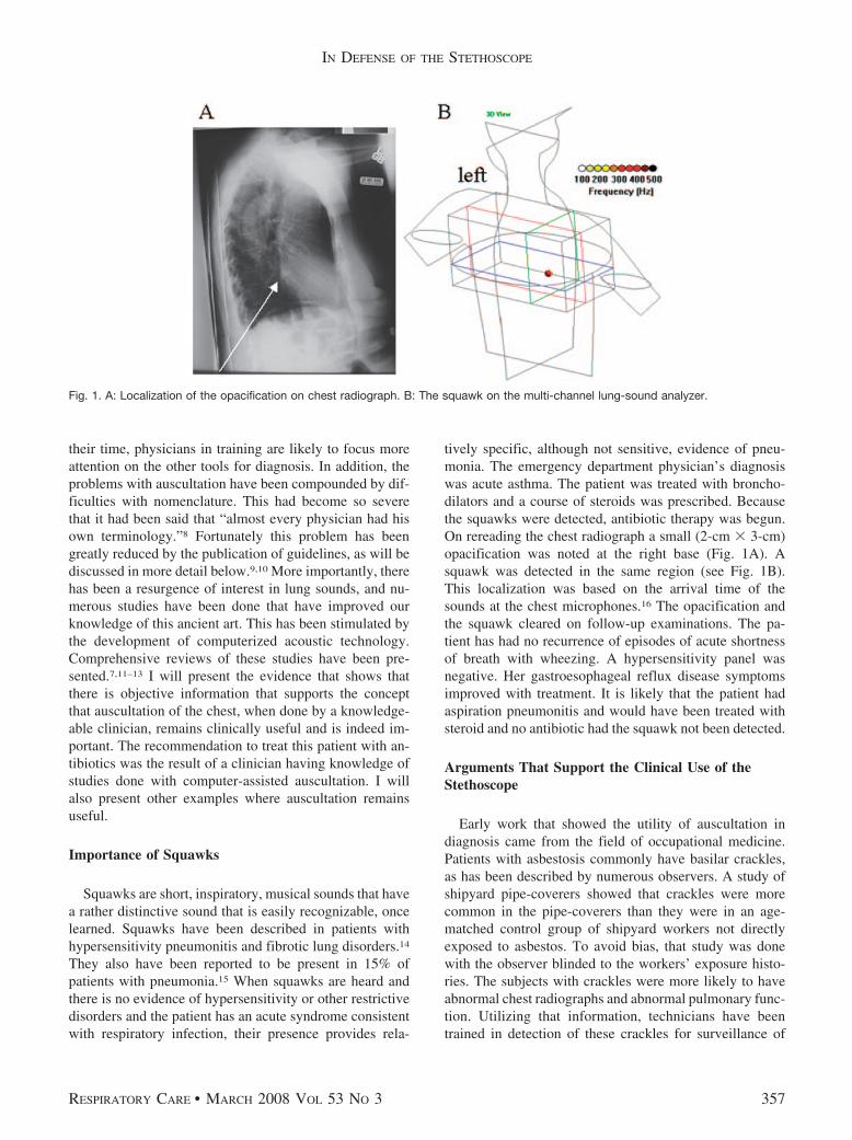
tively specific, although not sensitive, evidence of pneu-monia. The emergency department physician’s diagnosiswas acute asthma. The patient was treated with broncho-dilators and a course of steroids was prescribed. Becausethe squawks were detected, antibiotic therapy was begun.On rereading the chest radiograph a small (2-cm � 3-cm)opacification was noted at the right base (Fig. 1A). Asquawk was detected in the same region (see Fig. 1B).This localization was based on the arrival time of thesounds at the chest microphones.16 The opacification andthe squawk cleared on follow-up examinations. The pa-tient has had no recurrence of episodes of acute shortnessof breath with wheezing. A hypersensitivity panel wasnegative. Her gastroesophageal reflux disease symptomsimproved with treatment. It is likely that the patient hadaspiration pneumonitis and would have been treated withsteroid and no antibiotic had the squawk not been detected.
Arguments That Support the Clinical Use of theStethoscope
Early work that showed the utility of auscultation indiagnosis came from the field of occupational medicine.Patients with asbestosis commonly have basilar crackles,as has been described by numerous observers. A study ofshipyard pipe-coverers showed that crackles were morecommon in the pipe-coverers than they were in an age-matched control group of shipyard workers not directlyexposed to asbestos. To avoid bias, that study was donewith the observer blinded to the workers’ exposure histo-ries. The subjects with crackles were more likely to haveabnormal chest radiographs and abnormal pulmonary func-tion. Utilizing that information, technicians have beentrained in detection of these crackles for surveillance of
Fig. 1. A: Localization of the opacification on chest radiograph. B: The squawk on the multi-channel lung-sound analyzer.
IN DEFENSE OF THE STETHOSCOPE
RESPIRATORY CARE • MARCH 2008 VOL 53 NO 3 357
workers exposed to asbestos.17 Validation of these obser-vations became clearer after the development of comput-erized techniques. The detection of crackles via time-ex-panded waveform analysis has been shown, by British andFinnish investigators, to be as good as CT scans in detect-ing asbestosis.18,19
The accuracy of lung sounds for the identification ofdisease states in studies cited to follow the rules of evi-dence-based medicine were also confirmatory.20–22 Ex-pressed in terms of likelihood ratio, fine end-inspiratorycrackles were associated with pulmonary fibrosis with alikelihood ratio of �5.9. Other observations also cited tomeet these criteria include: wheezing during quiet breath-ing predicts asthma (likelihood ratio �6) or chronic bron-chitis (likelihood ratio �6). Fine or coarse early inspira-tory crackles were associated with chronic bronchitis(likelihood ratio �14 to �20); decreased breath soundswere associated with emphysema (likelihood ratio �10.2).Though the association of fine crackles with pulmonaryfibrosis is strong, as noted, fine and coarse crackles werealso found in congestive heart failure (likelihood ratio�3.4). This calls attention to the fact that there are dangersthat can arise from the misinterpretation of auscultationfindings. Patients with interstitial fibrosis are commonlygiven diuretics inappropriately. Their crackles are mis-taken for those of heart failure. Similarly, the cause ofwheezing may be misinterpreted. A diagnosis of cardiacasthma may be missed if the wheezing is mistakenly at-tributed to bronchial rather than cardiac asthma. Theseobservations reaffirm the importance of proper education
in physical diagnosis. It brings to mind the question oftenposed to medical students: What is the most important partof the stethoscope? The answer, of course, is the partbetween the ear-pieces.
Respiratory Acoustic Analysis
In the year 2000 the European Respiratory Societypointed out that over the past “30 years, computerizedmethods for recording analysis of respiratory sounds haveovercome the many limitations of simple auscultation. Re-spiratory acoustic analysis can now quantify changes inlung sounds, make permanent records of the measurementsmade, and produce graphical representations that help withthe diagnosis and management of patients suffering fromchest diseases.”10
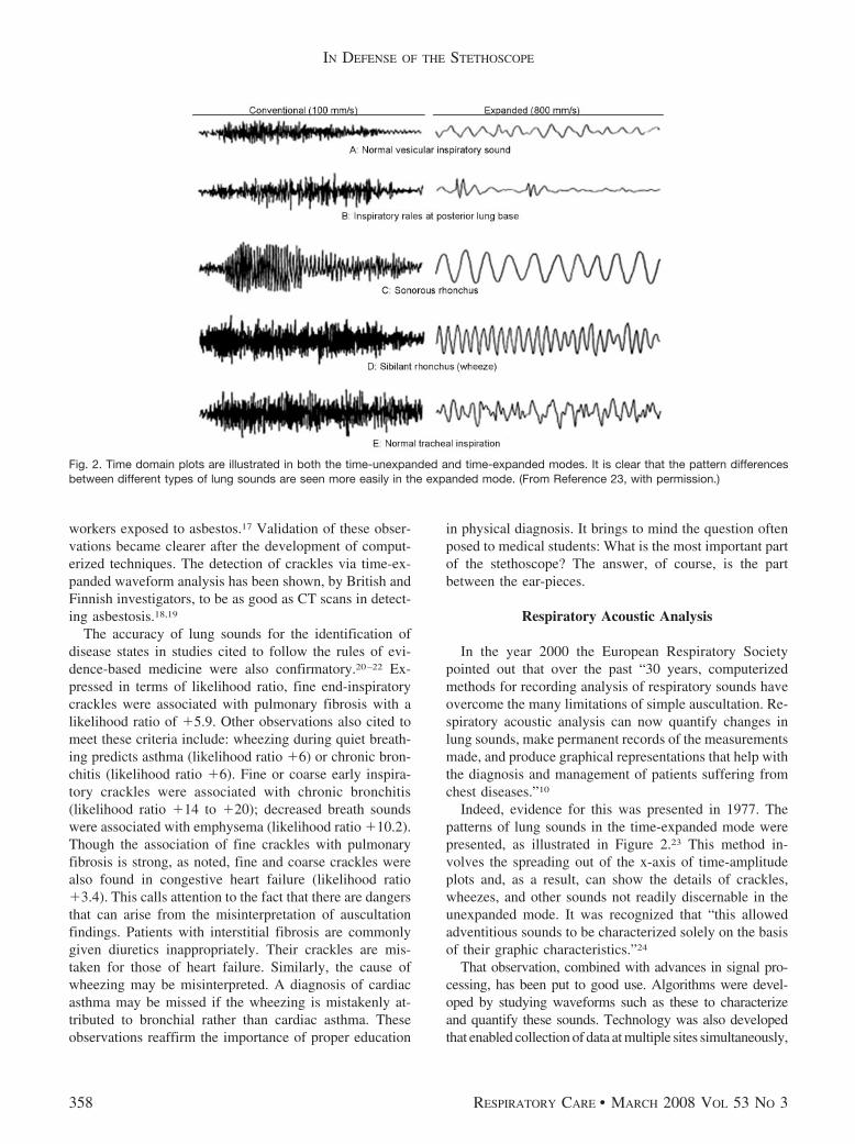
Indeed, evidence for this was presented in 1977. Thepatterns of lung sounds in the time-expanded mode werepresented, as illustrated in Figure 2.23 This method in-volves the spreading out of the x-axis of time-amplitudeplots and, as a result, can show the details of crackles,wheezes, and other sounds not readily discernable in theunexpanded mode. It was recognized that “this allowedadventitious sounds to be characterized solely on the basisof their graphic characteristics.”24
That observation, combined with advances in signal pro-cessing, has been put to good use. Algorithms were devel-oped by studying waveforms such as these to characterizeand quantify these sounds. Technology was also developedthat enabled collection of data at multiple sites simultaneously,
Fig. 2. Time domain plots are illustrated in both the time-unexpanded and time-expanded modes. It is clear that the pattern differencesbetween different types of lung sounds are seen more easily in the expanded mode. (From Reference 23, with permission.)
IN DEFENSE OF THE STETHOSCOPE
358 RESPIRATORY CARE • MARCH 2008 VOL 53 NO 3
which allows presentation of the patterns of distribution ofnormal and abnormal sounds on the chest.25 This provides amore organized approach to the study of the correlation ofacoustic findings with disease states and calls attention toboth the strengths and potential pitfalls of auscultation. Inaddition, this work led to the development of guidelines tohelp clear up the previously confused terminology.
Guidelines
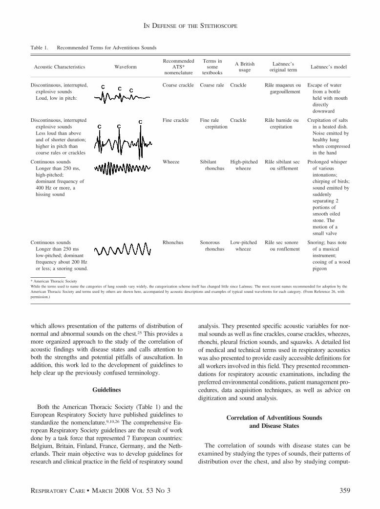
Both the American Thoracic Society (Table 1) and theEuropean Respiratory Society have published guidelines tostandardize the nomenclature.9,10,26 The comprehensive Eu-ropean Respiratory Society guidelines are the result of workdone by a task force that represented 7 European countries:Belgium, Britain, Finland, France, Germany, and the Neth-erlands. Their main objective was to develop guidelines forresearch and clinical practice in the field of respiratory sound
analysis. They presented specific acoustic variables for nor-mal sounds as well as fine crackles, coarse crackles, wheezes,rhonchi, pleural friction sounds, and squawks. A detailed listof medical and technical terms used in respiratory acousticswas also presented to provide easily accessible definitions forall workers involved in this field. They presented recommen-dations for respiratory acoustic examinations, including thepreferred environmental conditions, patient management pro-cedures, data acquisition techniques, as well as advice ondigitization and sound analysis.
Correlation of Adventitious Soundsand Disease States
The correlation of sounds with disease states can beexamined by studying the types of sounds, their patterns ofdistribution over the chest, and also by studying comput-
Table 1. Recommended Terms for Adventitious Sounds
Acoustic Characteristics WaveformRecommended
ATS*nomenclature
Terms insome
textbooks
A Britishusage
Laennec’soriginal term
Laennec’s model
Discontinuous, interrupted,explosive soundsLoud, low in pitch:
Coarse crackle Coarse rale Crackle Rale muqueux ougargouillement
Escape of waterfrom a bottleheld with mouthdirectlydownward
Discontinuous, interruptedexplosive soundsLess loud than aboveand of shorter duration;higher in pitch thancoarse rales or crackles
Fine crackle Fine ralecrepitation
Crackle Rale humide oucrepitation
Crepitation of saltsin a heated dish.Noise emitted byhealthy lungwhen compressedin the hand
Continuous soundsLonger than 250 ms,high-pitched;dominant frequency of400 Hz or more, ahissing sound
Wheeze Sibilantrhonchus
High-pitchedwheeze
Rale sibilant secou sifflement
Prolonged whisperof variousintonations;chirping of birds;sound emitted bysuddenlyseparating 2portions ofsmooth oiledstone. Themotion of asmall valve
Continuous soundsLonger than 250 mslow-pitched; dominantfrequency about 200 Hzor less; a snoring sound.
Rhonchus Sonorousrhonchus
Low-pitchedwheeze
Rale sec sonoreou ronflement
Snoring; bass noteof a musicalinstrument;cooing of a woodpigeon
* American Thoracic SocietyWhile the terms used to name the categories of lung sounds vary widely, the categorization scheme itself has changed little since Laennec. The most recent names recommended for adoption by theAmerican Thoracic Society and terms used by others are shown here, accompanied by acoustic descriptions and examples of typical sound waveforms for each category. (From Reference 26, withpermission.)
IN DEFENSE OF THE STETHOSCOPE
RESPIRATORY CARE • MARCH 2008 VOL 53 NO 3 359
erized analysis of the type, quantity, timing, and waveformdifferences in amplitude of the sounds.
Types of Sounds
Table 2 summarizes the findings obtained with a multi-channel lung-sound analyzer in a convenience sample ofpatients with common illnesses who were studied in acommunity teaching hospital. This device collects datawith microphones imbedded in a soft foam mat that isplaced on the back and the lateral bases, as has been de-scribed.25 It is illustrated in Figure 3. Fine and coarsecrackles, wheezes, rhonchi, and squawks are identified andcounted by this device.
Examination of Table 2 calls attention to an importantproblem with auscultation. There is considerable overlapin the presence of abnormal sounds in common illnesses.As a consequence, correlation of the type of sound withdisease states can be difficult. For example, as can be seenin this table, wheezing is more common in asthma than inthe other conditions. Fifty-nine percent of the patients withasthma had some wheezing at the time they were studied.However, inspection of the table also shows that wheezingwas not uncommon in pneumonia, chronic obstructive pul-monary disease, and congestive heart failure. This bringsto mind the adage, perhaps forgotten by the physicians inthis case, “All that wheezes is not asthma.”
There is also overlap in other sounds. Rhonchi are alsoseen to be almost equally common in pneumonia, conges-tive heart failure, chronic obstructive pulmonary disease,and asthma. Crackles are also seen to be present in allthese conditions. They occur even in those with no recog-nized cardiopulmonary disorder, as has been reported.27,28
Although it can be difficult to separate disease states bythe type of their adventitious sounds, there are some ex-ceptions. Expiratory crackles are more common in pneu-monia than they are in asthma, chronic obstructive pulmo-nary disease, or congestive heart failure; when detected inan acutely ill patient with no history of chronic lung dis-ease, the possibility of pneumonia should be considered.
The crackles in patients with interstitial pulmonary fi-brosis are fairly distinctive. As can be seen in the table, thehighest rates of crackles are seen in this disorder; the in-spiratory crackles averaged 25 � 12 per breath. Recogni-tion of this sound is often the first clue that interstitialpulmonary fibrosis is present.
Patterns of Distribution of Sounds
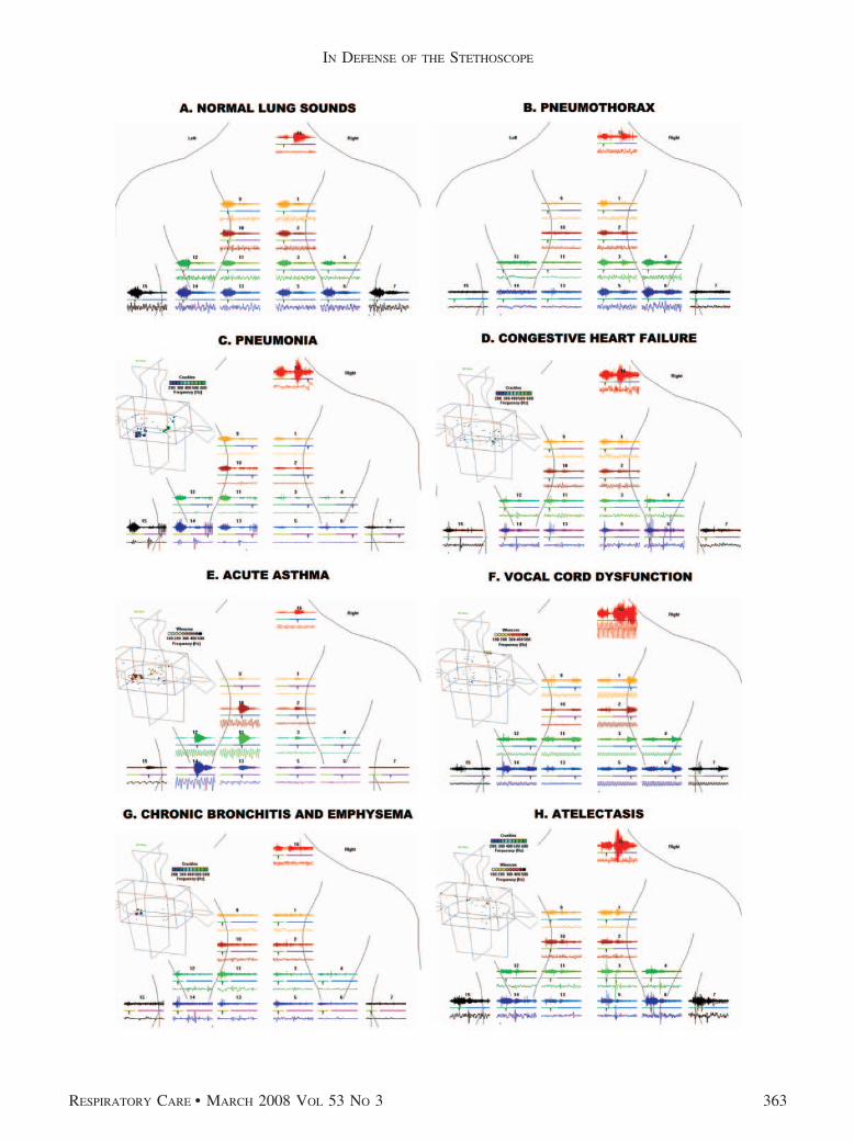
The pattern of distribution of sounds over the chest canalso be obtained with a multi-channel lung-sound ana-lyzer. Pattern differences are often helpful in separatingdiseases from each other. Typical patterns in common con-ditions are illustrated in Figure 4. The pattern seen in a
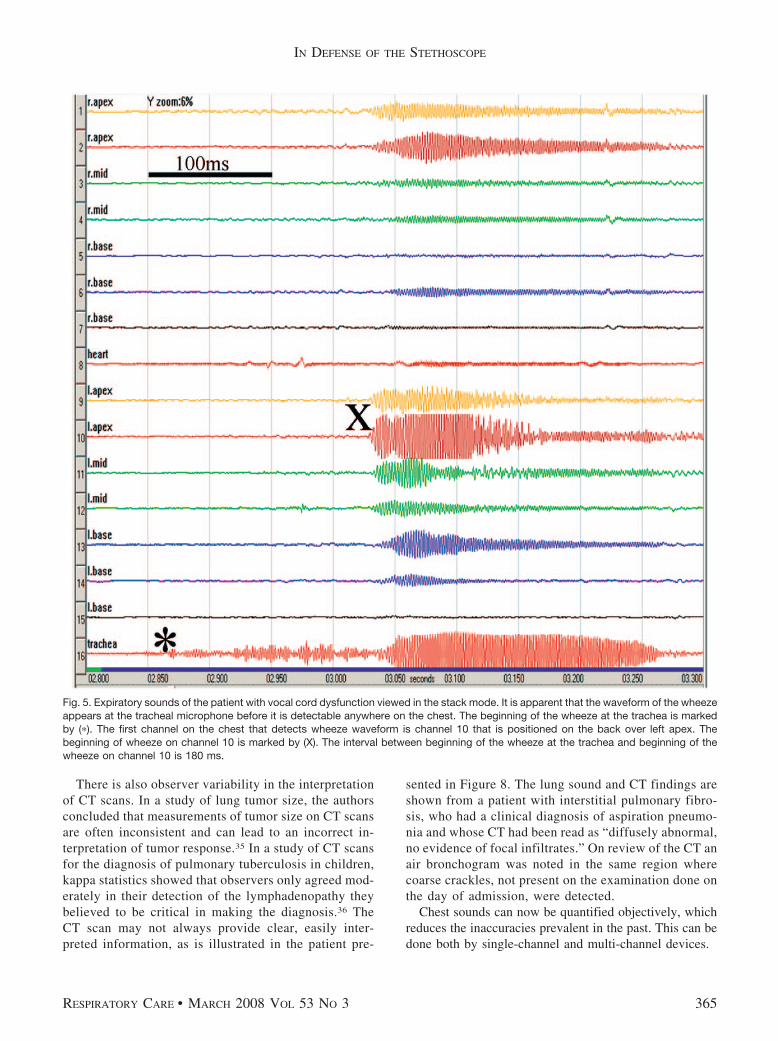
patient with vocal cord dysfunction is presented in Fig-ure 5. It illustrates the observation that presentation of thesimultaneously recorded waveforms in a stack mode pro-vides evidence as to the origin of the sound. In this casethe wheeze pattern is seen to appear first at the micro-phone placed over the trachea.
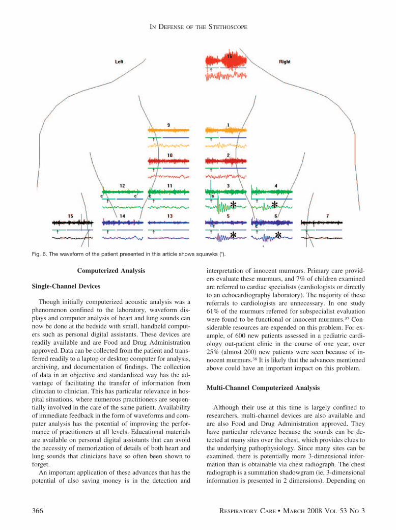
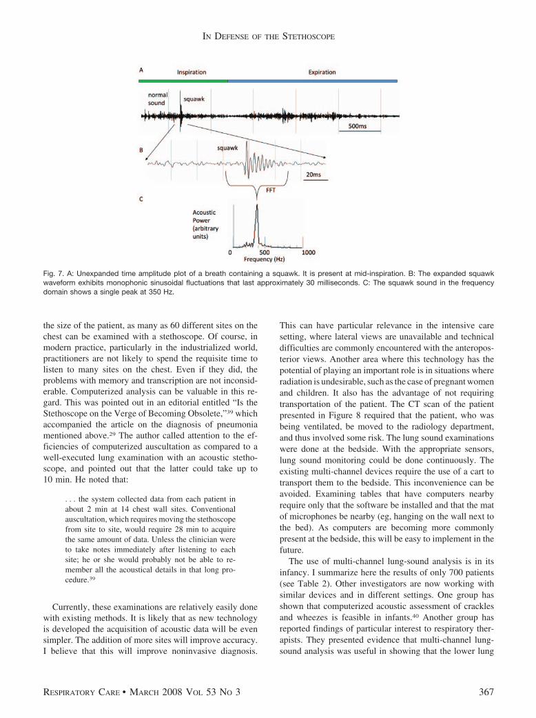
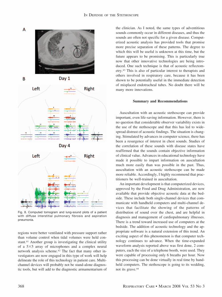
As noted, the automated analysis of the lung sounds ofthe patient presented in this article confirmed the obser-vations of one of the clinicians and provided objectiveevidence that supports the diagnosis of pneumonia. Thedetection of squawks was particularly helpful (Figure 6).Their waveforms are distinctive, as is illustrated in Fig-ure 7. Information can also be obtained from the way thesound patterns change. The patient whose CT is presentedin Figure 8 had interstitial pulmonary fibrosis. On beingintubated because of respiratory failure, she was observedto aspirate. On the 4th day of her stay in the intensive careunit she developed a fever and an elevated white-blood-cell count. Her CT was interpreted as showing “diffuseinfiltrates, no focal abnormalities.” She developed coarsecrackles over her left base, where in retrospect an air bron-chogram consistent with pneumonia was noted on the CT.There was some increased opacification consistent withpneumonia at her right base as well. Rhonchi were de-tected over the right lung that were not present on day 1.The rhonchi were probably due to phlegm in the airways,associated with the aspiration. They were probably not dueto the interstitial pulmonary fibrosis. As can be seen inTable 2, none of the 19 patients with interstitial pulmonaryfibrosis had rhonchi.
Computerized Detection and Quantification
The type, quantity, and waveform differences in ampli-tude and timing of the sounds, as well as other features,can be calculated automatically by computer. In one ex-ample of this approach, an algorithm for detecting pneu-monia separated patients diagnosed with this disease fromage-matched controls with a sensitivity of 0.78, a speci-ficity of 0.88, and a positive predictive power of 0.93.29 Inanother example, multi-channel lung-sound analysis wasshown to offer promise of help in separating the cracklesof interstitial pulmonary fibrosis from those of congestiveheart failure. The transmission of individual crackles overthe chest can be tracked by a computer and differs in the2 disease states.30
Utility of Auscultation With an Acoustic Stethoscope
Such objective measurements demonstrate that, despitethe difficulties with auscultation mentioned above, patterndifferences exist that can help in diagnosis of cardiopul-monary disorders. This provides support for the conceptthat useful information is obtainable via auscultation, when
IN DEFENSE OF THE STETHOSCOPE
360 RESPIRATORY CARE • MARCH 2008 VOL 53 NO 3
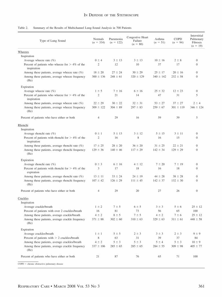
Table 2. Summary of the Results of Multichannel Lung-Sound Analysis in 700 Patients
Type of Lung SoundNormals
(n � 334)Pneumonia(n � 122)
Congestive HeartFailure
(n � 80)
Asthma(n � 51)
COPD(n � 94)
InterstitialPulmonary
Fibrosis(n � 19)
WheezesInspiration
Average wheeze rate (%) 0 � 4 3 � 13 3 � 13 10 � 16 2 � 8 0Percent of patients who wheeze for � 4% of the
inspiration2 12 10 37 17 0
Among these patients, average wheeze rate (%) 18 � 20 27 � 24 30 � 29 25 � 17 20 � 16 0Among these patients, average wheeze frequency
(Hz)300 � 136 248 � 61 320 � 129 340 � 142 232 � 58 0
ExpirationAverage wheeze rate (%) 1 � 5 7 � 16 6 � 16 25 � 32 12 � 23 0Percent of patients who wheeze for � 4% of the
inspiration2 21 14 47 31 5
Among these patients, average wheeze rate (%) 22 � 29 30 � 22 32 � 31 53 � 27 37 � 27 2 � 4Among these patients, average wheeze frequency
(Hz)309 � 122 304 � 89 297 � 83 259 � 67 301 � 119 346 � 124
Percent of patients who have either or both 4 29 16 59 39 5
RhonchiInspiration
Average rhonchi rate (%) 0 � 1 5 � 13 3 � 12 5 � 15 3 � 11 0Percent of patients with rhonchi for � 4% of the
inspiration2 16 8 16 15 0
Among these patients, average rhonchi rate (%) 17 � 25 28 � 20 36 � 20 31 � 25 22 � 21 0Among these patients, average rhonchi frequency
(Hz)129 � 36 140 � 46 117 � 29 142 � 34 129 � 29 0
ExpirationAverage rhonchi rate (%) 0 � 3 6 � 16 4 � 12 7 � 20 7 � 19 0Percent of patients with rhonchi for � 4% of the
expiration3 17 18 16 18 0
Among these patients, average rhonchi rate (%) 13 � 11 33 � 24 24 � 19 44 � 28 38 � 28 0Among these patients, average rhonchi frequency
(Hz)107 � 42 126 � 29 111 � 45 142 � 37 132 � 30 0
Percent of patients who have either or both 4 29 20 27 26 0
CracklesInspiration
Average crackle/breath 1 � 2 7 � 5 6 � 5 3 � 3 5 � 6 25 � 12Percent of patients with over 2 crackles/breath 16 81 73 56 65 100Among these patients, average crackle/breath 4 � 2 8 � 5 7 � 5 4 � 2 7 � 6 25 � 12Among these patients, average crackle frequency
(Hz)371 � 88 302 � 60 310 � 63 329 � 63 311 � 61 448 � 58
ExpirationAverage crackles/breath 1 � 1 5 � 5 2 � 3 3 � 3 2 � 3 9 � 9Percent of patients with � 2 crackles/breath 8 63 31 39 37 84Among these patients, average crackles/breath 4 � 2 5 � 3 5 � 3 5 � 4 5 � 3 10 � 9Among these patients, average crackle frequency
(Hz)337 � 106 285 � 65 285 � 65 284 � 55 309 � 98 405 � 77
Percent of patients who have either or both 21 87 76 65 71 100
COPD � chronic obstructive pulmonary disease
IN DEFENSE OF THE STETHOSCOPE
RESPIRATORY CARE • MARCH 2008 VOL 53 NO 3 361
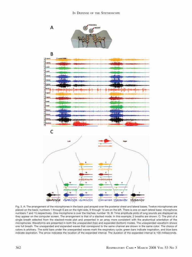
Fig. 3. A: The arrangement of the microphones in the back-pad arrayed over the posterior chest and lateral bases. Twelve microphones areplaced on the back; numbers 1 through 6 are on the right side, 9 through 14 are on the left. There is one on each lateral base: microphonenumbers 7 and 15 respectively. One microphone is over the trachea: number 16. B: Time amplitude plots of lung sounds are displayed asthey appear on the computer screen. The arrangement is that of a stacked mode. In this example, 2 breaths are shown. C: The plot of asingle breath selected from the stacked-mode plot and presented in an array more consistent with the anatomical orientation of themicrophones. Waveforms are presented in both the unexpanded (top) and expanded (bottom) modes. The unexpanded waveform showsone full breath. The unexpanded and expanded waves that correspond to the same channel are shown in the same color. The choice ofcolors is arbitrary. The solid bars under the unexpanded waves mark the respiratory cycle: green bars indicate inspiration, and blue barsindicate expiration. The arrow indicates the location of the expanded interval. The duration of the expanded interval is 100 milliseconds.
IN DEFENSE OF THE STETHOSCOPE
362 RESPIRATORY CARE • MARCH 2008 VOL 53 NO 3
IN DEFENSE OF THE STETHOSCOPE
RESPIRATORY CARE • MARCH 2008 VOL 53 NO 3 363
done by a knowledgeable clinician. It also helps explainthe apparent paradox of an unreliable instrument that con-tinues to be widely used.
Auscultation, particularly when combined with medicalhistory and the other modalities of physical diagnosis, canguide steps taken in patient management before any of theadvanced technologies are employed. Examples are as fol-lows.
Collapse of one lung can be detected by the absence ormarked decrease of lung sounds on one side. Treatment forthis condition is usually done immediately, particularly inthe severely ill, based on auscultation findings, and can belife-saving.
Stridor can be heard when foreign bodies are in largeairways. This is a potentially life-threatening situation, andaction can be taken to remove the offending material oncethis sound is recognized (eg, a Heimlich maneuver can beperformed to treat the “cocktail coronary”). Diffuse wheez-ing points toward a diagnosis of acute bronchial asthma,and in a patient with a known history of asthma broncho-dilators are often administered before other testing is done.
Fluid can be withdrawn from the pleural space reliably,based on physical findings. The presence of numerouscrackles on auscultation in a patient presenting with lungcongestion secondary to heart failure is used to guide theadministration of medications to reduce the fluid prior tomore definitive testing.
The stethoscope is not likely to be relegated to the mu-seum shelf in the near future. On the contrary, its useful-ness will be increased by application of recent technolog-ical advances, including the tools to facilitate instruction inits use.
Improvements in Education
As I point out, taking advantage of the important infor-mation that can be provided by auscultation requires aknowledgeable clinician. Fortunately, it is now easier forclinicians to become knowledgeable. The teaching of aus-
cultation has been improved because of the availability ofaudio recordings and advanced multimedia educational ma-terials. It has been shown that the availability of visual aswell as auditory presentations of sound has improved thelearning process.31 This takes advantage of the fact thatmany people are predominantly visual rather than predom-inantly auditory learners. In addition the information im-parted is now more accurate because it is based on acous-tic analysis, agreed-upon criteria, and correlation studiesof acoustic and clinical findings. The teaching of auscul-tation has also improved with other advances in technol-ogy. An interesting example is the use of video analysis tomonitor caregivers’ performance. In a case report that usedthis technology it was documented that the failure to useauscultation with a stethoscope was a likely contributor tothe prolonging of an uncorrected misplaced endotrachealtube.32 By giving direct feedback, the video aided in theeducation process.
The clinical benefit of acoustic pattern studies is illus-trated in the case study of the patient with aspiration pneu-monia. The reason that one clinician was able to detect andproperly interpret the squawk in the patient presented aboveis that he had studied the waveforms of many patients withpneumonia, as well as other illnesses. He was thus able to“rediscover” an observation made by Laennec.
Observer Variability in Medical Tests
It was recently pointed out that one of the reasons forthe demise of the physical examination is that physicianswant more precise tools.2 Although the problems with ob-server variability in detection of lung sounds are widelyrecognized, it is less widely recognized that there is sub-stantial observer variability in clinical data collection ingeneral, including the interpretation of chest radio-graphs.33,34 This was also illustrated in this case. Severalexperienced observers missed the small patch of retrocar-diac opacification.
Fig. 4. Waveforms in common cardiopulmonary conditions. A: Normal. In this subject there is no lung disease. There are no wheezes andno crackles. The time amplitude pattern seen over the chest is relatively uniform from site to site. The expiratory phase has its highestamplitude over the trachea and has relatively low amplitude at sites distant from large airways. The amplitude is of intermediate value atsites close to large airways (bronchovesicular). B: This waveform pattern was seen in a 26-year-old male who presented with chest pain.The amplitudes of the sounds on the left side were markedly decreased. This led to a diagnosis of spontaneous pneumothorax, prior to itsconfirmation via chest radiograph. C: In this patient with pneumonia, crackles are concentrated at the left base, where opacification wasdetected on radiograph. Both inspiratory and expiratory crackles are present. The insert shows 3-dimensional localization of abnormal lungsounds. The cubes indicate crackles. The spheres indicate wheezes and rhonchi. The color indicates the frequency of the sound. The sizeof the markers is proportional to the crackle and wheeze rate. D: In this patient with congestive heart failure, inspiratory crackles are presentat both bases. E: In this patient with acute bronchial asthma, substantial expiratory wheezing is present. F: In this patient with vocal corddysfunction, diffuse expiratory wheezing is also present. The intensity over the trachea is higher than that over the chest sites. G: The patternof the distribution of the sounds in a typical patient with chronic obstructive pulmonary disease shows that the amplitude tends to bemarkedly decreased, but the degree of this decrease varies from site to site. The expiratory phase is relatively prolonged. H: Bibasilar finecrackles as they appeared in this healthy male undergoing spinal anesthesia. They were not present preoperatively and cleared promptlyafter the patient took several deep breaths.
IN DEFENSE OF THE STETHOSCOPE
364 RESPIRATORY CARE • MARCH 2008 VOL 53 NO 3
There is also observer variability in the interpretationof CT scans. In a study of lung tumor size, the authorsconcluded that measurements of tumor size on CT scansare often inconsistent and can lead to an incorrect in-terpretation of tumor response.35 In a study of CT scansfor the diagnosis of pulmonary tuberculosis in children,kappa statistics showed that observers only agreed mod-erately in their detection of the lymphadenopathy theybelieved to be critical in making the diagnosis.36 TheCT scan may not always provide clear, easily inter-preted information, as is illustrated in the patient pre-
sented in Figure 8. The lung sound and CT findings areshown from a patient with interstitial pulmonary fibro-sis, who had a clinical diagnosis of aspiration pneumo-nia and whose CT had been read as “diffusely abnormal,no evidence of focal infiltrates.” On review of the CT anair bronchogram was noted in the same region wherecoarse crackles, not present on the examination done onthe day of admission, were detected.
Chest sounds can now be quantified objectively, whichreduces the inaccuracies prevalent in the past. This can bedone both by single-channel and multi-channel devices.
Fig. 5. Expiratory sounds of the patient with vocal cord dysfunction viewed in the stack mode. It is apparent that the waveform of the wheezeappears at the tracheal microphone before it is detectable anywhere on the chest. The beginning of the wheeze at the trachea is markedby (�). The first channel on the chest that detects wheeze waveform is channel 10 that is positioned on the back over left apex. Thebeginning of wheeze on channel 10 is marked by (X). The interval between beginning of the wheeze at the trachea and beginning of thewheeze on channel 10 is 180 ms.
IN DEFENSE OF THE STETHOSCOPE
RESPIRATORY CARE • MARCH 2008 VOL 53 NO 3 365
Computerized Analysis
Single-Channel Devices
Though initially computerized acoustic analysis was aphenomenon confined to the laboratory, waveform dis-plays and computer analysis of heart and lung sounds cannow be done at the bedside with small, handheld comput-ers such as personal digital assistants. These devices arereadily available and are Food and Drug Administrationapproved. Data can be collected from the patient and trans-ferred readily to a laptop or desktop computer for analysis,archiving, and documentation of findings. The collectionof data in an objective and standardized way has the ad-vantage of facilitating the transfer of information fromclinician to clinician. This has particular relevance in hos-pital situations, where numerous practitioners are sequen-tially involved in the care of the same patient. Availabilityof immediate feedback in the form of waveforms and com-puter analysis has the potential of improving the perfor-mance of practitioners at all levels. Educational materialsare available on personal digital assistants that can avoidthe necessity of memorization of details of both heart andlung sounds that clinicians have so often been shown toforget.
An important application of these advances that has thepotential of also saving money is in the detection and
interpretation of innocent murmurs. Primary care provid-ers evaluate these murmurs, and 7% of children examinedare referred to cardiac specialists (cardiologists or directlyto an echocardiography laboratory). The majority of thesereferrals to cardiologists are unnecessary. In one study61% of the murmurs referred for subspecialist evaluationwere found to be functional or innocent murmurs.37 Con-siderable resources are expended on this problem. For ex-ample, of 600 new patients assessed in a pediatric cardi-ology out-patient clinic in the course of one year, over25% (almost 200) new patients were seen because of in-nocent murmurs.38 It is likely that the advances mentionedabove could have an important impact on this problem.
Multi-Channel Computerized Analysis
Although their use at this time is largely confined toresearchers, multi-channel devices are also available andare also Food and Drug Administration approved. Theyhave particular relevance because the sounds can be de-tected at many sites over the chest, which provides clues tothe underlying pathophysiology. Since many sites can beexamined, there is potentially more 3-dimensional infor-mation than is obtainable via chest radiograph. The chestradiograph is a summation shadowgram (ie, 3-dimensionalinformation is presented in 2 dimensions). Depending on
Fig. 6. The waveform of the patient presented in this article shows squawks (*).
IN DEFENSE OF THE STETHOSCOPE
366 RESPIRATORY CARE • MARCH 2008 VOL 53 NO 3
the size of the patient, as many as 60 different sites on thechest can be examined with a stethoscope. Of course, inmodern practice, particularly in the industrialized world,practitioners are not likely to spend the requisite time tolisten to many sites on the chest. Even if they did, theproblems with memory and transcription are not inconsid-erable. Computerized analysis can be valuable in this re-gard. This was pointed out in an editorial entitled “Is theStethoscope on the Verge of Becoming Obsolete,”39 whichaccompanied the article on the diagnosis of pneumoniamentioned above.29 The author called attention to the ef-ficiencies of computerized auscultation as compared to awell-executed lung examination with an acoustic stetho-scope, and pointed out that the latter could take up to10 min. He noted that:
. . . the system collected data from each patient inabout 2 min at 14 chest wall sites. Conventionalauscultation, which requires moving the stethoscopefrom site to site, would require 28 min to acquirethe same amount of data. Unless the clinician wereto take notes immediately after listening to eachsite; he or she would probably not be able to re-member all the acoustical details in that long pro-cedure.39
Currently, these examinations are relatively easily donewith existing methods. It is likely that as new technologyis developed the acquisition of acoustic data will be evensimpler. The addition of more sites will improve accuracy.I believe that this will improve noninvasive diagnosis.
This can have particular relevance in the intensive caresetting, where lateral views are unavailable and technicaldifficulties are commonly encountered with the anteropos-terior views. Another area where this technology has thepotential of playing an important role is in situations whereradiation is undesirable, such as the case of pregnant womenand children. It also has the advantage of not requiringtransportation of the patient. The CT scan of the patientpresented in Figure 8 required that the patient, who wasbeing ventilated, be moved to the radiology department,and thus involved some risk. The lung sound examinationswere done at the bedside. With the appropriate sensors,lung sound monitoring could be done continuously. Theexisting multi-channel devices require the use of a cart totransport them to the bedside. This inconvenience can beavoided. Examining tables that have computers nearbyrequire only that the software be installed and that the matof microphones be nearby (eg, hanging on the wall next tothe bed). As computers are becoming more commonlypresent at the bedside, this will be easy to implement in thefuture.
The use of multi-channel lung-sound analysis is in itsinfancy. I summarize here the results of only 700 patients(see Table 2). Other investigators are now working withsimilar devices and in different settings. One group hasshown that computerized acoustic assessment of cracklesand wheezes is feasible in infants.40 Another group hasreported findings of particular interest to respiratory ther-apists. They presented evidence that multi-channel lung-sound analysis was useful in showing that the lower lung
Fig. 7. A: Unexpanded time amplitude plot of a breath containing a squawk. It is present at mid-inspiration. B: The expanded squawkwaveform exhibits monophonic sinusoidal fluctuations that last approximately 30 milliseconds. C: The squawk sound in the frequencydomain shows a single peak at 350 Hz.
IN DEFENSE OF THE STETHOSCOPE
RESPIRATORY CARE • MARCH 2008 VOL 53 NO 3 367
regions were better ventilated with pressure support ratherthan volume control when tidal volumes were held con-stant.41 Another group is investigating the clinical utilityof a 5�5 array of microphones and a complex neuralnetwork analysis scheme.42 The fact that many other in-vestigators are now engaged in this type of work will helpdelineate the role of this technology in patient care. Multi-channel devices will probably not be stand-alone diagnos-tic tools, but will add to the diagnostic armamentarium of
the clinician. As I noted, the same types of adventitioussounds commonly occur in different diseases, and thus thesounds are often not specific for a given disease. Comput-erized acoustic analysis has provided tools that promisemore precise separation of these patterns. The degree towhich this will be useful is unknown at this time, but thefuture appears to be promising. This is particularly truenow that other innovative technologies are being intro-duced. One such technique is that of acoustic reflectom-etry.43 This is also of particular interest to therapists andothers involved in respiratory care, because it has beenshown to be potentially useful in the immediate detectionof misplaced endotracheal tubes. No doubt there will bemany more innovations.
Summary and Recommendations
Auscultation with an acoustic stethoscope can provideimportant, even life-saving information. However, there isno question that considerable observer variability exists inthe use of the stethoscope and that this has led to wide-spread distrust of acoustic findings. The situation is chang-ing. Stimulated by advances in computer science, there hasbeen a resurgence of interest in chest sounds. Studies ofthe correlation of these sounds with disease states havereaffirmed that the sounds contain objective informationof clinical value. Advances in educational technology havemade it possible to impart information on auscultationmuch more easily than was possible in the past. Thus,auscultation with an acoustic stethoscope can be mademore reliable. Accordingly, I highly recommend that prac-titioners be well-trained in auscultation.
An important development is that computerized devices,approved by the Food and Drug Administration, are nowavailable that provide objective acoustic data at the bed-side. These include both single-channel devices that com-municate with handheld computers and multi-channel de-vices that facilitate the showing of the patterns ofdistribution of sound over the chest, and are helpful indiagnosis and management of cardiopulmonary illnesses.There is a trend toward increased use of computers at thebedside. The addition of acoustic technology and the ap-propriate software is a natural extension of this trend. Anexciting aspect of this phenomenon is that computer tech-nology continues to advance. When the time-expandedwaveform analysis reported above was first done, 2 com-puters, each the size of a telephone booth, were used. Theywere capable of processing only 6 breaths per hour. Nowthis processing can be done virtually in real time by hand-held computers. The stethoscope is going to its wedding,not its grave.44
Fig. 8. Computed tomogram and lung-sound plots of a patientwith diffuse interstitial pulmonary fibrosis and aspirationpneumonia.
IN DEFENSE OF THE STETHOSCOPE
368 RESPIRATORY CARE • MARCH 2008 VOL 53 NO 3
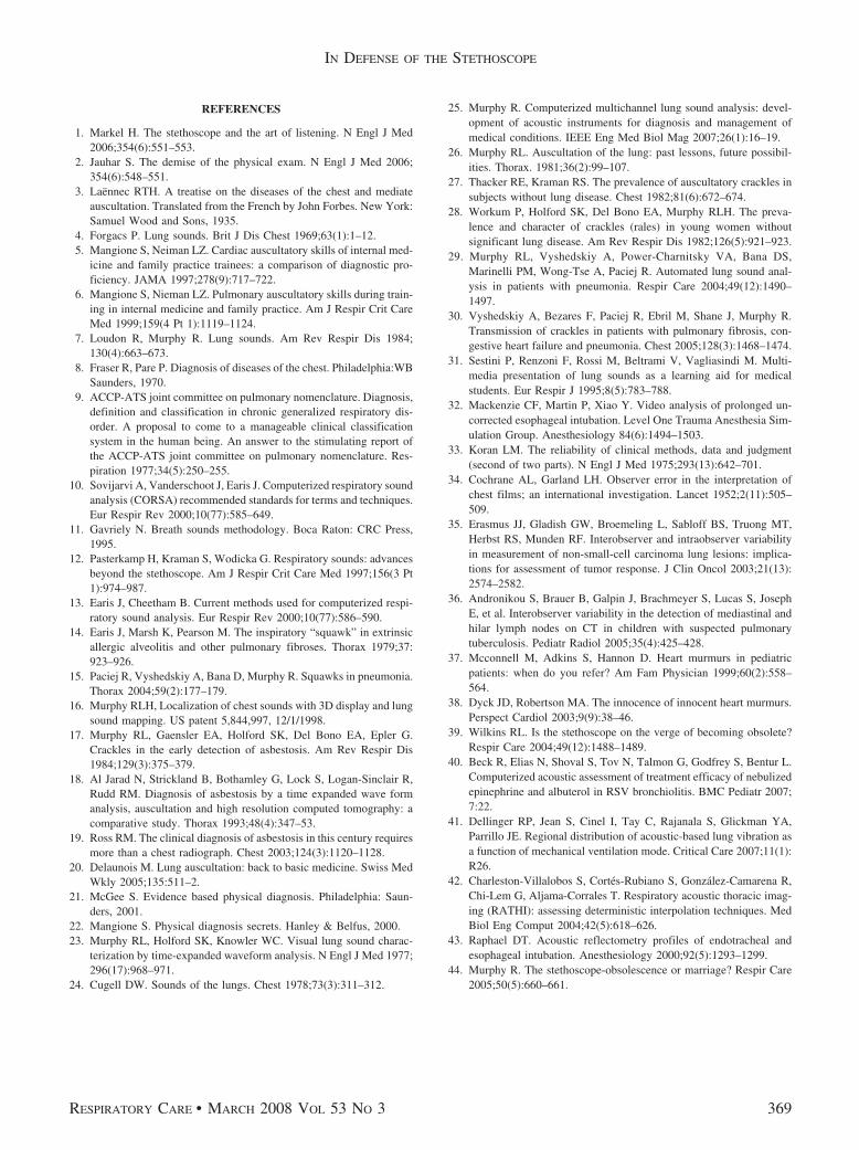
REFERENCES
1. Markel H. The stethoscope and the art of listening. N Engl J Med2006;354(6):551–553.
2. Jauhar S. The demise of the physical exam. N Engl J Med 2006;354(6):548–551.
3. Laennec RTH. A treatise on the diseases of the chest and mediateauscultation. Translated from the French by John Forbes. New York:Samuel Wood and Sons, 1935.
4. Forgacs P. Lung sounds. Brit J Dis Chest 1969;63(1):1–12.5. Mangione S, Neiman LZ. Cardiac auscultatory skills of internal med-
icine and family practice trainees: a comparison of diagnostic pro-ficiency. JAMA 1997;278(9):717–722.
6. Mangione S, Nieman LZ. Pulmonary auscultatory skills during train-ing in internal medicine and family practice. Am J Respir Crit CareMed 1999;159(4 Pt 1):1119–1124.
7. Loudon R, Murphy R. Lung sounds. Am Rev Respir Dis 1984;130(4):663–673.
8. Fraser R, Pare P. Diagnosis of diseases of the chest. Philadelphia:WBSaunders, 1970.
9. ACCP-ATS joint committee on pulmonary nomenclature. Diagnosis,definition and classification in chronic generalized respiratory dis-order. A proposal to come to a manageable clinical classificationsystem in the human being. An answer to the stimulating report ofthe ACCP-ATS joint committee on pulmonary nomenclature. Res-piration 1977;34(5):250–255.
10. Sovijarvi A, Vanderschoot J, Earis J. Computerized respiratory soundanalysis (CORSA) recommended standards for terms and techniques.Eur Respir Rev 2000;10(77):585–649.
11. Gavriely N. Breath sounds methodology. Boca Raton: CRC Press,1995.
12. Pasterkamp H, Kraman S, Wodicka G. Respiratory sounds: advancesbeyond the stethoscope. Am J Respir Crit Care Med 1997;156(3 Pt1):974–987.
13. Earis J, Cheetham B. Current methods used for computerized respi-ratory sound analysis. Eur Respir Rev 2000;10(77):586–590.
14. Earis J, Marsh K, Pearson M. The inspiratory “squawk” in extrinsicallergic alveolitis and other pulmonary fibroses. Thorax 1979;37:923–926.
15. Paciej R, Vyshedskiy A, Bana D, Murphy R. Squawks in pneumonia.Thorax 2004;59(2):177–179.
16. Murphy RLH, Localization of chest sounds with 3D display and lungsound mapping. US patent 5,844,997, 12/1/1998.
17. Murphy RL, Gaensler EA, Holford SK, Del Bono EA, Epler G.Crackles in the early detection of asbestosis. Am Rev Respir Dis1984;129(3):375–379.
18. Al Jarad N, Strickland B, Bothamley G, Lock S, Logan-Sinclair R,Rudd RM. Diagnosis of asbestosis by a time expanded wave formanalysis, auscultation and high resolution computed tomography: acomparative study. Thorax 1993;48(4):347–53.
19. Ross RM. The clinical diagnosis of asbestosis in this century requiresmore than a chest radiograph. Chest 2003;124(3):1120–1128.
20. Delaunois M. Lung auscultation: back to basic medicine. Swiss MedWkly 2005;135:511–2.
21. McGee S. Evidence based physical diagnosis. Philadelphia: Saun-ders, 2001.
22. Mangione S. Physical diagnosis secrets. Hanley & Belfus, 2000.23. Murphy RL, Holford SK, Knowler WC. Visual lung sound charac-
terization by time-expanded waveform analysis. N Engl J Med 1977;296(17):968–971.
24. Cugell DW. Sounds of the lungs. Chest 1978;73(3):311–312.
25. Murphy R. Computerized multichannel lung sound analysis: devel-opment of acoustic instruments for diagnosis and management ofmedical conditions. IEEE Eng Med Biol Mag 2007;26(1):16–19.
26. Murphy RL. Auscultation of the lung: past lessons, future possibil-ities. Thorax. 1981;36(2):99–107.
27. Thacker RE, Kraman RS. The prevalence of auscultatory crackles insubjects without lung disease. Chest 1982;81(6):672–674.
28. Workum P, Holford SK, Del Bono EA, Murphy RLH. The preva-lence and character of crackles (rales) in young women withoutsignificant lung disease. Am Rev Respir Dis 1982;126(5):921–923.
29. Murphy RL, Vyshedskiy A, Power-Charnitsky VA, Bana DS,Marinelli PM, Wong-Tse A, Paciej R. Automated lung sound anal-ysis in patients with pneumonia. Respir Care 2004;49(12):1490–1497.
30. Vyshedskiy A, Bezares F, Paciej R, Ebril M, Shane J, Murphy R.Transmission of crackles in patients with pulmonary fibrosis, con-gestive heart failure and pneumonia. Chest 2005;128(3):1468–1474.
31. Sestini P, Renzoni F, Rossi M, Beltrami V, Vagliasindi M. Multi-media presentation of lung sounds as a learning aid for medicalstudents. Eur Respir J 1995;8(5):783–788.
32. Mackenzie CF, Martin P, Xiao Y. Video analysis of prolonged un-corrected esophageal intubation. Level One Trauma Anesthesia Sim-ulation Group. Anesthesiology 84(6):1494–1503.
33. Koran LM. The reliability of clinical methods, data and judgment(second of two parts). N Engl J Med 1975;293(13):642–701.
34. Cochrane AL, Garland LH. Observer error in the interpretation ofchest films; an international investigation. Lancet 1952;2(11):505–509.
35. Erasmus JJ, Gladish GW, Broemeling L, Sabloff BS, Truong MT,Herbst RS, Munden RF. Interobserver and intraobserver variabilityin measurement of non-small-cell carcinoma lung lesions: implica-tions for assessment of tumor response. J Clin Oncol 2003;21(13):2574–2582.
36. Andronikou S, Brauer B, Galpin J, Brachmeyer S, Lucas S, JosephE, et al. Interobserver variability in the detection of mediastinal andhilar lymph nodes on CT in children with suspected pulmonarytuberculosis. Pediatr Radiol 2005;35(4):425–428.
37. Mcconnell M, Adkins S, Hannon D. Heart murmurs in pediatricpatients: when do you refer? Am Fam Physician 1999;60(2):558–564.
38. Dyck JD, Robertson MA. The innocence of innocent heart murmurs.Perspect Cardiol 2003;9(9):38–46.
39. Wilkins RL. Is the stethoscope on the verge of becoming obsolete?Respir Care 2004;49(12):1488–1489.
40. Beck R, Elias N, Shoval S, Tov N, Talmon G, Godfrey S, Bentur L.Computerized acoustic assessment of treatment efficacy of nebulizedepinephrine and albuterol in RSV bronchiolitis. BMC Pediatr 2007;7:22.
41. Dellinger RP, Jean S, Cinel I, Tay C, Rajanala S, Glickman YA,Parrillo JE. Regional distribution of acoustic-based lung vibration asa function of mechanical ventilation mode. Critical Care 2007;11(1):R26.
42. Charleston-Villalobos S, Cortes-Rubiano S, Gonzalez-Camarena R,Chi-Lem G, Aljama-Corrales T. Respiratory acoustic thoracic imag-ing (RATHI): assessing deterministic interpolation techniques. MedBiol Eng Comput 2004;42(5):618–626.
43. Raphael DT. Acoustic reflectometry profiles of endotracheal andesophageal intubation. Anesthesiology 2000;92(5):1293–1299.
44. Murphy R. The stethoscope-obsolescence or marriage? Respir Care2005;50(5):660–661.
IN DEFENSE OF THE STETHOSCOPE
RESPIRATORY CARE • MARCH 2008 VOL 53 NO 3 369