Embed Size (px)
Citation preview
123
Conventional Nuclear Medicine in Pediatrics
Maria Carmen GarganeseGiovanni Francesco Livio D’ErricoEditors
In collaboration with Milena Pizzoferro · Maria Felicia Villani
A Clinical Case-Based Atlas
Conventional Nuclear Medicine in Pediatrics
Maria Carmen Garganese Giovanni Francesco Livio D'Errico Editors
Conventional Nuclear Medicine in Pediatrics
A Clinical Case-Based Atlas
In collaboration with Milena Pizzoferro and Maria Felicia Villani
Editors Maria Carmen Garganese Diagnostic Imaging Dept-Nuclear Med IRCCS Bambino Gesù Paediatric Hospi Rome Italy
Giovanni Francesco Livio D'Errico Diagnostic Imaging Dept-Nucl Med Un Private Hospital "Pio XI" Rome Italy
ISBN 978-3-319-43179-6 ISBN 978-3-319-43181-9 (eBook) DOI 10.1007/978-3-319-43181-9
Library of Congress Control Number: 2016959035
© Springer International Publishing Switzerland 2017 This work is subject to copyright. All rights are reserved by the Publisher, whether the whole or part of the material is concerned, specifi cally the rights of translation, reprinting, reuse of illustrations, recitation, broadcasting, reproduction on microfi lms or in any other physical way, and transmission or information storage and retrieval, electronic adaptation, computer software, or by similar or dissimilar methodology now known or hereafter developed. The use of general descriptive names, registered names, trademarks, service marks, etc. in this publication does not imply, even in the absence of a specifi c statement, that such names are exempt from the relevant protective laws and regulations and therefore free for general use. The publisher, the authors and the editors are safe to assume that the advice and information in this book are believed to be true and accurate at the date of publication. Neither the publisher nor the authors or the editors give a warranty, express or implied, with respect to the material contained herein or for any errors or omissions that may have been made.
Printed on acid-free paper
This Springer imprint is published by Springer Nature The registered company is Springer International Publishing AG Switzerland The registered company address is Gewerbestrasse 11, 6330 Cham, Switzerland
To Professor Gianclaudio Ciofetta who dedicated his life to pediatric nuclear medicine and has contributed to our vocational education with humanity and scientifi c strength Our work is a way to remember him and thank him for what he has given us and all the young patients.
vii
That is a good book which is opened with expectation, and closed with delight and profi t. (Amos Bronson Alcott)
When Professor D’Errico and I decided to write this book, myriad of thoughts and feelings went through my mind.
The fi rst feeling was the delight of being able to share with him the writing of a work started when I was “young,” and he was the fi rst tutor when I was a resident in nuclear medicine: he asked me what I was able to do and I said “nothing at all”!
The second one was the sadness for the passing of Professor Gianclaudio Ciofetta, to whom this work is dedicated, who would have been proud to see his greatest wish realized.
The last one was the enthusiasm felt in translating in word a daily experience – made of kindness, of fears, of commitment, and of devotion to the children who pass every day through our unit – in order to create something that can help those who approach pediatric nuclear medicine and those who wish to fi nd a hint or a method and an answer I hope, a tangle of pos-sibilities that open up in front of a child and in front of a test to which a child is submitted.
Furthermore, the thoughts about the best and the most educational possible way to set up and to perform the various topics and the study of the various body districts, my wish is to provide to those who open the book, searching a help or an answer, more and more than they search, to fi nd a case similar to his one and an adequate answer to his question.
The work was long and it would not have started without the help of my colleagues, Dr. Milena Pizzoferro and Dr. Maria Felicia Villani, who, with utmost care and great detail, have researched the clinical cases, have made the scintigraphic images for publication, and supple-mented them with radiological images. They have left out no detail, no possible explanation, no images, and no texts. And all the iconographic documentation would not have been so complete and so made without the cooperation of the technical staff, Gaetano Masi, Stefano Chiapparelli, and Elisa Villanucci, and nurses – Consilia Lella and Filomena Petrucci –that, with professionalism and affection, have followed and cuddled the children and their parents during the execution of scintigraphic studies.
Performing quality studies on children without using anesthesia is a daily challenge; it is a goal that we try to achieve every day, keeping high our guard and also the commitment toward the standards that the “Bambino Gesù” Children Hospital child struggles to respect and to keep.
Dutiful thanks go to Professor Tomà and to all our fellow radiologists and clinicians in our hospital who actively participated in the drafting of the various chapters of the book but also to all those of which the name does not appear but who work and take part in our activities, col-laborating in the good success, each one for his role.
Special thanks also go to the colleagues of the Medical Physics Unit – Dr. Vittorio Cannatà and Dr. Elisabetta Genovese – who are alongside us every day, helping us to respect the con-tinued effi ciency of our instruments and handling all aspects of dosimetry.
Affectionate thanks to Professor Paolo Caione with whom we have shared countless patients and numerous scientifi c discussions.
Pref ace

viii
This book is the result of collaboration, and it is based on a daily teamwork. Each chapter is derived not only from our consolidated experience by now but also from the exchange of information and from the contact with the specialists who formulate the questions which we strive to answer in a complete, clear, and standardized way, making the interpretative doubts clear to them. It shows that we can and we must work well with humility. It strongly shows that conventional nuclear medicine can still give much if the studies are conducted properly and integrated with the supporting diagnostics.
We hope that the readers, who occasionally perform studies on children, fi nd in the book what we have proposed they will fi nd, and we hope that it is also useful to all the colleagues who deal with pediatric nuclear medicine as a part of their activities not dedicated to children.
Rome, Italy Maria Carmen Garganese
Preface
ix
It was in the early 1980s that I get interested in pediatric nuclear medicine and, in particular, of pediatric nephro-urology. In the 1990s, the young doctor Garganese meets me and “pediat-ric nuclear medicine,” and, since then, this fi eld has represented her “great love.”
Nuclear medicine, revealing the physiological processes in vivo, has had, in recent years, incredible progress, but, since the child is not a “young adult,” it is not enough to adapt proce-dures performed in adults to the age and size of the child. Considerable progress has been made in the fi eld of pediatric nuclear medicine due to the high capacity of earlier diagnosis, easier management of young patients, and treatment that have benefi ted children. Just to this reason, pediatric diagnostic paths have been developed internationally (e.g., health.wa.gov.au). The pediatric nuclear medicine has become an increasingly important tool both to follow the success of therapy and to assess the progression. In other words, the development of pediatric nuclear medicine is because it provides information about the condition of the child physiolog-ically, fast, safe, sensitive, and minimally invasive.
The novelty of this work consists in providing a systematic approach, as a textbook, with the simplicity of consultation of an atlas, on a niche topic in nuclear medicine, so important and widespread as the pediatrics.
This book, entitled Conventional Nuclear Medicine in Pediatrics: A Clinical Case-Based Atlas , fi lls a gap in the literature of a reference tool “clinical case based” in pediatric nuclear medicine imaging, by a collection of richly illustrated teaching cases and problem-solving, dealing with clinical history, technical informations, workfl ows, image descriptions, and pitfalls.
Each chapter describes the diagnostic nuclear medicine molecular imaging based on the availability of sensitive and relatively specifi c radiopharmaceuticals tailored for different tar-gets that can be expressed in this complex pediatric scenario.
Authors with different tasks (M. Pizzoferro, M.F. Villani, and M.C. Garganese as nuclear medicine physicians; G. Masi, S. Chiapparelli, and E. Villanucci as nuclear medicine technolo-gists; and C. Lella and F. Petrucci as pediatric nurses) contributed by writing Chap. 1 about the need for a dedicated “teamwork” and several informations on the management of children such as reception, administration, and interaction with parents.
In Chap. 2 (“radiation exposure”), V. Cannatà, M. Longo, and E. Genovese provide infor-mation about the activities to be administered, according to dosimetric considerations.
The role of the nuclear medicine physician, in the usual pediatric clinical scenarios, is care-fully covered, in Part 2, “Clinical Pediatric Practices,” with the important contribution of many coauthors, skilled clinicians in each fi eld.
N. Capozza, S. Nappo, G. Torino, E. Mele, G. Di Zazzo, G. Mosiello, M.L. Capitanucci, M. De Gennaro, and D. Barbuti, expert co-workers, treat, in Chaps. 3 , 4 , 5 , 6 , 7 , 8 , 9 , and 10 , the different aspects of nephro-urology.
Oncology was treated by F. Locatelli, L. Vinti, A. Mastronuzzi, A. Castellano, R. Lombardi, and D. Barbuti in Chaps. 11 , 12 , and 13 , while gastroenterology was dealt by L. Dall’Oglio, T. Caldaro, P. De Angelis, R. Tambucci, F. Torroni, G. Angelino, L. Del Prete, F. Rea, V. Balassone, A.C.I. Contini, E.F. Romeo, F. De Peppo, G. Torre, M. Candusso, M.S. Basso, A. Pietrobattista, A. Infante, and L. Monti in Chaps. 14 , 15 , 16 , 17 , 18 , and 19 .
Pref ace
x
F. De Benedetti, A. Insalaco, and A. Krzysztofi ak have contributed, in Chaps. 20 and 21 , interesting clinical cases about infl ammation–infection and rheumatology disease; L. Menchini, clinical cases about bronchopneumology (Chap. 27 ); and A. Grossi and G. Ubertini, clinical cases in endocrinology fi eld (Chaps. 22 , 23 , 24 , 25 ) and G. Natali and D. Barbuti, benign bone disease (Chap. 26 ).
We are very fortunate to have had the contributions of coauthors, also our colleagues, skilled clinicians in each fi eld, whose experiences enrich this atlas, and therefore, we are deeply indebted to all of them.
Our close collaborators, Dr. M. Pizzoferro and Dr. M.F. Villani, deserve special acknowl-edgments for the care given to provide images and clinical cases.
Rome, Italy Giovanni Francesco Livio D’Errico
Preface
xi
Part I General Considerations Paolo Tomà
1 Management of the Pediatric Patient: A Teamwork . . . . . . . . . . . . . . . . . . . . . . . . . 3 Milena Pizzoferro , Maria Felicia Villani , Maria Carmen Garganese , Gaetano Masi , Stefano Chiapparelli , Elisa Villanucci , Consilia Lella , and Filomena Petrucci
2 Radiation Risk . . . . . . . . . . . . . . . . . . . . . . . . . . . . . . . . . . . . . . . . . . . . . . . . . . . . . . . 11 Vittorio Cannatà , Elisabetta Genovese , and Mariaconcetta Longo
Part II Clinical Pediatric Practice
3 Focus on Children with Nephrological and Urological Problems . . . . . . . . . . . . . 19 Nicola Capozza and Giacomo Di Zazzo
4 Hydronephrosis . . . . . . . . . . . . . . . . . . . . . . . . . . . . . . . . . . . . . . . . . . . . . . . . . . . . . . 23 Maria Felicia Villani , Milena Pizzoferro , Simona Nappo , and Maria Carmen Garganese
5 Megaureter . . . . . . . . . . . . . . . . . . . . . . . . . . . . . . . . . . . . . . . . . . . . . . . . . . . . . . . . . 41 Maria Felicia Villani , Milena Pizzoferro , Giovanni Torino , and Maria Carmen Garganese
6 Nephrourology: Vesicoureteral Reflux . . . . . . . . . . . . . . . . . . . . . . . . . . . . . . . . . . . 57 Milena Pizzoferro , Maria Felicia Villani , Ermelinda Mele , and Maria Carmen Garganese
7 Nephrourology: Nephrological Problems . . . . . . . . . . . . . . . . . . . . . . . . . . . . . . . . . 81 Maria Felicia Villani , Milena Pizzoferro , Giacomo Di Zazzo , Domenico Barbuti , and Maria Carmen Garganese
8 Nephrourology: Urinary Tract Infections . . . . . . . . . . . . . . . . . . . . . . . . . . . . . . . . 91 Maria Felicia Villani , Milena Pizzoferro , Giacomo Di Zazzo , and Maria Carmen Garganese
9 Nephrourology: Focus On Child with Bladder Dysfunctions and Urodynamics . . . . . . . . . . . . . . . . . . . . . . . . . . . . . . . . . . . . . . . . . . . . . . . . . . . 105 Mario De Gennaro , Maria Luisa Capitanucci , and Giovanni Mosiello
10 Nephrourology: Bladder Dysfunctions . . . . . . . . . . . . . . . . . . . . . . . . . . . . . . . . . . 113 Maria Felicia Villani , Milena Pizzoferro , Mario De Gennaro , Maria Luisa Capitanucci , Giovanni Mosiello , and Maria Carmen Garganese
11 Oncology . . . . . . . . . . . . . . . . . . . . . . . . . . . . . . . . . . . . . . . . . . . . . . . . . . . . . . . . . . 123 Luciana Vinti , Angela Mastronuzzi , Giuseppe Maria Milano , Evelina Miele , Raffaele Cozza , and Franco Locatelli
Contents
xii
12 Oncology: Solid Mass in Pediatrics and Malignant Bone Involvement . . . . . . . . 127 Maria Felicia Villani , Milena Pizzoferro , Luciana Vinti , Angela Mastronuzzi , Antonella Cacchione , Roberta Lombardi , Domenico Barbuti , Evelina Miele , and Maria Carmen Garganese
13 Oncology: Neuroblastoma . . . . . . . . . . . . . . . . . . . . . . . . . . . . . . . . . . . . . . . . . . . . 171 Maria Felicia Villani , Milena Pizzoferro , Aurora Castellano , Roberta Lombardi , Annalisa Serra , Maria Carmen Garganese , and Franco Locatelli
14 Gastroenterology: Focus on Children with Gastrointestinal Problems . . . . . . . . 215 Luigi Dall’Oglio and Renato Tambucci
15 Gastroenterology: Gastric Emptying – Gastroesophageal Reflux . . . . . . . . . . . . 219 Tamara Caldaro , Paola De Angelis , Filippo Torroni , Francesca Rea , Giulia Angelino , Laura Del Prete , Erminia Francesca Romeo , Simona Faraci , Valerio Balassone , Anna Chiara Iolanda Contini , and Giovanni Federici di Abriola
16 Gastroenterology: Intestinal Bleeding . . . . . . . . . . . . . . . . . . . . . . . . . . . . . . . . . . 241 Milena Pizzoferro , Maria Felicia Villani , Tamara Caldaro , Valerio Balassone , Anna Chiara Iolanda Contini , Erminia Francesca Romeo , Simona Faraci , and Maria Carmen Garganese
17 Gastroenterology: Bronchoaspiration: Neurological Child . . . . . . . . . . . . . . . . . 265 Milena Pizzoferro , Maria Felicia Villani , Francesco De Peppo , and Maria Carmen Garganese
18 Gastroenterology: Focus on Children with Liver Problems . . . . . . . . . . . . . . . . . 275 Giuliano Torre
19 Gastroenterology: Biliary Atresia, Choledochal Cyst, Cystic Fibrosis . . . . . . . . 279 Milena Pizzoferro , Maria Felicia Villani , Lidia Monti , Amato Infante , Manila Candusso , Maria Sole Basso , Andrea Pietrobattista , and Maria Carmen Garganese
20 Rheumatology . . . . . . . . . . . . . . . . . . . . . . . . . . . . . . . . . . . . . . . . . . . . . . . . . . . . . . 297 Fabrizio De Benedetti and Antonella Insalaco
21 Benign Skeletal Disease: Bone Infection and Inflammation . . . . . . . . . . . . . . . . . 305 Maria Felicia Villani , Milena Pizzoferro , Antonella Insalaco , Andrzej Krzysztofi ak , and Maria Carmen Garganese
22 Endocrinology . . . . . . . . . . . . . . . . . . . . . . . . . . . . . . . . . . . . . . . . . . . . . . . . . . . . . . 329 Armando Grossi , Graziamaria Ubertini , and Milena Pizzoferro
23 Endocrinology: Thyroid Carcinoma . . . . . . . . . . . . . . . . . . . . . . . . . . . . . . . . . . . . 339 Milena Pizzoferro , Maria Felicia Villani , Armando Grossi , Debora De Pasquale , and Maria Carmen Garganese
24 Endocrinology: Congenital Hypothyroidism . . . . . . . . . . . . . . . . . . . . . . . . . . . . . 351 Milena Pizzoferro , Maria Felicia Villani , Graziamaria Ubertini , and Maria Carmen Garganese
25 Endocrinology: Hyperparathyroidism . . . . . . . . . . . . . . . . . . . . . . . . . . . . . . . . . . 357 Milena Pizzoferro , Maria Felicia Villani , Armando Grossi , Graziamaria Ubertini , and Maria Carmen Garganese
26 Benign Skeletal Disease . . . . . . . . . . . . . . . . . . . . . . . . . . . . . . . . . . . . . . . . . . . . . . 367 Milena Pizzoferro , Maria Felicia Villani , Domenico Barbuti , Gian Luigi Natali , and Maria Carmen Garganese
27 Bronchopneumology . . . . . . . . . . . . . . . . . . . . . . . . . . . . . . . . . . . . . . . . . . . . . . . . . 407 Milena Pizzoferro , Maria Felicia Villani , Laura Menchini , and Maria Carmen Garganese
Contents
xiii
Giulia Angelino Unit of Endoscopic Surgery , “Bambino Gesù” Children Hospital , Rome , Italy
Valerio Balassone Unit of Endoscopic Surgery , “Bambino Gesù” Children Hospital , Rome , Italy
Domenico Barbuti Imaging Department , “Bambino Gesù” Children Hospital , Rome , Italy
Maria Sole Basso Unit of Epathology , “Bambino Gesù” Children Hospital , Rome , Italy
Antonella Cacchione Oncoematology Department , “Bambino Gesù” Children Hospital , Rome , Italy
Tamara Caldaro Unit of Endoscopic Surgery , “Bambino Gesù” Children Hospital , Rome , Italy
Manila Candusso Unit of Epathology , “Bambino Gesù” Children Hospital , Rome , Italy
Vittorio Cannatà Unit of Medical Physics , “Bambino Gesù” Children Hospital , Rome , Italy
Maria Luisa Capitanucci Unit of Urology, Robotic Surgery and Urodynamic , “Bambino Gesù” Children Hospital , Rome , Italy
Nicola Capozza Unit of Urology , “Bambino Gesù” Children Hospital , Rome , Italy
Aurora Castellano Oncoematology Department , “Bambino Gesù” Children Hospital , Rome , Italy
Stefano Chiapparelli Nuclear Medicine Unit, Imaging Department , IRCCS “Bambino Gesù” Children Hospital , Rome , Italy
Anna Chiara Iolanda Contini Unit of Endoscopic Surgery , “Bambino Gesù” Children Hospital , Rome , Italy
Raffaele Cozza Oncoematology Department , “Bambino Gesù” Children Hospital , Rome , Italy
Luigi Dall’Oglio Unit of Endoscopic Surgery , “Bambino Gesù” Children Hospital , Rome , Italy
Paola De Angelis Unit of Endoscopic Surgery , “Bambino Gesù” Children Hospital , Rome , Italy
Fabrizio De Benedetti Unit of Rheumatology , “Bambino Gesù” Children Hospital , Rome , Italy
Mario De Gennaro Unit of Urology, Robotic Surgery and Urodynamic , “Bambino Gesù” Children Hospital , Rome , Italy
Debora De Pasquale Oncoematology Department , “Bambino Gesù” Children Hospital , Rome , Italy
Francesco De Peppo Unit of Pediatric Surgery , “Bambino Gesù” Children Hospital , Rome , Italy
Laura Del Prete Unit of Endoscopic Surgery , “Bambino Gesù” Children Hospital , Rome , Italy
Giacomo Di Zazzo Unit of Nephrology , “Bambino Gesù” Children Hospital , Rome , Italy
Simona Faraci Unit of Endoscopic Surgery , “Bambino Gesù” Children Hospital , Rome , Italy
Contributors
xiv
Gaetano Masi Nuclear Medicine Unit, Imaging Department , IRCCS “Bambino Gesù” Children Hospital , Rome , Italy
Maria Carmen Garganese Nuclear Medicine Unit, Imaging Department , IRCCS “Bambino Gesù” Children Hospital , Rome , Italy
Elisabetta Genovese Unit of Medical Physics , “Bambino Gesù” Children Hospital , Rome , Italy
Armando Grossi Unit of Endocrinoly , “Bambino Gesù” Children Hospital , Rome , Italy
Amato Infante Imaging Department , “Bambino Gesù” Children Hospital , Rome , Italy
Antonella Insalaco Unit of Rheumatology , “Bambino Gesù” Children Hospital , Rome , Italy
Andrzej Krzysztofi ak Unit of Infettivology , “Bambino Gesù” Children Hospital , Rome , Italy
Consilia Lella Nuclear Medicine Unit, Imaging Department , IRCCS “Bambino Gesù” Children Hospital , Rome , Italy
Franco Locatelli Oncoematology Department , “Bambino Gesù” Children Hospital , Rome , Italy
Roberta Lombardi Imaging Department , “Bambino Gesù” Children Hospital , Rome , Italy
Mariaconcetta Longo Unit of Medical Physics , “Bambino Gesù” Children Hospital , Rome , Italy
Angela Mastronuzzi Oncoematology Department , “Bambino Gesù” Children Hospital , Rome , Italy
Ermelinda Mele Unit of Urology , “Bambino Gesù” Children Hospital , Rome , Italy
Laura Menchini Imaging Department , “Bambino Gesù” Children Hospital , Rome , Italy
Evelina Miele Oncoematology Department , “Bambino Gesù” Children Hospital , Rome , Italy
Giuseppe Milano Oncoematology Department , “Bambino Gesù” Children Hospital , Rome , Italy
Lidia Monti Imaging Department , “Bambino Gesù” Children Hospital , Rome , Italy
Giovanni Mosiello Unit of Urology , “Bambino Gesù” Children Hospital , Rome , Italy
Simona Nappo Unit of Urology , “Bambino Gesù” Children Hospital , Rome , Italy
Gian Luigi Natali Imaging Department , “Bambino Gesù” Children Hospital , Rome , Italy
Filomena Petrucci Unit of Nuclear Medicine , “Bambino Gesù” Children Hospital , Rome , Italy
Andrea Pietrobattista Unit of Epathology , “Bambino Gesù” Children Hospital , Rome , Italy
Milena Pizzoferro Nuclear Medicine Unit, Imaging Department , IRCCS “Bambino Gesù” Children Hospital , Rome , Italy
Francesca Rea Unit of Endoscopic Surgery , “Bambino Gesù” Children Hospital , Rome , Italy
Erminia Francesca Romeo Unit of Endoscopic Surgery , “Bambino Gesù” Children Hospital , Rome , Italy
Annalisa Serra Oncoematology Department , “Bambino Gesù” Children Hospital , Rome , Italy
Renato Tambucci Unit of Endoscopic Surgery , “Bambino Gesù” Children Hospital , Rome , Italy
Paolo Tomà Imaging Department , “Bambino Gesù” Children Hospital , Rome , Italy
Giovanni Torino Unit of Urology , “Bambino Gesù” Children Hospital , Rome , Italy
Giuliano Torre Unit of Epathology , “Bambino Gesù” Children Hospital , Rome , Italy
Contributors

xv
Filippo Torroni Unit of Endoscopic Surgery , “Bambino Gesù” Children Hospital , Rome , Italy
Graziamaria Ubertini Unit of Endocrinoly , “Bambino Gesù” Children Hospital , Rome , Italy
Maria Felicia Villani Nuclear Medicine Unit, Imaging Department , IRCCS “Bambino Gesù” Children Hospital , Rome , Italy
Elisa Villanucci Nuclear Medicine Unit, Imaging Department , IRCCS “Bambino Gesù” Children Hospital , Rome , Italy
Luciana Vinti Oncoematology Department , “Bambino Gesù” Children Hospital , Rome , Italy
Contributors
1.1 Introduction to Pediatric Imaging in Multimodality Diagnostic Era
As a pediatric radiologist, I have seen the impact of SPECT and PET on the clinical care of children. Our imaging practice has had to change to accommodate the powerful nature of these new images.
Image fusion overlays two or more three-dimensional (3D) image sets of the same or differ-ent imaging modality that are in the same orientation in the same space. Anatomic and func-tional imaging is complementary; this is the metaphor of the relationships between radiologists and nuclear medicine physicians.
In my opinion, nuclear medicine is a crucial part of pediatric radiology. Pediatric radiology has peculiar characteristics depending on anatomical development and
specifi c clinical conditions related to the different stages of growth. Other peculiarities specifi -cally related to pediatric radiology include radiation safety and the need of dedicated environ-ments and approaches.
In both pediatric and adult patient care situations, there are family members with whom the imager must interact. However, in the pediatric setting, there are several unique features in the relationship among imager, patient, and family. Most of the complaints by parents and families are not related to technical errors; they are more commonly related to issues of professionalism and communication.
Providing child-friendly surroundings may help to ease a young child’s anxiety and cause him or her to be more cooperative. Paintings on the walls and equipment and cartoonish fi gures in the examination rooms can be helpful. Eliminating or minimizing painful portions of the examination can also be very helpful in keeping a young child cooperative.
In general, the physicians who choose to go into pediatric subspecialties, as well as health care workers who choose to work at pediatric institutions in general, have to be nice, gentle people. Aggressive, power- hungry people cannot work with children.
This edition of the book comes at a very propitious time. Revolutions in medical imaging are modeling and quantifi cation. Imaging of physiologic and cellular processes displays them in four dimensions (three- dimensional images over time).
The goal of the book is to provide a comprehensive text on the clinical applications of nuclear medicine in a pediatric population.
As some techniques have faded (e.g., scrotal scintigraphy), others have been added to the nuclear medicine arsenal. The most signifi cant development has been the dissemination of positron emission tomography (PET) into pediatric practice.
Writing a book is a task that requires time and commitment from many people. I express appreciation to MG, who has transformed their lives in a text. Her dedication is
refl ected in the high quality of the fi nal product. It is my sincere hope that readers will fi nd this work to be a cornerstone of pediatric imaging
and one that they will use frequently in their daily practices.
Part I
General Considerations
Paolo Tomà
3© Springer International Publishing Switzerland 2017M.C. Garganese, G.F.L. D’Errico (eds.), Conventional Nuclear Medicine in Pediatrics, DOI 10.1007/978-3-319-43181-9_1
Management of the Pediatric Patient: A Teamwork
Milena Pizzoferro , Maria Felicia Villani , Maria Carmen Garganese , Gaetano Masi , Stefano Chiapparelli , Elisa Villanucci , Consilia Lella , and Filomena Petrucci
1.1 Introduction
Pediatric nuclear medicine refers to imaging examinations performed in babies, young children, and teenagers. Carrying out nuclear medicine procedure on children requires a com-pletely different approach than on adults. The complexity of the examination varies depending on the age of the patient, the degree of cooperation, clinical conditions, and duration of scintigraphic acquisition. Depending on the increasing complexity of pediatric nuclear medicine studies, a higher level of department expertise is necessary for the delivery of good quality examination and safe patient care.
In a pediatric nuclear medicine department, all members must be profi cient in performing their competencies according to their role, and a trained teamwork is the winning strategy to achieve the main goal, getting a diagnostic imaging that allows to accurately respond to specifi c clinical question.
Within a framework of a well-coordinated staff (including specialized training pediatric nurse, technician, and nuclear medicine physician), it is easier to create a positive atmo-sphere that serves to take the child cooperation, limiting the sedation in few selected children.
Pediatric patient management requires considering the special status of these patients in relation to age and medical condition depending on the underlying disease.
Working with pediatric population requires adequate awareness: in order to obtain appropriate acquisition
standards, the team approach must be shaped to the child’s mind- set, rather than fi tting the child into the paradigm of adult examinations.
In fact, children are not miniature adults and, as well as the normal values of the vital parameters of infants and chil-dren are different than adults, even methods of communica-tion should be adequate for their developmental age.
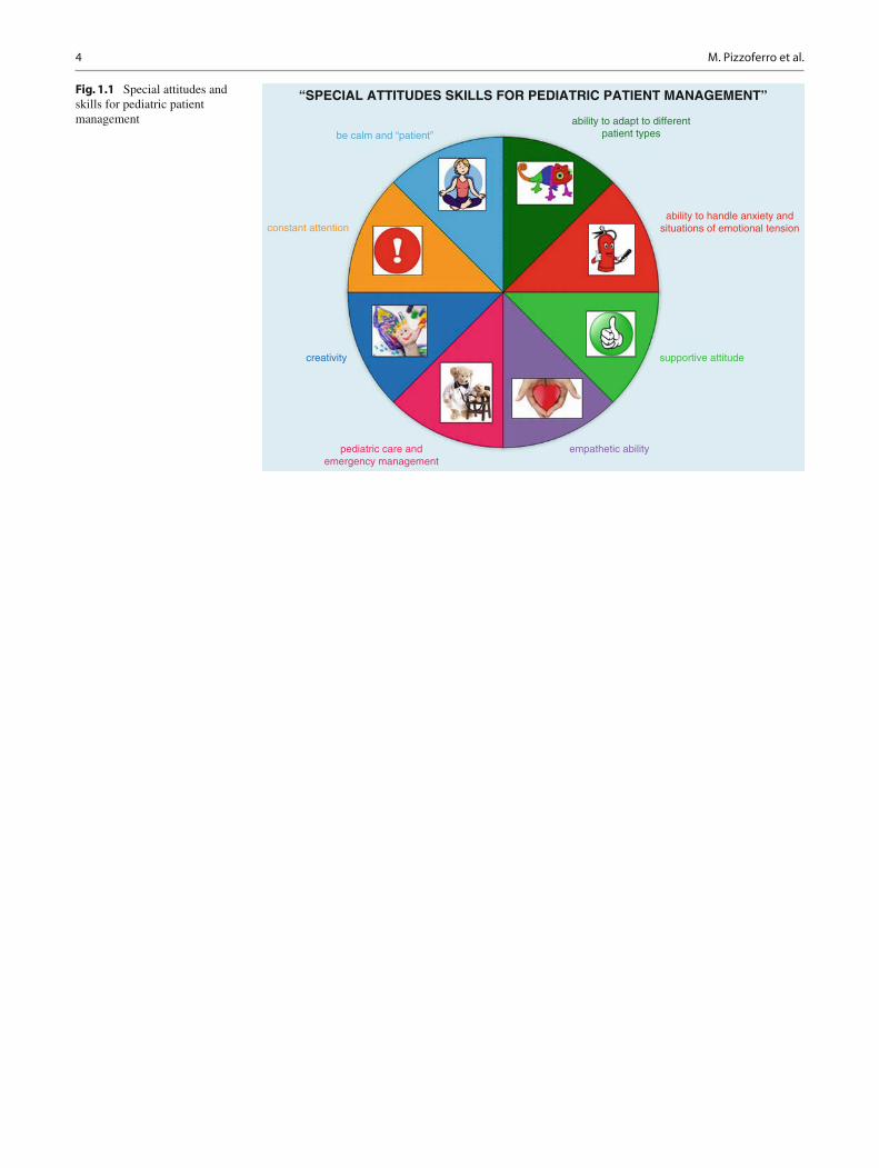
Besides, pediatric patients are children with special health care needs who could have any type of condition that may affect normal growth and development; this may include physical dis-ability, acute or chronic illness, peculiar clinical condition (pain, technological dependency, etc.). During the diagnostic proce-dure, even when the staff use distraction techniques to hold child still and calm, health care professionals must be ready to manage a possible worsening of the patient’s clinical status (Fig. 1.1 ).
Taking care of a pediatric patient includes parental man-agement, and the attitude of all staff members must be posi-tive toward the child and parent (or other family components). In “family-centered approach,” the family’s input is the major driving force to achieve a good cooperation of the child and a high degree of satisfaction of parents.
A department structured with colored paintings on the walls, playful environments (equipped with toys, books, video games, or DVDs) and child-friendly spaces could help foster a welcoming accommodation, but it does not replace the right atmosphere that the nuclear medicine staff must be able to create from the fi rst patient approach.
M. Pizzoferro , MD, PhD (*) • M. F. Villani , MD, PhD M. C. Garganese , MD, PhD • G. Masi • S. Chiapparelli E. Villanucci • C. Lella • F. Petrucci Nuclear Medicine Unit, Imaging Department , IRCCS “Bambino Gesù” Children Hospital , Rome , Italy e-mail: [email protected]
1
4
“SPECIAL ATTITUDES SKILLS FOR PEDIATRIC PATIENT MANAGEMENT”
ability to adapt to differentpatient types
ability to handle anxiety andsituations of emotional tension
supportive attitude
empathetic abilitypediatric care andemergency management
creativity
constant attention
be calm and “patient”
Fig. 1.1 Special attitudes and skills for pediatric patient management
M. Pizzoferro et al.
5
1.2 Accommodate the Child and the Parents
Nurses usually handle the reception of the child and the par-ent. Based on a fi rst quick observation, it is crucial to defi ne an individual approach evaluating a child’s personal features and familiar or sociocultural infl uences. In all cases, the fi rst child approach must be slowly and calm in order to set the basis of a good relationship with all the nuclear medicine staff. The reception of pediatric patients must include an initial evalua-tion of clinical condition to set up an appropriate assistance and monitoring level in case of special health care needs.
From the fi rst moment the child and parent enter the department, all members must be honest with both of them, in order to create a trusting relationship. In the case of both children and adolescents, the staff members must be able to adapt to different patient types, involving them in every moment and explaining the whole procedure in simple terms.
Starting from the fi rst contacts, parents and children must be introduced into the department’s environment by a spe-cifi c education about correct paths within the department (with particular regard to the use of bathrooms and disposal of radioactive diapers) and explanation of the use of dedi-cated tools available in the waiting room (bottle warmer, electronic devices, etc.). To avoid the risk of accidental falls, it is necessary to enforce the standards of child safety, invit-ing the parents to buckle up the stroller and constantly moni-toring the baby should not run or climb in the waiting room.
Parents are encouraged to stay with their children throughout every part of the examination (with the exception if the child’s mother is pregnant), emphasizing the importance of their role in supporting the child for a good outcome of the examination. Generally, the idea to perform a scintigraphic examination gen-erates fear in the child and the parent. Professionals in charge of the pediatric patient management have to be adequately trained handling anxiety and situations of emotional tension of child and the parents. This peculiar ability is closely related to a per-sonal attitude (prone to a supportive and empathetic approach) as well as the capability to provide clear information.
To gain the trust of the child, put him at ease and reduce anxiety; one of the most used tricks in pediatrics is entertain-ing the patient speaking about his everyday life or family members before starting with the diagnostic procedure. Explaining same symptoms by metaphors could be useful to create a direct line of communication (i.e., we usually describe the puncture syringe similar to a mosquito bite or a little pinch).
Providing clear information is also the best form to reduce anxiety for parents. During the interview for the anamnesis and the acquisition of the written informed consent, nuclear medicine physician must explain the entire scintigraphic procedure, stressing the clinical utility within the global diagnostic–therapeutic iter. Providing instructions on how parents can collaborate together with staff is a helpful way to motivate them to be part of the team in order to improve the impact of diagnostic examination on their child.
One of the most diffi cult steps is minimizing and com-municating radiation risk in pediatric nuclear medicine. Generally, parents can understand that their child will undergo an appropriate medical imaging test, but the expla-nation about radiation risk associated with nuclear medicine examination is always a critical point.
Nuclear medicine practitioners must be able to effectively communicate that the dose range associated with most nuclear medicine procedure results in a low radiation expo-sure and a very low risk of detrimental health effects. In case of scintigraphy, with a medium or high radiation exposure level, it is necessary to clarify that the potential risk associ-ated to ionizing radiation remains still low compared with the benefi ts derived by the diagnostic information, unattain-able using other imaging procedures.
Depending on the type of nuclear medicine exam, nuclear medicine physicians have to provide adequate radioprotec-tion instructions to avoid undue radiation exposure of other children or pregnant women; in order to reduce global patient radiation exposure, proper indications should be suggested (good hydration with water or whatever is pleasing to the child and frequent urination).
1 Management of the Pediatric Patient: A Teamwork
6
1.3 Administration of Radiopharmaceutical
Depending on the type of nuclear medicine exam, the radiotracer is either injected into the body, swallowed, or inhaled as a gas in order to evaluate the functional informa-tion of the organ system being examined. Except for intra-venous injections, most nuclear medicine procedures are painless and rarely associated with signifi cant discomfort or side effects.
1.3.1 Endovenous Administration
If the tracer is given intravenously, a small needle is used to inject the radiotracer and removed immediately after. At times, an indwelling intravenous catheter may be used if it is already present, in case of multiple phases of administration or provocative examination (i.e., stress myocardial scintigra-phy), when the patient is in poor clinical conditions. In onco-logical patients, the use of central venous catheter is limited only to certain types of scintigraphic examinations with proper antiseptic precautions.
For a successful intravenous injection, a team approach is necessary to distract the child, to properly immobilize the
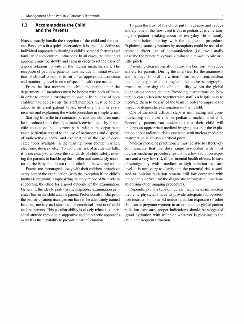
limb of the child, and to explain to the parent, as he may cooperate reassuring the child. During administration, a bed pad must be positioned under the injection site to limit potential contamination. Different immobilization tech-niques are used depending on the age of the child and administration site (Fig. 1.2 ).
1.3.2 Aerosol Administration
The radioaerosol can be inhaled through deep breaths, using a mask of appropriate size for age. In case of unco-operative patient, the radioaerosol can be adequately administered during the whole inspiration phase, even if the child cries. Immediately following the radioaerosol inhalation, the patients must rinse their mouth by gar-gling, when the child is able. Then, the patient is placed in the supine position, and a gamma camera detector is pos-teriorly positioned for acquiring lung radioactivity. After an initial qualitative evaluation, if a satisfactory distribu-tion is not obtained, a number of extra inspirations must be performed. Measurement of the administered radioac-tivity dose can be performed calculating the ratio of fi rst frame counts and a conversion ratio, specifi c for each gamma camera.
a b c
Fig. 1.2 The newborn is made to lie on the stretcher, the parent remains close to the head of the child, the nurse immobilizes the upper limb (arm or hand), while the doctor administers ( a ); in the case of adminis-tration to the foot, the use of sandbags is useful to facilitate the blocking
of the contralateral leg ( b ). The child is seated on the parent’s legs that blocks the baby’s legs between his, the nurse immobilizes the child’s arm or hand, while the doctor injects the radiopharmaceutical ( c )
M. Pizzoferro et al.
7
Oral Naso-gastric tube
PEG
Fig. 1.3 In case of oral feeding, one of the parents can administer the meal so that the child does not notice the difference from the usual. The nurse provides instructions to parents to avoid contamination, supervises the administration, and supports the parent in case of diffi culty ( a ). If a child is unable to feed orally, the nurse manages meal administration using nasogastric tube or percutaneous endoscopic gastrostomy when already
positioned ( b ). Meal administration is with the patient lying on the bed of gamma camera and, by monitor persistence, the nuclear medicine physi-cian evaluates gastric fi lling according to the patient’s capability. The right amount of meal is determined by the habits of the patient and by the pres-ence of indirect signs as high grade of gastroesophageal refl ux or the beginning of intestinal progression of the meal from the stomach
1.3.3 Meal Administration
1.3.3.1 Gastric Emptying Scintigraphy (GES) Protocol
For gastric emptying scintigraphy, radionuclide-labeled test meals are used. The test meal can be liquid, semiliquid, or semisolid, according to the normal meal that the patient under study would generally consume. When swallowed, the radiotracer has no taste. Labeled meals, milk scan as well as homogenized, yogurt or chocolate drinks, are used for gas-tric emptying studies in newborn, infants and children. The meal should have a pleasant taste to encourage a quick and complete recruitment of the meal, ensuring a good nutri-tional intake to obtain reliable functional information about gastric emptying parameters. A good amount of meal taken is also closely linked to the sensitivity of scintigraphy in detecting the presence of gastroesophageal refl ux. Meals must be consumed within a 10-min period, after which the scintigraphy study must be started to avoid the beginning of intestinal progression of the meal from the stomach.
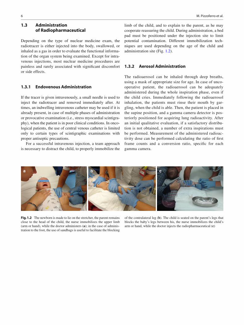
In case of oral feeding, one of the parents can administer the meal, so that the child does not notice the difference from the usual. The nurse provides instructions to parents to avoid contamination, supervises the administration, and supports the parent in case of diffi culty. The necessary precautions to limit contamination in case of vomiting must be taken always.
In such a condition, the technique is completely noninva-sive, whereas in evaluating children with feeding diffi culties, the nurse can place a nasogastric tube which can be removed after meal administration. If a child is unable to feed orally, the nurse manages meal administration using nasogastric tube or percutaneous endoscopic gastrostomy when already positioned. Meal administration is with the patient lying on the bed of gamma camera and, by monitor persistence, nuclear medicine physician evaluates gastric fi lling accord-ing to the patient’s capability. Habits of the patient and pres-ence of indirect signs (as high grade of gastroesophageal refl ux or the beginning of intestinal progression of the meal from the stomach) allow to determinate the right amount of meal (Fig. 1.3 ).
1 Management of the Pediatric Patient: A Teamwork
8
a b
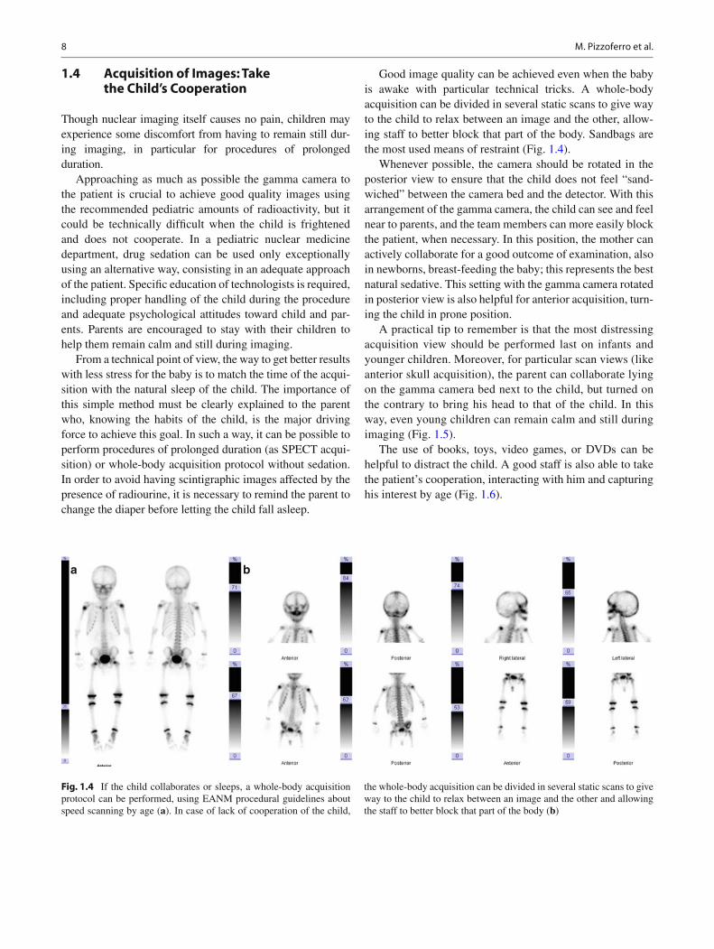
Fig. 1.4 If the child collaborates or sleeps, a whole-body acquisition protocol can be performed, using EANM procedural guidelines about speed scanning by age ( a ). In case of lack of cooperation of the child,
the whole-body acquisition can be divided in several static scans to give way to the child to relax between an image and the other and allowing the staff to better block that part of the body ( b )
1.4 Acquisition of Images: Take the Child’s Cooperation
Though nuclear imaging itself causes no pain, children may experience some discomfort from having to remain still dur-ing imaging, in particular for procedures of prolonged duration.
Approaching as much as possible the gamma camera to the patient is crucial to achieve good quality images using the recommended pediatric amounts of radioactivity, but it could be technically diffi cult when the child is frightened and does not cooperate. In a pediatric nuclear medicine department, drug sedation can be used only exceptionally using an alternative way, consisting in an adequate approach of the patient. Specifi c education of technologists is required, including proper handling of the child during the procedure and adequate psychological attitudes toward child and par-ents. Parents are encouraged to stay with their children to help them remain calm and still during imaging.
From a technical point of view, the way to get better results with less stress for the baby is to match the time of the acqui-sition with the natural sleep of the child. The importance of this simple method must be clearly explained to the parent who, knowing the habits of the child, is the major driving force to achieve this goal. In such a way, it can be possible to perform procedures of prolonged duration (as SPECT acqui-sition) or whole-body acquisition protocol without sedation. In order to avoid having scintigraphic images affected by the presence of radiourine, it is necessary to remind the parent to change the diaper before letting the child fall asleep.
Good image quality can be achieved even when the baby is awake with particular technical tricks. A whole-body acquisition can be divided in several static scans to give way to the child to relax between an image and the other, allow-ing staff to better block that part of the body. Sandbags are the most used means of restraint (Fig. 1.4 ).
Whenever possible, the camera should be rotated in the posterior view to ensure that the child does not feel “sand-wiched” between the camera bed and the detector. With this arrangement of the gamma camera, the child can see and feel near to parents, and the team members can more easily block the patient, when necessary. In this position, the mother can actively collaborate for a good outcome of examination, also in newborns, breast-feeding the baby; this represents the best natural sedative. This setting with the gamma camera rotated in posterior view is also helpful for anterior acquisition, turn-ing the child in prone position.
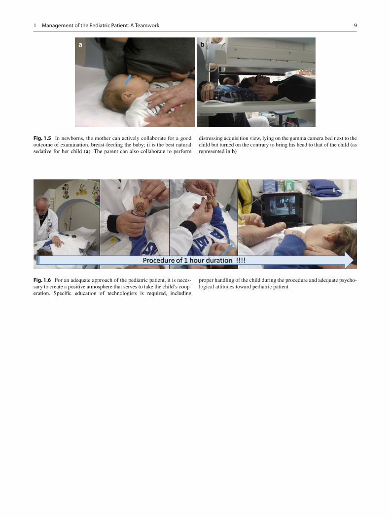
A practical tip to remember is that the most distressing acquisition view should be performed last on infants and younger children. Moreover, for particular scan views (like anterior skull acquisition), the parent can collaborate lying on the gamma camera bed next to the child, but turned on the contrary to bring his head to that of the child. In this way, even young children can remain calm and still during imaging (Fig. 1.5 ).
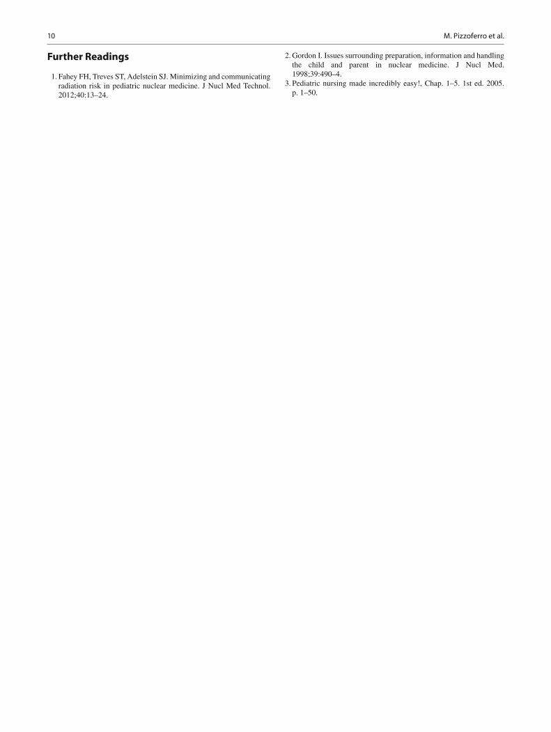
The use of books, toys, video games, or DVDs can be helpful to distract the child. A good staff is also able to take the patient’s cooperation, interacting with him and capturing his interest by age (Fig. 1.6 ).
M. Pizzoferro et al.
9
a b
Fig. 1.5 In newborns, the mother can actively collaborate for a good outcome of examination, breast-feeding the baby; it is the best natural sedative for her child ( a ). The parent can also collaborate to perform
distressing acquisition view, lying on the gamma camera bed next to the child but turned on the contrary to bring his head to that of the child (as represented in b )
Fig. 1.6 For an adequate approach of the pediatric patient, it is neces-sary to create a positive atmosphere that serves to take the child’s coop-eration. Specifi c education of technologists is required, including
proper handling of the child during the procedure and adequate psycho-logical attitudes toward pediatric patient
1 Management of the Pediatric Patient: A Teamwork
10
Further Readings
1. Fahey FH, Treves ST, Adelstein SJ. Minimizing and communicating radiation risk in pediatric nuclear medicine. J Nucl Med Technol. 2012;40:13–24.
2. Gordon I. Issues surrounding preparation, information and handling the child and parent in nuclear medicine. J Nucl Med. 1998;39:490–4.
3. Pediatric nursing made incredibly easy!, Chap. 1–5. 1st ed. 2005. p. 1–50.
M. Pizzoferro et al.
11© Springer International Publishing Switzerland 2017M.C. Garganese, G.F.L. D’Errico (eds.), Conventional Nuclear Medicine in Pediatrics, DOI 10.1007/978-3-319-43181-9_2
Radiation Risk
Vittorio Cannatà, Elisabetta Genovese, and Mariaconcetta Longo
2.1 Introduction
The practice of nuclear medicine leads to a potential risk of exposure for the patient. The activity of radiopharmaceutical should be administered in order to guarantee the correct bal-ance between risks and benefits. In the last years, the intro-duction of technological advances, the increased availability of scanning equipment, and new radiopharmaceuticals lead to an intensified use of nuclear medicine examinations. On the one hand, these improvements involved in a remarkable progress in image quality; on the other hand, technological advances do not necessarily imply a decrease in patient exposure to ionizing radiation. The implementation of radia-tion protection practices aimed to limit radiation exposure in nuclear medicine exams is an utmost need. For pediatric patient, a more attention has to be paid as they have higher tissue radiosensitivity and longer life expectancy.
2.2 Effects of Ionizing Radiations
A type of radiation which has enough energy to eject elec-trons from atoms or molecules is defined as ionizing radia-tion. It is well known that the interaction between ionizing radiation and biological tissues or organs may cause changes in cells which may later cause them to become malignant or bring about other detrimental functional changes in irradi-ated tissues and organs. It is important to note that irrespec-tive of the nature of the primary radiation (which may be composed of particles and/or electromagnetic waves), the energy transfer mechanism always occurs via the secondary electrons which are produced by interaction between the pri-mary radiation beam and the biological targets. At the micro-scopic level, when incident rays or particles interact with
orbital electrons within the atoms, two processes through which radiation interacts with matter can happen: one of these processes is the excitation, the other one is the ioniza-tion [14]. Excitation involves raising a bound electron to a higher energy state, leaving the atom in an excited state, while ionization happens when the electron receives suffi-cient energy to be ejected from its orbit and to leave the host atom. These physical interactions between radiation and spe-cific structures within the cells can cause more or less serious biological damages. These latter are associated to the inter-action of radiation with deoxyribonucleic acid (DNA) and can mainly occur through direct and indirect processes.
The direct interaction implies a direct damage of DNA structures after ionization of atoms or molecules, through a sequence of chemical events which can provoke the final biological damage. This is the dominant process for highly ionizing particles, i.e., heavy charged particles, proton and neutrons. On the contrary, the indirect interaction involves secondary electrons which are ejected during the ionization process. These secondary particles, energetic and unbound, are capable of migrating away from the site of their produc-tion giving up their energy to the surrounding medium, through a series of interactions with other atoms and mole-cules. This energy absorption process results in the forma-tion of free radicals and other chemical species, i.e., more reactive molecules which are the true causatives of damages of critical targets in the cells [2].
For example, when the radiation interaction happens with water molecules, the created highly unstable free radicals, such as water ions (H2O+) and hydroxyl (OH.), can spread through the cell interacting even with distant cellular target. The indirect interaction and its consequently biological det-riment are mainly caused by sparsely ionizing radiation, i.e., electrons or x-ray.
In the events timescale, the initial ionization event occurs instantaneously (~10−18 s) at the microscopic level, while the chemical changes may appear to operate over a timescale of about 10−5 s. Thus, the period during which the chemical damage is caused is relatively long on the microscopic scale.
V. Cannatà (*) • E. Genovese • M. LongoUnit of Medical Physics, Paediatric Bambino Gesù , Rome, Italye-mail: [email protected]
2
12
These events are the precursors to a chain of subsequent events which may eventually lead to the clinical (macro-scopic) manifestation of radiation damage. The clinically observable radiation effects, whose timescale may extend to years, are expressed as the results of the functional impair-ment after lethal damage inflicted to large numbers of cells or critical substructures [3].
Dealing with these macroscopic effects, an important dis-tinction has to be made between low and high dose effects, whose consequences on biological tissues are really differ-ent. This concept is highlighted by the NCRP Report No. 136 [15] and by the BEIR VII Report [10] where a funda-mental distinction is made: low to moderate doses encom-pass the values between 0 and 100 mSv, while high doses include values greater than 100 mSv.
Moreover, a distinction of the effects of ionizing radiation on biological tissues is often made according the required time for the effects to manifest. If an effect occurs within several hours or days after the exposure of the individual to extremely high doses, it is considered as an acute effect. Conversely, delayed or latent effects manifest several weeks or years after the exposure.
In some cases, the damaged component of the genetic material is essential for cell survival, and the cell may die or not be able to undergo proper mitosis. The removal of these cells will not contribute to late radiation effects such as car-cinogenesis. Instead, late effects occur when the cell survives the initial genetic damage. The consequences of this damage manifest later, perhaps decades after the initial exposure; such late effects may result from genomic instability due to the initial radiation damage. In particular, cells that are grow-ing rapidly and undergoing mitosis at a higher rate may be more susceptible to late radiation effects than those that are growing more slowly [9].
On the basis of these considerations, the radiation effects can also result in a radiation detriment, which is defined as the harm that would eventually be experienced by an exposed group and its descendants as a result of the group’s exposure to a radiation source [11].
The radiation damage may be classified as being either deterministic or stochastic.
Deterministic effects are characterized by a threshold dose level. These effects manifest themselves in the form of harm-ful tissue reactions, i.e., cataract induction, general radiation syndromes, bone marrow ablation, which could manifest after an exposure to high radiation doses. Above the threshold dose level, the severity of the effect is linearly dependent with dose: if the amount of radiation dose is increased, the lesion severity also grows depending on the number of damaged cells [11].
Stochastic effects, which include both carcinogenic and hereditary effects, are those for which the likelihood of occur-ring is dose related, but the severity of the resultant condition is not related to the dose received. They may occur without a threshold dose, and for them, an increase on radiation dose will result in a growth of the probability of occurring [11].
In the field of Nuclear Medicine (NM) diagnostic uses, stochastic effects have to be predominantly considered as potential side effects while, for radionuclide therapy applica-tions, the concerns relate to both stochastic and deterministic effects [12].
In addition, there are other parameters that influence the radiation effects and that need to be discussed. In fact, it is well established that the risk of ionizing radiation varies with both age and sex. In particular, for pediatric patients, the risk of radiation effect is higher than in adults. This behavior can be attributed to a twofold cause: on one hand, the tissues of younger subjects are more radiosensitive as they are actively growing and, on the other hand, life expec-tancy in young people is higher than in adults allowing a longer time for the risk to be realized. Moreover, girls dem-onstrated a higher risk for cancer induction than boys, which is, in large part, attributable to the excess risk of breast can-cer in this population [9].
2.2.1 Evaluation of Radiation Exposure in Nuclear Medicine
Nuclear medicine procedures involve the use of radio-pharmaceuticals that emit radiations such as γ-rays, α-particles, β-particles, and positron. These emissions expose the patient to ionizing radiation that might lead to detrimental health effects [11]. Nuclear Medicine offers the possibility to detect early stages diseases, and its non-invasive nature allows to use it as a powerful diagnostic tool in examinations involving children. The administered activities in nuclear medicine procedures are well estab-lished in many specialties including oncology, urology, cardiology, gastroenterology, and orthopedics. For pediat-ric patients, it is highly recommended that practitioners of pediatric nuclear medicine have to develop a knowledge in understanding radiation risk and dosimetry and how this risk may vary in children relative to adults. Using nuclear medicine procedures, expected clinical results can be guaranteed using the lowest possible administered activities and, thus, the minimum necessary risk for patients. To this purpose, as recommended by the Society of Nuclear Medicine and Molecular Imaging, the key to
V. Cannatà et al.

13
dose optimization is to perform the right test with the right dose on the right patient at the right time [8]. In order to estimate the dose received by organs and tissues during nuclear medicine procedures, the knowledge of bio-kinetic models about the incorporated radionuclides is needed.
The methodologies developed to assess dosimetric evalu-ations in nuclear medicine are mainly two: one of these mod-els was developed by the International Commission on Radiological Protection (ICRP) [12], and the other one by the Medical Internal Radiation Dose (MIRD) Committee of the United States Society of Nuclear Medicine [16]. Based on the same theoretical considerations, MIRD model is focused on biological endpoints for which the knowledge of intake is necessary, while ICRP also gives an estimation of the radiation detriment.
The theoretical approach of both methods will be dis-cussed in the following paying particular attention to how risk varies with age. Moreover, the radiation risk and dose calculation will be discussed later for pediatric nuclear medicine.
The calculation of the absorbed doses by the different organs or tissues is based on the definitions of sources and target organs. The target organs or tissues are those for whom the absorbed doses may arise as a result of radioactive decays occurring in other organs, the so-called source regions.
Thus, the absorbed dose in a particular organ or tissue is calculated as the sum of contributions from various sources, including the target organ or tissue itself.
In order to take into account the different radiosensitivity of organs or tissues, ICRP introduced a dosimetric quantity named effective dose. This definition allowed an overall can-cer risk computation for a situation in which different organs receive different doses, with or without external irradiation of the whole body.
According to ICRP model, the mean absorbed dose D(T ← S) to a target organ or tissue T is the sum of the con-tributions arising from nuclear transformations of the radio-nuclide in various source organs S and it is given by:
D T S
ME Y S T S
Ti i i←( ) = × = × ←( )∑ A A1
i
ϕ
where à is the time-integrated or cumulated activity, equal to the total number of nuclear transformations in S, and S T S←( ) is the absorbed dose in T per unit of cumulated activity in S.
The other symbols have the following meaning: MT is the mass of the target organ or tissue, Ei is the mean energy of
radiation type i, Yi is the yield of radiation type i per transfor-mation, φi is the absorbed fraction of energy of radiation type i.
à is the bio-kinetic component, S T S←( ) represents the physical-geometrical component, as it depends on the radia-tion type, on the energy emitted per transformation, on the mass of the target organ, and on the geometry of the mathe-matical phantoms representing the adult and children of vari-ous ages.
This model is essential to estimate the dose absorbed by the different target organs or tissues. However, if we wish to compare different procedures and the resulting patient doses for assessment of risk versus benefit, the more appropriate parameter to be consider is the effective dose, as it takes into account the different organs sensitivities. ICRP 106 also reports, for each radionuclide, the bio-kinetic model, the bio- kinetic data, the absorbed dose, and the correspondent effec-tive dose per unit of activity administered for different ages (Adult and 15, 10, 5, 1 years old) [12].
The MIRD Committee follows the same theoretical dosi-metric approach described above for ICRP. For each source organ, the radiation dose is calculated and summed to deter-mine the total dose to the target organ.
For pediatric patients, the radiopharmaceutical dose var-ies from that to an adult as organ masses of children differ from those of adults because they are smaller and closer together. S values for patients of different ages can be used to estimate the radiation dose to children. However, both ICRP and MIRD methods do not take into account individual dif-ferences in anatomy and physiology from the standard mod-els. The patient’s body may vary from the standard with respect to size, weight, shape, organ orientation, and dis-tances from other organs. These models also make assump-tions with respect to the amount of source organ radioactivity, including rates for uptake and clearance of the radiopharma-ceutical from that organ. These methods were developed for estimating the average dose to a population and should not be used to estimate the dose to a specific patient [11].
Even though models have traditionally used simple shapes representing the organs, more realistic voxel-based models have been developed with the aim to provide more accurate dose esti-mations [4]. Using these methods, the radiation dose to organs of patients of different sizes and ages can be estimated.
The software code Organ Level INternal Dose Assessment/EXponential Model (OLINDA/EXM) [17] has been devel-oped to facilitate automated and standardized internal dose calculations for nuclear medicine applications. The OLINDA/EXM code uses the same technical basis (phan-toms, organ masses, equations, relationships assumed, and other details) reported by MIRD [16].
2 Radiation Risk
14
2.2.2 Radiation Protection Principles and Considerations on Diagnostic Reference Levels
The ionizing properties of radiation and the correspondent biological effects have to be taken into account in order to implement some radiation protection measures. The ICRP on its 2007 Publication [11] states that practices involving the use of ionizing radiation are regulated by three funda-mental principles of radiological protection: justification, optimization, and limitation of doses.
According to the first principle, any medical practice involv-ing patient exposures must be justified: any decision that alters the radiation exposure situation should do more good than harm [11]. It should be in the right balance between risk and benefit, taking into account social, economic, and technical factors involving the realization of the procedure itself.
The second principle states that once the exposure to ion-izing radiation is justified, each examination must be per-formed so that individual doses should all be kept as low as reasonably achievable (ALARA), taking into account eco-nomic and societal factors [11].
Dose limits are established to ensure that no individual is exposed to radiation risk level exceeding the appropriate lim-its recommended by the ICRP. Medical exposure is not sub-jected to the third principle but to the first two only [5].
In 1997, the European Council of Ministers, following the recommendation of the ICRP in its Publication 73 [13], issued the Medical Exposure Directive (MED) [5] which introduced the Diagnostic Reference Levels (DRLs) for diagnostic exams.
DRLs are part of the quality assurance program and are defined as the dose levels in medical diagnostic practices or, in case of radiopharmaceuticals, levels of activity, for typical examinations for groups of standard-sized patients or stan-dard phantoms for broadly defined types of equipment [5]. DRLs are primarily intended to offer benchmark values as a rough guideline for appropriate practice and can be consid-ered as a useful tool to help physicians in realizing best prac-tices [1]. These levels are expected not to be exceed for standard procedures when good and normal practice regard-ing diagnostic and technical performance is applied.
In diagnostic nuclear medicine, DRLs are expressed in terms of administered activity. This latter is based on the administered activity necessary for a good image during a standard procedure. DRLs can be used for diagnostic exami-nations in clinical practice to different aims: set standards to identify average doses, compare local practice with peer institutions and national levels, provide required protocol settings for local practices, and provide legal justification in event of malpractice law suit. However, as administered activity is not expected to be exceeded in standard proce-dures, it should be approached as closely as possible to pro-duce optimized images [6]. This is the reason because in
nuclear medicine an “optimum” value for DRL is used. On the basis of the experience of the professional groups, it is recommended to nationally set reference levels for adminis-tered activities of radionuclides with the aim to obtain infor-mation for standard groups of patients (adults and children).
In therapeutic nuclear medicine, where all exposure of target tissues should be specially planned for each patient, so that the doses are as low as possible in nontarget tissues, a system of reference levels is not applicable.
The administered activities are highly dependent on the procedures used. Poorly functioning gamma camera or other chain imaging equipment, the calibration of the activity- meter, the nuclear medicine staff expertise can influence DRL values. Therefore, not only it is highly difficult to compare administered activities without knowing precisely the proto-col used, but also there is a large variation between DRLs given by different countries. Not all European Member States have still recommended DRLs for nuclear medicine [7].
Only the 64 % of the European countries set DRLs for NM exams, while the 33 % have no DRLs and the data of the remaining 3 % are unknown (Fig. 2.1).
Most European countries provided optimal values for almost all types of examinations produced by the profes-sional groups and approved by the competent authorities, giving national DRLs for NM procedures [7]. The existence of specific guidance showed that some countries had included references for the DRLs such as published guidance, reports or results of national surveys.
It should be noted that DRLs are based on administered activ-ities used for normal size patients (70 kg). If the adult patients are of a nonstandard size, the injected activities need to be corre-spondently adjusted. A pro-rata adjustment by patient weight is the simplest method to allow for patient size variation [6].
Regarding pediatric patients, it is highly important to give guidance for a dosage and the following effective dose.
For children the administered activity has to be a fraction of that for adults: this can be assessed on the basis of child weight or by age. Basing the evaluation simply on weight, the resulted activity uptake is comparable to that for adults of less weight, but for children aged under 10, it could not be the right strategy due to children smaller organ masses or to a shorter retention times. The European Association of Nuclear Medicine’s Task Group on Pediatrics has produced a list of fractions of adult activity (Table 2.1) which gives an accept-able image quality using nomograms for surface area [6].
These fractions are suitable for most nuclear medicine exam-inations. Both methods require a minimum activity of 1/10th of the adult value and should be used to ensure that imaging times are acceptable in young children (see Table 2.2) [6].
The employment of DRLs in nuclear medicine clinical practice can ensure the right balance between image quality for a specific diagnostic task and the administered activity, especially for pediatric patients.
V. Cannatà et al.
15
DRLs exist based partly ormainly on own nationalsurveys
DRLs exist but adoptedfrom other sources
No DRLs
No reply or source of dataunknown
tool by ammap.com
Fig. 2.1 Adoption of national Diagnostic Reference Levels for NM examinations in European countries [7]
Table 2.1 Fraction of adult administered activity for different age groups of children recommended by the Pediatric Task Group of the European Association of Nuclear Medicine [6]
kgFraction of adult administered activity kg
Fraction of adult administered activity kg
Fraction of adult administered activity
3 0.1 22 0.50 42 0.78
4 0.14 24 0.53 44 0.80
6 0.19 26 0.56 46 0.82
8 0.23 28 0.58 48 0.85
10 0.27 30 0.62 50 0.88
12 0.32 32 0.65 52–54 0.90
14 0.36 34 0.68 56–58 0.95
16 0.40 36 0.71 60–62 1.00
18 0.44 38 0.73 64–66
20 0.46 40 0.76
2 Radiation Risk
16
Bibliography
1. Alessio AM, Farrell MB, Fahey FH. Role of reference levels in nuclear medicine: a report of the SNMMI dose optimization task force. J Nucl Med. 2015;56(12):1960–4.
2. Attix FH. Introduction to radiological physics and radiation dosim-etry. New York: Wiley; 1986.
3. Bailey DL, Humm JL, Todd-Pokropek A, Van Aswegen A. Nuclear medicine physics: a handbook for teachers and students. Vienna: International Atomic Energy Agency; 2014.
4. Bolch WE, et al. MIRD pamphlet no. 17: the dosimetry of non- uniform activity distributions: radionuclide S values at the voxel level. J Nucl Med. 1999;40(Suppl):11S–36.
5. Council directive 97/43/EURATOM of 30 June 1997 on health pro-tection of individuals against the dangers of ionising radiation in relation to medical exposure. Off J Eur Comm, No L 180.
6. European Commission. Radiation protection n. 109. Guidance on diagnostic reference levels (DRLs) for medical exposures. 1999.
7. European Commission. Radiation protection n. 180. Diagnostic reference levels in thirty-six European countries. 2014.
8. Fahey FH, Stabin M. Dose optimization in nuclear medicine. Semin Nucl Med. 2014;44:193–201.
9. Fahey FH, Treves ST, Adelstein SJ. Minimizing and communicat-ing radiation risk in pediatric nuclear medicine. J Nucl Med. 2011;52(8):1240–51.
10. Health risks from exposure to low levels of ionizing radiation: BEIR VII Phase 2. 2006.
11. ICRP. The 2007 recommendations of the International Commission on Radiological Protection. ICRP publication 103. Ann ICRP. 2007;37(2–4):1–332.
12. ICRP. Radiation dose to patients from radiopharmaceuticals – addendum 3 to ICRP publication 53. ICRP publication 106. Ann ICRP. 2008;38(1–2):1–197.
13. ICRP. Radiological protection and safety in medicine. ICRP publi-cation 73. Ann ICRP. 1996;26(2).
14. Johns HE, Cunningham JR. The physics of radiology. 4th ed. Springfield: Charles C. Thomas; 1983.
15. National Council on Radiation Protection and Measurements, Report No. 136. Evaluation of the linear-nonthreshold dose- response model for ionizing radiation. 2001.
16. Siegel JA, et al. MIRD pamphlet no. 16: techniques for quantitative radiopharmaceutical biodistribution data acquisition and analysis for use in human radiation dose estimates. J Nucl Med. 1999;40:37S–61.
17. Stabin MG, Sparks RB, Crowe E. OLINDA/EXM: the second- generation personal computer software for internal dose assessment in nuclear medicine. J Nucl Med. 2005;46:1023–7.
Table 2.2 Minimum amounts of administered activities for children [6]
RadiopharmaceuticalMinimum administered activity for children (MBq)
Gallium-67-citrate 10
I-123-Amphetamine (brain) 18
I-123-Hippuran 10
I-123-Iodide (thyroid) 3
I-123-MIBG 35
I-131-MIBG 35
Tc-99m-albumin (cardiac) 80
Tc-99m-colloid (liver and spleen) 15
Tc-99m-colloid (marrow) 20
Tc-99m-colloid (gastric reflux) 10
Tc-99m-DTPA (kidneys) 20
Tc-99m-DMSA 15
Tc-99m-MDP (phosphonate) 40
Tc-99m-spleen (denatured RBC) 20
Tc-99m-HIDA (biliary) 20
Tc-99m-HMPAO (brain) 100
Tc-99m-HMPAO (WBC) 40
Tc-99m-MAA or microspheres 10
Tc-99m-MAG3 15
Tc-99m-pertechnetate (micturating-cystography)
20
Tc-99m-pertechnetate (first pass) 80
Tc-99m-pertechnetate (Meckel’s diverticulum/ectopic gastric mucosa)
20
Tc-99m-pertechnetate (thyroid) 10
Tc-99m-RBC (blood pool) 80
V. Cannatà et al.

Part II
Clinical Pediatric Practice