Embed Size (px)
Citation preview

Advances in Computational Sciences and Technology
ISSN 0973-6107 Volume 10, Number 1 (2017) pp. 91-101
© Research India Publications
http://www.ripublication.com
Improving the efficiency of Medical Image
Segmentation based on Histogram Analysis
A. Naresh1, G. Venkateswara Rao2 & Ch. Satyananda Reddy3
21GITAM University, 3Andhra University
Abstract
Image segmentation plays a vital role in medical field. There are various
clustering techniques for segmentation of medical images. In clustering
algorithms, centroids are initialized randomly and clusters are formed by
partitioning the pixels to its nearest centroid. If the centroids are not initialized
properly then the time taken for segmentation increases. In this paper a new
algorithm is proposed for centroid initialization for improving the standard
fuzzy c means clustering which reduces the number of iterations and CPU
time in segmentation. Based on histogram, consider the pixels intensities in a
continuous fashion ignoring the ends of the histogram if it has low averaged
pixel intensities in the end. Then divide the considered histogram into number
of partitions based on the required number of segments and from each
partition consider the highest intensity pixel as centroids. Segmentation of
simulated and real images works faster and better for real images and thus
shows the superiority of the proposed algorithm.
Keywords- image segmentation, clustering algorithm, histogram, medical
images, image filtering, fuzzy c means.
I. INTRODUCTION
Medical image segmentation is a challenging issue in the field of image processing as
these images are easily affected by noise. The medical images are generally acquired
by advanced imaging equipments such as CT, MRI and Ultrasound. The common
cause for noise and distortion of medical images are random disturbance of these
electron devices, the influence of ambient environment, and human factors during the
imaging process. An ample statistical analysis gives conclusion that salt-and pepper
noise (impulse noise) is one of the common noise sources. First step in image

92 A. Naresh, G. Venkateswara Rao & Ch. Satyananda Reddy
segmentation is removal of noise. The most widely used technique for removal of
salt-and-pepper noise is Median filter.
There are so many clustering techniques are available to segment the image. It is an
unsupervised classification of patterns into groups of similar objects. These are widely
used in medical diagnostic studies, image analysis, image processing, decision
making, machine learning situation etc [3]-[11]. While segmenting the image,
clustering techniques iteratively computes the characteristics of each cluster and
segment the image by classifying each pixel to the nearest cluster according to a
distance metric. Segmentation results that can be obtained by this process are better
but the major problem is over segmentation that must be faced. Image segmentation
using clustering algorithms have been applied in numerous applications including
medical applications, particularly in the biomedical image analysis. The objective of
medical image segmentation is to group it into various anatomical structures. Several
previous studies have proven that clustering algorithms is capable in segmenting and
determining certain regions of interest on medical images [12], [13]. It is because in
the biomedical image segmentation task, clustering algorithm is often suitable since
the number of clusters for the structure of interest is usually known from its
anatomical information [14]. The most frequent clustering-based segmentation
methods used for image segmentation are Fuzzy C Means. Fuzzy C Means algorithms
with spatial constraints have been proven effective in the field of image segmentation.
The rest of this paper is organized as follows: Section 2 presents the removal of salt
and pepper noise, section 3 presents FCM clustering algorithm, Section 4 presents the
proposed algorithm for centroid initialization for improved fuzzy c means, Section 5
presents experimental results and analysis and finally Section 6 report conclusion.
II. SALT AND PEPPER NOISE DETECTION AND CANCELLATION
Based on the assumption that an image corrupted with salt-and-pepper noise will
produce two peaks at the noisy image histogram [15], the detection stage begins by
searching for these two peak intensities from both ends. Let us consider an 8-bit gray
scale digital image with 256 gray levels in the interval [0, 255]. Generally, a Salt-and-
Pepper noise takes on the high-end and low-end intensities. It can either be positive or
negative where the intensity value for the positive impulse is near 255 (i.e., appears
white known as the salt), and the negative impulse with the intensity value of near 0
(i.e., appears black known as the pepper). These two Salt-and-Pepper noise intensities
will be used to identify possible ‘noise-pixels’ in the image. As in [1], [2], a binary
noise mask N(i, j) will be created to mark the location of ‘noise-pixels’ by using;

Improving the efficiency of Medical Image Segmentation based on Histogram Analysis 93
Where P(i, j) is the pixel intensity at the location (i, j) . N(i,j)=1 represents the ‘noise-
free’ pixel to be retained at the location (i,j) in the next clustering stage while N(i,j)=0
represents the pixel located at (i,j) is ‘noise’ pixel.
Noise cancellation and clustering:
In order to permit more adaptable and effective techniques of clustering-based
segmentation in noisy images, after the binary noise mask N(i, j) is created, a linearly-
fuzzy weighted correction value of ‘noise’ pixel is obtained using:
P’(i,j) = (1− F(i,j) P(i,j) + F(i,j) M(i,j) (2)
where P’(i, j) denotes the corrected ‘noise’ pixel value, and M (i, j) is the median
value of the pixel at location (i, j). Generally the window size we select is 3X3. In the
equation (2) F(i,j) is the fuzzy membership used to weigh the linear relationship
between the processing pixel, P(i,j), and the median pixel, M(i,j).
Prior to that, the median of the ‘noise’ pixels is extracted in a 3X3 window as follows:
M(i, j) = Median {P(i+x, j+y) with x, y (-1, 0, 1) (3)
After the median pixel is found, the absolute luminance difference, d(i, j), is
computed by using;
d(i+x,j+y)= |P(i+x,j+y)-P(i,j)| with (i+x,j+y)≠(i, j) (4)
Then the local information of the ‘noise’ pixels in 3X3 window is calculated by taking
the maximum value of the absolute luminance difference given by;
D(i, j) = Max{ d(i+x, j+y) } (5)
The choice of the maximum operator rather than minimum operator is justified in [2].
Then for the extracted local information D(i, j) the fuzzy concept is applied.
The fuzzy membership function F(i, j) is defined as;

94 A. Naresh, G. Venkateswara Rao & Ch. Satyananda Reddy
For optimal performance, the threshold values T1 and T2 are set to 10 and 30
respectively as described in [2]. Next, the noise pixel is corrected using (2).
III. FUZZY C-MEANS (FCM) CLUSTERING
The Fuzzy C-Means algorithm applies fuzzy partitioning such that a pixel can belong
to all clusters with varying membership grades between 0 and 1. The objective is to
find cluster centers that minimize dissimilarity function. By iteratively updating the
cluster centers and the membership grade for each pixel, the algorithm iteratively
moves the cluster centers to the “correct” location within the data set. The FCM
algorithm is used to group the data to nearest center.
Let N be the number of pixels to be clustered into nc clusters. Let vt be the tth pixel
where t=1, 2,…, N and ck is the kth center. For the conventional FCM, the objective
function of segmenting an image into ck clusters is given by [16].
c
n N2m
kt t k
k 1 t 1
J M v c
(7)
Where m>1, m is the fuzziness integer exponent. The new position for each center
is calculated using:
Nm
kt t
t 1k N
m
kt
t 1
M v
c
M
(8)
with each fuzzy membership function, Mktm, satisfying
n 2c
d m 1ktdlt
l 1
m
kt
1M if d 0, l, t
lt;
(9)
Where dkt=||vt-ck||2
All processes are repeated until the cluster centers or memberships for successive
iteration differ by more than some prescribed value ‘e’ (where ‘e’ is a termination
criterion value between 0 and 1).
Improving the efficiency of Medical Image Segmentation based on Histogram Analysis 95
IV. THE PROPOSED IMPROVED HISTOGRAM ANALYSIS FCM
(IHAFCM) CLUSTERING TECHNIQUE
Conventional FCM does not ensure that it converges to an optimal solution in an
optimal time, as the cluster centers (centroids) are initialized randomly. The
performance of Fuzzy C Means depends on initial centroids. Hence the selection of a
centroid is important in FCM.
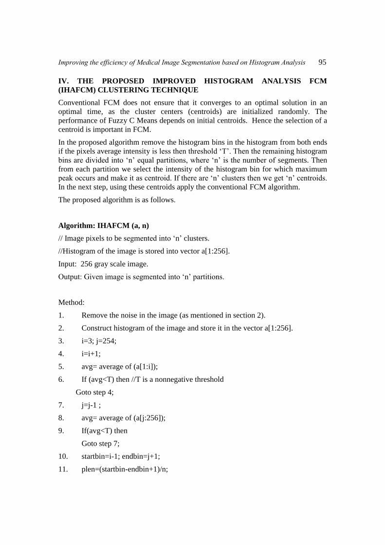
In the proposed algorithm remove the histogram bins in the histogram from both ends
if the pixels average intensity is less then threshold ‘T’. Then the remaining histogram
bins are divided into ‘n’ equal partitions, where ‘n’ is the number of segments. Then
from each partition we select the intensity of the histogram bin for which maximum
peak occurs and make it as centroid. If there are ‘n’ clusters then we get ‘n’ centroids.
In the next step, using these centroids apply the conventional FCM algorithm.
The proposed algorithm is as follows.
Algorithm: IHAFCM (a, n)
// Image pixels to be segmented into ‘n’ clusters.
//Histogram of the image is stored into vector a[1:256].
Input: 256 gray scale image.
Output: Given image is segmented into ‘n’ partitions.
Method:
1. Remove the noise in the image (as mentioned in section 2).
2. Construct histogram of the image and store it in the vector a[1:256].
3. i=3; j=254;
4. i=i+1;
5. avg= average of (a[1:i]);
6. If (avg<T) then //T is a nonnegative threshold
Goto step 4;
7. j=j-1 ;
8. avg= average of (a[j:256]);
9. If(avg<T) then
Goto step 7;
10. startbin=i-1; endbin=j+1;
11. plen=(startbin-endbin+1)/n;

96 A. Naresh, G. Venkateswara Rao & Ch. Satyananda Reddy
//plen represents each partition size.
12. c1=Max(a[startbin:startbin+plen-1]);
c2=Max(a[startbin+plen:startbin+2*len-1]);
c3=Max(a[startbin+2*len:startbin+3*plen-1]);
.
.
.
cn=Max(a[startbin+(n-1)*plen:endbin]);
13. Using centroids c1, c2, c3… cn apply the conventional FCM algorithm.
V. EXPERIMENTAL RESULTS AND ANALYSIS
In this section, the performance of the IHAFCM is compared with conventional FCM.
The experimental results are showcased on several standard images. In this
experiment, images corrupted with salt-and-pepper noise are taken to test the
effectiveness and the efficiency of the proposed IHAFCM algorithm. The experiments
were performed in a 2.99 GHz Intel Core 2 Duo processor, Windows 7 with 4 GB
RAM, using Matlab R2010a. The images are collected from the databases,
http://www.radiologyinfo.org, http://www.med.harvard.edu.
The proposed IHAFCM clustering algorithm and conventional FCM clustering with
varying number of clusters on images contaminated by different levels of salt-and-
pepper noise are executed to investigate the robustness of the algorithms.

Improving the efficiency of Medical Image Segmentation based on Histogram Analysis 97

98 A. Naresh, G. Venkateswara Rao & Ch. Satyananda Reddy
TABLE I
Comparison of number of iterations of fig 1,fig 2,fig 3 with 20% noise, fig 4 and fig 5
with 30% noise using conventional FCM and proposed algorithm for threshold T=15
Number of iterations
FCM
Number of iterations
IHAFCM
No. of
clusters
Fig.1 Fig. 2 Fig. 3 Fig. 4 Fig. 5 Fig. 1 Fig. 2 Fig. 3 Fig. 4 Fig. 5
3 39 25 44 39 59 21 19 31 31 36
4 26 37 86 31 38 16 25 71 19 16
5 106 157 74 141 80 80 131 69 127 26
TABLE II
Comparison of the CPU time taken by fig 1,fig 2,fig 3 with 20% noise, fig 4 and fig 5
with 30% noise using conventional FCM and proposed algorithm for threshold T=15.
CPU time in seconds
FCM IHAFCM
No. of
clusters
Fig.1 Fig. 2 Fig. 3 Fig. 4 Fig. 5 Fig. 1 Fig. 2 Fig. 3 Fig. 4 Fig. 5
3 4.1645 3.8594 7.4841 251.2197 154.3157 2.5412 2.25 3.7344 163.4531 92.2344
4 3.9844 8.4841 14.7497 271.5237 117.2969 2.152 3.75 10.3418 118.2188 49.1963
5 16.8945 26.7189 12.4127 1008.725 287.1563 11.6406 19.39 10.0186 841.573 90.5469
TABLE III
Comparison of number of iterations of 100 images with 20% noise using conventional
FCM and proposed algorithm for threshold T=15.
No. of clusters Average number of
iterations
FCM
Average number of
iterations
IHAFCM
3 40.6 26.23
4 46.3 30.45
5 117.2 85.3

Improving the efficiency of Medical Image Segmentation based on Histogram Analysis 99
TABLE IV
Comparison of the CPU time taken 100 images with 20% noise using conventional
FCM and proposed algorithm for threshold T=15
No. of
clusters
Average CPU time in
seconds
FCM
Average CPU time in
seconds
IHAFCM
3 420.2417 260.7491
4 481.4715 298.5712
5 1214.3761 847.5832
The data in table 1 and table 2 shows that the proposed algorithm reduces the
iterations to a large extent resulting in the reduction of running time for generating
segmented image. It can be observed that with the increased number of clusters the
proposed method has produced good results. It can be observe from fig. 4 and fig. 5 in
table 2 that if the image size is larger then there is vast CPU time difference between
convention FCM and proposed algorithm. It takes more number of iteration for
conventional FCM compared to proposed algorithm if the histogram bins towards
front end or rear end empty or average number of pixels less than some threshold ‘T’.
Most of the medical image histograms are like this so the proposed algorithm gives
good results for medical images.
VI. CONCLUSION
This paper presents a new algorithm named Improved Histogram Analysis Fuzzy-C
Means clustering algorithm for image segmentation, especially proposed for medical
images corrupted with salt-and-pepper noise. The proposed algorithm produces results
faster than the conventional FCM with the novel initialization method based on
histogram analysis to start the FCM clustering for segmentation of an image. This
algorithm is tested on several standard images, the results shows that the processing
time is reduced to segment the image. It also produces better results through its
inclusion of the noise detection and cancellation algorithm in its clustering process.
Furthermore, this finding suggests the IHAFCM clustering works as a novel method
for the segmentation of medical images and is efficient in terms of its computational
time.
REFERENCES
[1] K. K. V.Toh. and N. A. Mat-Isa, “Noise Adaptive Fuzzy Switching Median
Filter for Salt-and-Pepper Noise Reduction,” IEEE Signal Processing Letters, vol.17, no.3, pp 281-284, 2010.
100 A. Naresh, G. Venkateswara Rao & Ch. Satyananda Reddy
[2] K. K. V. Toh, H. Ibrahim, and M. N. Mahyuddin, “Salt-and-pepper noise
detection and reduction using fuzzy switching median filter,” IEEE Trans. Consumer Electron., vol. 54, no. 4, pp. 1956–1961, Nov.2008.
[3] M. E. Algorri and F. Flores-Mangas, “Classification of anatomical structures
in MR Brain Images using fuzzy parameters,” IEEE Transactions on Biomedical Engineering, vol. 51, pp. 1599-1608, 2004.
[4] C. Garcia and G. Tziritas, “Face detection using quantized skin color regions
merging and wavelet packet analysis,” IEEE Transactions on Multimedia, vol.
1, pp. 264-277, 1999.
[5] A. D. Doulamis, N. Doulamis, and S. Kollas, “Non-sequential video content
representation using temporal variation of feature vectors,” IEEE Transactions on Consumer Electronics, vol. 46, pp. 758-768, 2000.
[6] K. Nickel and R. Stiefelhagen, “Visual recognition of pointing gestures for
human-robot interaction,” Image and Vision Computing, vol. 25, pp. 1875-
1884, 2007.
[7] M. P. Segundo, L. Silva, O. R. P. Bellon and C. C. Queirolo, “Automatic Face
Segmentation and Facial Landmark Detection in Rang Images,” IEEE Transactions on Systems, Man, and Cybernetics, Part B: Cybernetics, vol. 40,
pp. 1319-1330, 2010.
[8] M. Setnes, “Supervised fuzzy clustering for rule extraction,” IEEE Transactions on Fuzzy Systems, vol. 8, pp. 416-424, 2000.
[9] K. K. Sung and T. Poggio, “Example-based learning for view-based human
face detection,” IEEE Transactions on Pattern Analysis and Machine Intelligence, vol. 20, pp. 39-51, 1998.
[10] S. N. Sulaiman and N. A. Mat Isa, “Adaptive fuzzy-K-means clustering
algorithm for image segmentation,” IEEE Transactions on Consumer Electronics, vol. 56, pp. 2661-2668, 2010.
[11] S. N. Sulaiman and N. A. Mat Isa, “Denoising-based clustering algorithms for
segmentation of low level salt-and-pepper noise corrupted images,” IEEE Transactions on Consumer Electronics, vol. 56, pp. 2702-2710, 2010.
[12] N. A. Mat-Isa, M.Y. Mashor, and N. Othman, “Pap Smear Image
Segmentation Using Modified Moving K-Mean Clustering,” International Conference on Biomedical Engineering. Kuala Lumpur Malaysia. 2002.
[13] N. A. Mat-Isa, M. Y. Mashor, N. H. Othman and S. N. Sulaiman, “Application
of Moving K-Means Clustering for Pap Smear Image Processing,” Proceeding of International Conference on Robotics, Vision, Information and Signal Processing, Penang Malaysia, 2002.
[14] N. A. Mat-Isa, A. S. Samy, U. K. Ngah, “Adaptive fuzzy moving Kmeans
algorithm for image segmentation,” IEEE Transactions on Consumer Electronics, vol. 55, no. 4, pp. 2145-2153, 2009.

Improving the efficiency of Medical Image Segmentation based on Histogram Analysis 101
[15] W. Luo, “Eficient removal of impulse noise from digital images,” IEEE Trans. Consumer Electron., vol. 52, no. 2, pp.523–527, May 2006.
[16] S. Chen, and D. Zhang, “Robust Image Segmentation Using FCM With
Spatial Constraints Based On New Kernel-Induced Distance Measure,” IEEE
Transactions on Cybernetics; Systems, Man, and Cybernetics, Part B, vol.34,
no.4, pp 1907-1916, 2004.
AUTHOR PROFILE
Mr. A. Naresh received B.Tech degree in Computer Science and
Engineering from J.N.T. University, India and M.Tech degree in
Information Technology from Andhra University, India in 2004 and
2007 respectively.
He is working as Assistant Professor in the department of IT in GITAM
University, Hyderabad and pursuing Ph.D. form JNTU, Kakinada.
Mr. G. Venkateswara Rao received M.Sc, M.Phil and M.Tech in
Computer Science and Engineering from Andhra University, India in
1993, 1996 and 1999 respectively and Ph.D. degree from Acharya
Nagarjuna University, India in 2010.
He is working as Associate Professor in the department of IT in GITAM
University, Visakhapatnam. He was awarded UGC major research
project in 2007.
Mr. Ch. Satyananda Reddy received M.Tech degree in Computer
Science and Engineering from JNTU, India and Ph.D. degree from
Andhra University, India in 2002 and 2010 respectively.
He is working as Associate Professor in the department of Computer
Science and Systems Engineering, Andhra University, Visakhapatnam.

102 A. Naresh, G. Venkateswara Rao & Ch. Satyananda Reddy