Embed Size (px)
Citation preview

Improving the Detection Rate of Congenital Heart Defects: Using a Real-Time Cardiac Assessment Technique in Routine Obstetrical
Screening
By
Donald Scott, Diploma (Medical Radiation Technology), MAppSc (Medical Ultrasound).
A thesis submitted in fulfilment of the requirements for the degree,
Doctor of Philosophy, Diagnostic Medical Ultrasound,
Faculty of Science, Charles Sturt University
2013
© Donald Scott

ii
Table of Contents
List of Tables ............................................................................................ iv
Certificate of Authorship ....................................................................... viii
Acknowledgements ................................................................................. ix
Verification of Authorship ....................................................................... xi
Ethics Approvals .................................................................................... xxi
Abstract ................................................................................................. xxii
Chapter 1 Prenatal Detection of Congenital Heart Disease ................... 1
Introduction ......................................................................................................... 1
Development of the Fetal Heart ....................................................................... 8
Fetal Cardiac Anatomy and Physiology ....................................................... 12
Causes of Congenital Heart Disease and Prevention ................................ 20
History of Screening for Congenital Heart Disease .................................... 27
Prevalence and Classification of Congenital Heart Disease ..................... 30
Sonographic Features of Severe Congenital Heart Disease (Cook et al., 2004). ................................................................................................................. 33
Chapter 2 Screening for Congenital Heart Disease .............................. 40
Two-Dimensional Imaging Review ................................................................ 40
Image quality ................................................................................................ 40
Pulse-echo Technique ................................................................................ 43
Image Formation .......................................................................................... 44
Image Processing ........................................................................................ 46
Doppler imaging ............................................................................................... 48
Doppler image processing .......................................................................... 50
Three dimensional imaging STIC and Matrix tools ..................................... 52
Acquiring a Three-Dimensional Volume ................................................... 54
STIC ............................................................................................................... 55
Live 3D........................................................................................................... 56
Volume Image Analysis .............................................................................. 58
Off-line Analysis ............................................................................................... 59
Summary ........................................................................................................... 60
Sonographic Features and Techniques for Early Detection ................ 61
Impact of Early Detection ............................................................................... 63
Current Techniques ......................................................................................... 66
Static Images ................................................................................................ 67
Importance of Cine-Loop Sweeps ............................................................. 69

iii
Colour Doppler ............................................................................................. 71
Three-Dimensional Imaging ....................................................................... 72
Proposed Research for this Thesis ............................................................... 76
Chapter 3 Assessment of the Fetal Heart during Routine Obstetrical Screening, a Standardized Method ....................................................... 83
Introduction ....................................................................................................... 83
Standardized Assessment .............................................................................. 86
Assessment ...................................................................................................... 92
Summary ........................................................................................................... 97
Chapter 4 Increasing the Detection Rate of Normal Fetal Cardiac Structures: A Real-Time Approach ....................................................... 99
Introduction ....................................................................................................... 99
Research Question/Objectives .................................................................... 102
Method ............................................................................................................. 103
Statistical Analysis...................................................................................... 109
Results ............................................................................................................. 109
Sample Images .............................................................................................. 112
Discussion ....................................................................................................... 115
Real-Time Methodology ................................................................................ 118
Study Limitations............................................................................................ 119
Conclusion ...................................................................................................... 119
Chapter 5 Increasing the Detection Rate of Congenital Heart Disease during Routine Obstetrical Screening Using Two Cine-Loop Sweeps .................................................................................................. 120
Materials and Methods .................................................................................. 123
Statistical Analysis ......................................................................................... 129
Results ............................................................................................................. 129
Discussion ....................................................................................................... 131
Conclusion ...................................................................................................... 135
Chapter 6 Increasing Recognition of Fetal Heart Anatomy using Online Tutorials and Mastery Learning compared with Classroom Instructional Methods .......................................................................... 137
Introduction ..................................................................................................... 137
Materials and Methods .................................................................................. 141
Results ............................................................................................................. 147
Discussion ....................................................................................................... 150
Conclusion ...................................................................................................... 153

iv
Chapter 7 State of the Art and Future Opportunities .......................... 155
Summary of Research Studies .................................................................... 155
Feedback loops .............................................................................................. 157
A model for Improving Screening Techniques .......................................... 159
References ............................................................................................ 161
Appendix A ............................................................................................ 170
Project Protocols and Design Considerations ........................................... 170
Project #1, pilot study, project proposal summary, protocol and sample size estimate .............................................................................. 170
Project #2, cine-loops sweeps, proposal summary, protocol and sample size estimate .............................................................................. 177
Project #3, “Find it First”, proposal summary, protocol and sample size estimate .............................................................................. 187
Appendix B ............................................................................................ 191
Fetal Heart Sonographic Assessment Tool ............................................... 191
Appendix C ............................................................................................ 196
Find it First web application ..................................................................... 196
Appendix D ............................................................................................ 202
Published papers ......................................................................................... 202
List of Tables
Table 1- 1 Anatomic structures examined during routine screening ............ 3
Table 1- 2 Sensitivity of screening for congenital anomalies ....................... 7
Table 1- 3 CHD and exposures by risk ratios ............................................ 22
Table 1- 4 Sonographic features of severe CHD ....................................... 33
Table 2- 1 Grey scale settings for imaging the fetal heart .......................... 48
Table 2- 2 Doppler settings and image quality .......................................... 52
Table 2- 3 Prenatal detection rate by view and incidence .......................... 68
Table 3- 1 Prenatal detection by protocol .................................................. 85
Table 4- 1 Clinical site survey data ......................................................... 102
Table 4- 2 Subject demographcs and acquisition time ............................ 103

v
Table 4- 3 Rate of detection by technique .............................................. 110
Table 4- 4 Detection rate of colour flow loops ......................................... 111
Table 4- 5 Detection rate differences by group ....................................... 112
Table 5- 1 Congenital heart disease characteristics ................................ 124
Table 5- 2 Detection rate by reviewer ..................................................... 130
Table 5- 3 Average detection rate by group ............................................ 130
Table 6- 1 Five points of assessment ..................................................... 148
Table 6- 2 Detection rate by group ......................................................... 149
List of Figures
Figure 1- 1 Cast of a fetal heart .................................................................. 5
Figure 1- 2 Cardiac looping ........................................................................ 9
Figure 1- 3 Fetal blood flow shunts ........................................................... 14
Figure 1- 4 Three vessel view ................................................................... 16
Figure 1- 5 Pulmonary veins ..................................................................... 16
Figure 1- 6 Right ventricular outflow tract ................................................. 18
Figure 1- 7 Left ventricular outflow tract .................................................... 19
Figure 1- 8 Transposition of the great arteries .......................................... 20
Figure 1- 9 Measurement of the nuchal translucency ............................... 26
Figure 1- 10 Tetralogy of Fallot ................................................................. 35
Figure 1- 11 Hypoplastic left heart syndrome ............................................ 36
Figure 2- 1 Colour Doppler aliasing .......................................................... 50
Figure 2- 2 Matrix array transducer ........................................................... 58
Figure 2- 3 Dextro-transposition of the great arteries ................................ 62
Figure 2- 4 Tetralogy of Fallot ................................................................... 70
Figure 2- 5 Atrioventricular septal defect .................................................. 71
Figure 3- 1 Grey scale sweep acquisition ................................................. 88
Figure 3- 2 Image optimization ................................................................. 90

vi
Figure 3- 3 Anatomic landmarks four chamber view .................................. 90
Figure 3- 4 Parallel outflow tracts .............................................................. 92
Figure 3- 5 Four chamber to outflow sweep .............................................. 97
Figure 4- 1 Static image of the four chamber view .................................. 104
Figure 4- 2 Static image of the LVOT view .............................................. 104
Figure 4- 3 Static image of the RVOT view ............................................. 105
Figure 4- 4 Static image of the abdomen ................................................ 106
Figure 4- 5 Four chamber view from cine-loop sweep ............................. 106
Figure 4- 6 LVOT view from cine-loop sweep .......................................... 107
Figure 4- 7 RVOT view from cine-loop sweep ......................................... 107
Figure 4- 8 Pulmonary veins from cine-loop sweep ................................. 113
Figure 4- 9 Four chamber view from colour Doppler sweep .................... 113
Figure 4- 10 Five chamber view from colour Doppler sweep ................... 114
Figure 4- 11 Three vessel view from colour Doppler sweep .................... 114
Figure 5- 1 Cine-loop acquisition ............................................................. 126
Figure 5- 2 Fetal heart sonographic assessment tool .............................. 128
Figure 5- 3 Ventricular septal defect........................................................ 134
Figure 6- 1 Quiz feedback ....................................................................... 144
Figure 6- 2 Final quiz .............................................................................. 146
Figure B- 1 .......................................................................................................... 191
Figure B- 2 .......................................................................................................... 192
Figure B- 3 .......................................................................................................... 192
Figure B- 4 .......................................................................................................... 193
Figure B- 5 .......................................................................................................... 193
Figure B- 6 .......................................................................................................... 194
Figure B- 7 .......................................................................................................... 194
Figure B- 8 .......................................................................................................... 195
Figure C- 1 .......................................................................................................... 197

vii
Figure C- 2 .......................................................................................................... 197
Figure C- 3 .......................................................................................................... 198
Figure C- 4 .......................................................................................................... 198
Figure C- 5 .......................................................................................................... 199
Figure C- 6 .......................................................................................................... 199
Figure C- 7 .......................................................................................................... 200
Figure C- 8 .......................................................................................................... 200
Figure C- 9 .......................................................................................................... 201
Figure C- 10 ........................................................................................................ 201

viii
Certificate of Authorship
I hereby declare that this submission is my own work and to the best
of my knowledge and belief, understand that it contains no material
previously published or written by another person, nor material
which to a substantial extent has been accepted for the award of any
other degree or diploma at Charles Sturt University or any other
educational institution, except where due acknowledgement is made
in the thesis [or dissertation, as appropriate]. Any contribution made
to the research by colleagues with whom I have worked at Charles
Sturt University or elsewhere during my candidature is fully
acknowledged.
I agree that this thesis be accessible for the purpose of study and
research in accordance with normal conditions established by the
Executive Director, Library Services, Charles Sturt University or
nominee, for the care, loan and reproduction of thesis, subject to
confidentiality provisions as approved by the University.
Name Ted Scott
Signature
Date 01/07/2014
ix
Acknowledgements
This PhD thesis employed three research projects in order to devise
a new method of screening the fetal heart during routine obstetrical
sonography and translate the method to clinical practice. The first
project was completed to determine the optimal imaging and
assessment protocol. The second project tested the imaging and
assessment protocol to determine if the method increased the
detection rate of congenital heart disease compared with current
methods. The third project was developed to demonstrate that the
new method of screening could be translated to clinical practice
using an online, master-based learning tool.
Project #1 Increasing the Detection Rate of Normal Fetal Cardiac Structures: A Real-Time Approach, Ted Scott, Hans Swan, Gerald Moran, Tapas Mondal, Judy Jones, Karm Guram, Jaime Huff, Journal of Diagnostic Medical Sonography 2008; 24; 63 This project was undertaken with the financial support of the SDMS
Education Foundation, Graduate Research Award and the Bracco
Diagnostics Research Award. Thanks are given to the following
Sonographers: Malka Glasner, Mandie Bird, Rob Noftle and Melissa
Wood for their efforts in acquiring the fetal cardiac images.
Appreciation is also expressed to Philips Medical Systems Canada
and Mohawk College for their support of this project. Equipment was
provided by Philips Medical Systems, Canada and laboratory space
was provided by Diane Barrafato associate dean, Medical Radiation
Sciences, School of Health Sciences, Mohawk College.

x
Project #2 Increasing the Detection Rate of Congenital Heart Disease During Routine Obstetric Screening Using Cine Loop Sweeps, Ted Scott, Judy Jones, Herschel Rosenberg, Andrea Thomson, Hournaz Ghandehari, Neil Rosta, Malka Stromer, KimJozkow, Journal of Ultrasound in Medicine, 2013; 32:973-979
This project was supported by a health research grant from the
Hamilton Community Foundation. This work was published as an
abstract and presented orally at the American Institute of Ultrasound
in Medicine 2010 Annual Convention; March 24–27, 2010; San
Diego, California. Normal cases were acquired from the Department
of Radiology, Grey Bruce Health Services, by Bertha Benninger,
Georgia Roach, Terri Dowling, and Alicia Leudke. Abnormal cases
were acquired with the help of Tapas Mondal, MD, from the
Department of Pediatric Cardiology, McMaster Children’s Hospital
(Hamilton,Ontario, Canada). The Fetal Heart Sonographic
Assessment Tool was developed by Andrew Connery, instructional
designer, Center for Teaching and Learning, Mohawk College.
Project #3
Increasing Recognition of Fetal Heart Anatomy using Online Tutorials and Mastery Learning Compared with Classroom Instructional Methods, Ted Scott, Laura Thomas, Keith Edwards, Judy Jones, Hans Swan, Andrew Wessels, Journal of Diagnostic Medical Sonography
The authors would like to acknowledge the efforts of University of
Hawaii at Hilo Computer Sciences 460 students, Joel Cook, Jacob
Shon, Kylie Gonsalves and Jermaine Vitales for their development of
the “Find it First” learning application.

xi
Verification of Authorship
I have contacted each of the co-authors relating to the publications
included in this thesis to verify that I have performed the role as 1st
author and I am responsible for the research concept, development
and execution of each project and published paper. The verification
email from each co-author is provided below with the exception of Dr.
Guram who could not be located.
Dear Dr. Moran,
I would like to acknowledge your co-author role in the publication “Increasing the Detection Rate of Normal Fetal Cardiac Structures: A Real-Time Approach” as advisor to the experimental design of this study. If you accept this description of your contribution and agree that as first author, I (Ted Scott) developed the concept and should be acknowledged as the creator and 1st author of the manuscript please reply with confirmation to this email.
Best regards,
Ted
From: MORAN, GERALD (NAM RC-CA) <[email protected]>
Sent: October-21-13 2:19 PM
To: Scott, Ted
Subject: Validation of Role in "Increasing the Detection Rate of Normal Fetal Cardiac Structures: A Real-Time Approach."
Yes, Thank you Ted.
Jer

xii
Dear Dr. Mondal,
I would like to acknowledge your co-author role in the publication “Increasing the Detection Rate of Normal Fetal Cardiac Structures: A Real-Time Approach.” as an image reviewer for this study. If you accept this description of your contribution and agree that as first author, I (Ted Scott) developed the concept and should be acknowledged as the creator and 1st author of the manuscript please reply with confirmation to this email.
Best regards,
Ted
From: Mondal, Tapas <[email protected]>
Sent: October-21-13 2:47 PM
To: Scott, Ted
Subject: Validation of role in ""Increasing the Detection Rate of Normal Fetal Cardiac Structures: A Real-Time Approach."
Hi Ted,
I confirm that you developed the concept of this paper and should be recognized as the creator and first author for this article.
Hope this clarifies the position.
Regards
Tapas
Dear Dr. Gandehari,
I would like to acknowledge your co-author role as image reviewer in the publication, “Increasing the Detection of Congenital Heart Disease During Routine Obstetrical Screening using Two Cine-Loop Sweeps. J. Ultrasound Med., 32(6).” If you accept this description of your contribution and agree that as first author, I (Ted Scott) developed the concept and should be acknowledged as the creator and 1st author of the manuscript please reply with confirmation to this email.
Best regards,
Ted

xiii
From: Hournaz Ghandehari <[email protected]>
Sent: October-21-13 3:54 PM
To: Scott, Ted
Subject: Re: re; verification of role
Hi Ted,
Yes I agree.
Thanks,
Hournaz
Sent from my iPhone
Dear Dr. Thomson,
I would like to acknowledge your co-author role as image reviewer in the publication, “Increasing the Detection of Congenital Heart Disease During Routine Obstetrical Screening using Two Cine-Loop Sweeps. J. Ultrasound Med., 32(6).” If you accept this description of your contribution and agree that as first author, I (Ted Scott) developed the concept and should be acknowledged as the creator and 1st author of the manuscript please reply with confirmation to this email.
Best regards,
Ted
From: Andrea Young <[email protected]>
Sent: October-21-13 3:35 PM
To: Scott, Ted
Subject: verification of role
Yes I agree.
Best wishes
Andrea Thomson
Sent from my iPhone

xiv
Dear Ms. Jozkow,
I would like to acknowledge your co-author role as image reviewer in the publication, “Increasing the Detection of Congenital Heart Disease During Routine Obstetrical Screening using Two Cine-Loop Sweeps. J. Ultrasound Med., 32(6).” If you accept this description of your contribution and agree that as first author, I (Ted Scott) developed the concept and should be acknowledged as the creator and 1st author of the manuscript please reply with confirmation to this email.
Best regards,
Ted
From: Kim Jozkow <[email protected]>
Sent: October-21-13 4:15 PM
To: Scott, Ted
Subject: RE: verification of role - Agree
Dear Ted,
I agree with your description.
All the best,
Kim Jozkow
Dear Ms. Stromer,
I would like to acknowledge your co-author role as image reviewer in the publication, “Increasing the Detection of Congenital Heart Disease During Routine Obstetrical Screening using Two Cine-Loop Sweeps. J. Ultrasound Med., 32(6).” If you accept this description of your contribution and agree that as first author, I (Ted Scott) developed the concept and should be acknowledged as the creator and 1st author of the manuscript please reply with confirmation to this email.
Best regards,
Ted

xv
From: Malka Stromer <[email protected]>
Sent: October-21-13 4:15 PM
To: Scott, Ted
Subject: Re: re; verification of role
Sounds fine.
Thanks ted.
Sent from my iPhone
Dear Dr. Rosta,
I would like to acknowledge your co-author role as image reviewer in the publication, “Increasing the Detection of Congenital Heart Disease During Routine Obstetrical Screening using Two Cine-Loop Sweeps. J. Ultrasound Med., 32(6).” If you accept this description of your contribution and agree that as first author, I (Ted Scott) developed the concept and should be acknowledged as the creator and 1st author of the manuscript please reply with confirmation to this email.
Best regards,
Ted
From: Rosta, Neil <[email protected]>
Sent: October-21-13 4:51 PM
To: Scott, Ted
Subject: RE: verification of role
Hi Ted, Thanks for the email. I agree that you developed the concept for the publication, “Increasing the Detection of Congenital Heart Disease during Routine Obstetrical Screening using Two Cine-Loop Sweeps. J. Ultrasound Med., 32(6)” and you should be acknowledged as the creator and 1st author of the manuscript.

xvi
Best Regards, Neil Rosta
Dear Ms. Thomas,
I have described your role as a study co-ordinator for the publication, “Increasing Recognition of Fetal Heart Anatomy using Online Tutorials and Mastery Learning Compared with Classroom Instructional Methods.” submitted to the Journal of Diagnostic Medical Sonography. If you accept this description of your contribution and agree that as first author, I (Ted Scott) developed the concept and should be acknowledged as the creator and 1st author of the manuscript please reply with confirmation to this email.
Best regards,
Ted
From: Thomas, Laura
Sent: October-21-13 6:33 PM
To: Scott, Ted
Subject: Verification of role
Ted,
Yes I accept the description of study co-ordinator.
Laura Thomas
From: Scott, Ted
Sent: Monday, October 21, 2013 2:48 PM
To: mailto:[email protected]
Subject: re: Validation of role in Ted's research publications
Hi Judy,
I have described your role and contribution to the publications that have been accepted and submitted to the Journal of Diagnostic Medical Sonography and the Journal of Ultrasound in Medicine, as listed below. If you accept this description of your contribution and

xvii
agree that as first author, I (Ted Scott) developed the concept and should be acknowledged as the creator and 1st author of the manuscript please reply with confirmation to this email.
1. Scott, T., Swan, H., Moran, G., Mondal, T., Jones, J., Guram, K., & Huff, J. (2008). Increasing the Detection Rate of Normal Fetal Cardiac Structures: A Real-Time Approach. Journal of Diagnostic Medical Sonography, 24(2), 63–71. doi:10.1177/8756479308315234
a. Judy Jones participated as an image reviewer in this study, Ted Scott, designed the study and was the primary author of the published manuscript.
2. Scott, Ted.;Jones, Judy.; Rosenberg, Herschel.; Ghandehari, Thomson, Andrea.; Hournaz.; Rosta, Neil.; Jozkow, Kim.; Stromer, Malka.;Swan, H. (2013). Increasing the Detection of Congenital Heart Disease During Routine Obstetrical Screening using Two Cine-Loop Sweeps. J. Ultrasound Med., 32(6).
a. Judy Jones participated as an image reviewer in this study, Ted Scott, designed the study and was the primary author of the published manuscript.
3. Scott, T. Thomas, L., Edwards K., Jones, J., Swan, H., Wessels, A. (n.d.). Increasing Recognition of Fetal Heart Anatomy using Online Tutorials and Mastery Learning Compared with Classroom Instructional Methods. Journal of Diagnostic Medical Sonography.
a. Judy Jones participated as a contributor to the educational tutorials used in this study, Ted Scott, designed the study and was the primary author of the published manuscript.
4. Scott, T.; Jones, J.; Swan, H. (n.d.). Assessment of the Fetal Heart During Routine Obstetrical Screening, A Standardized Method. Journal of Diagnostic Medical Sonography.
a. Judy Jones participated as editor of the manuscript, Ted Scott, was the primary author of the published manuscript.

xviii
5. Scott, T.; Jones, J.; Swan, H. (n.d.). Screening for Congenital Heart Disease: Sonographic Features and Techniques for Prenatal Detection. Journal of Diagnostic Medical Sonography.
a. Judy Jones participated as an editor of this manuscript, Ted Scott was the primary author of the published manuscript.
Best regards,
Ted
From: Judy Jones <[email protected]>
Sent: October-21-13 8:15 PM
To: Scott, Ted
Subject: Re: Validation of role in Ted's research publications
HI Ted- Yes, I agree to all.
Thanks,
Judy
On Mon, Oct 21, 2013 at 9:10 AM, Scott, Ted <[email protected]> wrote:
Dear Mr. Wessels,
I have described your role as a lead software application developer for the publication, “ Increasing Recognition of Fetal Heart Anatomy using Online Tutorials and Mastery Learning Compared with Classroom Instructional Methods.” submitted to the Journal of Diagnostic Medical Sonography. If you accept this description of your contribution and agree that as first author, I (Ted Scott) developed the concept and should be acknowledged as the creator and 1st author of the manuscript please reply with confirmation to this email.
Best regards,
Ted

xix
From: Andrew W. <[email protected]>
Sent: October-21-13 9:28 PM
To: Scott, Ted
Subject: Re: re; verification of role
Yeah, that sounds great, thanks.
Dear Dr. Rosenberg,
I would like to acknowledge your co-author role as image reviewer in the publication, “Increasing the Detection of Congenital Heart Disease During Routine Obstetrical Screening using Two Cine-Loop Sweeps. J. Ultrasound Med., 32(6).” If you accept this description of your contribution and agree that as first author, I (Ted Scott) developed the concept and should be acknowledged as the creator and 1st author of the manuscript please reply with confirmation to this email.
Best regards,
Ted
From: Herschel Rosenberg <[email protected]>
Sent: October-22-13 7:36 AM
To: Scott, Ted
Subject: Re: re; verification of role
That's fine Ted.
Dr. H.C. Rosenberg MD, FRCP

xx
Pediatric Cardiologist
Children's Hospital of Western Ontario
University of Western Ontario
On Mon, Oct 21, 2013 at 9:11 AM, Scott, Ted
<[email protected]> wrote:
Dear Dr. Edwards,
I have described your role as a software application development supervisor for the publication, “Increasing Recognition of Fetal Heart Anatomy using Online Tutorials and Mastery Learning Compared
with Classroom Instructional Methods.” submitted to the Journal of Diagnostic Medical Sonography. If you accept this description of your contribution and agree that as first author, I (Ted Scott) developed the concept and should be acknowledged as the creator and 1st author of the manuscript please reply with confirmation to this email.
Best regards,
Ted
From: H. Keith Edwards <[email protected]>
Sent: October-22-13 1:48 AM
To: Scott, Ted
Subject: verification of study role
Hello Ted: I have no issue with you appearing as the first author.
- H. Keith Edwards
Associate Professor - Computer Science
University of Hawaii - Hilo
New Phone (2013/10/20): (808) 932-7522

xxi
Ethics Approvals
Project #1 Increasing the Detection Rate of Normal Fetal Cardiac Structures: A Real-Time Approach. Approved for research by: Charles Sturt University, Human Research Ethics Committee,
protocol number 2013/032; Mohawk College Research Ethics Board,
TS 28.07.06
Project #2 Improving the Detection Rate of Congenital Heart Defects Using a Standardized, Real-Time Cardiac Assessment Technique in Routine Obstetrical Screening. Approved for research by: Charles Sturt University, Human Research Ethics Committee,
protocol number 2013/032; Mohawk College Research Ethics Board,
TS 28.07.06; Hamilton Health Sciences Research Ethics Board,
project #08-071; Grey Bruce Health Services, Research Ethics
Board.
Project #3 The Find it First project: Improving the ability of learners to identify congenital heart disease using a simulation based assessment in an online format, compared with traditional classroom instruction. Approved for research by:
Charles Sturt University, Human Research Ethics Committee,
protocol number 2013/032; McMaster University Research Ethics
Board, Project # 2013-005; Mohawk College Research Ethics Board,
RA 03.04.13.

xxii
Abstract
Improving the Detection Rate of Congenital Heart Defects Using a Real-Time Cardiac Assessment Technique in Routine Obstetrical
Screening Screening the fetal heart for the presence of congenital heart disease
(CHD) during routine obstetrical sonography has not translated to
early identification of CHD for the majority of patients (Friedberg et
al., 2009; Poole et al., 2013). Limitations have been identified in the
methods used to assess the fetal heart. A number of leading experts
have identified the need for a real-time assessment of the fetal heart
compared with the convention of relying on static images only for
interpretation (Mcgahan, Moon-grady, & Pahwa, 2007; Sklansky,
2007). In addition, a deep need has been identified for education to
improve the examiner’s and interpreting physician’s understanding of
the sonographic features of CHD (Friedberg et al., 2009; McBrien,
Sands, Craig, Dornan, & Casey, 2010).
Imaging the fetal heart is a challenging element of a routine
obstetrical sonogram. The fetal heart is a very small, complex and
highly dynamic structure with a normal heart rate ranging from 120-
160 bpm (Fredouille, 2007). The expression of CHD varies
considerably, depending on the specific congenital anomaly and the
time at which the exam is performed. (Cook, Yates, & Anderson,
2004). Some severe forms of CHD including Transposition of the
Great Arteries (TGA) can only be detected through visualization of
the outflow tracts, while other forms such as Hypoplastic Left Heart
Syndrome (HLHS) can be seen in the four chamber view (Chaoui,
2003).

xxiii
Although the practice guidelines for obstetric ultrasound imaging
explicitly identify the anatomic structure that should be examined
during screening, considerable variation in how these structures are
assessed exists (ACR Guidelines and Standards Committee,
American Institute of Ultrasound in Medicine, 2003)(American
Institute of Ultrasound in Medicine, 2013). The majority of screening
centers rely on a few static images to demonstrate the normalcy of
fetal heart including a four chamber view and both outflow tracts.
Alternative approaches have been identified and these include the
use of cine-loops, Doppler imaging, and three dimensional imaging,
as well as detailed anatomic planes (Chaoui & Heling, 2005; Chaoui
& McEwing, 2003; Mcgahan et al., 2007).
This PhD thesis includes three projects that have been completed to
address potentially increasing the rate of CHD detection. The first
project assessed the feasibility and efficacy of using cine-loop
sweeps, grey-scale cine-loop sweeps and colour Doppler cine
sweeps compared with static images only. The use of cine-loop
sweeps was found to be the most effective and practical technique.
The second project assessed the practicality of using cine-loop
sweeps and a standardized assessment technique compared with
static images only and demonstrated an increased detection rate of
CHD. The third project assessed the efficacy of an online, tutorial-
based tool for learning the standardized assessment technique of
cine-loop sweeps used in the first two projects.
Improving the detection rate of CHD during routine obstetrical
sonography can be achieved with the use of a standardized

xxiv
assessment of cine-loop sweeps. The translation of this technique to
clinical practice can be achieved using an online learning tool.

Chapter 1 Prenatal Detection of Congenital Heart Disease
Introduction The identification of congenital heart disease, (CHD) during the
prenatal period is an important screening objective of a routine
obstetrical sonogram. Despite the technological advances that have
occurred within the field of diagnostic medical sonography, the
detection of CHD has lagged behind the detection rates of other
congenital anomalies (Grandjean, Larroque, & Levi, 1999). The
opportunity for improving the detection of CHD will most likely
depend on two facets of the screening process; 1) optimization of the
image acquisition process and 2) the standardization of the
assessment protocol. The feasibility of implementation of the optimal
process and protocol will be an additional key consideration.
A thorough review of the literature was undertaken in order to fully
examine the state of the art as it relates to imaging the fetal heart.
This includes a review of: practice guidelines for assessment of the
fetal heart, the embryological and physiological basis for CHD; the
history of screening the fetal heart, the technical underpinnings of
image formation in two dimensions, grey-scale mode, colour and
spectral Doppler and three dimensional imaging tools. The analysis
of the imaging acquisition and assessment capabilities of modern
sonography systems as they relate to detecting CHD in-utero were
framed by the current imaging protocols and the relative ease of
implementation.

2
A significant limitation of current methods is the reliance on static
images for assessment by the interpreting physician. Given the
technical improvements in imaging systems, including image
processing, Doppler and three-dimensional techniques, it may be
possible to leverage these capabilities in a new image acquisition
and assessment protocol.
The use of ultrasound imaging for routine fetal screening in the
second trimester has been adopted by the Canadian health care
system (Van den Hof & Wilson, 2005). A complete screening
examination of the fetus will provide the parents and care providers
with a comprehensive report of the gestational status and viability of
the pregnancy. According to the American Institute of Ultrasound in
Medicine (AIUM) practice guidelines, the routine screening of the
fetus in the second trimester is expected to include documentation of
the gestational status and fetal anatomy as defined in Table 1-1
(American Institute of Ultrasound in Medicine, 2013).

3
Table 1- 1
Gestational status and anatomic structures documented in routine second trimester screening.(American Institute of Ultrasound in Medicine, 2013)
Anatomic structure Imaging parameters Comments Gestational status Fetal number,
presence of cardiac activity and presentation
Multiple gestations require further documentation
Gestational sac Placental location and assessment of amniotic fluid
Quantitative or semi-quantitative estimate of amniotic fluid volume should be reported
Fetal Biometry
Gestational age estimate, fetal weight estimate
Significant discrepancies between gestational age and fetal measurements may suggest fetal growth restriction or macrosomia.
Maternal adnexa
Uterus and adnexa
Allow recognition of incidental findings
Anatomic survey Head face and neck Cerebellum, choroid
plexus, cisterna magna, upper lip, lateral cerebral ventricles, midline falx and cavum septum pellucidi
Chest Fetal heart, four- chamber view, Left ventricular outflow tract ; and Right ventricular outflow tract
Abdomen Stomach (size, situs and presence), kidneys, bladder, umbilical cord insertion into the fetal abdomen, umbilical cord vessel number
Spine Cervical, thoracic, lumbar and sacral spine
Extremities Presence of legs and arms
Gender Indicated in low-risk pregnancies only for evaluation of multiple gestations

4
Compared with the assessment of most fetal anatomic structures, the
assessment of the fetal heart during routine screening represents a
diagnostic challenge for sonographers and interpreting physicians.
This is due to a number of factors; the size and complexity of the
heart, screening methods used and the need for focussed training to
enable more effective identification of heart defects. The assessment
and documentation of the fetal heart can very easily be
misinterpreted as normal based on the acquisition and interpretation
of static images only (Sklansky, 2007). The fetal heart is a relatively
small and complex structure as seen in an early second trimester
cast of a fetal heart in Figure 1-1. Key anatomical structures such as
the outflow tracts are only 2-6 mm in diameter (Allan, 2004). In some
forms of CHD, the abnormality may only be detectable within a
relatively small fraction of the heart volume. For example, congenital
heart disease relating to the outflow tracts such as dextro-
transposition of the great arteries may only be detected at their
origins and frequently are not detected by the four chamber view.

5
Figure 1- 1
Cast of a fetal heart in early second trimester, obtained from the McMaster University Anatomy laboratory with the assistance of Glenn Oomen.
The normal fetal heart beat ranges from 120-160 beats per minute;
each beat represents a systolic and diastolic phase equivalent to an
effective unidirectional movement of 240-320 cycles per second
(Pildner von Steinburg S, Boulesteix A-L, Lederer C, Grunow S,
Schiemeier S, Hatzmann W, 2013). Furthermore the impact of
congenital heart disease varies widely depending on the specific
form it takes. In some cases the ventricles of the heart may be
entirely normal in appearance and the defect may only be evidenced
in the outflow tracts of the heart or in a relatively small fraction of the
fetal heart volume.
Since static images of the fetal heart represent only very thin slices of
the fetal heart it is very important that the fetal heart is assessed in
real-time and interrogated completely in contiguous slices. The

6
diversity and relative complexity of congenital heart defects demands
considerable skill of the sonographer and interpreting physician to
ensure a complete examination.
According to a survey of 2758 referrals to the fetal cardiology unit at
Guy’s Hospital, London, UK, the low risk population is the source of
80% of Congenital Heart Defects (CHD) (Sharland, 2004). This
underscores the importance of routine screening for detection of
CHD. Routine screening provides a more accurate measure of
gestational age, earlier detection of multiple pregnancies and may
detect congenital malformations. The Eurofetus study identified
marked differences in sensitivity for detection of different
malformations, see Table 1-2.

7
Table 1- 2
Sensitivity of routine obstetrical screening for congenital anomalies, reported in the Eurofetus study (Grandjean et al., 1999).
Anomaly Number Type Sensitivity Urinary tract 138 Major 84.8 816 Minor 89.1 Total Musculoskeletal
954 Combined 88.5
349 Major 73.6 694 Minor 18.0 Total Respiratory system
1043 Combined 36.6
14 Major __
16 Minor __ Total 30 Combined 70.0 Central nervous system
Total
738
Combined
88.3
Heart and great vessels 366 Major 38.8 587
Minor 20.8
Total 953 Combined 27.7 Digestive system 8 Major __ 221 Minor 52.0 Total 229 Combined 53.7 Total 4615 Combined 56.2
Abnormalities of the urinary system and nervous system were most
frequently detected with a sensitivity of 88.5% and 88.3%
respectively (Grandjean et al., 1999). In this study cardiac
abnormalities were not detected in most cases, sensitivity for major
cardiac defects was 38.8% and minor defects 20.8% with a
combined sensitivity of 27.7% (Grandjean et al., 1999). In Canada,
the routine second trimester examination is performed in real-time
and documented as a series of static images archived electronically
or on film. Typically the interpretation of the cardiac images is
8
performed by a radiologist or obstetrician using the static images.
Present detection rates for CHD vary widely depending on a number
of factors: operator skills, maternal obesity, fetal position, inadequate
image optimization, image acquisition protocol and the nature of the
CHD (Chaoui, 2003). In order to clarify the recommended fetal heart
structures for examination the American College of Radiology (ACR),
the American College of Obstetrics and Gynecology (ACOG), the
Canadian Society of Gynecologists and the International Society of
Ultrasound in Obstetrics & Gynecology (ISUOG) have published
guidelines for routine second trimester screening (American Institute
of Ultrasound in Medicine, 2013; Reddy, Abuhamad, Levine, &
Saade, 2014; Van den Hof & Wilson, 2005). Both of these
publications require the four chamber view, also known as a basic
assessment and where possible views of the left ventricular outflow
tract (LVOT) and right ventricular outflow tracts (RVOT) commonly
referred to as an extended examination (ACR Guidelines and
Standards Committee, American Institute of Ultrasound in Medicine,
2003; Lee et al., 2008).
Development of the Fetal Heart Heart Tube Formation
The first organ to develop during embryogenesis is the fetal heart; it
completes development by the end of the first trimester (Lohr, Martin,
& Garry, 2012). The process of gastrulation describes molecular
signalling activities that result in the fusion of the anterior and lateral
mesoderm to form the heart tube in the midline of the embryo. This
single tube begins to beat at 22-23 days gestation and Doppler

9
imaging can demonstrate blood flow between four to five weeks
gestation (Lohr, Martin, & Garry, 2012). During the fourth and fifth
weeks of gestation, the epicardial cells arise from the proepicardium
and migrate over the heart tube to contribute to the connective tissue
of the myocardium and epicardium defining the coronary arteries
(Abdulla, Blew, & Holterman, 2004).
Formation of the Vascular System
The vascular system forms at the same time as the fetal heart. The
arterial and venous systems form bilaterally and they are symmetric
initially, anterior aortic arches, posterior and anterior cardinal veins
are paired. The paired dorsal aorta fuse into a single thoraco-
abdominal aorta early in embryogenesis, while the dual aortic arches
and paired venous return persist until six to eight weeks gestation
(Moore & Persaud, 2008).
Figure 1- 2
The heart tube is anchored at both ends. As the tube grows, it bends on itself to the right, the green circle represents the future right ventricle, the red square represents the future left ventricle. Illustrations prepared by Ken Wong Graphic Designer, Mohawk College.

10
Cardiac Looping
Elongation of the heart tube between 22-28 days gestation generates
the primitive outflow and inflow tracts. Rightward looping brings the
inflow region to a cranial position, this looping process establishes
the anatomic relationships needed for normal chamber formation and
septation. See Figure 1-2. Severe forms of congenital heart disease
and outflow tract defects are associated with disturbances in the
looping process such as, Transposition of the Great Arteries (TGA).
(Kirby & Waldo, 2002).
Chamber Formation and Septation
Septation begins at the same time as looping and the elongated
heart tube narrows to form the conotruncus (outflow tracts),
ventricular chambers, atrioventricular sulcus, primitive atrial chamber
and the sinus venosus. At the atrialventricular sulcus the myocardium
forms the endocardial cushion during the fifth week of gestation. The
endocardial cushion divides the heart into left and right
chambers(Abdulla et al., 2004). The atrial and ventricular septa fuse
with the endocardial cushion to complete the septation of the cardiac
chambers. The septum primum is a thin membrane which divides the
atria and fuses during the fifth week of gestation. This septum
develops an opening, the foramen secondum which provides access
for shunting of blood during gestation from the right to left side of the
heart. A thicker and more muscular septum secundum overlaps the
septum primum creating a flap like valve over the foramen ovale. The
ventricular septum also develops during this period, a muscular
septum grows through proliferation and trabeculation of the ventricles

11
while the inlet, membranous and outflow septa close with the fusion
of the bulbar ridges and the endocardial cushion fusing with the
muscular septum (Lohr, Martin, & Garry, 2012). The most common
form of congenital heart disease results from a failure of the septum
to close completely which results in an interventricular septal defect
(Hoffman & Kaplan, 2002).
Cardiac Outflow Tract Formation and Innervation
Parasympathetic innervation and septation of the outflow tracts in the
developing heart is governed by the cardiac neural crest (CNC) cells.
These cells migrate from the neural tube and have a unique
molecular signature. CNCs also contribute to the development of the
anterior parasympathetic plexus (Kirby & Waldo, 2002).
Formation of the Valves and Conduction System
The cardiac valves are formed at the atrioventricular canal and
outflow tracts, originating from the endocardial cushion. They are
fully developed by the tenth week of gestation. Structurally, the
valves are comprised of fibrous tissue which stretches and becomes
thin during development. Initially the myocardium generates cardiac
impulses, while the specialized conduction system develops during
the fifth week of gestation. The sinus and av nodes form and as the
chambers form and undergo septation, the bundle of HIS and
purkinje fibers are developed (Lohr, Martin, & Garry, 2012). The fetal
heart is completely formed and functional by the end of the first
trimester, although the sarcomeres continue to undergo significant
changes in metabolism and growth following birth (Cook et al., 2004).

12
Fetal Cardiac Anatomy and Physiology The fetal cardiovascular system is structurally and physiologically
different than the mature, postnatal cardiovascular system. The
presence of three vascular shunts, the ductus venosus, the foramen
ovale and the ductus arteriosus define a parallel circulation that relies
on the maternal placenta for gas exchange and metabolic needs.
The fetal heart pumps blood to the placenta through the right
ventricle, main pulmonary artery and the ductus arteriosus as well as
the left ventricle directly into the aorta. The right ventricle is dominant
and accounts for between 60-65% of the combined cardiac output.
(Cohen, 2001) During this time the pulmonary circulation is in
development and not yet functional. Pulmonary circulation is
characterized by a high degree of flow resistance in utero, and low
flow volume. The nature of a parallel circulation is to favour a low
resistance path and therefore only a small fraction of the fetal blood
supply passes through the left and right pulmonary arteries to the
lungs.
Deoxygenated blood returns to the placenta through the internal iliac
arteries into the umbilical cord and back to the placenta. Within the
capillary network of the placenta the returning fetal blood is
oxygenated and enriched with nutrients. The oxygen saturation of
85% returning from the placenta is less than that of the 100%
saturation of blood later in post natal life (Cohen, 2001; Kiserud &
Acharya, 2004). The relatively low saturation level is offset by a
higher percentage of hemoglobin in the fetal blood. Fetal blood flow
returns to the fetus by the umbilical vein and into the fetal cord

13
insertion entering the body through the abdominal wall becoming the
ductus venosus where it goes on to join the left portal vein in the
liver. The ductus venosus shunts approximately 30% of the blood
into the heart at mid-gestation with the remainder perfusing the liver
(Kiserud & Acharya, 2004). Mixing of oxygenated and deoxygenated
blood in the right atrium occurs as systemic venous blood returns
through the Superior Vena Cava (SVC) and the Inferior Vena Cava
(IVC) adding to the oxygenated blood provided by the placenta
through the ductus venosus. The architecture of the flow path into the
atrium favours a flow of oxygenated blood from the ductus venosus
across the right atrium and into the left atrium through the foramen
ovale with minimal mixing of oxygenated and deoxygenated blood.
This ensures that the fetal brain and myocardium receives blood flow
with a higher oxygen concentration relative to the blood flow mixed
with a higher concentration of systemic venous return provided to the
rest of the body through the ductus arteriosus and descending aorta
(Limperopoulos et al., 2010). At birth the ductus arteriosus and
foramen ovale shunts will close completely within 48 hours while the
ductus venosus and umbilical vein will close completely within one-
three weeks (Kiserud & Acharya, 2004).

14
Figure 1- 3
A para-sagittal plane illustrates in blue Doppler coding, blood flow in the Ductus Venosus, see arrow, panel A. A wide arrow points to the descending aorta, visualized in panel B. A transverse plane of shunting blood flow from the right atrium into the left atrium across the foramen ovale is depicted in blue Doppler coding, see arrow, in panel C. The blue Doppler coding at the arrow defines the shunting of blood through the ductus arteriosus in a para-sagittal place in panel C. The relationship between the ductal arch and the aortic arch is defined in panel D.
Images A, B and C, provided by Nick Arbic, Hospital for Sick Children, Toronto Ontario. Image D, Ken Wong Graphic Designer, Mohawk College
Exclusive of the shunting provided by the ductus arteriosus and
foramen ovale previously described in Figure 1-3, the fetal heart can
be defined by three sets of connections on each side of the heart in
D C
B A

15
alignment to function. The venous, atrioventricular and
ventriculoarterial connections are described as follows;
1. Right side of the heart, systemic venous blood returns to the
right atrium from the SVC and IVC while on the left side of the
heart, four pulmonary veins return blood from the lungs to the
left atrium.
2. The tricuspid valve provides a connection between the right
atrium and the right ventricle and the mitral valve creates an
opening between the left atrium and the left ventricle.
3. A pulmonary valve connects the right ventricle with the
pulmonary circulation and on the left side, the aortic valve
joins the left ventricle with the aorta.
The superior vena cava and inferior vena cava can be visualized
sonographically. The superior vena cava is best visualized in the
three vessel view along with the main pulmonary artery and
ascending aorta, see Figure 1-4. The pulmonary venous connections
can be frequently seen entering the left atrium in real-time see Figure
1-5. In a pilot study, Scott et al identified the pulmonary veins
entering the left atrium using cine-loop sweeps in 56% of cases
compared to only 3.6% of static images (Scott et al., 2008).

16
Figure 1- 4
The superior vena cava, the ascending aorta and the main pulmonary artery. Image provided by Nick Arbic, Hospital for Sick Children, Toronto Ontario. Illustrations by Ken Wong, Graphic Designer, Mohawk College.
Figure 1- 5
The pulmonary veins, indicated by the red arrow heads in A can be seen entering the left atrium in a posterior view of the fetal heart. Pulmonary veins are visible and identified by red arrowheads entering the left atrium in B.
Image A provided by, Ken Wong, Graphic Designer, Mohawk College
A B

17
The right ventricle is separated from the right atrium by the tricuspid
valve, comprised of inferior, septal and anteriorsuperior leaflets.
During the closure of the atrioventricular canal and creation of right
and left inlets, the right atrium drapes over the ventricular septum.
This has the effect of displacing the tricuspid valve towards the
apex relative to the mitral valve (Cook et al., 2004). The offset of the
tricuspid valve represents an important criterion of normal structure
since, a number of forms of CHD, including AVSD and Ebstein’s
anomaly will not share this feature. The surface of the right
ventricle is trabeculated and leaves a coarse appearance relative to
the left ventricle which is smooth walled. The moderator band is a
prominent muscle that traverses the apex of the right ventricle
further differentiating the right ventricle from the left ventricle. Both
ventricles are normally equal in size. The outlet of the right ventricle
is separated by a muscular structure known as the crista
supraventricularis or conus and this forms the subpulmonary
infundibulum as seen in Figure 1-6. The infundibulum is a tube of
muscle that supports the leaflets of the pulmonary valve and runs
obliquely right-left in an infero-superior direction over the aortic root
and valve towards the pulmonary trunk as seen in Figure 1.6. The
routing of the right ventricular outflow tract along this path results in
a vitally important relationship, the cross-over relative to the left
ventricular outflow tract at an angle of approximately 70 degrees. A
number of forms of congenital heart disease broadly characterized
as conotruncal defects will disturb this anatomical arrangement, see
Figure 1-8.

18
Figure 1- 6
In A, the anterior wall of the right ventricle has been removed and the right ventricular inflow and outflow tracts are visualized adjacent to the aorta. The region between the yellow shaded region defines the RVOT. An image representing the RVOT sonographically is seen in B.
Image A provided by, Ken Wong, Graphic Designer, Mohawk College
The left ventricle compared with the right ventricle is smooth walled
with the shape of a ballerina’s foot where the mitral valve
represents the back of the heel, the toes the apex and the aorta the
leg, see Figure 1-7 (Cook et al., 2004). The left ventricular outflow
A
B

19
tract and aortic valve is directed at an angle relative to the right
ventricular outflow tract passing posterior to the left ventricular
outflow tract and pulmonary valve.
Figure 1- 7
In A, a section exposes the left ventricle and outflow tract. The yellow circle denotes the interventricular septum. Double arrow defines the left ventricle and the region approaching the LVOT. The corresponding sonogram is seen in B.
B
A

20
Image A provided by, Ken Wong, Graphic Designer, Mohawk College.
Figure 1- 8
The blue arrows indicate blood flow out of the right ventricle while the red arrows describe flow leaving the left ventricle. The parallel configuration of the outflow tracts associated with the transposition spectrum of CHD is defined.
Images A provided by Judy Jones, London Health Sciences. Image B provided by Ken Wong, Graphic Designer, Mohawk College.
Causes of Congenital Heart Disease and Prevention There have been major developments in the understanding of
inherited causes of CHD over the last 15 years. In parallel, a
growing body of evidence has been accumulating to identify non-
inherited modifiable factors that may adversely affect the fetal heart
(Jenkins et al., 2007). The heart is one of the most deeply
examined organs and one of the most prone to disease (Srivastava,
2006). Historically, the occurrence of most CHD in populations has
been attributed to isolated cases of disease with no known causes
identifiable. Studies of recurrence and transmission risks have led
to the proposal of a unifying hypothesis that a multifactorial etiology
A B

21
exists (Pierpont et al., 2007). In this form of inheritance, genetic
predisposition of the individual combined with subsequent
environmental interactions result in disruptions that influence the
patterning and morphogenesis of the fetal heart (Meyer-Wittkopf,
2002). While the proportion of cases of CHD that can be prevented
by modifications to the fetal environment are unknown, a study by
Wilson et al. suggests that the fraction attributable to identifiable
and modifiable factors may be as high as 30% for some types of
CHD (Wilson, 1998). A summary of linkages established between
specific forms of CHD and various environmental exposures by
Jenkins et al is described in Table 1-3 (Jenkins et al., 2007). Note
that the analysis of the exposure risk compares the likelihood of
exposure versus non-exposure as a ratio by defect. The exposure
period includes three months prior to pregnancy and the first
trimester of pregnancy.

22
Table 1- 3
Congenital defect and associated exposures by risk ratio (Jenkins et al., 2007).
Congenital Heart Defect Exposure Risk Ratio
Conotruncal Pregestational diabetes 5.55 Febrile illness 1.55 Influenza 1.74 Anticonvulsants 4.2 Ibuprofen 1.86 Sulfasalazine 3.4 Thalidomide * Trimethoprim-Sulfonamide 2.1-4.8 Vitamin A congeners/retionoids * Organic solvents 2.3-3.9 Ventricular septal defect Phenylketonuria >6 Febrile illness 1.8 Influenza 2.0 Maternal rubella * Epilepsy * Anticonvulsants 4.2 Ibuprofen 1.9 Sulfasalazine 3.4 Thalidomide * Trimethoprim-Sulfonamide 2.1-4.9 Vitamin A congeners/retionoids * Organic solvents * Marijuana 1.9 AVSD Pregestational diabetes 10.6 Organic solvents 5.6 Ibuprofen (Down Syndrome) 2.4 D-TGA Pregestational Diabetes 3.8-27.2 Influenza 2.1 Ibuprofen 2.5 Organic solvents (with intact ventricular
septum) 3.4
Ebstein’s anomaly Marijuana 2.4 Organic solvents 3.6 All defects Pregestational diabetes 3.1-18 Phenylketonuria >6 Febrile illness 1.8-2.9 Influenza 2.1 Maternal rubella * Epilepsy * Anticonvulsants 4.2 Ibuprofen 1.86 Sulfasalazine 3.4 Thalidomide * Trimethoprim-Sulfonamide 2.1-4.8 Vitamin A congeners/retinoids *

23
The management of maternal conditions including pre-gestational
diabetes, Phenylketonuria, epilepsy, febrile illness and a variety of
infections including, rubella and influenza is an important dimension
of prenatal healthcare. In particular, uncontrolled pre-gestational
diabetes and untreated phenylketonuria present relatively high risk
conditions for the development of congenital heart disease.
Fortunately both of these conditions can be treated or controlled.
Early identification of pre-gestational diabetes is very important to
ensure successful management since most malformations due to
diabetes will occur before the seventh week of gestation (Jenkins et
al., 2007).
In addition to the maternal exposure to the conditions described in
Table 1-3, the risk of developing congenital heart disease can be
extended to include two additional categories, familial and fetal.
Familial risks include: previous child with CHD, paternal CHD,
Mendelian syndromes (tuberous sclerosis, noonan syndrome, and
DiGeorge /velocardiofacial syndrome (Small & Copel, 2004). Fetal
extracardiac anomalies including, omphalacele, duodenal atresia,
spina bifida, VACTERAL, trisomies, DiGeorge/velocardiofacial
syndrome, increased NT measurement, nonimmune hydrops,
arrthmyias, abnormal four chamber view, and abnormal cardiac axis
(Small & Copel, 2004). All of the risk factors defined above as
maternal, fetal, and familial indicate follow-up with comprehensive
fetal echocardiography.
During routine second trimester screening, eight sonographic, soft
markers should be evaluated, five of which:
24
1. Thickened nuchal fold
2. Echogenic bowel
3. Mild ventriculomegaly
4. Choroid plexus cyst
5. Echogenic focus in the heart
are associated with an increased risk of fetal aneuploidy and
possibly non-chromsomal problems (Ali, Shazly, Ali, Abdelbadee, &
Abbas, 2012; Van den Hof & Wilson, 2005). The remaining three,
single umbilical artery, enlarged cisterna magna and pyelectasis are
usually associated with non-chromosomal abnormalities (Van den
Hof & Wilson, 2005).
Echogenic IntraCardiac Focus (EICF) is defined as a focus of
echgenicity comparable to fetal bone in the region of the papillary
muscle in either or both ventricles of the fetal heart, however it is
primarily seen in the left ventricle (Alfred Z. Abuhamad, 2010; Van
den Hof & Wilson, 2005). The guidelines for diagnosing EICF were
defined by Winn et al. and include four criteria in order to avoid
false positive identification and include the following:
1. Located within the ventricle at the papillary muscle
2. Visible from multiple angles
3. Visible from non-specular incidence
4. Does not demonstrate an entrance-exit reflection (Winn,
Sonson, & Filly, 2003).
EICF is not associated with congenital heart disease (Van den Hof
& Wilson, 2005; Winn et al., 2003). Society of Obstetricians and

25
Gynecologists of Canada practice guidelines recommend that
assessment of the fetal heart for EICF be included in all sonograms
performed between 16-20 weeks. For those women with an isolated
EICF and a risk of fetal aneuploidy of greater than 1/600 by
maternal age or maternal serum screening, should be offered
counselling regarding fetal karyotyping.
Up to 33% of CHD are associated with fetal aneuploidy
(Wimalasundera & Gardiner, 2004). Wimalasundera et al., found
that aneuploidy conditions were linked to CHD as follows:
• Trisomy 21 and Trisomy 18 are most closely linked to AVSD,
VSD, Tetralogy of Fallot, DORV and Coarctation of the aorta.
• Trisomy 18 is also linked to HLHS
• Trisomy 21 is most closely linked to Coarctation, HLHS and
DORV
• 22q11 deletion, linked to, interrupted aortic arch type b, VSD,
Tetralogy of Fallot and Truncus Arteriosus (Wimalasundera
& Gardiner, 2004).
Nuchal translucency (NT) is defined as the sonolucent region seen
in the posterior aspect of the fetus during the first trimester (Hyett,
2004). An increased thickness of this tissue may be associated with
chromosomal abnormality. It is possible to measure this thickness
and apply thresholds above which chromosomal abnormalities can
be detected. It has proven to be particularly effective for screening
trisomy 21 (Snijders et al., 1998). See Figure 1-9. The fetal nuchal
translucency measurement is made in a sagittal section with a

26
magnification such that the fetus occupies at least 75% of the
image and the maximum thickness of the subcutaneous
translucency between the skin and soft tissue overlying the spine is
measured (Snijders et al., 1998). Early evidence describes an
association between increased NT and major cardiac defects in
fetuses with NT greater than 2.5 mm and is similar to the increased
risk associated with maternal diabetes or family history of CHD
(Hyett, 2004). A study led by Westin et al, explored the possibility
that NT evaluation could be useful as a screening method for
isolated CHD. In their examination of 16,383 fetuses measuring the
NT, 127 cases with a diagnosis of CHD; 52 of these were confirmed
as isolated with a sensitivity of only 13.5% (Westin et al., 2006).
The poor performance of NT in identifying CHD suggests that it is
not a viable alternative to sonographic screening.
Figure 1- 9
Red arrows represent the ideal location for measuring the nuchal translucency. Illustration prepared by Ken Wong Graphic Designer, Mohawk College.

27
Two strategies have been suggested to reduce the burden of CHD
on populations. The first and preferred strategy is primary
prevention (Botto & Correa, 2003). Primary prevention is a means
of identifying causes and removing or avoiding exposures to known
causes in order to reduce the incidence of the disease within the
population. A number of conditions and teratogens including,
maternal diabetes, Phenylketonuria and anticonvulsants have been
summarized in Table 1-3 that are amenable to post-operative care
following early and accurate diagnostic ultrasound imaging, therapy
or avoidance. The use of vitamin supplements may support
prevention. Jenkins et al have reported several trials that
demonstrate significant risk reduction through the use of
multivitamins containing folic acid (Jenkins et al., 2007). The
second strategy is an attempt to mitigate the impact of those
affected by CHD. This involves complex clinical care and execution
of leading edge surgical practices and post-operative care.
History of Screening for Congenital Heart Disease The first reported use of sonographic technology to assess the fetal
heart in North America was described by Winsberg at McGill
University in Montreal, Canada (Winsberg, 1972). Winsberg applied
a real-time B-scanning technique and m-Mode imaging to derive
estimates of left ventricular output in 13 fetuses and 11 neonates.
His early work along with others, (Lange et al., 1980) provided
some evidence that sonographic techniques might contribute to the
diagnostic assessment and management of the fetal heart. Early
work documenting congenital heart disease advanced in the 1980s,

28
(Allan, Crawford, Anderson, & Tynan, 1985; Kleinman et al., 1983).
In the eighties, the focus was on high risk pregnancies and the four
chamber view was the primary plane of assessment. In the late
eighties and early nineties, colour and pulsed Doppler were applied
to the examination in order to assess the functional status of the
fetal heart and better appreciate the pathophysiology of congenital
heart disease (Hornberger, 2008). In the nineties, major
improvements in computer processing power along with innovations
in transducer design translated into a variety of powerful beam
formation and image processing capabilities. Modern ultrasound
imaging systems are capable of vastly superior contrast and spatial
resolution compared with the systems available in the seventies
and eighties. Image quality improvements based on broad
bandwidth signal processing has provided improved contrast
resolution using, tissue harmonic signal processing, spatial and
frequency compounding, and adaptive filters. Transducer design
and construction also contributed to improvements in spatial
resolution and contrast resolution. Many of the digital image
processing and transducer design innovations of the nineties have
provided the technical requirements to generate three-dimensional
images in real-time. These innovations resulted in significant
improvements in spatial resolution, volume manipulation and
assessment. Image quality improvements in the nineties made the
attainment of high quality diagnostic images much more accessible
to sonographers and interpreting physicians. As the millennium
approached, two distinct approaches to three-dimensional imaging

29
of the fetal heart became available to examiners and interpreting
physicians. The Spatial-Temporal Image Correlation (STIC)
technology, demonstrated a three-dimensional view of the fetal
heart based on the acquisition of image data integrated over time,
typically five to ten seconds of real-time data (Devore,
Falkensammer, Sklansky, & Platt, 2003). A real-time, three
dimensional imaging technology also became available which is
called four dimensional imaging. This approach acquired three-
dimensional data in real-time using a matrix of transducers (Stetten
& Tamburo, 2001).
Similarly enhancements during this period in Digital Imaging and
Communications in Medicine (DICOM) and computer networks
have enabled imaging facilities to store vast amounts of image data
in a fully integrated electronic medical record. The first decade of
this century has seen a maturation of these technical innovations
and a significant consolidation of equipment manufacturers in the
global marketplace. Despite these technical advances,
improvements commensurate with the new capabilities provided by
sonographic imaging systems failed to translate to a significant rise
in the detection rate of congenital heart disease (Friedman,
Kleinman, & Copel, 2002). Recent trends in equipment product
development have shifted towards design of hand held devices to
serve the growing use of the technology by non-experts including,
emergency physicians, anesthetists, sports medicine physicians
and related professionals.

30
The application of sonographic imaging technology to the fetal heart
has matured from an adjunct clinical tool into a field of its own, with
pediatric cardiology, at the intersection of perinatology and pediatric
cardiology (Hornberger, 2008). The prenatal diagnosis of congenital
heart disease is critically important for a number of reasons;
1. To allow early access to high risk clinical expertise
2. To determine specific preparations for delivery
3. To assuage parental expectations
4. To allow the patient termination considerations
5. To ease parental anxiety when confirming normality.
Prevalence and Classification of Congenital Heart Disease
In an early, large prospective study, Mitchell, Korones & Berenedes
(1971) defined a congenital heart defect as a, “gross structural
abnormality of the heart or intrathoracic great vessels that is
actually or potentially of functional significance” (Mitchell, Korones,
& Berendes, 1971). Abnormalities of systemic veins and arteries
along with, arrthymias unassociated with structural malformations
are also excluded. A patent ductus arteriosus is considered normal
for the first 14 days of the neonatal period. Congenital heart disease
can also be defined by the potential impact on fetal viability and
practical implications for survival. A critical CHD is one that requires
invasive intervention within the first month of life (Gazit, Huddleston,
Checchia, Fehr, & Pezzella, 2010).
Typically, the incidence of disease is defined by the number of
newly affected individuals expressed as a unit of time or per

31
population. Within the context of congenital heart disease this is
usually defined by thousands or millions of live births in a year. The
prevalence is defined as the difference between those born and
living with the disease and those who have died (Hoffman &
Kaplan, 2002). Estimating the incidence of CHD to a high degree of
accuracy is quite challenging since some mild forms will go
undetected and more severe forms may die early after birth and not
be diagnosed via autopsy (Hoffman & Kaplan, 2002). Incidence is
usually estimated based on one of two approaches: 1) large
numbers of live births recording the number of those born with CHD
and 2) smaller numbers of live births usually from a single regional
centre. In the first case, recording all of the cases of CHD is unlikely
to occur leading to a potential for underestimation. In the second
case, smaller numbers within the sample may be inadequate to
accurately represent the true incidence of congenital heart disease
within the population (Hoffman & Kaplan, 2002). According to a
systematic review by van der Linde et al, globally the prevalence of
CHD has increased over time from 0.6 per 1000 live births in 1930-
1934 to 9.1 per 1000 live births after 1995 (van der Linde et al.,
2011). Prevalence in Europe was significantly higher, 8.2 per 1000
live births compared with 6.9 per 1000 live births in North America
(van der Linde et al., 2011). The distribution of congenital heart
disease can be defined as severe for those that will require expert
care, moderate for those that will require care, and mild for those
that may resolve spontaneously. Based on the analysis of 44
studies, Hoffman et al have determined that the incidence of

32
severe CHD is stable at 2.5-3 per 1000 live births, moderately
severe accounting for another 3 per 1000 live births and 13 per
1000 live births will have bicuspid aortic valves that may eventually
need care (Hoffman & Kaplan, 2002). Severe forms of CHD that
can be further defined as cyanotic and acyanotic. Cyanotic forms
are those that result in hypoxia at birth usually due to an intact
interventricular septum. Acyanotic defects are those where left to
right mixing of oxygenated blood and deoxygenated blood is
possible either through the atrial or ventricular septum or a patent
ductus arteriosus. Common severe and mild forms of CHD are
listed in Table 1-4.

33
Table 1- 4
Sonographic Features of Severe Congenital Heart Disease (Cook et al., 2004).
Anomaly by classification Sonographic Feature(s) Severe-cyanotic D-TGA Parallel outflow tracts Tetralogy of Fallot Overriding aorta, large membranous
VSD and the pulmonary artery is smaller than the aorta
HLHS The left ventricle is small relative to the right ventricle
• Aortic atresia Hypertrophy of LV , non contractile or poorly contracting LV
• Mitral atresia HRHS The right ventricle is small relative to
the left ventricle • Ebsteins anomaly Apical displacement of the Tricuspid
valve • Pulmonary atresia and intact
Ventricular septum Pulmonary valve is replaced by a non opening membrane of tissue
• Tricuspid atresia Tricuspid valve is replaced by a non opening band of tissue
Single Ventricle Single ventricle – one large ventricle,
second ventricle may be present but extremely small
DORV Parallel outflow tracts, emerging from the RV, often with some override
Truncus Arteriosus Single outflow tract overriding both ventricles
TAPVR Small left atrium and often left ventriclepulmonary veins not visualized entering the LA
Pulmonary stenosis (critical) Valve thickening and RV hypertophy Severe- acyanotic AVSD (complete) Large defect of the lnlet septum and
absent interatrial septum, single Atrioventricular valve spanning across the septal defects
Ventricular septal defect (large) Large defect in the ventricular septum Aortic Stenosis (critical)
Thickening of the valve, LV may hypertrophy, then dilate and become poorly contractile
Pulmonary stenosis (severe) Thickening of the valve, may include hypertrophy of the RV.
Coarctation (critical) Narrowing of aorta, usually distal to the left subclavian artery
Mild Ventricular septal defect (small) Small defect in the ventricular septum Pulmonary stenosis (mild) Mild thickening of the leaflets Bicuspid Aortic Valve (BAV) Two cusps form the aortic valve often
leading to stenosis

34
The transposition of the great arteries is one of the most common
forms of CHD occurring in 2.5-5% of all cases (Gazit et al., 2010).
TGA with an intact ventricular septum represents about 75% of
these cases and results in parallel and separated circulatory paths
between the pulmonary and systemic circulatory systems. In the
absence of significant mixing at the level of the ductus arteriosus
and atrial septum the potential for severe cyanosis is very high.
The four chamber view in the majority of these cases, will appear
normal. Detection of TGA is usually made on the basis of
visualizing the outflow tracts in parallel rather than in their
characteristic crossing over orientation. This form of CHD has been
found to be frequently missed in prenatal screening (Yeo et al.,
2011). Yates et al, using data provided by the British Paediatric
Cardiac Association found that less than 20% of these are identified
with prenatal screening (Yates, 2004). This finding is in agreement
with a study by Jaeggi comparing the pattern, management and
outcome of pre-versus post natally diagnosed major CHD. In this
study, on average only 6.5% of CHD that require visualization of the
outflow tracts were detected before birth (Jaeggi, Sholler, Jones, &
Cooper, 2001).

35
Figure 1- 10
The sonogram displays a Tetralogy of Fallot which is evident in the sonogram above as an abnormal cardiac axis and ventricular septal defect. In the adjacent line drawing, the narrowing of the pulmonary artery, the overriding aorta, right ventricular hypertrophy and ventricular septal defect are depicted. Line drawings provided by Ken K. Wong, Graphic Designer, Mohawk College.
Tetralogy of Fallot is the most common form of cyanotic CHD
representing 3-5% of all infants born with CHD (Apitz, Webb, &
Redington, 2009). TF is defined as: 1) right ventricular outflow
obstruction; 2) right ventricular hypertrophy; 3) over-riding aorta
and; 4) ventricular septal defect (VSD). It was first attributed to
Canadian, Maude Abbott in 1924 (Apitz et al., 2009). Identification
of a ventricular septal defect and over-riding aorta are key
sonographic features, see Figure 1-10. In addition the pulmonary
artery is usually smaller than the aorta. Inclusion of outflow tract
views, in addition to the four chamber view, greatly enhances the
likelihood of prenatal detection.

36
Figure 1- 11
In this image the asymmetry in the chamber size is defined in the four-chamber view. This is an example of Hypoplastic Left Heart Syndrome (HLHS). Line drawings provided by Ken K. Wong, Graphic Designer, Mohawk College.
Hypoplastic Left Heart Syndrome (HLHS) is one of the most
common and easily diagnosed severe forms of CHD. It is better
described as a spectrum of hypoplasia where one end of the
spectrum is defined by a slit-like left ventricle and atretic mitral and
aortic valve. The opposite end of the spectrum is defined by a
patent mitral valve, aortic valve stenosis or atresia and a thick-
walled ventricle, calcified and enveloped in endocardial
fibroelastosis (Cook et al., 2004). The standard four chamber view
demonstrates a small left heart or an echogenic left ventricle due to
endocardial fibroelastosis. A small or difficult to visualize left
ventricular outflow tract is usually present, see Figure 1-11
(Tworetzky et al., 2001). Alternatively, an enlarged left atrium with a
non-contractile, large blood-filled left ventricle may present, which

37
does not grow as the pregnancy progresses, leaving the left
ventricle hypoplastic at birth ( Lee & Comstock, 2006).
Hypoplastic Right Heart Syndrome (HRHS) is rarer than HLHS and
is defined by an underdeveloped right side of the heart including the
ventricle, pulmonary artery, tricuspid and pulmonary valves (Rajiah,
Mak, Dubinksy, & Dighe, 2011). Most commonly this is due to
pulmonary atresia. Sonographic features include a small,
hypertrophied right ventricle with a small or absent pulmonary artery
or valve.
A single ventricle is usually (85%) the morphologic left ventricle and
represents 2% of congenital heart defects (Rajiah et al., 2011).
There is often a second chamber but it is a small rudimentary
ventricle which is non-functional. Alternative diagnosis include,
large VSD, HLHS or HRHS.
A condition where most of the aorta arises from the right ventricle
along with the pulmonary artery and a ventricular septal defect is
known as Double Outlet Right Ventricle (DORV). It may be
mistaken for TF or TGA with a ventricular septal defect. Frequently
the aorta is parallel and to the right of the pulmonary artery (Rajiah
et al., 2011). Visualization of the outflow tracts is very important to
enable identification of this defect.
A common arterial trunk arising from the base of the heart is
commonly known as truncus arteriosus. Typically a truncus
overrides a VSD and in the most frequently seen form, Type 1, the
pulmonary arteries arise from the trunk. It can be difficult to

38
differentiate sonographically between truncus arteriosus and
pulmonary valve atresia with VSD (Lee & Comstock, 2006).
Total Anomalous Pulmonary Venous Return (TAPVR) represents a
rare form of congenital heart disease that, in its obstructed form,
presents as a surgical emergency for the newborn (Jaquiss &
Tweddell, 2005). The anomalous circulation is characterized by all
four pulmonary veins returning to the right atrium rather than to the
left atrium. This results in a post natal closed pulmonary circulation
where oxygenated blood recirculates between the right and left
heart but never to the systemic circulation. A PFO may allow some
mixing of oxygenated and dexoxygenated blood (Lee & Comstock,
2006). This anomaly is very difficult to detect in utero and can most
reliably be excluded through documentation of pulmonary venous
flow into the left atrium (Lee & Comstock, 2006).
Pulmonary stenosis is difficult to diagnose sonographically and may
show thickening of the valve leaflets and elevated blood flow
velocities across the valve. Consequences of the stenosis may
include right ventricle hypertrophy, and a dilated pulmonary artery
considered post stenotic dilatation.
A common atrioventricular junction in lieu of separate mitral and
tricuspid valve orifices is described as a complete AtrioVentricular
Septal Defect (AVSD). It is also referred to as an endocardial
cushion defect, or AV canal defect. The sonographic features are
defined by a common AV valve and absence of the normal offset of

39
the tricuspid valve, as well as an inlet VSD and a primum ASD, see
Figure 2-4 (Rajiah et al., 2011).
Coarctation of the aorta is a discrete narrowing of the aortic arch
distal to the left subclavian artery. It can be difficult to diagnose,
while the ductus arteriosus is patent, as it is in the fetus, as this can
mask the severity of the narrowed arch. Direct visualization of the
transverse arch and aortic isthmus is the best predictor, although
indirect evidence includes dilatation of the right heart compared to
the left heart (Lee & Comstock, 2006). The ascending aorta is small
and the coarcation progresses in utero with increased gestational
age (Rajiah et al., 2011).

40
Chapter 2 Screening for Congenital Heart Disease
Two-Dimensional Imaging Review Imaging the fetal heart is a very demanding assignment for the
ultrasound examiner and interpreting physician. Many variables,
both technical and non-technical, will impact the success of the
image examination and diagnostic interpretation. Maternal imaging
characteristics can profoundly influence exam quality: the fetal lie
may make imaging the fetal heart difficult particularly if the fetus is
in a downward position facing the maternal spine; low levels of
amniotic fluid will reduce the quality of the acoustic window and
reduce image quality; and, finally, a very active fetus will hinder the
examination of the fetal heart. It may be necessary in some cases
to change the maternal position or wait until the fetus changes
position to gain access to an adequate acoustic window before the
fetal heart examination can be completed. There are however, a
number of imaging system controls and settings that can be used to
ensure a high quality examination of the fetal heart assuming an
adequate acoustic window and suitable fetal lie.
Image quality The optimisation of image quality depends on the effective
management of many technical factors including:
• pre and post processing grey scale
• tissue harmonic processing
• image magnification
• image frame rate

41
Image quality represents the intersection of three dimensions of
image resolution: spatial, contrast and temporal. Spatial resolution
is concerned with the ability to define detail in the image. This can
be measured or evaluated in all three axes of the acoustic beam,
the lateral plane, elevational (thickness) plane and the axial (pulse
length) (Hedrick, 2005; Kremkau, 2011; Miele, 2006). Typically
acoustic beams are not isotropic, which means that the planes of
the acoustic beam are variable in their dimensions and
consequently image quality will vary between the axes of the
reflectors. The axial dimension is determined primarily by the spatial
pulse length which is the product of the transducer frequency and
the damping characteristic of the transducer array (Kremkau, 2011).
The transducer array typically employs an electronic focus to
minimize the beam width and frequently a mechanical or fixed focus
is defined for the elevational plane. Acoustic beams are typically
narrowest at the focal point and this is where the lateral beam axis
can approach the sharpness of the axial plane (Miele, 2006). In
nearly all cases the thickness plane is optimized at the focal point
but it is typically greater in dimension than either the axial or lateral
planes. Optimal imaging of fetal heart structures will depend on the
sonographer’s ability to set the focus of the beam to the appropriate
anatomic structure to ensure that the structure is visualized
(Fredouille, 2007). The sonographer will typically use the highest
possible frequency setting to minimize the spatial pulse length and
maximize the axial resolution. Inadequate spatial resolution can
lead to image “drop out” or blurring of the image.

42
In terms of contrast resolution, the representation of a visible
difference between adjacent reflectors will determine the likelihood
of visualization the anatomic structure. Image contrast is a function
of image signal relative to image noise (Miele, 2006). The image
signal processing is defined as the electronic processing of echo
amplitudes representative of anatomic structures. The noise in the
image signal can be generated by a variety of acoustic beam
interactions in the tissue and signal processing processes
(Kremkau, 2011). Noise introduces random fluctuations in the
image signal that reduce the image contrast and reduce image
quality. In terms of contrast resolution the sonographer will optimize
contrast by reducing noise wherever possible and optimizing the
signal to enhance the contrast of the relevant image signal
elements (Fredouille, 2007). In order to display the magnitude of the
echo received by the transducer, the sonographer will apply the
dynamic range control. This will determine the amplitude of the
image signal and the relative difference in grey shades expressed
in the image. The fetal heart is a tubular structure with fluid-filled
cavities. The image contrast should be adjusted to accentuate the
difference in the visualization of the fluid relative to the walls and
outflow tracts. The grey scale can be manipulated at the receiver of
the ultrasound system using a dynamic range control. This control
will establish the priority of the amplification of signals. It can be
adjusted to accentuate the amplification of strong reflectors and
minimize the amplification of weak reflectors to ensure a high level
of contrast between the blood and the chambers. A secondary

43
control can also be adjusted on the post processing (after signal
storage) function of the ultrasound system. This control can be
referred to as grey scale mapping. This signal processing tool
represents a manipulation of the grey scale window and level that
simultaneously establishes the number of grey shades depicted and
selects the range of grey shades presented relative to the image
signal. A map that favours a narrow range of signals aligned to the
strong amplitudes received from the fetal heart walls and diminishes
the weak amplitudes received from the within the chambers will
optimize the contrast.
Pulse-echo Technique Sonographic imaging systems use the pulse-echo technique to
create real-time images. The imaging transducer, typically a two-
dimensional array of piezo-electric crystals emits a series of pulses
from the transducer face. Each pulse is transmitted into the soft
tissue and at every tissue interface an echo is reflected back to the
transducer face based on a difference in acoustic impedance
(Kremkau, 2011). Acoustic impedance is the product of the
propagation speed of the material and the density. The imaging
system assumes an average propagation speed of 1540 m/s and
calculates the position of the reflectors based on a simple range
equation (Hedrick, 2005). The distance from the source of the
acoustic beam is equal to the propagation speed multiplied by the
pulse flight time divided by two. Each pulse will generate one scan
line. A series of scan lines can be used to construct a two-
dimensional image or frame. Multiple frames create a real-time

44
representation of the tissues. Based on the pulse-echo principle,
the maximum frame rate relates to the depth selected by the
system user. Typically the maximum number of pulses that can be
used in a second is determined by the following relationship:
𝑃𝑃𝑃𝑃𝑃𝑃 = 𝑃𝑃𝑃𝑃𝑃𝑃𝑃𝑃𝑃𝑃 𝑃𝑃𝑃𝑃𝑅𝑅𝑃𝑃𝑅𝑅𝑅𝑅𝑅𝑅𝑅𝑅𝑅𝑅𝑅𝑅 𝑃𝑃𝐹𝐹𝑃𝑃𝐹𝐹𝑃𝑃𝑃𝑃𝑅𝑅𝐹𝐹𝐹𝐹 (𝐻𝐻𝐻𝐻)
𝐹𝐹 = 𝑃𝑃𝐹𝐹𝑅𝑅𝑅𝑅𝑃𝑃𝑃𝑃𝑃𝑃𝑅𝑅𝑅𝑅𝑅𝑅𝑅𝑅 𝑃𝑃𝑅𝑅𝑃𝑃𝑃𝑃𝑠𝑠 (𝐹𝐹𝑐𝑐/𝑃𝑃)
𝑠𝑠 = 𝐷𝐷𝑃𝑃𝑅𝑅𝑅𝑅ℎ (𝐹𝐹𝑐𝑐)
𝑷𝑷𝑷𝑷𝑷𝑷 = 𝒄𝒄 ÷ 𝟐𝟐𝟐𝟐
The assignment of scan lines/frame is usually determined by the
manufacturer. The frame rate depends on the relationship between
the PRF and depth, maintaining the minimum depth setting possible
to adequately visualize the relevant anatomy to support the
maintenance of a high frame rate,
To ensure adequate visualization of the movement of the fetal heart
with a minimum of blurring, the frame rate should be set as high as
possible (Fredouille, 2007). It is important to note that the depth
setting is the key input to determine the number of scan lines
available in a second and this means it can be a useful proxy for
frame rate. Minimizing the depth setting provides the system with
the maximum available frame rate and this defines the limits of
temporal resolution.
Image Formation Image formation is based on the registration of the acoustic scan
lines as a two-dimensional matrix or frame. Each echo is translated
into a voltage-amplitude based on a piezo-electric transformation at

45
the transducer face. The representation of amplitudes on the image
display depends on the nature of the acoustic beam tissue
interactions (Hedrick, 2005).
In general terms, three acoustic beam/tissue interactions create
echos which are used to form the image:
1. Specular (mirror) reflections
2. Scattering reflections
3. Rayleigh Scatter
Specular reflectors are smooth and large relative to the wave front
of the acoustic beam. Specular reflectors are strong reflectors and
generate angle-dependent reflections (Kremkau, 2011). Tissue
parenchyma scatters the acoustic beam generating weak
reflections that create coherent wave interference patterns in the
image.
The dominant forms of reflection relative to imaging the fetal heart
are specular and Rayleigh scatter. Since specular reflectors behave
as acoustic mirrors they are highly sensitive to the angle of acoustic
beam insonation.
Red blood cells interact with the acoustic beam as very weak
Rayleigh scatter (Kremkau, 2011). This form of reflection generates
very low amplitude reflections that are highly sensitive to the
frequency of the acoustic beam. The intensity of the reflection is
proportional to the frequency to the fourth power (Miele, 2006).
Typically, for grey-scale imaging, Rayleigh scatter is depicted as
anechoic.
46
Image Processing Most frequently fetal cardiac imaging is performed by using a 2-5
MHz curvi-linear transducer array employing broad bandwidth
signal processing. Typical frame rates can vary between 15-90 Hz
depending on the depth and preset parameters. Image enlargement
is a very important capability to provide adequate visualization of
the fetal heart. Ideally the fetal thorax will fill 2/3 of the image
display. There are two methods for image enlargement, write zoom
and read zoom. Write zoom magnification defines an image matrix
solely based on the zoom box size(Kremkau, 2011). This method
preserves optimal spatial resolution but it requires the sonographer
to perform the enlargement of the image in real-time. Alternatively,
the image maybe zoomed in real-time or after the image is frozen
by using a “read” zoom approach which multiplies stored pixel
values to create a larger image. This method will rapidly sacrifice
image quality as the image increases in size (Kremkau, 2011).
Images stored in the system memory can be recorded on film or
disk as static frames representing the curvilinear field of view.
Alternatively, a series of images can be stored as a series of frames
over time typically 1-10 seconds duration. These are typically
described as cine-loops or cine-clips and may include colour and/or
spectral Doppler along with grey scale image information. A grey
scale, four second cine-loop generates an AVI file size of
approximately 35-50 megabytes, which can be easily compressed
to a 1-3 megabyte MP4 format file.

47
Tissue harmonic imaging is a powerful signal processing tool that
reduces acoustic noise and enhances contrast resolution (Miele,
2006). As the acoustic beam propagates in soft tissue, the central
axis of the acoustic beam where the intensity of the beam is
greatest distorts the wave creating harmonic frequencies that echo
back to the transducer array as multiples of the fundamental
frequency. It is possible to extract the harmonic echoes using either
pulse inversion or RF processing as distinctly different from the
fundamental frequency (Miele, 2006). Since the harmonic
frequencies are generated in tissue, they are only distorted on the
path back to the transducer array compared with the fundamental
frequency which is distorted as it passes through the tissue and
echoes back. Furthermore, the harmonic echoes return only along
the path of the acoustic beam and do not contribute off-axis
acoustic noise to the image (Miele, 2006).
Optimal imaging of the fetal heart requires effective management of
image contrast resolution, temporal resolution and spatial resolution
(Fredouille, 2007). The optimal image settings to balance the image
quality parameters described above are summarized in Table 2-1.
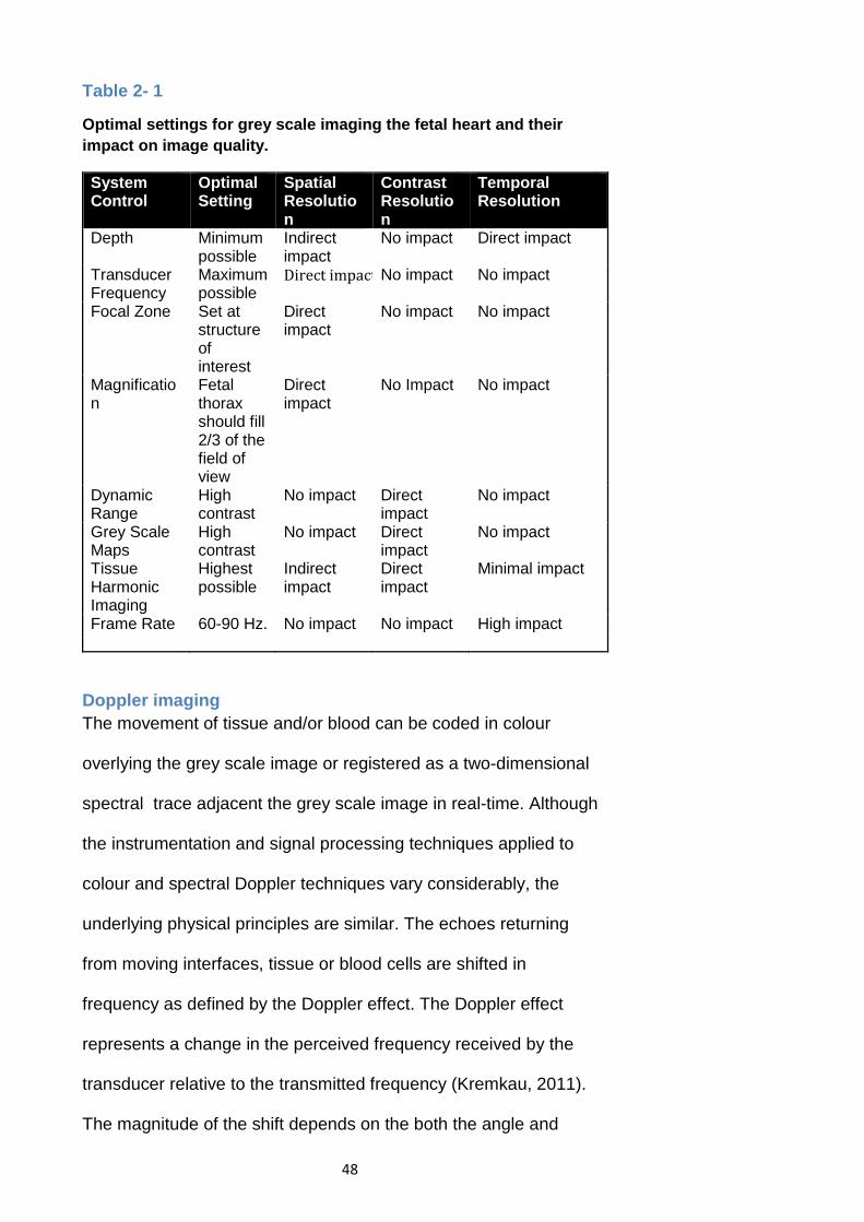
48
Table 2- 1
Optimal settings for grey scale imaging the fetal heart and their impact on image quality.
System Control
Optimal Setting
Spatial Resolution
Contrast Resolution
Temporal Resolution
Depth Minimum possible
Indirect impact
No impact Direct impact
Transducer Frequency
Maximum possible Direct impact No impact No impact
Focal Zone Set at structure of interest
Direct impact
No impact No impact
Magnification
Fetal thorax should fill 2/3 of the field of view
Direct impact
No Impact No impact
Dynamic Range
High contrast
No impact Direct impact
No impact
Grey Scale Maps
High contrast
No impact Direct impact
No impact
Tissue Harmonic Imaging
Highest possible
Indirect impact
Direct impact
Minimal impact
Frame Rate 60-90 Hz. No impact No impact High impact
Doppler imaging The movement of tissue and/or blood can be coded in colour
overlying the grey scale image or registered as a two-dimensional
spectral trace adjacent the grey scale image in real-time. Although
the instrumentation and signal processing techniques applied to
colour and spectral Doppler techniques vary considerably, the
underlying physical principles are similar. The echoes returning
from moving interfaces, tissue or blood cells are shifted in
frequency as defined by the Doppler effect. The Doppler effect
represents a change in the perceived frequency received by the
transducer relative to the transmitted frequency (Kremkau, 2011).
The magnitude of the shift depends on the both the angle and

49
speed of the moving reflector. The Doppler equation summarizes
the key variables that influence the magnitude of the perceived shift
as follows:
𝑃𝑃𝑠𝑠 = Doppler shift (Hz)
𝑓𝑓 = Transmitted frequency (Hz)
θ = the angle between the acoustic beam and the reflector direction
𝑣𝑣 = Reflector speed (cm/s)
𝐹𝐹 = Propagation speed (cm/s)
𝑷𝑷𝟐𝟐 = 𝒇𝒇 × 𝐜𝐜𝐜𝐜𝐜𝐜 𝜽𝜽(𝟐𝟐 × 𝒗𝒗) ÷ 𝒄𝒄
As in all signal sampling processes, aliasing of the Doppler signal is
a phenomenon that occurs when the Nyquist limit is violated. The
Nyquist limit specifies the minimum number of samples that must
be acquired relative to the frequency of the image signal. The
minimum number of samples/cycle is equal to two (Miele, 2006).
Nyquist Limit can also be defined as the maximum detectable
Doppler shift without aliasing.
𝑷𝑷𝟐𝟐(𝒎𝒎𝒎𝒎𝒎𝒎𝒎𝒎𝒎𝒎𝒎𝒎𝒎𝒎) = 𝑷𝑷𝑷𝑷𝑷𝑷 ÷ 𝟐𝟐
When the sampling frequency falls below the minimum threshold,
the displayed image will not register the full frequency shift. This
can be observed in both colour and spectral modes of observation
as illustrated in Figure 2-1.

50
Figure 2- 1
Aliasing can be observed in the colour coding of the aqua-blue segments of the colour map in this image. The PRF is set at 1500 Hz and where blood flow generates a Doppler shift > than 750 Hz, the colour coding reverses on the colour spectrum. A similar phenomenon is observed in spectral Doppler waveforms.
Doppler image processing Image formation during Doppler processing relies on the pulse
repetition frequency which is equal to the propagation speed
divided by two times the depth setting. This inverse relationship
implies that an increase in depth reduces the pulse repetition
frequency and hence the number of samples that can be used to
form the two dimensional image and sample the Doppler shift.
For this reason, most frequently sonographers will alternate
between the live two dimensional image and the live spectral image
and this technique is commonly referred to as duplex imaging;
simultaneous display of colour and spectral images is described as
triplex imaging. Triplex imaging is not often used for imaging at
depths greater than 5-10 cm since the frame rate loss creates a

51
loss of coherence between the two modes of imaging. Colour
sampling is usually set at a pulse packet of between 4-32
pulses/scan-line. Since colour imaging is performed at the same
time as the grey scale image is displayed, the likelihood of aliasing
is much greater compared with spectral Doppler imaging. The
relationship between pulse packet size, image depth, frame rate
and aliasing relative to image resolution are evident in Table 2-2.
Colour Doppler makes it possible to provide a map of tissue
movement or blood flow. In most instances, colour Doppler is used
to map blood flow and this is a very helpful tool to distinguish the
presence of flow, quality of flow, turbulent or laminar and the speed
of flow. Spectral Doppler can provide quantitative values
representative of blood flow speed (Kremkau, 2011).
An alternative to colour or directional Doppler imaging is power or
amplitude imaging. This technique is very similar to colour Doppler
imaging except that flow speed and direction are not provided.
Instead of splitting the Doppler shift signal in to two channels to
provide directional information, the entire signal is processed and
mapped according to the amplitude of the echoes (Miele, 2006).
This technique is typically used to detect low or slow blood flow due
to the enhanced sensitivity associated with the signal processing
methods.
While colour Doppler is a very useful tool to map blood flow and
describe flow dynamics, it does have significant limitations and
drawbacks which must be considered. The use of colour Doppler

52
constrains the frame rate and limits the temporal resolution
available for assessing fetal heart. Furthermore, inappropriate
colour gain or PRF will result in significant noise overlaying the grey
scale image and this may limit the assessment of important
anatomical structures including the interventricular septum and the
chamber walls.
Optimal Doppler imaging parameters for the fetal heart are
described below in Table 2-2
Table 2- 2
Doppler system controls and the impact on image quality (Kremkau, 2011).
System Control
Optimal Setting Spatial Resolution
Contrast Resolution
Temporal Resolution
Depth Minimum possible
Indirect impact
No impact Direct impact
PRF/ Velocity Scale
As high as needed to avoid aliasing
Direct impact No impact Direct impact
Sector width
Set at structure of interest
Direct impact No impact Direct impact
Pulse Packet Size
Set low Direct impact No Impact Direct impact
Colour gain
Just high enough to fill the vessel or chamber
Direct impact No impact No impact
Threshold
Just enough to fill the vessel or chamber
Direct impact No impact No impact
Three dimensional imaging STIC and Matrix tools Although two-dimensional imaging of the fetal heart can be routinely
expected to provide high quality images and a real-time
perspective, the complexity and relationships of fetal heart anatomy
can be difficult to resolve consistently as single, static images for

53
review by the interpreting physician. A three-dimensional view of
the fetal heart would resolve some key limitations of two-
dimensional imaging including:
• Inability to display views of the fetal heart obstructed by the
fetal thorax, primarily the coronal axis
• Requirement to mentally transform two-dimensional
relationships into three-dimensional impressions, a highly
subjective and user-dependent process
• Increased variability of measurements (Fenster, Downey, &
Cardinal, 2001).
Many different approaches have been used over the past twenty
years to acquire three-dimensional images. These can be
categorized as, mechanical, free-hand and a unique free hand
approach employing a two-dimensional phased array of transducers
(Fenster et al., 2001). Mechanical methods of three-dimensional
imaging have fallen out of favour in recent years due to their size,
bulkiness and advances in free-hand techniques. Within the realm
of fetal imaging, two free hand imaging techniques have become
dominant: Real-Time three-dimensional imaging and Spatial-
Temporal Image Correlation (STIC).
Three new developments have emerged from the maturation of
sonographic, three-dimensional imaging technology. Two of these
relate to the method of image acquisition and the third relates to the
off-line analysis of the image volume. The Spatial-Temporal Image
Correlation (STIC) acquisition technique generates a three-

54
dimensional cine-loop while a live, three-dimensional imaging
method uses a two-dimensional matrix of transducers to generate a
live, three-dimensional image. Distinctive advantages and
disadvantages can be identified for the STIC and live three
dimensional techniques respectively while the ability to analyze the
fetal heart in three dimensions in real-time, off-line provides the
specialist with the necessary data to identify congenital heart
defects after the patient has left the imaging facility or from a
location geographically distant from the site of acquisition.
Development of techniques for imaging the fetal heart in three
dimensions in real-time have addressed at least two of the key
limitations of two-dimensional imaging. Visualizing the anatomy in
multiple planes simultaneously provides the clinician with the tools
necessary to establish the spatial relationships between the
relevant cardiac structures such as the cross-over of the RVOT and
LVOT. In addition the real-time characteristics, wall motion and
synchronicity can be established in real-time.
Acquiring a Three-Dimensional Volume Formation of a three dimensional image using acoustic technology
poses unique challenges for engineers and physicists. Sonographic
images are two dimensional slices of variable thickness. The axial,
lateral and thickness planes vary in dimensions over depth and
when they are used to produce volume data sets they generate
asymmetric voxels. The thickness plane is typically the largest
dimension and reconstruction of this plane generates the lowest
image quality of the three planes.

55
Presently there are two different techniques that can be used to
acquire volume datasets of the fetal heart. The most well-known
technique is known as spatial-temporal image correlation (STIC)
and typically uses an internally motorized single-dimensional array
of crystals. Another technique that has been popular for imaging
adult hearts is known as a matrix array, capable of live three-
dimensional imaging. Although the matrix array was developed to
image adult hearts, numerous investigators have begun the process
of assessing its value in imaging fetal hearts. Each of these
techniques can be defined by the strengths and weaknesses that
impact their effectiveness in imaging fetal hearts.
STIC The STIC technique acquires a volume of image data using an
automated scan head that slowly sweeps through a pre-selected
arc (DeVore, Falkensammer, Sklansky, & Platt, 2003).
As described in the seminal article by Devore et al., the linear array
of elements acquires image data at a very high frame rate of
approximately150 frames/s. The acquisition time can be adjusted
between 7.5 – 15 seconds. Acquisition over a longer period of time
allows the algorithm to acquire more images and optimize image
quality. As an example, a 10 second acquisition at a frame rate of
150 frames/s will generate 1500 image slices of the fetal heart as it
moves through 23 cardiac cycles assuming a fetal heart rate of 140
bpm. The STIC algorithm analyzes the periodic motion of the heart
as it contracts and expands to determine the average heart rate.
Multiple cardiac volumes will be generated from the correlation

56
between image slices and the heart rate. The image slices are
registered according to the phase of the cardiac cycle. A complete
acquisition represents a cine-loop of multiple cardiac volumes. For
this reason, significant changes in fetal heart rate or fetal movement
will result in a failed reconstruction (DeVore et al., 2003). The STIC
acquisition technique generates a cine-loop comprised of multiple
volume reconstructions. This allows the viewer to interrogate the
heart in three-dimensions as a real-time cine-loop.
Image analysis involves the use of the Multi-planar Reconstruction
(MPR) technique. The volume can be interrogated in any plane and
the relationship between orthogonal planes is maintained (Yagel,
Cohen, Shapiro, & Valsky, 2007). It is possible to reconstruct
images of the cardiac outflow tracts and establish their relationship
without rescanning the patient. Additional tools allow the viewer to
subtract structures from the image and improve visualization of
targeted anatomical structures.
Live 3D The main differences between the STIC technique and the matrix
transducer are related to the beam formation and processing of
acquired image slices. The matrix uses up to three thousand tiny
crystals to simultaneously transmit and receive acoustic pulses, see
Figure 2-2. The beam is electronically controlled and steered in
real-time acquiring pyramidal volumes (Stetten & Tamburo, 2001).
A significant development enabling the success of this technique
relates to the number of miniaturized circuit boards embedded
within the probe handle providing the capacity to perform signal

57
processing prior to the signal returning to the main sonography
system. The sheer number of slices required to generate sufficient
pyramidal volumes for real-time display imposes a significant signal
processing challenge. Parallel processing the acoustic beam at a
rate of 16:1 allows the system to acquire sufficient slices to
generate real-time pyramidal volumes at clinically useful depths of
penetration (Appareti, 2003). The electronic beam formation
generates nearly symmetric voxels but suffers from lower SNR
relative to the STIC technique.

58
Figure 2- 2
Tiny transducer elements can be seen relative to the size of a human hair and the size of miniature circuit boards embedded within the probe handle are defined. Line drawings provided by Ken K. Wong, Graphic Designer, Mohawk College.
Volume Image Analysis Due to the complexity of congenital heart defects, and in many
cases their subtle characteristics, expert consultation is usually
required. The standard image acquisition techniques usually involve
static image slices stored on film or electronically. In more rare
instances video or real-time sequences are acquired. In any case
consultation usually requires a rescan at the referral center, a
process that extends the period of diagnostic uncertainty and may
add significant emotional stress to the prospective parents. In
addition to the emotional distress, the additional resources and
59
travel may significantly add cost to the patient and healthcare
system.
Off-line Analysis Acquisition of scanned volumes may be able to alleviate some of
these limitations through the application of telemedicine for
consultation. The three-dimensional volume can be transmitted and
reviewed remotely using a broadband internet connection. The
consultant is then able to examine the fetal heart in multiple planes
and in motion in much the same way the sonographer was able to
visualize the anatomy in real-time. A number of studies have trialed
this concept (Chan, Soong, Lessing, Watson, Concotta, Baker, et
al. 2000). In 2000 Kratochwil, Lee and Schoisswohl were able to
demonstrate the feasibility of this concept and found that 20:1 and
30:1 wavelet image compression techniques could be applied
without a discernable loss of information (Kratochwil, Lee, &
Schoisswohl, 2000). In another study performed in Australia, image
volumes were successfully transmitted from Townsville to Brisbane
a distance of 1500 km. This eliminated the need for physical referral
of 24/71 patients resulting in a significant cost saving to the patients
and healthcare system. In the most comprehensive evaluation
published to date, Vinals et al were able to demonstrate that STIC
volumes could be acquired by operators inexperienced in fetal
echocardiography and transmit them through the internet from
multiple remote locations in Chile to a referral centre in Santiago
Chile (Viñals, Tapia, & Giuliano, 2002). This allowed the specialists
the opportunity to rule out and confirm a diagnosis remotely. In

60
addition a web camera and e-mail were used to advise patients as
to the benefits of referral to tertiary care centers for those with
CHDs diagnosed. Evidence is rapidly accumulating to support the
concept of using telemedicine and three dimensional volume
analysis as a means of gaining an expert consultation without the
need for physical referral or rescanning. For many countries this is
a technique that may alleviate the challenge of providing advanced
healthcare to populations geographically distant from specialists
located in tertiary health centers.
Summary The development of two distinct three-dimensional imaging
techniques coupled with the capability of remote analysis via
telemedicine has the potential to transform the methods used to
manage patients that otherwise require referral to specialized fetal
treatment centers. The benefits of imaging the fetal heart using
three dimensional sonographic imaging may include increased
detection of CHD and enhanced diagnostic capabilities. These
imaging capabilities may be offset by the increased cost and
accessibility of three dimensional imaging systems and the
relatively steep learning curve associated with their use.

61
Sonographic Features and Techniques for Early Detection The impact of congenital heart disease varies widely depending on
the specific form it takes. In some cases the chambers of the heart
may be entirely normal in appearance and the defect may only be
evidenced in the outflow tracts of the heart. For example, congenital
heart disease relating to the outflow tracts such as dextro-
transposition of the great arteries may only be detected at their
origins and frequently are not detected by the four chamber view,
see Figure 2-3.
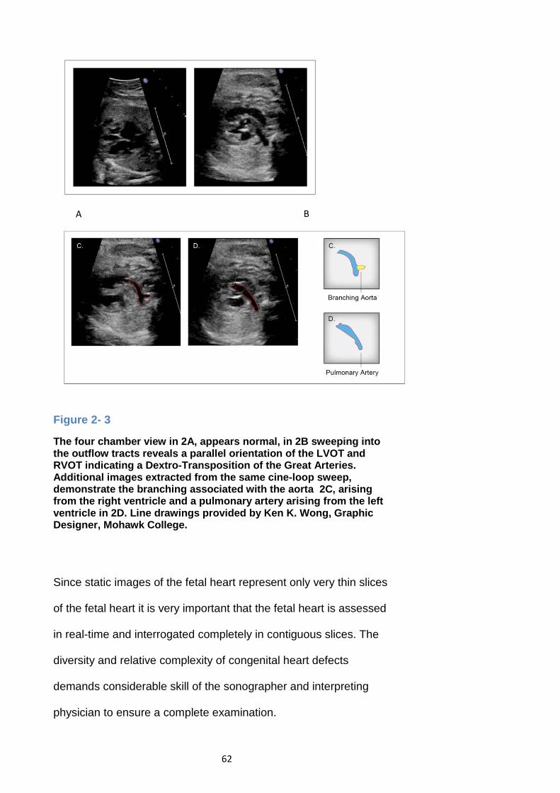
62
Figure 2- 3
The four chamber view in 2A, appears normal, in 2B sweeping into the outflow tracts reveals a parallel orientation of the LVOT and RVOT indicating a Dextro-Transposition of the Great Arteries. Additional images extracted from the same cine-loop sweep, demonstrate the branching associated with the aorta 2C, arising from the right ventricle and a pulmonary artery arising from the left ventricle in 2D. Line drawings provided by Ken K. Wong, Graphic Designer, Mohawk College.
Since static images of the fetal heart represent only very thin slices
of the fetal heart it is very important that the fetal heart is assessed
in real-time and interrogated completely in contiguous slices. The
diversity and relative complexity of congenital heart defects
demands considerable skill of the sonographer and interpreting
physician to ensure a complete examination.
A B

63
Impact of Early Detection The majority of deaths from congenital defects in childhood can be
attributed to congenital heart disease (CHD). CHD is six times more
common than chromosomal abnormalities and four times more
common than neural tube defects (Carvalho, Mavrides,
Shinebourne, Campbell, & Thilaganathan, 2002). The most
common cause of birth defects, CHD is most frequently reported
with an incidence of 0.8% of all live births (Hoffman & Kaplan,
2002). Improvements based on technical advances in the
management and therapy of CHD has resulted in a shifting of the
outcomes such that adults living with CHD outnumber the number
of children with CHD (Hoffman, Kaplan, & Liberthson, 2004).
Prenatal detection of CHD provides a number of advantages
relative to postnatal identification.
1. Counselling, and the continuance of a viable pregnancy
2. Monitoring of pregnancy and treatment
3. Assessment for extracardiac disease and genetic
associations
4. Decrease morbidity in some cases, reduction in fetal
neurological impact
Some of the benefits of routine obstetrical screening have been
found to include maternal psychological effects, as well as an
increased connection between the unborn child and enhanced
maternal-fetal bond (Sklansky et al., 2002). The visualization of the
fetus along with a normal screening result has been found to
decrease maternal anxiety and increase satisfaction (Sklansky et
64
al., 2002). Conversely, an abnormal result has the effect of
decreasing maternal happiness and increasing anxiety. As stated
by Menahem et al., ‘‘Diagnosis was made at 17 weeks. The baby
was active.’’ so that attachment has already begun between the
mother and her unborn infant. As one father in the same study
observed ‘‘my wife loved the baby so many months and worked
hard to keep it, to find out things were not right—is difficult beyond
belief” (Menahem & Grimwade, 2003).
In the long term, compared with women who were not diagnosed
prenatally, there was a more favourable impact on maternal
happiness. Mothers felt less responsibility for the CHD and tended
towards improved relationships with the infant’s fathers after
prenatal diagnosis (Sklansky et al., 2002). The early detection of
CHD ensures that there will be an opportunity to provide a clear and
accurate picture of the prognosis and outline treatment options to
the parents. The clinician must be able to base the prognosis on a
number of factors, not solely on the diagnosis. These factors
include; the security of the diagnosis, stage in gestation, potential
for change, and association with extracardiac malformations and
known results of treatment (Allan & Huggon, 2004). The counselling
opportunity provided by prenatal detection expands the range of
options to parents which may include continuance of the pregnancy,
continuance with therapy and/or possibly termination of the
pregnancy. Gestational age at the time of diagnosis is one of the
strongest predictors of pregnancy termination. An early diagnosis is
associated with termination in up to 70% of cases, the probability of
65
termination is inversely related to the percentage of early diagnosis
in a population (Germanakis & Sifakis, 2006).
For those parents that choose to continue the pregnancy, early
detection provides the opportunity to meet with perinatologists, and
cardiac specialists engaging in relaxed discussions related to
prospective interventions. These discussions may provide the
parents with a greater sense of control, while the healthcare team
may be in a better position to minimize potential complications,
adjusting the timing of the delivery either by induction, caesarean
section or the use of tocolytic agents to allow greater maturation of
the lungs, if required (Jaquiss & Tweddell, 2005). Often the infant is
able to make a much less disruptive entry into post natal life
through the provision of prostaglandin infusions, respiratory support
and intensive monitoring as required (Jaquiss & Tweddell, 2005).
A number of severe forms of CHD have been found to benefit from
prenatal diagnosis. Tworetzky et al, reported that surgical outcomes
for HLHS improved after fetal diagnosis. In a study of 88 patients,
all 33 cases that were diagnosed prenatally survived surgery
whereas only 25/38 that were postnatally diagnosed survived
(Tworetzky et al., 2001). Without prenatal diagnosis, most infants
born with HLHS experience delays in diagnosis and appropriate
resuscitation. This may lead to systemic hypoperfusion, shock and
organ damage hindering the long term survival prospects
(Tworetzky et al., 2001). These findings are supported by Satomi et
al in their study of HLHS, which discussed finding improvements in

66
preoperative conditions and avoidance of ductal shock (Satomi,
Yasukochi, Shimizu, Takigiku, & Ishii, 1999).
Bonnet et al, determined that prenatal detection of TGA reduced
neonatal morbidity and mortality. A ten year study comparing 68
neonates with prenatal diagnosis and 250 neonates without
prenatal diagnosis, identified significant delays in access to the
pediatric cardiology unit, increased postoperative mortality and
longer postoperative stays for those diagnosed post natally (Bonnet
et al., 1999). Prenatal diagnosis of coarctation of the aorta was
found to improve survival rates and reduce morbidity (Franklin,
2002). An examination of 217 neonates with hypoplastic left heart
syndrome and 422 with transposition of the great arteries found that
those with a prenatal diagnosis who underwent surgery had
objective indicators of lower severity of illness preoperatively
(Kumar, Newburger, Gauvreau, Kamenir, & Hornberger, 1996).
Although the prenatally diagnosed group experienced improved
preoperative condition, in this study, postoperative outcome did not
differ between the two groups (Kumar et al., 1996).
Current Techniques The type of CHD and the medical practice performing the screening
are identified as key factors by a number of studies (Friedberg et
al., 2009; Jaeggi et al., 2001; Wong, Chan, Cincotta, Lee-Tannock,
& Ward, 2003). Not surprisingly a number of studies have shown
increased detection of CHD at tertiary care centres as compared to
community screening centers (Friedberg et al., 2009; Grandjean et
al., 1999; Jaeggi et al., 2001; Stumpflen, Stumpflen, Wimmer,

67
1996). The screening of CHD can range from a basic exam,
extended exam to include additional views such as the three vessel
view, and the inclusion of Doppler and three-dimensional imaging
techniques.
Static Images The basic exam consists of a recording of the four-chambers of the
fetal heart as a cross-sectional image. As noted by Chaoui, this
approach has not led to widespread improvement in the detection of
heart defects (Chaoui, 2003). In his analysis he identified four
reasons for this outcome, the primary one being the fact that many
defects cannot be visualized in the four-chamber view, see Table 2-
3 (Chaoui, 2003). In an extended effort to identify cono-truncal
defects, the extended exam includes views of the left and right
outflow tracts (ACR Guidelines and Standards Committee,
American Institute of Ultrasound in Medicine, 2003; Lee et al.,
2008). Yagel et al proposed the acquisition and review of five cross-
sectional images as a means of improving and simplifying the
screening process (Yagel, Cohen, & Achiron, 2001). This approach
ensures that many of the key anatomic structures are represented
beginning with the abdominal situs and ending with a view of the
three vessels, the pulmonary trunk, the aorta and the superior vena
cava (Yagel et al., 2001). In a similar line of reasoning, Yoo et al,
have proposed the inclusion of the three vessel view as an element
of every screening examination (Yoo, Lee, Kim, Ryu, Choi, Cho,
1997). The three vessel view is very effective in identifying many of
the abnormalities associated with the outflow tracts and great

68
arteries (Chaoui & McEwing, 2003; Viñals, Heredia, & Giuliano,
2003; Yagel, Arbel, Anteby, Raveh, & Achiron, 2002).
Table 2- 3
Prenatal Detection Rate of Anomalies by View and Frequency of Incidence. (Jaeggi et al., 2001)
Anomaly Four chamber view (%)
Outflow tracts (%)
Subjects total
Detected by Four chamber view Complete Atrioventricular Septal Defect
12 (15.4)
78
Hypoplastic Left Heart 12 (44) 37 Total Anomalous Pulmonary Return 0 23 Mitral or Tricuspid atresia 11 (50) 22 AV septal defects 7 (44) 16 Pulmonary atresia (intact ventricular septum)
5 (31) 16
Ebsteins anomaly 7 (46.7) 15 Miscellaneous 5 (50) 10 Complete heart block 5 (83.3) 6 Corrected transposition 2 (40) 5 Total 66 (29) 229 Detected by Outflow views
Tetralogy of Fallot 11 (10) 110 Aortic arch obstruction 2 (2.6) 78 Dextro-Transposition great arteries 0 77 Ventricular septal defect 4 (5.3) 75 Double outlet right ventricle 5 (18.5) 27 Aortic stenosis 5 (20) 25 Pulmonary stenosis 2 (8.7) 23 Truncus arteriosus 0 13 Aortic-pulmonary window 0 2 Total 29 (6.7) 430
Although the use of multiple cross-sectional planes and the three
vessel view have the potential to demonstrate a great deal of the
fetal heart anatomy, these views have not been adopted by a
majority of screening centres. Acquiring static images of the outflow
tracts has proven to be more difficult than acquiring the four-

69
chamber view where all of the anatomy can be seen in one plane
(Sklansky, 2007). Furthermore, static images do not provide a real-
time perspective of the fetal heart and this remains a significant
limitation of this imaging mode. Sklansky has proposed a modified
approach using cine-loops that may address some of the
challenges associated with acquiring multiple static images
(Sklansky, 2007).
Importance of Cine-Loop Sweeps The sonographic features of severe CHD can usually be captured
as static images, once the disease is identified; however, it is a
challenge in many cases to identify CHD using only static images.
In Figure 2-3 the presence of parallel outflow tracts is documented
as a series of static images. It is worth noting that in isolation, some
of these static images may appear normal whereas in the context of
a cine-loop the aggregate set of images defines a relationship
between the outflow tracts that is more difficult to establish with
static images only. The four chamber image in this set is normal in
terms of the situs, axis and relative size of the fetal heart compared
with the thorax. The image of the LVOT in panel D of Figure 2-3
could also be misinterpreted as normal, key static images are
depicted in B and C. Should these images be unavailable, the
identification of CHD could be missed.

70
Figure 2- 4
In this image pair A and B static images taken from the same cine-loop sweep represent very different structural realities. A ventricular septal defect associated with Tetralogy of Fallot is indicated by the shaded area in image B, the septum appears to be intact in image A extracted from the same cine-loop sweep, Transducer frequency, 2.3-4.6 MHz, Tissue Harmonic Imaging, 80 Hz frame rate. Line drawings provided by Ken K. Wong, Graphic Designer, Mohawk College.
Another example where a cine-loop provides more value compared
with only a set of static images is illustrated in Figure 2-4. In this
example it is clear that the ventricular septal defect is evident in the
anterior aspect of the chamber and not the posterior aspect. This
could be easily misrepresented as a normal case if only the static
image as in figure A is provided for interpretation. Similarly, in
Figure 2-5, the atrioventricular septal defect apparent in image B
could easily be overlooked if only image A is provided for
interpretation. Sweeping the acoustic beam through the fetal heart,
acquiring thirty or more images/second, provides a more complete
set of images for interpretation. In addition, the cine-loop sweeps
provide many more opportunities to assess the relationship
between fetal cardiac structures and to assess the dynamics of the

71
fetal heart, myocardial contractility, valve motion and the synchrony
of the chambers.
Figure 2- 5
In this image pair taken from the same cine-loop sweep the interventricular septum appears intact in A, in the adjacent image B, the septum is not intact. This represents an Atrioventricular Septal Defect (AVSD). Line drawings provided by Ken K. Wong, Graphic Designer, Mohawk College.
Colour Doppler The inclusion of colour Doppler imaging in every fetal cardiac
examination was proposed by Chaoui & McEwing in order to
increase the speed and accuracy of the examination (Chaoui &
McEwing, 2003). In their review article, they identify three cross-
sectional planes to simplify the colour Doppler examination; the four
chamber, five chamber view and three vessel views (Chaoui &
McEwing, 2003). Copel et al, found that in 29% of cases the use of
colour Doppler was essential for an accurate diagnosis (Copel,
Joshua, Morotti, Hobbins, 1991). The inclusion of colour Doppler
cine-loops has been found to assist in clearing the screening views
of the heart in difficult cases (Poole et al., 2013). In a pilot study,
Scott et al examined the potential for including a colour Doppler

72
cine-loop of the three vessel view in a routine obstetrical screening
protocol (Scott et al., 2008). The results of this study determined
that the colour cine-loop lengthened the examination time
significantly and adversely affected the quality of the grey scale
features due to bleeding of colour and challenges associated with
optimizing the colour Doppler controls (Scott et al., 2008).
Three-Dimensional Imaging The Spatio-Temporal Image Correlation (STIC) technology enabling
the three-dimensional imaging of the fetal heart as a cine-loop
volume has been commercially available for approximately ten
years (Goncalves, 2005). The STIC methodology uses a slow
sweeping acquisition of many image planes over a 7.5-15 second
time interval. The mean heart rate is calculated and the images are
reformatted according to their correlation to the cardiac cycle
(Chaoui & Heling, 2005; DeVore et al., 2003). This technique
generates a cardiac volume which can be reviewed as a cine-loop
at any speed and images can be generated from the volume in any
plane (Chaoui & Heling, 2005; DeVore et al., 2003; Gonçalves et
al., 2003). Interrogating the fetal heart in any plane within a moving
cine-loop provides a number of advantages compared to two-
dimensional imaging. This would include the ability to review
unlimited images, correlate images between orthogonal planes, and
accessing novel planes such as the enface view of the cardiac
valves obtained through the coronal plane (Goncalves, 2005).
A number of closely related innovations have been designed to
enhance the relationship between anatomic structures, particularly

73
the outflow tracts. Those that leverage the STIC technology include,
Doppler and Doppler angiography and tomographic imaging.
Doppler imaging relies on the phase shift associated with the
movement of reflectors within the path of the acoustic beam (York &
Kim, 1999). The phase shift is angle dependent, reflections from
moving reflectors at 90 degrees relative to the acoustic beam axis
do not shift the phase of the frequency and are typically not
visualized with Doppler processing (York & Kim, 1999). In contrast,
Power Doppler or Doppler Angiography techniques process the
magnitude of the phase shift and do not determine directionality of
the reflectors. The loss of directional information is compensated by
significantly enhanced sensitivity to the presence of moving
reflectors (Chaoui & Kalache, 2001). In 2009, Tonni et al examined
the feasibility of including three-dimensional and Doppler
angiography of the great arteries in the second trimester
examination (Tonni, Centini, & Taddei, 2009). It was found that
three-dimensional imaging and Doppler can enhance the
visualization rate of the great vessels, although it is perceived to be
premature to include these techniques in routine examinations
(Tonni et al., 2009). According to Tonni et al., the technique should
be reserved for targeted cases where the two-dimensional
examination is incomplete (Tonni et al., 2009).
Tomographic imaging leverages the unique volume dataset
provided by the STIC technology. Tomographic imaging provides
the examiner with the ability to visualize an array of image slices
represented as a panel with a reference image available to

74
correlate the position of the slices relative to the image volume
(Espinoza et al., 2008). Espinoza et al provide a standardized
method that can be used to reproduce the most clinically relevant
and valuable images using commercial available software
processing (Espinoza et al., 2008). In a step further to minimize
operator training and dependency, Abuhamad et al evaluated an
automated software program that demonstrated the capability of
auto generating tomographic images from standardized volumes
(Abuhamad, Falkensammer, Reichartseder, & Zhao, 2008). In this
evaluation the software demonstrated a successful depiction of
tomographic slices in over 90% of each of the three cardiac planes:
the five chamber view, the aorta, the right ventricular outflow plane,
and the pulmonary artery, along with the abdominal circumference,
and the plane capturing the stomach (Abuhamad et al., 2008).
Summary
The detection of CHD during routine obstetrical screening remains
a diagnostic challenge. Unfortunately, improvements in sonography
instrumentation and systems including colour Doppler and three-
dimensional imaging techniques, have not resulted in improvements
in the detection of CHD (Shen & Yagel, 2010). Friedberg et al
reports that where screening is performed in nearly all patients in
northern California the detection of major CHD’s ranges from 20%-
82%, with an average rate of 28% detection (Friedberg et al., 2009)
In some cases, the early detection of CHD has significant impact on
the morbidity of neonates born with CHD (Bonnet et al., 1999;
Franklin, 2002; Satomi et al., 1999). For parents, early detection

75
can provide an opportunity for counselling and improved
understanding of the prognostic details of their pregnancy. Early
detection also provides an opportunity for improved management of
the delivery and the ability to possibly minimize complications.
A number of methods have been proposed to improve the detection
rate of CHD including the three vessel view by Yoo, the use of
colour Doppler as proposed by Chaoui and a variety of three-
dimensional imaging techniques (Chaoui & McEwing, 2003;
DeVore, Polanco, Sklansky, & Platt, 2004). Unfortunately, wide
spread adoption of each of these techniques has been limited.
Access to three-dimensional sonography systems is limited and the
optimal use of these systems to consistently acquire three-
dimensional images of the fetal heart requires significant additional
training (Roberts, 2008; Tonni et al., 2009).
The reliance on static images for the assessment of the fetal heart
imposes a significant limit to the screening of CHD during routine
obstetrical examinations. Using static images only, the interpreting
physician will not be able to assess the function or anatomic
relationships between cardiac structures. Alternatively, using cine-
loop sweeps provides the sonographer and the interpreting
physician with the opportunity to perform a dynamic assessment of
the fetal heart.
The use of cine-loops and real-time assessment of the fetal heart
has been suggested by a number of authors as a means of
improving the screening process (Mcgahan et al., 2007; Sklansky,

76
2007). The use of cine-loop sweeps will provide many more images
of the fetal heart, establishing critically important anatomical
relationships, and cardiac dynamics. Furthermore, this approach
has the potential to improve the detection of CHD during screening
by reducing the variability in the image data recorded and
standardizing the assessment of the fetal heart.
The acquisition of cine-loop sweeps through the fetal heart in lieu of
static images only has the potential to increase the detection of
CHD without imposing additional requirements for examiner training
or additional imaging techniques such as colour Doppler or three-
dimensional imaging. Standardizing the assessment of the fetal
heart and evaluating the functional characteristics of the fetal heart
through cine-loop acquisition during routine screening may provide
significant advantages compared with the interpretation of static
images only.
Proposed Research for this Thesis The objective of my research is to increase the detection of
congenital heart disease during routine second trimester obstetrical
screening. The proposed research will include three projects;
• to develop an optimal real-time image acquisition and
assessment technique;
• to test the rate at which the optimal real-time image
acquisition and assessment technique can be leveraged to
detect CHD;

77
• to design an online learning tool which will translate the
image acquisition and assessment technique to clinical
practice.
The first project will compare and evaluate the efficacy of two
different real-time approaches relative to the current method using
static images only. The second project will measure the rate at
which CHD is detected using the real-time technique selected in
project one compared with the current method using static images
only. Finally, a third project will develop an online training tool for
teaching sonographers and interpreting physicians to use a
standardized assessment tool. A quasi-experimental study was
completed to evaluate the sonographers and interpreting
physicians’ ability to use the online tool to learn a standardized
assessment protocol.
Project One
The first project will identify the optimal imaging protocol for the
routine assessment of the fetal heart during obstetrical screening
taking into account the limitations of time and technology imposed
on the sonographer during routine second trimester screening. The
proposed technique will increase the amount and quality of
diagnostic image information by acquiring and storing real-time
cine-loop sweeps, four-seconds in length using a minimum of 30
frames/second of the fetal heart. This project is entitled,
“Comparison of static, real-time and real-time, colour Doppler
imaging techniques applied to the assessment of the fetal heart in
routine screening”. This study will determine the optimal cine-loop

78
sweep imaging technique by comparing the performance of the
conventional static images against two different approaches. The
first will employ grey scale imaging only and the second will include
colour Doppler imaging. The grey scale technique will use two cine-
loop sweeps through the fetal heart beginning in the transverse
plane with the fetal stomach visible. The first sweep will provide the
interpreting physician with an opportunity to observe the
relationship of the fetal heart with the stomach, liver and abdominal
vessels in the upper abdomen. Since the first sweep will be
completed at the four chamber view and the second sweep will
begin at the four chamber view there will be some overlap in
visualizing the morphology of the cardiac chambers. The outflow
tracts will be the primary focus of the second sweep and this cine-
loop sweep will expose the anatomical relationship between each
outflow tract and its respective ventricle as well as the critically
important cross-over of the outflow tracts. The second approach will
apply the same imaging protocol as the first with the addition of a
colour Doppler map overlaying the grey scale and a third colour
cine-loop of the three vessel view. In order to provide a more
standardized review process, interpreting physicians will be
provided an anatomic checklist to guide and record decision
making. The research design assumes the ultrasound systems that
are used are capable of Colour Doppler processing and are
configured to store or transmit real-time loops to a network.

79
Research hypothesis:
Assessment of the fetal heart in routine screening will demonstrate
increased visualization of normal fetal cardiac anatomy by acquiring
cine-loop sweeps using either a grey scale,( protocol B) or colour
Doppler technique, (protocol C) compared with using static images,
(protocol A).
Research Aims
Demonstrate the ability to acquire the experimental
protocols, B and C in less than five minutes
Demonstrate an increased rate of detection of cardiac
structures using real-time and real-time colour Doppler loops
compared with static images
Establish the presence of antegrade flow between the atria
and ventricles
Confirm the presence of forward flow in the AV valves, aorta
and pulmonary trunk
It is anticipated that the real-time cine-loop sweeps will add
considerable diagnostic value to the screening process since there
will be two interpretations performed on every exam, the preliminary
review by the examiner and the summary review by the interpreting
physician. In addition using the real-time loops will allow the
interpreting physician the opportunity to review the images
repeatedly in real-time and evaluate structure at different points
during the cardiac cycle. The ability to assess structure during the

80
diastolic phase has been found by Chaoui et al to be helpful in
detecting atrioventricular septal defects (Chaoui, 2003). Further, it is
well established that observation of moving structures provides a
more sensitive means of detecting small differences in form (Ivry &
Cohen, 1992). A cine loop of a sweep through the cardiac outflow
tracts will allow the interpreting physician to better appreciate the
relationship between the LVOT and RVOT which is crucial for ruling
out outflow discrepancies and conotruncal abnormalities. The
advantages of using three basic Doppler planes have been well
documented by Chaoui (Chaoui & McEwing, 2003). Adding these
Doppler images aids in the identification of outflow discrepancies,
conotruncal abnormalities, valvular insufficiency and may reveal
more subtle lesions such as ventricular septal defects (Chaoui &
McEwing, 2003).
Project Two
The second project is entitled, “Increasing the Detection Rate of
Congenital Heart Disease during Routine Obstetrical Screening
using Two Cine-Loop Sweeps”. The goal of this study will be to
demonstrate an improved detection rate of congenital heart defects
using the technique identified in the first project.
Research hypothesis:
Using two cine-loop sweeps will increase the detection rate of
congenital heart disease compared with the acquisition and review
of three static images, the conventional technique.

81
Research Aims:
Demonstrate increased detection of CHD
Complete the review and interpretation of cine-loop sweeps
in an equivalent period of time or less than the static images
Maintain an equivalent or lesser rate of false positive
identification of CHD using real-time cine-loop sweeps
compared with static images
Maintain an equivalent or lesser rate of indeterminate cases
using cine-loop sweeps compared with static images
Project Three
The third project is entitled, “Increasing Recognition of Fetal Heart
Anatomy using Online Tutorials and Mastery Learning compared
with Classroom Instructional Methods”. The objective of the
proposed research is to determine if the standardized assessment
of the fetal heart in real-time method is a more effective tool to be
used by students when learning how to identify congenital heart
disease. Using a randomized, two group, quasi-experimental
design, we will address the hypothesis: "Training students to use a
simulation based standardized assessment technique online
compared with traditional classroom instruction will increase their
ability to identify congenital heart disease and increase their
confidence and knowledge in assessment of the fetal heart."
Research Aims:
Increased ability to identify normal fetal cardiac structures.
Increased ability to identify abnormal fetal cardiac structures.

82
Increased ability to identify abnormal cases.
The first two projects will identify a new standardized acquisition
and assessment technique for evaluating the fetal heart and
demonstrate that the technique is superior to the current methods of
evaluating the fetal heart. A third project will devise an online
learning tool that will be used to translate the new technique to
clinical practice.

83
Chapter 3 Assessment of the Fetal Heart during Routine Obstetrical Screening, a Standardized Method
Introduction The majority of deaths from congenital defects in childhood can be
attributed to congenital heart disease (CHD). CHD is six times more
common than chromosomal abnormalities and four times more
common than neural tube defects (Carvalho, Mavrides,
Shinebourne, Campbell, & Thilaganathan, 2002). The most
common cause of birth defects, CHD is most frequently reported
with an incidence of 0.8% of all live births (Hoffman & Kaplan,
2002). Improvements based on technical advances in the
management and therapy of CHD has resulted in a shifting of the
outcomes such that adults living with CHD outnumber the number
of children with CHD (Hoffman, Kaplan, & Liberthson, 2004).
Typically the role of the sonographer during routine obstetrical
screening is to identify normal and abnormal features of the fetal
heart and provide morphologic images of these for the interpreting
physician. The routine second trimester examination is typically
performed in real-time and documented as a series of static images
archived electronically or on film. Interpretation of the cardiac
images is then made by a radiologist or obstetrician using the static
images. Present detection rates for CHD may vary widely
depending on a number of factors: gestational age, imaging centre
protocol, operator skills, maternal obesity, fetal position, image
acquisition protocol and the nature of the CHD (Chaoui, 2003). In
order to clarify the recommended fetal heart structures for

84
examination the American College of Radiology (ACR), the
American College of Obstetrics and Gynecology and the
International Society of Ultrasound in Obstetrics & Gynecology
(ISUOG) have published guidelines for routine second trimester
obstetric screening (American Institute of Ultrasound in Medicine,
2013; Reddy et al., 2014). Both of these publications require the
four chamber view, also known as, a basic assessment, views of
the left ventricular outflow tract (LVOT) and right ventricular outflow
tracts (RVOT) commonly referred to as an extended examination
(ACR Guidelines and Standards Committee, American Institute of
Ultrasound in Medicine, 2003; Lee et al., 2008). Prenatal detection
rates using these methods are summarized in Table 3-1.

85
Table 3- 1
Prenatal Screening Detection Rate by protocol and population.
Author Year Study Design
Protocol
Subjects (n)
Detection (%)
Friedberg et al., 2009
Prospective, multicenter, major defects only
Basic and extended views
309, low risk
98, (36)
Tegnander et al., 2006
Prospective, single site
Basic and Extended Views
29,460, unselecte
d
55, (57)
Ogge et al.2006 Prospective,
multicenter
Basic and extended views
6368, unselecte
d
38, (65)
Todros et al., 1997 Prospective
, multicenter
Basic only
8299. low risk
6, (15)
Buskens et al., 1996 Prospective
, multicenter
Basic only
5319, low risk
7, (16.7)
Stumpflen et al., 1996
Prospective, single center (tertiary)
Basic and extende
d
3085, unselecte
d
46, (85.5)
Ott et al., 1995 Prospective single center
Basic and LVOT
1136, low risk
2 (14.3)
Rustico et al., 1995 Prospective, single center
Basic and extende
d
3079, low risk, experts
14, (45.2)
3945, low risk, non-experts
9, (26.5)
Tegnander 1995 Prospective single center (tertiary)
Basic only
7459, low risk
26, (39)
The range of detection varies widely by author, equipment
improvements, fetal age and study type. Those studies that relied
solely on the basic protocol ranged in detection from 15-39% while

86
those that included the extended protocol demonstrated a higher
rate of detection 26.5-86.5%. It is widely accepted that the extended
examination will improve the detection rate of CHD compared with
the basic examination only (Eik-Nes, 2006; Michelfelder & Cnota,
2009; Tegnander, Williams, Johansen, Blaas, & Eik-Nes, 2006).
Despite the inclusion of outflow tracts as part of an extended
examination of the fetal heart, only two of the six studies listed in
Table 3-1 achieved a detection rate greater than 60% (Oggè,
Gaglioti, Maccanti, Faggiano, & Todros, 2006; Stumpflen,
Stumpflen, Wimmer, 1996). There remains considerable room for
improvement in detection rates, especially considering the impact of
missed congenital heart disease during prenatal screening.
Standardized Assessment Congenital heart disease is based on structural and functional
deviations from normal embryological development. Screening the
fetal heart, unlike other fetal organs, requires an evaluation of
dynamic normalcy in functional terms as it beats and to assess the
synchrony of the valves, chambers and conduction system. Further
an enhanced exam of the fetal heart utilizing the relationship of the
ventricles to the outflow tracts, the outflow tracts with respect to one
another crossing at approximately 70 degrees and clarify the
function of other important structures such as the cardiac valves
(Allan, 2003). A thorough assessment of the continuity of these
structures and their function is not feasible as a series of static
images.

87
Current practices in routine obstetrical screening are limited in a
number of important ways. Limited screening time is a significant
constraint and the design of a standardized method to detect CHD
that enables adoption by sonographers and interpreting physicians
must take this into account. The practice guidelines provided by the
American Institute of Ultrasound in Medicine and the American
College of Radiology clearly articulate what fetal cardiac structures
should be imaged, however, they do not specify how or to what
standard they should be documented (American Institute of
Ultrasound in Medicine, 2013). For example the guidelines do not
specify that the interpreting physician interpret a real-time
assessment of the fetal heart, nor do they establish any specific
relationships that must be observed such as that of the outflow
tracts relative to one another and their continuity with the ventricles.
The interpretation of the practice guidelines could lead to a less
than optimum screening of CHD.
A number of authors have identified the use of cine-loops as a
practical means of improving the assessment of the fetal heart
(Mcgahan et al., 2007; Poole et al., 2013; Scott et al., 2008;
Sklansky, 2007). Acquiring cine-loops as compared to static images
certainly has the potential to increase the visualization rate of
normal fetal heart structures.(Scott et al., 2008) A standardized
assessment can be performed if the image acquisition process is
well structured and includes cine-loops.

88
Image Acquisition
Figure 3- 1
The first cine-loop sweep begins at the level of the stomach in the transverse plane sweeping into the four chamber view of the fetal heart. The second cine-loop sweep begins at the level of the four chamber heart indicated by the dotted arrow and continues through the outflow tracts. Line drawings provided by Ken K. Wong, Graphic Designer, Mohawk College.
A real-time assessment of the fetal heart can be completed using
two sweeps and storing them as cine-loops, see Figure 3-1. The
situs to four chamber sweep begins in the transverse abdominal
plane at the level of the stomach and ends at the four-chamber
view. The four-chamber to outflows sweep begins in the four
chamber view and slides through the ventricular outflow tracts.
Together these two sweeps will provide the examiner and the

89
interpreting physician with a more complete set of images in real-
time for assessment compared with static images.
Sonographic system settings should ensure that the fetal thorax fills
50-75% of the field of view, see Figure 3-2. The focus and
frequency settings, including tissue harmonic processing should be
optimized. Frame rate should be set between 30-100 Hz, with a
minimum threshold of 30Hz (Fredouille, 2007). Cine-loops can be
stored as DICOM files in either a MiniPACS or Radiology PACS
application. Sharing cine-loops with experts can be accomplished
within an enterprise application or as an exported video file,
formatted as an AVI, MPEG, Quick Time or similar standard.
Typical video file sizes for a four- second, grey scale cine-loop as a
Quick Time and MPEG movie files range from 1-2 megabytes, while
AVI files can be much larger in size, 40-50 megabytes. Given the
storage capacity available as a local server, enterprise server or
cloud storage solution, cine-loops do not impose a burden on the
storage capacity for most PACS applications.

90
Figure 3- 2
The fetal thorax fills more than 50% of the field of view, transducer frequency and focus are optimized for spatial resolution. The tissue harmonic signal processing option is selected and the frame rate is set at 51 Hz.
A
B
Figure 3- 3
Static images representing key landmarks demonstrate normal situs with both stomach and cardiac apex on the left beginning with the situs-four chamber cine-loop sweep in A-B. Line drawings provided by Ken K. Wong, Graphic Designer, Mohawk College.

91
To acquire the situs to four-chamber sweep, position the transducer
in a transverse plane at the level of the stomach, slide the
transducer cephalad from the abdominal circumference into the four
chamber view, without changing angles to the fetus. The stomach
and fetal heart should be visible on the left side of the fetus. A good
four chamber level will include one complete rib (Fredouille, 2007).
Acquisition of the cine-sweep should begin showing situs, and
capture at least 2 beats of the situs in order to allow the viewer to
be able to appreciate the pulsatility of the aorta. Next, slowly slide
the transducer cephalad without changing the angle, see Figure 3-
3. The Inferior Vena Cava (IVC) should be shown to enter into the
right atrium as the four chamber view approaches. The acquisition
should end with at least two beats of the four chambers visible.
Practice may be required to obtain a complete sweep. It is a good
practice to acquire several sweeps and keep the best one. From the
four chamber view, rotate slightly toward fetal left shoulder and
angled cephalad, see Figure 3-4. The first outflow tract visible is the
left ventricular outflow tract, see Figure 3-5. Angling the transducer
more cephalad will enable visualization of the right ventricular
outflow tract (Drose, 2013) The pulmonary artery is the most
anterior structure in the heart so often the bulk of the heart is not
visible at this level. The importance of the relationships between
these structures is depicted in Figure 3-4. In this example, a form of
transposition of the great vessels is identified by the abnormally
parallel course of the outflow tracts.

92
Figure 3- 4
The blood flows out of the right ventricle through the LVOT while blood flow leaving the left ventricle passes through the RVOT. The parallel configuration of the outflow tracts associated with the transposition spectrum of CHD is defined.
Image provided by Judy Jones, London Health Sciences. Line drawings provided by Ken K. Wong, Graphic Designer, Mohawk College.
Assessment Before an assessment of the fetal heart is made, it is important to
determine the fetal lie in order to enable identification of fetal left
and right anatomy. Once this is determined a five step assessment
can be undertaken to determine normalcy, see Table 3-2.
Step 1
Assess the situs, axis and size of the fetal heart. In a transverse
section through the fetal abdomen, identify the unilateral organs,
see Figure 3-3;
a. Toward the left a descending aorta and stomach; aorta is
circular and can be identified by its pulsations
corresponding to the fetal heart rate.
b. A right sided IVC, slightly anterior and to the right of the
aorta; IVC is circular/oval in shape, is not pulsatile but is

93
collapsible, and it may be seen to change size over the
cardiac cycle in response to changes in cardiac
pressures. The size can also be influenced by pressure
exerted by surrounding organs. Careful inspection is
required to locate this vessel since it may be difficult to
see due to its small size. The IVC should be seen to enter
the RA when performing the situs sweep
Use the spine as a guide to the centre of body; an imaginary line
can be drawn from spine to anterior abdominal wall to form the
antero-posterior axis; this axis divides the transverse section into
left and right halves.
There are three types of cardiac Situs that can be identified, Situs
Solitus, Situs Inversus and Situs Ambiguous. Situs Solitus
represents the normal arrangement as described above. Situs
Inversus represents the complete inverse of normal anatomy, it is
very important that the fetal left and right is identified to confirm this
condition. There are two common forms of Situs Ambiguous, right
atrial isomerism also known as asplenia and left atrial isomerism
also known as polysplenia. When the aorta and IVC are found on
the same side of the body, the arrangement is referred to right atrial
isomerism, the liver is usually midline and the stomach can be
found on either side of the body. Left atrial isomerism occurs when
the IVC is absent in the abdomen and replaced by an azygous vein
which runs parallel to the aorta and slightly posterior on either the
left or right side.

94
The transverse cine sweep provides the examiner with the
opportunity to assess the cardiac axis, stomach and heart
relationship. The cardiac axis can be defined as levocardia,
mesocardia or dextrocardia. Levocardia is the normal position of the
heart in the left chest with the apex of the heart leftward and the
interventricular septum forming a 45 degree angle relative to the
anterior-posterior axis of the chest. A heart positioned centrally in
the chest is referred to as mesocardia. Dextrocardia is a condition
where the heart is found in the right chest. Dextrocardia can be
defined by three types; 1) dextroversion, where the apex is
rightward, 2) dextroposition, usually due to a space occupying
lesion, apex remaining leftward and 3) mirror image dextrocardia,
the heart is completely inverted in the right chest.
Step 2
It will be possible to assess the four chambers in real-time using
both sweeps. See Figure 3-3. Two atria should be visualized
approximately equal in size and separated by an atrial septum, the
foramen ovale comprises approximately one half of the septum
primum moving into and back from the left atrium, this septum
primum forming the superior aspect of the crux of the heart. The left
atrium is located anterior to the descending aorta and spine. The
descending aorta should be the only vessel (seen as a pulsatile
circle) between the left atrium and the spine. In over 50% of normal
studies, at least 2 of the 4 pulmonary veins will be seen, usually one
from each lung (Anteby, E Y, Shimonovitz, S., Yagel, 1994; Scott et

95
al., 2008). Two ventricles should be seen, approximately the same
size, with symmetry in wall thickness and contractility. The right
ventricle will become slightly larger than the left ventricle as
pregnancy progresses. The ventricles will be divided by a complete
interventricular septum, tricuspid and mitral valves should be about
the same size and open freely, and the tricuspid valve is inserted
slightly closer to the apex than the mitral valve. This offset is best
seen when the apex is facing toward or away from the transducer,
due to a specular reflection of the sound against the perpendicular
valves. Identification of this feature is significant as the tricuspid
valve is always attached to the RV and the mitral valve is always
attached to the LV. The left ventricle will be conical in shape, and its
walls will be relatively smooth. The right ventricle is closest to the
anterior chest wall, pyramidal in shape and its walls will be rougher
due to the presence of trabeculae. The apex of the right ventricle
may appear more “filled in” compared to the apex of the left
ventricle due to the presence of the moderator band.
Step 3
The four-chamber to outflows sweep will expose the outflow tracts
and their origin relative to their respective ventricles. See Figure 3-
5. In this step, the Left Ventricular Outflow Tract (LVOT) will be
assessed to confirm that the aorta is seen arising from the left
ventricle. The inter-ventricular septum should continue as an
uninterrupted wall and continuous with the anterior aortic root wall.
The base of the anterior mitral valve should be in close proximity to
the point where the posterior aortic root begins and the aortic valve
96
leaflets should be thin and open well, appearing to disappear in
systole.
Step 4
In this step, the normal position of the Right Ventricular Outflow
Tract (RVOT) will be confirmed. The RVOT should be seen anterior
to the LVOT, the pulmonary artery is seen arising from the right
ventricle. The pulmonary artery and the aorta cross at their origins,
further downstream they do run parallel for a short distance. The
pulmonary artery trifurcates into the ductus arteriosus and two
smaller arteries, the right and left branches. The pulmonary valve
leaflets are thin and open well, appearing to disappear in systole.
Step Five
This step will confirm that the outflow tracts intersect at their origins
at approximately 70 degrees. For example, the presence of a
parallel orientation of the outflow tracts indicates an abnormal fetal
heart. The pulmonary artery is slightly larger than the aorta; this
becomes more evident later in gestation. The cine-loop will provide
the opportunity to review the frames repeatedly to ensure that the
orientation of the outflow tracts is normal.
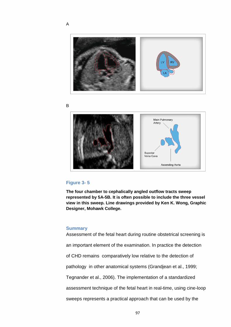
97
A
B
Figure 3- 5
The four chamber to cephalically angled outflow tracts sweep represented by 5A-5B. It is often possible to include the three vessel view in this sweep. Line drawings provided by Ken K. Wong, Graphic Designer, Mohawk College.
Summary Assessment of the fetal heart during routine obstetrical screening is
an important element of the examination. In practice the detection
of CHD remains comparatively low relative to the detection of
pathology in other anatomical systems (Grandjean et al., 1999;
Tegnander et al., 2006). The implementation of a standardized
assessment technique of the fetal heart in real-time, using cine-loop
sweeps represents a practical approach that can be used by the

98
majority of screening centres (Scott et al. 2013) . The standardized
technique is limited to those centres with the capability of storing
cine-loops as part of the clinical record. The basic obstetric
examiner must also become familiar with acquiring cine-loops in a
systematic way ensuring that the cine-loop sweep start and end-
points are observed enhancing the benefits of an extended
examination in real-time. However if these constraints can be
overcome, the benefits of a real-time assessment and an extended
examination can be fully realized.

99
Chapter 4 Increasing the Detection Rate of Normal Fetal Cardiac Structures: A Real-Time Approach
Introduction The use of ultrasound imaging for routine fetal screening in the
second trimester has been adopted by the Canadian health care
system and a number of European countries as a standard of care
(Rumack, Wilson, Charboneau, 2005). According to a survey of
2758 referrals to the fetal cardiology unit at Guy’s Hospital, London,
UK, 80% of congenital heart defects emerge from women at low
risk for congenital heart defects (Sharland, 2004). This fact
underscores the importance of routine screening for detection of
these defects. Routine screening provides a more accurate
measure of gestational age, earlier detection of multiple
pregnancies and may detect congenital malformations. The
Eurofetus study identified marked differences in sensitivity for
detection of different malformations. Abnormalities of the urinary
system and nervous system were most easily detected with a
sensitivity of 88.5% and 88.3% respectively (Grandjean et al.,
1999). In this study cardiac abnormalities were not well detected;
sensitivity for major cardiac defects was 38.8% and minor defects
20.8% (Grandjean et al., 1999). In Canada, an obstetric screening
examination is performed in real-time and documented as a series
of static images archived electronically or on film. The interpretation
of the cardiac images is performed by a Radiologist or Obstetrician
using the static images. Present detection rates for congenital heart
defects (CHD) vary widely depending on a number of factors:
operator skills, maternal obesity, fetal position, inadequate image

100
optimization, gestational age and visualization of the four chamber
view only (Sharland, 2004). The American College of Radiology
(ACR) and the International Society of Ultrasound in Obstetrics &
Gynecology (ISUOG) have published guidelines for routine second
trimester screening that require the four chamber view and views of
the left ventricular outflow tract (LVOT) and right ventricular outflow
tract (RVOT) to address this limitation (American Institute of
Ultrasound in Medicine, 2013). Acquisition of static images of the
fetal heart provides a very limited quantity of diagnostic information,
in that the identification of normal cardiac structures cannot be
completely demonstrated as a series of a few static slice images.
The interpretation of key structural relationships will only be
completed during a real-time interrogation. In practice, the detection
of the critical normal structures is accomplished solely by the
sonographer. This approach limits the interpreter’s ability to confirm
the findings of the sonographer. The main objective of this pilot
study is to demonstrate that acquiring real-time loops of the fetal
heart can be accomplished with minimal additional acquisition time
and skill. In addition, static and real-time techniques will be
compared in an effort to demonstrate the additional anatomical
information that is accessible using a real-time acquisition.
The real-time loops will add considerable diagnostic value to the
screening process since there will be two interpretative sweeps
performed on every exam: the preliminary review by the
sonographer and the summary review by the sonologist. Another
advantage of using the real-time loops is the opportunity it provides

101
the sonologist to review the images repeatedly in real-time, allowing
them to evaluate structures at different points during the cardiac
cycle. It is well established that observation of moving structures
provides a more sensitive means of detecting small differences in
form (Ivry 1992). The ability to assess structures during the diastolic
phase has been found to be helpful in detecting atrial-ventricular
septal defects (Chaoui, 2003). A cine-loop of a sweep through the
cardiac outflow tracts will allow the sonologist to better appreciate
the cardiac morphologic relationship such as the LVOT and RVOT
which is crucial for ruling out conotruncal abnormalities. Hosono et
al, (2002) found the use of tissue harmonic imaging (THI) to be
advantageous in imaging fetal cardiac tumors due to improvements
in image contrast (Hosono, Chiba, Kanai, & Kanagawa, 2002). A
comparative study was performed using two techniques,
conventional, 3.5 MHz frequency compared with a tissue harmonic
signal of 3.6 MHz. THI was used to improve the visualization of the
cardiac tumors. Using THI could improve the quality of the images
and may influence the detection rate of cardiac structures as well
(Paladini, Vassallo, Tartaglione, Lapadula, & Martinelli, 2004).

102
Table 4- 1
Clinical site survey data.
Obstetrical Exams/Day
Number of sites (%)
Routine Views
Number of sites
(%)
Operating PACS (%)
0-5
7 (33)
Four
Chamber
21 (100)
20 (95)
5-10
9 (42)
LVOT
18 (86)
10-15
2 (10)
RVOT
18 (86)
15-25
1 (5)
Aortic Arch (long axis)
7 (33)
25-30
1 (5)
Ductal Arch (long axis)
3 (14)
30-35
Great Vessels
(short axis)
6 (29)
>35 1 (5) Septum 1 (5) Total 21 (100) 21 (100) 21 (100)
A survey of 21 local screening centers illustrated in Table 4-1 found
that 95% of centres acquired and stored images using a Picture
Archiving Communications System (PACS). Most centres acquired
three static images of the fetal heart: the four chamber view, the
LVOT and RVOT view. See Table 4-1. For the purpose of this pilot
study, this combination of static images represents the conventional
technique, protocol A.
Research Question/Objectives This study compared static and real-time imaging techniques
applied to the assessment of the fetal heart in routine screening.
RESEARCH AIMS
• Demonstrate the ability to acquire the experimental protocol
B in less than five minutes

103
• Demonstrate an increased rate of detection of cardiac
structures using real-time cine-clips compared with static
images
Method The research protocol was reviewed and approved for use by the
ethics committee of Charles Sturt University (Wagga Wagga, NSW,
Australia) and the ethics committee of Mohawk College (Hamilton,
Ontario, Canada). Exclusion criteria included a family history of
congenital heart defects or an abnormal second trimester obstetric
sonogram. All 28 subjects had a normal second trimester sonogram
prior to participation in this study. Images were acquired using the
Philips HDI 5000, Envisor, Medical Systems, Bothell, WA, USA,
sonography systems. Images were acquired using a curvilinear
array transducer with a fetal cardiac preset using tissue harmonic
image processing, with a bandwidth of 2.5-5.0 MHz.
Table 4- 2
Subject demographics and protocol acquisition times.
Subjects (n=28) Average 95% Confidence Interval
Gestational Age 27 weeks 0 days
24 weeks 5 days – 28 weeks 6 days
Maternal BMI 27.8 25.8-29.8
Maternal Body Wall (cm)
1.8 1.6-1.9
Acquisition Time, Protocol A (00:00)
3:49 2:39-4:58
Acquisition Time, Protocol B (00:00)
4:18 3:19-5:17
Acquisition Time, Protocol C (00:00)
6:32 5:14-7:51
Subjects were scanned using three different techniques: Protocol A,
consisting of three images of the fetal heart including the four

104
chamber view, the LVOT and the RVOT view. See Figures 4-1, 4-2,
and 4-3. The average acquisition time for each technique
independent from the others is described in Table 4-2.
Figure 4- 1
Static image of the four chamber view.
Figure 4- 2
Static image of the LVOT view.
LVOT

105
Figure 4- 3
Static image of the RVOT view.
Protocol B, the real-time sweep, was comprised of two grey scale
sweeps of 4 four seconds duration each through the abdomen and
fetal heart. The first sweep began at the level of the transverse
abdominal circumference with a visible stomach and swept into the
transverse four chamber view. The second sweep began at the four
chamber plane and swept through the LVOT and RVOT. See
Figures 4-5, 4-6 and 4-7. Protocol C was comprised of three real-
time colour cine-loops of four seconds duration. The first was a
sweep from the level of the transverse abdominal circumference
with a visible stomach into the transverse four chamber view. The
second sweep began at the four chamber view and swept through
the LVOT and RVOT. The third was a colour cine-loop of the three
vessel view.
RVOT

106
Figure 4- 4
Abdominal circumference from real-time sweep #1.
Figure 4- 5
Four chamber view from real-time sweep #1.
ST

107
Figure 4- 6
LVOT view from real-time sweep #2.
Figure 4- 7
RVOT view from real-time sweep #2.
Informed consent was obtained and subjects were randomly
assigned a sonographer who completed either protocol A, B or C.
LVOT
RVOT

108
A total of five sonographers acquired images for this project. All
sonographers had a minimum of three years’ experience performing
obstetrical sonography and were ARDMS credentialed as RDMS
(obstetrics and gynecology). The sonographer recorded the scan
time for the acquisition using an electronic timer. Every subject was
scanned by at least two different sonographers. Images were
acquired and stored on the Philips Medical Systems Xcelera
Minipacs. No additional training was provided to save the real-time
loops compared with the static images. Sonographers were limited
to a maximum of 10 minutes to complete each separate protocol in
an effort to replicate the practical limitations of the clinical
environment.
The image sequences acquired were reviewed independently by
two different fetal cardiac sonography experts blinded to each
others’ results. The reviewers are expert in acquiring and
interpreting fetal cardiac sonograms. The first was a sonographer
(EX1) with 16 years pediatric cardiac experience registered with
RDCS (pediatric and fetal echo) credentials and the second a
Pediatric Cardiologist (EX2). In addition to the expert reviewers, two
non-experts reviewed the images independently and were blinded
to each others’ results. The first was a radiology resident in year
three of his training (NE1) and the second was a cardiac
sonographer with RDCS credentials and less than one year of
experience in performing fetal cardiac studies (NE2). Each reviewer
recorded the detection of five cardiac structures listed in table 3 as:
detected = 1 or, missed = 0. A complete normal assessment

109
required a score of 5, an incomplete assessment is recorded as any
total less than 5.
Statistical Analysis
Inter-observer differences were tested using a Kappa score for
agreement between detection of structures. A one-way ANOVA was
performed to test for agreement between the means of total
detection rate scores and the means of the acquisition times.
Detection rate differences for complete and incomplete
assessments were analyzed using the McNemar Chi-square test.
Results The subjects’ average gestational age was 27 weeks 0 days, with
an average BMI = 27.8 and an average body wall thickness = 1.8
cm, see Table 4-2. The acquisition time for the static images,
Protocol A, is statistically equivalent to the acquisition time for
Protocol B. The mean time for both is less than 5 minutes. See
Table 4-2. The acquisition of protocol C exceeded 5 minutes with a
mean time of 6:32, statistically different than protocols A and B.
Using Kappa scores the inter- observer agreement between experts
for detection of cardiac structures using static images were found to
be equal to 0.19, +/- 0.09, 95% confidence interval for the four
chamber view, 0.14, +/- 0.10, 95% confidence interval for the LVOT
view, 0.16, +/- 0.10, 95% confidence interval for the RVOT view and
0.20, +/- 0.12, 95% confidence interval for the Cross over view. The
inter-observor agreement between non-experts for detection of
cardiac structures using static images were found to be equal to
0.45, +/- 0.12, 95 confidence interval for the four chamber view,
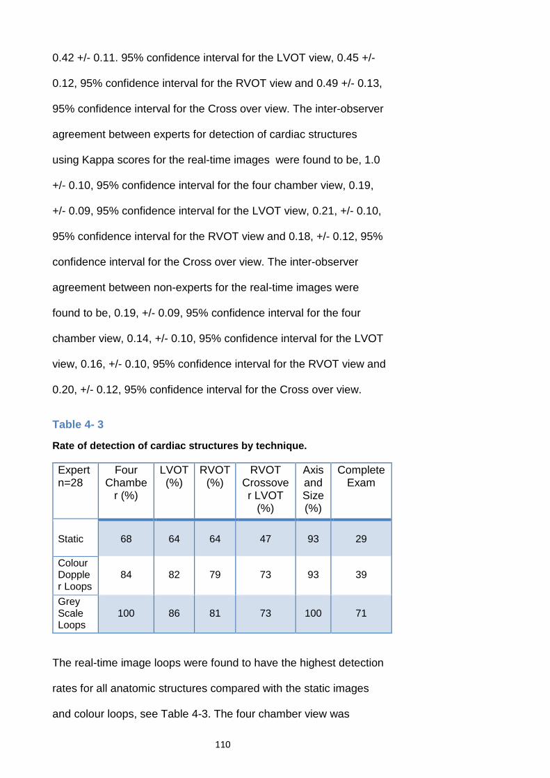
110
0.42 +/- 0.11. 95% confidence interval for the LVOT view, 0.45 +/-
0.12, 95% confidence interval for the RVOT view and 0.49 +/- 0.13,
95% confidence interval for the Cross over view. The inter-observer
agreement between experts for detection of cardiac structures
using Kappa scores for the real-time images were found to be, 1.0
+/- 0.10, 95% confidence interval for the four chamber view, 0.19,
+/- 0.09, 95% confidence interval for the LVOT view, 0.21, +/- 0.10,
95% confidence interval for the RVOT view and 0.18, +/- 0.12, 95%
confidence interval for the Cross over view. The inter-observer
agreement between non-experts for the real-time images were
found to be, 0.19, +/- 0.09, 95% confidence interval for the four
chamber view, 0.14, +/- 0.10, 95% confidence interval for the LVOT
view, 0.16, +/- 0.10, 95% confidence interval for the RVOT view and
0.20, +/- 0.12, 95% confidence interval for the Cross over view.
Table 4- 3
Rate of detection of cardiac structures by technique.
Expert n=28
Four Chambe
r (%)
LVOT (%)
RVOT (%)
RVOT Crossover LVOT
(%)
Axis and Size (%)
Complete Exam
Static
68
64
64
47
93
29
Colour Doppler Loops
84
82
79
73
93
39
Grey Scale Loops
100
86
81
73
100
71
The real-time image loops were found to have the highest detection
rates for all anatomic structures compared with the static images
and colour loops, see Table 4-3. The four chamber view was

111
identified in 100% of all real-time loops and in only 68% of static
images, and 84% of colour loops. The LVOT and RVOT cross-over
was detected in 73% of real-time loops and only 47% of static
images. Similar differences were found for the LVOT and RVOT as
individual structures.
Assessing the static images, 39% of studies generated a score of 5
for complete assessment compared with the real-time technique
that was able to completely assess the normal structures in 71% of
studies. This generated a statistically significant difference between
proportions = 0.321, p= 0.0352 using the McNemar Chi-square test.
The colour Doppler loops provided a complete assessment in only
39% of cases. The colour Doppler loops did provide the ability to
assess the blood flow through the atrial-ventricular valves and semi-
lunar valves in the majority of cases and this represents additional
diagnostic information compared with the grey scale static and real-
time loops. The colour Doppler loops were able to assess the four
chamber view and five chamber view in the majority of cases. The 3
vessel view was only achieved in 45% of cases, see Table 4-4.
Table 4- 4
Detection rate of colour flow using colour Doppler loops by view.
Expert n=28 Four Chamber
(%)
Five Chamber
(%)
Three Vessel View (%)
Assessment of Valves for
Regurgitation (%)
Colour Doppler Loops 91 84 45 86

112
Table 4- 5
Detection rate differences between experts and non-experts.
Expert/ Non-expert Difference
Four Chambe
r (%)
LVOT (%)
RVOT (%)
RVOT Crossover LVOT
(%)
Axis and Size (%)
Complete Exam
Static
0
13
4
8
7
5
Colour Doppler Loops
11
4
0
7
2
28
Grey Scale Loops
14
4
4
5
2
15
The expert reviewers and non-expert reviewers were found to agree
closely in their assessment of static images. The largest difference
between these groups was in evaluation of the LVOT with a
difference of 13% between groups, see Table 4-5. All other
structures differed by less than 10% respectively. There was close
agreement between the experts and non-experts interpreting the
grey scale and colour loops with the exception of the four chamber
view where the grey scale loop differed by 14% and the colour loop
differed by 11%. Assessment of a complete exam was in close
agreement between experts and non-experts for the static images
with a greater difference of 15% and 28% demonstrated in Table 4-
5, with the grey scale and colour loops respectively.
Sample Images The static image of the four chamber view allows the observer to
distinguish the axis, size and left/right chambers under ideal
conditions, see Figure 4-1. The static images of the LVOT and
RVOT, see Figures 4-2 and 4-3, demonstrate continuity between
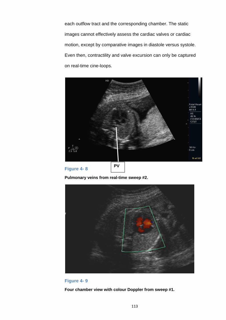
113
each outflow tract and the corresponding chamber. The static
images cannot effectively assess the cardiac valves or cardiac
motion, except by comparative images in diastole versus systole.
Even then, contractility and valve excursion can only be captured
on real-time cine-loops.
Figure 4- 8
Pulmonary veins from real-time sweep #2.
Figure 4- 9
Four chamber view with colour Doppler from sweep #1.
PV

114
Figure 4- 10
Five chamber view with colour Doppler sweep #2.
Figure 4- 11
Three vessel view from colour Doppler cine-loop #3.
LVOT
DA AA
SVC

115
For the sake of this thesis, static images have been extracted from
the real-time loops in an attempt to demonstrate the added value
loops provide. The first sweep sample begins at the transverse
abdominal circumference, see Figure 4-4. The four chamber view is
visualized in Figure 4-5. The second sweep demonstrates the
LVOT in Figure 4-6 and the RVOT is visualized in Figure 4-7. In
addition, the real-time sweeps provided continuity between the
stomach and abdominal circumference in 100% of real-time studies.
One or more pulmonary veins were visualized as contiguous
structures entering the left atrium in 56% of real-time cases
compared with 3.6% of cases with static images, see Figure 4-8. A
colour image extracted from a sweep from the abdominal
circumference to the four chamber view demonstrated blood flow
through the atrial-ventricular valves in Figure 4-9. The blood flow
through the LVOT is evident as an image acquired during the
sweep, from the four chamber through the outflow tracts, see Figure
4-10. In Figure 4-11 the great vessels demonstrate antegrade blood
flow in a three vessel view. If adequately seen, the three vessel
view can eliminate the need for ascending aorta and ductal arch
views (L Allan, 2004)
Discussion The subjects were a representative sample of expectant mothers in
the late second trimester and early third trimester between 24
weeks, five days and 28 weeks, six days. The requirement of a
previous normal diagnostic second trimester sonogram result, as a
prerequisite for participation in this study, resulted in the mean

116
gestational age of 27 weeks 0 days. The grey scale, real-time
sweeps, mean acquisition time (4:18) were acquired quickly like the
static images, mean acquisition time (3:49), and very little additional
expertise was required to capture the real-time loops.
The structure of the heart is complex due to relationships between
structures, such as the crossover of the outflow tracts, which is
critical for determining normalcy. Three static views of the four
chamber and outflow tracts are inadequate for complete
assessment in the majority of cases. Static images are much less
likely to be able to demonstrate subtle anatomic structures, such as
the moderator band, atrio-ventricular valve offset, that are useful for
distinguishing the left and right chambers. Static images cannot
assess the valves and their motion effectively, and this limits
observer confidence in detecting outflow function and their
surrounding morphologic relationships. The real-time loops are able
to detect these subtle anatomic structures and their motion; this fact
is reflected in the ability of the real-time loops to detect a completely
normal heart in 71% of studies compared with 39% of static studies.
In addition to the identification of the four chamber and outflow
tracts, the real-time sweeps provided the continuity of the left sided
stomach in the abdomen, as the sweep translates from the
abdomen to the chest in 100% of cases.
In a few cases, the relationship between the outflow tracts could not
be established in static images or real-time at normal speed:
however, use of a frame by frame analysis established that
relationship. This affirms the value of acquiring loops of information,

117
rather than individual slices as well as the need to re-evaluate the
heart in real-time after the image acquisition process.
The colour Doppler loops provided useful blood flow information in
most cases but this advantage was limited to some degree by the
fact that colour blooming had the effect of obscuring part of the
anatomy represented in grey scale. Unfortunately, our PACS and
ultrasound systems did not permit the suppression of colour.
Another drawback to the colour Doppler was the need for additional
operator expertise to fine tune the colour velocity scale or pulse
repetition frequency (PRF) and colour gain. The three vessel view
was detected in only 45% of cases compared with 91% and 84% for
the four chamber and five chamber views. This difference most
likely reflects the relative difficulty in acquiring the three vessel view
compared with the four chamber and five chamber views.
It is interesting to note that in nearly all cases there were only minor
differences in the detection rates between the expert image
reviewers and non-expert reviewers. Review of the static images
resulted in the least difference between these groups, likely due to
the fact that reviewers assessed only one image/view in
comparison with the real-time loops that generated a series of 120
images from a four second acquisition. The cine-loops provided the
reviewers with so much additional information it had the effect of
introducing a wider range of interpretation. Based on these findings,
it is clear that the identification of the four chambers, outflow tracts,
outflow tract cross over, size, situs, and position can be achieved
consistently by experts and non-experts.

118
Real-Time Methodology Typically second trimester screening exams are scheduled for
approximately 30 minutes in which time the sonographer is obliged
to interrogate the maternal pelvic and fetal anatomy, as well as
acquire representative images and measurements to estimate
gestational age. Fetal cardiac anatomy, owing to its complex shape
and rapid movement, represents the sonographer’s greatest
diagnostic challenge. Given these constraints, a robust screening
technique needs to be able to provide the maximum relevant
anatomical information with the minimum operator skill and time.
For most screening centers a reasonable goal would be to identify
an abnormal heart and refer the patient to a pediatric cardiology
laboratory to diagnose the specific nature of the defect based on a
standardized assessment. The use of colour Doppler cine-loops
provided additional blood flow information in the majority of cases
but this advantage was off-set by the additional time required to
acquire these loops. Additional operator skill is needed to apply
colour Doppler effectively and colour blooming may obscure
visualization of some grey scale anatomy unless it is suppressed.
For these reasons it is proposed that the two sweep cine-loop
acquisition technique tested in this study be used for assessing the
fetal heart in the second trimester. Applying this method of
acquisition will allow the image interpreter to identify the key cardiac
anatomical landmarks and relationships that were assessed in this
study in the majority of cases. This method will provide the most
effective assessment of fetal cardiac anatomy with minimal
additional time and skill.

119
Study Limitations It was not possible to blind the observer to the techniques used for
acquisition, since a static image is inherently different to a real-time
loop. The manipulation of the real-time loops as a frame by frame
analysis was left to the discretion of the individual reviewers. While
this may have limited the standardization of the image presentation
the manipulation of frame rate by the user is a key advantage of the
technique.
Conclusion Real-time sweeps through the fetal heart are easily and rapidly
acquired with minimal additional operator skill. Acquiring four-
second, real-time loops provides the sonologist with enhanced
information to more effectively assess the fetal heart. This study
was able to demonstrate a complete cardiac assessment in 71% of
studies using a real-time acquisition technique compared with 39%
of static image acquisitions. The use of colour Doppler provided
blood flow information of the atrial-ventricular valves in 86% of
subjects but was not compared to static colour images. A
standardized two sweep acquisition through the fetal heart can be
acquired quickly and with minimal additional operator skill. In the
future, studies comparing the performance of this technique
between normal and abnormal fetal hearts will be needed to
demonstrate the clinical value of real-time loops for detecting
congenital heart disease.

120
Chapter 5 Increasing the Detection Rate of Congenital Heart Disease during Routine Obstetrical Screening Using Two Cine-Loop Sweeps
The majority of deaths from congenital defects in childhood can be
attributed to congenital heart disease (CHD) (Lindsey Allan, 2010;
Carvalho et al., 2002). The most common cause of birth defects,
CHD is most frequently reported with an incidence of 0.8% of all live
births (Gilboa, Salemi, Nembhard, Fixler, & Correa, 2010; Hoffman
& Kaplan, 2002). The prenatal diagnosis of congenital heart disease
can improve clinical outcomes of those affected after birth (Bull,
1999; Hornberger, 2008; Jaeggi et al., 2001; Yates, 2004). Prenatal
diagnosis of congenital heart disease is critically important to
ensure optimal management of the pregnancy, optimal delivery of
the baby and provision of support for parental decision making. In
addition, those at high risk for CHD may be relieved of additional
anxiety and fears due to possible CHD by confirmation of normality
(Kitchiner, 2004).
Prenatal screening performance has been documented in the
literature extensively over the past 25 years and although the
reports vary in study design, they consistently demonstrate poor
performance in the low risk population using a four chamber
assessment with detection rates ranging from 5% to 48% (Achiron ,
Glaser, Gelernter, Hegesh, 1992; Buskens, Grobbee, 1996;
Friedberg et al., 2009; Oggè et al., 2006; Tegnander, Eik-Nes,
Johanesen, 1995). More recently, a number of prospective studies
that included the outflow tracts and substantial additional operator

121
training, found that some improvements were observed with
sensitivities ranging from 57% to 78% (Achiron, Glaser, Gelernter,
Hegesh, 1992; Carvalho et al., 2002; Tegnander et al., 2006).
While the inclusion of outflow tracts as an extension to a basic
exam of the four chambers has provided encouraging results,
further improvements in screening protocols are needed. The
interpretation of static images of the heart used by the majority of
routine obstetric screening centres is a significant barrier to the
detection of CHD (Sklansky, 2007). In some instances, identification
of an atrioventricular septal defect for example, is increased by
assessing the four chamber view during the diastolic and systolic
phases of the cardiac cycle (Chaoui, 2003). Reliance on static
images only for interpretation assumes that the sonographer will
recognize CHD in the course of their real-time assessment of the
fetal heart and record an image that clearly depicts the CHD. In the
event that the sonographer misses the abnormality, it is unlikely that
the CHD will be demonstrated on the static images since image
acquisition is operator dependent. However, several
instrumentation alternatives are available to interpreting physicians.
Dynamic three-dimensional volume sets of the fetal heart can be
stored for review and interpretation using Spatial-Temporal Image
Correlation (STIC) technology, (Chaoui, 2003; Sklansky, 2007).
Three-dimensional imaging has the potential to improve the
detection rate of CHD since it provides unlimited opportunities to
review the fetal heart as an acquired volume or as a cine-loop of
real-time slices (DeVore et al., 2003). Acquisition of three-

122
dimensional volumes of the fetal heart requires additional training
and resources (Sklansky, 2007). The need for additional
investments in training and equipment is mandatory. Though three-
dimensional assessment of the fetal heart has not become a
standard practice in routine obstetrical screening it is well on its way
to becoming the most effective tool to assess the fetal anatomy.
The use of cine-loop sweeps to assess the fetal heart however, has
been recommended as a practical means of increasing the
detection rate of CHD (Sklansky, 2007). Additionally the use of
cine-loop sweeps to examine the criss-cross of the outflow tracts in
an effort to increase the likelihood of detecting conotruncal defects
is well published (McGahan et al., 2007; Poole et al., 2013;
Sklansky, 2007). No new equipment is required other than the
storage capacity to store cine-loops within the imaging facility and
very little additional sonographer training is needed since the
sweeps can be incorporated into existing imaging protocols with
little effort. Short axis cuts through the fetal heart yields morphologic
information far superior to mere four-chamber and outflow tract
views. Extension to the three-vessel view, though not purposefully
examined on two-dimensional static imaging, will often be seen on
a cine-loop sweep. The first cine-loop sweep begins at the level of
the stomach in the transverse plane and moves superiorly to the
four chamber view. The second sweep begins at the four chamber
view in transverse and sweeps upward to include the outflow tracts.
The objective of this study was to determine if using two cine-loop

123
sweeps would improve the detection of congenital heart disease
compared with static images.
Materials and Methods Subjects
This study was approved by the Charles Sturt University Ethics in
Human Research Committee, Mohawk College research ethics
board, the Hamilton Health Sciences/Faculty of Health Sciences
research ethics board and the Grey Bruce Health Services research
ethics board. Informed parental consent was obtained for every
case. Normal images including static images and cine-loop sweeps
of the fetal heart from 79 expectant mothers during the course of
their routine second trimester screening sonogram were obtained.
Static images and cine-loop sweeps of the fetal heart from 14
expectant mothers referred to the McMaster Children’s Hospital,
pediatric cardiology laboratory for follow-up of suspected congenital
heart defects were also gathered. Diagnostic accuracy was
confirmed by post natal exams, excluding those newborns that
were not followed. See Table 5-1.

124
Table 5- 1
Congenital Heart Disease Characteristics and Gestational Age
n=14 Frequency Weeks
Hypoplastic Left Heart 4 19-22
Hypoplastic Left Heart and Atrioventricular Septal Defect
1 21
Atrioventricular Septal Defect 3 18-22
Large Heart 1 21
Rhabdomyoma 1 26
Double Outlet Right Ventricle with pulmonary stenosis
1 24
Supraventricular Tachycardia 1 32
Double Outlet Right Ventricle, tetralogy spectrum
1
19
Tetralogy of Fallot 1
20-25
Image Acquisition
In an effort to ensure a realistic evaluation of this approach, images
were acquired using a variety of manfuacturers’ sonographic
imaging equipment and transducers including; Toshiba America
Medical Systems Inc. (Tustin, CA), 4.0 and 5.0 MHz, curvi-linear
transducer frequencies, Philips Medical Systems Inc. (Bothell, WA),
5-1 MHz, curvilinear transducer, General Electric Healthcare
(Waukesha, WI), 4-8 MHz, curvilinear transducer and Siemens
Medical Solutions USA, Inc. (Malvern, PA) 6-2 MHz curvilinear

125
transducer. Equipment settings were optimized to include a high
frame rate, 30-100 Hz and harmonic imaging.
For both normal and abnormal cases three static images and two
cine-loop sweeps were acquired. The static images included; the
four chamber view, the left ventricular outflow tract view and the
right ventricular outflow tract view. These images represented a
conventional approach to image acquisition.
Using the same equipment settings as used in the conventional
method and where possible the same acoustic window and fetal
position two cine-loop sweeps were acquired. Each cine-loop
sweep was acquired using a four second electronic capture stored
to the local hard disk. The first cine-loop sweep is described as the
situs-four chamber sweep. The technique used is described as
follows; using a transverse abdominal section, the transducer was
moved cephalad from the abdominal circumference into the four
chamber view, without changing angles to the fetus, see Figure 5-1
.

126
Figure 5- 1
Cine-loop Acquisition
Orientation of cine-loop sweeps relative to the sagittal plane of the fetus. The first cineloop sweep begins at the abdominalcircumference and continues cephalad into the four chamber view. The second sweep begins in the four chamber view and with a slight rotation towards the fetal left shoulder, angles cephalad through the outflow tracts.
Acquisition of the image should begin showing situs, capture at
least two beats of situs in order to allow the interpreting physician to
be able to appreciate the pulsatility of the aorta, then slowly slide
the transducer cephalad without changing the angle. The
acquisition should then end after documenting two cardiac cycles
using the four chamber view. The second cine-loop sweep is
described as the four chamber-outflows sweep. To acquire this cine
sweep, the following technique was used: from the four chamber
view, the transducer is rotated slightly toward the fetal right
shoulder and angled cephalad; the LVOT is visible first with

127
cephalic angulation. The transducer is then angled more cephalad
and to the left shoulder to visualize the right ventricular outflow.
Image Review
Both abnormal images and normal images were collated into an
image data set representing 79 normal cases and 14 abnormal
cases which totals to 93 distinct pregnancies. An online computer
generated randomizer (random.org) was used to compile these
image data sets in random order. There were seven image
reviewers divided into three classes: expert, non-expert radiology
residents and sonographer. There were two experts, a pediatric
cardiologist, (E2) with more than ten years clinical experience and a
pediatric cardiac sonographer with more than ten years’ experience,
(E1), three non-experts, radiologists (NE1, NE2 and NE3), and two
sonographers each with more than five years’ experience (S1, S2).
Each of the image reviewers, blinded to one another’s results,
worked through 93 sets of static images sequentially using the
same randomized series. Upon completion of their review and
interpretation of these they went on to review and interpret 93 sets
of the abdomen to four chamber and four chamber to outflow cine-
loop sweeps. Their image review was enabled through the use of a
software program, Fetal Heart Sonographic Assessment Tool
(FSAT), see Figure 5-2. This software allowed them to electronically
record their assessments using an electronic check list that
confirms demonstrations of five key points; 1) Situs, Position, Size;
2) Four chamber morphology; 3) Left ventricular continuity; 4) Right
ventricular continuity and 5) the LVOT/RVOT relationship.

128
A
B
Figure 5- 2
Fetal heart Sonographic Assessment Tool (FSAT),
The images displayed as statics or cine-loop sweeps in panel A. Decision making tool depicted in panel B, N represents a normal finding, A, an abnormal finding and NS means the structure or relationship was not seen.

129
A second electronic record was made to determine one of 3
outcomes: normal, abnormal or indeterminate. In order to set a
comparable decision making process, each reviewer was advised
that a normal decision would imply no further imaging or follow-up,
an indeterminate selection would imply a follow-up ultrasound exam
and an abnormal decision would be referred to the pediatric
cardiology laboratory for further examination.
Statistical Analysis The statistical significance of the detection rates provided by the
image reviewers was determined using the McNemar chi-square
test for a difference of proportions with significance level, p < .001.
All other data was analyzed using a one-way analysis of variance
(ANOVA). The analysis was performed using the Analyse-it
Software, Ltd. (Leeds, UK).
Results The expert reviewer (E1) detected 50% of the CHD using the static
images and detected 93%, (13/14) of them using the cine-loop
sweeps, see Table 5-2. The detection rate varied most within the
cine-loop sweeps in both the expert and non-expert groups, where
the detection rate varied by 29% and 28% respectively. The
greatest improvement in detection between assessment methods
was found in the non-expert group (NE3) with an improvement of
64%, the least improvement was found in the sonographer group
(S2) with no difference observed. For all other reviewers the
detection rate improved from 7%-64%.

130
Table 5- 2
Detection Rate by Reviewer, Experts (E), Non-Experts (NE), Sonographers (S), Number (%)
n=14 (E1)* (E2) (NE1) (NE2) (NE3)* (S1) (S2)
Static 7 (50) 5 (36) 3 (21) 2 (14) 1 (7) 1 (7) 4 (29)
Sweep 13 (93) 9 (64) 6 (43) 6 (43) 10 (71) 2 (14) 4 (29)
*Statistically significant, p<.001
The experts detected the most CHD with an average rate of 43%
for the static images and 79% using the cine-loop sweeps, see
Table 5-3 as an example. The non-expert group identified on
average only 14% of CHD using static images and 52% using the
cine-loop sweeps. Sonographers identified only 18% using the
static images and nearly the same detection rate, 21% for the cine-
loop sweeps.
Table 5- 3
Average Detection Rate by Group, Experts (E), Non-Experts (NE), Sonographers (S), Number (%)
n=14 (E) (2) (NE) (3) (S) (2) All Reviewers (7)
Static 6 (43) 2 (14) 2.5 (18) 3.4 (24)
Sweeps 11 (79) 7.3 (52) 3 (21) 7.1 (51)
Change +5 (36) +5.3 (38) +0.5 (3) +3.8 (27)
The number of false positive cases identified overall by image
reviewers was quite low with an average of 5.4% observed in the

131
static image review compared with 3.9% using the cine-loop
sweeps.
Discussion The analysis of the cine-loop sweeps resulted in an improved
detection rate compared with the static images for expert and non-
experts of 36% and 38% respectively. The nearly equivalent rate of
improvement found in both groups suggests that the value of the
cine-loop sweeps is equally applicable to the expert and non-expert
interpreting physician. Within the non-expert group the variation in
detection rate was 14% for the static image review and 28% for the
cine-loop sweeps. This pattern of increased variation in detection
rate for the cine-loop sweeps was also observed in the expert
group. In this group the variation in detection rate for the static
image review was 14% and 29% for the cine-loop sweeps. The
increased variation between the reviewers may be explained by the
fact that the cine-loop sweeps provided significantly more image
information for analysis allowing individual interpretive skills to be
demonstrated to a greater degree than the static images. The
detection rate of non-expert reviewers using static images was only
14%. This result is in alignment with the reality that the static
images of the four chamber and outflow tracts provide a very
minimal amount of diagnostic information to the interpreting
physician during the image review process. However, the expert
reviewers were able to identify 43% of CHD using the same static
images while maintaining a low rate of false positive identification.
This difference suggests that the importance of training and

132
experience makes a significant impact on the detection rate of
CHD. Surprisingly, the sonographers’ improvement in detection rate
using the cine-loop sweeps compared with the static images was
only 3% and this finding was not consistent with that of the expert
and non-experts results. The sonographers detection rates overall
were low, 21% using the cine-loop sweeps and 18% for the static
images. Compared with the performance of the expert and non-
experts, this low detection rate suggests that the interpretative
expertise of the less experienced sonographers was not as refined
compared with the physicians. This is not surprising since the role
of a beginner sonographer is primarily image acquisition, initial
review and limited interpretation skill, whereas; the interpreting
physician’s role is primarily analysis and interpretation. These
results indicate that it is essential that the interpreting physician
have access to real-time sweeps of the heart if the goal is to detect
CHD in routine obstetrical screening, although diagnostic findings
would be improved by a full fetal exam which this research did not
address. Furthermore, the cine-loop sweeps can be shared
electronically via a secure network connection should the
interpreting physician request expert consultation.
The average rate of false positive identification of CHD for all
reviewers was 5.4% using the static images and 3.9% using cine-
loop sweeps. This suggests that the reviewers understood the
potential emotional impact and stress that a positive finding would
have on the parents and families affected and they set the
threshold for a positive finding relatively high. This might help

133
explain why the experts and non-expert detection rates did not
exceed 80% even with the use of cine-loop sweeps.
All fourteen abnormal cases were identified using the cine-loop
sweeps and two cases, a large heart and a hyoplastic left heart
were identified by all seven reviewers. In contrast, two cases,
atrioventricular septal defect and double outlet right ventricle with
tetralogy spectrum were missed by all seven reviewers using the
static images. An example is described below where the static
images provided a false impression of normalcy compared with the
cine-loop sweeps. In order to illustrate this, a frame was grabbed
from the cine-loop sweep using a VLC media player 2.0.3
Twoflower© to demonstrate the pathology. In Figure 5-3, a normal
looking four chamber view is contrasted with a more abnormal
looking view extracted from the cine-loop sweep. This was
confirmed as a Tetralogy of Fallot. Only one of the seven reviewers
identified this pathology on the static images. These findings further
underscore the need for an assessment in real-time.

134
A
B
Figure 5- 3
The ventricular septal defect associated with this example of Tetralogy of Fallot is best demonstrated in image A, a frame selected from the cine-loop sweep as compared with image B a static image acquired as a single image using the conventional technique.

135
While the results of this study uniformly point towards the increased
value of the cine-loop sweeps as compared with the static images,
due to the relatively small sample of 14 cardiac defects, statistical
significance was achieved in the results in only two of the seven
reviewers. Not included in our study however, was the three vessel
view which has been found to be a very effective method of
assessing the ventricular outflow tracts (Yoo, Lee, Kim, Ryu, Choi,
& Cho, 1997). In a pilot study it was found that the majority of
screening centres in our region were acquiring the four chamber
and outflows as three static images, and the acquisition of the three
vessel view increased the acquisition time significantly compared
with the two sweeps described above (Scott et al., 2008). Our
approach is intended to incorporate cine-loop sweeps into a routine,
second trimester sonogram with a minimally added acquisition time
and sonographer training. It was for these reasons we chose not to
include the three-vessel view. The abnormal cases were acquired
in a prospective fashion based on availability to the researchers and
therefore they do not represent a comprehensive sample of CHD.
Hypoplastic left heart cases are over represented along with atrial
ventricular septal defects in this sample.
Conclusion The use of cine-loop sweeps is a practical and standardized
approach to the assessment of the fetal heart during routine
obstetrical sonograms. This technique poses several advantages
compared with review of static images, it has been shown to
increase the rate of detection of CHD while minimizing the false

136
positive identification. The acquisition process is a relatively simple
procedure and does not require additional resources other than the
capacity to store cine-loops within the clinical facility.
The results of this study support the use of standardized
procedures, both for acquisition of images as well as their review
and interpretation.
By including real-time cine-loop sweeps through the fetal heart in a
standardized format, the interpreting physician gains access to
additional critically important evidence not obtained by three basic
images to guide their decision making. This study demonstrates a
possible improvement in prenatal detection of congenital heart
disease when standardized cine loops and checklists are included
in the prenatal cardiac screening review by expert and non-expert
clinicians. Despite the small sample size, statistically significant
improvement in congenital heart defect detection was demonstrated
with two out of seven reviewers. On the basis of these findings,
further study with a larger sample size and a more complex
research design may support inclusion of cine-loop sweeps as well
as increased two-dimensional views in routine prenatal screening
programmes.

137
Chapter 6 Increasing Recognition of Fetal Heart Anatomy using Online Tutorials and Mastery Learning compared with Classroom Instructional Methods
Introduction The majority of deaths from congenital defects in childhood can be
attributed to congenital heart disease (CHD) (Gilboa et al., 2010).
The most common cause of birth defects, CHD is most frequently
reported with an incidence of 0.8% of all live births (Dolk, Loane, &
Garne, 2011). The prenatal diagnosis of congenital heart disease
can improve clinical outcomes of those affected after birth (Brown et
al., 2006; Cass, 2011; Fuchs et al., 2007). Prenatal diagnosis of
congenital heart disease is critically important to ensure: optimal
management of the pregnancy, optimal delivery of the baby and
provision of support for parental decision making. In addition, those
at high risk for CHD may be relieved of additional anxiety and fears
due to the possibility of CHD by confirmation of normality (Allan &
Huggon, 2004).
The role of the sonographer in prenatal diagnosis of congenital
heart disease is to assess the fetal heart in real-time including the
four chambers and the outflow tracts (American Institute of
Ultrasound in Medicine, 2013; Eik-Nes, 2006). Typically the
obstetric sonographer will document normal or abnormal findings as
a preliminary report with corresponding static images. The
interpreting physician will review the preliminary report and
associated images confirming the determination of a normal,
abnormal or indeterminate finding.

138
The formal training for diagnostic medical sonographers in the
detection of congenital heart defects is provided in didactic form
usually within an obstetrics and gynecology sonography course.
The clinical practicum is most often provided through clinical
facilities linked to the college program. Imaging and assessing the
fetal heart is a challenging task for sonography students. Unlike
abdominal, pelvic or other sonography exams, students usually do
not have an opportunity to practice obstetrical sonography in their
college imaging laboratories. It is also difficult for education
programs to acquire a robust library of sample cases representative
of the diversity of congenital heart disease. Assessing the fetal
heart and acquiring images is a complex task demanding an
integrated understanding of the cardiac embryology of the fetal
heart and morphologic changes with pathology. Image recognition
skills for identifying rapidly moving structures such as the outflow
tracts on the order of 2-6 mm in diameter are intimidating to an
imaging learner (Allan, 2004).
Addressing complex learning objectives that demand significant skill
development by the learner is a challenge for the traditional
classroom setting. Skill development demands significant formative
assessment and structured feedback which can be difficult to
provide to a group in a classroom environment. For teachers at all
levels of education, it remains a challenge to provide customized
instruction and high quality feedback to individuals in large classes.
In a seminal article in 1984, Benjamin Bloom articulated a number
of methods to address the two sigma problem. Bloom found that

139
using both mastery learning and tutorial based instruction compared
with conventional classroom instruction, students in the tutorial and
mastery learning groups on average increased their achievement
levels by two sigma (standard deviations)(Bloom, 1984). In other
words, the average student in the tutorial and mastery group
outperformed 98% of the students in the classroom group (Bloom,
1984). Mastery based instruction proposes that learning should
provide formative evaluation with guided or corrective feedback
such that students achieve mastery of all of the learning objectives
(Gagné & Bloom, 1988). Tutorial based instruction assumes that
students are working regularly with a good tutor, in a face to face
mode. Two forms of instruction have emerged to address the
opportunity for learning using the theory of the two sigma problem
(Corbett, 2001). These forms include a method of automated
feedback based on student performance during practice exercises
and mastery learning, where students work at their own pace to
achieve mastery of learning (Corbett, 2001; Svinicki, 1999).
In the realm of digitally enhanced education that exists in many
postsecondary institutions today, there exists a significant
opportunity to provide students with opportunities to leverage the
educational theories and applications established by Bloom. Some
very popular solutions have emerged from outside the traditional
postsecondary sector. Khan Academy is a very popular electronic
platform for learning mathematics that uses tutorial-based
instruction and mastery learning principles (Khan, 2012). Students
are able to traverse the entire spectrum of mathematics necessary

140
to achieve successful preparation for entry into postsecondary
institutions. Besides the pedagogical advantages associated with
the two sigma problem, the use of electronic learning tutorials and
practice exercises provide numerous advantages compared with
traditional classroom learning. These advantages include:
• Ensure a consistent and high quality learning opportunity
• Self-paced progress through the learning objectives
• Opportunity to repeat tutorials without reliance on a human
tutor
• Ability to access experts on niche topics
• Expand the learning opportunity to a global audience in any
language
• Provide tutorial and learning tool designers valuable
quantitative feedback regarding the learning experience
• Feedback can be used to improve future iterations of the
tutorials and tools
The objective of the research was to determine if online tutorials
and practice exercises using a standardized assessment of the fetal
heart in real-time is a more effective approach to be used by
students when learning how to identify normal fetal heart structures
and congenital heart disease. The hypothesis was that "Training
students to use a standardized assessment technique using
tutorials and practice exercises online compared with traditional
classroom instruction will increase the student’s ability to identify
normal fetal heart structures and congenital heart disease."

141
Materials and Methods This study was approved by the Charles Sturt University Ethics in
Human Research Committee, the McMaster University Research
Ethics Board (MREB) and the Mohawk College Research Ethics
Board.
The participants in this study were drawn from the Medical
Radiation Science, Obstetrical and Gynecologic Ultrasonography 3
course. Sonography students were in the third year of study within
a collaborative four year, Mohawk-McMaster Medical Radiation
Sciences program. All students in the class chose to participate in
this study. The total number of all participants was 33. From the
initial 33 participants, 27 students completed all of the learning
activities and the final quiz, 15 students from the experimental
group and 12 students from the control group.
Three learning objectives for the experimental and control groups
are defined as follows:
1. Increase ability to identify normal fetal cardiac structures
2. Increase ability to identify abnormal fetal cardiac structures
3. Increase ability to identify abnormal cases
Students were randomly divided in to two groups. There were 17
students assigned to the classroom learning group (control) and 16
were assigned the online learning, (experimental) group. Students
assigned to the control group were not permitted access to the
online tutorials and practice exercises. Students assigned to the

142
experimental group were not permitted to attend the four teacher
led classes and single online tutorial with the course instructor. The
online exam was administered within two weeks of the onset of the
experiment.
Students enrolled in the control group participated in four classroom
lecture periods of 50 minutes each and one online tutorial. The
lectures reviewed the anatomy of the fetal heart, the fetal blood flow
and the sonographic views used to image the heart. They also
included real time loops with tips and suggestions on how to identify
a normal heart. Lectures included review of some of the more
common congenital heart abnormalities using real time loops. The
tutorial was delivered online with labeled anatomy images with
focused questions pertaining to the diagrams. The time for
instruction was approximately 240 minutes and an allowance for
independent study time of approximately 90 minutes for a total of
330 minutes of time committed to learning.
The experimental group was provided with the “Find it First” tool
consisting of 13 brief online tutorials in the use of a five step method
of the fetal heart assessment prepared by the study investigator,
amounting to approximately 75 minutes of instruction in total.
Students completed practice exercises online as formative
assessment with electronic feedback that required an additional 150
minutes of learning time. The total time committed to learning for
the experimental group was approximately 225 minutes.

143
The students were also provided practice cases for each step of the
technique and these exercises included immediate electronic
feedback. Students were required to demonstrate mastery of each
step before continuing to the next step of assessment. A screen
shot depicting the user interface for the practice exercises provided
by module two is provided in Figure 6-1. Students worked through
five online modules representing five steps of analysis considering
the anatomic features and relationships as follows;
1. Cardiac situs, position and size
2. Chamber morphology and function
3. Left Ventricular Outflow Tract (LVOT) continuity with the
left ventricle
4. Right Ventricular Outflow Tract (RVOT) continuity with the
right ventricle
5. Normal cross-over of outflow tracts at their origins

144
Figure 6- 1
During formative assessment the feedback is provided immediately. All cases that are incorrect must be repeated after the first practice series is completed before continuing to the next learning module.
Each of these features and relationships could be defined as
normal, abnormal or not seen. Each module consisted of a brief
tutorial to provide examples and instruction on how to assess the

145
relevant anatomic feature. Following the review of each tutorial the
students were required to complete 17 sample cases as depicted in
Figure 6-1. At the end of every series, all of the missed cases were
compiled and the learner re-assessed the missed cases until all 17
cases were accurately reviewed. The student could advance to the
next module only after successfully demonstrating the ability to
analyze the anatomic features covered by the module. As students
advanced through the modules, additional steps were added to the
analysis until the student reached the fifth module where all five
steps and corresponding features were examined representing a
review and analysis of a total of 85 cases.
In order to assess the performance of the students in both groups,
an online quiz was developed. Each student was provided with a
unique and secure user identification and password. Students’
ability to identify normal and abnormal structures was measured by
their review of 25 cases of fetal cardiac anatomy. A randomized
sequence of 10 abnormal and 15 normal cases were presented.
Each case consisted of two cine-loop sweeps through the fetal
heart, the abdomen to four chamber sweep began in the transverse
abdominal plane at the level of the stomach and ended at the four-
chamber view. The four-chamber to outflows sweep began in the
four chamber view and swept through the ventricular outflow tracts.
Students in both groups completed the online examination within
two weeks of completing the classroom or online instruction. As
described in Figure 6-2, students were required to identify 5

146
anatomic features for each case and determine whether that case is
either, normal, abnormal or indeterminate.
Figure 6- 2
The final quiz includes the assessment of all five indicators and the final decision.
A completely accurate assessment was one that scored each
feature accurately and correctly identified the case as normal,
abnormal or indeterminate. The decision framework was
standardized for the students using a protocol that defined the
following outcomes: a normal result implies no further assessment,
an abnormal determination will result in a referral to a pediatric
cardiologist, and an indeterminate finding will result in a follow-up

147
reassessment in the obstetric screening center. This evaluation
required approximately one hour for the students to complete.
Data Analysis
The Chi Square test was performed to evaluate the statistical
significance of the image analysis results. The null hypothesis for
this test was defined by the following statement, “After using the
computerized learning system (experimental group) or gaining
instruction in a traditional face to face classroom environment
(control group), there will be no statistical difference in students’
ability to identify normal and abnormal fetal heart structures.”
Differences on the multiple choice evaluations and the confidence
levels were compared using the Student t-test for statistical
significance.
Results The students were evaluated on their ability to correctly assign one
of three conditions, normal, abnormal or not seen to five unique
anatomic criteria and relationships and correctly define the case as
normal abnormal or indeterminate. For each case to be scored as
completely correct, all five anatomic criteria and the assignment of
normal, abnormal or indeterminate must be correct. This required a
total of six correct decisions for each case, by the student. The
students in the control group on average assessed correctly all five
anatomic features and determined the normal or abnormal status of
only 4.7/25, +/- 4.4 (standard deviation) compared with the
experimental group mean score, 9.9/25, +/- 2.7 (standard
deviation), P<.01. Students in the control group identified 19% of

148
the cases correctly compared with 39% for the experimental group.
In terms of mean performance difference between the two groups
there was a one sigma effect. This means that the average score in
the experimental group was greater than 84% of the students in the
control group. Furthermore, as seen in Table 6-1, the experimental
group outperformed the control group in every category of
assessment.
Table 6- 1
Five points of assessment of the fetal heart and the percentage of complete exams correctly identified as normal, abnormal or not seen. Students in the experimental group (Series 1) used the online tool compared with students in the control group (Series 2) who participated in classroom learning.

149
Table 6- 2
Detection rate by group according to the individual cases of congenital heart disease.
Case CHD, n=10 Experimental Group, n=15
Control Group, n=12
Difference
Detected (%) Detected (%)
(%)
1 Large Heart 13 (93) 7 (58) (35)
2 Truncus Arteriosus 12 (80) 9 (75) (5)
3 Hypoplastic Left Heart 12 (80) 6 (50) (30)
4 D-TGA 11 (73) 7 (58) (15)
5 Single Ventricle 10 (67) 6 (50) (17)
6 AVSD 7 (47) 5 (42) (5)
7 Tetralogy of Fallot 7 (47) 2 (17) (30)
8 Hypoplastic Left Heart 7 (47) 7 (58) (-11)
9 Tetralogy of Fallot 3 (20) 4 (33) (-13)
10 Ebsteins Anomaly 1 (7) 4 (33) (-26)
Total 83 (55) 57 (48)
The abnormal cases as depicted in Table 6-2, represent a typical
range of serious congenital heart disease. One or more abnormal
anatomical features were evident in the cine-loops provided to the
students for review. As defined in Table 6-2, the experimental group
outperformed the control group in 7/10 abnormal cases, p<.01.
Specifically in five abnormal cases, D-TGA, Single Ventricles, Large
Heart, HLHS and Truncus Arteriosus, a greater than 2/3 majority of
experimental students identified these cases whereas the control
group only identified 1 case, Truncus Arteriosus by a 2/3 majority of
students, p<.01. The experimental group performed particularly

150
poorly in detecting Ebsteins anomaly, 7% detection compared with
33% for the control group.
Discussion The strategy adopted by most sonography programs for teaching
students to identify the presence of pathology during abdominal or
pelvic examinations is typically based on providing students with
clinical practice and exposure to normal sonographic appearances
of anatomic structures. In teaching students to identify the presence
of pathology, sonography programs progress from a focus of
normal findings towards a framework for pathology such that
students can focus their attention on links between the patient’s
history and the probable associated pathology. Part of the
challenge for detecting congenital heart disease in-utero is the fact
that most often it is found unexpectedly in the low-risk population
(Sharland, 2004). This makes it more difficult to detect because one
cannot usually rely on clues from the patient history to focus the
examination. Furthermore, as a screening examination, the inherent
bias is an expectation of normalcy. Standardizing imaging
procedures and assessment protocols represent an important
consideration in this regard and one that was adopted for this
learning tool.
In addition to the two sigma pedagogy, the online learning tutorials
and practice exercises focused nearly exclusively on exposing
students in the experimental group to as many normal and
abnormal cases as possible in a step-wise learning methodology.
The focus for the learner was on practicing the decision making for

151
each step in each module until mastery of the concept was
achieved. In the classroom learning group, a broader focus was
taken and students were given more contextual information related
to the normal and abnormal heart. There was less emphasis on skill
development with feedback in the classroom setting.
Bloom’s two sigma theory was not replicated in this study; however,
a one sigma improvement was found and this represents a two-fold
increase in the percentage of cases accurately classified by
students in the experimental group, 39% which was compared with
19% in the control group. Further, the experimental group did not
benefit from any face to face learning opportunities. This might also
explain why a two sigma effect was not achieved. The ability to
correctly classify, anatomic features and relationships was
consistently demonstrated across all five categories. This suggests
that students acquired improved image interpretation skills in the
experimental group as compared to the control group.
Seven out of ten of the congenital heart defects were identified with
greater frequency by the experimental group compared with the
control group. In addition, the experimental group was much more
consistent as a group in identifying five out of ten defects with a
greater than 2/3 majority of the group compared with the control
group which only achieved a 2/3 majority correct identification for
one abnormal case. These results suggest that the use of mastery
based instruction in the form of practice exercises that are framed in
a step-wise protocol might lead to a more consistent development
of image interpretation skills. The particularly poor performance of

152
the experimental group with respect to the Ebstein’s Anomaly may
be due to the fact that it was not included as an example during the
online tutorials and it represents the singular isolated abnormality of
an apical displacement of the tricuspid valve. The online tutorials
emphasized more frequently encountered structural abnormalities
including the presence of ventricular septal defects, discontinuity of
the outflow tracts, disparity in chamber size, heart size and
angulation. Future versions of this learning tool will correct this
oversight.
This study had significant limitations. The number of participants
was relatively small and representative of a single cohort of
sonography students. Five of the 17 students in the control group
did not complete the study while only one of the students in the
experimental group did not complete the study. The students in the
experimental group may have persisted through the final evaluation
and completed the study in greater number due to their familiarity
with the “Find it First” learning tool, Some of the students from the
classroom (control) group may have found the final evaluation
provided on the “Find it First” tool to be unfamiliar and this may
have been enough to deter them from completing the study.
Another limitation may be that the online learning tutorials and
practice exercises were first generation and improvements in image
quality and consistency, and technical terms can be better achieved
in future generations of this application. Furthermore the
performance of the students in the control (classroom) group is

153
dependent on a single instructor which could bias the outcome of
the students’ learning in either a positive or negative direction.
Conclusion In this study, it was shown that, students’ abilities to correctly
classify normal and abnormal fetal cardiac anatomy can be
improved using principles of mastery learning and tutorial-based
instruction in an online format. Further, the opportunity to exploit the
benefits of online learning may provide students with greater access
to high quality learning opportunities. Students may benefit from a
self-paced approach to learning with the opportunity to review
tutorial sessions as much as desired.
Assessment of the fetal heart for congenital heart disease is a
challenging task and one that demands significant educational
expertise in the domains of cardiovascular and obstetrical practice
areas. For topics that demand complex analysis and clinical
expertise at the intersection of clinical skills such as fetal cardiac
assessment, an online tutorial-based approach may prove to be
very effective means of addressing this teaching and learning
challenge.
This study demonstrated that students using the online tutorial and
practice exercises had improved performance by one standard
deviation compared with students using traditional classroom based
learning methods. Further, examination of this approach including
quantitative analysis of the user experience may identify
opportunities to refine and improve learning approaches specific to
assessments of the fetal heart. Future studies may demonstrate

154
benefits to other stakeholders including practicing clinical
sonographers and interpreting physicians. It may prove worthwhile
for other educational applications to be developed using this
methodology, particularly those areas of clinical ultrasound that are
not fully addressed through traditional curriculum.

155
Chapter 7 State of the Art and Future Opportunities
Summary of Research Studies Increasing the detection of CHD during routine obstetrical
sonograms remains a challenge for sonographers and interpreting
physicians. The impact of early detection is significant, creating the
opportunity for counseling, enhanced management and treatment
of the pregnancy, potential identification of additional pathologies
and possible decreased morbidity (Allan & Huggon, 2004; Bonnet et
al., 1999; Sklansky et al., 2002) .
The interpreting physician’s reliance on limited static images of the
fetal heart during the routine obstetric sonogram has not been a
successful approach for identifying most CHD to date (Oggè et al.,
2006; Stumpflen, Stumpflen & Wimmer, 1996). This PhD research
describes a new approach to address the limitations associated
with reliance on only static images to identify CHD. A pilot study
was completed to compare three techniques: 1) conventional
technique capturing only three static images of the fetal heart; 2)
two grey-scale sweeps through the fetal heart and; 3) two grey
scale with colour Doppler sweeps. The greyscale sweeps were
found to provide enhanced information to more effectively assess
the fetal heart compared with static images only. A complete
cardiac assessment was achieved in 71% of studies using grey
scale sweeps technique compared with 39% of static image
acquisitions. The colour Doppler technique provided additional
blood flow information of the atrial-ventricular valves in 86% of
subjects. However, the use of colour Doppler was found to
156
negatively impact the visualization of other normal fetal heart
structures, and add significant examination time to the sonogram.
The grey scale sweeps imaging technique coupled with a five step
assessment method has been shown to be a practical and
standardized approach to the assessment of the fetal heart during
routine obstetrical sonograms. This technique poses several
advantages compared with the review of limited static images only.
It has been shown to increase the rate of detection of CHD while
minimizing the false positive identification of CHD. Furthermore the
acquisition and assessment process is a relatively simple procedure
and does not require additional resources, other than the capacity
to store cine-loops within the clinical facility.
By providing cine-loop sweeps instead of static images only, the
physician gains access to critically important evidence to guide their
decision-making. For cases that are not easily classified as normal
or abnormal, cine-loop sweeps can be shared with expert
consultants in order to provide greater clarity and certainty in the
decision-making process. Standardizing the assessment process
ensures that a consistent and thorough examination of the fetal
heart is conducted.
Enabling sonographers and interpreting physicians with the
opportunity to learn and practice the five step standardized
assessment depends on the availability of suitable learning
resources. An online learning tool, the “Find it First” web based
application was developed to ensure that sonographers,

157
interpreting physicians working in the field, and students would
have access to tutorials, practice exercises and quizzes to
enhance image recognition skills. The “Find it First” learning tool,
was deployed to evaluate the impact of online tutorials with practice
exercises and automated feedback using mastery-based instruction
compared with traditional classroom instruction in a quasi-
experimental study. It was shown that students’ ability to correctly
classify normal and abnormal fetal cardiac anatomy can be
improved using principles of mastery learning and tutorial-based
instruction. The tool is widely accessible as a web-based
application and exploits the strengths of online learning providing
students with unlimited, self-paced learning opportunities.
Feedback loops Both the sonographer and the interpreting physician have roles and
responsibilities related to the identification of abnormalities during
routine obstetrical screening. The sonographer is typically
responsible for completing an extensive survey of both maternal
and fetal anatomy. The objective of this survey is to document
normal features and identify abnormal findings. The role of the
interpreting physician is to review the sonographer’s findings most
often in the form of an electronic check list or hand written notes
along with static images. This creates a feedback loop between the
sonographer and interpreting physician which works well for most of
the obstetric examination. Due to the motion of the fetal heart and
its complexity, the review of static images only compromises the
quality of this feedback loop. Where there is discordance between

158
the sonographer and interpreting physician, the physician may
rescan the patient or request a follow-up examination. The real-time
standardized assessment proposed in this thesis has the potential
to improve the detection rate of congenital heart disease and
increase productivity by reducing the discordance between the
sonographer and the interpreting physician.
Standardized protocols for acquisition and assessment of real-time
cine-loop sweeps provide stronger feedback loops between the
sonographer and the interpreting physician. In a sense, the
interpreting physician will now have access to the same quality of
information as the sonographer. The cine-loop sweeps become a
common reference point that can elicit a higher quality feedback
loop between the sonographer and the interpreting physician.
Another critically important feedback loop can be created for the
learning process. Using the “Find it First” web application, learners
including sonographers and interpreting physicians receive
immediate feedback on their classification of fetal cardiac anatomy
and the determination of the case as normal, abnormal or
indeterminate. This ensures that the learner is able to identify their
mistakes and knowledge gaps as they work through the practice
exercises and re-visit tutorials and practice exercises. The ability to
review their work and identify gaps in their knowledge online
provides learners with a self-paced learning opportunity. Using
standardized tools provides the learners with the practice and
feedback needed to improve their image recognition skills until they
achieve mastery.

159
A model for Improving Screening Techniques In summary, the likelihood of increasing the detection rate of CHD
during routine obstetrical sonograms depends on the commitment
by the screening center staff to standardize a high quality imaging
process. Optimal screening for the presence of congenital heart
disease demands a commitment to five key facets of the process:
1. Image optimization
2. Acquisition standards
3. Image review
4. Interpretation protocol
5. Regular image recognition skills development with
structured, consistent feedback.
Assessment of the fetal heart for congenital heart disease is a
challenging task and one that demands significant educational
expertise in the domains of cardiovascular and obstetrical practice
areas. Fetal cardiac assessment demands complex analysis and
clinical expertise. Clinical skills development utilizing an online
tutorial-based approach may prove to be a very effective means of
addressing this teaching and learning challenge.
Future studies may demonstrate benefits to other imaging
examinations, particularly those areas of practice that are not fully
addressed through traditional curriculum. Examples where online
tutorial-based instruction might be developed include other areas of

160
sonography including, but not limited to, musculoskeletal imaging,
and volume imaging.
Given that screening for the presence of specific congenital
anomalies or disease is conducted on other body parts and
systems such as breast and vascular structures, other screening
programs may benefit from adopting the model described above. In
other screening programs such as carotid or abdominal aortic artery
screening, standard imaging protocols are used and, in some
cases, governed by accreditation processes. At present the
availability of image recognition and skills-oriented learning tools
online is very limited. A gap exists for opportunities to receive self-
paced, mastery-oriented imaging learning opportunities. In the
future, many such applications will be developed to enable access
to high quality learning opportunities.

161
References
ACR Guidelines and Standards Committee, American Institute of Ultrasound in Medicine, A. C. of O. and G. (2003). ACR PRACTICE GUIDELINE FOR THE PERFORMANCE OF ANTEPARTUM OBSTETRICAL ULTRASOUND, 781–787.
Abdulla, R., Blew, G. a, & Holterman, M. J. (2004). Cardiovascular embryology. Pediatric cardiology, 25(3), 191–200. doi:10.1007/s00246-003-0585-1
Abuhamad, a, Falkensammer, P., Reichartseder, F., & Zhao, Y. (2008). Automated retrieval of standard diagnostic fetal cardiac ultrasound planes in the second trimester of pregnancy: a prospective evaluation of software. Ultrasound in obstetrics & gynecology : the official journal of the International Society of Ultrasound in Obstetrics and Gynecology, 31(1), 30–6. doi:10.1002/uog.5228
Achiron Reuwan, Glaser, Joram, Gelernter Ilana, Hegesh, J. S. Y. (1992). Extended Fetal Echocardiographic Examination For Detecting Cardiac Malformations In Low Risk Pregnancies. British medical journal ( …, 304(14), 671–674.
Alfred Z. Abuhamad, R. C. (2010). A Practical Guide to Fetal Echocardiography: Normal and Abnormal Hearts (2nd ed., pp. 44–45). Philadelphia: Lippincott Williams & Williams.
Ali, M. K., Shazly, S. a., Ali, A. H., Abdelbadee, A. Y., & Abbas, A. M. (2012). Ultrasonographic soft markers of aneuploidy in second trimester fetuses. Middle East Fertility Society Journal, 17(3), 145–151. doi:10.1016/j.mefs.2012.04.007
Allan, L. (2004). Technique of fetal echocardiography. Pediatric cardiology, 25(3), 223–33. doi:10.1007/s00246-003-0588-y
Allan, L D, Crawford, D. C., Anderson, R. H., & Tynan, M. (1985). Spectrum of congenital heart disease detected echocardiographically in prenatal life. British heart journal, 54(5), 523–6. Retrieved from http://www.pubmedcentral.nih.gov/articlerender.fcgi?artid=481940&tool=pmcentrez&rendertype=abstract
Allan, Lindsey. (2010). 30 th Anniversary Issue of Prenatal Diagnosis Fetal cardiac scanning today, (March), 639–643. doi:10.1002/pd
Allan, Lindsey D. (2003). Cardiac anatomy screening: what is the best time for screening in pregnancy? Current opinion in obstetrics & gynecology, 15(2), 143–6. doi:10.1097/01.gco.0000063541.93768.74
Allan, Lindsey, & Huggon, I. (2004). Counselling following a diagnosis of congenital heart disease. Prenatal diagnosis, 24(13), 1136–42. doi:10.1002/pd.1071
American Institute of Ultrasound in Medicine AIUM practice guideline for the performance of obstetric ultrasound examinations. , 32 Journal of ultrasound in medicine : official journal of the American Institute of Ultrasound in Medicine 1083–1101 (2013). doi:10.7863/ultra.32.6.1083
Anteby, E Y, Shimonovitz, S., Yagel, S. (1994). Fetal Echocardiography: the identification of two of the pulmonary veins from the four-chamber view during the second trimester of pregnancy. Ultrasound Obstet. Gynecol., 4, 208–210.
Apitz, C., Webb, G. D., & Redington, A. N. (2009). Tetralogy of Fallot. Lancet, 374(9699), 1462–71. doi:10.1016/S0140-6736(09)60657-7
Appareti, K. (Philips U. (2003). Philips prepares to launch system upgrade capable of true real-time 3D echo. Diagnostic Imaging SCAN, 16(18), 2002–2003.
Bloom, B. (1984). The 2 Sigma Problem : The Search for Methods of Group Instruction as Effective as One-to-One Tutoring. Educational Researcher, 4–15.

162
Bonnet, D., Coltri, a., Butera, G., Fermont, L., Le Bidois, J., Kachaner, J., & Sidi, D. (1999). Detection of Transposition of the Great Arteries in Fetuses Reduces Neonatal Morbidity and Mortality. Circulation, 99(7), 916–918. doi:10.1161/01.CIR.99.7.916
Brown, K. L., Ridout, D. a, Hoskote, a, Verhulst, L., Ricci, M., & Bull, C. (2006). Delayed diagnosis of congenital heart disease worsens preoperative condition and outcome of surgery in neonates. Heart (British Cardiac Society), 92(9), 1298–302. doi:10.1136/hrt.2005.078097
Bull, C. (1999). C u rrent and potential impact of fetal diagnosis on prevalence and s p e c t rum of serious congenital heart disease at term in the UK, 354, 1242–1247.
Buskens, Erik, Grobbee DE, F.-M. I. (1996). Efficacy of routine fetal ultrasound screening for congenital heart disease in normal pregnancy. Circulation, 94, 67–72.
Carvalho, J. S., Mavrides, E., Shinebourne, E. a, Campbell, S., & Thilaganathan, B. (2002). Improving the effectiveness of routine prenatal screening for major congenital heart defects. Heart (British Cardiac Society), 88(4), 387–91. Retrieved from http://www.pubmedcentral.nih.gov/articlerender.fcgi?artid=1767391&tool=pmcentrez&rendertype=abstract
Cass, D. L. (2011). Impact of prenatal diagnosis and therapy on neonatal surgery. Seminars in fetal & neonatal medicine, 16(3), 130–8. doi:10.1016/j.siny.2011.03.007
Chan, F.Y., Soong, B., Lessing, K., Watson, D., Concotta, R., Baker, S., Smith, M., Green, E., Whitehall, J. (2000). Clinical Value of Real-Time Tertiary Fetal Ultrasound Consultation by Telemedicine: Preliminary Evaluation. Telemedicine Journal, 6(2), 237–242. doi:10.1089/107830200415171.
Chaoui, R. (2003). The four-chamber view: four reasons why it seems to fail in screening for cardiac abnormalities and suggestions to improve detection rate. Ultrasound in obstetrics & gynecology : the official journal of the International Society of Ultrasound in Obstetrics and Gynecology, 22(1), 3–10. doi:10.1002/uog.187
Chaoui, R., & Heling, K. S. (2005). New developments in fetal heart scanning: three- and four-dimensional fetal echocardiography. Seminars in fetal & neonatal medicine, 10(6), 567–77. doi:10.1016/j.siny.2005.08.008
Chaoui, R., & Kalache, K. D. (2001). Picture of the Month Three-dimensional power Doppler ultrasound of the fetal great vessels, 455–456.
Chaoui, R., & McEwing, R. (2003). Three cross-sectional planes for fetal color Doppler echocardiography. Ultrasound in obstetrics & gynecology : the official journal of the International Society of Ultrasound in Obstetrics and Gynecology, 21(1), 81–93. doi:10.1002/uog.5
Cohen, M. S. (2001). FETAL DIAGNOSIS AND MANAGEMENT OF CONGENITAL, 28(1), 11–29.
Cook, A. C., Yates, R. W., & Anderson, R. H. (2004). Normal and abnormal fetal cardiac anatomy. Prenatal diagnosis, 24(13), 1032–48. doi:10.1002/pd.1061
Copel, Joshua A, Morotti R, Hobbins JC, K. C. (1991). The antenatal diagnosis of congenital heart disease using fetal echocardiography: is color flow mapping necessary? Obstet Gynecol, 78, 1–8.
Corbett, A. (2001). Cognitive Computer Tutors : Solving the Two-Sigma Problem, 137–147.
DeVore, G. R., Falkensammer, P., Sklansky, M. S., & Platt, L. D. (2003). Spatio-temporal image correlation (STIC): new technology for evaluation of the fetal heart. Ultrasound in obstetrics & gynecology : the official journal of the International Society of Ultrasound in Obstetrics and Gynecology, 22(4), 380–7. doi:10.1002/uog.217

163
DeVore, G. R., Polanco, B., Sklansky, M. S., & Platt, L. D. (2004). The “spin” technique: a new method for examination of the fetal outflow tracts using three-dimensional ultrasound. Ultrasound in obstetrics & gynecology : the official journal of the International Society of Ultrasound in Obstetrics and Gynecology, 24(1), 72–82. doi:10.1002/uog.1085
Dolk, H., Loane, M., & Garne, E. (2011). Congenital heart defects in Europe: prevalence and perinatal mortality, 2000 to 2005. Circulation, 123(8), 841–9. doi:10.1161/CIRCULATIONAHA.110.958405
Drose, J. A. (2013). Fetal echocardiography. Elsevier Health Sciences.
Eik-Nes, S. I. E. C. (2006). Cardiac screening examination of the fetus: guidelines for performing the “basic” and “extended basic” cardiac scan. Ultrasound in obstetrics & gynecology : the official journal of the International Society of Ultrasound in Obstetrics and Gynecology, 27(1), 107–13. doi:10.1002/uog.2677
Espinoza, J., Romero, R., Kusanovic, J. P., Gotsch, F., Lee, W., Gonçalves, L. F., & Hassan, S. S. (2008). Standardized views of the fetal heart using four-dimensional sonographic and tomographic imaging. Ultrasound in obstetrics & gynecology : the official journal of the International Society of Ultrasound in Obstetrics and Gynecology, 31(2), 233–42. doi:10.1002/uog.5250
Fenster, a, Downey, D. B., & Cardinal, H. N. (2001). Three-dimensional ultrasound imaging. Physics in medicine and biology, 46(5), R67–99. Retrieved from http://www.ncbi.nlm.nih.gov/pubmed/11384074
Franklin, O. (2002). Prenatal diagnosis of coarctation of the aorta improves survival and reduces morbidity. Heart, 87(1), 67–69. doi:10.1136/heart.87.1.67
Fredouille Catherine, D.-M. J.-E. (2007). Fetal Heart Ultrasound, How, Why and When. Churchill Saunders Elsevier.
Friedberg, M. K., Silverman, N. H., Moon-Grady, A. J., Tong, E., Nourse, J., Sorenson, B., Lee, J., et al. (2009). Prenatal detection of congenital heart disease. The Journal of pediatrics, 155(1), 26–31, 31.e1. doi:10.1016/j.jpeds.2009.01.050
Fuchs, I. B., Müller, H., Abdul-Khaliq, H., Harder, T., Dudenhausen, J. W., & Henrich, W. (2007). Immediate and long-term outcomes in children with prenatal diagnosis of selected isolated congenital heart defects. Ultrasound in obstetrics & gynecology : the official journal of the International Society of Ultrasound in Obstetrics and Gynecology, 29(1), 38–43. doi:10.1002/uog.3900
Gagné, R. M., & Bloom, B. (1988). Mastery Learning and Instructional Design 6, 4(1968).
Gazit, A. Z., Huddleston, C. B., Checchia, P. a, Fehr, J., & Pezzella, a T. (2010). Care of the pediatric cardiac surgery patient--part 1. Current problems in surgery, 47(3), 185–250. doi:10.1067/j.cpsurg.2009.11.006
Germanakis, I., & Sifakis, S. (2006). The impact of fetal echocardiography on the prevalence of liveborn congenital heart disease. Pediatric cardiology, 27(4), 465–72. doi:10.1007/s00246-006-1291-6
Gilboa, S. M., Salemi, J. L., Nembhard, W. N., Fixler, D. E., & Correa, A. (2010). Mortality resulting from congenital heart disease among children and adults in the United States, 1999 to 2006. Circulation, 122(22), 2254–63. doi:10.1161/CIRCULATIONAHA.110.947002
Goncalves Luis F. (2005). Prenatal Evaluation of the Fetal Heart Using Spaciotemporal lmage Correlation. Business Briefing: Women’s Healthcare, 11–12.
Gonçalves, L. F., Lee, W., Chaiworapongsa, T., Espinoza, J., Schoen, M. L., Falkensammer, P., Treadwell, M., et al. (2003). Four-dimensional ultrasonography of the fetal heart with spatiotemporal image correlation. American Journal of Obstetrics and Gynecology, 189(6), 1792–1802. doi:10.1016/S0002-9378(03)00913-X

164
Grandjean, H., Larroque, D., & Levi, S. (1999). The performance of routine ultrasonographic screening of pregnancies in the Eurofetus Study. American journal of obstetrics and gynecology, 181(2), 446–54. Retrieved from http://www.ncbi.nlm.nih.gov/pubmed/10454699
Hedrick, H. and S. (2005). Ultrasound Physics and Instrumentation (Fourth.). St. Louis Missouri: Elsevier Mosby.
Hoffman, J. I. E., & Kaplan, S. (2002). The incidence of congenital heart disease. Journal of the American College of Cardiology, 39(12), 1890–900. Retrieved from http://www.ncbi.nlm.nih.gov/pubmed/12084585
Hoffman, J. I. E., Kaplan, S., & Liberthson, R. R. (2004). Prevalence of congenital heart disease. American heart journal, 147(3), 425–39. doi:10.1016/j.ahj.2003.05.003
Hornberger, L. K. (2008). Fetal diagnosis and imaging in cardiology. Expert Opinion on Medical Diagnostics, 2(11), 1291–1307. doi:10.1517/17530059.2.11.1291
Hosono, T., Chiba, Y., Kanai, H., & Kanagawa, T. (2002). Initial experiences of tissue harmonic imaging in the diagnosis of fetal cardiac tumors. Ultrasound in obstetrics & gynecology : the official journal of the International Society of Ultrasound in Obstetrics and Gynecology, 19(4), 400–2. doi:10.1046/j.1469-0705.2002.00664.x
Hyett, J. (2004). Does nuchal translucency have a role in fetal cardiac screening? Prenatal diagnosis, 24(13), 1130–5. doi:10.1002/pd.1070
Ivry Richard B, C. A. (1992). Asymmetry in Visual Search for Targets Defined by Differences in Movement Speet. Journal of Experimental Psychology: Human Perception and Performance, 18(4), 1045–1047.
Jaeggi, E. T., Sholler, G. F., Jones, O. D., & Cooper, S. G. (2001). Comparative analysis of pattern, management and outcome of pre- versus postnatally diagnosed major congenital heart disease: a population-based study. Ultrasound in obstetrics & gynecology : the official journal of the International Society of Ultrasound in Obstetrics and Gynecology, 17(5), 380–5. doi:10.1046/j.1469-0705.2001.00398.x
Jaquiss, R. D. B., & Tweddell, J. S. (2005). The neonate with congenital heart disease: what the cardiac surgeon needs to know from the neonatologist and the cardiologist. Clinics in perinatology, 32(4), 947–61, ix. doi:10.1016/j.clp.2005.09.002
Jenkins, K. J., Correa, A., Feinstein, J. a, Botto, L., Britt, A. E., Daniels, S. R., Elixson, M., et al. (2007). Noninherited risk factors and congenital cardiovascular defects: current knowledge: a scientific statement from the American Heart Association Council on Cardiovascular Disease in the Young: endorsed by the American Academy of Pediatrics. Circulation, 115(23), 2995–3014. doi:10.1161/CIRCULATIONAHA.106.183216
Khan, S. (2012). The One World Schoolhouse: Education Reimagined (p. 400). Hachette Digital Inc. doi:October 2, 2012
Kiserud, T., & Acharya, G. (2004). The fetal circulation. Prenatal diagnosis, 24(13), 1049–59. doi:10.1002/pd.1062
Kitchiner, D. (2004). Antenatal detection of congenital heart disease. Current Paediatrics, 14(1), 39–44. doi:10.1016/j.cupe.2003.10.001
Kleinman, C. S., Donnerstein, R. L., Jaffe, C. C., Devore, G. R., Weinstein, E. M., Lynch, D. C., Talner, N. S., et al. (1983). The American Fetal Echocardiography A Tool for Evaluation of In Utero Cardiac Arrhythmias and Monitoring of In Utero Therapy : Analysis of 71 Patients, 237–243.
Kratochwil, a, Lee, a, & Schoisswohl, a. (2000). Networking of three dimensional sonography volume data. Ultrasound in obstetrics & gynecology : the official journal of the International Society of Ultrasound in Obstetrics and Gynecology, 16(4), 335–40. doi:10.1046/j.1469-0705.2000.00191.x

165
Kremkau, F. W. (2011). Sonography Principles and Instruments (eigth edit., p. 521). St. Louis Missouri: Saunders.
Kumar, R. K., Newburger, J. W., Gauvreau, K., Kamenir, S. A., & Hornberger, L. K. (1996). Comparison of Outcome When Hypoplastic Left Heart Syndrome and Transposition of the Great Arteries Are Diagnosed Prenatally Versus When Made Only Postnatally, 9149(99), 1649–1653.
Lange, L. W., Sahn, D. J., Allen, H. D., Goldberg, S. J., Anderson, C., & Giles, H. (1980). Qualitative real-time cross-sectional echocardiographic imaging of the human fetus during the second half of pregnancy. Circulation, 62(4), 799–806. Retrieved from http://www.ncbi.nlm.nih.gov/pubmed/7408152
Lee, W, Allan, L., Carvalho, J. S., Chaoui, R., Copel, J., Devore, G., Hecher, K., et al. (2008). ISUOG consensus statement: what constitutes a fetal echocardiogram? Ultrasound in obstetrics & gynecology : the official journal of the International Society of Ultrasound in Obstetrics and Gynecology, 32(2), 239–42. doi:10.1002/uog.6115
Lee, Wesley, & Comstock, C. H. (2006). Prenatal Diagnosis of Congenital Heart Disease: Where Are We Now? Ultrasound Clinics, 1(2), 273–291. doi:10.1016/j.cult.2006.01.005
Limperopoulos, C., Tworetzky, W., McElhinney, D. B., Newburger, J. W., Brown, D. W., Robertson, R. L., Guizard, N., et al. (2010). Brain volume and metabolism in fetuses with congenital heart disease: evaluation with quantitative magnetic resonance imaging and spectroscopy. Circulation, 121(1), 26–33. doi:10.1161/CIRCULATIONAHA.109.865568
McBrien, a, Sands, a, Craig, B., Dornan, J., & Casey, F. (2010). Impact of a regional training program in fetal echocardiography for sonographers on the antenatal detection of major congenital heart disease. Ultrasound in obstetrics & gynecology : the official journal of the International Society of Ultrasound in Obstetrics and Gynecology, 36(3), 279–84. doi:10.1002/uog.7616
Mcgahan, J. P., Moon-grady, A. J., & Pahwa, A. (2007). Potential Pitfalls and Methods of Improving In Utero Diagnosis of Transposition of the Great Arteries , Including the Baby Bird ’ s Beak Image. Journal of ultrasound in medicine : official journal of the American Institute of Ultrasound in Medicine, 26, 1499–1510.
Menahem, S., & Grimwade, J. (2003). Pregnancy termination following prenatal diagnosis of serious heart disease in the fetus. Early Human Development, 73(1-2), 71–78. doi:10.1016/S0378-3782(03)00078-1
Meyer-Wittkopf, M. (2002). Interventional fetal cardiac therapy -- possible perspectives and current shortcomings. Ultrasound in obstetrics & gynecology : the official journal of the International Society of Ultrasound in Obstetrics and Gynecology, 20(6), 527–31. doi:10.1046/j.1469-0705.2002.00858.x
Michelfelder, E. C., & Cnota, J. F. (2009). Prenatal diagnosis of congenital heart disease in an era of near-universal ultrasound screening: room for improvement. The Journal of pediatrics, 155(1), 9–11. doi:10.1016/j.jpeds.2009.02.057
Miele, F. R. (2006). Ultrasound Physics & Instrumentation (fourth.). Forney Texas: Miele Enterprises LLC.
Mitchell, S, Korones, S, Berendes, H. (1971). Congenital Heart Disease in 56,109 Births Incidence and Natural History. Circulation, 43(March), 323–332.
Oggè, G., Gaglioti, P., Maccanti, S., Faggiano, F., & Todros, T. (2006). Prenatal screening for congenital heart disease with four-chamber and outflow-tract views: a multicenter study. Ultrasound in obstetrics & gynecology : the official journal of the International Society of Ultrasound in Obstetrics and Gynecology, 28(6), 779–84. doi:10.1002/uog.3830

166
Paladini, D., Vassallo, M., Tartaglione, a, Lapadula, C., & Martinelli, P. (2004). The role of tissue harmonic imaging in fetal echocardiography. Ultrasound in obstetrics & gynecology : the official journal of the International Society of Ultrasound in Obstetrics and Gynecology, 23(2), 159–64. doi:10.1002/uog.962
Pierpont, M. E., Basson, C. T., Benson, D. W., Gelb, B. D., Giglia, T. M., Goldmuntz, E., McGee, G., et al. (2007). Genetic basis for congenital heart defects: current knowledge: a scientific statement from the American Heart Association Congenital Cardiac Defects Committee, Council on Cardiovascular Disease in the Young: endorsed by the American Academy of Pediatrics. Circulation, 115(23), 3015–38. doi:10.1161/CIRCULATIONAHA.106.183056
Pildner von Steinburg S, Boulesteix A-L, Lederer C, Grunow S, Schiemeier S, Hatzmann W, et al. (2013). What is the “normal” fetal heart rate? Peer Journal. doi:January 2013
Poole, P. S., Chung, R., Lacoursiere, Y., Palmieri, C. R., Hull, A., Engelkemier, D., Rochelle, M., et al. (2013). Two-dimensional sonographic cine imaging improves confidence in the initial evaluation of the fetal heart. Journal of ultrasound in medicine : official journal of the American Institute of Ultrasound in Medicine, 32(6), 963–71. doi:10.7863/ultra.32.6.963
Rajiah, P., Mak, C., Dubinksy, T. J., & Dighe, M. (2011). Ultrasound of fetal cardiac anomalies. AJR. American journal of roentgenology, 197(4), W747–60. doi:10.2214/AJR.10.7287
Reddy, U. M., Abuhamad, A. Z., Levine, D., & Saade, G. R. (2014). Fetal imaging: Executive summary of a Joint Eunice Kennedy Shriver National Institute of Child Health and Human Development, Society for Maternal-Fetal Medicine, American Institute of Ultrasound in Medicine, American College of Obstetricians and Gynecolog. American journal of obstetrics and gynecology, 210(5), 387–97. doi:10.1016/j.ajog.2014.02.028
Roberts, D. (2008). How best to improve antenatal detection of congenital heart defects. Ultrasound in obstetrics & gynecology : the official journal of the International Society of Ultrasound in Obstetrics and Gynecology, 32(7), 846–8. doi:10.1002/uog.6279
Rumack C, Wilson S, Charboneau J., J. J. (2005). Overview of Obstetric Sonography. Diagnostic Ultrasound (3rd ed., pp. 1039–1059). St. Louis Missouri: Elsevier Mosby.
Satomi, G., Yasukochi, S., Shimizu, T., Takigiku, K., & Ishii, T. (1999). Has fetal echocardiography improved the prognosis of congenital heart disease? Comparison of patients with hypoplastic left heart syndrome with and without prenatal diagnosis. Pediatrics international : official journal of the Japan Pediatric Society, 41(6), 728–32. Retrieved from http://www.ncbi.nlm.nih.gov/pubmed/10618902
Scott, T., Swan, H., Moran, G., Mondal, T., Jones, J., Guram, K., & Huff, J. (2008). Increasing the Detection Rate of Normal Fetal Cardiac Structures: A Real-Time Approach. Journal of Diagnostic Medical Sonography, 24(2), 63–71. doi:10.1177/8756479308315234
Scott, Ted., Jones, Judy., Rosenberg, Herschel., Ghandehari, Hournaz., Thomson, Andrea., Rosta, Neil., Jozkow, Kim., Swan, H. (2013). Increasing the Detection of Congenital Heart Disease During Routine Obstetrical Screening using Two Cine-Loop Sweeps. J. Ultrasound Med., 32(6).
Sharland, G. (2004). Routine fetal cardiac screening: what are we doing and what should we do? Prenatal diagnosis, 24(13), 1123–9. doi:10.1002/pd.1069
Shen, O., & Yagel, S. (2010). The added value of 3D/4D ultrasound imaging in fetal cardiology: has the promise been fulfilled? Ultrasound in obstetrics & gynecology : the official journal of the International Society of Ultrasound in Obstetrics and Gynecology, 35(3), 260–2. doi:10.1002/uog.7569

167
Sklansky, M. (2007). Prenatal Screening for Congenital Heart Disease A Moving Proposal. J. Ultrasound Med., 26, 1–3.
Sklansky, M., Tang, A., Levy, D., Grossfeld, P., Kashani, I., Shaughnessy, R., & Rothman, A. (2002). Maternal psychological impact of fetal echocardiography. Journal of the American Society of Echocardiography, 15(2), 159–166. doi:10.1067/mje.2002.116310
Small, M., & Copel, J. a. (2004). Indications for fetal echocardiography. Pediatric cardiology, 25(3), 210–22. doi:10.1007/s00246-003-0587-z
Snijders, R. J. M., Noble, P., Sebire, N., Souka, A., Nicolaides, K. H., Medicine, F., & First, F. (1998). Articles UK multicentre project on assessment of risk of trisomy 21 by maternal age and fetal nuchal-translucency thickness at 10 – 14 weeks of gestation, 352, 343–346.
Srivastava, D. (2006). Making or breaking the heart: from lineage determination to morphogenesis. Cell, 126(6), 1037–48. doi:10.1016/j.cell.2006.09.003
Stetten, G., & Tamburo, R. (2001). Real-time three-dimensional ultrasound methods for shape analysis and visualization. Methods (San Diego, Calif.), 25(2), 221–30. doi:10.1006/meth.2001.1236
Stumpflen I, Stumpflen A, Wimmer M, B. G. (1996). Effect of detailed fetal echocardiography as part of routine prenatal ultrasonographic screening on detection of congenital heart disease. Lancet, 348(9031), 854–7. Retrieved from http://www.ncbi.nlm.nih.gov/pubmed/8826811
Svinicki, M. D. (1999). New Directions in Learning and Motivation. New Directions for Teaching and Learning, 1999(80), 5–27. doi:10.1002/tl.8001
Tegnander, E., Williams, W., Johansen, O. J., Blaas, H.-G. K., & Eik-Nes, S. H. (2006). Prenatal detection of heart defects in a non-selected population of 30,149 fetuses--detection rates and outcome. Ultrasound in obstetrics & gynecology : the official journal of the International Society of Ultrasound in Obstetrics and Gynecology, 27(3), 252–65. doi:10.1002/uog.2710
Tegnander, Eik-Nes SH, Johanesen OJ, L. D. (1995). Prenatal detection of heart defects at the fetal examination at 18 weeks in a non-selected population. Ultrasound in obstetrics & gynecology : the official journal of the International Society of Ultrasound in Obstetrics and Gynecology, 5, 372–380.
Tonni, G., Centini, G., & Taddei, F. (2009). Can 3D ultrasound and doppler angiography of great arteries be included in second trimester ecocardiographic examination? A prospective study on low-risk pregnancy population. Echocardiography (Mount Kisco, N.Y.), 26(7), 815–22. doi:10.1111/j.1540-8175.2008.00874.x
Tworetzky, W., McElhinney, D. B., Reddy, V. M., Brook, M. M., Hanley, F. L., & Silverman, N. H. (2001). Improved surgical outcome after fetal diagnosis of hypoplastic left heart syndrome. Circulation, 103(9), 1269–73. Retrieved from http://www.ncbi.nlm.nih.gov/pubmed/11238272
Van den Hof, M. C., & Wilson, R. D. (2005). Fetal soft markers in obstetric ultrasound. Journal of obstetrics and gynaecology Canada : JOGC = Journal d’obstétrique et gynécologie du Canada : JOGC, 27(6), 592–636. Retrieved from http://www.ncbi.nlm.nih.gov/pubmed/16100637
Viñals, F., Heredia, F., & Giuliano, a. (2003). The role of the three vessels and trachea view (3VT) in the diagnosis of congenital heart defects. Ultrasound in obstetrics & gynecology : the official journal of the International Society of Ultrasound in Obstetrics and Gynecology, 22(4), 358–67. doi:10.1002/uog.882
Viñals, F., Tapia, J., & Giuliano, a. (2002). Prenatal detection of ductal-dependent congenital heart disease: how can things be made easier? Ultrasound in obstetrics

168
& gynecology : the official journal of the International Society of Ultrasound in Obstetrics and Gynecology, 19(3), 246–9. doi:10.1046/j.1469-0705.2002.00651.x
Westin, M., Saltvedt, S., Bergman, G., Almström, H., Grunewald, C., & Valentin, L. (2006). Is measurement of nuchal translucency thickness a useful screening tool for heart defects? A study of 16,383 fetuses. Ultrasound in obstetrics & gynecology : the official journal of the International Society of Ultrasound in Obstetrics and Gynecology, 27(6), 632–9. doi:10.1002/uog.2792
Wimalasundera, R. C., & Gardiner, H. M. (2004). Congenital heart disease and aneuploidy. Prenatal diagnosis, 24(13), 1116–22. doi:10.1002/pd.1068
Winn, V. D., Sonson, J., & Filly, R. A. (2003). Echogenic Intracardiac Focus, 1207–1214.
Winsberg, F. (1972). Echocardiography of the Fetal and Newborn Heart. Investigative Radiology, 7(May-June), 152–158.
Wong, S. F., Chan, F. Y., Cincotta, R. B., Lee-Tannock, a, & Ward, C. (2003). Factors influencing the prenatal detection of structural congenital heart diseases. Ultrasound in obstetrics & gynecology : the official journal of the International Society of Ultrasound in Obstetrics and Gynecology, 21(1), 19–25. doi:10.1002/uog.7
Yagel, S., Arbel, R., Anteby, E. Y., Raveh, D., & Achiron, R. (2002). The three vessels and trachea view (3VT) in fetal cardiac scanning. Ultrasound in obstetrics & gynecology : the official journal of the International Society of Ultrasound in Obstetrics and Gynecology, 20(4), 340–5. doi:10.1046/j.1469-0705.2002.00801.x
Yagel, S., Cohen, S. M., & Achiron, R. (2001). Examination of the fetal heart by five short- axis views : a proposed screening method for, 367–369.
Yagel, S., Cohen, S. M., Shapiro, I., & Valsky, D. V. (2007). 3D and 4D ultrasound in fetal cardiac scanning: a new look at the fetal heart. Ultrasound in obstetrics & gynecology : the official journal of the International Society of Ultrasound in Obstetrics and Gynecology, 29(1), 81–95. doi:10.1002/uog.3912
Yates, R. S. (2004). The influence of prenatal diagnosis on postnatal outcome in patients with structural congenital heart disease. Prenatal diagnosis, 24(13), 1143–9. doi:10.1002/pd.1072
Yeo, L., Romero, R., Jodicke, C., Kim, S. K., Gonzalez, J. M., Oggè, G., Lee, W., et al. (2011). Simple targeted arterial rendering (STAR) technique: a novel and simple method to visualize the fetal cardiac outflow tracts. Ultrasound in obstetrics & gynecology : the official journal of the International Society of Ultrasound in Obstetrics and Gynecology, 37(5), 549–56. doi:10.1002/uog.8841
Yoo S-J, Lee Y_H, Kim ES, Ryu HM, Choi H-K, Cho KS, K. A. (1997). Three-vessel view of the upper mediastinum: an easy means of detecting abnormalities of the ventricular outflow tracts and great arteries during obstetric screening. Ultrasound in obstetrics & gynecology : the official journal of the International Society of Ultrasound in Obstetrics and Gynecology, 9, 173–182.
York, G., & Kim, Y. (1999). Ultrasound processing and computing: review and future directions. Annual review of biomedical engineering, 1, 559–88. doi:10.1146/annurev.bioeng.1.1.559
van der Linde, D., Konings, E. E. M., Slager, M. a, Witsenburg, M., Helbing, W. a, Takkenberg, J. J. M., & Roos-Hesselink, J. W. (2011). Birth prevalence of congenital heart disease worldwide: a systematic review and meta-analysis. Journal of the American College of Cardiology, 58(21), 2241–7. doi:10.1016/j.jacc.2011.08.025
(Candidate authored papers)
Scott, T., Swan, H., Moran, G., Mondal, T., Jones, J., Guram, K., & Huff, J. (2008). Increasing the Detection Rate of Normal Fetal Cardiac Structures: A Real-Time

169
Approach. Journal of Diagnostic Medical Sonography, 24(2), 63–71. doi:10.1177/8756479308315234
Scott, Ted.;Jones, Judy.; Rosenberg, Herschel.; Ghandehari, Thomson, Andrea.; Hournaz.; Rosta, Neil.; Jozkow, Kim.; Stromer, Malka.;Swan, H. (2013). Increasing the Detection of Congenital Heart Disease During Routine Obstetrical Screening using Two Cine-Loop Sweeps. J. Ultrasound Med., 32(6).
Scott, T. Thomas, L., Edwards K., Jones, J., Swan, H., Wessels, A. (n.d.). Increasing Recognition of Fetal Heart Anatomy using Online Tutorials and Mastery Learning Compared with Classroom Instructional Methods. Journal of Diagnostic Medical Sonography.
Scott, T.; Jones, J.; Swan, H. (n.d.). Assessment of the Fetal Heart During Routine Obstetrical Screening, A Standardized Method. Journal of Diagnostic Medical Sonography.
Scott, T.; Jones, J.; Swan, H. (n.d.). Screening for Congenital Heart Disease: Sonographic Features and Techniques for Prenatal Detection. Journal of Diagnostic Medical Sonography.

170
Appendix A
Project Protocols and Design Considerations
Project #1, pilot study, project proposal summary, protocol and sample size estimate
Improving the Detection Rate of Congenital Heart Defects Using a Real-Time Cardiac Assessment Technique in Routine Obstetrical
Screening
ABSTRACT
My PhD thesis is entitled, “Improving the Detection Rate of
Congenital Heart Defects Using a Real-Time Cardiac Assessment
Technique in Routine Obstetrical Screening”. I have planned two
projects that will allow me to successfully complete this program of
study. I have funding for this work from the SDMS Educational
Foundation, Graduate Research Award and the Bracco Ultrasound
Research Award for the first of these projects. The first is described
as phase one and the second one is described as phase two. The
phase one project is entitled, “Comparison of static, real-time and
real-time, colour Doppler imaging techniques applied to the
assessment of the fetal heart in routine screening”. This project will
identify the optimal imaging protocol for the routine assessment of
the fetal heart during obstetrical screening taking into account the
limitations of time and technology imposed on the sonographer
during routine second trimester screening. The phase two project
will be much larger in scope and it is entitled, “Comparison of the
basic extended fetal cardiac screening technique with a real-time
technique for the detection of congenital heart defects”. The goal of
this study will be to demonstrate an improved detection rate of
congenital heart defects using the technique identified in phase
one.

171
SYNOPSIS OF LITERATURE
The use of ultrasound imaging for routine fetal screening in the
second trimester has been adopted by the Canadian health care
system and a number of European countries as a standard of care
(Johnson 2005). According to a survey of 2758 referrals to the fetal
cardiology unit at Guy’s Hospital, London, UK, the low risk
population for congenital heart defects is the source of 80% of
congenital heart defects (Sharland 2004). This underscores the
importance of routine screening for detection of congenital heart
defects. Routine screening provides a more accurate measure of
gestational age, earlier detection of multiple pregnancies and may
detect congenital malformations. The Eurofetus study identified
marked differences in sensitivity for detection of different
malformations, abnormalities of the urinary system and nervous
system were most easily detected with a sensitivity of 88.5% and
88.3% respectively (Johnson 2005). In this study cardiac
abnormalities were not well detected, sensitivity for major cardiac
defects was 38.8% and minor defects 20.8% (Johnson 2005). In
Canada, the examination is performed in real-time and documented
as a series of still images archived electronically or on film. The
interpretation of the cardiac images is performed by a radiologist or
obstetrician using the still images. Present detection rates for
congenital heart defects (CHD) vary widely depending on a number
of factors, operator skills, maternal obesity, fetal position,
inadequate image optimization, visualization, of the four chamber
view only. The American College of Radiology (ACR) and the
International Society of Ultrasound in Obstetrics & Gynecology
(ISUOG) have published new guidelines for routine second
trimester screening that require the four chamber view and where
possible views of the left ventricular outflow tract (LVOT) and right
ventricular outflow tracts (RVOT) to address this limitation (ISUOG
2006, ACR 2003).

172
The objective of my research is to attempt to minimize operator
dependence by providing the sonologist with real-time loops of fetal
cardiac structures. My research assumes the ultrasound systems
that are used are capable of Colour Doppler processing and are
configured to store or transmit real-time loops to a network. The
real-time loops will add considerable diagnostic value to the
screening process since there will be two interpretations performed
on every exam, the preliminary review by the sonographer and the
summary review by the sonologist. In addition using the real-time
loops will allow the sonologist the opportunity to review the images
repeatedly in real-time and evaluate structure at different points
during the cardiac cycle. The ability to assess structure during the
diastolic phase has been found by Chaoui (2003) to be helpful in
detecting atrioventricular septal defects. Further, it is well
established that observation of moving structures provides a more
sensitive means of detecting small differences in form (Ivry &
Cohen, 1992). A cine loop of a sweep through the cardiac outflow
tracts will allow the sonologist to better appreciate the relationship
between the LVOT and RVOT which is crucial for ruling out
conotruncal abnormalities. Hosono et al, (2002) found the use of
tissue harmonic imaging to be advantageous in imaging fetal
cardiac tumours due to improvements in image contrast. Using THI
will improve the quality of the images and may influence the
detection rate of cardiac structures as well. The advantages of
using three basic Doppler planes have been well documented by
Chaou, (2003). Adding these Doppler images aids in the
identification of conotruncal abnormalities, valvular insufficiency and
may reveal more subtle lesions such as ventricular septal defects
(Chaoui, 2003).
PHASE ONE RESEARCH QUESTION/OBJECTIVES
Comparison of static, real-time and real-time, colour Doppler imaging techniques applied to the assessment of the fetal heart in routine screening

173
Research Aims
Demonstrate the ability to acquire the experimental
protocols, B and C in less than five minutes
Demonstrate an increase in the image quality of the atria,
ventricles and outflow tracts using THI and spatial
compounding in subjects with a BMI >30 compared with
conventional imaging
Demonstrate an increased rate of detection of cardiac
structures using real-time and real-time colour Doppler loops
compared with static images
Establish the presence of antegrade flow between the atria
and ventricles
Evaluate the ventricular septum for shunts
Determine patency of the AV valves and semilunar valves
Methodology and Sample Size
There will be three protocols tested, acquisition of three static
cardiac images, the four chamber view and longitudinal images of
the LVOT and RVOT. This experimental protocol will be known as
protocol A. An experimental known as protocol B will use THI and
spatial compounding while acquiring two real-time loops, one of the
four chamber view and a second that sweeps through the outflow
tracts. A second experimental protocol C, will acquire three colour
Doppler loops using the four chamber, five chamber and three
vessel view.
The study sample size has been chosen to be 28 which is
sufficiently large to allow identification of the normal cardiac
structures and their relationships in a minimum of 19 subjects using
the conventional technique and 9 cases using the experimental
technique which gives a difference of 10 between two independent
proportions with a power of 80% and α =.05 (UCLA Department of
Statistics, 2004).

174
Image Acquisition Guidelines
Patient name entry
No MRN or patient name to be entered. Only enter the study number in the MRN field
Example:
Surname: Fetal
First name: Study
MRN: Study 100
Real-time and Static images
• Images are acquired using a field of view that represents the fetal thorax as 75% of the field of view. The entire thorax must be visible in the field of view in all cases.
• Image presets are consistent from case to case. Use harmonics and maintain a frame rate of 30 frames/s minimum. Most fetal echo presets should provide this.
Real-time images:
• Configure the loops as 4 seconds in length • Save as DICOM files to the hard disk
Images to acquire
• 3 still images: a. 4 chamber b. LVOT (long axis) c. RVOT (long axis)
• 2 loops (4 seconds) each a. Sweep from AC 4 Chamber
Sweep from 4 chamber outflows

175
Reference ACR, 2003, ‘ACR practice guideline for the performance of antepartum obstetrical
ultrasound’, Antepartum Obstetrical Ultrasound, pp. 781-787. Bronshtein, M, Zimmer, E, 2002, ‘The sonographic approach to the detection of feta
l cardiac anomalies in early pregnancy’ Ultrasound Obstet Gynecol, vol. 19, pp.360-365.
Chaoui, R & McEwing, R 2003, ‘Three cross-sectional planes for fetal color
Doppler echocardiography’, Ultrasound Obstet Gynecol,, vol. 21, pp. 81-93.
Chaoui, R 2003, ‘The four chamber view: four reasons why it seems to fail in screening
for cardiac abnormalities and suggestions to improve detection rate’, Ultrasound Obstet Gynecol, vol. 22, pp.3-10
Garne, E, Stoll, C, Clementi, M, and THE EUROSCAN GROUP, 2001, ‘Evaluation of
prenatal diagnosis ofcongenital heart diseases by ultrasound: experience from 20 European registries’, Ultrasound Obstet Gynecol, vol. 17, pp.386-391.
Hendler, I, Blackwell, S, Bujold, E, Treadwell, M, Mittal, P, Sokol, R, Sorokin, Y 2005
‘Suboptimal Second-Trimester Ultrasonographic Visualization of the Fetal Heart in Obese Women’, J Ultrasound Med, vol. 24, pp. 1205-1209.
Hornberger, l, Jaeggi, E, Trines, J, ‘The Fetal Heart”, 2005, in Diagnostic Ultrasound,
Rumack, C, Wilson, S, Charboneau, (3rd ed.), Elsevier Mosby, St.Louis Missouri, USA
Hosono, T, Chiba, Y, Kanai, H, Kanagawa, T, 2002, ‘Initial experiences of tissue
harmonic imaging in the diagnosis of fetal cardiac tumors’, Ultrasound Obstet Gynecol, vol. 19, pp. 400-402.
ISUOG Education Committee, ‘Cardiac screening of the fetus: guidelines for performing
the ’basic’ and ‘extended basic’ cardiac scan’, Ultrasound Obstet Gynecol, vol. 27, pp. 107-113
Ivry, R, Cohen, A, 1992, ‘Asymmetry in Visual search for Targets Defined by Differences
in Movement Speed’, Journal of Experimental Psychology Human Perception and Performance, vol.18. No.4, pp. 1045-1057.
Kovalchin, J, Silverman, N, 2004, ‘The Impact of Fetal Echocardiography’, Pediatr
Cardiol, vol. 25, pp. 299-306.

176
Satomi, G, Satoshi, Y, Shimizu, T, Takigiku, K, 1999, ‘Has fetal echocardiography
improved the prognosis of congenital heart disease? Comparison of patients with hypoplastic left heart syndrome with and without prenatal diagnosis’, Pediatrics International, vol. 41, pp. 728-732.
Sharland, G 2004, ‘Routine fetal cardiac screening: what are we doing and what should
we do?’, Prenat Diagn, vol. 24, pp.1123-1129.
Tegnander, E, Williams, W, Johansens, O, Blaas, H, Eik-nes, S, 2006, ‘Prenatal
detection of heart defects in a non-selected population of 30 149 fetuses – detection rates and outcome’, Ultrasound Obstet Gynecol, vol. 27, pp. 252-265.
UCLA Department of Statistics 2004, Binomial Power Calculations: Binomial Distribution
2-sample Arcsine Approximation, electronic calculator, http://calculators.stat.ucla.edu/powercalc/binomial/b-2-arcsine/
Wong, S, Chan, F, Cincotta, R, Lee-Tannock, A, Ward, C, 2003, ‘Factors influencing the
prenatal detection of structural heart diseases’, Ultrasound Obstet Gynecol, vol 21, pp. 19-25.
Yates, R, 2004, ‘The influence of prenatal diagnosis on postnatal outcome in patients
with structural congenital heart disease’, Prenat Diagn, vol. 24, pp. 1143-1149.

177
Project #2, cine-loops sweeps, proposal summary, protocol and sample size estimate
1.0 INTRODUCTION
The use of ultrasound imaging for routine fetal screening in
the second trimester has been adopted by the Canadian health
care system and a number of European countries as a standard of
care1. According to a survey of 2758 referrals to the fetal cardiology
unit at Guy’s Hospital, London, UK, the low risk population for
congenital heart defects is the source of 80% of congenital heart
defects.2 This fact underscores the importance of routine screening
for detection of congenital heart defects. Routine screening
provides a more accurate measure of gestational age, earlier
detection of multiple pregnancies and may detect congenital
malformations. The Eurofetus study identified marked differences
in sensitivity for detection of different malformations. Abnormalities
of the urinary system and nervous system were most easily
detected with a sensitivity of 88.5% and 88.3% respectively.1 In this
study cardiac abnormalities were not well detected, sensitivity for
major cardiac defects was 38.8% and minor defects 20.8%.1
1.1 LIMITATIONS OF SCREENING
In Canada, an obstetric screening examination is performed
in real-time and documented as a series of still images archived
electronically or on film. The interpretation of the cardiac images is
performed by a radiologist or obstetrician using the still images.
Present detection rates for congenital heart defects (CHD) vary
widely depending on a number of factors, operator skills, maternal
obesity, fetal position, inadequate image optimization, and
visualization of the four chamber view only.3 The American College
of Radiology (ACR) and the International Society of Ultrasound in
Obstetrics & Gynecology (ISUOG) have published guidelines for
routine second trimester screening that require the four chamber
view and where possible, views of the left ventricular outflow tract

178
(LVOT) and right ventricular outflow tract (RVOT) to address this
limitation.4,5
Acquisition of static images of the fetal heart provides a very
limited quantity of diagnostic information, as the identification of
normal cardiac structures cannot be completely demonstrated as a
series of a few static slice images. The interpretation of key
structural relationships can only be completed during a real-time
interrogation. In practice the detection of the critical normal
structures is accomplished solely by the sonographer. This
approach limits the interpreter’s ability to confirm the findings of the
sonographer.
1.2 A REAL-TIME APPROACH
Acquiring real-time loops will add considerable diagnostic
value to the screening process since there will be two
interpretations performed on every exam, the preliminary review by
the sonographer and the summary review by the sonologist. In
addition using the real-time loops will provide the sonologist the
opportunity to review the images repeatedly in real-time and
evaluate structures at different points during the cardiac cycle. The
ability to assess structures during the diastolic phase has been
found by Chaoui (2003) to be helpful in detecting atrioventricular
septal defects.6 Further, it is well established that observation of
moving structures provides a more sensitive means of detecting
small differences in form.7 A cine loop of a sweep through the
cardiac outflow tracts will allow the sonologist to better appreciate
the relationship between the LVOT and RVOT which is crucial for
ruling out conotruncal abnormalities.
179
1.3 PILOT STUDY
A PhD student identified for this project, Mr. Ted Scott,
recently completed a pilot study (in press) that used a digital
imaging network to store cine-loops of the fetal heart.8 Acquisition
of real-time loops assumes the ultrasound systems that are used
are capable of storing and transmitting real-time loops to a network.
A survey of 21 local screening centers found that 95% of centers
were acquiring and storing images electronically using a Picture
Archiving Communications System (PACS).8 The majority of
centers acquired three static images of the foetal heart, the four
chamber view, the LVOT and RVOT view. For the purpose of this
study this combination of static images represents the conventional
technique, protocol A.
The purpose of this study was to determine if acquiring real-time
sweeps of the foetal heart would be a more effective method of
identifying normal cardiac structures compared with using static
images during routine 2nd trimester obstetric sonograms. Subjects
were scanned using three different techniques. The static image
acquisition, protocol A, included three images of the foetal heart.
Protocol B used two grey scale sweeps through the foetal heart.
Protocol C acquired three colour loops of the foetal heart. The
sweeps demonstrated a complete normal cardiac assessment in
71% of studies compared with the static image and colour Doppler
techniques that completed a normal cardiac assessment in only
39% of studies respectively.8 The real-time technique detected four
chambers, LVOT, RVOT, the LVOT/RVOT cross over, size and axis
of the heart with a greater frequency than the static images and
colour loops in all cases.8 In addition the real-time technique was
able to demonstrate the pulmonary veins in 56% of cases
compared with 3.6% for static images.8 The colour Doppler
acquisition demonstrated blood flow through the atrial-ventricular
and semi-lunar valves in 86% of cases.8
180
Research Proposal
2.0 OBJECTIVES
The primary objectives of this research are 1) to test a
standardized real-time acquisition technique for the detection of
cardiac defects in routine sonographic screening of the fetus in the
2nd trimester and 2) to develop an image database of normal and
abnormal fetal heart images for the purpose of training sonologists
and sonographers to improve detection rates of congenital heart
defects.
2.1 HYPOTHESES
1. During routine 2nd trimester screening, acquisition of cine-loops of the fetal heart will increase the detection rate of cardiac defects compared with reviewing static images.
2. Real-time assessment of fetal heart images will result in an improved rate of detection of cardiac defects by expert as well as novice image interpreters.
3. Using an image database training tool comprised of normal and abnormal fetal heart cine-loops will result in a measurable improvement in observer interpretation skill.
2.2 MILESTONES
To meet these objectives and test these hypotheses,
milestones are identified: 1) The acquisition of normal and
abnormal fetal cardiac images in the form of static images and cine-
loops; 2) The development of an image database; 3) Review of the
images to determine the rate of detection of cardiac defects by
technique - static image vs. cine-loop; and 4) Training of year 3

181
radiology residents and year 3 sonography students to improve
interpretation skills.
2.3 Study Population and Sample Size
The participants providing the normal fetal heart images will
be selected from the low risk population scanned routinely in the
radiology department at McMaster Children’s hospital. All patients
with a normal sonogram will be included in the normal study
population any patients with an abnormal sonogram will be
excluded. The abnormal heart images will be acquired from the
population of patients referred to the pediatric cardiology
department. All patients with normal exams will be excluded from
this group, only patients with abnormal heart exams will be included
in this group.
The sample size was selected to include 14 patients with
abnormal hearts and 79 patients with normal hearts. Based on the
results of the pilot study conducted on 28 normal pregnancies,
“Increasing the Detection Rate of Normal Fetal Cardiac Structures:
A Real-Time Approach” (in press) it is expected that there will be a
statistically significant difference in the detection rate of heart
defects using the experimental method compared with the
conventional technique.8
2.4 IMAGE ACQUISITION (4 Sonographers, 1 Pediatric
Cardiologist and 1 PhD student)
The collection of images will be facilitated by Ted Scott, a
PhD student who is also a Mohawk College instructor in the
McMaster University-Mohawk College, collaborative Medical
Radiation Sciences program. His experience includes design and
delivery of courses in applied physics and instrumentation of
sonographic systems, digital image management and applied
cardiovascular physiology. In addition to his academic role he has
extensive clinical experience and he has exercised leadership in the

182
national and provincial professional societies as a board member
and committee organizer.
The Hamilton Health Sciences, radiology department at the
McMaster University site will provide the opportunity to recruit 79
subjects participating in routine 2nd trimester obstetrical screening.
Consent from each subject will be obtained by the sonographer
assigned to the patient and then each fetus will be scanned by the
sonographer to acquire three conventional static images (protocol
A) and two (four second) cine-loop sweeps through the heart
(protocol B). These images will be acquired using at least 3 different
sonographic systems (platforms). The images will be interpreted to
verify normalcy and then will be exported from the Picture Archive
Communication System (PACS) as anonymous DICOM images.
The Hamilton Health Sciences, pediatric cardiology
department at the McMaster University site will provide the
opportunity to recruit 14 subjects participating in the diagnosis of
congenital heart defects. Consent from each subject will be
obtained by the sonographer assigned to the patient and then each
fetus will be scanned as above according to both protocols A and B
by Dr. Mondal (pediatric cardiologist). These images will be
acquired using at least one of the same sonographic systems
(platform) as was used in the collection of normal images to ensure
similarity of image presentation between the normal and abnormal
categories. Each of these will diagnosed and categorized as
individual cardiac defects. An array of different types of cardiac
defects will be acquired in an effort to represent the different
structural and functional effects they represent. Once the diagnosis
is confirmed they will be exported from the PACS as anonymous
DICOM images. Once these babies are born the diagnosis of the
cardiac defects will be confirmed to validate the in utero diagnosis.

183
2.5 IMAGE DATABASE DEVELOPMENT (1 PhD Student)
Ted Scott (the graduate student) will collect the images
acquired from the radiology and pediatric cardiology departments
and import them into the Mohawk College MiniPACS, known as
Xcelera. The Xcelera software and affiliated hardware provides the
necessary tools to organize and store the images. The image
reviewers will be able to assess the images using a standardized
set of review tools including: adjustable cine-loop speed, frame by
frame review, and limited grey-scale adjustment. Once developed,
the database will be available for testing the hypotheses and skills
training (see section 3.0 below).
2.6 IMAGE INTERPRETATION (2 Experts, 2 Non-experts)
The images will be reviewed by two experts as well as two
novice interpreters. The reviewers will be given a brief orientation to
the image database software and will assess all images blinded to
each others results and to the actual diagnosis. The static and real-
time images will be reviewed separately on different days to ensure
that the interpretation of one set of images does not influence the
interpretation of another. Within each group (real-time and static)
the normal and abnormal cases will be randomly distributed. Each
reviewer will be asked to identify five key anatomical structures: four
chambers, the LVOT, the RVOT, the LVOT/RVOT crossover and
cardiac situs/size. For each case the reviewers will be required to
define the images as normal, abnormal or indeterminate. No
attempt to identify the specific nature of a cardiac defect will be
made. Once this data is collected it will be compared with the
known standard obtained from the radiology and pediatric
cardiology departments respectively. Using the diagnosis provided
from the pediatric cardiology and radiology departments as a gold
standard the sensitivity, specificity and accuracy of the respective
techniques will be calculated. However validation of the normal
radiology diagnosis will be completed as described in section 1.6
below to ensure the normalcy of the radiology diagnosis.

184
2.7 VALIDATION OF NORMAL IMAGES (2 Sonographers, 1 PhD
Student)
The subjects recruited during their second trimester of
pregnancy will be requested to return to the Mohawk College-
McMaster University, Institute for Applied Health Sciences
sonography laboratory once their babies are born, in the first 3
months of the post natal period. At this time, their babies will be
scanned using a pediatric, three-dimensional sonographic system in
an effort to validate the normalcy of their hearts. The babies will be
scanned using a Philips Medical Systems, IU22 sonographic
system and its two-dimensional matrix array transducer. The
images will be analyzed using Q-Lab, a 3 dimensional image
analysis software.
3.0 TRAINING
The development of an image database will provide the
opportunity to train sonographers, radiology residents and
sonography students to better detect abnormal hearts. At present
the training for both these groups occurs within the Mohawk-
McMaster Institute for Applied Health Sciences (IAHS). Ted Scott is
responsible for leading a sonography seminar series for 3rd year
residents in the McMaster University Radiology Residency program.
He is also the sonography program co-coordinator for the
collaborative Medical Radiation Sciences program between
Mohawk College and McMaster University. In this role he will be
able to use the data base as a training tool for 3rd year sonography
students. Residents and sonography students will be given access
to the images and asked to view the images and detect normal
cardiac structures and determine which are abnormal. Their
respective scores will be compared with their performance after a
training exercise to determine the efficacy of the learning exercise.
Further the imaging and assessment technique will be developed
as a training course for local clinical facilities to be hosted at the

185
IAHS. In order to make this accessible to a wider audience it will be
provided as a digital, multimedia program for use by sonographers
and sonologists at distant sites in Canada and United States.
4.0 LIMITATIONS
Due to limitations in the number of cardiac defects included
in the study it will only be possible to test the imaging technique
across a representative sample of fetal cardiac defects. It will not be
possible to evaluate the performance against all types of fetal
cardiac defects. Due to limitations in the number of image
reviewers, (2 expert and 2 non-expert) it will not be possible to test
the technique across a wide array of potential interpreting
physicians.
5.0 SIGNIFICANCE
Early detection of cardiac defects offers the obstetrician and
delivery team the necessary time to schedule the delivery at an
appropriate tertiary care center. Since all pregnant mothers are
offered at least one sonogram and fetal cardiac defects occur with a
frequency of nearly one in one-hundred pregnancies, the potential
for improvement in the management of babies born with congenital
heart defects is high. In addition, detection of a cardiac defect may
signal the presence of other genetic anomalies such as Downs
Syndrome. Identification of cardiac defects during pregnancy allows
the parents to receive counseling and guidance to assist their
decision making.

186
Reference:
1. Johnson, J, ‘Overview of Obstetric Sonography”, 2005, in Diagnostic Ultrasound, Rumack, C, Wilson, S, Charboneau, (3rd ed.), Elsevier Mosby, St.Louis Missouri, USA
2. Sharland, G 2004, ‘Routine fetal cardiac screening: what are we
doing and what should we do?’, Prenat Diagn, vol. 24, pp.1123-1129.
3. Wong, S, Chan, F, Cincotta, R, Lee-Tannock, A, Ward, C, 2003,
‘Factors influencing the prenatal detection of structural heart diseases’, Ultrasound Obstet Gynecol, vol 21, pp. 19-25.
4. ACR, 2003, ‘ACR practice guideline for the performance of
antepartum obstetrical ultrasound’, Antepartum Obstetrical Ultrasound, pp. 781-787.
5 ISUOG Education Committee, ‘Cardiac screening of the fetus: guidelines for performing the ’basic’ and ‘extended basic’ cardiac scan’, Ultrasound Obstet Gynecol, vol. 27, pp. 107-113
6. Chaoui, R 2003, ‘The four chamber view: four reasons why it
seems to fail in screening for cardiac abnormalities and suggestions to improve detection rate’, Ultrasound Obstet Gynecol, vol. 22, pp.3-10
7. Ivry, R, Cohen, A, 1992, ‘Asymmetry in Visual Search for Targets
Defined by Differences in Movement Speed’, Journal of Experimental Psychology Human Perception and Performance, vol.18. No.4, pp. 1045-1057.
8. Scott T, Mondal T, Jones J, Guram K, Moran G, Huff J, Swan H,
2008, “Increasing the Detection Rate of Normal Fetal Cardiac Structures: A Real-Time Approach” Journal of Diagnostic Medical Sonography, in press

187
Project #3, “Find it First”, proposal summary, protocol and sample size estimate
Study title:
Improving the ability of learners to identify congenital heart disease
using a simulation based assessment in an online format,
compared with traditional classroom instruction.
Abstract
The objective of the research is to demonstrate a more effective
tool for students learning how to identify congenital heart disease.
Using a randomized, two group experimental design, we will
address the hypothesis: "Training students to use a simulation based standardized assessment technique online compared with traditional classroom instruction will increase their ability to identify congenital heart disease and increase their confidence and knowledge in assessment of the fetal heart."
The control group is defined as those students who receive four
classroom lectures and related classroom learning activities. The
time for instruction will be approximately 240 minutes and an
allowance for independent study time of 90 minutes for a total of
330 minutes of time committed to learning.
The experimental group will receive approximately, 15 online
tutorials amounting to 75 minutes of instruction and they will be
performing 15 practice exercises online as formative assessment
with feedback that will consume another 150 minutes. Total time
committed to learning for this group will be 225 minutes.
Both groups will be evaluated on their ability to identify normal and
abnormal structures using an electronic tool that will require
students to review 25 cases of fetal cardiac anatomy. 10 of these
cases will be abnormal and 15 will be normal, the sequence of the
cases will be randomized. Students will identify 5 anatomic features
for each case and determine that the case is either, normal,

188
abnormal or indeterminate. A normal result implies no further
assessment, an abnormal determination will result in a referral to a
pediatric cardiologist and an indeterminate finding will result in a
follow-up reassessment in the obstetric screening centre. For each
decision, the students will self- assess their level of confidence in
the decision using a 10 point likert scale.
The null hypothesis, is the condition where no difference in
student skills, knowledge or confidence is found between the two
groups. The Chi Square test will be performed to evaluate the
statistical significance of the results of the image analysis.
Differences on the post-test evaluations will be tested using the T
test for statistical significance.
Research Objectives
Students enrolled in the experimental protocol will be tutored in the
use of a five step, 13 point inspection method for assessment of the
fetal heart. These students will also be provided practice cases for
each step of the technique and these will include immediate
feedback. Students will be required to demonstrate mastery of each
step before continuing to the next step of assessment. Three
learning objectives are defined as follows:
4. Increased ability to identify normal fetal cardiac structures
5. Increased ability to identify abnormal fetal cardiac
structures
6. Increased confidence and knowledge related to
assessment of the fetal heart during routine obstetrical
screening.
Significance
• Teaching and learning to assess the fetal heart during
routine obstetrical screening is difficult

189
• Screening examinations should be sensitive and specific
relative to the presence of disease
• Screening examinations must be conducted by non-experts
to be effective
• Fetal cardiac structures are complex, small and rapidly
moving
• Identification of abnormal structures requires a complete and systematic methodology in real-time to ensure a high yield of congenital heart disease during screening
• Current resources in this field are lacking in the area of
training protocols and practice exercises
• Training of clinical sonographers has been demonstrated to
resulted in substantial improvements in the yield of
congenital heart disease during screening examinations
Outcome measures
• Improved scores in the knowledge of screening for CHD
• Increased rate of correctly identifying normal fetal heart
structures
• Increased rate of identifying CHD and confidence level
Methods
• Classroom instruction, define common learning objectives for
control group and experimental group
• Online learning, focus on five step, 12 point inspection with
practice exercises for each step and examples of common
pathologies
• Post-test will assess objective and subjective measures of
knowledge and confidence
• A set of 25 cases will be used to quantitatively differentiate
the detection rate of normal structures and CHD
Statistical methods, sample size
Sample size calculations for categorical and continuous data are
provided below. A sample of 15 students in each group will provide

190
80% power and 5% significance level assuming a difference
between groups of 0.5 using Chi Square analysis of correctly
identified normal structures and CHD. The formula used is
described below. The calculations were completed using the online
statistical calculator provided by the Department of Obstetrics and
Gynecology, The Chinese University of Hong Kong.
http://department.obg.cuhk.edu.hk/researchsupport/Sample_size_C
omp2Prop.asp
Reference:
Casagrande JT, Pike MC, Smith PG. An improved approximate formula
for calculating sample sizes for comparing two binomial distributions.
Biometrics 1978;34:483-486.
Fleiss JL. Statistical Methods for Rates and Proportions (2nd edition).
New York: Wiley 1981.
The data collected from the multiple choice questions and likert scores will
be statistically significant according to the sample size defined as 9
students in each group. This is sufficient to detect a difference of 0.2
between mean scores with a standard deviation = 0.15 providing a power
value of 0.8 and statistical significance of .05.
The calculations were completed using the online statistics calculator,
University of British Columbia
(http://stat.ubc.ca/~rollin/stats/ssize/n2.html)

191
Appendix B
Fetal Heart Sonographic Assessment Tool
This tool was developed in order to enable a completely electronic
process for reviewing the static and real-time images of the fetal
heart. The tool provided the image reviewers with a simple and
effective method for scoring each set of images and coming to a
decision of normal, abnormal or indeterminate status for the case.
The tool facilitated data collection through a simple export function
using a .CSV file to an excel data sheet. It also ensured that all
reviewers shared a common experience and quality of review,
standardizing the data analysis process.
Figure B- 1
Log in screen, user identification and authentication page.

192
Figure B- 2
Application menu, links to the demonstration and image databases.
.
Figure B- 3
The demonstration tutorial page for static images defines the image controls and navigation tools.

193
Figure B- 4
The demonstration tutorial page for cine-loop images defines the image controls and navigation tools.
Figure B- 5
Analysis page, allows the user to select anatomical status as normal, abnormal or not seen. These are then considered in making the decision of normal, abnormal or indeterminate for the case.

194
Figure B- 6
Menu page, provides users access to the static image database, video database and demonstration tutorial.
Figure B- 7
The anatomical reference points are visible in the header of this web page as a reminder for the image review. Scroll bar can be moved to scrub the frames.

195
Figure B- 8
The anatomical reference points are visible in the header of this web page as a reminder for the image review.The static image views can be toggled through all three frames, the four chamber view, the LVOT and RVOT views.

196
Appendix C
Find it First web application
The Find it First web application was developed to provide
sonographers and interpreting physicians with the opportunity to
learn a standardized assessment of the fetal heart in real-time. The
tool employs tutorial based instruction, mastery learning and
automated feed-back. This approach leverages a web based
approach which makes the learning tool accessible to a global
audience. Learners are able to practice each skill as many times as
they need to in order to achieve mastery. The application can be
accessed using the link provided below and the log in and
password credentials that follow. Screen shots of many of the
application’s key web pages are provided in figures below.
Link: http://rustynoob.com/FiF/
User identification: guest user
Password: finditfirst

197
Figure C- 1
Landing page for the “Find it First” application.
Figure C- 2
The introductory module provides a brief explanation of the objectives for the learning activities.

198
Figure C- 3
The second module uses a white board to define the embryological underpinnings of congenital heart disease.
Figure C- 4
This tutorial defines the first step in analyzing the fetal heart, the situs to four chamber sweep.

199
Figure C- 5
Each tutorial is followed by a series of 15-20 practice cases. Feedback is provided immediately. All practice cases must be successfully completed before moving to the next learning step.
Figure C- 6
Each module includes a tutorial that narrates the analysis of a number of abnormal cases. These cases provide the learner with examples that reinforce the screening methods.

200
Figure C- 7
After completing all five modules, the learner completes a final exam, including 25 clinical cases and eight multiple choice questions. Abnormal cases are randomly distributed.
Figure C- 8
In this sample case, all five attributes are scored and a final decision must be rendered before moving to the next case.

201
Figure C- 9
A sample multiple choice question is displayed. The multiple choice questions are used to define basic skills.
Figure C- 10
A user forum was developed to enable learners to draw upon their peers to address specific questions and issues that might arise from the learning activities.

202
Appendix D
Published papers
I have included three articles that I published in 2008 and 2013
respectively within this appendix. The following articles identified as
numbers one and three are reprinted in whole or in part with
permission from the Journal of Diagnostic Medical Sonography.
Article number two was reprinted with permission from the
American Institute of Ultrasound in Medicine.
1. “Increasing the Detection Rate of Normal Fetal Cardiac
Structures: A Real-Time Approach.”
2. “Increasing the Detection Rate of Congenital Heart Disease
During Routine Obstetric Screening using Cine Loop
Sweeps.”
3. “Increasing Recognition of Fetal Heart Anatomy Using Online
Tutorials and Mastery Learning Compared With Classroom
Instructional Methods.”


Increasing the Detection Rate of CongenitalHeart Disease During Routine ObstetricScreening Using Cine Loop Sweeps
ost deaths from congenital defects in childhood can beattributed to congenital heart disease.1 The most commoncause of birth defects, congenital heart disease, is most fre-
quently reported, with an incidence of 0.8% of all live births.2 Theprenatal diagnosis of congenital heart disease can improve clinicaloutcomes of those affected after birth.3–6 Prenatal diagnosis of con-genital heart disease is critically important to ensure optimal man-agement of the pregnancy, optimal delivery of the neonate, andprovision of support for parental decision making. In addition, thoseat high risk for congenital heart disease may be relieved of additionalanxiety and fears due to the possibility of congenital heart disease byconfirmation of normality.7
Ted E. Scott, MAppSci, Judy Jones, BSc, RDMS, RDCS, Herschel Rosenberg, MD, Andrea Thomson, MD,Hournaz Ghandehari, MD, Neil Rosta, MD, Kim Jozkow, BSc, RDCS, RDMS, RVT, Malka Stromer, MEd, BSc, RDMS, Hans Swan, PhD
Received October 1, 2012, from the Department ofMedical Radiation Sciences, Mohawk College,Hamilton, Ontario, Canada (T.E.S.); Depart-ment of Pediatric Cardiology, London Health Sci-ences, London, Ontario, Canada (J.J., H.R.);Department of Radiology, Hamilton Health Sci-ences, Hamilton, Ontario, Canada (A.T., H.G.);Department of Radiology, Grey Bruce HealthServices, Owen Sound, Ontario, Canada (N.R.);Department of Cardiology, St Mary’s GeneralHospital, Kitchener, Ontario, Canada (K.J.);Department of Healthcare, Gateway CommunityCollege, Phoenix, Arizona USA (M.S.); andDepartment of Dentistry and Health Sciences,Charles Sturt University, Wagga Wagga, NewSouth Wales, Australia (H.S.). Revision requestedOctober 22, 2012. Revised manuscript acceptedfor publication October 26, 2012.
This project was supported by a healthresearch grant from the Hamilton CommunityFoundation. This work was published as anabstract and presented orally at the AmericanInstitute of Ultrasound in Medicine 2010 AnnualConvention; March 24–27, 2010; San Diego,California. Normal cases were acquired from theDepartment of Radiology, Grey Bruce HealthServices, by Bertha Benninger, Georgia Roach,Terri Dowling, and Alicia Leudke. Abnormal caseswere acquired with the help of Tapas Mondal,MD, from the Department of Pediatric Cardiol-ogy, McMaster Children’s Hospital (Hamilton,Ontario, Canada). The Fetal Heart SonographicAssessment Tool was developed by AndrewConnery, instructional designer, Center for Teach-ing and Learning, Mohawk College.
Address correspondence to Ted E. Scott,MAppSci, Applied Research Center, MohawkCollege, PO Box 2034, Hamilton, ON L8N 3T2,Canada.
E-mail: [email protected]
AbbreviationsLVOT, left ventricular outflow tract; RVOT,right ventricular outflow tract
M
©2013 by the American Institute of Ultrasound in Medicine | J Ultrasound Med 2013; 32:973–979 | 0278-4297 | www.aium.org
ORIGINAL RESEARCH
Objectives—The purpose of this study was to demonstrate an increase in the detectionrate of fetal cardiac defects using 2 cine loop sweeps.
Methods—Image reviewers examined a series of 93 cases randomly sorted, including 79studies with normal findings and 14 studies with abnormal findings. All of the imageswere assessed by 5 standard criteria. Cases were classified as normal, abnormal, or inde-terminate. Reviewers using the conventional approach reviewed 3 still images: the 4-chamber, left ventricular outflow tract, and right ventricular outflow tract views.Reviewers using the cine loop sweeps viewed 2 grayscale sweeps through the fetal heart inreal time. The image sequences were reviewed independently by 2 experts, 3 nonexperts,and 2 sonographers blinded to each others’ results.
Results—The cine loop sweeps had an increased detection rate of 38% for the nonexpertsand 36% for the experts compared with the conventional approach. The cine loopsweeps allowed identification of all cardiac defects by at least 2 of the 7 reviewers; thepercentage of cases with false-positive findings was 3.9%. With the conventionalapproach, 2 defects went undetected by all reviewers, and 4 defects were found by only1 reviewer; the percentage of cases with false-positive findings was 5.4%.
Conclusions— The use of cine loop sweeps has the potential to increase the detectionof fetal cardiac defects without increasing the rate of false- positive findings or increasingthe interpretation and decision-making times.
Key Words—cine loop; congenital heart disease; prenatal; real time; screening; sonog-raphy; sweeps
Videos online at www.jultrasoundmed.org
doi:10.7863/ultra.32.6.973

Prenatal screening performance has been documentedin the literature extensively over the past 25 years, andalthough the reports vary in study design, they consistentlydemonstrate poor performance in the low-risk populationusing a 4-chamber assessment, with detection rates rangingfrom 5% to 48%.8–10 More recently in a number of prospec-tive studies that included the outflow tracts and additionaloperator training, some improvements have been observed,with sensitivities ranging from 57% to 78%.1,10,11
Although the inclusion of outflow tracts as an exten-sion to a basic examination of the 4 chambers has providedencouraging results, further improvements in screeningprotocols are needed. The interpretation of still images ofthe heart used by most routine obstetric screening centersis a considerable barrier to detection of congenital heart dis-ease.12 In some instances, identification of congenital heartdisease, such as an atrioventricular septal defect, is increasedby assessing the 4-chamber view during the diastolic andsystolic phases of the cardiac cycle.13 Reliance on still imagesonly for interpretation assumes that the sonographer willrecognize congenital heart disease in the course of a real-time assessment of the fetal heart and record an image thatclearly depicts the disease. In the event that the sonogra-pher misses the presence of congenital heart disease, it isunlikely that the disease will be shown on the still images.However, several alternatives are available to interpretingphysicians. Moving 3-dimensional images of the fetal heartcan be stored for review and interpretation using spa-tiotemporal image correlation technology.12,13 Three- dimensional imaging has the potential to improve thedetection rate of congenital heart disease because it pro-vides unlimited opportunities to review the fetal heartin motion either as a volume or as a cine loop of slices.14
Acquisition of 3-dimensional volumes of the fetal heartrequires additional training and resources.12 The need foradditional investments in training and equipment mayexplain why 3-dimensional assessment of the fetal heart hasnot become a standard practice in routine obstetric screening.
The use of cine loop sweeps to assess the fetal hearthas been recommended as a practical means of increasingthe detection rate of congenital heart disease.12 Acquiringcine loop sweeps through the fetal heart is a simpler alter-native compared with acquiring 3-dimensional images.McGahan et al15 recommended the use of cine loop sweepsto examine the crisscross of the outflow tracts in an effortto increase the likelihood of detecting conotruncal defects.No new equipment is required other than the storagecapacity to store cine loops within the imaging facility.Very little additional sonographer training is neededbecause the sweeps can be incorporated into existing imag-
ing protocols with little effort. Our approach includes 4 ofthe 5 short-axis views described by Yagel et al.16 To mini-mize additional sonographer training, we did not includethe 3-vessel view, described as the fifth short-axis view.The first cine loop sweep begins at the level of the stomachin the transverse plane and moves superiorly to the 4-chamberview. The second sweep begins at the 4-chamber view inthe transverse plane and sweeps upward to include the out-flow tracts. The objective of this study was to determinewhether using 2 cine loop sweeps would improve the detec-tion of congenital heart disease compared with still images.
Materials and Methods
Patients This study was approved by the Charles Sturt UniversityEthics in Human Research Committee, Hamilton HealthSciences/Faculty of Health Sciences Research EthicsBoard, and Grey Bruce Health Services Research EthicsBoard. Informed parental consent was obtained for everycase. Normal images including still images and cine loopsweeps of the fetal heart from 79 expectant mothers duringthe course of their routine second-trimester screeningsonographic examinations were obtained. Still images andcine loop sweeps of the fetal heart from 14 expectant moth-ers referred to the McMaster Children’s Hospital pediatriccardiology laboratory for follow-up of suspected congeni-tal heart defects were also gathered, and their diagnoseswere confirmed by postnatal examinations; the neonateswere not followed (Table 1).
Image Acquisition In an effort to ensure a realistic evaluation of this approach,images were acquired with a variety of ultrasound equip-ment and transducers from different manufacturers,including Toshiba America Medical Systems, Inc (Tustin,CA), 4.0- and 5.0-MHz curvilinear transducers; PhilipsHealthcare (Bothell, WA), 5–1-MHz curvilinear transducer;GE Healthcare (Waukesha, WI), 4–8-MHz curvilineartransducer; and Siemens Medical Solutions (Malvern, PA),6–2-MHz curvilinear transducer. Equipment settings wereoptimized to include a high frame rate and harmonic imaging.
For both normal and abnormal cases, 3 still imagesand 2 cine loop sweeps were acquired. The still imagesincluded the 4-chamber view, the left ventricular outflowtract (LVOT) view, and the right ventricular outflow tract(RVOT) view. These images represented a conventionalapproach to image acquisition. With the same equipmentsettings used in the conventional method and, when pos-sible, the same acoustic window and fetal position, 2 cine
Scott et al—Detection of Fetal Congenital Heart Disease Using Cine Loop Sweeps
J Ultrasound Med 2013; 32:973–979974
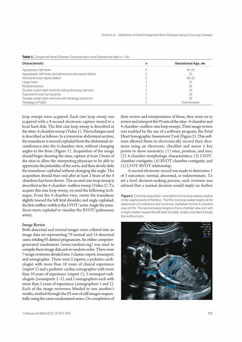
loop sweeps were acquired. Each cine loop sweep wasacquired with a 4-second electronic capture stored to alocal hard disk. The first cine loop sweep is described asthe situs–4-chamber sweep (Video 1). The technique usedis described as follows: In a transverse abdominal section,the transducer is moved cephalad from the abdominal cir-cumference into the 4-chamber view, without changingangles to the fetus (Figure 1). Acquisition of the imageshould begin showing the situs, capture at least 2 beats ofthe situs to allow the interpreting physician to be able toappreciate the pulsatility of the aorta, and then slowly slidethe transducer cephalad without changing the angle. Theacquisition should then end after at least 2 beats of the 4chambers has been shown. The second cine loop sweep isdescribed as the 4-chamber–outflow sweep (Video 2). Toacquire this cine loop sweep, we used the following tech-nique: From the 4-chamber view, rotate the transducerslightly toward the left fetal shoulder and angle cephalad;the first outflow visible is the LVOT/aorta. Angle the trans-ducer more cephalad to visualize the RVOT/pulmonaryartery.
Image ReviewBoth abnormal and normal images were collated into animage data set representing 79 normal and 14 abnormalcases, totaling 93 distinct pregnancies. An online computer-generated randomizer (www.random.org) was used tocompile these image data sets in random order. There were7 image reviewers divided into 3 classes: expert, nonexpert,and sonographer. There were 2 experts, a pediatric cardi-ologist with more than 10 years of clinical experience(expert 2) and a pediatric cardiac sonographer with morethan 10 years of experience (expert 1); 3 nonexpert radi-ologists (nonexperts 1–3), and 2 sonographers each withmore than 5 years of experience (sonographers 1 and 2).Each of the image reviewers, blinded to one another’sresults, worked through the 93 sets of still images sequen-tially using the same randomized series. On completion of
their review and interpretation of these, they went on toreview and interpret the 93 sets of the situs–4-chamber and4-chamber–outflow cine loop sweeps. Their image reviewwas enabled by the use of a software program, the FetalHeart Sonographic Assessment Tool (Figure 2). This soft-ware allowed them to electronically record their deci-sions using an electronic checklist and assess 5 keypoints to show normalcy; (1) situs, position, and size;(2) 4-chamber morphologic characteristics; (3) LVOT-chamber contiguity; (4) RVOT-chamber contiguity; and(5) LVOT-RVOT relationship.
A second electronic record was made to determine 1of 3 outcomes: normal, abnormal, or indeterminate. Toset a level decision-making process, each reviewer wasadvised that a normal decision would imply no further
J Ultrasound Med 2013; 32:973–979 975
Scott et al—Detection of Fetal Congenital Heart Disease Using Cine Loop Sweeps
Table 1. Congenital Heart Disease Characteristics and Gestational Age (n = 14)
Characteristic n Gestational Age, wk
Hypoplastic left heart 4 19–22
Hypoplastic left heart and atrioventricular septal defect 1 21
Atrioventricular septal defect 3 18–22
Large heart 1 21
Rhabdomyoma 1 26
Double-outlet right ventricle with pulmonary stenosis 1 24
Supraventricular tachycardia 1 32
Double-outlet right ventricle with tetralogy spectrum 1 19
Tetralogy of Fallot 1 2nd trimester
Figure 1. Cine loop acquisition: orientation of cine loop sweeps relative
to the sagittal plane of the fetus. The first cine loop sweep begins at the
abdominal circumference and continues cephalad into the 4-chamber
view (4 CH). The second sweep begins in the 4-chamber view and, with
a slight rotation toward the left fetal shoulder, angles cephalad through
the outflow tracts.

imaging or follow-up; an indeterminate decision wouldimply follow-up sonography; and an abnormal decisionwould be referred to the pediatric cardiology laboratoryfor further examination.
Statistical AnalysisThe statistical significance of the detection rates providedby the image reviewers was determined with the McNemarχ2 test for a difference of proportions with a significance levelof P < .001. All other data were analyzed with a 1-way analy-sis of variance. The analysis was performed with Analyse-itsoftware (Analyse-it Software, Ltd, Leeds, England).
Results
Expert reviewer 1 detected 50% of the congenital heartdiseases using the still images and detected all but 1 ofthem using the cine loop sweeps (Table 2). The detec-tion rates varied most within the cine loop sweeps in boththe expert and nonexpert groups. Here, the detectionrates varied by 29% and 28%, respectively. The greatestimprovement in detection between assessment methodswas found in the nonexpert group (nonexpert 3), with animprovement of 64%; the least improvement was foundin the sonographer group (sonographer 2), with no differ-ence observed. For all other reviewers, the detection ratesimproved from 7% to 64%.
The experts detected the most congenital heart dis-eases, with average rates of 43% for the still images and 79%for the cine loop sweeps (Table 3). The nonexpert groupidentified on average only 14% of congenital heart diseasesusing the still images and 52% using the cine loop sweeps.The sonographers identified only 18% using the stillimages and had nearly the same detection rate, 21%, forthe cine loop sweeps. The number of cases with false-positive findings identified overall by the image reviewerswas quite low, with an average of 5.4% observed in the stillimage review compared with 3.9% for the cine loop sweeps.
Discussion
The analysis of the cine loop sweeps resulted in improveddetection rates compared with the still images for expertsand nonexperts of 36% and 38%, respectively. The nearlyequivalent rates of improvement found in both groupssuggest that the value of the cine loop sweeps is equallyapplicable to expert and nonexpert interpreting physicians.Within the nonexpert group, the variations in detectionrates were 14% for the still image review and 28% for thecine loop sweeps. This pattern of increased variation in
detection rates for the cine loop sweeps was also observedin the expert group. In this group, the variation in detectionrates for the still image review was 14%, and the variationfor the cine loop sweeps was 29%. The increased variationbetween the reviewers may be explained by the fact thatthe cine loop sweeps provided considerably more imageinformation for analysis, allowing individual interpretiveskills to be demonstrated to a greater degree than for thestill images. The detection rate for the nonexpert reviewersusing the still images was only 14%. This result is in align-ment with the reality that the still images of the 4-chamberand outflow tract views provide a very minimal amount ofdiagnostic information to the interpreting physician duringthe image review process. However, the expert reviewerswere able to identify 43% of congenital heart diseases usingthe same still images while maintaining a low rate of false-positive identification. This difference suggests that the
Scott et al—Detection of Fetal Congenital Heart Disease Using Cine Loop Sweeps
J Ultrasound Med 2013; 32:973–979976
Figure 2. Fetal Heart Sonographic Assessment Tool. A, Image display,
in which still images or cine loop sweeps are shown. B, Decision-
making tool. A indicates abnormal finding; N, normal finding; and NS,
structure or relationship not shown.
A
B

importance of training and experience can have a substantialimpact on the detection rate of congenital heart disease.
Surprisingly, the sonographers’ improvement in thedetection rate using the cine loop sweeps compared withthe still images was only 3%, and this finding was notconsistent with that of the expert and nonexpert results.The sonographers’ detection rates overall were low: 21%for the cine loop sweeps and 18% for the still images.Compared with the performance of the experts and non-experts, these low detection rates suggest that the inter-pretative expertise of the sonographers was not as refinedas that of the physicians. This finding was not surprisingbecause the role of a sonographer is primarily imageacquisition, initial review, and interpretation, whereas theinterpreting physician’s role is primarily analysis and inter-pretation. The training and experiences for each of thesehealth care professionals reflect these differences in roles.These results indicate that it is essential that the interpret-ing physician have access to real-time sweeps of the heartif the goal is to detect congenital heart disease in routineobstetric screening. Furthermore, the cine loop sweeps can beshared electronically via a secure network connection shouldthe interpreting physician request expert consultation.
The average rates of false-positive identification of con-genital heart disease for all reviewers were 5.4% for the stillimages and 3.9% for the cine loop sweeps. This finding sug-gests that the reviewers understood the potential emotionalimpact and stress that a positive finding would have on theparents and families affected, and they set a relatively highthreshold for a positive finding. This factor might helpexplain why the expert and nonexpert detection rates didnot exceed 80% even with the use of cine loop sweeps.
All 14 abnormal cases were identified on the cine loopsweeps, and 2 cases, a large heart and a hypoplastic left
heart, were identified by all 7 reviewers. In contrast, 2 cases,an atrioventricular septal defect and a double-outlet rightventricle with tetralogy spectrum, were missed by all 7reviewers using the still images. An example is describedbelow in which the still images provided a false impressionof normalcy compared with the cine loop sweeps. To illus-trate this example, a frame was grabbed from the cine loopsweep on a Twoflower 2.0.3 VLC media player (VideoLANOrganization, Paris, France) to show the abnormality.In Figure 3, a normal-looking 4-chamber view is contrastedwith a more abnormal-looking view extracted from thecine loop sweep. This was confirmed as tetralogy of Fallot.Only 1 of the 7 reviewers identified this abnormality on thestill images. These findings further express the need for anassessment in real time.
Although the results of this study uniformly pointtoward the increased value of the cine loop sweeps com-pared with the still images, because of the relatively smallsample of 14 cardiac defects, statistical significance wasachieved in the results for only 2 of the 7 reviewers.Not included in our study, however, was the 3-vessel view,which has been found to be a very effective method ofassessing the ventricular outflow tracts.17 In a pilot study,it was found that most screening centers in our region wereacquiring the 4-chamber and outflow views as 3 still images,and the acquisition of the 3-vessel view increased the acqui-sition time substantially compared with the 2 sweepsdescribed above.18 Our approach is intended to incorpo-rate cine loop sweeps into a routine second-trimester sono-graphic examination with a minimum of additionalacquisition time and sonographer training, and for this rea-son, we chose not to include the 3-vessel view. The abnor-mal cases were acquired in a prospective fashion based onavailability to the researchers; therefore, they do not rep-
J Ultrasound Med 2013; 32:973–979 977
Scott et al—Detection of Fetal Congenital Heart Disease Using Cine Loop Sweeps
Table 2. Detection Rates by Reviewer (n = 14)
Image Type Expert 1a Expert 2 Nonexpert 1 Nonexpert 2 Nonexpert 3a Sonographer 1 Sonographer 2
Still 7 (50) 5 (36) 3 (21) 2 (14) 1 (7) 1 (7) 4 (29)
Sweep 13 (93) 9 (64) 6 (43) 6 (43) 10 (71) 2 (14) 4 (29)
Data are presented as number (percent).aStatistically significant (P < .001).
Table 3. Average Detection Rates by Group (n = 14)
Image Type Experts (2) Nonexperts (3) Sonographers (2) All (7)
Still 6 (43) 2 (14) 2.5 (18) 3.4 (24)
Sweep 11 (79) 7.3 (52) 3 (21) 7.1 (51)
Change +5 (36) +5.3 (38) +0.5 (3) +3.8 (27)
Data are presented as number (percent).
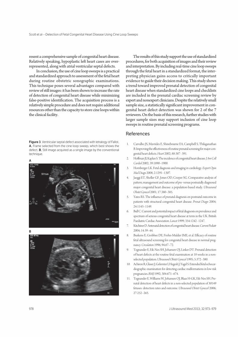
resent a comprehensive sample of congenital heart disease.Relatively speaking, hypoplastic left heart cases are over-represented, along with atrial ventricular septal defects.
In conclusion, the use of cine loop sweeps is a practicaland standardized approach to assessment of the fetal heartduring routine obstetric sonographic examinations.This technique poses several advantages compared withreview of still images: it has been shown to increase the rateof detection of congenital heart disease while minimizingfalse-positive identification. The acquisition process is arelatively simple procedure and does not require additionalresources other than the capacity to store cine loops withinthe clinical facility.
The results of this study support the use of standardizedprocedures, for both acquisition of images and their reviewand interpretation. By including real-time cine loop sweepsthrough the fetal heart in a standardized format, the inter-preting physician gains access to critically importantevidence to guide their decision making. This study showsa trend toward improved prenatal detection of congenitalheart disease when standardized cine loops and checklistsare included in the prenatal cardiac screening review byexpert and nonexpert clinicians. Despite the relatively smallsample size, a statistically significant improvement in con-genital heart defect detection was shown for 2 of the 7reviewers. On the basis of this research, further studies withlarger sample sizes may support inclusion of cine loopsweeps in routine prenatal screening programs.
References
1. Carvalho JS, Mavrides E, Shinebourne EA, Campbell S, ThilaganathanB. Improving the effectiveness of routine prenatal screening for major con-genital heart defects. Heart 2002; 88:387–391.
2. Hoffman JI, Kaplan S. The incidence of congenital heart disease. J Am CollCardiol 2002; 39:1890–1900.
3. Hornberger LK. Fetal diagnosis and imaging in cardiology. Expert OpinMed Diagn 2008; 2:1291–1307.
4. Jaeggi ET, Sholler GF, Jones OD, Cooper SG. Comparative analysis ofpattern, management and outcome of pre- versus postnatally diagnosedmajor congenital heart disease: a population-based study. UltrasoundObstet Gynecol 2001; 17:380–385.
5. Yates RS. The influence of prenatal diagnosis on postnatal outcome inpatients with structural congenital heart disease. Prenat Diagn 2004;24:1143–1149.
6. Bull C. Current and potential impact of fetal diagnosis on prevalence andspectrum of serious congenital heart disease at term in the UK. BritishPaediatric Cardiac Association. Lancet 1999; 354:1242–1247.
7. Kitchiner D. Antenatal detection of congenital heart disease. Current Pediatr2004; 14:39–44.
8. Buskens E, Grobbee DE, Frohn-Mulder IME, et al. Efficacy of routinefetal ultrasound screening for congenital heart disease in normal preg-nancy. Circulation 1996; 94:67–72.
9. Tegnander E, Eik-Nes SH, Johansen OJ, Linker DT. Prenatal detectionof heart defects at the routine fetal examination at 18 weeks in a non-selected population. Ultrasound Obstet Gynecol 1995; 5:372–380.
10. Achiron R, Glaser J, Gelernter I, Hegesh J, Yagel S. Extended fetal echocar-diographic examination for detecting cardiac malformations in low riskpregnancies. BMJ 1992; 304:671–674.
11. Tegnander E, Williams W, Johansen OJ, Blaas H-GK, Eik-Nes SH. Pre-natal detection of heart defects in a non-selected population of 30149fetuses: detection rates and outcome. Ultrasound Obstet Gynecol 2006;27:252–265.
Scott et al—Detection of Fetal Congenital Heart Disease Using Cine Loop Sweeps
J Ultrasound Med 2013; 32:973–979978
Figure 3. Ventricular septal defect associated with tetralogy of Fallot.
A, Frame selected from the cine loop sweep, which best shows the
defect. B, Still image acquired as a single image by the conventional
technique.
A
B

12. Sklansky MS. Prenatal screening for congenital heart disease: a movingproposal. J Ultrasound Med 2007; 26:1–3.
13. Chaoui R. The four-chamber view: four reasons why it seems to fail inscreening for cardiac abnormalities and suggestions to improve detectionrate. Ultrasound Obstet Gynecol 2003; 22:3–10.
14. DeVore G, Falkensammer P, Sklansky MS, Platt LD, Spatio-temporalimage correlation (STIC): new technology for evaluation of the fetalheart. Ultrasound Obstet Gynecol 2003; 22:380–387.
15. McGahan JP, Moon-Grady AJ, Pahwa A, et al. Potential pitfalls and meth-ods of improving in utero diagnosis of transposition of the great arteries,including the baby bird’s beak image. J Ultrasound Med 2007; 26:1499–1510.
16. Yagel S, Cohen SM, Achiron R. Examination of the fetal heart by fiveshort-axis views: a proposed screening method for comprehensive car-diac evaluation. Ultrasound Obstet Gynecol 2001; 17:367–369.
17. Yoo SJ, Lee YH, Kim ES, et al. Three-vessel view of the fetal upper medi-astinum: an easy means of detecting abnormalities of the ventricular out-flow tracts and great arteries during obstetric screening. Ultrasound ObstetGynecol 1997; 9:173–182.
18. Scott T, Swan H, Moran G, et al. Increasing the detection rate of normalfetal cardiac structures: a real-time approach. J Diagn Med Sonography2008; 24:63–71.
J Ultrasound Med 2013; 32:973–979 979
Scott et al—Detection of Fetal Congenital Heart Disease Using Cine Loop Sweeps

http://jdm.sagepub.com/Sonography
Journal of Diagnostic Medical
http://jdm.sagepub.com/content/early/2013/12/20/8756479313517297The online version of this article can be found at:
DOI: 10.1177/8756479313517297
published online 27 December 2013Journal of Diagnostic Medical SonographyTed Edward Scott, Laura Thomas, Keith Edwards, Judy Jones, Hans Swan and Andrew Wessels
Compared With Classroom Instructional MethodsIncreasing Recognition of Fetal Heart Anatomy Using Online Tutorials and Mastery Learning
Published by:
http://www.sagepublications.com
On behalf of:
Society of Diagnostic Medical Sonography
can be found at:Journal of Diagnostic Medical SonographyAdditional services and information for
http://jdm.sagepub.com/cgi/alertsEmail Alerts:
http://jdm.sagepub.com/subscriptionsSubscriptions:
http://www.sagepub.com/journalsReprints.navReprints:
http://www.sagepub.com/journalsPermissions.navPermissions:
What is This?
- Dec 27, 2013OnlineFirst Version of Record >>
at Society of Diagnostic Medical Sonography Member Access on January 6, 2014jdm.sagepub.comDownloaded from at Society of Diagnostic Medical Sonography Member Access on January 6, 2014jdm.sagepub.comDownloaded from

Journal of Diagnostic Medical Sonography201X, Vol XX(X) 1 –8© The Author(s) 2013 Reprints and permissions: sagepub.com/journalsPermissions.navDOI: 10.1177/8756479313517297jdms.sagepub.com
Energizing Education
Introduction
The majority of deaths from congenital defects in child-
hood can be attributed to congenital heart disease (CHD).1
The most common cause of birth defects, CHD is most
frequently reported with an incidence of 0.8% of all live
births.2 Numerous studies have shown, however, that the
prenatal diagnosis of congenital heart disease can improve
clinical outcomes of those affected after birth.3–5 Prenatal
diagnosis of congenital heart disease is critically impor-
tant to ensure optimal management of the pregnancy,
optimal delivery of the baby, and provision of support for
parental decision making. In addition, those at high risk
for CHD may be relieved of additional anxiety and fears
caused just by the possibility of CHD by the confirmation
of normality.6
The role of the sonographer in prenatal diagnosis of
congenital heart disease is to assess the fetal heart in real
time, including the four chambers and whenever possible
the outflow tracts.7 Typically the sonographer will docu-
ment normal or abnormal findings as a preliminary report
with corresponding still images. The interpreting physi-
cian will review the preliminary report and associated
images confirming the determination of a normal, abnor-
mal, or an indeterminate finding.
The formal training for diagnostic medical sonogra-
phers in the detection of congenital heart defects is pro-
vided in didactic form usually within an obstetrics and
gynecology sonography course. The clinical training is
most often provided within a practicum through clinical
517297 JDMXXX10.1177/8756479313517297Journal of Diagnostic Medical SonographyScott et al.research-article2013
1Medical Radiation Sciences, Mohawk College, Hamilton, ON, Canada2Department of Computer Science, University of Hawaii, Hilo, HI, USA3Pediatric Cardiology, London Health Sciences, London, ON, Canada4Dentistry and Health Sciences, Charles Sturt University, Wagga Wagga, NSW, Australia
Corresponding Author:
Ted Edward Scott, MAppSc, Mohawk-McMaster Institute for Applied Health Sciences, 1400 Main Street West, Hamilton Ontario L8S 1C7, Canada. Email: [email protected]
Increasing Recognition of Fetal Heart Anatomy Using Online Tutorials and Mastery Learning Compared With Classroom Instructional Methods
Ted Edward Scott, MAppSc1, Laura Thomas, RDMS, CRGS1, Keith Edwards, PhD2, Judy Jones, BS, RDMS, RDCS3, Hans Swan, PhD4, and Andrew Wessels2
Abstract
Assessment of the fetal heart is a challenging part of any routine obstetrical sonogram. The practice of teaching and learning this skill demands expertise in the visualization and interpretation of normal sonographic appearances of the fetal heart. Recognition of the pathological features associated with commonly seen congenital heart diseases is also very important. In the study reported here, students in an obstetrical sonography course were randomly assigned to two groups. The students in the control group (classroom instruction) on average assessed correctly all five anatomic cardiac features but determined the normal or abnormal status of only 19% of cases, 4.7of 25 ± 4.4 SD. The experimental group (online tutorials) correctly identified 39% of cases, 9.9 of 25 ± 2.7 SD, P < .01. The average score in the experimental group was greater than 84% of the students in the control group, a one sigma effect. These data support the conclusion that students learning to assess the fetal heart for the presence of congenital heart disease using a series of online tutorials and practice exercises compared with students receiving conventional classroom instruction demonstrated an improved ability to correctly identify normal and abnormal fetal heart structures.
Keywords
online education, tutorial, mastery learning, fetal heart assessment, instructional technology, hybrid education
at Society of Diagnostic Medical Sonography Member Access on January 6, 2014jdm.sagepub.comDownloaded from

2 Journal of Diagnostic Medical Sonography XX(X)
facilities linked to the college program. Imaging and
assessing the fetal heart is a challenging task for sonogra-
phy students. Acquiring images and assessing the fetal
heart is a complex task demanding an integrated under-
standing of the anatomy and physiology of the fetal heart
and image recognition skills of rapidly moving structures
such as the outflow tracts on the order of 2 to 6 mm in
diameter.8 Unlike abdominal, pelvic, or other sonography
exams, students usually do not have any significant
opportunity to practice obstetrical sonography in their
college imaging laboratories. It is also difficult for educa-
tion programs to acquire a robust library of sample cases
representative of the broad diversity of congenital heart
disease.
Addressing complex learning objectives that demand
significant skill development from the learner is a chal-
lenge for the traditional classroom setting. Skill develop-
ment demands significant formative assessment and
structured feedback, which can be difficult to provide to a
group in a classroom environment. For teachers at all lev-
els of education, it remains a challenge to provide cus-
tomized instruction and high-quality feedback to
individuals in large classes.
In a seminal article in 1984, Benjamin Bloom articu-
lated a number of methods to address the two-sigma prob-
lem.9 Bloom found that when using both mastery learning
and tutorial-based instruction as compared with conven-
tional classroom instruction, students in the tutorial and
mastery learning groups on average increased their
achievement levels by two-sigma (two standard devia-
tions). In other words, the average student in the tutorial
and mastery group outperformed 98% of the students in
the classroom group. Mastery-based instruction proposes
that learning should provide formative evaluation with
guided or corrective feedback such that students achieve
mastery of all of the learning objectives.10 Tutorial-based
instruction assumes that students are working regularly
with a good tutor, in a face-to-face mode. Two forms of
instruction have emerged to address the opportunity for
learning using the theory of the two-sigma problem.11
These forms include a method of automated feedback
based on student performance during practice exercises,
and mastery learning, where students work at their own
pace to achieve mastery of learning.11,12
In the realm of digitally enhanced education that exists
in many postsecondary institutions today, there exists a
significant opportunity to provide students with opportu-
nities to leverage the educational theories and applica-
tions established by Bloom. Some very popular solutions
have emerged from outside the traditional postsecondary
sector. Khan Academy is a very popular electronic plat-
form for learning mathematics based on tutorial-based
instruction and mastery learning principles.13 Students
are able to traverse the entire spectrum of mathematics
necessary to achieve successful preparation for entry into
postsecondary institutions. Besides the pedagogical
advantages associated with the two-sigma problem, the
use of electronic learning tutorials and practice exercises
provide numerous advantages compared with traditional
classroom learning. These advantages include:
self-paced progress through the learning
objectives,
opportunity to repeat tutorials anytime anywhere
without reliance on a human tutor,
ability to access experts on niche topics,
expand the learning opportunity to a global audi-
ence in any language,
ensure a consistent and high-quality learning
opportunity,
provide the tutorial and learning tool designers
with valuable quantitative feedback regarding the
learning experience that can be used to improve
future iterations of the tutorials and tools.
The objective of this study was to determine if a series
of online tutorials and practice exercises using a stan-
dardized assessment of the fetal heart in real time would
be a more effective approach to be used by students when
learning how to identify normal fetal heart structures and
congenital heart disease. The researchers addressed the
following hypothesis
Hypothesis 1: Training students to use a standardized
assessment technique using tutorials and practice
exercises online, compared with traditional classroom
instruction, will increase the students’ ability to iden-
tify normal fetal heart structures and congenital heart
disease.
Materials and Methods
This study was approved by the Charles Sturt University
Ethics in Human Research Committee, the McMaster
University Research Ethics Board (MREB), and the
Mohawk College Research Ethics Board prior to initia-
tion. The participants in this study were drawn from the
Medical Radiation Science Obstetrical and Gynecologic
Ultrasonography 3 class. These sonography students
were in the third year of study within a collaborative four-
year, Mohawk-McMaster Medical Radiation Sciences
program. All 33 students in the class volunteered to par-
ticipate in this study.
Students were randomly divided into two groups.
There were 17 students assigned to the classroom learn-
ing (control) group, and 12 of these students completed
all of the learning activities. Of the 16 originally assigned
to the online learning (experimental) group, 15 students
at Society of Diagnostic Medical Sonography Member Access on January 6, 2014jdm.sagepub.comDownloaded from
Scott et al. 3
completed all of the learning activities. The 27 students
who completed all of the learning activities formed the
basis for final data analysis. Three learning objectives for
the experimental and control groups were defined as
follows:
1. increased ability to identify normal fetal cardiac
structures,
2. increased ability to identify abnormal fetal car-
diac structures,
3. increased ability to identify abnormal cases.
Students assigned to the control group were not per-
mitted access to the online tutorials and practice exer-
cises. Students assigned to the experimental group were
not permitted to attend the four face-to-face classes and
single online tutorial with the course instructor. The
online examination was administered within two weeks
of the onset of the experiment.
Students enrolled in the control group participated in
four classroom lecture periods of 50 minutes each and
one online tutorial. The lectures reviewed the anatomy of
the fetal heart, the fetal blood flow, and the sonographic
views used to image the heart. They also included real-
time cineloops with tips and suggestions on how to iden-
tify a normal heart. Lectures included review of some of
the more common congenital heart abnormalities using
real-time cineloops. The tutorial was delivered online
with labeled anatomy images, with focused questions
pertaining to the diagrams. The time for instruction was
approximately 240 minutes, with an allowance for inde-
pendent study time of 90 minutes, for a total of 330 min-
utes of time committed to learning.
The experimental group was provided with the “Find
It First” tool consisting of 13 brief online tutorials in the
use of a five-step method of assessment of the fetal heart
prepared by the study investigator, amounting to approxi-
mately 75 minutes of instruction in total. Students com-
pleted practice exercises online as formative assessment
with electronic feedback that required an additional 150
minutes of learning time. The total time committed to
learning for the experimental group was approximately
225 minutes.
The students were also provided practice cases for
each step of the technique, and these exercises included
immediate electronic feedback. Students were required
to demonstrate mastery of each step before continuing
to the next step of assessment. A screen shot depicting
the user interface for the practice exercises provided by
module two is provided in Figure 1. Students worked
through five online modules representing five steps of
analysis considering the anatomic features and relation-
ships as follows:
1. cardiac situs, axis, and size;
2. chamber morphology and function;
3. left ventricular outflow tract (LVOT) continuity
with the left ventricle;
4. right ventricular outflow tract (RVOT) continuity
with the right ventricle;
5. cross-over of outflow tracts at their origins.
Each of these features and relationships could be
defined as normal, abnormal, or not seen. Each module
consisted of a brief tutorial to provide examples and
instruction on how to assess the relevant anatomic fea-
ture. Following the review of each tutorial the students
were required to complete 17 sample cases similar to the
example depicted in Figure 1. At the end of every series,
all of the missed cases were compiled, and the learner
reassessed the missed cases until all 17 cases were accu-
rately reviewed. The student could advance to the next
module only after successfully demonstrating the ability
to analyze the anatomic features covered by the module.
As students advanced through the modules, additional
steps were added to the analysis until the student reached
the fifth module where all five steps and corresponding
features were examined, representing a review and analy-
sis of a total of 85 cases.
In order to assess the performance of the students in
both groups an online quiz was developed. Each student
was provided with a unique and secure user identification
and password. Students’ ability to identify normal and
abnormal structures was measured by their review of 25
cases of fetal cardiac anatomy. A randomized sequence of
10 abnormal and 15 normal cases were presented. Each
case consisted of two cine-loop sweeps through the fetal
heart. The situs to four chamber sweep began in the trans-
verse abdominal plane at the level of the stomach and
ended at the four chamber view. The four chamber to out-
flows sweep began in the four chamber view and swept
through the ventricular outflow tracts. Students in both
groups completed the online examination within two
weeks of completing the classroom or online instruction.
As described in Figure 2, students were required to identify
five anatomic features for each case and determine whether
that case was normal, abnormal, or indeterminate. A com-
pletely accurate assessment was one that scored each fea-
ture accurately and correctly identified the case as normal,
abnormal, or indeterminate. The decision framework was
standardized for the students using a protocol that defined
the following outcomes: a normal result implies no further
assessment, an abnormal determination will result in a
referral to a pediatric cardiologist, and an indeterminate
finding will result in a follow-up reassessment in the
obstetric screening center. This evaluation required
approximately one hour for the students to complete.
at Society of Diagnostic Medical Sonography Member Access on January 6, 2014jdm.sagepub.comDownloaded from

4 Journal of Diagnostic Medical Sonography XX(X)
Data Analysis
The chi square test was performed to evaluate the statisti-
cal significance of the results of the image analysis. The
null hypothesis for this test was defined by the following
statement: “After using the computerized learning system (experimental group) or gaining instruction in a tradi-tional face to face classroom environment (control group), there will be no statistical difference in students’ ability to identify normal and abnormal fetal heart
Figure 1. A screen shot depicting the user interface for the practice exercises provided by module two of the “Find It First” tool. During formative assessment the feedback is provided immediately. All cases that are incorrect must be repeated after the first practice series is completed before continuing to the next learning module.
at Society of Diagnostic Medical Sonography Member Access on January 6, 2014jdm.sagepub.comDownloaded from

Scott et al. 5
structures.” Differences on the multiple choice evalua-
tions and the confidence levels were tested using the two-
tailed t-test for statistical significance.
Results
The students were evaluated on their ability to correctly
assign one of three conditions, normal, abnormal, or not
seen, to five unique anatomic criteria and relationships
and correctly define the case as normal, abnormal, or inde-
terminate. For each case, to be scored as completely cor-
rect, all five anatomic criteria and the assignment of
normal, abnormal, or indeterminate had to be correct,
which required a total of six correct decisions for each
case. The students in the control group on average assessed
correctly all five anatomic features but determined the
normalcy or abnormal status of only 19% of cases, 4.7 of
25 ± 4.4 SD compared with the experimental group mean
score of 39% of correctly identified cases, 9.9 of 25 ± 2.7
SD (P < .01). In terms of mean performance difference
between the two groups there was a one-sigma (one stan-
dard deviation) effect. This can be interpreted to mean that
the average score in the experimental group was greater
than 84% of the students in the control group. As seen in
Figure 3, the experimental group outperformed the control
group in every category of assessment.
The abnormal cases depicted in Table 1 represent a
typical range of serious congenital heart disease with
one or more abnormal anatomical features evident in the
Figure 2. Screen shot of an example taken from the final quiz. The final quiz includes the assessment of all five indicators and the final decision.
Figure 3. Five points of assessment of the fetal heart and the percentage of complete examinations correctly identified as normal, abnormal, or not seen. Students in the experimental group (Series 1) used the online tool compared with students in the control group (Series 2) who participated in classroom learning.
at Society of Diagnostic Medical Sonography Member Access on January 6, 2014jdm.sagepub.comDownloaded from

6 Journal of Diagnostic Medical Sonography XX(X)
cine-loops provided to the students for review. As shown
in the table, the experimental group outperformed the
control group in 7 of 10 abnormal cases (P < .01).
Specifically in five abnormal cases, D-TGA, single ven-
tricles, large heart, HLHS, and truncus arteriosus, a
greater than two-thirds majority of experimental stu-
dents identified these cases whereas the control group
only identified 1 case, truncus arteriosus by a two-thirds
majority of students (P < .01). The experimental group
performed particularly poorly in detecting Ebstein’s
anomaly, displacement of the septal leaflet of the tricus-
pid valve typically causing regurgitation, with only a
7% detection rate compared with 33% for the control
group.
Discussion
The strategy adopted by most sonography programs for
teaching students to identify the presence of pathology
during abdominal or pelvic examinations is typically
based on providing students with a significant amount of
supervised practice and exposure to the normal sono-
graphic appearances of anatomic structures. In teaching
students to identify the presence of pathology, sonogra-
phy programs begin with a focus on the normal anatomi-
cal appearance of the structure. In this way, students
develop expectations in terms of the visualization of rela-
tionships and structures that establish normalcy. Once
understanding of normalcy is firmly grasped and rooted
in the student’s experience, most programs and courses
attempt to provide a framework for pathology such that
students can focus their attention on links between the
patient’s history and the probable associated pathology.
For example, a history of right upper quadrant pain and
elevated liver enzymes would draw the sonographer’s
attention to the liver and biliary tree.
To put this educational paradigm in simpler terms, if
the signal is the presence of pathology and the factors
contributing to missing the detection of pathology are
defined as noise, a sonographer can think about enhanc-
ing the signal to noise ratio in order to identify pathology
more frequently. To simplify this analogy, if the pathol-
ogy is the signal, and the factors that influence missed
detection of a pathology are the noise, then the sonogra-
pher’s task is to enhance the signal to noise ratio in an
effort to increase the detection rate of pathology. A deeper
understanding of frequent sonographic appearances of
pathology relative to patient history will strengthen the
signal. The sonographer’s ability to optimize image qual-
ity, perform a complete real-time interrogation, and apply
systematic examination techniques will help to reduce the
noise. In this way, the presence of pathology (signal) can
be enhanced during the sonographic examination.
Part of the challenge for detecting congenital heart dis-
ease in utero is the fact that most often it is found in the
low-risk population.14 This makes it more difficult to
detect because one cannot usually rely on clues from the
patient history (signal) to focus the examination.
Furthermore, as a screening examination, the inherent
bias is an expectation of normalcy. In this setting the abil-
ity to reduce noise to enhance what is a weak signal
becomes a very important consideration. Standardizing
imaging procedures and assessment protocols represents
an important consideration in this regard and one that was
adopted for this learning tool.
In addition to the two-sigma pedagogy, the online
learning tutorials and practice exercises focused nearly
exclusively on exposing students in the experimental
group to as many normal and abnormal cases as possible
in a step-wise learning methodology. The focus for the
learner was on practicing the decision making for each
step in each module until mastery of the concept was
Table 1. Detection rate by group according to the individual cases of congenital heart disease.
Experimental Group, n = 15
Control Group, n = 12 Difference
Case CHD, n = 10 Detected (%) Detected (%) %
1 Large heart 13 (93) 7 (58) 352 Truncus arteriosus 12 (80) 9 (75) 53 Hypoplastic left heart 12 (80) 6 (50) 304 D-TGA 11 (73) 7 (58) 155 Single ventricle 10 (67) 6 (50) 176 AVSD 7 (47) 5 (42) 57 Tetralogy of fallot 7 (47) 2 (17) 308 Hypoplastic left heart 7 (47) 7 (58) –119 Tetralogy of fallot 3 (20) 4 (33) –13
10 Ebstein’s anomaly 1 (7) 4 (33) –26 Total 83 (55) 57 (48)
at Society of Diagnostic Medical Sonography Member Access on January 6, 2014jdm.sagepub.comDownloaded from

Scott et al. 7
achieved. In the classroom learning group, a broader
focus was taken and students were given more contextual
information related to the normal and abnormal heart.
There was less emphasis on skill development with feed-
back in the classroom setting.
Bloom’s two-sigma theory was not replicated in this
study; however, a one-sigma improvement was found and
this represents a twofold increase in the percentage of
cases accurately classified by students in the experimen-
tal group, 39% compared with 19% in the control group.
Further, the experimental group did not benefit from any
face-to-face learning opportunities. This might also
explain why a two-sigma effect was not achieved. The
ability to correctly classify anatomic features and rela-
tionships was consistently demonstrated across all five
categories. This suggests that students acquired improved
image interpretation skills in the experimental group as
compared to the control group.
The majority of congenital heart defects (7/10) were
identified with greater frequency by the experimental
group compared with the control group. In addition, the
experimental group was much more consistent as a group
in identifying 5 of 10 defects with a greater than two-
thirds majority of the group compared with the control
group, which only achieved a two-thirds majority correct
identification for one abnormal case. These results sug-
gest that the use of mastery-based instruction in the form
of practice exercises framed in a step-wise protocol might
lead to a more consistent development of image interpre-
tation skills. The particularly poor performance of the
experimental group with respect to the Ebstein’s anomaly
may be due to the fact that it was not included as an
example during the online tutorials and it represents the
singular isolated abnormality of an apical displacement
of the tricuspid valve. The online tutorials emphasized
more frequently encountered structural abnormalities,
including presence of ventricular septal defects, disconti-
nuity of the outflow tracts, disparity in chamber size,
heart size and angulation, among other factors. Future
versions of this learning tool can be expanded to correct
this oversight.
This study was limited in a number of ways. The
number of participants was relatively small and repre-
sentative of a single cohort of sonography students. Five
of the 17 students in the control group did not complete
the study while only one of the students in the experi-
mental group did not complete the study. The students in
the experimental group may have persisted through the
final evaluation and completed the study in a greater
percentage due to their familiarity with the “Find It
First” learning tool. Some of the students from the class-
room (control) group may have found the final evalua-
tion provided on the “Find It First” tool to be unfamiliar
and this may have been enough to deter them from com-
pleting the study. The online learning tutorials and prac-
tice exercises were first generation and improvements in
content and technical terms can be achieved in future
generations of this application. Finally, the performance
of the students in the control (classroom) group was
dependent on a single instructor and this could bias the
outcome of the student’s learning in either a positive or
negative direction.
Conclusion
In this study it was shown that students’ ability to cor-
rectly classify normal and abnormal fetal cardiac anat-
omy can be improved using principles of mastery learning
and tutorial-based instruction in an online format. Further,
the opportunity to exploit the benefits of online learning
may provide students with greater access to high-quality
learning opportunities. Students may benefit from a self-
paced approach to learning with the opportunity to review
tutorial sessions anytime, anywhere as much as desired.
Assessment of the fetal heart for congenital heart dis-
ease is a challenging task and one that demands signifi-
cant educational expertise in the domains of cardiovascular
and obstetrical practice areas. For topics that demand
complex analysis and clinical expertise at the intersection
of clinical skills such as fetal cardiac assessment, an
online tutorial-based approach may prove to be a very
effective means of addressing this teaching and learning
challenge.
In this study, students using the online tutorial and
practice exercises had improved performance by one
standard deviation compared with students using tradi-
tional classroom-based learning methods. Further exami-
nation of this approach, including quantitative analysis of
the user experience, may identify opportunities to refine
and improve this approach to learning how to assess the
fetal heart. Future studies may demonstrate benefits to
other stakeholders including practicing clinical sonogra-
phers and interpreting physicians. It may prove worth-
while for other educational applications to be developed
using this methodology as well, particularly those areas
of sonographic practice that are not fully addressed
through traditional curriculum. Examples where online
tutorial-based instruction might be developed include
musculoskeletal imaging, volume imaging, and imaging
of the bowel.
Acknowledgments
The authors would like to acknowledge the efforts of University
of Hawaii at Hilo Computer Sciences 460 students, Joel Cook,
Jacob Shon, Kylie Gonsalves, and Jermaine Vitales for their
development of the “Find It First” learning application.
at Society of Diagnostic Medical Sonography Member Access on January 6, 2014jdm.sagepub.comDownloaded from
8 Journal of Diagnostic Medical Sonography XX(X)
Declaration of Conflicting Interests
The authors declared no potential conflicts of interest with
respect to the research, authorship, and/or publication of this
article.
Funding
The authors received no financial support for the research,
authorship, and/or publication of this article.
References
1. Gilboa SM, Salemi JL, Nembhard WN, et al: Mortality
resulting from congenital heart disease among children
and adults in the United States, 1999 to 2006. Circulation
2010;122(22):2254–63.
2. Dolk H, Loane M, Garne E. Congenital heart defects in
Europe: prevalence and perinatal mortality, 2000 to 2005.
Circulation 2011;123(8):841–849.
3. Fuchs IB, Müller H, Abdul-Khaliq H, et al: Immediate and
long-term outcomes in children with prenatal diagnosis
of selected isolated congenital heart defects. Ultrasound Obstet Gyn 2007;29(1):38–43.
4. Cass DL. Impact of prenatal diagnosis and therapy on neonatal
surgery. Sem Fetal Neonatal Med 2011;16(3):130–138.
5. Brown KL, Ridout D a, Hoskote a, et al: Delayed diag-
nosis of congenital heart disease worsens preoperative
condition and outcome of surgery in neonates. Heart 2006;92(9):1298–1302.
6. Allan LD, Huggon IC. Counselling following a diagnosis of
congenital heart disease. Prenatal Diag 2004;24(13):1136–
1142.
7. Considerations G. Cardiac screening examination of
the fetus: guidelines for performing the “basic” and
“extended basic” cardiac scan. Ultrasound Obstet Gyn
2006;27(1):107–113.
8. Allan L. Technique of fetal echocardiography. Ped Cardiology 2004;25(3):223–233.
9. Bloom BS. The search for methods of group instruc-
tion as effective as one-to-one tutoring. Educ Leadership
1984;41(8):4-17.
10. Gagné RM. Mastery learning and instructional design.
Perform Improvement Quart 1988;1:7-18.
11. Corbett A. Cognitive computer tutors: solving the two-
sigma problem. In: Bauer M, Gmytrasiewicz PJ, Vassileva
J (eds): User Modeling. Berlin, Springer-Verlag, 2001, pp.
137–147.
12. Svinicki MD. New directions in learning and motivation.
New Directions Teach Learn 1999;1999:5–27.
13. Khan S. The One World Schoolhouse: Education Reimagined. London, Hodder & Stoughton Ltd, 2012.
14. Sharland G. Routine fetal cardiac screening: what
are we doing and what should we do? Prenatal Diag
2004;24(13):1123–1129.
at Society of Diagnostic Medical Sonography Member Access on January 6, 2014jdm.sagepub.comDownloaded from


![Hospital-based Birth Defects · PDF fileHospital-based birth defects surveillance: ... Birth defects are one type of congenital conditions. [Slides 5-6] ... • Congenital malformations:](https://img.pdfslide.us/doc/110x75/5ab1551c7f8b9a7e1d8c4658/hospital-based-birth-defects-birth-defects-surveillance-birth-defects-are-one.jpg)