Embed Size (px)
Citation preview
Original Article PROGRESS in MEDICAL PHYSICS 27(3) Sept 2016httpdxdoiorg1014316pmp2016273105
pISSN 2508-4445 eISSN 2508-4453
- 105 -
This research was financially supported by the Ministry of Trade
Industry amp Energy (MOTIE) Korea Institute for Advancement of Tech-
nology (KIAT) and Gangwon Institute for Regional Program Evaluation
(GWIRPE) through the Economic and Regional Cooperation Industry
Following are results of a study on the Leades INdustry-university
Cooperation Project supported by the Ministry of Education (MOE)
Received 7 July 2016 Revised 21 September 2016 Accepted 22
September 2016
Correspondence Hee-Joung Kim (hjk1yonseiackr)
Tel 82-33-760-2983 Fax 82-33-760-2562cc This is an Open-Access article distributed under the terms of the Creative Commons
Attribution Non-Commercial License (httpcreativecommonsorglicensesby-nc40) which
permits unrestricted non-commercial use distribution and reproduction in any medium
provided the original work is properly cited
Improvement of Analytic Reconstruction Algorithms Using a Sinogram Interpolation Method for Sparse-angular Sampling
with a Photon-counting Detector
Dohyeon Kim Byungdu Jodagger Su-Jin Parkdagger Hyemi Kimdagger Hee-Joung Kimdagger
Departments of Radiation Convergence Engineering daggerRadiological Science College of Health Science Yonsei University Wonju Korea
Sparse angular sampling has been studied recently owing to its potential to decrease the radiation exposure from
computed tomography (CT) In this study we investigated the analytic reconstruction algorithm in sparse angular
sampling using the sinogram interpolation method for improving image quality and computation speed A
prototype of the spectral CT system which has a 64-pixel Cadmium Zinc Telluride (CZT)-based photon-counting
detector was used The source-to-detector distance and the source-to-center of rotation distance were 1200
and 1015 mm respectively Two energy bins (23sim33 keV and 34sim44 keV) were set to obtain two reconstruction
images We used a PMMA phantom with height and radius of 500 mm and 175 mm respectively The phantom
contained iodine gadolinium calcification and lipid The Feld-kamp-Davis-Kress (FDK) with the sinogram
interpolation method and Maximum Likelihood Expectation Maximization (MLEM) algorithm were used to
reconstruct the images We evaluated the signal-to-noise ratio (SNR) of the materials The SNRs of iodine
calcification and liquid lipid were increased by 16703 15793 and 4177 respectively with the 23sim33
keV energy bin using the sinogram interpolation method The SNRs of iodine calcification and liquid state lipid
were also increased by 10701 1358 and 2739 respectively with the 34sim44 keV energy bin using the
sinogram interpolation method Although the FDK algorithm with the sinogram interpolation did not produce better
results than the MLEM algorithm it did result in comparable image quality to that of the MLEM algorithm We
believe that the sinogram interpolation method can be applied in various reconstruction studies using the analytic
reconstruction algorithm Therefore the sinogram interpolation method can improve the image quality in
sparse-angular sampling and be applied to CT applications985103985103985103985103985103985103985103985103985103985103985103985103985103985103985103985103985103985103985103985103Key Words Sinogram interpolation Computed tomography (CT) Image reconstruction Low-dose Photon-
counting detector
Introduction
Computed tomography (CT) is widely used in the medical
field Recently sparse angular sampling in CT image re-
construction has been widely studied because it reduces patient
radiation exposure and scanning time12) Sparse-angular re-
construction does not offer high spatial resolution and it gen-
erates streak artifacts caused by inadequate projection data
However it offers reasonable image quality which extends the
use of CT imaging to applications such as radiation therapy
for treatment planning or industrial applications Various re-
construction algorithms can be used to acquire reconstruction
images in sparse tomographic imaging4) In sparse-angle CT
views the iterative reconstruction algorithm has been ex-
tensively studied and developed5) However long reconstruct-
ing time makes it difficult to clinically apply the iterative re-
Dohyeon Kim et alImprovement of Analytic Reconstruction Algorithms Using a Sinogram Interpolation Method for Sparse-angular Sampling with a Photon-counting Detector
- 106 -
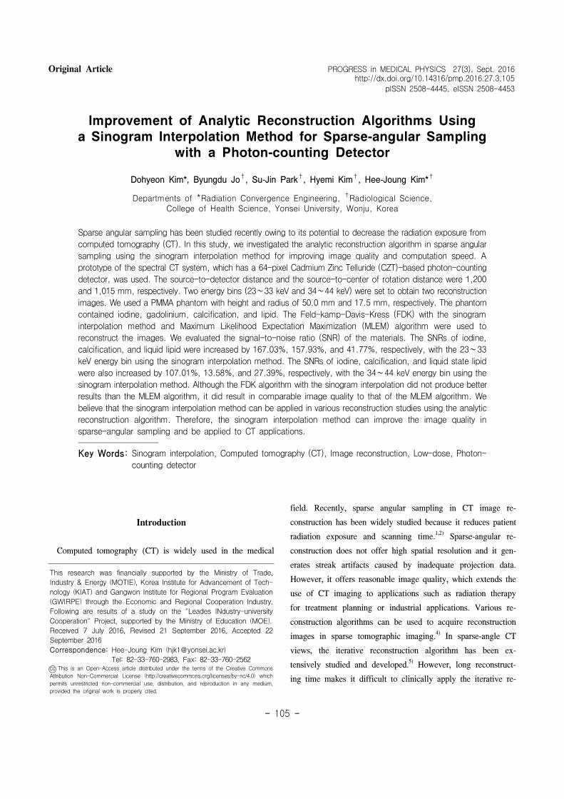
Fig 1 Flowchart of the proposed
sinogram interpolation method
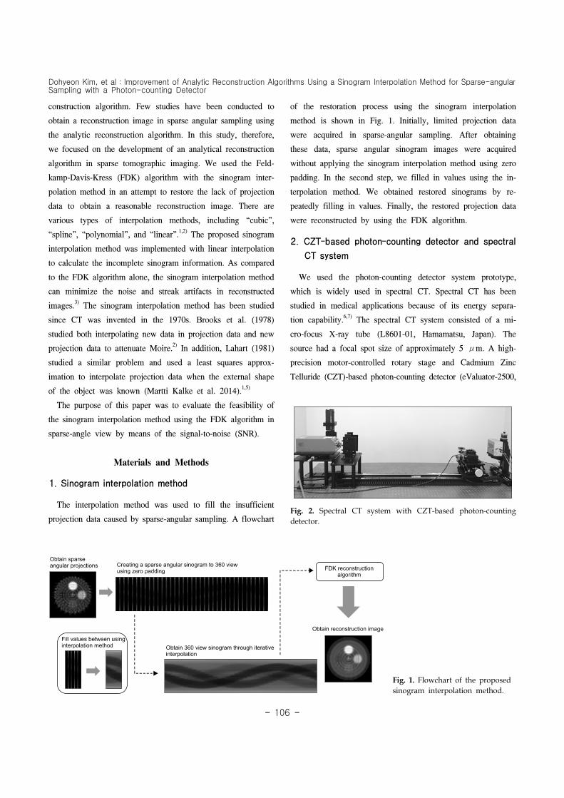
Fig 2 Spectral CT system with CZT-based photon-counting
detector
construction algorithm Few studies have been conducted to
obtain a reconstruction image in sparse angular sampling using
the analytic reconstruction algorithm In this study therefore
we focused on the development of an analytical reconstruction
algorithm in sparse tomographic imaging We used the Feld-
kamp-Davis-Kress (FDK) algorithm with the sinogram inter-
polation method in an attempt to restore the lack of projection
data to obtain a reasonable reconstruction image There are
various types of interpolation methods including ldquocubicrdquo
ldquosplinerdquo ldquopolynomialrdquo and ldquolinearrdquo12) The proposed sinogram
interpolation method was implemented with linear interpolation
to calculate the incomplete sinogram information As compared
to the FDK algorithm alone the sinogram interpolation method
can minimize the noise and streak artifacts in reconstructed
images3) The sinogram interpolation method has been studied
since CT was invented in the 1970s Brooks et al (1978)
studied both interpolating new data in projection data and new
projection data to attenuate Moire2) In addition Lahart (1981)
studied a similar problem and used a least squares approx-
imation to interpolate projection data when the external shape
of the object was known (Martti Kalke et al 2014)15)
The purpose of this paper was to evaluate the feasibility of
the sinogram interpolation method using the FDK algorithm in
sparse-angle view by means of the signal-to-noise (SNR)
Materials and Methods
1 Sinogram interpolation method
The interpolation method was used to fill the insufficient
projection data caused by sparse-angular sampling A flowchart
of the restoration process using the sinogram interpolation
method is shown in Fig 1 Initially limited projection data
were acquired in sparse-angular sampling After obtaining
these data sparse angular sinogram images were acquired
without applying the sinogram interpolation method using zero
padding In the second step we filled in values using the in-
terpolation method We obtained restored sinograms by re-
peatedly filling in values Finally the restored projection data
were reconstructed by using the FDK algorithm
2 CZT-based photon-counting detector and spectral
CT system
We used the photon-counting detector system prototype
which is widely used in spectral CT Spectral CT has been
studied in medical applications because of its energy separa-
tion capability67) The spectral CT system consisted of a mi-
cro-focus X-ray tube (L8601-01 Hamamatsu Japan) The
source had a focal spot size of approximately 5 μm A high-
precision motor-controlled rotary stage and Cadmium Zinc
Telluride (CZT)-based photon-counting detector (eValuator-2500
PROGRESS in MEDICAL PHYSICS Vol 27 No 3 September 2016
- 107 -
Fig 3 Illustration of the PMMA
phantom containing four materials
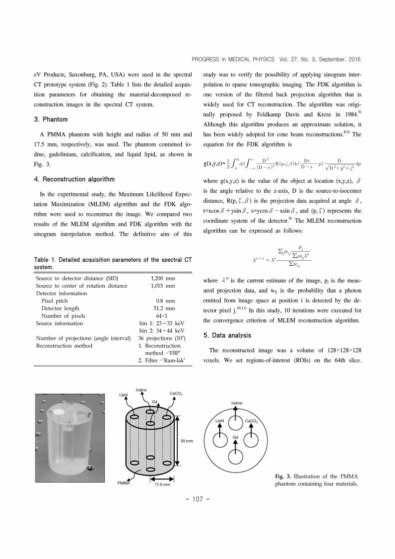
Table 1 Detailed acquisition parameters of the spectral CT
system
Source to detector distance (SID) 1200 mm
Source to center of rotation distance 1015 mm
Detector information
Pixel pitch 08 mm
Detector length 512 mm
Number of pixels 64times1
Source information bin 1 23sim33 keV
bin 2 34sim44 keV
Number of projections (angle interval) 36 projections (10o)
Reconstruction method 1 Reconstruction
method ndashlsquoFBPrsquo
2 Filter ndashlsquoRam-lakrsquo
eV Products Saxonburg PA USA) were used in the spectral
CT prototype system (Fig 2) Table 1 lists the detailed acquis-
ition parameters for obtaining the material-decomposed re-
construction images in the spectral CT system
3 Phantom
A PMMA phantom with height and radius of 50 mm and
175 mm respectively was used The phantom contained io-
dine gadolinium calcification and liquid lipid as shown in
Fig 3
4 Reconstruction algorithm
In the experimental study the Maximum Likelihood Expec-
tation Maximization (MLEM) algorithm and the FDK algo-
rithm were used to reconstruct the image We compared two
results of the MLEM algorithm and FDK algorithm with the
sinogram interpolation method The definitive aim of this
study was to verify the possibility of applying sinogram inter-
polation to sparse tomographic imaging The FDK algorithm is
one version of the filtered back projection algorithm that is
widely used for CT reconstruction The algorithm was origi-
nally proposed by Feldkamp Davis and Kress in 19849)
Although this algorithm produces an approximate solution it
has been widely adopted for cone beam reconstructions89) The
equation for the FDK algorithm is
g(xyz)=
infin
infin
where g(xyz) is the value of the object at location (xyz) β
is the angle relative to the z-axis D is the source-to-isocenter
distance R(pζβ) is the projection data acquired at angle β
t=xcosβ+ysinβ s=ycosβminusxsinβ and (pζ) represents the
coordinate system of the detector8) The MLEM reconstruction
algorithm can be expressed as follows
sumsumsum
where λn is the current estimate of the image pj is the meas-
ured projection data and wij is the probability that a photon
emitted from image space at position i is detected by the de-
tector pixel j1011) In this study 10 iterations were executed for
the convergence criterion of MLEM reconstruction algorithm
5 Data analysis
The reconstructed image was a volume of 128times128times128
voxels We set regions-of-interest (ROIs) on the 64th slice
Dohyeon Kim et alImprovement of Analytic Reconstruction Algorithms Using a Sinogram Interpolation Method for Sparse-angular Sampling with a Photon-counting Detector
- 108 -
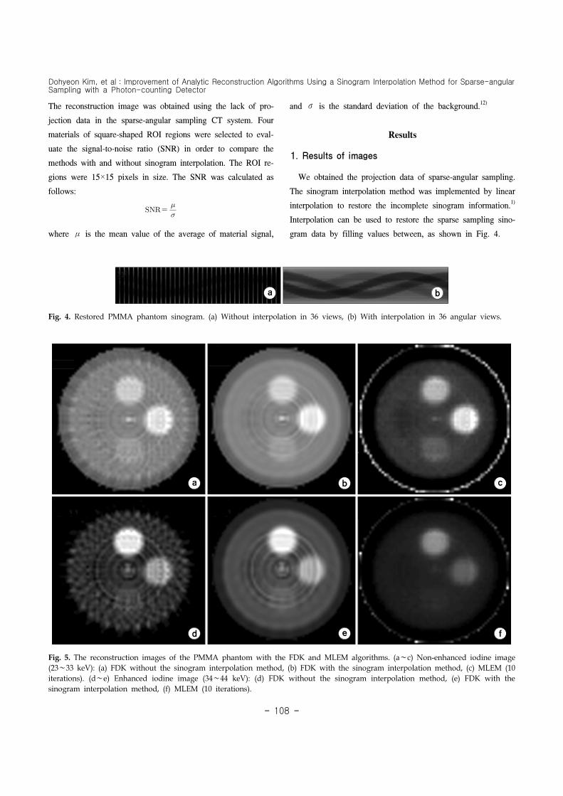
Fig 5 The reconstruction images of the PMMA phantom with the FDK and MLEM algorithms (asimc) Non-enhanced iodine image
(23sim33 keV) (a) FDK without the sinogram interpolation method (b) FDK with the sinogram interpolation method (c) MLEM (10
iterations) (dsime) Enhanced iodine image (34sim44 keV) (d) FDK without the sinogram interpolation method (e) FDK with the
sinogram interpolation method (f) MLEM (10 iterations)
Fig 4 Restored PMMA phantom sinogram (a) Without interpolation in 36 views (b) With interpolation in 36 angular views
The reconstruction image was obtained using the lack of pro-
jection data in the sparse-angular sampling CT system Four
materials of square-shaped ROI regions were selected to eval-
uate the signal-to-noise ratio (SNR) in order to compare the
methods with and without sinogram interpolation The ROI re-
gions were 15times15 pixels in size The SNR was calculated as
follows
where μ is the mean value of the average of material signal
and σ is the standard deviation of the background12)
Results
1 Results of images
We obtained the projection data of sparse-angular sampling
The sinogram interpolation method was implemented by linear
interpolation to restore the incomplete sinogram information1)
Interpolation can be used to restore the sparse sampling sino-
gram data by filling values between as shown in Fig 4
PROGRESS in MEDICAL PHYSICS Vol 27 No 3 September 2016
- 109 -
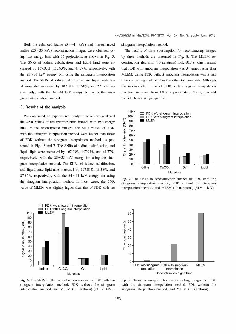
Fig 6 The SNRs in the reconstruction images by FDK with the
sinogram interpolation method FDK without the sinogram
interpolation method and MLEM (10 iterations) (23sim33 keV)
Fig 8 Time consumption for reconstructing images by FDK
with the sinogram interpolation method FDK without the
sinogram interpolation method and MLEM (10 iterations)
Fig 7 The SNRs in reconstruction images by FDK with the
sinogram interpolation method FDK without the sinogram
interpolation method and MLEM (10 iterations) (34sim44 keV)
Both the enhanced iodine (34sim44 keV) and non-enhanced
iodine (23sim33 keV) reconstruction images were obtained us-
ing two energy bins with 36 projections as shown in Fig 5
The SNRs of iodine calcification and liquid lipid were in-
creased by 16703 15793 and 4177 respectively with
the 23sim33 keV energy bin using the sinogram interpolation
method The SNRs of iodine calcification and liquid state lip-
id were also increased by 10701 1358 and 2739 re-
spectively with the 34sim44 keV energy bin using the sino-
gram interpolation method
2 Results of the analysis
We conducted an experimental study in which we analyzed
the SNR values of the reconstruction images with two energy
bins In the reconstructed images the SNR values of FDK
with the sinogram interpolation method were higher than those
of FDK without the sinogram interpolation method as pre-
sented in Figs 6 and 7 The SNRs of iodine calcification and
liquid lipid were increased by 16703 15793 and 4177
respectively with the 23sim33 keV energy bin using the sino-
gram interpolation method The SNRs of iodine calcification
and liquid state lipid also increased by 10701 1358 and
2739 respectively with the 34sim44 keV energy bin using
the sinogram interpolation method In most cases the SNR
value of MLEM was slightly higher than that of FDK with the
sinogram interpolation method
The results of time consumption for reconstructing images
by three methods are presented in Fig 8 The MLEM re-
construction algorithm (10 iterations) took 607 s which means
that FDK with sinogram interpolation was 34 times faster than
MLEM Using FDK without sinogram interpolation was a less
time consuming method than the other two methods Although
the reconstruction time of FDK with sinogram interpolation
has been increased from 18 to approximately 216 s it would
provide better image quality
Dohyeon Kim et alImprovement of Analytic Reconstruction Algorithms Using a Sinogram Interpolation Method for Sparse-angular Sampling with a Photon-counting Detector
- 110 -
Discussion and Conclusion
In the real experimental study the SNR values with the si-
nogram interpolation method were higher than those without
the sinogram interpolation method In two energy bins the si-
nogram interpolation method increased the material signals and
reduced the background noise The FDK algorithm with the si-
nogram interpolation method did not result in better results
than the MLEM algorithm in sparse-angular sampling However
it produced comparable SNR results to those of the MLEM
algorithm These results suggest that the sinogram interpolation
method can improve image quality of sparse-angular sam-
pling1314)
Overall the sinogram interpolation method can add value to
the reconstruction process after sparse-angular sampling which
improves the image quality of the analytic reconstruction
algorithm Reconstructed images obtained using the sinogram
interpolation method agreed well with those obtained using
360 projection data By applying the sinogram interpolation to
the FDK algorithm we expect that the analytic reconstruction
algorithm can obtain reasonable image quality of the iterative
algorithm level Furthermore the analytic reconstruction algo-
rithm using the sinogram interpolation method can be used in
place of an iterative algorithm with a long reconstruction time
We believe that the sinogram interpolation method can be ap-
plied in various reconstruction studies using the analytic re-
construction algorithm
In conclusion our results suggest that the sinogram inter-
polation method can be useful in low dose CT applications
such as radiation therapy for treatment planning or industrial
CT applications
References
1 Kalke M Siltanen S Sinogram interpolation method for
sparse-angle tomography Appl Math 5(3)423-441 (2014)
2 Brooks RA Weiss GH Talbert AJ A new approach to in-
terpolation in computed tomography J Comput Assisted Tomogr
2(5)577-585 (1978)
3 Kim D Park S-J Jo B Kim H Kim H-J Investigation of
sparse-angle view in cone beam computed tomography (CBCT)
reconstruction algorithm using a sinogram interpolation method
IFMBE Proceedings 2015 Canada pp 74
4 Siltanen S Kolehmainen V Jaumlrvenpaumlauml S et al Statistical
inversion for medical x-ray tomography with few radiographs I
General theory Phys Med Biol 48(10)1465-1490 (2003)
5 Lahart MJ Estimation of reconstructions in computed
tomography J Opt Soc Am 71(10)1155-1161 (1981)
6 Shikhaliev PM Xu T Molloi S Photon counting CT
Concept and initial results Med Phys 32(2)427-436 (2005)
7 Lundqvist M Cederstrom B Chmill V Danielsson M
Hasegawa B Evaluation of a photon counting X-ray imaging
system IEEE Trans Nucl Sci 48(4)1530-1536 (2001)
8 Sarkar V Shi C Rassiah-Szegedi P et al The effect of
a limited number of projections and reconstruction algorithms on
the image quality of megavoltage digital tomosynthesis J Appl
Clin Med Phys 10(3)155-172 (2009)
9 Feldkamp LA Davis LC Kress JW Practical cone-beam
algorithm J Opt Soc Am A 1(6)612-619 (1984)
10 Shepp LA Vardi Y Maximum likelihood reconstruction for
emission tomography IEEE Trans Med Imag 1(2)113-122
(1982)
11 Dempster AP Laird NM Rubin DB Maximum likelihood
from incomplete data via the EM algorithm J R Statist Soc B
39(1)1-38 (1977)
12 httpsenwikipediaorgwikiSignal-to-noise_ratio_(imaging)
13 Bertram M Wiegert J Schaumlfer D Aach T and Rose G
Directional View Interpolation for Compensation of Sparse
Angular Sampling in Cone-Beam CT IEEE Trans Med Imag
28(7)1011-1022 (2009)
14 Bertram M Rose G Schaumlfer D Wiegert J and Aach
T Directional interpolation of sparsely sampled cone-beam CT
sinogram data IEEE Int Symp Biomed Imag 1928-931 (2004)
Dohyeon Kim et alImprovement of Analytic Reconstruction Algorithms Using a Sinogram Interpolation Method for Sparse-angular Sampling with a Photon-counting Detector
- 106 -
Fig 1 Flowchart of the proposed
sinogram interpolation method
Fig 2 Spectral CT system with CZT-based photon-counting
detector
construction algorithm Few studies have been conducted to
obtain a reconstruction image in sparse angular sampling using
the analytic reconstruction algorithm In this study therefore
we focused on the development of an analytical reconstruction
algorithm in sparse tomographic imaging We used the Feld-
kamp-Davis-Kress (FDK) algorithm with the sinogram inter-
polation method in an attempt to restore the lack of projection
data to obtain a reasonable reconstruction image There are
various types of interpolation methods including ldquocubicrdquo
ldquosplinerdquo ldquopolynomialrdquo and ldquolinearrdquo12) The proposed sinogram
interpolation method was implemented with linear interpolation
to calculate the incomplete sinogram information As compared
to the FDK algorithm alone the sinogram interpolation method
can minimize the noise and streak artifacts in reconstructed
images3) The sinogram interpolation method has been studied
since CT was invented in the 1970s Brooks et al (1978)
studied both interpolating new data in projection data and new
projection data to attenuate Moire2) In addition Lahart (1981)
studied a similar problem and used a least squares approx-
imation to interpolate projection data when the external shape
of the object was known (Martti Kalke et al 2014)15)
The purpose of this paper was to evaluate the feasibility of
the sinogram interpolation method using the FDK algorithm in
sparse-angle view by means of the signal-to-noise (SNR)
Materials and Methods
1 Sinogram interpolation method
The interpolation method was used to fill the insufficient
projection data caused by sparse-angular sampling A flowchart
of the restoration process using the sinogram interpolation
method is shown in Fig 1 Initially limited projection data
were acquired in sparse-angular sampling After obtaining
these data sparse angular sinogram images were acquired
without applying the sinogram interpolation method using zero
padding In the second step we filled in values using the in-
terpolation method We obtained restored sinograms by re-
peatedly filling in values Finally the restored projection data
were reconstructed by using the FDK algorithm
2 CZT-based photon-counting detector and spectral
CT system
We used the photon-counting detector system prototype
which is widely used in spectral CT Spectral CT has been
studied in medical applications because of its energy separa-
tion capability67) The spectral CT system consisted of a mi-
cro-focus X-ray tube (L8601-01 Hamamatsu Japan) The
source had a focal spot size of approximately 5 μm A high-
precision motor-controlled rotary stage and Cadmium Zinc
Telluride (CZT)-based photon-counting detector (eValuator-2500
PROGRESS in MEDICAL PHYSICS Vol 27 No 3 September 2016
- 107 -
Fig 3 Illustration of the PMMA
phantom containing four materials
Table 1 Detailed acquisition parameters of the spectral CT
system
Source to detector distance (SID) 1200 mm
Source to center of rotation distance 1015 mm
Detector information
Pixel pitch 08 mm
Detector length 512 mm
Number of pixels 64times1
Source information bin 1 23sim33 keV
bin 2 34sim44 keV
Number of projections (angle interval) 36 projections (10o)
Reconstruction method 1 Reconstruction
method ndashlsquoFBPrsquo
2 Filter ndashlsquoRam-lakrsquo
eV Products Saxonburg PA USA) were used in the spectral
CT prototype system (Fig 2) Table 1 lists the detailed acquis-
ition parameters for obtaining the material-decomposed re-
construction images in the spectral CT system
3 Phantom
A PMMA phantom with height and radius of 50 mm and
175 mm respectively was used The phantom contained io-
dine gadolinium calcification and liquid lipid as shown in
Fig 3
4 Reconstruction algorithm
In the experimental study the Maximum Likelihood Expec-
tation Maximization (MLEM) algorithm and the FDK algo-
rithm were used to reconstruct the image We compared two
results of the MLEM algorithm and FDK algorithm with the
sinogram interpolation method The definitive aim of this
study was to verify the possibility of applying sinogram inter-
polation to sparse tomographic imaging The FDK algorithm is
one version of the filtered back projection algorithm that is
widely used for CT reconstruction The algorithm was origi-
nally proposed by Feldkamp Davis and Kress in 19849)
Although this algorithm produces an approximate solution it
has been widely adopted for cone beam reconstructions89) The
equation for the FDK algorithm is
g(xyz)=
infin
infin
where g(xyz) is the value of the object at location (xyz) β
is the angle relative to the z-axis D is the source-to-isocenter
distance R(pζβ) is the projection data acquired at angle β
t=xcosβ+ysinβ s=ycosβminusxsinβ and (pζ) represents the
coordinate system of the detector8) The MLEM reconstruction
algorithm can be expressed as follows
sumsumsum
where λn is the current estimate of the image pj is the meas-
ured projection data and wij is the probability that a photon
emitted from image space at position i is detected by the de-
tector pixel j1011) In this study 10 iterations were executed for
the convergence criterion of MLEM reconstruction algorithm
5 Data analysis
The reconstructed image was a volume of 128times128times128
voxels We set regions-of-interest (ROIs) on the 64th slice
Dohyeon Kim et alImprovement of Analytic Reconstruction Algorithms Using a Sinogram Interpolation Method for Sparse-angular Sampling with a Photon-counting Detector
- 108 -
Fig 5 The reconstruction images of the PMMA phantom with the FDK and MLEM algorithms (asimc) Non-enhanced iodine image
(23sim33 keV) (a) FDK without the sinogram interpolation method (b) FDK with the sinogram interpolation method (c) MLEM (10
iterations) (dsime) Enhanced iodine image (34sim44 keV) (d) FDK without the sinogram interpolation method (e) FDK with the
sinogram interpolation method (f) MLEM (10 iterations)
Fig 4 Restored PMMA phantom sinogram (a) Without interpolation in 36 views (b) With interpolation in 36 angular views
The reconstruction image was obtained using the lack of pro-
jection data in the sparse-angular sampling CT system Four
materials of square-shaped ROI regions were selected to eval-
uate the signal-to-noise ratio (SNR) in order to compare the
methods with and without sinogram interpolation The ROI re-
gions were 15times15 pixels in size The SNR was calculated as
follows
where μ is the mean value of the average of material signal
and σ is the standard deviation of the background12)
Results
1 Results of images
We obtained the projection data of sparse-angular sampling
The sinogram interpolation method was implemented by linear
interpolation to restore the incomplete sinogram information1)
Interpolation can be used to restore the sparse sampling sino-
gram data by filling values between as shown in Fig 4
PROGRESS in MEDICAL PHYSICS Vol 27 No 3 September 2016
- 109 -
Fig 6 The SNRs in the reconstruction images by FDK with the
sinogram interpolation method FDK without the sinogram
interpolation method and MLEM (10 iterations) (23sim33 keV)
Fig 8 Time consumption for reconstructing images by FDK
with the sinogram interpolation method FDK without the
sinogram interpolation method and MLEM (10 iterations)
Fig 7 The SNRs in reconstruction images by FDK with the
sinogram interpolation method FDK without the sinogram
interpolation method and MLEM (10 iterations) (34sim44 keV)
Both the enhanced iodine (34sim44 keV) and non-enhanced
iodine (23sim33 keV) reconstruction images were obtained us-
ing two energy bins with 36 projections as shown in Fig 5
The SNRs of iodine calcification and liquid lipid were in-
creased by 16703 15793 and 4177 respectively with
the 23sim33 keV energy bin using the sinogram interpolation
method The SNRs of iodine calcification and liquid state lip-
id were also increased by 10701 1358 and 2739 re-
spectively with the 34sim44 keV energy bin using the sino-
gram interpolation method
2 Results of the analysis
We conducted an experimental study in which we analyzed
the SNR values of the reconstruction images with two energy
bins In the reconstructed images the SNR values of FDK
with the sinogram interpolation method were higher than those
of FDK without the sinogram interpolation method as pre-
sented in Figs 6 and 7 The SNRs of iodine calcification and
liquid lipid were increased by 16703 15793 and 4177
respectively with the 23sim33 keV energy bin using the sino-
gram interpolation method The SNRs of iodine calcification
and liquid state lipid also increased by 10701 1358 and
2739 respectively with the 34sim44 keV energy bin using
the sinogram interpolation method In most cases the SNR
value of MLEM was slightly higher than that of FDK with the
sinogram interpolation method
The results of time consumption for reconstructing images
by three methods are presented in Fig 8 The MLEM re-
construction algorithm (10 iterations) took 607 s which means
that FDK with sinogram interpolation was 34 times faster than
MLEM Using FDK without sinogram interpolation was a less
time consuming method than the other two methods Although
the reconstruction time of FDK with sinogram interpolation
has been increased from 18 to approximately 216 s it would
provide better image quality
Dohyeon Kim et alImprovement of Analytic Reconstruction Algorithms Using a Sinogram Interpolation Method for Sparse-angular Sampling with a Photon-counting Detector
- 110 -
Discussion and Conclusion
In the real experimental study the SNR values with the si-
nogram interpolation method were higher than those without
the sinogram interpolation method In two energy bins the si-
nogram interpolation method increased the material signals and
reduced the background noise The FDK algorithm with the si-
nogram interpolation method did not result in better results
than the MLEM algorithm in sparse-angular sampling However
it produced comparable SNR results to those of the MLEM
algorithm These results suggest that the sinogram interpolation
method can improve image quality of sparse-angular sam-
pling1314)
Overall the sinogram interpolation method can add value to
the reconstruction process after sparse-angular sampling which
improves the image quality of the analytic reconstruction
algorithm Reconstructed images obtained using the sinogram
interpolation method agreed well with those obtained using
360 projection data By applying the sinogram interpolation to
the FDK algorithm we expect that the analytic reconstruction
algorithm can obtain reasonable image quality of the iterative
algorithm level Furthermore the analytic reconstruction algo-
rithm using the sinogram interpolation method can be used in
place of an iterative algorithm with a long reconstruction time
We believe that the sinogram interpolation method can be ap-
plied in various reconstruction studies using the analytic re-
construction algorithm
In conclusion our results suggest that the sinogram inter-
polation method can be useful in low dose CT applications
such as radiation therapy for treatment planning or industrial
CT applications
References
1 Kalke M Siltanen S Sinogram interpolation method for
sparse-angle tomography Appl Math 5(3)423-441 (2014)
2 Brooks RA Weiss GH Talbert AJ A new approach to in-
terpolation in computed tomography J Comput Assisted Tomogr
2(5)577-585 (1978)
3 Kim D Park S-J Jo B Kim H Kim H-J Investigation of
sparse-angle view in cone beam computed tomography (CBCT)
reconstruction algorithm using a sinogram interpolation method
IFMBE Proceedings 2015 Canada pp 74
4 Siltanen S Kolehmainen V Jaumlrvenpaumlauml S et al Statistical
inversion for medical x-ray tomography with few radiographs I
General theory Phys Med Biol 48(10)1465-1490 (2003)
5 Lahart MJ Estimation of reconstructions in computed
tomography J Opt Soc Am 71(10)1155-1161 (1981)
6 Shikhaliev PM Xu T Molloi S Photon counting CT
Concept and initial results Med Phys 32(2)427-436 (2005)
7 Lundqvist M Cederstrom B Chmill V Danielsson M
Hasegawa B Evaluation of a photon counting X-ray imaging
system IEEE Trans Nucl Sci 48(4)1530-1536 (2001)
8 Sarkar V Shi C Rassiah-Szegedi P et al The effect of
a limited number of projections and reconstruction algorithms on
the image quality of megavoltage digital tomosynthesis J Appl
Clin Med Phys 10(3)155-172 (2009)
9 Feldkamp LA Davis LC Kress JW Practical cone-beam
algorithm J Opt Soc Am A 1(6)612-619 (1984)
10 Shepp LA Vardi Y Maximum likelihood reconstruction for
emission tomography IEEE Trans Med Imag 1(2)113-122
(1982)
11 Dempster AP Laird NM Rubin DB Maximum likelihood
from incomplete data via the EM algorithm J R Statist Soc B
39(1)1-38 (1977)
12 httpsenwikipediaorgwikiSignal-to-noise_ratio_(imaging)
13 Bertram M Wiegert J Schaumlfer D Aach T and Rose G
Directional View Interpolation for Compensation of Sparse
Angular Sampling in Cone-Beam CT IEEE Trans Med Imag
28(7)1011-1022 (2009)
14 Bertram M Rose G Schaumlfer D Wiegert J and Aach
T Directional interpolation of sparsely sampled cone-beam CT
sinogram data IEEE Int Symp Biomed Imag 1928-931 (2004)
PROGRESS in MEDICAL PHYSICS Vol 27 No 3 September 2016
- 107 -
Fig 3 Illustration of the PMMA
phantom containing four materials
Table 1 Detailed acquisition parameters of the spectral CT
system
Source to detector distance (SID) 1200 mm
Source to center of rotation distance 1015 mm
Detector information
Pixel pitch 08 mm
Detector length 512 mm
Number of pixels 64times1
Source information bin 1 23sim33 keV
bin 2 34sim44 keV
Number of projections (angle interval) 36 projections (10o)
Reconstruction method 1 Reconstruction
method ndashlsquoFBPrsquo
2 Filter ndashlsquoRam-lakrsquo
eV Products Saxonburg PA USA) were used in the spectral
CT prototype system (Fig 2) Table 1 lists the detailed acquis-
ition parameters for obtaining the material-decomposed re-
construction images in the spectral CT system
3 Phantom
A PMMA phantom with height and radius of 50 mm and
175 mm respectively was used The phantom contained io-
dine gadolinium calcification and liquid lipid as shown in
Fig 3
4 Reconstruction algorithm
In the experimental study the Maximum Likelihood Expec-
tation Maximization (MLEM) algorithm and the FDK algo-
rithm were used to reconstruct the image We compared two
results of the MLEM algorithm and FDK algorithm with the
sinogram interpolation method The definitive aim of this
study was to verify the possibility of applying sinogram inter-
polation to sparse tomographic imaging The FDK algorithm is
one version of the filtered back projection algorithm that is
widely used for CT reconstruction The algorithm was origi-
nally proposed by Feldkamp Davis and Kress in 19849)
Although this algorithm produces an approximate solution it
has been widely adopted for cone beam reconstructions89) The
equation for the FDK algorithm is
g(xyz)=
infin
infin
where g(xyz) is the value of the object at location (xyz) β
is the angle relative to the z-axis D is the source-to-isocenter
distance R(pζβ) is the projection data acquired at angle β
t=xcosβ+ysinβ s=ycosβminusxsinβ and (pζ) represents the
coordinate system of the detector8) The MLEM reconstruction
algorithm can be expressed as follows
sumsumsum
where λn is the current estimate of the image pj is the meas-
ured projection data and wij is the probability that a photon
emitted from image space at position i is detected by the de-
tector pixel j1011) In this study 10 iterations were executed for
the convergence criterion of MLEM reconstruction algorithm
5 Data analysis
The reconstructed image was a volume of 128times128times128
voxels We set regions-of-interest (ROIs) on the 64th slice
Dohyeon Kim et alImprovement of Analytic Reconstruction Algorithms Using a Sinogram Interpolation Method for Sparse-angular Sampling with a Photon-counting Detector
- 108 -
Fig 5 The reconstruction images of the PMMA phantom with the FDK and MLEM algorithms (asimc) Non-enhanced iodine image
(23sim33 keV) (a) FDK without the sinogram interpolation method (b) FDK with the sinogram interpolation method (c) MLEM (10
iterations) (dsime) Enhanced iodine image (34sim44 keV) (d) FDK without the sinogram interpolation method (e) FDK with the
sinogram interpolation method (f) MLEM (10 iterations)
Fig 4 Restored PMMA phantom sinogram (a) Without interpolation in 36 views (b) With interpolation in 36 angular views
The reconstruction image was obtained using the lack of pro-
jection data in the sparse-angular sampling CT system Four
materials of square-shaped ROI regions were selected to eval-
uate the signal-to-noise ratio (SNR) in order to compare the
methods with and without sinogram interpolation The ROI re-
gions were 15times15 pixels in size The SNR was calculated as
follows
where μ is the mean value of the average of material signal
and σ is the standard deviation of the background12)
Results
1 Results of images
We obtained the projection data of sparse-angular sampling
The sinogram interpolation method was implemented by linear
interpolation to restore the incomplete sinogram information1)
Interpolation can be used to restore the sparse sampling sino-
gram data by filling values between as shown in Fig 4
PROGRESS in MEDICAL PHYSICS Vol 27 No 3 September 2016
- 109 -
Fig 6 The SNRs in the reconstruction images by FDK with the
sinogram interpolation method FDK without the sinogram
interpolation method and MLEM (10 iterations) (23sim33 keV)
Fig 8 Time consumption for reconstructing images by FDK
with the sinogram interpolation method FDK without the
sinogram interpolation method and MLEM (10 iterations)
Fig 7 The SNRs in reconstruction images by FDK with the
sinogram interpolation method FDK without the sinogram
interpolation method and MLEM (10 iterations) (34sim44 keV)
Both the enhanced iodine (34sim44 keV) and non-enhanced
iodine (23sim33 keV) reconstruction images were obtained us-
ing two energy bins with 36 projections as shown in Fig 5
The SNRs of iodine calcification and liquid lipid were in-
creased by 16703 15793 and 4177 respectively with
the 23sim33 keV energy bin using the sinogram interpolation
method The SNRs of iodine calcification and liquid state lip-
id were also increased by 10701 1358 and 2739 re-
spectively with the 34sim44 keV energy bin using the sino-
gram interpolation method
2 Results of the analysis
We conducted an experimental study in which we analyzed
the SNR values of the reconstruction images with two energy
bins In the reconstructed images the SNR values of FDK
with the sinogram interpolation method were higher than those
of FDK without the sinogram interpolation method as pre-
sented in Figs 6 and 7 The SNRs of iodine calcification and
liquid lipid were increased by 16703 15793 and 4177
respectively with the 23sim33 keV energy bin using the sino-
gram interpolation method The SNRs of iodine calcification
and liquid state lipid also increased by 10701 1358 and
2739 respectively with the 34sim44 keV energy bin using
the sinogram interpolation method In most cases the SNR
value of MLEM was slightly higher than that of FDK with the
sinogram interpolation method
The results of time consumption for reconstructing images
by three methods are presented in Fig 8 The MLEM re-
construction algorithm (10 iterations) took 607 s which means
that FDK with sinogram interpolation was 34 times faster than
MLEM Using FDK without sinogram interpolation was a less
time consuming method than the other two methods Although
the reconstruction time of FDK with sinogram interpolation
has been increased from 18 to approximately 216 s it would
provide better image quality
Dohyeon Kim et alImprovement of Analytic Reconstruction Algorithms Using a Sinogram Interpolation Method for Sparse-angular Sampling with a Photon-counting Detector
- 110 -
Discussion and Conclusion
In the real experimental study the SNR values with the si-
nogram interpolation method were higher than those without
the sinogram interpolation method In two energy bins the si-
nogram interpolation method increased the material signals and
reduced the background noise The FDK algorithm with the si-
nogram interpolation method did not result in better results
than the MLEM algorithm in sparse-angular sampling However
it produced comparable SNR results to those of the MLEM
algorithm These results suggest that the sinogram interpolation
method can improve image quality of sparse-angular sam-
pling1314)
Overall the sinogram interpolation method can add value to
the reconstruction process after sparse-angular sampling which
improves the image quality of the analytic reconstruction
algorithm Reconstructed images obtained using the sinogram
interpolation method agreed well with those obtained using
360 projection data By applying the sinogram interpolation to
the FDK algorithm we expect that the analytic reconstruction
algorithm can obtain reasonable image quality of the iterative
algorithm level Furthermore the analytic reconstruction algo-
rithm using the sinogram interpolation method can be used in
place of an iterative algorithm with a long reconstruction time
We believe that the sinogram interpolation method can be ap-
plied in various reconstruction studies using the analytic re-
construction algorithm
In conclusion our results suggest that the sinogram inter-
polation method can be useful in low dose CT applications
such as radiation therapy for treatment planning or industrial
CT applications
References
1 Kalke M Siltanen S Sinogram interpolation method for
sparse-angle tomography Appl Math 5(3)423-441 (2014)
2 Brooks RA Weiss GH Talbert AJ A new approach to in-
terpolation in computed tomography J Comput Assisted Tomogr
2(5)577-585 (1978)
3 Kim D Park S-J Jo B Kim H Kim H-J Investigation of
sparse-angle view in cone beam computed tomography (CBCT)
reconstruction algorithm using a sinogram interpolation method
IFMBE Proceedings 2015 Canada pp 74
4 Siltanen S Kolehmainen V Jaumlrvenpaumlauml S et al Statistical
inversion for medical x-ray tomography with few radiographs I
General theory Phys Med Biol 48(10)1465-1490 (2003)
5 Lahart MJ Estimation of reconstructions in computed
tomography J Opt Soc Am 71(10)1155-1161 (1981)
6 Shikhaliev PM Xu T Molloi S Photon counting CT
Concept and initial results Med Phys 32(2)427-436 (2005)
7 Lundqvist M Cederstrom B Chmill V Danielsson M
Hasegawa B Evaluation of a photon counting X-ray imaging
system IEEE Trans Nucl Sci 48(4)1530-1536 (2001)
8 Sarkar V Shi C Rassiah-Szegedi P et al The effect of
a limited number of projections and reconstruction algorithms on
the image quality of megavoltage digital tomosynthesis J Appl
Clin Med Phys 10(3)155-172 (2009)
9 Feldkamp LA Davis LC Kress JW Practical cone-beam
algorithm J Opt Soc Am A 1(6)612-619 (1984)
10 Shepp LA Vardi Y Maximum likelihood reconstruction for
emission tomography IEEE Trans Med Imag 1(2)113-122
(1982)
11 Dempster AP Laird NM Rubin DB Maximum likelihood
from incomplete data via the EM algorithm J R Statist Soc B
39(1)1-38 (1977)
12 httpsenwikipediaorgwikiSignal-to-noise_ratio_(imaging)
13 Bertram M Wiegert J Schaumlfer D Aach T and Rose G
Directional View Interpolation for Compensation of Sparse
Angular Sampling in Cone-Beam CT IEEE Trans Med Imag
28(7)1011-1022 (2009)
14 Bertram M Rose G Schaumlfer D Wiegert J and Aach
T Directional interpolation of sparsely sampled cone-beam CT
sinogram data IEEE Int Symp Biomed Imag 1928-931 (2004)
Dohyeon Kim et alImprovement of Analytic Reconstruction Algorithms Using a Sinogram Interpolation Method for Sparse-angular Sampling with a Photon-counting Detector
- 108 -
Fig 5 The reconstruction images of the PMMA phantom with the FDK and MLEM algorithms (asimc) Non-enhanced iodine image
(23sim33 keV) (a) FDK without the sinogram interpolation method (b) FDK with the sinogram interpolation method (c) MLEM (10
iterations) (dsime) Enhanced iodine image (34sim44 keV) (d) FDK without the sinogram interpolation method (e) FDK with the
sinogram interpolation method (f) MLEM (10 iterations)
Fig 4 Restored PMMA phantom sinogram (a) Without interpolation in 36 views (b) With interpolation in 36 angular views
The reconstruction image was obtained using the lack of pro-
jection data in the sparse-angular sampling CT system Four
materials of square-shaped ROI regions were selected to eval-
uate the signal-to-noise ratio (SNR) in order to compare the
methods with and without sinogram interpolation The ROI re-
gions were 15times15 pixels in size The SNR was calculated as
follows
where μ is the mean value of the average of material signal
and σ is the standard deviation of the background12)
Results
1 Results of images
We obtained the projection data of sparse-angular sampling
The sinogram interpolation method was implemented by linear
interpolation to restore the incomplete sinogram information1)
Interpolation can be used to restore the sparse sampling sino-
gram data by filling values between as shown in Fig 4
PROGRESS in MEDICAL PHYSICS Vol 27 No 3 September 2016
- 109 -
Fig 6 The SNRs in the reconstruction images by FDK with the
sinogram interpolation method FDK without the sinogram
interpolation method and MLEM (10 iterations) (23sim33 keV)
Fig 8 Time consumption for reconstructing images by FDK
with the sinogram interpolation method FDK without the
sinogram interpolation method and MLEM (10 iterations)
Fig 7 The SNRs in reconstruction images by FDK with the
sinogram interpolation method FDK without the sinogram
interpolation method and MLEM (10 iterations) (34sim44 keV)
Both the enhanced iodine (34sim44 keV) and non-enhanced
iodine (23sim33 keV) reconstruction images were obtained us-
ing two energy bins with 36 projections as shown in Fig 5
The SNRs of iodine calcification and liquid lipid were in-
creased by 16703 15793 and 4177 respectively with
the 23sim33 keV energy bin using the sinogram interpolation
method The SNRs of iodine calcification and liquid state lip-
id were also increased by 10701 1358 and 2739 re-
spectively with the 34sim44 keV energy bin using the sino-
gram interpolation method
2 Results of the analysis
We conducted an experimental study in which we analyzed
the SNR values of the reconstruction images with two energy
bins In the reconstructed images the SNR values of FDK
with the sinogram interpolation method were higher than those
of FDK without the sinogram interpolation method as pre-
sented in Figs 6 and 7 The SNRs of iodine calcification and
liquid lipid were increased by 16703 15793 and 4177
respectively with the 23sim33 keV energy bin using the sino-
gram interpolation method The SNRs of iodine calcification
and liquid state lipid also increased by 10701 1358 and
2739 respectively with the 34sim44 keV energy bin using
the sinogram interpolation method In most cases the SNR
value of MLEM was slightly higher than that of FDK with the
sinogram interpolation method
The results of time consumption for reconstructing images
by three methods are presented in Fig 8 The MLEM re-
construction algorithm (10 iterations) took 607 s which means
that FDK with sinogram interpolation was 34 times faster than
MLEM Using FDK without sinogram interpolation was a less
time consuming method than the other two methods Although
the reconstruction time of FDK with sinogram interpolation
has been increased from 18 to approximately 216 s it would
provide better image quality
Dohyeon Kim et alImprovement of Analytic Reconstruction Algorithms Using a Sinogram Interpolation Method for Sparse-angular Sampling with a Photon-counting Detector
- 110 -
Discussion and Conclusion
In the real experimental study the SNR values with the si-
nogram interpolation method were higher than those without
the sinogram interpolation method In two energy bins the si-
nogram interpolation method increased the material signals and
reduced the background noise The FDK algorithm with the si-
nogram interpolation method did not result in better results
than the MLEM algorithm in sparse-angular sampling However
it produced comparable SNR results to those of the MLEM
algorithm These results suggest that the sinogram interpolation
method can improve image quality of sparse-angular sam-
pling1314)
Overall the sinogram interpolation method can add value to
the reconstruction process after sparse-angular sampling which
improves the image quality of the analytic reconstruction
algorithm Reconstructed images obtained using the sinogram
interpolation method agreed well with those obtained using
360 projection data By applying the sinogram interpolation to
the FDK algorithm we expect that the analytic reconstruction
algorithm can obtain reasonable image quality of the iterative
algorithm level Furthermore the analytic reconstruction algo-
rithm using the sinogram interpolation method can be used in
place of an iterative algorithm with a long reconstruction time
We believe that the sinogram interpolation method can be ap-
plied in various reconstruction studies using the analytic re-
construction algorithm
In conclusion our results suggest that the sinogram inter-
polation method can be useful in low dose CT applications
such as radiation therapy for treatment planning or industrial
CT applications
References
1 Kalke M Siltanen S Sinogram interpolation method for
sparse-angle tomography Appl Math 5(3)423-441 (2014)
2 Brooks RA Weiss GH Talbert AJ A new approach to in-
terpolation in computed tomography J Comput Assisted Tomogr
2(5)577-585 (1978)
3 Kim D Park S-J Jo B Kim H Kim H-J Investigation of
sparse-angle view in cone beam computed tomography (CBCT)
reconstruction algorithm using a sinogram interpolation method
IFMBE Proceedings 2015 Canada pp 74
4 Siltanen S Kolehmainen V Jaumlrvenpaumlauml S et al Statistical
inversion for medical x-ray tomography with few radiographs I
General theory Phys Med Biol 48(10)1465-1490 (2003)
5 Lahart MJ Estimation of reconstructions in computed
tomography J Opt Soc Am 71(10)1155-1161 (1981)
6 Shikhaliev PM Xu T Molloi S Photon counting CT
Concept and initial results Med Phys 32(2)427-436 (2005)
7 Lundqvist M Cederstrom B Chmill V Danielsson M
Hasegawa B Evaluation of a photon counting X-ray imaging
system IEEE Trans Nucl Sci 48(4)1530-1536 (2001)
8 Sarkar V Shi C Rassiah-Szegedi P et al The effect of
a limited number of projections and reconstruction algorithms on
the image quality of megavoltage digital tomosynthesis J Appl
Clin Med Phys 10(3)155-172 (2009)
9 Feldkamp LA Davis LC Kress JW Practical cone-beam
algorithm J Opt Soc Am A 1(6)612-619 (1984)
10 Shepp LA Vardi Y Maximum likelihood reconstruction for
emission tomography IEEE Trans Med Imag 1(2)113-122
(1982)
11 Dempster AP Laird NM Rubin DB Maximum likelihood
from incomplete data via the EM algorithm J R Statist Soc B
39(1)1-38 (1977)
12 httpsenwikipediaorgwikiSignal-to-noise_ratio_(imaging)
13 Bertram M Wiegert J Schaumlfer D Aach T and Rose G
Directional View Interpolation for Compensation of Sparse
Angular Sampling in Cone-Beam CT IEEE Trans Med Imag
28(7)1011-1022 (2009)
14 Bertram M Rose G Schaumlfer D Wiegert J and Aach
T Directional interpolation of sparsely sampled cone-beam CT
sinogram data IEEE Int Symp Biomed Imag 1928-931 (2004)