Embed Size (px)
Citation preview

Improvement in the TensileBond Strength Between
Resin Cement and DentinSurfaces After Temporary
Cement Application
f//seie Kazumi Watanabe, DDSAtsushi Yamashita, DDS, PhD''Hirofumi Yatani, DDS, PhD''Kunlo ¡shikawa, PhD''Kazuomi Suzuki, PhD^
Purpose:Theaimof fhis study was to evaluate the efficacy of three footh condifioners in restoringthe reduced bond strengtii between resin cement and teeth resuiting from remaining temporarycement. Materials and Methods: Ethyl dihydrogen piiosphafe, methacryloxyeihyl dihydrogenphosphate, and 2-methai:ryioxyethyl hydrogen maléate were evaluated as conditioners. Aftereliminating the temporary cement with a curette from the hovinc dentin surface, the conditionerswere applied onto the surface ofthe specimen and a resin cement was adhered. Stepwise scanningelectron microscopic observation and tensile bond strength measurement were carried out.Results: Granular substances were present on the dentin surface even vvhen the temporary cementwas carefully eiiminated with a curette. When primer and resin cement were applied on this surtacewithout conditioner application, no resin tag or hyhrid layer was observed and the mean bondingStrength between tooth and adhesive resin cement was 1.8 MPa. In contrast, resin tag and hybridiayer were observed after primer and resin cement application when the dentinai surface wastreafed wifh conditioner. Mean tensile bone strength values increased to 6.2 MPa i or specimenstreated with 20% methacryloxyethyl dihydrogen phosphate i or 60 seconds. Conclusion: The authorsrecommend methacryloxyethyl dihydrogen phosphate as it provides high tensile bond strengthvalues and requires no additional rinse step. !nt! Prosfhodont 1998:11:203-211.
Temporary cementation on dentin surfaces is animportant procedure to avoid pain and intection
and lo restore function and esthetics.' ^ Althoughtemporary cement has to be removed before the fol-lowing adhesion procedure, compiete elimination oftemporary cement from the dentin surface is diffi-cult. Unfortunately it has been demonsfrated thatresidual temporary cements on the dentinai surfacecause a decrease in bond strength befween dentin
^Doctoral Student, Depatinen! of Fixed Pmsthodonrics.aksyama
University Dentai Scliooi, ak^yama, Japan.''Professor, Depärliner>l of Fixed Proítliodonticí, akayamaUniversity Oentai Sciiooi, Okayama, ¡apan.
'Associale Profeííor, Oepanrnent of Fixed Pronhoönntics, akayamsUniversity Déniai Sciioal, Okayama, Japan.
''Asiociate Professor, Depanmer>t of Dental Mater'iaii, akayama
University Dental Schooi, Okayama, lapan'Professor, Departmeni of Demal Materiais, Oiiayama UniversityDentai School, Okayama, japan.
Reprint requests: Or Bisele Kazumi Watanabe, Depaivnent of Fixed
Prostfiodontics, akayama University Dentai Schooi, 2-5-1 Shikata-
cho, Okayama 700-8525, lapan.
and composite resin.''"'^ Therefore, many attemptshave been made fo eliminate the remaining tempo-rary cement. The efficacy of cleaning the dentai sur-face with a pumice has shown contradictions,^-^"^and fhe use of soap with pumice presented an in-verse resuit decreasing the bond strength.'°Reetching showed some efficacy, although some ofthe dentinai fubules were still partiaiiy occupied bythe temporary cement" and only slightly improvedthe contact angies of distilied water on dentin sur-t'aces.'- The use of a conditioner that can eliminatethe adverse effects of temporary cement appiicationand that does not need an additional rinsing stepwould be more practical tor clinical use.
In the present study, three organic acids wereselecfed as foofh conditioners: ethyl dihydrogenphosphate (EPl, methacryloxyethyl dihydrogen phos-phate (MEP), and 2-methacryloxyethyl hydrogenmaléate (MEM). These organic acids were evaluatedas to whether they could restore the bond strength ofresin cement thaf had been reduced by temporatycement application, because of remaining temporary
11. Numtjer 3, 1998 2 0 3 The Irternational lournai of Pcosthodorlics

Soiiíl Streiiglli Alter Temporaiy CemerU Applic
cement. Methacryloxyethyl dihydrogen phosphateand MEM contain methacrylate groups, and thuscan be classified as self-etching, self-polymerizingconditioners. Ethyl dihydrogen phosphate does notcontain methacrylate, and thus must be rinsed afterapplication. Ethyl dihydrogen phosphate and MEPare monoester phosphoric acids, whereas MEM is amaleic acid with conjugative carboxylic acids.
Materiais and Methods
Ethyl dihydrogen phosphate and MEP were ob-tained commercially (lohoku) and used without fur-ther purification. The MEM was prepared by the ad-dit ion of maleic anhydride to 2-hydroxyethylmethacrylate, as reported previously.'^ Ail ot thechemicals were dissolved in distilled water with theminimum volume of ethanol necessary to make thesolution clear, resulting in 10 wt7ö and 20 wt7o so-lutions.
Bovine mandibular incisors, frozen immediateiyalter extraction, had the radicular portion cut andthe pulp removed. The vestibular area of the coro-nal portion was then ground by a diamond disk toexpose the dentin. The obtained area was polishedflat to 600 grit by a silicon carbide waterproof abra-sive paper (Nihon Kenshi) under running water.Some of the specimens were worn in a rectangularform and suppiied for observation with scanningelectron microscopy (SEM; DS-720, Topcon) andanalysis with energy dispersive x-ray spectroscopy(EDS; Voyager, Noran Instruments).
Test Procedure
The specimens obtained were divided into twogroups: control group (C group) and temporary ce-ment application group (TC group).
In the TC group, an acrylic resin plate (Unifast II,GC) measuring 1 0 x 5 X 2 mm was cementedto the dentin surtace with temporary cement(HY-Bond Temporary Cement Hard, Shofu). After 10minutes, the specimens were immersed in distilledwater at 37°C for 48 hours. The acrylic resin plateswere removed, and the remaining temporary ce-ment on the dentin surface was then removed with acurette. The C group received no treatment withtemporary cement. At this stage, some of the speci-mens in each group were used for the SEM observa-tion and EDS analysis. The remaining specimens ineach group were further divided into two subgroupsbased on the presence and absence of conditionerapplication, as shown in Eig 1. The presence andabsence of conditioner application was abbreviatedto "cond" and "none," respectively.
Tbe following 10 combinations of conditionertype, concentration, and period of conditioner appi-cation were used: (JJ 10% EP for 10 seconds; Í2>10% EP for 30 seconds; (3) 10% MEP for 30 sec-onds; (4) 10% MEP for 60 seconds; (5) 20% MHP for30 seconds; (6) 207o MEP tor 60 seconds; (7) 10%MEM for 30 seconds; (6) 10% MEM for 60 seconds;(9j 20% MEM for 30 seconds; (10) 207o MEM for 60seconds. The type and/or concentration and/or pe-riod of conditioner application is stated in parenthe-ses instead of "cond" to specify the details of condi-tioner application when appropriate. Eor exampie,TC (10% EP 30 seconds) represents specimens towhich temporary cement was appiied and removed,and to which 10% EP was applied for 30 seconds.
Specimens of the other subgroups, ie, the C(none] and TC (none) subgroups, were used for thenext step without conditioner application. The sam-ples that had their dentin surfaces treated with EPwere rinsed with running water for 10 seconds andair-stream dried for 10 seconds, whereas the speci-mens treated with MEP or MEM were air-streamdried without water rinsing, since these condition-ers can be ciassified as self-etching primers. Somespecimens in the C (cond) and TC (cond) subgroupswere used tor SEM obsen/ation to examine the ef-fects of the conditioners.
All specimens underwent the next "ED Primerapplication" step. ED Primer (a methacrylate-basedself-etching primer present in the Panavia 21 set,Kuraray) was applied to the specimen surfaces for60 seconds (as indicated by the manufacturer), andthe specimens were then air-stream dried for 10seconds. Some specimens of the C (none) and TC(none) subgroups were reserved for SEM observa-tion to see the effect of ED Primer.
The remaining specimens were used for the finalstep, "Panavia 21 cementation." An acrylic resinplate measuring 1 0 x 5 X 2 mm was cemented tothe dentin surface with Panavia 21 for SEM obser-vation of the adhesive interface. A 4-mm diameterstainless steel rod (SUS-304) sandblasted with50-|jm aluminum oxide was also cemented for ten-sile bond strength (TBS) measurement.
SEM Observation
For SEM observation of the dentin surfaces afterconditioner application, the specimens were alsowashed with water for 10 seconds (otherwise thesurface wouid be covered by the primer substance,hiding its effect on the dentin surface^) and driedwith an air stream (three-way syringe WS-9, Morita)for 10 seconds. Tbe specimens were gold coated inthe ion coater (IB-5, Eiko Engineering).
The Inlernalinnal Journal of Prosthodonlii 204 , Number3, 1998
Watanabe et al Bond Strength After Temporary Cemenl Appiication
c group
Dentin surface 1
^ TC group
H Y-Bond applicatonRemovai of IHY-Bond with an excavator
Conditioner appiics
r '
tion
\
r \
SEM Observation
EDS analysis
ED Primer application
/ SEM observation
Panavia 21 cementation
- - Í - -C (none] TC (cond)
SEtui observation
TBS measurement
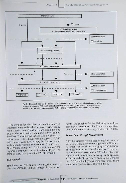
Fig 1 Researcti design: the treatment ot the control (C) specimens and specimens to whichtemporary cements IJC) were appiied is shown. EDS = Energy dispersive x-ray spectroscopy;SEM = scanning electron microscopy; TBS = tensile bond strength; and cond = conditioning.
The samples for SEM observation of fhe adhesiveinterface were embedded in slow-curing epoxyresin (Epofix, Sfruers) and sectioned along the longaxis of the tooth wifh a diamond cutter (Isomef,Buehler]. The obtained surface was polished to amirror state with silicon carbide paper {< 1,000grit), treated with 37% HjPO^ for 7 seconds andwith sodium hypochlorite solution (NeoCleaner,Neo Pharmaceutic) for 10 minutes to remove theorganic compound on the interfacial layer. Thesamples were gold plated for SEM observation.
EDS Analysis
Specimens for EDS analysis were carbon coated(Polaron CC7650 Carbon Coater, Fisons Instru-
ments) and supplied for the EDS analysis with anaccelerating voltage of 15 keV and an acquisitiontime of 300 seconds at a magnification of X 1,000.
Tensile Bond Strength Measurement
After the samples were placed in distilled water at37°C for 24 hours, they were supplied for TBS mea-surement. Ih brief, an autograph {DCS-2000,Shimadzu) at a cross-head speed of 2 tnm perminute was used to obtain tbe TBS values of at leasteight specimens in each group or subgroup.Approximately 50 specimens each in the C (none)and TC (none) subgroups were measured. Exactnumbers of specimens are shown in Fig 6.
unie 11, Number 3, 1598 205 The Intern aliona i Journal of Prosthodonlics
W-ntansbe et al
k^=5 TC(none)
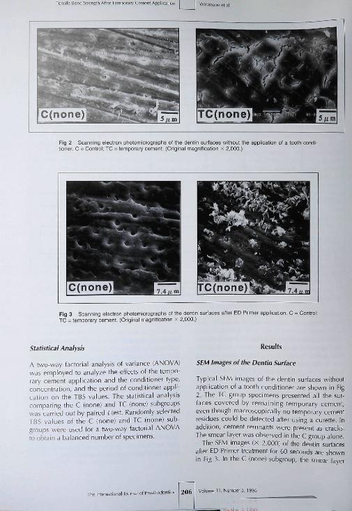
Fig 2 Scanning electron phctomicrographs of the dentin surfaces without the application ol a tooth condi-tioner. C = Control; TC = temporary cement. (Original magniticaticn x 2,000.)
Fig 3 Scanning electron photomicrographs ot the denlin surfaces atter ED Primar application. C = Control;TC = temporary cement. (Original magnification x 2,000.)
Statistical Anaiysis
A two-way factorial anaiysis of variance (ANOVA)was employed to analyze ihe effects ot fhe tempo-rary cement application and the conditioner type,concentration, and fine period ot conditioner appli-cation on the TBS values. The statistical analyiiscomparing the C (none) and TC (none) subgroupswas carried out by paired f test. Randomly selectedTBS values of the C (none) and TC (none) sub-groups were used for a two-way tactorial ANOVAto obtain a balanced number of specimens.
Results
SEM Images of the Dentin Surface
Typical SEM images of the dentin surfaces withoutappiication of a tooth conditioner are shown in Fig2. The TC group specimens presented all the sur-faces covered by remaining temporary cement,even though macroscopically no temporary cementresidues couid be detected atter using a curette. Inaddition, cement remnants were present as craci<s.The smear layer was observed in the C group alone.
The SEM images (x 2,000) of lhc> denlin surfacesafter ED Primer treatment for 60 seconds are shownin Fig 3. in the C (none) subgroup, the smear layer
I of Prostfiodortli 206 Volumen, Number 3. 1998
ary Ce
TC(20%MEP6Ôs)
TC(20%MEM60s)
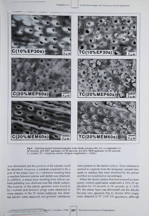
Fig 4 Scanning electron photomicrographs of the dentin surfaoes after 10% EP application for30 seconds, 20% MEP application for 60 seconds, and 20% MEM appiication tor 60 seconds.C = Control: TC = temporary cement. (Original magnification x 2,000.)
was eliminated and the posifion of the tubules couldbe identified. However, a substrate su ipected to be apart of fbe smear layer or a substance resulting froma reaction between primer and dentin was observed.In addition, a smear layer resulting from silicon car-bide polishing was observed over the whole surface.The majority of tbe tubule apertures were found tobe covered and dentinal plugs were observed insome tubules. In the TC (none) subgroup, few denti-nal tubules were observed and granular substances
were present on the dentin surface. These substancesseemed to originate from the temporary cement rem-nants or residues that were dissolved by the primerand then recrystallized or recombined.
When the dentin surface that had received no tem-porary cement application underwent a 10% EP ap-plication for 10 seconds or 30 seconds, ie, C (iO%EP), the smear layer was eliminated and the tubulesbecame very apparent (Eig 4), Similar SEM imageswere obtained in TC (10% EP) specimens, although
Qlume 11, Number 3, 1998 207 Journal ot Prosttiodontics

¡Me Bonri Slrengtii Afler Temporary Cement Appiit Watanabeelai
some dentinai tubuies were still occluded and granu-lar substances were present on tbe dentin surface.
The amount of granular substances present onthe dentin surface of the TC (2O'Ï . MEP 60 seconds)specimens was greater than that of the TC (10% EP30 seconds) specimens, buf much loss than that ofthe TC (none) specimens (Fig 4). In the SEM imagesof the C (MEPl specimens, the smear layer had beeneliminated and tbe tubule openings were apparent.However, some of the dentinai tubuies were stiiioccluded. The application time and the concentra-tion level did nof produce any visibie differenceson the SEM surface in either subgroup.
Similar to the specimens treated with MEP condi-tioner, granular substances were present on thedentin surface ot the TC (MEM) specimens (Fig 4).Most ofthe dentinai tubules were occluded, in con-frast to the specimens treated with the EP or MEPconditioners, which showed many opened dentina!tubuies. The application time and the concentrationlevel did not affect the SEM appearance. In every Csubgroup, the smear layer had been eliminated andthe majority of tubules were apparent. These fea-tures were more evident at the higher concentrationlevels and application times.
EDS Results
Peaks of Zn, absent in the C group, were apparentin the TC group, even when the temporary cementwas removed with a curette, confirming the pres-ence of temporary cement remnants.
5EM Images ofthe Adhesive Interface
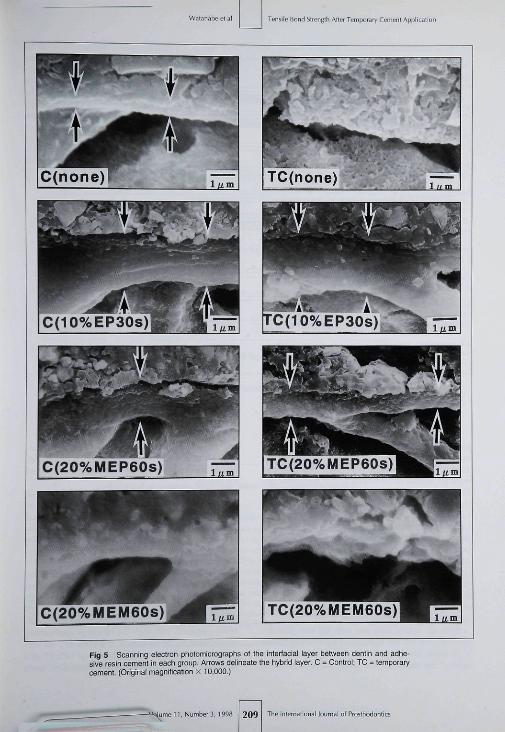
Specimens of the C (none) subgroup presented resintags that infiltrated and polymerized in ihe dentinaitubules. A hybrid layer about 0.5 \im thick was evi-dent (Eig 5). In the TC (none) subgroup specimens,both the resin tags and hybrid layer seen in the C(none) specimens were absent (Eig 51.
When each conditioner was applied to the denti-nai surfaces after elimination of temporary cement,only the specimens treafed with EP and MEPshowed resin fags and hybrid layers (Fig 5). How-ever, the frequencies of resin tags and the hybridlayer were lower compared with the frequencies inthe C (none) subgroup. These frequencies were verylow in the case of TC (MEP) at all concentrations ofMEP. In addition, only the TC (MEP 60 seconds)specimens showed resin tags and hybrid layers, butneither resin tags nor hybrid layer was observedwhen the MEP treatment period was 30 seconds.
In contrast to the TC (cond) specimens, theC (cond) specimens showed resin tags in all
specimens. A hybrid iayer was also observed m theC (EP) and C (MEP) specimens. No hybrid layer wasobserved when the specimens were conditionedwith MEM at any concentration or at any treatmentperiod of MEM. The hybrid layer of the C (EP)specimens was thicker than that in the C (MEP)specimens. The thickness of the hybrid layer foundin the C (EP) specimens was 1.5 pm and 2 to 3 pmfor the treatment period of 10 and 30 seconds, re-spectively. Among the C (MEP) specimens, thethickness was approximately 1.0 pm at all concen-trations and treatment periods.
Tensile Bond Strength
The TBS values are summarized in Eig 6. The TC(none) subgroup showed TBS values significantlylower than those of the C (none) subgroup {P =0.03). In ofher words, the denfin specimens appliedwith temporar>' cement presented reduced bondingstrength even though the femporary cement was re-moved with a curette. The strong TBS-reducing ef-fect of the temporary cement application was alsoobserved in the TC (none) and TC (cond) groupscompared with the C group (P = 0.0001 ).
The C (cond) subgroup showed TBS values signif-icantly higher (P < 0.05) than those oi the C (none)subgroup, except for the C (10% MEM) and C (20%MEM 60 seconds) specimens. Therefore, condi-tioner application was useful to strengthen the TBSbefween resin cement and the tooth even when notemporary cement application was carried out. TheC (EP) or C (MEP) specimens showed significantlyhigher (P < 0.05) TBS values, whereas the C (MEM)specimens had a relatively limited increase in TBScompared to the C (none) subgroup.
The TBS vaiues of the TC group were signifi-cantly smaller (P < 0.05) than the correspondingTBS values of the C group, except for the 10% EPand 20% MEP 60-second specimens. In otherwords, the 10% EP or 20% MEP 60-second treaf-ments had enough ability to counteract the effect oftemporary cement application.
Discussion
The results of (his study clearly demonstrated theusefulness of a tooth conditioner to strengthen fhebonding strength of adhesive resin cement to tooththat has been reduced because of remaining tempo-rary cement. One of the reasons for the inhibitoryeffect of remaining temporary cement on adhesionmay be the iow mechanical strength of temporarycement. The temporary cement is a weak layer,even if it can bind tightly with composite resin and
The International |oii i ol Prostliodonlics 208 Volume 11, Number 3, 1998
Watarabe et al Tensile Bond Slrengld After Temporary Cement Applicati
C(20%MEM60s)
Fig 5 Scanning electron photorricrographs of the intertaciai layer between dentin and adhe-sive resin cement in each group. Arrows delineate the hybrid iayer. C = Contrci; TC = temporarycement (Original magnitication x tO.OOO.]
11, Number 3, 199B 2 0 9 '^^^ International Journal of Prostliotlontics

Tensile Bond St'engtii After Tempora'y Cement Applicaiion Waianabe el ;
tO%EP(1Os)
t0%EP(30s]
10%MEP(30s)
10%MEP(60s)
20% MEP (30 s)
20% MEP (60 s)
10% MEM (30 s]
tO% ÍViEM (60s]
20% MEM (30 s]
20% MEM (60 s)
Fig 6 Tensiie bond strength vaiues of resin cement to bovine dentin in each group (mean + SD).
tooth. In addition, polymerizatiort of the primer/adhesive resin cement is inhibited because of thecomponents of the temporary cement. For example,eugenol inhibits polymerization.""^ The temporarycement used in this study, HY-bond carboxylatetemporary cement, contains tannin-fluoride prepa-ration (HY-agent). hlY-agent reduces secondarycaries by conversion of collagen and powdereddentin to collagenase-resistant forms and by obtura-tion of dentinal tubulesJ'^''^ However, these cario-static properties of HY-agent become shortcomingsif it remains on the dentinal surface. The TC re-duces tbe infiltration of adhesive substances intothe dentinal tubules, which plays an important rolein the adhesion between dentin and adhesive resincement by forming the resin tag.
Although hybrid layer and resin tag formation arenot sufficient but are necessary conditions to obtainhigh bond strength of adhesive resin cement totootb,'^ tbe materials that could inhibit the hybrid
layer and/or resin tag formation (eg, temporary ce-ment remnants) should be eliminated to ensure ad-equate bonding strength between resin cement andthe dentin.
According to the results of this study, the use ofa conditioner before the cementation process re-stored the reduced bonding strength to its originalvalue or higher. Also, it is suggested that TBS val-ues increased when a conditioner was used, evenwhen no temporary cementation process was car-ried out.
Regarding the conditioner type, concentration,and length of application, the 10% EP 30-secondand 20% MEP 60-second specimens showed highTBS values (over 6 MPa). Therefore, the use of 20%MEP for 60 seconds is recommended as tbe condi-tioning process. Although the length of application,60 seconds, is twice that of the 10% EP, 30 sec-onds, it should be noted that the use of 10% EP as aconditioner requires an additional rinse step, since
nai of Proslhodontici 210 Volume i 1 , Number 3, 1998

Waranabe el al
EP does not contain a methacrylate group and thuscannot be classified as a self-etching primer.
In conclusion, MEP was found to have a restora-tive effect on the bonding strength of resin cementto dentin reduced because of temporary cement ap-plication. Further studies of MEP are pianned basedon the initial results obtained in this study.
Acknowledgment
This invesligation wai supporled in part by a Cranl-in-Aid forScientific Research from Ihe Ministry of Educalion, Science,Sports, and Culture, lapan.
References
1. Vahidi F. The provisional restoration. Dent Ciin North Am!9B7;31:363-3ai.
2. Christensen CJ. Provisional restorations for flxed prosthodon-tíC5.J Am Dem Assod 996:127:249-252.
3. Hornbrook DS. Provipont & Provilink. Maximizing aestheticsand funct ion when fabricat ing provisional restorations.Signature 1995:10-16.
4. Harien EK, Asmussen E. Influence of temporary fiiiing materi-als on eftect of dentir-bonding agents. Scand | Dent Res 1987;95:51 &-520.
5. Woody TL, Davis RD. The effect of eugenol-containing andeugenoi-free temporary cements on microieakage in resinbonded restorations. Ope: Dent 1992;1 7:1 75-180.
6. Watanabe EK, Suzuiil K, Yamashita A, Shigeta N, Imai M,Yatani H, et al. Irfiuence oí temporary cement on the adhe-siveness of resin cement to dentin. | | Dent Mater 1996:15:187-191.
7. Watanabe EK, Yamashita A, Imai M, Yatani H, Suzuki K.Temporary cement remnants as an adhesion inhibiting factorin the interface between resin cements and bovine dentin. int JProsthodDnt1997;10:44CM52.
Tensile Bond Strengrli After Temporary Cemeni Application
Schwartz R, Davis R, Hilton T|. Effectoftempoiary cements onihe bond strength of a resin cement. Am ] Dent 1992;5:147-150.
Paul S|, Scliarer P. Effect of provisional cementî on the bondstrength of various adhesive bonding systems on dentine. ICrai Rehabil 1997;24:8-i4.
Bachmanr M, Paui SJ, Lijthy H, Schärer P. Effect of cleaningdentine with soap and pumice or shear bond strength of den-tine-bonding agents. | Oral Rehabil 1997;24:433^3B.Xie I, Powers |M, McGiickin RS. In vitro bond strength of ^voadhesives to enamel and dentin under normal and contami-nated conditions. Dent Mater 1993:9:295-299.Terata R. Characterization of enamei and dentin sun'aces afterremoval of temporary cement—Study on removal of tempo-rary cement. Dent Mater] 1993:12:13-28.Fukushima T, Inoue Y, i-loribe T. Synthesis of 2-methacry-ioxyethyl hydrogen maieate and its bonding to tooth surfaces.J i Dent Mater 1988;7:675-679.
Boviien RL, Argentar H. A stabi l iz ing comonomer: I I .Stabilization and poiymerization characteristics. | Dent Res1972,51:1614-1618.
Hume WR. An analysis of the release and the diffusionthrough denlin of eugenol from ïinc oxide-eugenoi mixtures. |Dent Res 1984;63:881-884.
Tanizaki K. Effect of tannin-fluoride preparation on the reduc-tion of secondary dental caries. Jap J Conserv Dent ¡978;21:279-296.
Yamaga M. Koide T, Hieda T, Daito M. Obturation of dentinaitubules wilh tannin-fluoride preparation (HY agent) incorpo-rated into glass ionomer cement. J Osai<a Dent Univ 1993;27:77-87.
Nakabayashi N, Ashizawa M, Nakamura M. Identification of aresin-dentin hybrid iayer in vitai human dentin created in vivo:Durable bonding to vital dentin. Quintessence int 1992:23:135-141.
Lileraiwe Abstract
Association of residual ridge résorption with systemic factors in
home-living eideriy subjects
This study is part ot the Heisinki Aging Study ct 76-. 81 -. and 86-year-old patients. Ail
subjects edentulous in one or both arches (46 men and 139 women) were included. The
heights of the residual ridges were measured on panoramic radiographs, t68 maxiliae and
126 mandibles. The residual ridge résorption was calcuiated and used as tfie dependent
variabie in multivariate regression models. The percentage ot bone reduction in the
mandibie was signiticantiy associated with the femaie gender when adjusted for age and
duration of edentulousness but with no other independent variable. Severe résorption in
residual ridge (both arches) was significantly associated with the women but also with
asthma. An astonishing tinding was that in the maxiiia severe ridge résorption was inversely
related to alcohol consumption (meaning that low intake was associated with severe résorp-
tion) when adjusted for gender, age. and body mass index.
Xie Q, Afnamo A, Tilvis R. Ada Odontol Scand 1997;55:299-305. References; 52.
Reprints: DrsQiufei Xie and Anja Ainamo, Institute of Dentistry, P.O. Box 41, FIN-00014
University of Helsinki, Finland.—SP
11, Number 3, 1995 211 The International lojrnal of Frosthodontic