Embed Size (px)
Citation preview
Improved Visualization of Femoroacetabular Impingement Cartilage Damage with Multi-band Simultaneous Multi-Slice Acceleration
Casey P. Johnson1; Luning Wang1; Shelly Marette1; Takashi Takahashi1; Patrick Morgan2; Kâmil Uğurbil1;
Dingxin Wang1,3; Jutta Ellermann1
1 Department of Radiology (CMRR), University of Minnesota, Minneapolis, MN, USA 2 Department of Orthopaedic Surgery, University of Minnesota, Minneapolis, MN, USA 3 Siemens Medical Solutions USA Inc., Minneapolis, MN, USA
Introduction
Femoroacetabular impingement
(FAI) is a common source of hip pain
in adults caused by a pathological
abutment of the head-neck junction
of the femur and the acetabular rim
of the hip [6]. This abutment leads
to mechanical friction, which can
in turn cause labral and chondral
lesions and lead to osteoarthritis (OA)
[1, 2, 4, 5, 8]. The recommended
treatment for FAI is joint preservation
surgery if a labral tear has occurred,
cartilage damage is not severe, and
patient symptoms cannot be managed
conservatively with physical therapy
[3, 10, 11]. In FAI, cartilage damage is
typically limited to the acetabulum and
occurs deep within the tissue as a
debonding of articular cartilage from
underlying bone [2]. This so-called
‘cartilage delamination’ is the hallmark
of the disease. If cartilage damage is
severe, as would be the case if there
was prevalent cartilage delamination,
then joint preservation surgery will
fail and total hip replacement will
be necessary. Therefore, in order to
appropriately recommend treatment
for FAI, imaging is needed to evaluate
the hip joint for both labral tears and
cartilage delamination.
However, cartilage delamination is
not seen using current clinical imaging
protocols because spatial resolution is
insufficient. The necessary resolution
cannot be achieved due to prohibi-
tively long acquisition times. Addition-
ally, labral tears can be difficult to
diagnose, also due to limited spatial
resolution. Specifically, slices of 3 to
4 mm are too thick, leading to prob-
lematic volumetric averaging within
voxels along the curved surface
of the acetabulum that obscures
the cartilage and labral pathology.
Multiband Simultaneous Multi-
Slice (SMS) acceleration technology
[7, 9] can be used to improve the
spatial resolution while not increas-
ing the acquisition time (or, equiva-
lently, reducing the acquisition
time for a given spatial resolution).
Multiband allows multiple imaging
slices to be excited simultaneously,
thereby enabling more slices (and
thus higher spatial resolution) to be
acquired within a given acquisition
time. In this study, we evaluated the
potential clinical benefits of utilizing
a multiband SMS accelerated turbo
spin echo (TSE) sequence1 to either
improve diagnostic accuracy of carti-
lage delamination and labral tears
or save scan time while maintaining
diagnostic image quality.
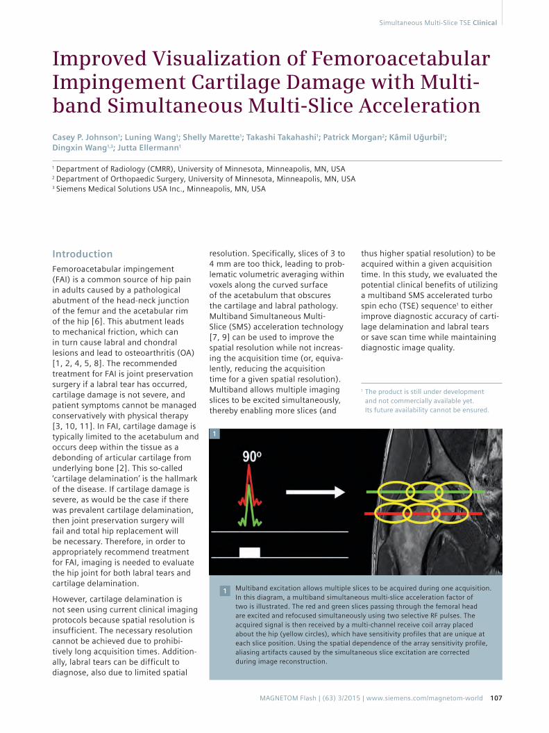
Multiband excitation allows multiple slices to be acquired during one acquisition.
In this diagram, a multiband simultaneous multi-slice acceleration factor of
two is illustrated. The red and green slices passing through the femoral head
are excited and refocused simultaneously using two selective RF pulses. The
acquired signal is then received by a multi-channel receive coil array placed
about the hip (yellow circles), which have sensitivity profiles that are unique at
each slice position. Using the spatial dependence of the array sensitivity profile,
aliasing artifacts caused by the simultaneous slice excitation are corrected
during image reconstruction.
1
1
1 The product is still under development
and not commercially available yet.
Its future availability cannot be ensured.
Simultaneous Multi-Slice TSE Clinical
MAGNETOM Flash | (63) 3/2015 | www.siemens.com/magnetom-world 107
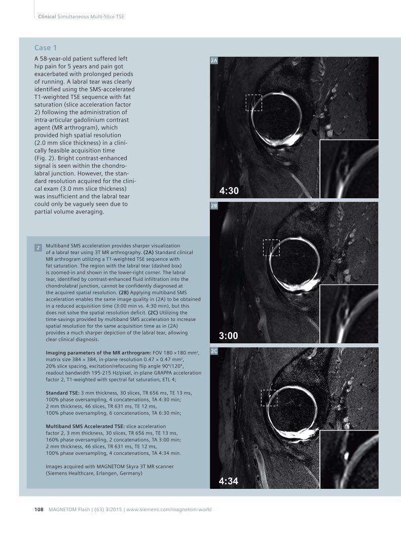
Case 1
A 58-year-old patient suffered left
hip pain for 5 years and pain got
exacerbated with prolonged periods
of running. A labral tear was clearly
identified using the SMS-accelerated
T1-weighted TSE sequence with fat
saturation (slice acceleration factor
2) following the administration of
intra-articular gadolinium contrast
agent (MR arthrogram), which
provided high spatial resolution
(2.0 mm slice thickness) in a clini-
cally feasible acquisition time
(Fig. 2). Bright contrast-enhanced
signal is seen within the chondro-
labral junction. However, the stan-
dard resolution acquired for the clini-
cal exam (3.0 mm slice thickness)
was insufficient and the labral tear
could only be vaguely seen due to
partial volume averaging.
Multiband SMS acceleration provides sharper visualization
of a labral tear using 3T MR arthrography. (2A) Standard clinical
MR arthrogram utilizing a T1-weighted TSE sequence with
fat saturation. The region with the labral tear (dashed box)
is zoomed-in and shown in the lower-right corner. The labral
tear, identified by contrast-enhanced fluid infiltration into the
chondrolabral junction, cannot be confidently diagnosed at
the acquired spatial resolution. (2B) Applying multiband SMS
acceleration enables the same image quality in (2A) to be obtained
in a reduced acquisition time (3:00 min vs. 4:30 min), but this
does not solve the spatial resolution deficit. (2C) Utilizing the
time-savings provided by multiband SMS acceleration to increase
spatial resolution for the same acquisition time as in (2A)
provides a much sharper depiction of the labral tear, allowing
clear clinical diagnosis.
Imaging parameters of the MR arthrogram: FOV 180 ×180 mm2,
matrix size 384 × 384, in-plane resolution 0.47 × 0.47 mm2,
20% slice spacing, excitation/refocusing flip angle 90º/120º,
readout bandwidth 195-215 Hz/pixel, in-plane GRAPPA acceleration
factor 2, T1-weighted with spectral fat saturation, ETL 4;
Standard TSE: 3 mm thickness, 30 slices, TR 656 ms, TE 13 ms,
100% phase oversampling, 4 concatenations, TA 4:30 min;
2 mm thickness, 46 slices, TR 631 ms, TE 12 ms,
100% phase oversampling, 6 concatenations, TA 6:30 min;
Multiband SMS Accelerated TSE: slice acceleration
factor 2, 3 mm thickness, 30 slices, TR 656 ms, TE 13 ms,
160% phase oversampling, 2 concatenations, TA 3:00 min;
2 mm thickness, 46 slices, TR 631 ms, TE 12 ms,
100% phase oversampling, 4 concatenations, TA 4:34 min.
Images acquired with MAGNETOM Skyra 3T MR scanner
(Siemens Healthcare, Erlangen, Germany)
2
2A
2C
2B
Clinical Simultaneous Multi-Slice TSE
108 MAGNETOM Flash | (63) 3/2015 | www.siemens.com/magnetom-world
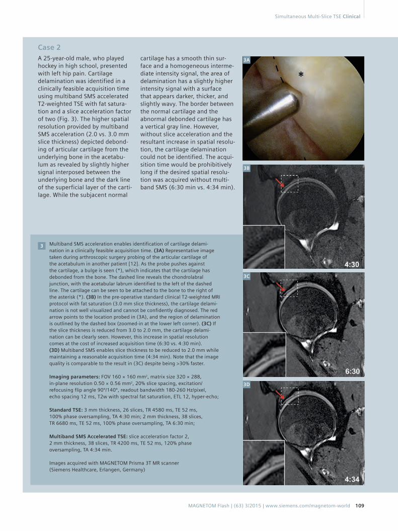
Case 2
A 25-year-old male, who played
hockey in high school, presented
with left hip pain. Cartilage
delamination was identified in a
clinically feasible acquisition time
using multiband SMS accelerated
T2-weighted TSE with fat satura-
tion and a slice acceleration factor
of two (Fig. 3). The higher spatial
resolution provided by multiband
SMS acceleration (2.0 vs. 3.0 mm
slice thickness) depicted debond-
ing of articular cartilage from the
underlying bone in the acetabu-
lum as revealed by slightly higher
signal interposed between the
underlying bone and the dark line
of the superficial layer of the carti-
lage. While the subjacent normal
Multiband SMS acceleration enables identification of cartilage delami-
nation in a clinically feasible acquisition time. (3A) Representative image
taken during arthroscopic surgery probing of the articular cartilage of
the acetabulum in another patient [12]. As the probe pushes against
the cartilage, a bulge is seen (*), which indicates that the cartilage has
debonded from the bone. The dashed line reveals the chondrolabral
junction, with the acetabular labrum identified to the left of the dashed
line. The cartilage can be seen to be attached to the bone to the right of
the asterisk (*). (3B) In the pre-operative standard clinical T2-weighted MRI
protocol with fat saturation (3.0 mm slice thickness), the cartilage delami-
nation is not well visualized and cannot be confidently diagnosed. The red
arrow points to the location probed in (3A), and the region of delamination
is outlined by the dashed box (zoomed-in at the lower left corner). (3C) If
the slice thickness is reduced from 3.0 to 2.0 mm, the cartilage delami-
nation can be clearly seen. However, this increase in spatial resolution
comes at the cost of increased acquisition time (6:30 vs. 4:30 min).
(3D) Multiband SMS enables slice thickness to be reduced to 2.0 mm while
maintaining a reasonable acquisition time (4:34 min). Note that the image
quality is comparable to the result in (3C) despite being >30% faster.
Imaging parameters: FOV 160 × 160 mm2, matrix size 320 × 288,
in-plane resolution 0.50 × 0.56 mm2, 20% slice spacing, excitation/
refocusing flip angle 90º/140º, readout bandwidth 180-260 Hz/pixel,
echo spacing 12 ms, T2w with spectral fat saturation, ETL 12, hyper-echo;
Standard TSE: 3 mm thickness, 26 slices, TR 4580 ms, TE 52 ms,
100% phase oversampling, TA 4:30 min; 2 mm thickness, 38 slices,
TR 6680 ms, TE 52 ms, 100% phase oversampling, TA 6:30 min;
Multiband SMS Accelerated TSE: slice acceleration factor 2,
2 mm thickness, 38 slices, TR 4200 ms, TE 52 ms, 120% phase
oversampling, TA 4:34 min.
Images acquired with MAGNETOM Prisma 3T MR scanner
(Siemens Healthcare, Erlangen, Germany)
3
cartilage has a smooth thin sur-
face and a homogeneous interme-
diate intensity signal, the area of
delamination has a slightly higher
intensity signal with a surface
that appears darker, thicker, and
slightly wavy. The border between
the normal cartilage and the
abnormal debonded cartilage has
a vertical gray line. However,
without slice acceleration and the
resultant increase in spatial resolu-
tion, the cartilage delamination
could not be identified. The acqui-
sition time would be prohibitively
long if the desired spatial resolu-
tion was acquired without multi-
band SMS (6:30 min vs. 4:34 min).
3B
3C
3D
3A
Simultaneous Multi-Slice TSE Clinical
MAGNETOM Flash | (63) 3/2015 | www.siemens.com/magnetom-world 109
Methods
Patients with hip pain undergoing
a clinical MR evaluation (current
standard of care) for FAI were imaged
using both a clinical and a multiband
SMS accelerated sagittal multi-slice
2D TSE sequence with fat saturation
under an Institutional Review Board
approved protocol for which
informed consent was obtained.
Imaging was done using Siemens
3T MRI systems (MAGNETOM Skyra
and MAGNETOM Prisma, Siemens
Healthcare, Erlangen, Germany)
with an 18-channel flex body coil
wrapped about either the right or
left hip in combination with an inte-
grated 32-channel spine coil array.
This enabled unilateral imaging
of the hip joint for higher spatial
resolution by limiting the receive
coil sensitivity to one side of the
body to avoid signal wrap.
Multiband technology was used to
simultaneously excite and acquire
more than one imaging slice simulta-
neously (Fig. 1). Multiband RF pulses
were generated for simultaneous
multi-slice excitation and echo refo-
cusing, and the VERSE technique was
applied to the RF pulses to reduce
peak power and SAR. A low-resolu-
tion multi-slice 2D GRE scan inte-
grated into the SMS TSE sequence
was used as the reference scan to
obtain the coil sensitivities. Image
reconstruction techniques based on
parallel imaging methodology were
then used to unalias the signal
acquired for the multiple slices.
Discussion
Accurate assessment of the acetabu-
lar cartilage is fundamental to the
evaluation of and the clinical deci-
sion-making for patients with symp-
tomatic FAI. Patients with moderate
to advanced cartilage degeneration
will fail arthroscopic repair, leading
to total hip arthroplasty. Multiband
SMS acceleration technology enables
higher spatial resolution to be
acquired with minimal impact on
image quality and no increase in
acquisition time. As shown in the
two clinical cases, this technique pro-
vides improved diagnostic informa-
tion that can better inform treatment
decisions. In our initial clinical
experience, we found that the multi-
band SMS accelerated TSE sequence
can provide 30% higher spatial reso-
lution within a given acquisition time
to improve diagnostic accuracy of
cartilage delamination and labral
tears, or alternatively over 30% time
savings for a given spatial resolution
while maintaining diagnostic image
quality. In general, multiband SMS
acceleration will enable higher-
quality imaging protocols for clinical
3T applications by targeting higher
resolution to allow for more diagnos-
tic accuracy in a standard clinical
setting.
Contact
Jutta M. Ellermann, M.D.
Associate Professor
Department of Radiology (CMRR)
University of Minnesota
420 Delaware St., S.E. MMC 292
Minneapolis, MN 55455
USA
Phone: +1 612-626-3342
References
1 Allen D, Beaule PE, Ramadan O, Doucette
S. Prevalence of associated deformities
and hip pain in patients with cam-type
femoroacetabular impingement. J Bone
Joint Surg Br. 2009;91(5):589-594.
2 Beck M, Kalhor M, Leunig M, Ganz R. Hip
morphology influences the pattern of
damage to the acetabular cartilage:
femoroacetabular impingement as a cause
of early osteoarthritis of the hip. J Bone
Joint Surg Br. 2005;87(7):1012-1018.
3 Beck M, Leunig M, Parvizi J, Boutier V,
Wyss D, Ganz R. Anterior femoroace-
tabular impingement: part II. Midterm
results of surgical treatment. Clin Orthop
Relat Res. 2004(418):67-73.
4 Dudda M, Albers C, Mamisch TC, Werlen S,
Beck M. Do normal radiographs exclude
asphericity of the femoral head-neck
junction? Clin Orthop Relat Res.
2009;467(3):651-659.
5 Ganz R, Leunig M, Leunig-Ganz K,
Harris WH. The etiology of osteoarthritis
of the hip: an integrated mechanical
concept. Clin Orthop Relat Res.
2008;466(2):264-272.
6 Ganz R, Parvizi J, Beck M, Leunig M,
Notzli H, Siebenrock KA. Femoroacetabular
impingement: a cause for osteoarthritis of
the hip. Clin Orthop Relat Res.
2003(417):112-120.
7 Larkman DJ, Hajnal JV, Herlihy AH, Coutts
GA, Young IR, Ehnholm G. Use of multicoil
arrays for separation of signal from multiple
slices simultaneously excited. J Magn
Reson Imaging. 2001;13(2):313-317.
8 Leunig M, Beaule PE, Ganz R. The concept
of femoroacetabular impingement:
current status and future perspectives.
Clin Orthop Relat Res.
2009;467(3):616-622.
9 Moeller S, Yacoub E, Olman CA, et al.
Multiband multislice GE-EPI at 7 tesla,
with 16-fold acceleration using partial
parallel imaging with application to high
spatial and temporal whole-brain fMRI.
Magn Reson Med. 2010;63(5):1144-1153.
10 Philippon M, Schenker M, Briggs K,
Kuppersmith D. Femoroacetabular
impingement in 45 professional athletes:
associated pathologies and return to sport
following arthroscopic decompression.
Knee Surg Sports Traumatol Arthrosc.
2007;15(7):908-914.
11 Philippon MJ, Briggs KK, Yen YM,
Kuppersmith DA. Outcomes following
hip arthroscopy for femoroacetabular
impingement with associated chondro-
labral dysfunction: minimum two-year
follow-up. J Bone Joint Surg Br.
2009;91(1):16-23.
12 Ellermann J, Ziegler C, Nissi MJ, Goebel R,
Hughes J, Benson M, Holmberg P,
Morgan P. Acetabular cartilage assessment
in patients with femoroacetabular
impingement by using T2* mapping
with arthroscopic verification.
Radiology 2014;271(2):512-23.
Clinical Simultaneous Multi-Slice TSE
110 MAGNETOM Flash | (63) 3/2015 | www.siemens.com/magnetom-world