Embed Size (px)
Citation preview

i
Improved miniaturised solid
phase extraction
by
Esme Candish PhD
School of Physical Sciences
University of Tasmania June 2015

ii
Declaration
This thesis contains no material which has been accepted for a degree or diploma by the
University or any other institution, except by way of background information and duly
acknowledged in the thesis, and to the best of the my knowledge and belief no material
previously published or written by another person except where due acknowledgement
is made in the text of the thesis, nor does the thesis contain any material that infringes
copyright.
The publishers of the papers in this thesis hold the copyright for that content, and access
to the material should be sought from the respective journals. The remaining non
published content of the thesis may be made available for loan and limited copying and
communication in accordance with the Copyright Act 1968.
The research associated with this thesis abides by the international and Australian codes
on human and animal experimentation, the guidelines by the Australian Government's
Office of the Gene Technology Regulator and the rulings of the Safety, Ethics and
Institutional Biosafety Committees of the University
Esme Candish June 2015

iii
Acknowledgements
Throughout my PhD candidature I have been assisted, advised and guided by my three
supervisors Prof. Emily Hilder, A/Prof. Robert Shellie and Dr. Andrew Gooley. I am
very grateful to them for their support throughout the project and for providing me with
the means to learn, conduct experiments and publish my work. I would like to
acknowledge the support of SGE Analytical Science and Trajan Scientific and Medical
for providing me with funding, unique networking opportunities as well as opportunities
to gain experience by attending conferences to meeting interesting scientists.
I offer special acknowledgement to all the friends that I have made during this roller
coaster of a journey. I am very grateful to Han-Jürgen Wirth for research guidance,
support and friendship throughout my candidature. I am especially thankful to Artchie
Kazarian for continued love, support, food and the occasional hard words of
perspective. A super duper special thanks to my very bestest friends Tim (TJ) Causon
and Ryan Nai, Mari Egeness, Eli Fornells Vernet (1/2 of Smeli), Joan Marc Cabot
Canyelles, Amin Khodabandeh, Anton Peristyy, Daniel Gostoettenmayr, Naama Karu
and Laura Parsley for support, chats and laughs. Also to the students, post docs and
staffs in ACROSS and the Department of Chemistry for offering their assistance and
friendships throughout my candidature. Thank you for making my working life an
enjoyable one. Everything is and was awesome!
Last but not least, I would like to thank my family for the constant support that they
have given me.

iv
Statement of co-authorship
The following people and institutions contributed to the publication of work undertaken as part
of this thesis:
Esme Candish, School of Physical Sciences, UTAS = Candidate
Robert A. Shellie, School of Physical Sciences, UTAS = Author 1
Hans-Jürgen Wirth, Trajan Scientific and Medical = Author 2
Andrew Gooley, Trajan Scientific and Medical = Author 3
Emily F. Hilder, School of Physical Sciences, UTAS = Author 4
Peter A. Dawes, EPrep Pty. Ltd. = Author 5
Marion Gaborieau, Molecular Medicine Research Group, UWS =Author 6
Thomas Rodemann· Central Science Laboratory, UTAS = Author 7
Author details and their roles:
Paper 1 <Recent advances in the diverse selectivity's, formats and applications of organic
porous polymer monoliths for sample preparation>
Located in chapter 1
Candidate was the primary author (65%) with author 1 (15 %). Author 3 and 4 (10%
respectively) assisted with refinement and presentation.
Paper 2, < A simplified approach to direct SPE-MS >
Located in chapter 2
Candidate was the primary author (65%) with author 2 (5%). Author 3, 4 and 5 (10%
respectively) contributed to the idea, and its formalisation and development. Author 2, 2, 4
assisted with refinement and presentation

v
Paper 3, <A comprehensive evaluation of highly crosslinked poly(divinyl benzene) and
hypercrosslinked polymer monoliths for solid phase extraction>
Located in chapter 3
Candidate was the primary author (70%) with author 2 (15%). Author 1,3,5 (5%
respectively) contributed to the idea, and its formalisation and development. Author 1, 3, 4
assisted with refinement and presentation
Paper 4, < Surface modified monolithic poly(divinyl benzene) restricted access adsorbents
and their application for miniaturized solid phase extraction>
Located in chapter 4
Candidate was the primary author (70%) with author 4 (15%). Author 1 (10%) and author
3 (5%) contributed to the idea and its formalisation and development. Author 6, 7 provided
assistance with specialised instrumentation. Author 1, 3 4, 6 assisted with refinement and
presentation
We the undersigned agree with the above stated “proportion of work undertaken” for each
of the above published (or submitted) peer-reviewed manuscripts contributing to this
thesis:
Signed :

vi
List of publications and presentations
1. Candish. E., Shellie, R. A. Gooley, A., & Hilder, E. F. 2013. Recent advances in
the diverse selectivity’s, formats and applications of organic porous polymer
monoliths for sample preparation. J. Chrom. A. Submitted (Chapter 1)
2. Candish. E. Gooley, A., Wirth, H-J., Dawes, P. A., Shellie, R. A. & Hilder, E. F.
2012. A simplified approach to direct SPE-MS. J. Sep. Sci. 35, 2399–2406.
(Chapter 2)
3. Candish, E. Gooley, A., Shellie, R. A. & Hilder, E. F. 2014. A comprehensive
evaluation of highly crosslinked poly(divinyl benzene) and hypercrosslinked polymer
monoliths for solid phase extraction. J. Chrom. A. Submitted (Chapter 3)
4. Candish, E. Shellie, R. A. Gooley, A., Gaborieau, M., Rodemann, T., & Hilder,
E. F. Hydrophilic monolithic poly(divinyl benzene) restricted access adsorbents
and their application for miniaturized solid phase extraction. J. Chrom. A. In
preparation (Chapter 4)
5. Candish. E. Gooley, A., Shellie, R. A. & Hilder, E. F. 2014. Eliminating
chromatography? Screening metabolites with µSPE-MS. G.I.T. Laboratory
Journal, Europe, 18(3-4), 32-34.
6. Candish. E. Gooley, A., Wirth, H-J., Shellie, R. A. & Hilder, E. F. 2014. Rapid
sample preparation using miniaturized solid phase extraction (oral presentation).
5th Australia New Zealand Micro and nanofluidics symposium. Hobart. Australia.
7. Candish. E. Gooley, A., Wirth, H-J., Shellie, R. A. & Hilder, E. F. 2014. Rapid
sample preparation using miniaturized solid phase extraction (oral presentation).
13th Hyphenated Techniques in Chromatography. Bruges. Belgium.
8. Candish. E. Gooley, A., Wirth, H-J., Dawes, P. A., Shellie, R. A. & Hilder, E. F.
2012. Rapid sample preparation protocols using microextraction by packed

vii
sorbent (oral presentation). 14th International Symposium on Extraction
Technologies. Messina. Italy.
9. Candish. E. Gooley, A., Wirth, H-J., Shellie, R. A. & Hilder, E. F. 2015.
Miniaturized solid phase extraction for rapid sample preparation (poster
presentation). ASMS Conference on Security and Forensic Applications of Mass
Spectrometry. Clearwater. USA.
10. Candish. E. Hon, W. B. Gooley, A., Wirth, H-J., Shellie, R. A. & Hilder, E. F.
2014. Microsample preparation devices for filtration, digestion and extraction of
whole blood (poster presentation). 62nd ASMS Conference on Mass
Spectrometry and Allied Topics. Baltimore. USA.
11. Candish. E. Gooley, A., Wirth, H-J., Shellie, R. A. & Hilder, E. F. 2013. High
surface area polymer monoliths as adsorbents for solid phase extraction (poster
presentation). 40th HPLC. Hobart. Australia.
12. Candish. E. Gooley, A., Wirth, H-J., Dawes, P. A., Shellie, R. A. & Hilder, E. F.
2013. At-line approach to direct solid phase extraction - mass spectrometry
(poster presentation). 61st ASMS Conference on Mass Spectrometry and Allied
Topics. Minneapolis. USA.
13. Candish. E. Gooley, A., Wirth, H-J., Dawes, P. A., Shellie, R. A. & Hilder, E. F.
2013. At-line approach to direct solid phase extraction - mass spectrometry
(poster presentation). 24th Biannual Conference Australia and New Zealand
Society for Mass Spectrometry. Parkville. Victoria.
14. Candish. E. Gooley, A., Wirth, H-J., Dawes, P. A., Shellie, R. A. & Hilder, E. F.
2011. Rapid sample preparation and direct validation of codeine urinary
metabolites using eVol-MEPS (poster presentation). 11th Asia Pacific
International Symposium on Microscale Separations and Analysis. Hobart,
Australia

viii
Abstract
The thesis focuses on the development of miniaturised solid phase extraction (SPE)
technologies for the rapid and effective processing of complex biological samples prior
to mass spectrometry (MS) analysis.
The first section focuses on the format and operation of miniaturised solid phase
extraction devices. The existing technology was queried and improved for a more
efficient operation. The miniaturised SPE technology, microextraction by packed
sorbent (MEPS), was explored as a representative format due to ease of operation. A
superior format of MEPS was developed that incorporates a two-way valve in the
syringe barrel for efficient sample and solvent introduction to the adsorbent bed.
Controlled directional flow (CDF)-MEPS allows fluid to be introduced directly into the
syringe barrel, bypassing the adsorbent bed entirely. Matching extraction workflows
demonstrated a reduction in carryover from 65% for conventional MEPS to only 1% for
CDF-MEPS. The developed technology was directly hyphenated with electrospray
ionisation (ESI)-MS and sharp, concentrated sample bands were revealed.
The second section of this thesis explores the concept of organic polymer monolith
adsorbents for improved miniaturised SPE. Polymer monoliths are widely described as
adsorbents for SPE but appropriate characterisation of physical characteristics is rarely
explored to probe any observed advantages and disadvantages over alternative
adsorbents. Fabrication of large surface area adsorbent involved a high percentage of
the crosslinking monomer, divinyl benzene (DVB), or the hypercrosslinking of pre-
formed polymer. Frontal analysis studied the adsorption of probes; anisole, phenol and
cortisone. Extraction performance was compared with conventional polymer particulate
adsorbents. The polymer monolith adsorbents demonstrated a clear advantage for the

ix
small probes, as a high extraction performance (high recovery) could be achieved
independent of flow rate. Limited retention of cortisone was seen for both polymer
monolith adsorbents as the surface area was predominantly provided by micropores
inaccessible to the larger probe cortisone.
The size exclusion mechanism of the microporous large surface area poly(DVB) was
exploited as it presented a physical barrier restricting proteins from accessing the
adsorbent’s internal surface. A hydrophilic layer of poly(ethylene glycol) methacrylate
(PEGMA) was grafted to the residual vinyl groups of the poly(DVB) and their
suitability as restricted access materials was explored. PEGMA monomers with glycol
chain lengths of Mn 360 and 950 were studied. The external hydrophilic layer was
delicately balanced to prevent protein binding while preserving the hydrophobic
capacity and rapid analyte mass transfer. Sharp breakthrough curves confirmed that
both hydrophobic capacity and rapid analyte mass transfer were maintained for
poly(DVB)-g-PEGMA950, while the adsorbent displayed a substantial reduction in
protein binding. Ibuprofen was extracted from human plasma (diluted 20% v/v), using
both poly(DVB) and poly(DVB)-g-PEGMA950. The extracts were analysed by at-line
ESI-MS. The sample from prepared with the biocompatible poly(DVB)-g-PEGMA950
provided extracts with reduced protein content resulting in a more sensitive and
improved at-line ESI-MS analyses.

x
Table of content
Declaration ii
Acknowledgements iii
Statement of co-authorship iv
List of publications and presentations vi
Abstract viii
Table of content x
List of abbreviations xiii
Preface 1
Scope of this thesis 6
References 7
1. Literature review: recent advances and future perspectives for the design and
application of porous organic polymer monoliths for sample preparation 10
1.1. Introduction 10
1.2. Polymer monoliths 11
1.3. Structural requirements of adsorbents sample preparation 15 1.3.1 Extraction and pre-concentration of low molecular weight compounds 15 1.3.2 Sample preparation of high molecular weight compounds 17
1.4. Sample preparation formats using polymer monoliths 18
1.5. Diverse functionalities 27 1.5.1. Non-specific interactions 28 1.5.2. Affinity interactions 39
1.6. Immobilised enzyme reactors for protein digestion. 47
1.7. Concluding remarks 54
1.8. References 56

xi
2. Direct solid phase extraction - mass spectrometry: a simplified at-line approach
63
2.1 Introduction 63
2.2 Experimental section 66 2.2.1 Chemicals and materials 66 2.2.2 Sample collection 66 2.2.3 Instrumentation 67 2.2.4 At-line miniaturised SPE-ESI-MS 67 2.2.5 Method optimisation 68 2.2.6 Method performance 69
2.3 Results and discussion 69 2.3.1 At-line miniaturised SPE-ESI-MS 69 2.3.2 Processes for rapid method optimisation 72 2.3.3 At-line miniaturised SPE-ESI-MS method performance 76 2.3.4 At-line miniaturised SPE-ESI-MS for drug metabolite screening 78
2.4 Concluding remarks 80
2.5 References 80
3. Polymer monoliths with large surface area for solid phase extraction: a
comprehensive evaluation of their suitability 83
3.1. Introduction 83
3.2. Experimental section 85 3.2.1 Chemicals and materials 85 3.2.2 Instrumentation 86 3.2.3 Preparation of the polymer monolith SPE adsorbents 87 3.2.4 Adsorbent performance 89
3.3. Results and discussion 90 3.3.1 Large surface area polymer monoliths 90 3.2 Frontal analysis 90 3.3.3 Characterisation of pore structure 97 3.3.4 Polymer monoliths for SPE 103
3.4. Concluding remarks 106
3.5. References 107

xii
4. Hydrophilic monolithic poly(divinyl benzene) restricted access adsorbents and
their application for miniaturised solid phase extraction 109
4.1. Introduction 109
4.2. Experimental section 112 4.2.1 Chemicals and materials 112 4.2.2 Sample collection 113 4.2.3 Instrumentation 113 4.2.4 Preparation of the polymer monolith SPE adsorbents 116 4.2.5 Adsorbent performance 117 4.2.6 Fluorescence assay of protein adsorption 117 4.2.7 RAM polymer monoliths for SPE 118
4.3. Results and discussion 119 4.3.1 Characterisation of large surface area polymer monolith 119 4.3.2 Hydrophilic functionalisation of poly(DVB) adsorbents 120 4.3.3 Frontal analysis 127 4.3.4 Protein adsorption 129 4.3.5 RAM polymer monoliths for SPE 130
4.4. Concluding remarks 133
4.5 References 134
5. General conclusions and future perspectives 137
5.1 Controlled directional flow miniaturised solid phase extraction 137 5.1.1 Conclusions 137 5.1.2 Future work 138
5.2 Polymer monoliths for solid phase extraction 141 5.2.1 Conclusions 141 5.2.2 Future work 143
5.3 References 145

xiii
List of abbreviations
Acronym Representation
AAm acylamide
AIBN 2,2’-azo-bis-isobutironitrile
APPBA 3-acrylamidophenylboronic acid
ATR-FTIR attenuated total reflectance fourier transform infrared
BAEE N-α-benzoyl-L-arginine ethyl ester
BET Brunauer, Emmett and Teller
BIN barrel insert needle
BMA butyl methacrylate
BSA bovine serum albumin
CIM® convective interactive media
CDF controlled directional flow
cMWNT carboxylated multi-wall carbon nanotube
CP-MAS NMR cross-polarization magic angle spinning nuclear magnetic
resonance
DART direct analysis real time
DCE 1, 2-dichloroethane
DVB divinyl benzene
EDA ethylene dimethacrylate
EDMA ethylene glycol dimethacrylate
ESI electrospray ionization
GMA glycidyl methacrylate
GlyMA glycerol monomethacrylate
HEMA 2-hydroxyethyl methacrylate
HPLC high performance liquid chromatography
HSA human serum albumin
IMER immobilised enzyme reactor
ISEC inverse size exclusion chromatography
LLE liquid-liquid extraction
MS mass spectrometry
META [2-(methacryloyloxy)ethyl] trimethylammonium chloride
MBA N,N′ methylenebisacrylamide
xiv
Acronym Representation
MAA methacrylic acid
MALDI matrix assisted laser desorption ionisation
MEPS microextraction by packed sorbent
MYO myoglobin,
NAS N-acryloxysuccinimide
NLDFT non-localized density functional theory
PE polyethylene
PEG poly(ethylene glycol)
PEGDA poly(ethylene glycol) diacrylate
PEGMA360 poly(ethylene glycol) methacrylate Mn 360
PEGMA950 poly(ethylene glycol) methyl ether methacrylate Mn 950
PETRA pentaerythritol triacrylate
PDMS polydimethylsiloxane
RAM restricted access material
SEM scanning electron microscopy
SPE solid phase extraction
SBSE stir bar sorptive extraction
SPME solid phase microextraction
Sty styrene
TEGMA triethylene glycol dimethacrylate
TOF time of flight
UV ultraviolet
VAL 4,4-dimethyl-2-vinyl azlactone
VBC 4-vinylbenzyl chloride
VLP N-vinylpyrrolidone

Preface
1
Preface
The analysis of exogenous and endogenous compounds and their respective metabolites
in biological matrices such as urine, serum, plasma, whole blood or saliva is a necessity
for many fields of science. These fields, which include drug development, forensic
analysis and the monitoring of therapeutics medications, drugs of abuse and diagnostic
biomarkers, require qualitative and often quantitative determination of a broad range of
target analytes. A typical pre-analytical workflow first involves sample collection, next
the sample must be prepared for analysis, typically some type of separation and
detection, and finally the data must be interpreted. The last decade has seen a number of
improvements in the analytical phase of the workflows but it has been the
improvements in mass spectrometry (MS) instrumentation where the impact has been
most significant. MS enables highly selective and sensitive analyses with sensitivities
routinely lower than parts per billion (ppb) [1]. For assays targeting only a small
number of pre-defined analytes it becomes feasible to eliminate the bench-top
chromatograph from the analytical workflow to facilitate a rapid analytical outcome.
Eliminating the bench-top chromatograph from the analytical workflow also permits the
potential to realise at-line analysis using MS instrumentation with the longer term
objective of point-of-test analysis with portable or fieldable formats of MS
instrumentation such as the 908 Devices M908 and Microsaic 4000 MID. Bringing the
laboratory to a patient for point-of-care testing and diagnostics either, within a clinic or
even at a patient’s home, initiates customised healthcare scenarios; such as rural and
remote communities [2]. However, the matrix components of complex biological
samples remain the “Achilles’ Heel” of an MS analysis [3]. Matrix components
complicate data, foul analytical instruments, and significantly suppress ionisation,

Preface
2
reducing assay sensitivity and selectivity. Highly effective sample preparation becomes
critical for fast and accurate on-site MS analysis to be achieved, but the required
combination of technologies to couple sample preparation with MS analysis has not yet
been achieved. Sample preparation technologies must be developed that are, not only
reliable but reproducible and simplified, particularly in the decentralised laboratory
environment.
The basic principle of sample preparation is to selectively enrich the analyte in a single
liquid or a solid phase for manipulation. Solid-liquid approaches are generally more
robust due to the practicalities of operation. A solid phase can be packed into various
embodiments (columns, cartridges, disks) and the liquid phase can be percolated
through the solid phase adsorbent bed. Today, the exhaustive solid phase extraction
(SPE) cartridge formats are universally adopted as one of the most reliable and robust
sample preparation processes available, Agilent’s BondElut®, Water’s Oasis® and
Phenomenex’s Strata® are standard consumables in many laboratories. However, they
are largely incompatible with on-site analysis as the workflow(s) are time intensive,
highly laborious and often require significant volumes of harmful organic solvent [4].
To circumvent the limitations of traditional SPE one logical extension is to miniaturise
the whole SPE process; modern analytical instruments are certainly sensitive and
selective enough to enable routine ppb analysis.
One successful implementation of miniaturised SPE is in the field of proteomics where
peptides generated from pure proteins are concentrated using SPE pipette tips, here a
small mass of sorbent material is encased in a disposable polypropylene pipette tip.
Peptide digests are easily manipulated with a standard laboratory autopipette, peptides
are extracted from the salty digest solution and eluted in volumes less than 1 µL directly

Preface
3
onto a targets plate for matrix assisted laser desorption ionisation time of flight MS
(MALDI-TOF) [5, 6]. This technique was universally adopted and subsequently many
laboratories went on to develop their own particular version of this miniatuirised SPE
pipette tip technology [7-9].
Despite successful adoption by MALDI-TOF MS laboratories, interfacing the pipette
tip SPE embodiment directly with electrospray ionisation (ESI) remains a practical
challenge and additional hardware is required [10, 11]. An alternative format for
miniaturised SPE is microextraction by packed sorbent (MEPS) [12]. This technology
originally described the addition of standard SPE adsorbents into the barrel of an
analytical syringes but was subsequently modified in the commercial embodiment
where the adsorbent was embedded in a modified analytical syringe needle that houses
approximately 2-4 mg of the SPE adsorbent (Figure 1). The modified needle can be
inserted into a conventional analytical syringe to realise the extraction, washing and
elution steps of an SPE method. The stainless steel needle format enables clean sample
effluent to be efficiently infused into an ESI source using a simple zero dead volume
union. Unlike the pipette tip based SPE, which is single use extraction technology,
MEPS has been developed to be reusable.
Figure 1. The modified syringe needle and the MEPS device.
Using a miniaturised SPE approach can maximise the efficiency of the extraction and
obtaining a sensitive assay becomes possible if a high analyte recovery and low sample

Preface
4
carryover are achieved. In the pursuit of high analyte recovery and increased assay
sensitivity the vast majority of the MEPS publications demonstrate considerable effort
to optimise each component of the sample preparation workflow [13-16].
Unfortunately, miniaturised exhaustive SPE technologies, such as MEPS, present
significant operational shortcomings that necessitate these lengthy extraction
optimisation protocols (relative to the time required for the extraction). The
shortcomings stem from the fundamental format of the technology that dictates the
mode of operation. For traditional SPE devices the fluid makes only a single pass of the
adsorbent bed; fluid is applied to the top of the adsorbent bed and percolated through
the device using positive pressure or vacuum. The operation of miniaturised MEPS SPE
embodiment is distinctly different as the flow of fluid is bi-directional. To aspirate, the
plunger is withdrawn creating a negative pressure drawing the fluid into the device and
through the adsorbent bed. To dispense the plunger is pushed down, resulting in the
fluid making a second pass through the adsorbent bed prior to being expelled out the
needle. The principles of normal partitioning equilibrium ultimately dictate a
diminished recovery that reduces the sensitivity of the assay. The high amount of
analyte remaining on the adsorbent bed must be eliminated for reuse and extensive
washing protocols must be developed to reuse the adsorbent bed. These precautionary
steps have been largely successful but they add steps to both the sample preparation and
the optimisation workflows, increasing the sample preparation time [17].
A second constraint of miniaturised SPE technology is backpressure limitations of the
devices that necessitate use of a highly permeable adsorbent bed. Conventionally, large
diameter porous particulate adsorbents on the order of 40-100 µm are employed to
provide adequate bed permeability [18]. This is problematic because large diameter
particles lead to slow diffusion-driven mass transfer as the liquid phase moves though

Preface
5
the interstitial voids between the packed particles. Analyte mass transfer is slow and
considerable time is required for the analyte to interact with the adsorbent bed. If
equilibrium is not achieved the reduced assay efficiency is exposed by a diminished
recovery and a high sample carryover, this occurrence is common when using higher
flow rates. Unfortunately, reducing the flow rate can dramatically increase the time
required for analysis. To overcome these limitations extensive protocols can be
developed which involve optimising the operational flow rate [19]. If miniaturised
MEPS SPE is to be routinely adopted, adsorbents that present structural morphologies
more suited towards rapid and highly efficient sample preparation are required.
There are four essential design considerations underlying the development of new SPE
adsorbent phases. First, as mentioned above is the interaction between the adsorbent bed
and analyte should be efficient and largely independent of the operational flow rate
enabling high-throughput without compromising assay sensitivity [20]. Second, the
adsorbent should be highly permeable to enable the fast flow of fluid through the bed
for rapid and high through-put sample processing, Third, the adsorbent should retain a
high capacity for the analyte(s) of interest and a large, interactable surface is critical for
achieving maximum sensitive assay [20]. Finally, the application of highly complex
biological sample matrices to the adsorbent bed should not foul the cartridge as this
subsequently diminishes sample preparation performance [21].
The unique structural architecture of porous organic polymer monoliths (known
hereafter as polymer monoliths) is highly appealing for SPE. Structurally, the material
is composed of a porous interconnected network of fused microglobules. The
microglobules possess a highly crosslinked non-porous core therefore any interactable
surface is accessible by a small diffusion distance which enables high flow rates without
Preface
6
compromising the extraction efficiency [22]. In addition, the presence of the precipitant
or the pore forming solvent in the polymerisation mixtures inherently provides the
resultant adsorbents with macroporous (>50 nm) cavities or voids for highly permeable
fluid flow [23]. Consequently, polymer monoliths have been extensively explored as
adsorbents for sample preparation [24-26] .
While polymer monoliths satisfy two of the qualifying criteria for SPE adsorbents the
surface area of these materials is generally substantially smaller than particulate
materials and monolithic silica adsorbents, where surface areas of 300-1000 m2 g-1 are
common. In addition, the approaches to fabricate large surface area polymer monolithic
adsorbents typically incorporate highly hydrophobic monomers. The hydrophobic
adsorbent beds are often subject to non-specific protein interactions that lead to
adsorbent fouling and reduced sample preparation efficiency. If these materials are to be
adopted into routine analysis these shortcomings must be addressed, the performance
must be understood and superiority over existing alternatives clearly demonstrated.
Scope of this thesis
The goal of this thesis is to develop a sample preparation approach capable of being
interfaced directly with MS analysis for at-line analysis. For this to be realised the
inherent problems of miniaturised SPE formats must be overcome to provide a
technology which does not require extensive optimisation protocols. Three distinctly
separate limitations must be addressed to realise the goals of this dissertation. First, the
device itself must be redesigned to address the flow path of not only the sample but the
wash and elution buffers. Second, the extraction efficiency must be independent of the
extraction flow rate. This necessitates that the adsorbent bed must be carefully re-

Preface
7
designed to meet this requirement. Finally, for improved assay sensitivities
biocompatible absorbents will be developed to improve the extraction of target analytes
from complex biological matrices such as plasma or whole blood.
The first chapter of this thesis evaluates the broad potential of polymer monoliths for
miniaturised sample preparation to reveal the limitations and necessary future
developments of this field. The second chapter of this thesis deals with improving the
miniaturised SPE device to provide a suitable format for polymer development. The
approach investigates the incorporation of a simple “push-pull” valve into the syringe to
provide control over the application of the fluid to the adsorbent bed. The technology
will be implemented for at-line ESI-MS applications. The third chapter examines the
adsorbent bed for fast and efficient extractions. Polymer monoliths, which display large
surface area, will be investigated and characterised to evaluate their suitability for high
capacity extractions. The final chapter investigates approaches to fabricate high capacity
biocompatible adsorbents for more efficient sample preparation of highly complex
biological fluids.
References
[1] Hopfgartner, G., Bourgogne, E. Mass Spectrom. Rev. 2003, 22, 195–214.
[2] Tirimacco, R. Clin Biochem Rev 2010, 31, 75–80.
[3] Taylor, P. J. Clin Biochem 2005, 38, 328–334.
[4] Pawliszyn, J. Anal. Chem. 2003, 75, 2543–2558.
[5] Gobom, J., Nordhoff, E., Mirgorodskaya, E., Ekman, R., Roepstorff, P. J. Mass
Spectrom. 1999, 34, 105–116.
[6] Pluskal, M. G. Nat. Biotechnol. 2000, 18, 104–105.

Preface
8
[7] Zhang, Y., Kang, X., Chen, L., Pan, C., Yao, Y., Gu, Z.-Z. Anal. Bioanal.
Chem. 2008, 391, 2189–2197.
[8] Miyazaki, S., Morisato, K., Ishizuka, N., Minakuchi, H., Shintani, Y., Furuno,
M., Nakanishi, K. J. Chromatogr. A 2004, 1043, 19–25.
[9] Altun, Z., Skoglund, C., Abdel-Rehim, M. J. Chromatogr. A 2010, 1217, 2581–
2588.
[10] Henion, J. D., Kurz, T. U.S. Patent No. 8,546,752. 2013.
[11] Huang, Y.-Q., You, J.-Q., Yuan, B.-F., Feng, Y.-Q. Analyst 2012, 137, 4593.
[12] Abdel-Rehim, M. J. Chromatogr. B 2004, 801, 317–321.
[13] Chaves, A. R., Leandro, F. Z., Carris, J. A., Queiroz, M. E. C. J. Chromatogr. B
2010, 878, 2123–2129.
[14] Gonçalves, J., Mendes, B., Silva, C. L., Câmara, J. S. J. Chromatogr. A 2012,
1229, 13–23.
[15] Kaur, R., Rani, S., Malik, A. K., Aulakh, J. S. J. Sep. Sci. 2014, 37, 966-973.
[16] Mendes, B., Silva, P., Mendonça, I., Pereira, J., Câmara, J. S. Talanta 2013,
116, 164–172.
[17] Miyaguchi, H., Iwata, Y. T., Kanamori, T., Tsujikawa, K., Kuwayama, K.,
Inoue, H. J. Chromatogr. A 2009, 1216, 4063–4070.
[18] Poole, C. F., Gunatilleka, A. D., Sethuraman, R. J. Chromatogr. A 2000, 885,
17–39.
[19] Prieto, A., Schrader, S., Möder, M. J. Chromatogr. A 2010, 1217, 6002–6011.
[20] Simpson, N. J. K. Solid-Phase Extraction; CRC Press, 2000.
[21] Li, Y., Lee, M. L. J. Sep. Sci. 2009, 32, 3369–3378.
[22] Svec, F., Tennikova, T. B., Deyl, Z. Monolithic Materials; Elsevier, 2003.
[23] Buchmeiser, M. R. Polymer 2007, 48, 2187–2198.
[24] Svec, F. J. Chromatogr. B 2006, 841, 52–64.

Preface
9
[25] Potter, O. G., Hilder, E. F. J. Sep. Sci. 2008, 31, 1881–1906.
[26] Namera, A., Nakamoto, A., Saito, T., Miyazaki, S. J. Sep. Sci. 2011, 34, 901–
924.

Chapter 1 Literature review
10
1. Literature review: recent advances and future perspectives
for the design and application of porous organic polymer
monoliths for sample preparation
1.1. Introduction
Increasing importance is being placed on the processes of preparing a crude sample for
analysis since sample preparation often influences identification, confirmation and
quantification of the analyte. Sample preparation is a umbrella term used to describe the
procedures of modifying a sample from its original format to one that is suitable for
analysis [1]. There are three primary objectives of sample preparation; (1) extraction of
analytes from interfering matrix components for enhanced assay selectivity, (2)
enrichment of low concentration analytes to increase assay sensitivity and (3)
conversion of the analyte to a form that is suitable for analysis, such as a
chromatographic separation and detection. Sample preparation processes are generally
considered the most labour-intensive, time-consuming and error-prone steps in
analytical workflows, often consuming more than 80% of the total analysis time [2-4].
The basic principle of sample preparation is to localise (and enrich) the analyte in a
single liquid or a solid phase for manipulation. Solid-liquid approaches are generally
considered more robust due to the practicalities of operation and the ability to automate
[2]. A solid phase can be packed into various embodiments (columns, cartridges, disks,
pipette tips) and the liquid phase can be percolated through the solid phase adsorbent
bed. The morphology of the adsorbent phase can play an integral role in the sample
preparation efficiency. In this context efficiency is defined as recovery, enrichment

Chapter 1 Literature review
11
factor, or percentage of analyte conversion. Particulate-based adsorbents are routinely
employed but upper pressure limitations of sample preparation devices necessitates the
use of large diameter porous particulate adsorbents on the order of 40-100 µm to
provide adequate bed permeability [5]. This is problematic because the vast majority of
the interactable surface of these large diameter particles is provided by the internal
network of pores; this leads to slow diffusion-driven mass transfer as the liquid phase
moves though the interstitial voids between the packed particles. Considerable time is
required to achieve equilibrium between the analyte and the adsorbent bed and flow
rates that are not optimal often result in reduced assay efficiency. When the assay is
miniaturised or the sample is limited the pursuit of highly efficient sample preparation is
increasingly important for assay sensitivity.
Considerable attention has been placed on the development of adsorbents that present
structural morphologies more suited towards rapid and highly efficient sample
preparation. Of the contenders porous monoliths are perhaps the most widely
investigated, these adsorbents can be fabricated from either silica or organic polymer
based materials. However, the ease of synthesis and diversity of functionality of the
organic polymer monoliths has lent them more towards sample preparation than the
silica counterparts [6, 7]. Consequently, the discussion will focus solely on organic
polymer monoliths henceforth referred to as polymer monoliths. The benefits of
polymer monoliths and their advantageous physical properties are described herein.
1.2. Polymer monoliths
Polymer monoliths were first introduced as an alternative to particulate-based
chromatographic stationary phases for high performance liquid chromatography
(HPLC) in the late 1980’s when Hjerten pondered the possibility of designing a single

Chapter 1 Literature review
12
piece of continuous sorbent bed in situ using bulk polymerisation [8]. Significant
pioneering work undertaken by Svec and Fréchet in the 1990’s further laid the
foundation for introduction and development of polymer monoliths in separation
science [9, 10]. Visually these materials appear as a macroporous (pores > 50 nm) series
of interconnected clustered microglobules (typically 100s of nanometers in diameter)
cross-linked together as a single rigid piece (Figure 1.1). Polymer monoliths have been
predominantly explored as stationary phases for HPLC [11, 12], and have also been
widely explored for use in sample preparation [4], micro-mixing [13] and solid phase
chemistry [14]. Despite intense investigation, there is limited demonstrable evidence of
polymer monoliths having advantages over particulate-based stationary phases. The
chromatographic performance of polymer monoliths is compromised by the degree of
bed heterogeneity, an intrinsic limitation of free radical polymerisation, and this is
particularly problematic for fast-diffusing low molecular weight compounds [15, 16].
Performance of sample preparation is less sensitive to a heterogeneous bed structure, so
exploration of polymer monoliths for this purpose is a justifiable pursuit.

Chapter 1 Literature review
13
Figure 1.1. The SEM of poly(4-vinylbenzyl chloride-co-styrene-co-divinyl benzene)
with a small surface area (A-C) and poly(divinyl benzene) adsorbent containing a
textured structure and a large surface area (D-F). Top) 10 000 x magnification. Middle)
20 000 x magnification. Bottom) 80 000 x magnification.
B
C
A
E
F
D

Chapter 1 Literature review
14
The structural morphology of polymer monoliths presents a number of unique features
that enable rapid and efficient sample preparation. First, the macroporous structure is
highly permeable, allowing fast fluid flows while maintaining low backpressures.
Second, the polymeric microglobules possess non-porous highly crosslinked cores for
improved solute mass transfer kinetics. Unlike particulate adsorbent where the
interactable surface area or active ligands are deep inside the particle, the surface of a
polymer monolith adsorbent is accessible by a small diffusion distance, which enables
high flow rates without compromising efficiency [17].
Svec and Fréchet described the first use of polymer monoliths for a sample preparation
in 1996 for the digestion of proteins using a poly(glycidyl methacrylate-co-ethylene
glycol dimethacrylate) (GMA-co-EDMA) adsorbent to which trypsin was covalently
immobilized [18]. The proteolytic activity of the polymer monolith with immobilised
trypsin was compared to 10 µm poly(GMA-co-EDMA) particles with trypsin
immobilised thereon. Trypsin activity was greater using the polymer monolith
adsorbent since the immobilised enzyme was more accessible to the relatively large
molecular weight analyte (cytochrome c - ~12 kDa). Later, a large surface area (400 m2
g-1) polymer monolithic adsorbent was prepared from a high percentage of the
crosslinking monomer divinyl benzene (DVB) and used for the extraction of phenolic
compounds from water samples [19]. These first demonstrations of polymer monoliths
for sample preparation set the scene for steady expansion. A wide variety of sample
preparation applications and platforms have been described [20]. The current literature
review focuses on the most recent developments and trends in the design and
application of polymer monoliths for sample preparation. This includes a discussion on
the structural requirements, adsorbent selectivity considerations, and provides an
overview of formats currently explored.

Chapter 1 Literature review
15
1.3. Structural requirements of adsorbents sample preparation
1.3.1 Extraction and pre-concentration of low molecular weight compounds
Sample preparation of low molecular weight compounds centres on SPE principles for
purification and pre-concentration of analytes in complex matrices. A large interactable
surface is critical for achieving a high extraction capacity. However, the surface area of
polymer monoliths is generally substantially smaller than particulate materials and
monolithic silica adsorbents, where surface areas of 300-1000 m2 g-1 are common.
Typically, particulate materials and monolithic silica adsorbents possess a high degree
of mesoporosity (2 to 50 nm) that accounts for the vast majority of the surface area
available for interaction. In contrast, polymer monoliths generally display very small
surface areas (< 20 m2g-1) and low adsorption capacity, as they are largely absent of any
mesopores or micropores (0.3 to 2 nm).
Approaches to increase the surface area of a polymer monoliths structure must be
explored. Producing an adsorbent with an increased number of small diameter
microglobules increases the surface area. Approaches for achieving this goal include
high temperature polymerisation, or increased concentrations of the solvating porogen
(microporogen) [21, 22]. Unfortunately, these approaches can limit permeability as the
void size between the globules (macropores size) is reduced. For these reasons it is
generally accepted that there is a trade-off between the surface area and permeability
[21] and this places substantial limitation on the applicability of polymer monoliths for
SPE.
Several approaches exist for preparation of polymer monoliths with bimodal porous
structure. These materials are highly macroporous while exhibiting micro - and
mesoporous features [23]. Several SPE adsorbents have been produced with surface

Chapter 1 Literature review
16
areas as high as 650 m2 g-1 [24-26] by incorporating a high degree of internal
crosslinking from an elevated percentage of crosslinking monomer [27, 28]. An
alternative approach involves augmenting the surface area through extensive post-
crosslinking of a pre-formed polymer using a Davankov reaction [29]. Here a preformed
poly(Styrene-co-DVB) (Sty) adsorbent is reacted using Friedel-Craft’s alkylation to
produce structural bridges between neighbouring phenyl groups. Dramatically increased
surface area up to 900 m2 g-1 has been described using this approach [30-32]. As both
approaches provide adsorbents with large surface areas their superiority for high
capacity extractions is often assumed. A detailed characterisation is still necessary to
support these assumptions.
Researchers often choose to characterise their newly developed materials by the
macropore size, determined by mercury intrusion porosimetry and surface areas
obtained by BET N2 adsorption/desorption. While the macropore size distribution is
routinely reported graphically, the BET isotherm is rarely provided. This is unfortunate
as the isotherms provide considerable detail regarding pore structure, size and even
some insight into shape. While nitrogen is routinely explored as the adsorbate in these
studies, investigation using alternative gases such as argon, carbon dioxide or even
benzene could reveal additional information about the pore size, shape and pore size
distribution. The use of argon at 87K and carbon dioxide at ambient temperature could
reveal the presence of ultramicropores (< 1 nm) in large surface area polymer monoliths
[33]. The use of benzene as a comparison to nitrogen or argon could provide interesting
information regarding pore accessibility given the larger cross-sectional area of
benzene. In addition, benzene may swell the polymer monolith and provide pore
information in the swollen state [33]. Exploring non-local density functional theory the
more accurate approach to estimate pore size and pore size distribution, will also
Chapter 1 Literature review
17
generate a better understanding of the polymer monolith adsorbents and help identify
the features that promote highly efficient extraction.
However, while there are considerable benefits of a detailed characterisation in the dry
state the short-comings of these techniques has long been lamented for not adequately
reflecting the performance under operational conditions (solvated state) [34].
Characterisation in the solvated state is time consuming and often avoided, but it is
critical to truly understand the performance, and it can no longer be ignored. If these
materials are to be adopted into routine analysis the performance must be understood
and superiority over existing alternatives clearly demonstrated.
1.3.2 Sample preparation of high molecular weight compounds
Polymer monoliths represent an ideal structure for sample preparation of high molecular
weight species. A highly porous architecture largely devoid of small pores enables
convective mass transfer to efficiently transport high molecular weight species through
the adsorbent bed [35]. Due to sample complexity and typically low concentration of
high molecular weight analytes, selective extractions based on molecular recognition
are often necessary. Adsorbent developments commonly focused on covalently
attaching biorecognition ligands or nanoparticles to preformed reactive polymer
monoliths. The alternative approach is to introduce affinity monomers or nanoparticles
directly into the polymerisation mixture, which often yields adsorbents with widely
different macroporous properties. Despite broad claims of enhanced performance, the
experimental designs used in many literature reports makes it difficult to disentangle the
specific benefits of molecular recognition agents from changes in macroporosity to
observed performance benefits. Yang et al. the utilised a porogenic template to enable
the systematic study of the effects of incorporating a functional monomer for boronate

Chapter 1 Literature review
18
affinity extraction of glycoproteins from a bovine serum albumin (BSA) solution, using
poly(3-acrylamidophenylboronic acid-co-ethylene dimethacrylate) (APPBA-co-EDA)
[36]. The metal-organic gel porogenic template was fabricated at 45 to 80°C to produce
templates of different size. Porogenic templates are attractive as they provide a unique
opportunity to adjust the pore size independently from composition of the
polymerisation mixture.
1.4. Sample preparation formats using polymer monoliths
Polymer monolith polymerisation takes place within a mould that dictates the adsorbent
shape. Consequently, polymer monoliths present flexible opportunities to develop
unique formats for sample preparation. Polymerisation can be initiated using a range of
methods including thermal [10], ultraviolet (UV) [37], redox [38], γ-radiation [39], or
electron beam [40]. To date the investigations of adsorbent beds for sample preparation
has been largely limited to UV or thermal initiation. Alternative approaches to induce
polymerisation of sample preparation adsorbents remain largely unexplored and this
presents exciting possibilities for researchers to probe unique and obscure formats.
Potter and Hilder discussed the use of polymer monoliths for sample preparation [41],
highlighting formats for on-line sample preparation, where the polymer monolith
adsorbent is directly coupled with HPLC and MS for sample clean up, or with CE for
pre-concentration. On-line sample preparation approaches facilitate high-throughput,
improve assay reproducibility and reduce human error. Despite this, the formats and
operations of on-line sample preparation have seen little advance in the last five years.
Research has primarily focused around opportunities to develop adsorbents for targeted
applications through the exploration of adsorbent functionality. The operational

Chapter 1 Literature review
19
challenges, particularly for CE, have proved difficult to overcome and many researchers
have focused on alternative approaches for analyte pre-concentration.
One area of on-line sample preparation where polymer monoliths have seen noteworthy
advances is the development of microfluidic lab-on-a-chip technologies that aim to
integrate all aspects of the analytical workflow into a single device. While considerable
advances have been seen in this field, complex samples pose a considerable challenge,
and they are often processed and purified off-line prior to analysis. Efforts are now
being focused to incorporate sample preparation onto the device. Polymer monoliths
present attractive practicalities as they can be formed directly into the microchannel of
such devices. Implementing a UV mediated irradiation polymerisation enables the
polymer monolith to be photopatterned in a desired location without the need of frits to
hold the sorbent in place [42]. To date only a handful of researchers have explored the
potential of polymer monolith adsorbents beds for chip-based sample preparation.
Coupling microfluidic devices to mass spectrometry (MS) provides a route to highly
sensitive and selective analysis [43]. Commercial platforms (e.g. Chip Cube, IonKey)
have been developed for microchip-based separations but these currently lack the
flexibility required for high throughput microscale sample preparation. Harrison and
coworkers reported a series of works aiming to integrate fractionation, pre-
concentration, tryptic digestion and desalting into a single chip-based platform for
proteomics [44]. Electrokinetic pumping drives fluid flow through the device, thus it is
necessary to incorporate a charged monomer to maintain appropriate fluid flow over the
adsorbent bed. In their first example the channel walls were coated with the
polycationic coating and zones of poly([2-(methacryloyloxy)ethyl] trimethylammonium
chloride-co-BMA-co-EDMA) (META), and were fabricated via a co-polymerisation.

Chapter 1 Literature review
20
Proteins were extracted based on hydrophobic interactions (maximum capacity of 11.5
mg mL-1 for cytochrome c). Immobilised proteins were digested by flushing with a
trypsin solution, and substantial improvements in digestions were achieved (15 min
compared with 24 h for the solution based assay). By extension devices with multiple
channels were fabricated for fractionation (6-8 channels) for elution into a single exit
port for detection [45]. The importance of the META (0.45 %) was authenticated in this
multiplexed device, the cationic monomer enabled the flow of sample to be controlled
preventing any cross-contamination between channels. The 6-channel device was used
to demonstrate the channel-to-channel reproducibility where the recovery of model
proteins varied by only 8 % RSD. Further work demonstrated a 36-channel device
where electrokinetic pumping was generated by native silanol groups on the channel
wall, hence incorporation of the negatively charged monomer (2-acrylamido-2-
methylpropane sulfonic acid) maintained fluid flow over the adsorbent bed [46].
Wheeler and co-workers offered a simplified format to circumvent any challenges of
microchannel fabrication wherein a digital microfluidic device involves in situ
fabrication of a 2 mm poly(butyl acrylate-co-1,3- butanediol diacrylate-co-lauryl
acrylate) adsorbent disk directly on an insulated electrode (Figure 1.2) [47]. Extraction
was implemented by sequentially driving the solvent and sample from five individual
wells over the polymer monolith disk by application of an electrostatic force. The
device showed highly promising results for the recovery of fluorescamine-labeled
angiotensin IV when compared with C18 ZipTips® with 93 ± 14% and 92 ± 5%
respectively. The effectiveness was further explored by assessing the ability to desalt a
peptide solution for off-line nano electrospray ionisation (ESI)-MS, angiotensin II
solution prepared in 100 mM NaCl. The signal of a non-extracted sample was
completely suppressed, whereas desalting with the device ensured a strong signal at the

Chapter 1 Literature review
21
corresponding m/z. The work has been further extended to strong cation-exchange
extraction of proteins and peptides [48]. While the microfluidic formats appear highly
promising current work has been limited to model proteins in aqueous solution and a
demonstration of device utility and compatibility with real complex samples (spiked or
otherwise) is necessary. Preparation of highly complex samples using microfluidic
platforms is non-trivial and we are eagerly waiting for this to be demonstrated.
Figure 1.2. The digital microfluidic device for SPE using polymer monolith disks.
Reprinted with permission from ref. [47] Copyright 2011 American Chemical Society.

Chapter 1 Literature review
22
Adaption of off-line sample preparation tools into routine laboratories is more realistic
than emerging on-line formats and new devices for off-line sample preparation have
received substantial attention in the last five years. In-tube flow through devices have
received the largest amount of attention of all off-line formats investigated. Feng and
co-workers introduced polymer monolith microextraction in which the adsorbent is
fabricated in a wide-bore capillary (0.2 – 2 cm × 0.53 mm i.d), which is subsequently
attached via a pinhole connection to a 1 mL plastic syringe to drive the extraction [49].
This format has been investigated using a wide variety adsorbents to suit targeted
applications including antibiotics in chicken and antidepressants in human plasma [49,
50]. The primary advantage of this format is the cost effective manufacture from
general laboratory consumables as well as the use of a conventional laboratory syringe
pump to drive extraction [49]. Pietrzynska et al. reported a robust flow through
extraction device where the adsorbent was prepared in stainless steel needle for
subsequent attachment to a 10 mL syringe [51]. Miniaturised flow through extraction
devices are frequently described as solid phase microextraction (SPME) in the
literature. As Potter and Hilder [41] stated the term SPME is strongly associated with
the equilibrium partitioning governed extractions described by Pawliszyn [52] and
therefore should not be used in the context of exhaustive extraction technologies.
While dispensing fluid over a polymer monolith is not generally problematic, fluid
aspiration is challenging due to the restriction created by the polymer monolith
adsorbent bed. Filling the syringe with fluid prior to attaching the extraction capillary
has typically circumvented this limitation. Regrettably, this introduces several handling
steps that are difficult to automate. Consequently, workflows are labour intensive
making them unsuitable for routine analysis. Unfortunately, this means that such
formats cannot be considered as anything other than a tool for polymer monolith

Chapter 1 Literature review
23
development. We believe the solution is to engineer a device combatable with a wide
range of adsorbent permeability.
Mugo et al. increased adsorbent surface to volume ratio while preserving permeability
by employing an extruded multichannel silica optical fibers for porous layer open
tubular microextraction (Figure 1.3) [53]. The intention of this work was to fabricate a
1 µm thick layer of poly(Sty-co-DVB) on the walls of each of the 168 channels to
permit fast extraction kinetics while still maintaining a high surface area for an
increased extraction capacity. Unfortunately, the authors were unable to reproducibly
form the polymer monolith in all of the 168 channels. Although a polymer monolith
layer formed in some channels, others were either completely filled or remained empty.
Nevertheless, the use of multichannel optical fiber for improved sample preparation is
an exciting concept that warrants further investigation.
Micropipette tip devices are an alternative format for flow through sample preparation,
in which a short adsorbent bed is cast in a polypropylene pipette tip. There are a number
of attractive features about this format, namely polypropylene pipette tips span a broad
size range. In addition, they are inexpensive consumables making them amenable to
single use application. Further, they can be introduced to any lab equipped with an auto
pipette. Equally, micropipette tips are readily automated with robotic liquid handling
devices that facilitate simultaneous extraction of 8 to 96 samples. Prior to 2010 Abdel
Rehim et al. demonstrated 96 array poly(BMA-co-EDMA) micropipette tips for high
throughput sample preparation in the pharmaceutical industry [54]. Permeability of the
adsorbent bed is particularly critical in this format as there is little scope to introduce
sample and solvent into the pipette tip above the adsorbent bed. Developers have often
chosen to introduce a flow-channel through the adsorbent bed to achieve unrestricted

Chapter 1 Literature review
24
fluid flow. One may assume that this would be detrimental for extraction efficiencies
but Xie et al. found that increasing the diameter flow channel (from 250 - 760 µm)
improved analyte recovery [55]. The alternative is to develop a micropipette format for
the extraction of large biomolecules where a highly macroporous architecture is
preferred. Examples include affinity supports where incorporated ligands or
nanoparticles provide highly specific interaction sites for the analyte while the polymer
monolith itself merely acts as a scaffold [56-59].
Figure 1.3. Multichannel porous layer open tubular poly(Sty-co-DVB) polymer
monolith devices. Reproduced from ref. [53] with permission from The Royal Chemical
Society.
A unique device was prepared by Peroni et al., where hydrophobic poly(Sty-co-DVB)
and poly(BMA-co-EDMA) adsorbents were employed to phase separate the solvents of

Chapter 1 Literature review
25
a liquid-liquid extraction (LLE) [60]. Phase separation was realised by flushing the LLE
sample through a chamber, a capillary (3 mm × 0.53 mm i.d) containing the polymer
monolith was inserted perpendicular to fluid flow. Solvents were separated by
pressurising the chamber; the non-polar organic solvent migrated through the pores of
the hydrophobic adsorbent while water was completely excluded from the capillary and
directed to waste. The poly(Sty-co-DVB) macropore size was varied over a range of 0.9
to 14.7 µm and, as expected, 14.7 µm was most suitable as it enabled the highest flow
of the organic solvent, n-hexane. The phase separator was employed for the LLE
extraction of polyaromatic hydrocarbons in wastewater where recoveries of 93 - 114%
were achieved.
While flow through extraction devices require highly permeable adsorbent beds,
polymer monolith stir bar sorptive extraction (SBSE) presents operational flexibility
over a wide range of macropore sizes. SBSE involves a layer of polymer monolith
deposited over a magnetic rod (10 mm × 1.2 mm) encased in vinylised glass tubing
[61]. Alternatively the polymer monolith may be adhered by physical absorption to a
stir bar encased in a metal spring [25]. The stir-bar is directly exposed to the stirred
(300-600 ppm) sample matrix and then manually rinsed to remove matrix contaminants.
Finally, the analytes are desorbed by stirring in a desorption buffer [62]. As polymer
monoliths can be brittle and prone to damage, Huang et al. inserted an ex situ formed
polymer monolith adsorbent in a magnetic holder (Figure 1.4) to minimise the physical
damage induced by high-speed collisions with the walls of the extraction vessel [63].

Chapter 1 Literature review
26
Figure 1.4. SBSE, the protective housing to encase the polymer monolith disk.
Reproduced from ref. [63] Copyright 2011 from Wiley.
Recently, Takahashi et al. prepared an epoxide polymer monolith disk for the
enrichment of polar organic compounds, followed by thermal desorption in an on-line
chamber attached to gas chromatography-MS [64]. Rather than using the polymer
monolith itself to agitate the solution the disks were submerged in an aqueous sample
and subjected to sonication. Pyrolysis analysis of the epoxide polymer monolith from
100 - 700°C revealed that the material began to decompose at 300°C, so 250°C was
selected for thermal desorption. The epoxy polymer monolith was demonstrated for the
extraction from red wine where the adsorbent demonstrated selectivity towards the polar
elements in the sample.
Hilder et al. has demonstrated the use of planar hydrophilic polymer monolith as a
storage medium for dried blood spot sampling [65]. The poly(HEMA-co-EDMA)
adsorbents were prepared on a flexible backing polymer fibre material, a format suited
to automation [66]. By developing a material with a macropore size greater than 7 µm
whole blood was able to soak instantly into the adsorbent. The performance of the flat

Chapter 1 Literature review
27
poly(HEMA-co-EDMA) was compared with a commercially available cellulose based
adsorbents and a glass fiber-based adsorbent. Spot size variability was compared for
samples with hematocrit levels from 20 to 80%. Additionally, analyte diffusion across
the spot was assessed to determine the feasibility sampling the same spot multiple
times. In both cases the polymer monolith afforded superior performance over the
commercially available counterparts. The polymer monolithic adsorbents also present
further opportunities to introduce functionality for improved sample stability, selective
extraction and elimination of matrix components.
1.5. Diverse functionalities
The polymer monolith surface functionality plays an important role in interactions
between the analyte and the adsorbent. There are a number of approaches to introduce
functionality to a polymer monolithic adsorbent, with each approach having distinct
advantages and disadvantages. Co-polymerisation is by far the most common
methodology to impart functionality. It is the simplest approach as the functional
monomer(s) is directly incorporated into the polymerisation mixture. Unfortunately,
depending on monomer reactivity the functional group may be buried within the non-
porous microglobules of the bulk scaffold, and will be inaccessible for analyte
interaction. Careful consideration of the reactivity of the functional and crosslinking
monomers can uncover adsorbents with accessible functionality. To date this has not
adequately been explored and it would be encouraging to see these considerations
demonstrated. A more practical disadvantage is the need to re-optimise the conditions of
polymerisation for every particular monomer to maintain suitable scaffold porosity.
Changes in porosity occurring through functional monomer incorporation also means it

Chapter 1 Literature review
28
can be challenging to truly assign any performance improvements solely to surface
functionality.
An alternative approach is to directly functionalise the pre-formed polymer through a
post-polymerisation modification. While this approach for adsorbent production is more
involved, the structural integrity is largely maintained so the net process is beneficial.
There are two general post-polymerisation modification routes; the first exploits the
reactivity of the functional monomer such as GMA or 4,4-dimethyl-2-vinylazlactone
(VAL) for covalent attachment of the desired functionality (both non-specific and
affinity ligands). However, post-polymerisation modification reactions can be time
consuming and a range of functional ligands available for attachment are limited to
those with complementing chemistry. The second approach involves a thin layer of
reactive polymer chains UV photografted onto the surface of the preformed polymer.
Again this post-polymerisation modification approach is limited to compatible
monomers [67]. Adsorbents decorated with functionalised nanoparticles have recently
gained status for use in sample preparation as can they provide increased points of
interaction with unique chemistry [68, 69]. Nanoparticles can be introduced by direct
embedding in the polymer monolith structure or by covalent immobilisation. These
approaches deliver the same advantages and drawbacks as functional monomer
incorporation [70, 71]. Functionalised adsorbents, which have been discussed for both
non-specific and specific interactions, are listed in Tables 1-4.
1.5.1. Non-specific interactions
The popularity of polymer monolith adsorbents for SPE can be in part attributed to the
diverse range of functional monomers commercially available for exploration [6, 72,
73]. This presents endless opportunities to tailor the surface chemistry of the adsorbent

Chapter 1 Literature review
29
to suit the desired application. Table 1.1 and Table 1.2 summarise the incorporation of
functional monomers and nanoparticles into polymer monolith SPE adsorbents as
discussed in this section of the review.
Polymer monolithic adsorbents are suitable materials for SBSE, for which, until
recently polydimethylsiloxane (PDMS) was the only available adsorbent. PDMS is
highly retentive for apolar analytes, but retention can be limited when polar moieties are
displayed. Subsequently, the diverse range of functional monomers has facilitated
development of polymer monolith SBSE adsorbents that provide hydrophilic and ion
exchange functionality [74]. Huang and coworkers fabricated a range of SBSE for polar
and mid-polar analytes using a hydrophobic crosslinker (EDMA or DVB) with the
hydrophilic functional monomers vinylpyridine, vinylamidazole, and vinylpyrrolidone
(VLP) [75, 76, 62]. Here, a high concentration of the crosslinking monomer was utilised
(>85% of the monomer concentration), which can provide an abundance of small pores,
and avail a large surface area for analyte interaction. The extraction mechanism for
polymer monolith SBSE is adsorption rather than equilibrium partitioning, and
comparison with commercial PDMS exhibited impressive improvements in the
extraction capacity of polyaromatic amines and steroid sex hormones [75, 76, 62]. The
dramatic increase in the surface-to-volume ratio of the polymer monolith adsorbent
compared to PDMS likely contributes substantially to the increased capacity.
Bratkowska et al. utilised poly(VLP-co-DVB) to simulate OASIS® HLB, to promote
more efficient surface contact with aqueous samples [25]. Large surface area adsorbents
(650 m2 g-1) were employed for the extraction of personal care products from waste
water and impressive performance improvements were achieved when comparing with
PDMS. These performance gains were attributed to increased surface area, and to

Chapter 1 Literature review
30
interactions with the functional and crosslinking monomers (π-π interactions). To
further probe retention mechanisms a series of adsorbents were fabricated;
poly(HEMA-co-pentaerythritol triacrylate) (PETRA) a hydrophilic adsorbent with a
small surface area, poly(HEMA-co-DVB) a hydrophobic adsorbent with a large surface
area, and poly(poly(ethylene glycol) methacrylate-co-PETRA) (PEGMA) a non-porous
polymer that swells considerably in solvent. Surprisingly, the swollen non-porous
poly(PEGMA-co-PETRA) polymer monolith provided comparable recoveries to the
poly(HEMA-co-DVB) polymer monolith, which the authors rationalised by the high
number of polar sites [77]. When compared with new commercially available EG
Slicone Twister® and Acrylate Twister® polar stir-bars, the polymer monolith afforded
higher recoveries over a wider range of analytes [78].
Preparation of polymer monoliths as adsorbents for SPE based on ion exchange
interactions has been extensively investigated. However, polymer particulate-based ion
exchange or mixed mode (reversed phase/ion exchange resins) resins inherently possess
a higher ion exchange capacity as recipes permit a greater concentration of functional
monomer which provide more points of interaction and subsequently greater capacities
[69]. Adsorbents fabricated from poly(methacrylic acid-co-DVB) (MAA) (25%
MAA:75% DVB) provided a large interactable surface area (500 m2 g-1). Attempts were
made to exploit the carboxylic acid functionality for cation exchange extraction of
amines, but despite the presence of MAA, better recoveries were obtained for
deprotonated analytes [24]. Caution must be taken when comparing the high accessible
surface area and the capacity of functional groups available for interaction. Many
researchers quote the dry state surface area obtained by nitrogen adsorption but there are
far fewer examples of ion exchange capacity of the materials being provided. Where ion
exchange capacity has been demonstrated it appears there is a lack of convention in the

Chapter 1 Literature review
31
experimental design that makes it difficult to draw comparisons between materials. It is
clear that there is a considerable challenge around the development of polymer
monoliths which possess a higher density of functional groups but unless a standard
approach is adopted to adequately benchmark the ion exchange capacities, the task of
improving these materials become more onerous as we continue to work blindly. Both a
sound understanding of the mechanistic properties of these polymer monolith
adsorbents and creativity in their synthesis is the key overcoming these challenges.

32
Table 1.1. Polymer monoliths adsorbents incorporating functional monomers for sample extraction and preconcentration based on non-specific
interactions
Monomer Type Functionality Application Embodiment Ref.
poly(META-co-BMA-co-EDMA) hydrophobic protein immobilisation microchip [44]
poly(AMPS-co-BMA-co-EDMA) hydrophobic protein immobilisation microchip [46]
poly(butyl acrylate-co-1,3- butanediol
diacrylate-co-lauryl acrylate) hydrophobic protein immobilisation microchip
[47]
poly(GMA-co-EDMA) strong cation
exchange protein immobilisation microchip
[48]
poly(MAA-co-EDMA) hydrophobic antidepressants in urine and plasma capillary - syringe driven [49]
poly(MAA-co-EDMA) hydrophobic antibiotics in chicken capillary - syringe driven [50]
poly(Sty-co-DVB) hydrophobic phenolics in water stainless steel needle -
syringe driven
[51]
poly(Sty-co-DVB) hydrophobic polyaromatic hydrocarbons multichannel fiber optic
capillary - syringe driven
[53]
poly(Sty-co-DVB) and poly(BMA-co-EDMA) hydrophobic phase separator capillary - on-line [60]
poly(VLP-co-EDMA) hydrophobic steroid sex hormones in waste water SBSE [75]

33
Monomer Type Functionality Application Embodiment Ref.
poly(vinylamidazole-co-DVB) hydrophobic polar aromatic amines in water SBSE [62]
poly(vinylpyrrolidone-co-DVB) hydrophobic apolar and polar organic compounds
and heavy metal ions SBSE
[76]
poly(VLP-co-DVB) hydrophobic personal care products in river water SBSE [25]
poly(PEGMA-co-PETRA) hydrophilic polar analytes SBSE [77]
poly(HEMA-co-DVB) hydrophilic polar analytes SBSE [77]
poly(HEMA-co-PETRA) hydrophilic polar analytes SBSE [77, 78]
diglycidyl ether of bisphenol A hydrophilic polar organic compounds in aqueous
media thermal extraction disk
[64]
poly(HEMA-co-EDMA) hydrophilic pharmaceuticals in dried blood spot flat sheet [65]

Chapter 1 Literature review
34
Thabano et al. developed an approach to improve the ion-exchange capacity of a
poly(MAA-co-EDMA) adsorbent by directing the orientation of the carboxylic acid
group to the pore surface [79]. The intention of the work was to employ 80 nm silica
nanoparticles to template small macroporous cavities into the microglobule surface to
improve surface area. The silica nanoparticles were removed from the adsorbent
scaffold by etching. While scanning electron micrograph images provided no visual
indication of a templated surface, the adsorbent displayed a 10-fold increase in ion-
exchange capacity. The authors suggested this is due to a re-orientation of the
carboxylic acid groups caused by hydrogen bonding to silica nanoparticles in the
porogenic solvent, making them more accessible to the surface. This work provides an
exciting insight to the development of novel porogenic solvent compositions that
ameliorate the problem of the monovinyl monomer functionality being buried in the
bulk polymer. Fabrications of materials using approaches to orient the location of the
functional group afford exciting possibilities and warrant further investigation
Alternatively, nanoparticles have been incorporated into the adsorbent structure in an
attempt to overcome the low density of reactive functional groups of polymer
monoliths. In a series of works Jia and coworkers have studied the incorporation of
graphene nanosheets, graphene oxidide, γ-alumina and β-cyclodextrin / nanocuprous
oxide into methacrylate adsorbents [80-84]. The effect of directly embedding the
hydrophobic graphene nanosheets and β-cyclodextrin / nanocuprous oxide nanoparticles
into poly(BMA-co-EDMA) was studied to determine the influence on the extraction of
glucocorticoids from cosmetics and pesticides [81, 85, 84]. Direct incorporation of
nanoparticles into the polymerisation mixture influences microglobule formation, and
often decreased globule size, providing a structure different from the control adsorbent
[86]. When the nanoparticles possess a similar retention mechanism to the bulk polymer

Chapter 1 Literature review
35
it can be difficult to truly ascertain whether the improved performance results from the
nanoparticles themselves, or by way of altered porous architecture of the adsorbent.
Covalent attachment of nanoparticles to the pore surface using the reactive poly(GMA-
co-EDMA) adsorbent is an alternative fabrication methodology. The GMA epoxide
group (oxirane) was reacted with diethylamine to provide amine surface chemistry for
covalent attachment of graphene oxide and subsequently graphene nanosheets. The
extraction of sarosine, a urine-derived prostate cancer biomarker, was demonstrated.
However, an assessment with a wider variety of more commentary probes is necessary
to truly ascertain the reason of any performance improvements [80].
Zhou et al. presented a unique approach to evenly disperse carboxylated multi-wall
carbon nanotubes (cMWNT) into the poly(MAA-co-EDMA) adsorbent as seen in
Figure 1.5 [87]. The initiated thermal polymerisation was stopped after 20 min and a
solution of cMWNT was added, after which polymerisation was continued until 24 h.
The oligomers formed during the first 20 min facilitated even distribution of the
cMWNT through the polymer. BET surface area of poly(MAA-co-EDMA-cMWNT)
adsorbent increased from 14 m2 g-1 to 86 m2 g-1 without a dramatic reduction in
permeability. The adsorbents displayed a substantial improvement in the cation-
exchange retention of basic proteins measured by frontal elution chromatography. The
amount of basic proteins hemoglobin and cytochrome c loaded on the column increased
from 0.26 mg mL-1 and 0.26 mg mL-1 respectively for poly(MAA-co-EDMA) to 6.4 mg
mL-1 and 6.1 mg mL-1 for the poly(MAA-co-EDMA-cMWNT). The utility of the
adsorbent was demonstrated for the effective enrichment of the basic protein,
hemoglobin, from whole human blood.

Chapter 1 Literature review
36
Figure 1.5. Oligomer stabilised distribution of carbon nanotubes through a poly(MAA-
co-EDMA) adsorbent. Reproduced from ref. [87] with permission from The Royal
Chemical Society.
Xie et al. investigated approaches to increase the capacity of the poly(EDMA)
adsorbent by directly embedding 60 µm Oasis® HLB particles into the adsorbent [55].
Here, the polymer monolith largely assumes the role of a support scaffold for the large
surface area 60 µm particles which improved the capacity by approximately 50%.
While this may increase the capacity of the adsorbent bed, any improvements in mass
transfer are lost since analyte diffusion distance requires considerable time for the
analyte to partition in and out of the large particle to reach the interaction sites.
A vast number of additional articles published in this area outline a detailed
investigation to reveal the optimal extraction conditions, recoveries, and limits of
detection, which are typically compared with benchmark metrics gathered from the
literature. Unfortunately, this type of comparison does not accurately probe the
performance of the adsorbent materials themselves. To truly benchmark the developed

Chapter 1 Literature review
37
polymer monolith adsorbents it is necessary to provide a direct comparison with
alternative technologies by analysing all extracts under identical chromatographic and
detection conditions, but this is time consuming and therefore rarely undertaken.
Researchers must place greater emphasis on investigating mechanistic aspects rather
than superficial exploration of the application of functional monomers if the field is to
genuinely advance.

38
Table 1.2. Polymer monoliths incorporating nanoparticles for sample preparation based on non-specific interactions
Monomer Type Particle Approach Functionality Application Embodiment Ref.
poly(MAA-co-EDMA) silica nanoparticle template weak cation
exchange
neurotransmitters in
aqueous solution capillary – on-line
[79]
Poly(BMA-co-EDMA) graphene nanosheets embedded hydrophobic glucocorticoids in waste
water
capillary - syringe
driven
[81]
Poly(GMA-co-EDMA) graphene nanosheets &
graphene oxide
covalent
attachment hydrophobic sarcosine in urine
capillary-syringe
driven
[80]
Poly(BMA-co-EDMA) β-cyclodextrin /
nanocuprous oxide embedded hydrophobic
polychlorinated
biphenyls in water
capillary - syringe
driven
[84]
poly(N-isopropylacrylamide-co-
MBA) γ-alumina embedded hydrophobic
synthetic food dyes in
soft drinks
capillary - syringe
driven
[82]
poly(N-isopropylacrylamide-co-
GMA-co-MBA) γ-alumina
covalent
attachment hydrophobic sudan dyes in wine
capillary - syringe
driven
[83]
poly(MAA-co-EDMA) cMWNT embedded cation exchange proteins in blood stainless steel
column
[87]
poly(EDMA) 60 µm OASIS HLB embedded hydrophobic pharmaceuticals in
human plasma pipette tip
[55]

Chapter 1 Literature review
39
1.5.2. Affinity interactions
Highly specific sample preparation typically utilises affinity interactions based on a
three-dimensional chemical and spatial recognition between an immobilised ligand and
the target analyte. The use of polymer monoliths as adsorbents for affinity
chromatography is prevalent throughout the literature and a number of recent reviews
detail the array of specific affinity ligands and applications [88-90]. As such, only a
brief discussion on the current trends to fabricate affinity adsorbents for sample
preparation will follow, the summary of this section is tabluted in Table 1.3.
Boronic acid functionality has been exploited to selective extract analytes possessing
1,2 and 1,3-cis-vicinal diol moieties (saccharides, nucleosides, and glycoproteins)
through the reversible formation of cyclic aromatic esters. Recent examples involved
the co-polymerisation of a poly(4-vinyl phenylboronic acid-co-EDMA) adsorbent for
the extraction of catecholamines [26]. A high concentration of crosslinking monomer
provided a large interactable surface (239 m2 g-1) and a maximum adsorption capacity
of approximately 14 mg g-1 for epinephrine. The capacity of this adsorbent was not
appreciably greater than poly(APPBA-co-EDA), which displayed only a small surface
area material (20 m2 g-1) and an adsorption capacity of 8.2 mg g-1 for the large
biomolecule ovalbumin. As suggested above, it can be problematic to prepare
adsorbents with a high density of functional groups available for interaction. An
alternative approach is molecularly imprinted polymers, templated structures which
comprises three-dimensional recognition cavities for analytes or classes of analytes of
specific shape and functionality. The inherent disadvantages of these materials are well
known, namely they suffer poor extraction kinetics and it can be difficult to completely
remove the template molecule. This has largely limited molecularly imprinted polymer

Chapter 1 Literature review
40
monoliths as a topic of academic interest. Recent developments have been application-
focused and are therefore not discussed in this literature review [91-93].
The fabrication of adsorbents for highly selective macromolecule extractions almost
exclusively involves a co-polymerisation with GMA to provide a location for the
covalent attachment of bioligands. The well characterised poly(GMA-co-EDMA)
adsorbents possess the oxirane functionality that can react with the nucleophilic amino
groups of many affinity ligands. However, as the reaction kinetics are slow this
approach is generally avoided. The most common approach (Figure 1.6) is to react the
oxirane group with the nucelophilic ethylenediamine followed by introducing a
difunctional spacer ligand (glutaraldehyde), and covalent attachment of the affinity
ligand [18]. The glutaraldehyde spacer not only provides a point of attachment but also
limits interactions between the adsorbent surface and the affinity ligand, which
maintains its structural integrity and therefore function. Some of the most elegant work
in this field has been demonstrated for the commercially available Convective
Interactive Media (CIM®) disks manufactured by BIA Separations (Ljubljana, Slovenia)
[94, 95].

Chapter 1 Literature review
41
Figure 1.6. A commonly employed reaction scheme used for the immobilisation of
affinity ligands and enzymes to poly(GMA-co-EDMA). The three-step reaction
involves the amination of GMA’s oxirane ring for the attachment of the difunctional
glutaraldehyde linker. The affinity ligand or enzyme can then be covalently bound to
the glutaraldehyde moieties.
An alternative approach employed the reactive monomer glyceryl monomethacrylate
(GlyMA). This monomer possesses 1,2-diol functionality typically obtained through a
hydrolysis ring opening reaction of the oxirane group of GMA. A poly(GlyMA-co-
EDMA) adsorbent was prepared and the 1,2-diol was oxidised to an aldehyde for the
immobilisation of the lectin family of affinity ligands: wheat germ agglutinin,
concanavalin A and Ricinus communis agglutinin-I. Three polymer monolith affinity
columns were placed in series for the selective extraction of glycoproteins from human
serum [96]. While, the use of GlyMA simplifies adsorbent fabrication, GMA cannot be

Chapter 1 Literature review
42
substituted directly with the hydrophilic GlyMA as drastically different pore forming
solvents are necessary to retain a highly porous architecture and optimisation processes
can be tedious. The use of a pore templating porogenic solvent may overcome this
disadvantage.
An exciting approach to introduce the affinity ligand to the porous surface of a
hydrophilic poly(PEGMA-co-triethylene glycol dimethacrylate) (TEGMA) was
demonstrated using electron beam irradiation induced polymerisation and grafting [56].
The hydrophilic poly(PEGMA-co-TEGMA) adsorbent scaffold was selected to prevent
non-specific protein interactions. However, since hydrophilic adsorbents can be prone
to shrinkage, hydrophilic silica particles were incorporated into the polymerisation
mixture, to limit shrinkage and facilitate efficient attachment to walls a polypropylene
pipette tip housing. The graft solution, of poly(allyamine), was evenly dispersed
throughout the adsorbent bed using centrifugation to yield a pore surface modified with
amine functionality. Next, lectin cocanavalin A was immobilised to the surface using a
glutardialdehyde spacer/linkage ligand in a reaction that only required 1.5 h. The
selective function of the adsorbent material was demonstrated using a mixture of the
glycosylated protein ovalbumin and BSA, then analysed using matrix-assisted laser
desorption/ionisation (MALDI)-MS. This polymer monolith outperformed commercial
AffinSpin ConA adsorbent, with recoveries of 75% and 42% respectively, despite the
polymer monolith cocanavalin A adsorbent possessing 5% less of the affinity ligand.
The improved performance was attributed to the polymer monolith structures and the
high accessibility of the binding sites.
Instead of utilising affinity ligands Krenkova et al. developed a series of polymer-
nanoparticle hybrids for the selective extraction of phosphopeptides to facilitate

Chapter 1 Literature review
43
sensitive MS analysis. The first approach reported the hydrophilic poly(HEMA-co-
EDMA) adsorbent embedded with 50 nm rod shaped hydroxyapatite nanoparticles for
extraction based on immobilised metal affinity chromatography interactions (Figure
1.7A) [86]. Again, hydrophilic poly(HEMA-co-EDMA), fabricated in-capillary, was
specifically selected to limit non-specific protein interactions. The capacity of the
polymer-monolith hybrid was 14.6 mg g-1 of hydroxyapatite, which is comparable to the
commercially available hydroxyapatite adsorbents. The utility of the poly(HEMA-co-
EDMA-hydroxyapatite) was demonstrated to isolate the phosopeptides from a tryptic
digest of the α- and β-caseins. Direct MALDI-MS analysis (with no extraction) yielded
a spectrum dominated by highly-abundant non-phosphorylated peptides. In contrast, the
extraction spectrum for the β-casein digest displayed a significant increase for the three
phosphopeptides, while the more complex sample of digested α-casein displayed 10
enriched phosophopeptides. Another approach was demonstrated by electrostatically
immobilising 20 nm iron oxide particles on the pore surface of a poly(GMA-co-EDMA)
adsorbent in-capillary for selective isolation of phosphopeptides using interactions
based on metal oxide affinity chromatography [97]. The process of fabrication involved
a reaction with the oxirane moieties of GMA with diethylamine followed by a further
alkylation with iodoethane to generate a quaternary amine for particle immobilisation.
The binding capacity was measured with adenosine-5-triphosphate and determined to be
86 mmol mL-1. For both α- and β-casein the poly(GMA-co-EDMA-iron oxide)
outperformed poly(HEMA-co-EDMA)-hydroxyapatite adsorbent by isolating the 3
phosphopeptides for β-casein and 13 for α-casein. The work was extended to the format
of pipette tips but the described process of preparing the poly(GMA-co-EDMA-iron
oxide) adsorbents was not compatible with the polypropylene pipette tips. A redesigned
poly(HEMA-co-EDMA) adsorbent was photografted with META to provide the amine
functionality for iron oxide immobilisation [59]. Again the polymer monolith with

Chapter 1 Literature review
44
electrostatically bound iron oxide outperformed the directly embedded hydroxyapatite
adsorbent. Both polymer monolith-nanoparticle hybrids showed improved results over
the commercially available TiO tip, although it must be acknowledged that the optimum
reaction conditions where not used for the TiO tip. It would be exciting to extend this
work and showcase these materials for use with more complex samples and
phosphorylated samples less abundant than the caseins.
Alwael et al. took a different approach where gold nanoparticles were immobilised on
the surface of a poly(EDMA) polymer monolith to anchor the affinity ligand Erythrina
cristagalli lectin (ECL) for the selective extraction of galactosylated glycoproteins [58].
A complex multistep fabrication first involved polymerisation of the poly(EDMA)
scaffold which was then photografted with the reactive monomer VAL [98]. VAL
presents a superior alternative to GMA as the azalactone readily reacts with a variety of
functionalities (thiols, amines and alcohols) facilitating efficient immobilisation of
nanoparticles and bioligands [99]. Gold nanoparticles were electrostatically
immobilised on VAL (Figure 1.7B). The bifunctional linker di(N-hydroxysuccinimide
ester) was introduced to the gold nanoparticle surface to provide a stable covalent
attachment with the Erythrina cristagalli lectin. Finally, to prevent non-specific
binding, tris(hydroxymethyl)amino-methane was used to block both the remaining
succinimidyl groups and any bare gold adsorption sites. In spite of the complex
fabrication, di(N-hydroxysuccinimide ester) readily interacts with any bio-recognition
molecules thus presenting the possibility of a universal scaffold with unlimited
applications. The adsorbent function was demonstrated with a complex mix of proteins
where it displayed a high selectivity towards the glycoproteins containing a terminal
galactose (desialylated transferrin and desialylated thyroglobulin) where recovery of the
desialylated transferrin was > 86%. Finally, the adsorbent was applied to an Escherichia

Chapter 1 Literature review
45
coli cell lysate sample spiked with galactosylated glycoproteins to successfully
demonstrate matrix tolerance.
Figure 1.7. Polymer monolith-nanoparticle hybrids, (A) poly(HEMA-co-EDMA)
directly embedded with hydroxyapatite nanoparticles. Reprinted with permission from
ref. [86] Copyright 2010 American Chemical Society. (B). poly(EDMA)-g-VAL with
covalently attached gold nanoparticles. Reproduced from ref. [58] with permission from
The Royal Chemical Society.

46
Table 1.3. Polymer monoliths adsorbents based on affinity interactions
Monomer Type Ligand/Nanoparticle Functionality Application Embodiment Ref.
poly(APPBA-co-EDA)
boronate affinity glycoprotein stainless steel
column
[36]
poly(4-vinyl phenylboronic
acid-co-EDMA) boronate affinity catecholamines microchip
[26]
poly(GlyMA-co-EDMA) wheat germ agglutinin, concanavalin A
and Ricinus communis agglutinin lectin affinity glycoprotein
stainless steel
column
[96]
poly(PEGMA-co-TEGMA)-g-
poly(allyamine) cocanavalin A lectin affinity glycoprotein plastic pipette
[56]
poly(HEMA-co-EDMA) hydroxyapatite immobilised
metal affinity phosphopeptide
capillary and
pipette tip
[86]
poly(GMA-co-EDMA) iron oxide metal oxide
affinity phosphopeptide capillary
[97]
poly(HEMA-co-EDMA)-g-
META iron oxide
metal oxide
affinity phosphopeptide pipette tip
[59]
poly(EDMA)-g-VAL
gold nanoparticles, di(N-
hydroxysuccinimide ester), Erythrina
cristagalli lectin
lectin affinity
galactosylated glycoproteins
spiked Escherichia coli cell
lysate
pipette tip
[58]

Chapter 1 Literature review
47
1.6. Immobilised enzyme reactors for protein digestion.
Polymer monolith immobilised enzyme reactors (IMER) is an area of sample
preparation receiving considerable attention to facilitate rapid streamlined workflows.
The highly porous architecture provides an idea structure of the immobilisation of
enzyme macromolecules, e.g. the endoproteinase trypsin (23 kDa). Fabrication of IMER
devices has been extensively reviewed in the last five years for the vast majority support
materials [100-102]. The discussion here will be focused on the current movement of
fabricating monoliths for IMER. For a summary of the materials discussed in this
section please see Table 1.4.
Development of biocompatible adsorbents resistant to protein fouling is one major area
of interest for IMER scaffolds. Proteins and peptides, in particular the serum albumins,
can adsorb non-specifically to surfaces through hydrophobic, hydrogen bonding and
ion-exchange interactions and despite extensive washing protocols they can be difficult
to remove. Protein fouling caused by the non-specific adsorption leads to considerable
disadvantages, active sites can be blocked limiting the re-usability of the IMER device
and the carryover of protein and peptide residues can make quantitative proteome
analysis challenging. As the introduction of hydrophilic surface functionality can
eliminate much of the non-specific adsorption various approaches to prepare
hydrophilic adsorbents have been investigated. Hydrophilic materials can be decisive in
preserving the proteolysis activity, whereas hydrophobic materials are not favorable.
Again, a wide variety of commercially available monomers provides numerous
opportunities to realise an adsorbent with the ideal surface chemistries for IMER.

Chapter 1 Literature review
48
IMERs fabricated from crosslinked GMA are by far the most popular choice for
protease immobilisation. Krenkova et al. conducted an extensive study to eliminate non-
specific binding on these materials. Starting with in-capillary poly(GMA-co-EDMA),
the adsorbent was sequentially modified to improve hydrophilicity (Figure 1.8) [103].
The oxirane moieties were first hydrolysed to generate the diol surface functionality, but
even in 20% acetonitrile the bed appeared to visibly adsorb a substantial amount of the
fluorescently labeled BSA. A surface layer of PEGMA was photografted on the
adsorbent to improve hydrophilicity [98]. The PEGMA surface demonstrated reduced
BSA fouling with just 5% acetonitrile. For covalent endoproteinase attachment (trypsin
and endoproteinase LysC) a second layer of functional monomer VAL was photografted
to the adsorbent. Unlike GMA, the nucleophilic enzyme is covalently bound to the open
azalactone surface in a matter of minutes. Digestion of cytochrome c could be achieved
in just 1.5 min using a trypsin IMER, while the large globular protein BSA required
only 4.5 min digestion time, with a sequence coverage of 93 and 80% respectively. The
utility of trypsin and endoproteinase LysC IMERs was also assessed for the 150 kDa
protein polyclonal human IgG. While the trypsin IMER provided sequence coverage
comparable the solution digestion the endoproteinase LysC was much lower and here
the only advantage was the improved reaction kinetics (6 min digestion). However, if
the device reproducibly cleaves a series of unique marker peptides that can be assigned
using multiple reaction monitoring MS the low sequence coverage is of little
consequence.

Chapter 1 Literature review
49
Figure 1.8. The non-specific protein binding with fluorescently labeled BSA
poly(GMA-co-EDMA) adsorbent sequentially modified to improve the hydophilicity of
the surface, on the left the oxirane moiety has been modified to provide a diol and on
the right the surface has been photografted with PEGMA. Reprinted with permission
from ref. [103] Copyright 2009 American Chemical Society.
Krenkova et al. extended this platform for the immobilisation of peptide-N-glycosidase
F for the deglycosylation of model proteins [104]. The analysis of deglycosylation
product from human IgG was compared for IMER and the in solution assay using off-
line MALDI and no observable difference in the reaction products were displayed
(Figure 1.9). The IMER was integrated into an on-line system comprising on-line

Chapter 1 Literature review
50
glycan release followed by chromatographic separation and detection with ESI-MS.
This enabled sample preparation and analysis to be achieved in only 5.5 min, which is
inconceivable for the solution-based assay.
Figure 1.9. The identical MALDI mass spectrum of the N-linked glycans released from
hIgG using: (A) PNGase F immobilised on the poly(GMA-co-EDMA) adsorbent (B)
soluble PNGase F. Reprinted ref. [104] Copyright 2009 with permission from Elsevier.
In a simplified fabrication process the hydrophilic poly(N-acryloxysuccinimide-co-
poly(ethylene glycol) diacrylate) (NAS-co-PEGDA) adsorbent was fabricated in a
microfluidic platform. The succinamide reactivity was exploited to achieve single step
trypsin immobilisation in just 4 h [105]. Non-specific binding was also assessed in this
approach using fluorescently labeled BSA. When compared with a diol functionalised
poly(GMA-co-EDMA), the poly(NAS-co-PEGDA) displayed much lower fouling. A
particulate C18 packed electrospray emitter was attached to the microfluidic platform to

Chapter 1 Literature review
51
interface the digest directly with MS. Again the effectiveness of the device was
demonstrated by comparison with a solution-based digestion. Depending on the flow
rate the digestion could be achieved in 12-71 sec with a sequence coverage of 61% for
myoglobin (MYO). In a commendable move the authors then demonstrated the utility
of their platform for the analysis of a randomly selected RPLC fraction of Escherichia
coli extracts and in three consecutive runs 28 proteins could be identified repeatably.
As the covalent attachment of the enzyme can be time consuming, Gao et al.
demonstrated an interesting approach to introduce the enzyme to the poly(AAm-co-
N,N′ -methylenebisacrylamide) (MBA) adsorbent [106]. Trypsin was reacted with NAS
under mild polymerization conditions to introduce vinyl functionality to the enzyme
itself. The vinylised trypsin was included directly to the polymerisation mixture and
crosslinked with the poly(AAm-co-MBA). Polymerisation using the initiators
tetramethylethylenediamine and ammonium persulfate was investigated at room
temperature to prevent enzyme denaturation. Proteolytic activity was assessed using N-
α-benzoyl-L-arginine ethyl ester (BAEE) as the model substrate and enzyme
encapsulation did not appear to inhibit the enzyme’s function. The 49 s digests for
MYO and BSA provided sequence coverage of 94 and 29 percent respectively. The
utility of the IMER was demonstrated in off-line mode by the digestion of weak anion
exchange/RPLC fractions of the real proteome sample, a human liver extract and 16
unique peptides corresponding to 3 proteins could be identified.
Calleri et al. presented an important systematic study involving poly(GMA-co-
diethylene glycol dimethacrylate) IMERs, specifically investigating the effects of
adsorbent porosity and the introduction of the hydrophilic monomers GlyMA and
acrylamide (AAm) with diol and amine functionalities respectively [39]. The trypsin

Chapter 1 Literature review
52
was bound to the adsorbents and the digestion yields were studied by using BAEE as a
model peptide. A three-fold improvement in catalytic activity was demonstrated when
the poly(GMA-co-EDMA) displayed increased porosity suggesting greater access of the
substrate to the site of the immobilised enzyme. A stronger affinity between the BAEE
substrate and the enzyme for GlyMA / AAm absorbents was demonstrated due to
improved protein-surface contacts. However, for proteins the authors found that surface
hydrophilicity may be detrimental to the affinity between enzyme-substrate as reduced
interactions with the hydrophobic proteins was seen.
Almost two decades since the first demonstration of polymer monolith IMER newly
developed adsorbents are still compared to in-solution digestion and the time saving
advantages reported accordingly; this seems a redundant study as these benefits are well
established. Credibly, Hahn et al. compared the performance of a relatively
hydrophobic poly(GMA-co-DVB) IMER fabricated in pipette tips against commercially
available IMER tips from two vendor sources [57]. The poly(GMA-co-DVB) IMER
outperformed commercially available tips from both vendors (based on monolithic
silica) for the model proteins α-casein, MYO and BSA with sequence coverage of 83,
89 and 78 respectively. The robustness of the poly(GMA-co-DVB) was confirmed by
the digestion of α-casein in milk and as well as for the highly abundant proteins (human
serum albumin, apolipoprotein A II and IgG) in human serum and for both these
applications the sequence coverage for the developed IMER were comparable to the in
solution digestion. Researchers must undertake more systematic studies such as the one
of Hahn et al. to compare newly developed adsorbents IMER to alternative IMERs as
only this will truly drive this technology forward and demonstrate the superiority of
polymeric monolithic IMER.

53
Table 1.4. Polymer monoliths adsorbents for IMER
Monomer Type Enzyme Application Embodiment Ref.
poly(GMA-co-EDMA)-g-PEGMA & VAL trypsin and
endoproteinase LysC digestion of model proteins capillary
[103]
poly(GMA-co-EDMA)-g-PEGMA & VAL peptide-N-glycosidase
F deglycosylation of model proteins capillary
[104]
poly(NAS-co-PEGDA) trypsin digestion proteins extracted from fraction of
Escherichia coli capillary
[105]
poly(AAm-co-MBA) trypsin covalently
linked to NAS digestion of model proteins capillary
[106]
poly(GMA-co-diethylene glycol dimethacrylate)
co-polymerised with GlyMA & AAm trypsin digestion of model proteins capillary
[39]
poly(GMA-co-DVB) trypsin α-casein in milk and human serum albumin,
apolipoprotein A II and IgG in plasma pipette tips
[57]

Chapter 1 Literature review
54
1.7. Concluding remarks
Polymer monoliths have now been demonstrated as SPE substrates and IMERs for close
to two decades and a wide range of chemistries and functionalities have been explored
in detail. Despite extensive investigations the number of commercially available sample
preparation products based on organic polymer monoliths remains limited. It is clear
that these materials present ideal adsorbents for sample preparation of biological
macromolecules as their porous properties enable more efficient mass transfer through
the bed, while the well established chemistries enable effective immobilisation of
affinity ligands and nanoparticles. Numerous examples have been detailed through this
review where these adsorbents, when compared with alternative materials, have a
distinct advantage due to the pore structure of the polymer monolith enabling improved
accessibility of biological macromolecule analytes to the active sites on the adsorbent
bed. In many cases the improved performance was demonstrated regardless of the
polymer monoliths possessing a lower polymer immobilised density of the functional
ligand. For these reasons, it is well established that polymer monolith adsorbents are
ideal for the preparation and purification of biological macromolecules. Exciting
possibilities now emerge to investigate these materials in different formats that facilitate
both the high throughput sample preparation and analysis. However, the integration of
polymer monolith adsorbents into routine laboratories will not be realised if we
continue to rely solely on further polymer development. The answer lies in engineering
suitable formats and devices to complement these polymer monolithic adsorbents.
IMER displays some of the most appealing performance benefits, but in spite of the
innovative and attractive developments in adsorbents and devices this field has
stagnated. It is clear that the adequate demonstration of polymer monolith IMER

Chapter 1 Literature review
55
requires comparison not only with alternative materials but between polymer monolith
adsorbents themselves. In addition, any developed technologies have solely focused on
enzymatic digestion, as it is the most time-consuming component of the workflow.
However, to achieve efficient digestion most proteins must first be reduced and
alkylated and these process still occur in solution, which can be time consuming. It may
prove interesting to immobilise these chemistries, particularly for on-line and lab-on-a-
chip proteomic workflows.
In contrast, both the structure and the inability to effectively prepare adsorbents with a
high density of functional groups for interaction have limited the success of polymer
monoliths for SPE of low molecular weight species. Regardless, a vast number of
publications have been demonstrated but often the only advantage of the polymer
monolith is the ease of fabrication and the wide variety of functional monomers
available for exploitation. In fact, in this review alone over 20 different types of
functional monomer are discussed. It is clear that conventional polymer monoliths do
not possess the ideal structure for SPE. While efficient analyte interactions can be
achieved, the small surface area available for interaction limits their suitability. While it
is possible to fabricate polymer monoliths with large surface areas, little work has been
undertaken to identify and optimise the exact pore structure characteristics that provide
improved capacities. Developing polymer monoliths with improved structural
characteristics and a high density of functionality remains a practical challenge and
innovation is required to drive this technology forward.
Chapter 1 Literature review
56
1.8. References
[1] Smith, R. M. J. Chromatogr. A 2003, 1000, 3–27.
[2] Chen, Y., Guo, Z., Wang, X., Qiu, C. J. Chromatogr. A 2008, 1184, 191–219.
[3] Costa, R. Crit. Rev. Anal. Chem. 2014, 44, 299–310.
[4] Saunders, K. C., Ghanem, A., Boon Hon, W., Hilder, E. F., Haddad, P. R.
Anal. Chim. Acta 2009, 652, 22–31.
[5] Poole, C. F., Gunatilleka, A. D., Sethuraman, R. J. Chromatogr. A 2000, 885,
17–39.
[6] Namera, A., Nakamoto, A., Saito, T., Miyazaki, S. J. Sep. Sci. 2011, 34, 901–
924.
[7] Nema, T., Chan, E. C. Y., Ho, P. C. J. Pharmaceut. Biomed. Anal. 2014, 87,
130–141.
[8] Hjerten, S., Liao, J. L., Zhang, R. J. Chromatogr. A 1989, 473, 273–275.
[9] Tennikova, T. B., Svec, F., Belenkii, B. G. J. Liq. Chromatogr. 1990, 13, 63–
70.
[10] Svec, F., Fréchet, J. M. J. Anal. Chem. 1992, 64, 820–822.
[11] Arrua, R. D., Causon, T. J., Hilder, E. F. Analyst 2012, 137, 5179–5189.
[12] Arrua, R. D., Talebi, M., Causon, T. J., Hilder, E. F. Anal. Chim. Acta 2012,
738, 1–12.
[13] Mair, D. A., Schwei, T. R., Dinio, T. S., Svec, F., Fréchet, J. M. J. Lab Chip
2009, 9, 877–883.
[14] Deverell, J. A., Rodemann, T., Smith, J. A., Canty, A. J., Guijt, R. M. Sensor.
Act. B-Chem. 2011, 155, 388–396.
[15] Guiochon, G. J. Chromatogr. A 2007, 1168, 101–168.
[16] Nischang, I., Brüggemann, O., Svec, F. Anal. Bioanal. Chem. 2010, 397, 953–
960.

Chapter 1 Literature review
57
[17] Svec, F., Tennikova, T. B., Deyl, Z. Monolithic Materials; Elsevier, 2003.
[18] Petro, M., Svec, F., Fréchet, J. M. J. Biotechnol. Bioeng. 1996, 49, 355–363.
[19] Xie, S., Svec, F., Frechet, J. M. Chem. Mater. 1998, 10, 4072–4078.
[20] Svec, F. J. Chromatogr. B 2006, 841, 52–64.
[21] Viklund, C., Svec, F., Fréchet, J. M. J., Irgum, K. Chem. Mater. 1996, 8, 744–
750.
[22] Svec, F., Fréchet, J. M. J. Chem. Mater. 1995, 7, 707–715.
[23] Svec, F. J. Chromatogr. A 2012, 1228, 250–262.
[24] Bratkowska, D., Fontanals, N., Cormack, P. A. G., Borrull, F., Marcé, R. M.
J. Chromatogr. A 2012, 1225, 1–7.
[25] Bratkowska, D., Marcé, R. M., Cormack, P. A. G., Borrull, F., Fontanals, N.
Anal. Chim. Acta 2011, 706, 135–142.
[26] Cakal, C., Ferrance, J. P., Landers, J. P., Caglar, P. Anal. Chim. Acta 2011,
690, 94–100.
[27] Sýkora, D., Peters, E. C., Svec, F. Macromol. Mater. Eng. 2000, 275, 42–47.
[28] Lubbad, S. H., Buchmeiser, M. R. J. Chromatogr. A 2010, 1217, 3223–3230.
[29] Davankov, V. A., Tsyurupa, M. P. React. Polym. 1990, 13, 27–42.
[30] Urban, J., Svec, F., Fréchet, J. M. J. Anal. Chem. 2010, 82, 1621–1623.
[31] Urban, J., Svec, F., Fréchet, J. M. J. J. Chromatogr. A 2010, 1217, 8212–
8221.
[32] Maya, F., Svec, F. Polymer 2013, 1–7.
[33] Landers, J., Gor, G. Y., Neimark, A. V. Colloid. Surface. A 2013, 437, 3–32.
[34] Nischang, I. J. Chromatogr. A 2013, 1287, 39–58.
[35] Peters, E. C., Svec, F., Fréchet, J. M. J. Adv. Mater. 1999, 11, 1169–1181.
[36] Yang, F., Lin, Z., He, X., Chen, L., Zhang, Y. J. Chromatogr. A 2011, 1218,
9194–9201.
Chapter 1 Literature review
58
[37] Peterson, D. S., Rohr, T., Svec, F., Frechet, J. M. Anal. Chem. 2003, 75,
5328–5335.
[38] Yao, K., Yun, J., Shen, S., Wang, L., He, X., Yu, X. J. Chromatogr. A 2006,
1109, 103–110.
[39] Calleri, E., Temporini, C., Gasparrini, F., Simone, P., Villani, C., Ciogli, A.,
Massolini, G. J. Chromatogr. A 2011, 1218, 8937–8945.
[40] Svec, F. J. Chromatogr. A 2010, 1217, 902–924.
[41] Potter, O. G., Hilder, E. F. J. Sep. Sci. 2008, 31, 1881–1906.
[42] Rohr, T., Ogletree, D. F., Svec, F., Frechet, J. M. Adv. Funct. Mater. 2003,
13, 264–270.
[43] Wang, X., Yi, L., Mukhitov, N., Schrell, A. M., Dhumpa, R., Roper, M. G. J.
Chromatogr. A 2015, 1382, 98–116.
[44] Hua, Y., Jemere, A. B., Harrison, D. J. J. Chromatogr. A 2011, 1218, 4039–
4044.
[45] Hua, Y., Jemere, A. B., Dragoljic, J., Harrison, D. J. Lab Chip 2013, 13, 2651.
[46] Wang, Z., Jemere, A. B., Harrison, D. J. Electrophoresis 2012, 33, 3151–
3158.
[47] Yang, H., Mudrik, J. M., Jebrail, M. J., Wheeler, A. R. Anal. Chem. 2011, 83,
3824–3830.
[48] Mudrik, J. M., Dryden, M. D. M., Lafrenière, N. M., Wheeler, A. R. Can. J.
Chem. 2014, 92, 179–185.
[49] Wei, F., Fan, J., Zheng, M.-M., Feng, Y.-Q. Electrophoresis 2010, 31, 714–
723.
[50] He, H.-B., Lv, X.-X., Yu, Q.-W., Feng, Y.-Q. Talanta 2010, 82, 1562–1570.
[51] Pietrzyńska, M., Voelkel, A., Bielicka-Daszkiewicz, K. Anal. Chim. Acta
2013, 1–21.

Chapter 1 Literature review
59
[52] Arthur, C. L., Pawliszyn, J. Anal. Chem. 1990, 62, 2145–2148.
[53] Mugo, S. M., Huybregts, L., Mazurok, J. Anal. Methods 2014, 6, 1291.
[54] Altun, Z., Skoglund, C., Abdel-Rehim, M. J. Chromatogr. A 2010, 1217,
2581–2588.
[55] Xie, W., Mullett, W., Pawliszyn, J. Bioanalysis 2011, 3, 2613–2625.
[56] Reichelt, S., Elsner, C., Prager, A., Naumov, S., Kuballa, J., Buchmeiser, M.
R. Analyst 2012, 137, 2600.
[57] Hahn, H. W., Rainer, M., Ringer, T., Huck, C. W., Bonn, G. K. J. Proteome
Res. 2009, 8, 4225–4230.
[58] Alwael, H., Connolly, D., Clarke, P., Thompson, R., Twamley, B., O'Connor,
B., Paull, B. Analyst 2011, 136, 2619.
[59] Krenkova, J., Foret, F. Anal. Bioanal. Chem. 2013, 405, 2175–2183.
[60] Peroni, D., Vanhoutte, D., Vilaplana, F., Schoenmakers, P. J., de Koning, S.,
Janssen, H.-G. Anal. Chim. Acta 2012, 720, 63–70.
[61] Huang, X., Yuan, D. J. Chromatogr. A 2007, 1154, 152–157.
[62] Huang, X., Qiu, N., Yuan, D., Lin, Q. J. Chromatogr. A 2009, 1216, 4354–
4360.
[63] Huang, X., Chen, L., Lin, F., Yuan, D. J. Sep. Sci. 2011, 34, 2145–2151.
[64] Takahashi, T., Odagiri, K., Watanabe, A., Watanabe, C., Kubo, T., Hosoya,
K. J. Sep. Sci. 2011, 34, 2925–2932.
[65] Hilder, E. F. Aust. J. Chem. 2011, 64, 843–843.
[66] Hilder, E. F., Hon, W. B. U.S. Patent Application. No. 012766. 2014.
[67] Rohr, T., Hilder, E. F., Donovan, J. J., Svec, F., Frechet, J. M.
Macromolecules 2003, 36, 1677–1684.
[68] Hilder, E. F., Svec, F., Fréchet, J. M. J. J. Chromatogr. A 2004, 1053, 101–
106.

Chapter 1 Literature review
60
[69] Hutchinson, J. P., Hilder, E. F., Shellie, R. A., Smith, J. A., Haddad, P. R.
Analyst 2006, 131, 215–221.
[70] Connolly, D., Currivan, S., Paull, B. Proteomics 2012, 12, 2904–2917.
[71] Krenkova, J., Foret, F., Svec, F. J. Sep. Sci. 2012, 35, 1266–1283.
[72] Xu, L., Shi, Z.-G., Feng, Y.-Q. Anal. Bioanal. Chem. 2010, 399, 3345–3357.
[73] Camino-Sánchez, F. J., Rodríguez-Gómez, R., Zafra-Gómez, A., Santos-
Fandila, A., Vílchez, J. L. Talanta 2014, 130, 388–399.
[74] Gilart, N., Marcé, R. M., Borrull, F., Fontanals, N. TRAC Trend. Anal. Chem.
2014, 54, 11–23.
[75] Huang, X., Lin, J., Yuan, D., Hu, R. J. Chromatogr. A 2009, 1216, 3508–
3511.
[76] Huang, X., Qiu, N., Yuan, D., Huang, B. Talanta 2009, 78, 101–106.
[77] Gilart, N., Marcé, R. M., Cormack, P. A. G., Fontanals, N., Borrull, F. J. Sep.
Sci. 2014, 37, 2225–2232.
[78] Gilart, N., Cormack, P. A. G., Marcé, R. M., Borrull, F., Fontanals, N. J.
Chromatogr. A 2013, 1295, 42–47.
[79] Thabano, J. R. E., Breadmore, M. C., Hutchinson, J. P., Johns, C., Haddad, P.
R. J. Chromatogr. A 2009, 1216, 4933–4940.
[80] Tong, S., Zhou, X., Zhou, C., Li, Y., Li, W., Zhou, W., Jia, Q. Analyst 2013,
138, 1549.
[81] Tong, S., Liu, Q., Li, Y., Zhou, W., Jia, Q., Duan, T. J. Chromatogr. A 2012,
1253, 22–31.
[82] Li, W.-J., Zhou, X., Tong, S.-S., Jia, Q. Talanta 2013, 105, 386–392.
[83] Li, W., Zhou, X., Ye, J., Jia, Q. J. Sep. Sci. 2013, 36, 3330–3337.
[84] Zheng, H., Liu, Q., Jia, Q. J. Chromatogr. A 2014, 1343, 47–54.
[85] Qi, R., Jiang, H., Liu, S., Jia, Q. Anal. Methods 2014, 6, 1427–1434.
Chapter 1 Literature review
61
[86] Krenkova, J., Lacher, N. A., Svec, F. Anal. Chem. 2010, 82, 8335–8341.
[87] Zhou, C., Du, Z., Li, G., Zhang, Y., Cai, Z. Analyst 2013, 138, 5783.
[88] Arrua, R. D., Alvarez Igarzabal, C. I. J. Sep. Sci. 2011, 34, 1974–1987.
[89] Sproß, J., Sinz, A. J. Sep. Sci. 2011, 34, 1958–1973.
[90] Li, H., Liu, Z. TRAC Trend. Anal. Chem. 2012, 37, 148–161.
[91] Li, J., Zhai, H., Chen, Z., Zhou, Q., Liu, Z., Su, Z. J. Sep. Sci. 2013, 36,
3608–3614.
[92] Su, Z., Zhai, H., Chen, Z., Zhou, Q., Li, J., Liu, Z. Anal. Bioanal. Chem.
2013, 406, 1551–1556.
[93] Szumski, M., Grzywiński, D., Prus, W., Buszewski, B. J. Chromatogr. A
2014, 1364, 163–170.
[94] Mönster, A., Hiller, O., Grüger, D., Blasczyk, R., Kasper, C. J. Chromatogr.
A 2011, 1218, 706–710.
[95] Brgles, M., Clifton, J., Walsh, R., Huang, F., Ručević, M., Cao, L., Hixson,
D., Müller, E., Josic, D. J. Chromatogr. A 2011, 1218, 2389–2395.
[96] Selvaraju, S., Rassi, El, Z. J. Sep. Sci. 2012, 35, 1785–1795.
[97] Krenkova, J., Foret, F. J. Sep. Sci. 2011.
[98] Stachowiak, T. B., Svec, F., Fréchet, J. M. J. Chem. Mater. 2006, 18, 5950–
5957.
[99] Coleman, P. L., Walker, M. M., Milbrath, D. S., Stauffer, D. M., Rasmussen,
J. K., R Krepski, L., Heilmann, S. M. J. Chromatogr. A 1990, 512, 345–363.
[100] Calleri, E., Ambrosini, S., Temporini, C., Massolini, G. J. Pharmaceut.
Biomed. Anal. 2012, 69, 64–76.
[101] Safdar, M., Sproß, J., Jänis, J. J. Chromatogr. A 2014, 1324, 1–10.
[102] Krenkova, J., Svec, F. J. Sep. Sci. 2009, 32, 706–718.
[103] Krenkova, J., Lacher, N. A., Svec, F. Anal. Chem. 2009, 81, 2004–2012.

Chapter 1 Literature review
62
[104] Krenkova, J., Lacher, N. A., Svec, F. J. Chromatogr. A 2009, 1216, 3252–
3259.
[105] Liang, Y., Tao, D., Ma, J., Sun, L., Liang, Z., Zhang, L., Zhang, Y. J.
Chromatogr. A 2011, 1218, 2898–2905.
[106] Gao, M., Zhang, P., Hong, G., Guan, X., Yan, G., Deng, C., Zhang, X. J.
Chromatogr. A 2009, 1216, 7472–7477.

This article has been removed for copyright or proprietary reasons.
Candish. E. Gooley, A., Wirth, H-J., Dawes, P. A., Shellie, R. A. & Hilder, E. F. 2012. A simplified approach to direct SPE-MS. J. Sep. Sci. 35, 2399–2406

Chapter 3 Large surface area polymer monoliths for extraction
83
3. Polymer monoliths with large surface area for solid phase
extraction: a comprehensive evaluation of their suitability
3.1. Introduction
Polymer monoliths have been extensively explored as an alternative to particulate
materials for chromatography and sample preparation due to the unique features of their
structural morphology. These adsorbents can be tailored to present a highly permeable
macroporous structure, permitting fast fluid flows while maintaining low backpressures.
Further, the polymeric microglobules possess non-porous highly crosslinked cores
therefore any interactable surface is accessible by a small diffusion distance [1].
Consequently, these materials can provide improved solute mass transfer kinetics.
Despite these attractive features bed heterogeneity has limited the realisation of these
materials as genuinely competitive materials for high efficiency chromatographic
applications [2]. Unlike chromatographic separations, sample preparation seeks binary
analyte interactions (on/off; retained/unretained); thus is less sensitive to the
heterogeneous adsorbent structure. Polymer monoliths have the potential to offer
genuine benefit in the sample preparation domain.
Typically, polymer monoliths have a lower extraction capacity than particles, the
capacity of an adsorbent bed is strongly related to the surface area available for
interaction, thus a greater surface area affords improved assay sensitivity. Whereas
surface areas of up to 1000 m2 g-1 are common for polymer particles, polymer monoliths
generally display very small surface areas (< 20 m2 g-1). While micro (0.3 to 2 nm) and
mesopores (2 to 50 nm) have been suggested in polymer monolith structures their
quantity is substantially lower than for particulate adsorbents. Approaches to prepare a

Chapter 3 Large surface area polymer monoliths for extraction
84
polymer monolith with a bimodal porous structure and an associated large surface area
can be divided into two distinct categories; the first approach is to incorporate a high
degree of internal crosslinking in the polymer network. Increased crosslinking at an
early stage in the polymerisation results in a large number of swollen nuclei that
aggregate to form the globular structure typical of a polymer monolith [3]. Voids
between aggregated nuclei deliver micro- and mesoporosity that drive up the surface
area of the polymer monolith [3]. Increased crosslinking has been demonstrated using
an elevated percentage of crosslinking monomer [4-6] and by prematurely terminating
the polymerisation reaction [7-9]. Polymer monoliths, which contain a high
concentration of the crosslinking monomer divinyl benzene (DVB), possess surface
areas as large as 490 m2g-1 [10]. The second approach to generate a large surface area
polymer monolith is through extensive post-crosslinking of a pre-formed polymer using
a Davankov reaction [11]. The reactive monomer 4-vinylbenzyl chloride (VBC) can be
incorporated into the precursor polymer to act as an internal electrophile, followed by a
Friedel-Craft’s alkylation to produce structural bridges between neighboring phenyl
groups for increased surface porosity [12, 13]. Urban et al. recently applied this method
to polymer monoliths and demonstrated the increased surface area of a polymer
monolith precursor from 29 m2 g-1 to 663 m2 g-1 [14, 15]. This was extended by Maya et
al. who used an external crosslinker to prepare polymer monoliths with surface areas as
great as 900 m2 g-1 [16]. Given the large obtainable surface areas of these polymer
monoliths their superiority for SPE is often assumed. Despite these assumptions the
suitability of large surface area polymer monoliths for SPE still remains inconclusive
and a detailed characterisation is necessary uncover any cause-effect relationships that
may provide benefits or disadvantages for SPE.

Chapter 3 Large surface area polymer monoliths for extraction
85
In this study large surface area polymer monoliths were investigated and a detailed
investigation of their physical properties was performed to accurately determine the
adsorbents’ suitability for SPE applications. Two classes of polymer monoliths (the first
prepared from a high concentration of crosslinking monomer and the second using a
hypercrosslinking reaction) were selected as representative approaches for the
fabrication. The resulting polymer monoliths were investigated using frontal analysis
with the probe analytes anisole, phenol and cortisone to explore adsorption behavior,
capacity and surface area. In addition, the porous properties of these materials were
characterised by mercury intrusion porosimetry, scanning electron microscopy (SEM)
and argon adsorption/desorption to any unravel links between physical characteristics
and extraction performance. Additionally, we have benchmarked the performance of the
polymer monolith adsorbents against commercially available polymer particulate
adsorbents. This information may help in promoting better understanding polymer
monolith morphologies and could be of assistance in further tailoring their physical
properties as SPE adsorbents.
3.2. Experimental section
3.2.1 Chemicals and materials
DVB (containing 80% 1,3-DVB + 1,4-DVB and 20% -ethyl-3-vinyl benzene + 1-ethyl-
4-vinyl benzene), (97%), Styrene (Sty) (99%), VBC (99%), 1-dodecanol, toluene,
benzophenone (99%), 1,2 dichloroethane (DCE) (anhydrous 99.8%), ferric chloride,
anisole, cortisone, and phenol were all purchased from Sigma Aldrich (Castle Hill,
Australia). Inhibitors were removed by passage through a packed bed of basic alumina.
The initiator 2,2’-azo-bis-isobutyronitrile (AIBN) was obtained from MP Biomedicals
(Santa Ana, CA, USA) and purified by recrystallisation with methanol. High

Chapter 3 Large surface area polymer monoliths for extraction
86
performance liquid chromatography (HPLC) grade methanol was purchased Sigma
Aldrich and water was purified with a Milli-Q system (Millipore, Bedford, MA, USA).
Polyethylene (PE) tubing (1.57 mm i.d.) was obtained from SDR Scientific
(Chatswood, Australia). The MEPS cartridge assembly included frits, shank and needle
hub components. The particulate MEPS cartridge contained a 2.5 mm PE inner lining
and was packed with 85 µm poly(Sty-co-DVB); both were provided by SGE Analytical
Science (Ringwood, Australia).
3.2.2 Instrumentation
A Spectrolinker XL-1000 UV Crosslinker fitted with six 254 nm bulbs was employed
for photografting PE tubing (VWR Scientific, Murarrie, Australia). All polymerisations
were undertaken by thermal initiation in a water bath (PolyScience, Niles, IL, USA).
The performance of the SPE adsorbents was assessed using an ICS3000 system
(ThermoScientific, Scoresby, Australia) consisting of two quaternary solvent pumps, an
autosampler and an Ultraviolet (UV) detector with an 11 µL flow cell. A ProteCol C18
(4.6 mm i.d. × 250 mm) HPLC column was employed to analyse analyte recovery from
the off-line SPE extraction (SGE Analytical Science). All MEPS assemblies comprised
a cartridge coupled to 100 µL controlled directional flow (CDF)-MEPS syringe driven
by a hand held semi automated analytical syringe (SGE Analytical Science).
Chromatograms were achieved isocratically using 80:20 methanol:water v/v at a flow
rate of 1 mL min-1.
The macroporous properties of the adsorbents were measured using an Autopore IV
mercury intrusion porosimeter. The Brunauer–Emmett–Teller (BET) surface area and

Chapter 3 Large surface area polymer monoliths for extraction
87
microporosity were assessed using a Tristar II analyser for the argon
adsorption/desorption at 77 K (Particle and Surface Science, Gosford, Australia).
Microporous surface area was determined using t-plots and the pore size was assessed
using the non-localised density functional theory (NLDFT), as no specific model is
available for large surface area polymer monoliths either a cylindrical or slit pore
geometry was assumed and the model was selected based on the goodness of fit of the
data.
The surface morphologies of the polymer monoliths were analysed using a Hitachi SU-
70 field emission scanning electron microscope (SEM) in the Central Science
Laboratory, University of Tasmania. The polymer monoliths were either sputter-coated
with platinum or imaged directly.
3.2.3 Preparation of the polymer monolith SPE adsorbents
PE tubing surface was modified using the single step photografting approach described
by Rohr et al. [17]. Briefly, the PE tubing was filled with a 1:1 w/w mixture of MMA
and EDMA with 3% w/w benzophenone. The tubing was then irritated with at 254 nm
for 15 min. All residual polymerisation mixture was rinsed from the tube with methanol
and air-dried.
The compositions of poly(VBC-co-Sty-co-DVB) were adapted from Urban and
coworkers [15]; polymer monoliths prepared from poly(DVB) were based on an
approach described by Sýkora et al [4]. Briefly, the poly(VBC-co-Sty-co-DVB) was
prepared from 18% w/w toluene, 42% w/w dodecanol and 24% w/w DVB. The
composition of VBC and Sty was varied to provide 8% w/w, 12% w/w and 16% w/w
VBC. The poly(DVB) adsorbents were prepared from 8% w/w toluene, 52% w/w
Chapter 3 Large surface area polymer monoliths for extraction
88
dodecanol and 40% w/w DVB. All polymerisation mixtures were prepared in glass vials
and sonicated for 2 min then purged with nitrogen for 10 min.
Polymerisations were carried out at a temperature of 60 °C for 20 h for poly(VBC-co-
Sty-co-DVB). For the poly(DVB) adsobent 70°C was employed and the polymerization
was terminated at 90, 180 and 360 mins. Soxhlet extraction with acetone for 12 h was
employed to remove any residual polymerisation mixture from all formed polymer
monoliths, whether prepared in glass vials or in PE tubing. The polymers were then
dried under vacuum at 25 °C overnight.
The post-polymerisation hypercrosslinking reaction of the poly(VBC-co-Sty-co-DVB)
has been previously described by Urban et al [15]. The bulk polymer monolith was
swollen in 10 mL of DCE for 2 h and the PE tubes containing polymer monolith were
flushed with DCE for 2 h at a flow rate of 120 µL h-1. A saturated solution of FeCl3 (1
g) was prepared in 10 mL of DCE. A 250 µL aliquot was flushed over the polymer
monoliths in PE tube for 2 h at a flow rate of 120 µL h-1. The solution of swollen bulk
polymer monolith was placed in an ice bath and the FeCl3 solution was added and left
for 2 h. Both the bulk and the polymer monoliths in tubes were reacted at 80 °C for 20
h. The products were Soxhlet extracted with acetone for 12 h and dried under vacuum
overnight.
For testing in the MEPS format, the MEPS cartridges were filled with polymer monolith
sections weighing 2 ± 0.2 mg and once assembled the cartridges were flushed with 8
mL of 95:5 methanol:water v/v to remove any residual polymerisation mixture.

Chapter 3 Large surface area polymer monoliths for extraction
89
3.2.4 Adsorbent performance
Frontal analysis was employed to determine breakthrough times, adsorption behavior
and capacities of the polymer monolith SPE adsorbents. The MEPS cartridge was
inserted between the dual quaternary pump and the UV detector. Pump 1 delivered the
solvent solutions while Pump 2 delivered the aqueous analyte solution. Unless
otherwise stated, a methanol solution refers to methanol:water (95:5 v/v) and aqueous
solution refers to water:methanol (95:5 v/v). The adsorbent was first conditioned with
methanol solution using a continuous flow of 2.5 mL at 1 mL min-1 then equilibrated
with 2.5 mL aqueous solution at the same flow rate. The analyte in aqueous solution
was continuously pumped over the adsorbent bed and the cartridge effluent was
monitored at 254 nm. Uracil was employed as the t0 marker compound to determine the
void volume of the system.
Recovery was determined by off-line extraction. The MEPS cartridge was attached to a
100 µL CDF-MEPS syringe which was driven by a digital syringe drive (see Appendix
B). Sample and solvent were aspirated into the syringe barrel with the valve in position
2 and dispensed with the syringe valve in position 1 via the modified MEPS needle
[18]. The flow rate to aspirate was 3.5 mL min-1 (the highest programmable speed, 1.2
second aspiration) and dispense was achieved at 0.428 mL min-1 (66 second total
extraction time) and 1.0 mL min-1 (36 second total extraction time). The flow rates for
dispense were selected to replicate flow rates for frontal analysis. The extraction
workflow involved first preconditioning with 100 µL of methanol and then 100 µL
water. Next, a 100 µL aliquot of the 400 mg L-1 anisole in aqueous solution was applied
to the adsorbent, any unretained compounds were removed with 100 µL water. The
analyte was eluted with 100 µL methanol. The amount recovered was determined by

Chapter 3 Large surface area polymer monoliths for extraction
90
comparing the peak area for an extracted anisole solution with a non-extracted anisole
solution.
3.3. Results and discussion
3.3.1 Large surface area polymer monoliths
Three categories of adsorbents were selected to investigate the suitability of polymer
monoliths as SPE adsorbents. Polymer monoliths were prepared in bulk as well as in
situ for characterisation. Category I comprised poly(VBC-co-Sty-co-DVB) precursor
adsorbents that contained 8, 12, or 16% VBC. These polymer monoliths provided a
small surface area performance benchmark. Category II adsorbents were prepared by
swelling the poly(VBC-co-Sty-co-DVB) adsorbents in DCE followed by reaction with
FeCl3 at 80°C for 20 h to introduce external crosslinking to the adsorbent. Category III
polymer monoliths were prepared from poly(DVB) to achieve an extensive internally
crosslinked polymer network. As the highest grade DVB is only 80% (the rest is
predominately ethyl styrene), termination of the polymerisation reaction at 90 min, 180
min and 360 min was investigated to prevent the formation of a lightly crosslinked
monovinyl surface layer which may block the analyte access to any micro- and
mesopores [19].
3.2 Frontal analysis
Frontal analysis was employed to evaluate both analyte adsorption and capacity of the
adsorbent using anisole as a probe molecule, to provide a comparison between all
polymer monolith adsorbents, and to aid selection of the best performing adsorbent
from each class. The void volume of the system was determined to be approximately 80

Chapter 3 Large surface area polymer monoliths for extraction
91
µL. Analyte breakthrough (VB) is defined as the volume corresponding to 1% of the
maximum signal, calculated from Equation 3.1,
𝑉𝑉𝐵𝐵 = 𝑉𝑉𝑅𝑅 − 2𝜎𝜎𝑣𝑣 (3.1)
where VR is the retention volume determined from the inflection point of the curve and
σ v is the standard deviation of the derivative curve as described by Bielicka-
Daszkiewicz and coworkers [20]. Figure 3.1 shows the breakthrough curves for all
investigated adsorbents using 400 mg L-1 of anisole in aqueous solution and a flow rate
of 0.5 mL min-1.
For all small surface area precursor adsorbents the breakthrough curve for anisole
mirrored the breakthrough of the unretained uracil (80 μL) (Figure 3.1A), highlighting
the necessity of a large available surface area for analyte interaction. These small
surface area polymer monoliths were considered unsuitable for SPE and were not
assessed further. Following the hypercrosslinking reaction of poly(VBC-co-Sty-co-
DVB) materials, breakthrough did not occur until approximately 700 µL of the aqueous
anisole solution had been passed through the cartridge. Interestingly, all
hypercrosslinked poly(VBC-co-Sty-co-DVB) polymer monoliths yielded the same
breakthrough time for anisole (Figure 3.1B). To confirm this result the analyte
concentration was reduced to 40 mg L-1 and the assay was repeated for hypercrosslinked
poly(VBC-co-Sty-co-DVB) with 12 and 16%. Again, for the two adsorbents, the
breakthrough occurred at very similar times 7.32 mL and 7.43 mL respectively. Urban
et al. have reported that the dry state surface area increases as the content of VBC is
raised in the polymerisation mixture from 8% to 16% (300 m2g-1 to 600 m2g-1) and our
results reveal the same trend (see Appendix C) [15]. Given that the increase in the
analyte loading capacity was not equivalent with the increase in dry state surface area
only the hypercrosslinked poly(VBC-co-Sty-co-DVB) adsorbent containing 16% VBC

Chapter 3 Large surface area polymer monoliths for extraction
92
is discussed further. Poly(DVB) adsorbents displayed increasing anisole VB with longer
polymerisation times (Figure 3.1C). This is likely due to an increased amount of small
voids between forming microglobules that may provide additional sites for interaction.
The 360 min polymerised poly(DVB) adsorbent had the best performance of all
assessed adsorbents with an anisole VB = 1.15 mL.

93
Figure 3.1. The anisole (400 mg L-1) breakthrough curves with adsorbents from each of the three categories. A) Small surface area precursors 8% (---),
12% ( )and 16% (-∙-) VBC. B) Hypercrosslinked poly(VBC-co-Sty-co-DVB) adsorbents 8 (---), 12 ( ) and 16 (-∙-) % VBC. C) poly(DVB) -
polymerization terminated at 90 (---), 180 (-∙-) and 360 min ( ).

Chapter 3 Large surface area polymer monoliths for extraction
94
The best performing polymer monoliths hypercrosslinked poly(VBC-co-Sty-co-DVB)
with 16% VBC and poly(DVB) polymerised for 360 mins were further probed using
anisole adsorption. The time of retention (tR) was obtained for anisole concentrations of
400, 300, 200, 100 and 40 mg L-1 in aqueous solution and used to determine the amount
of analyte adsorbed (qc),
𝑞𝑞𝑐𝑐 = (𝑡𝑡𝑅𝑅 − 𝑡𝑡0)𝑓𝑓𝑓𝑓 (3.2)
where t0 is the void time, f is flow rate and C is analyte concentration. The adsorption
isotherm for each adsorbent was constructed and can be seen in Figure 3.2. The semi-
reciprocal plot was employed to linearise the data and validate the applicability of the
Langmuir equation (Equation 3.3),
𝑞𝑞𝑐𝑐 = 𝑞𝑞𝑚𝑚𝐾𝐾𝑎𝑎𝐶𝐶1+𝐾𝐾𝑎𝑎𝐶𝐶
(3.3)
where qm is maximum capacity, and Ka is binding constant. The high correlation
coefficients of the linearised data indicate that adsorption of anisole on all adsorbents
can be expressed by the Langmuir isotherm (Table 3.1).
In the case of poly(DVB) the lower model fit may be due to textural surface
heterogeneity. The maximum capacity (qm) of the polymer monoliths and the binding
constant (Ka) were determined from a direct least squares analysis of the adsorption
data. The hypercrosslinked poly(VBC-co-Sty-co-DVB) displays a steeper initial slope
of the adsorption isotherm for the anisole probe (Figure 3.2) which is expressed by the
higher Ka. The enhanced affinity is connected to the large surface area and the
subsequent high volume of small micro and mesopores which lead to intensified π- π
interactions [21, 22]. Despite the greater the analyte affinity (Ka) for the
hypercrosslinked poly(VBC-co-Sty-co-DVB) this adsorbent displayed a lower
maximum adsorption capacity than the poly(DVB) adsorbent.

Chapter 3 Large surface area polymer monoliths for extraction
95
Figure 3.2. A) Langmuir isotherms of anisole on hypercrosslinked poly(VBC-co-Sty-
co-DVB) (), and poly(DVB) (▲).
The specific surface area (S) of each adsorbent was calculated by,
𝑆𝑆𝑆𝑆𝑆𝑆 =𝑞𝑞𝑚𝑚
𝑀𝑀𝑀𝑀�𝛼𝛼𝑎𝑎𝑎𝑎𝑎𝑎𝑎𝑎𝑎𝑎𝑎𝑎𝑎𝑎𝑁𝑁𝐴𝐴𝐴𝐴𝑎𝑎
(3.4)
where MW is the molecular weight of the well retained probe molecule (anisole), α
anisole is the occupied surface area of one anisole molecule (0.32 nm2, assuming the
anisole molecule is lying flat on the adsorbent surface, 6.84 × 4.67 = 32 Å2) and NAvo is
the Avogadro number (6.023 × 10²³ mol-1). The poly(DVB) revealed a surface area
substantially greater than for the hypercrosslinked poly(VBC-co-Sty-co-DVB) (Table
3.1).

Chapter 3 Large surface area polymer monoliths for extraction
96
Table 3.1. The parameters of the Langmuir equation and calculated specific surface
area determined from the adsorption of anisole on adsorbents solvated in an aqueous
environment.
Adsorbent r2 qm Ka SSA
(mg g-1) (L mg-1) (m2 g-1)
Hypercrosslinked 0.998 191 60.97 341
DVBmonolith 0.993 298 13.91 518
The ability for an adsorbent to effectively extract an analyte from an aqueous solution
significantly depends on both the chemical structure of the analyte and the adsorbent
phase itself. Therefore, a broader study of the adsorbent suitability was conducted using
a range of analytes. Retention factor (k’), VB and VR as well as the qc were investigated.
The performance was evaluated for both polymer monoliths and compared with a
conventional SPE particulate adsorbent, 85 µm poly(Sty-co-DVB). The adsorption data
for 100 mg L-1 anisole (0.23 – 0.42 nm2), phenol (0.20 – 0.36 nm2) and cortisone (0.47-
0.97 nm2) in aqueous solutions is provided in Table 3.2. Both polymer monolith
adsorbents, displayed a high VR for the small aromatic’s, anisole and phenol. However,
the VR of the higher molecular weight analyte, cortisone, was very low. By reducing the
cortisone concentration to 10 mg L-1 the VR was improved to 832 µL and 980 µL for
poly(VBC-co-Sty-co-DVB) and poly(DVB) respectively. This indicates that the surface
area available to cortisone was saturated at the higher analyte concentration. In contrast,
the retention of cortisone from the 100 mg L-1 solution was substantially higher for the
particulate poly(Sty-co-DVB) adsorbent. This highlights that a greater understanding
into the pore structure of large surface area polymer monoliths is necessary to fully
understand their utility for SPE.

Chapter 3 Large surface area polymer monoliths for extraction
97
Table 3.2. Capacity factor, breakthrough volume and retention volume and amount of
analyte adsorbed for adsorbents.
Adsorbent k' VB VR qc
(µL) (µL) (mg g-1)
Hypercrosslinked
Anisole 39 1937 3127 142
Phenol 12 332 951 43
Cortisone 0 0 18 1
DVBmonolith
Anisole 35 2450 2828 127
Phenol 6 285 442 20
Cortisone 1 25 78 3
Sty-co-DVBparticle
Anisole 33 0 2793 139
Phenol 4 0 327 16
Cortisone 12 0 992 49
3.3.3 Characterisation of pore structure
The porous morphology of the large surface area polymer monoliths was investigated
with mercury intrusion porosimetry, SEM and argon adsorption/desorption. Low
resistance to flow is an essential criterion for SPE to ensure high flow rates can be
employed without exceeding the backpressure limitations of the extraction device.
Mercury intrusion porosimetry was employed to characterise the macroporous porous
properties of the adsorbent. Table 3.3 lists the median pore size of both the adsorbents
investigated; both have a pore size greater than 5 µm to facilitate fast operational fluid
flow.

Chapter 3 Large surface area polymer monoliths for extraction
98
Table 3.3. Characteristics of adsorbents in the dry state obtained from argon
adsorption/desorption at 77K and mercury intrusion porosimetry.
Adsorbent SBET SMicropore
Pore
volume
Micropore
volume
Macropore
size
(m2g-1) (m2g-1) (cm3g-1) (cm3g-1) (µm)
Hypercrosslinked 817 810 0.381 0.368 12
DVBmonolith 531 505 0.295 0.252 7
Sty-co-DVBparticle 969 234 1.463 0.101
Scanning electron micrographs display the clustered globule morphology typical of
organic polymer monoliths. Both materials are made up of interconnected globules of
approximately 5 µm in diameter (top Figure 3.3). At higher magnification the surface
of the hypercrosslinked poly(VBC-co-Sty-co-DVB) polymer monolith appears smooth
and homogenous (Figure 3.3A). In contrast, the surface morphology of poly(DVB)
appears considerably textured, with small polymeric microspheres on the order of 5 –
10 nm agglomerating to provide a rough surface to the globular structure (Figure 3.3B).
These textural voids between the polymeric microspheres could be considered as
heterogeneous mesoporosity. As the platinum coating can mask these small features the
differences between the adsorbents was confirmed by imaging without the platinum
coating. The bottom micrograph in Figure 3.3A-B confirm that the surface of the
poly(DVB) is considerably more textured.

Chapter 3 Large surface area polymer monoliths for extraction
99
Figure 3.3. The SEM images of polymer monoliths prepared from A) hypercrosslinked
poly(VBC-co-Sty-co-DVB) and B) poly(DVB). In the top (10 000 x magnification) and
middle micrographs (80 000 x magnification) the adsorbents have been platinum
coated. In the bottom (1200 000 x magnification) micrographs the adsorbents were
imaged directly without a platinum coating.

Chapter 3 Large surface area polymer monoliths for extraction
100
The dry state surface area of the polymer monoliths was measured by BET for
comparison with anisole’s Langmuir adsorption isotherm measured in the solvated
state. Figure 3.4 compares argon adsorption/desorption isotherms of the
hypercrosslinked poly(VBC-co-Sty-co-DVB) and poly(DVB) polymer monolith and
identifies the observable similarities and differences with the conventional particulate
poly(Sty-co-DVB) adsorbent. The isotherm for hypercrosslinked poly(VBC-co-Sty-co-
DVB) can be classified as Type I, indicative of micropores, with Type H4 hysteresis that
is characteristic of narrow slit pores in the micropore region. Further analysis of the
pore size by NLDFT indicates a homogenous pore size distribution with pores of
approximately 1 nm. The pore structure of hypercrosslinked adsorbents has been
likened to a microporous netting [23]. The isotherm for poly(DVB) is also classified by
a Type I isotherms. However, following the micropore saturation, the knee of the
isotherm is wide and no clear plateau attained. This hysteresis loop can be classified as
a Type H2, indicating the presence of disordered mesopores with a broad size
distribution and no distinguishable shape [24]. Pore size analysis by NLDFT indicated
more heterogeneous pore size distribution with pores ranging from 1 to 6 nm. This
supports the data visually obtained by SEM.
The isotherm of the particulate poly(Sty-co-DVB) adsorbent is in stark contrast to the
polymer monolith adsorbents. Figure 3.4C is described as a Type IV isotherm typical of
an adsorbent consisting of both meso- and micropores. This isotherm possesses two
sharp inclines, the first at low relative pressures (P/P0) highlights the substantial amount
of micropores. The second distinct incline near saturation pressure indicates a high
density of mesopores. The hysteresis loop can also be classified as Type H2 suggesting
a wide distribution in mesopore size, further analysis by NLDFT reveals a substantial

Chapter 3 Large surface area polymer monoliths for extraction
101
portion of microposity at 1-2 nm along with a dominant portion of mesopores ranging
from 2 nm to 100 nm.
Figure 3.4. Argon adsorption/desorption isotherms of argon at 77 K for adsorbents A)
hypercrosslinked poly(VBC-co-Sty-co-DVB), B) poly(DVB) and for comparison C)
poly(Sty-co-DVB) particulate.
An elevated surface area of large surface area polymer monoliths is often attributed to
the presence of mesoporous. In this study, the vast majority of the surface area of
polymer monoliths is in fact recognised as micropores (Table 3.3). The microporous
structure of the large surface area polymer monoliths provides a key insight to the
reduced VR of the higher molecular weight analtye, cortisone seen in Table 3.2. The
results suggest a size exclusion mechanism prevents cortisone from penetrating the
microporous structure, which accounts for the vast majority of the surface area. To gain
a precise insight into the molecular weight limitations of these large surface area

Chapter 3 Large surface area polymer monoliths for extraction
102
polymer monoliths in the aqueous environment a detailed assessment using aqueous
inverse size exclusion chromatography (ISEC) is necessary. This was avoided in this
study due to the innate difficulties of microscale ISEC [25]. However, these results
presented herein suggest that polymer monoliths with a permanent mesoporous
structure are yet to be successfully fabricated.
Table 3.3 summarises the physical properties of both polymer monoliths,
hypercrosslinked poly(VBC-co-Sty-co-DVB) and poly(DVB), as well as the particulate
poly(Sty-co-DVB). The measured dry state surface area of the poly(DVB) is similar to
the estimated surface area in the solvated state measured from anisole adsorption (Table
3.1). Previous studied have provided compelling evidence to suggest that the dry state
characteristics of polymers does not directly translate to the behaviors observed when
the polymer is in a solvated environment due to the appearance gel-type micropores
[26-28]. Loosely crosslinked polymer chains on the outer surface of the polymer
microglobule swell in a good solvent promoting the formation of a permeable layer.
However, here we find the surface area of poly(DVB) in the dry state is very similar to
the surface area estimated in the aqueous environment. This is not unexpected as the
wettability of the hydrophobic poly(DVB) surface is poor thus the adsorbent is unlikely
to swell.
Contrary, the BET surface area of the hypercrosslinked poly(VBC-co-Sty-co-DVB)
adsorbent is approximately 2.5 times greater than the surface area estimated in the
solvated state by the Langmuir adsorption isotherm of anisole (Table 3.1 & 3.3). It is
well known that hypercrosslinked materials swell in all solvents, including water to
reduce the inner strain induced through the formation of the methylene bridges [29].
When solvated, a conformational rearrangement of the hypercrosslinked network may

Chapter 3 Large surface area polymer monoliths for extraction
103
occur blocking pores that may be otherwise accessible in the dry state [23]. As such, the
solvated surface area does not correspond to values measured in the dry state. This
testifies that a measure of dry state surface area alone is not adequate to determine an
adsorbent’s suitability for SPE.
3.3.4 Polymer monoliths for SPE
Despite the molecular weight limitations of the polymer monoliths both the poly(DVB)
and hypercrosslinked poly(VBC-co-Sty-co-DVB) adsorbents displayed exciting
potentials for the SPE of small aromatic analytes. The SPE performance of the
poly(DVB) was benchmarked against the particulate 85 µm poly(Sty-co-DVB). Anisole
was employed as the probe analyte at a concentration of 400 mg L-1 in aqueous solution.
The shape of the breakthrough curve was compared for both the polymer monolith and
particles at three flow rates, 1 mL min-1, 0.5 mL min-1 and 0.1 mL min-1 (Figure 3.5).
For the particulate poly(Sty-co-DVB), at a flow rate of 1 mL min-1 anisole is
immediately detected at the cartridge outlet, passing through the cartridge without
interaction as most of the interactable surface is inside the porous particle. The shallow
breakthrough curve is due to poor analyte diffusion-driven mass transfer, it takes
considerable time for the analyte to partition in and out of the 85 µm particles.
Saturation of the adsorbent occurs after 6 min. A similar curve can be seen for the flow
rate of 0.5 mL min-1. A decrease in flow rate to 0.1 mL min-1 improves the
analyte/adsorbent interactions, as slower flow rates provide sufficient time for the
analyte to reach equilibrium with the particulate adsorbent, as such the analyte is not
seen at the column outlet until 2.5 min. These results highlight that slow operational
flow rates are necessary to achieve the highest extraction performance of the particulate
poly(Sty-co-DVB) materials. Therefore, these materials are not suitable for rapid SPE

Chapter 3 Large surface area polymer monoliths for extraction
104
workflows as the use of fast flow rates leads to low extraction recoveries and high
sample carryover.
Regardless of the operational flow rate (0.1 - 1 mL min-1) the analyte is well retained on
the poly(DVB) polymer monolith adsorbent and a sharp breakthrough curve is observed
when the adsorbent reaches saturation (Figure 3.5A-C). These observations indicate
fast mass transfer, and suggest adsorption is neither mass transfer limited nor flow rate
dependent. The porous morphology of large surface area polymer monoliths visualised
in Figure 3.3 supports these empirical observations. The fused interconnected globules
are composed of agglomerated microspheres, which possess a compact non-porous
core. The polymer monolith has an approximate globule size of only 5 µm and unlike
the deeply penetrating pores of particulate adsorbent the micro- and mesoporous
structure of large surface area polymer monoliths is represented by the series of shallow
voids formed between the agglomerated microspheres. Any interactable surface is
accessible by a small diffusion distance and this enables the use of fast flow rates
without compromising efficiency. These vastly different characteristics have important
implications on the development and use of SPE workflows, since the need to optimise
the speed of extraction to maximise analyte recovery and minimise carryover is largely
eliminated. This represents a significant breakthrough for the use of polymer monoliths
for SPE.

Chapter 3 Large surface area polymer monoliths for extraction
105
Figure 3.5. Breakthrough curves obtained by frontal analysis for poly(DVB) monolith
(---), poly(Sty-co-DVB) 85 µm particulate (---). A) 1 mL min-1 B) 0.5 mL min-1 C) 0.1
mL min-1.
To further corroborate observations of enhanced suitability of polymer monoliths for
SPE, a comparative study of recovery was undertaken. Analyte recovery is perhaps the
most important performance characteristic of SPE. Recovery of 400 mg L-1 anisole
using both materials was compared at two different extraction flow rates of 1 mL min-1
and 0.5 mL min-1. The off-line SPE was achieved by attaching the MEPS cartridge to a
100 µL CDF syringe and the extraction was driven by the digital syringe drive. Sample
and solvent were applied to the adsorbent bed from above via the syringe barrel. With
an extraction flow rate of approximately 0.5 mL min-1 similar recoveries of 88% ± 0.03
and 90% ± 0.10 was achieved for the polymer monolith poly(DVB) and the particulate
poly(Sty-co-DVB) respectively. An increase in extraction flow rate to 1 mL min-1
reduced analtye recovery to 80% ± 0.08 when using the particulate poly(Sty-co-DVB)
adsorbent. The extraction efficiency of the polymer monolith poly(DVB) remained
stable at 89% ± 0.05. This again confirms that polymer monolith are superior adsorbents

Chapter 3 Large surface area polymer monoliths for extraction
106
for SPE of low molecular weight analytes, as the need to optimise flow rates becomes
obsolete facilitating both rapid and efficient sample preparation.
3.4. Concluding remarks
Polymer monoliths with enhanced micro- and mesoporosity displayed a high analyte
capacity and have been shown to be suitable for SPE. However, while an enhanced
surface area is important, a detailed characterisation of the physical properties is
essential to adequately demonstrate utility and to advance this technology by unraveling
the key physical characteristics which provide the observed advantages over alternative
adsorbents. Despite assumptions that the pore structure of large surface area polymer
monoliths is mesoporous it has been demonstrated that in aqueous environments surface
is are almost exclusively composed of micropores. As the vast majority of the surface
area is within the porous network the utility of large surface area polymer monoliths is
limited to analytes that can penetrate these small pores. This work highlights that there
is still considerable need to gain a broader understanding of polymer monolith
morphologies to avoid assumptions of their suitability. Further, the physical limitations
of large surface area polymer monoliths described in this work provide an insight that is
crucial for future developments in appropriately tailoring adsorbents for the SPE of
analytes over a broader molecular weight range.
Large surface area polymer monoliths have excellent properties as adsorbents for the
SPE of low molecular weight species. While the extraction performance of the
poly(Sty-co-DVB) particulate adsorbent was highly dependent on the extraction flow
rate, the performance of the polymer monolith was independent of flow rate due to its
open porous structure that provides improved analyte mass transfer. The fast mass

Chapter 3 Large surface area polymer monoliths for extraction
107
transfer properties improve extraction efficiency without the need to optimise extraction
flow rates demonstrating that these polymer monoliths are superior adsorbents for SPE
of small molecules.
3.5. References
[1] Svec, F., Tennikova, T. B., Deyl, Z. Monolithic Materials; Elsevier, 2003.
[2] Guiochon, G. J. Chromatogr. A 2007, 1168, 101–168.
[3] Buchmeiser, M. R. Polymer 2007, 48, 2187–2198.
[4] Sýkora, D., Peters, E. C., Svec, F. Macromol. Mater. Eng. 2000, 275, 42–47.
[5] Li, Y., Tolley, H. D., Lee, M. L. J. Chromatogr. A 2010, 1217, 4934–4945.
[6] Lubbad, S. H., Buchmeiser, M. R. J. Chromatogr. A 2010, 1217, 3223–3230.
[7] Trojer, L., Bisjak, C. P., Wieder, W., Bonn, G. K. J. Chromatogr. A 2009,
1216, 6303–6309.
[8] Greiderer, A., Trojer, L., Huck, C. W., Bonn, G. K. J. Chromatogr. A 2009,
1216, 7747–7754.
[9] Nischang, I., Teasdale, I., Brüggemann, O. J. Chromatogr. A 2010, 1217,
7514–7522.
[10] Smirnov, K. N., Dyatchkov, I. A., Telnov, M. V., Pirogov, A. V., Shpigun, O.
A. J. Chromatogr. A 2011, 1218, 5010–5019.
[11] Davankov, V. A., Tsyurupa, M. P. React. Polym. 1990, 13, 27–42.
[12] Veverka, P., Jerabek, K. React. Funct. Polym. 1999, 41, 21–25.
[13] Ahn, J.-H., Jang, J.-E., Oh, C.-G., Ihm, S.-K., Cortez, J., Sherrington, D. C.
Macromolecules 2006, 39, 627–632.
[14] Urban, J., Svec, F., Fréchet, J. M. J. Anal. Chem. 2010, 82, 1621–1623.
[15] Urban, J., Svec, F., Fréchet, J. M. J. J. Chromatogr. A 2010, 1217, 8212–8221.
[16] Maya, F., Svec, F. Polymer 2013, 1–7.

Chapter 3 Large surface area polymer monoliths for extraction
108
[17] Rohr, T., Hilder, E. F., Donovan, J. J., Svec, F., Frechet, J. M. Macromolecules
2003, 36, 1677–1684.
[18] Candish, E., Gooley, A., Wirth, H.-J., Dawes, P. A., Shellie, R. A., Hilder, E. F.
J. Sep. Sci. 2012, 35, 2399–2406.
[19] Okay, O. Prog. Polym. Sci. 2000, 25, 711–779.
[20] Bielicka-Daszkiewicz, K., Voelkel, A. Talanta 2009, 80, 614–621.
[21] Jerabek, K., Hankova, L., Prokop, Z. React. Polym. 1994, 23, 107–112.
[22] Fontanals, N., Galià, M., Cormack, P. A. G., Marcé, R. M., Sherrington, D. C.,
Borrull, F. J. Chromatogr. A 2005, 1075, 51–56.
[23] Davankov, V. A., Tsyurupa, M. P. Hypercrosslinked Polymeric Networks and
Adsorbing Materials; Elsevier, 2010.
[24] Lowell, S. Characterization of Porous Solids and Powders: Surface Area, Pore
Size and Density; Springer, 2004.
[25] Jarmalavičienė, R., Kornyšova, O., Westerlund, D., Maruška, A. Anal. Bioanal.
Chem. 2003, 377, 902–908.
[26] Jerabek, K. Anal. Chem. 1985, 57, 1598–1602.
[27] Nevejans, F., Verzele, M. J. Chromatogr. A 1987, 406, 325–342.
[28] Nischang, I., Teasdale, I., Brüggemann, O. Anal. Bioanal. Chem. 2010, 400,
2289–2304.
[29] Zhang, X., Shen, S., Fan, L. J Mater Sci 2007, 42, 7621–7629.

Chapter 4 Polymer monolith as restricted access materials
109
4. Hydrophilic monolithic poly(divinyl benzene) restricted
access adsorbents and their application for miniaturised solid
phase extraction
4.1. Introduction
Highly selective and sensitive analysis of drugs and metabolites by mass spectrometry
(MS) has emerged as an essential tool for clinical chemistry, forensic toxicology and
pharmaceutical research. Blood, plasma and serum are among the most difficult samples
to analyse as they are poorly compatible with MS [1]. Matrix components, including
cellular material, proteins and non-volatile lower molecular weight solutes, can severely
reduce assay sensitivity. Non-volatile solutes can impede analyte ionisation and the
highly abundant protein fraction can substantially suppress the signal of lower
molecular weight target analytes [2]. Furthermore, matrix components complicate data
and foul the instrument. The key to achieving highly sensitive and accurate analysis is
to present a purified sample to the MS.
Adsorbents based on hydrophobicity and ion-exchange are extremely efficient for
removal of salts and non-adsorbed matrix compounds but protein rich matrices remain
problematic. Proteins and peptides exhibit both hydrophobic and ionic interactions
resulting in their non-specific surface adsorption and precipitation [3]. Non-specific
adsorption can diminish the extraction performance, block the SPE cartridge,
complicate data, and reduce assay sensitivity [3, 4]. To circumvent, multi-step sample
pretreatment procedures including centrifugation, protein precipitation and filtration are
routinely introduced into the workflow prior to SPE. Multistep procedures nullify the

Chapter 4 Polymer monolith as restricted access materials
110
benefits of the miniaturised SPE formats, since the additional processes are labour
intensive, and time and sample/solvent consuming. Alternatively, the morphology and
chemistry of the SPE adsorbent can be engineered for reduced protein adsorption.
Restricted access materials (RAMs) facilitate rapid and efficient purification of
biological materials; non-adsorbed low molecular weight matrix compounds and the
protein rich matrix can be eliminated using a single adsorbent [5, 6]. Whereas small
molecules are retained, a diffusion barrier limits the accessibility of proteins to the
adsorbent’s surface. RAMs are classified according to the mechanism for protein
exclusion, a physical barrier of small pores prevents proteins from interacting with the
adsorbent by a size exclusion mechanism [7]. Hagestam and Pinkerton used 6 nm
porous silica particles to prevent protein interaction, here the silica surface was grafted
with a hydrophobic tripeptide (glycine-l-phenylalanine-l-phenylalanine) [5, 8-10]. The
external surface was rendered hydrophilic by an enzymatic cleavage of the peptide at
the glycine unit to completely eliminate protein interaction. Similarly, adsorbents with
more traditional C18, C8 and C4 internal pore functionalities are common [7, 11].
Adsorbents with a hydrophilic chemical mesh barrier based on poly(ethylene glycol)
(PEG) make up the second category of RAMs. PEG is well known to inhibit protein
interaction, demonstrated approaches have embed hydrophobic functionality within the
hydrophilic mesh itself [12, 13]. Alternatively, the underlying hydrophobic C18 and C8
particulate can be shielded by a PEG mesh layer [14].
Widespread adoption of RAMs remains limited, despite their introduction 30 years ago.
RAMs are almost exclusively employed as pre-columns for on-line sample purification.
Unfortunately, substantial skills are necessary for operation, on-line formats of sample
preparation can require highly complicated systems, expensive instrumentation and

Chapter 4 Polymer monolith as restricted access materials
111
intensive method development [15]. Off-line tools for sample preparation are generally
considered a more realistic approach for routine laboratory use. A marriage between
miniaturised SPE devices and RAMs for analyte purification from biological materials
affords considerable potential. Unfortunately, RAMs are almost exclusively based
around particulate adsorbents, which is problematic for the operation of miniaturised
SPE devices. To adopt RAMs for fast and efficient sample preparation the
morphologies of the adsorbent must be amended to suit miniaturised SPE devices.
The unique structural morphologies of porous polymer monoliths present a number of
desirable attributes for miniaturised SPE. First, the macroporous structure can be highly
permeable, enabling fast fluid flows while maintaining low backpressures. Second, the
highly crosslinked non-porous core of the polymeric microglobules means that any
interactable internal surface area is accessible by a small diffusion distance for
improved solute mass transfer kinetics [16]. To date, the few examples of polymeric
monolithic RAMs are limited to on-line sample clean up. The hydrophobic
polyacrylamide RAMs described by Maruška and co-workers possessed a hydrophilic
dextran coating to prevent protein binding [17, 18]. Dong et al. also described a
hydrophobic poly(ethylhexyl methacrylate-co-ethylene dimethacrylate) polymer
monolith column grafted with a hydrophilic glycerol monomethacrylate film [19]. In
both examples, the hydrophilic chemical shield successfully reduced the non-specific
protein adsorption. However, the aforementioned polymer monoliths possess the typical
morphology largely absent of micro (0.2-2 nm) and mesoporosity (2-50 nm) and this
limits the analyte capacity. For a highly sensitive miniaturised SPE assay the adsorbent
must possesses a permanent large surface area for high capacity analyte interaction.
High capacity large surface area polymer monoliths can be fabricated by increasing the
internal crosslinking density [20, 21]. Using this approach to fabricate the typical

Chapter 4 Polymer monolith as restricted access materials
112
globular structure of a polymer monolith presents a textured surface of aggregated
microglobules, voids between the microglobules deliver permanent micro- and
mesoporosity to the structure [22]. In Chapter 3 it is demonstrated that large surface
area polymer monoliths adsorbents based on poly(divinyl benzene) (DVB) are highly
promising for the SPE extraction of small molecules. The inherent structural
morphology may be exploited to determine if the adsorbents possess a physical
mechanism for protein exclusion and in this Chapter suitability of polymer monolith
poly(DVB) adsorbents as RAMs is explored. A hydrophilic coating is introduced to the
poly(DVB) by anchoring poly(ethylene glycol) methacrylate to the surface. The density
of the surface layer was delicately balanced to prevent proteins from accessing to
hydrophobic surface while preserving hydrophobic capacity and fast analyte mass
transfer. The structural properties, extraction performance and non-specific protein
binding are then extensively evaluated before and after the introduction of the
hydrophilic chemical barrier. The suitability of the resultant RAMs was demonstrated
for the miniaturised SPE purification of highly complex, protein rich biological
samples.
4.2. Experimental section
4.2.1 Chemicals and materials
DVB (containing 80% 1,3-DVB + 1,4-DVB and 20% 1-ethyl-3-vinyl benzene + 1-
ethyl-4-vinyl benzene), poly(ethylene glycol) methyl ether methacrylate average Mn 950
(PEGMA950), poly(ethylene glycol) methacrylate average Mn 360 (PEGMA360),
poly(ethylene glycol) diacrylate average Mn 258 (PEGDA258), ethylene glycol
dimethacrlyate (EDMA) (98%), methyl methacrylate (MMA) (99%), anisole, 1-
dodecanol, benzophenone (99%), ibuprofen, human serum albumin lyophilized powder

Chapter 4 Polymer monolith as restricted access materials
113
≥97% (HSA), fluorescamine, ammonium hydroxide and sodium tetraborate were all
purchased from Sigma Aldrich (Castle Hill, Australia). Monomer inhibitors were
removed by passage through a packed bed of basic alumina. The initiator 2,2’-azo-bis-
isobutyronitrile (AIBN) was obtained from MP Biomedicals (Santa Ana, CA, USA) and
purified by recrystallisation with methanol. High performance liquid chromatography
(HPLC) grade, acetone, acetonitrile, methanol and toluene were purchased from Sigma
Aldrich and water was purified with a Milli-Q system (Millipore, Bedford, MA, USA).
Polyethylene (PE) tubing (1.57 mm i.d.) was obtained from SDR Scientific
(Chatswood, Australia). The MEPS cartridge assembly included frits, shank and needle
hub components (SGE Analytical Science, Ringwood, Australia).
4.2.2 Sample collection
Blood samples were obtained by finger lancet (Acc-Chek Softclix, Roche Diagostics,
Castle Hill, Australia) from a healthy female volunteer and stored in EDTA MiniCollect
tubes (ThermoScientific, Scoresby, Australia). All blood samples were centrifuged to
obtain the plasma fraction. The plasma was diluted with water (20% v/v) and spiked
with 50 ng mL-1 ibuprofen.
4.2.3 Instrumentation
A Spectrolinker XL-1000 UV Crosslinker fitted with six 254 nm bulbs was employed
for photografting (VWR Scientific, Murarrie, Australia). All polymerisations were
undertaken by thermal initiation in a water bath (PolyScience, Niles, IL, USA). An
inverted fluorescence microscope (Nikon, Eclipse Ti-U, Japan) was employed to
determine protein binding with violet pass excitation (lex at 380-420 nm) and emission

Chapter 4 Polymer monolith as restricted access materials
114
(lem at 450 nm) filters (Semrock, Rochester, NY, USA) operated with NIS-Elements
BR 3.10 software (Melville, NY, USA).
The Brunauer–Emmett–Teller (BET) surface area and microporosity were assessed
using a Tristar II analyzer for the argon adsorption/desorption at 77 K (Particle and
Surface Science, Gosford, Australia). Microporous surface area was determined using t-
plots and the pore size was assessed using the non-localised density functional theory
(NLDFT). The macroporous properties of the adsorbent materials were measured using
an Autopore IV mercury intrusion porosimeter. The surface morphologies of the
adsorbents were analyzed using a Hitachi SU-70 field emission scanning electron
microscope (SEM); the polymer monoliths were sputter-coated with platinum.
Chemical functionality of the adsorbents was determined using solid-state 13C cross-
polarisation magic angle spinning nuclear magnetic resonance (CP-MAS NMR)
spectroscopy and attenuated total reflectance Fourier transform infrared (ATR-FTIR)
spectroscopy. 13C CP-MAS NMR was carried out to determine the presence of vinyl
groups on the poly(DVB) using a Bruker DPX 200 spectrometer (Bruker, Alexandria,
Australia) operating at 50 MHz Larmor frequency for 13C, with a 4 mm solid-state MAS
NMR probehead and at a MAS rotational frequency of 10 kHz. Spectra were recorded
using a 4 μs 90° pulse, a 4 ms contact time, a 3 s repetition delay, and 15360 transients.
The 1H and 13C pulses were calibrated with adamantane and a mixture of 3 singly 13C
labelled alanines. The 13C chemical shift scale was externally referenced to
tetramethylsilane (TMS) at 0.0 ppm using adamantane by setting the CH resonance to
38.5 ppm [23]. All data was collected using Topspin software (Bruker, Alexandria,
Australia). ATR-FTIR spectroscopy was used to determine the functional groups on the
adsorbents surface. The spectra were recorded on Vertex 70 (Bruker Optic, Ettlingen,

Chapter 4 Polymer monolith as restricted access materials
115
Germany) spectrometer in 500–4000 cm−1 region, 32 scans were signal-averaged with a
spectral resolution of 4 cm−1 using a single reflection diamond ATR attachment,
Platinum ATR (Bruker Ettlingen, Germany).
SPE adsorbent performance was assessed using an ICS3000 system (ThermoScientific,
Scoresby, Australia) consisting of two quaternary solvent pumps, an autosampler and an
ultraviolet (UV) detector with an 11 µL flow cell. Analyte recovery and protein
adsorption from the off-line SPE extraction was analysed using a ProteCol C8 (3 µm
particles and 1000 Å pores, 4.6 mm i.d. × 250 mm) HPLC column (SGE Analytical
Science) using gradient elution with mobile phase A (95:5 0.1% trifluoroacetic
acid:methanol v/v) and mobile phase B (95:5 methanol: 0.1% trifluoroacetic acid v/v).
The elution involved ramping from 60% mobile phase A to 100% mobile phase B in 10
min using a flow rate of 0.6 mL min-1. All MEPS assemblies comprised a cartridge
coupled to 100 µL or 50 µL controlled directional flow (CDF)-MEPS syringe driven by
an eVol hand held semi automated analytical syringe (SGE Analytical Science,
Australia).
MS experiments were performed using a micrOTOF-Q mass spectrometer (Bruker
Daltonics, Brenman, Australia) equipped with an electrospray ionisation (ESI) source.
The instrument was operated as follows: +3.5 kV capillary potential (negative ion
mode), nitrogen nebuliser gas at 1.7 bar, nitrogen dry gas at 220°C and a flow rate of 4
L min-1. Calibration of the time of flight (TOF)-MS was carried out prior to each
analysis by direct infusion of multimode tune mix low concentration (Agilent
Technologies, Mulgrave, Australia) at 5 μL min-1. Data were collected between 100-600
m/z at an acquisition rate of 0.5 Hz, using micrOTOF control 2.2. Ibuprofen was
monitored using a single extracted ion: 205.12 ±0.01 m/z. The analyte identity was

Chapter 4 Polymer monolith as restricted access materials
116
confirmed using standard addition. All ion traces were processed using Compass Data
Analysis 4.0 (Bruker, Preston, Australia) and smoothed using the Gauss smoothing
algorithm at 2.003 s.
4.2.4 Preparation of the polymer monolith SPE adsorbents
PE tubing surface was modified using the single step photografting approach described
by Rohr et al. [24]. Briefly, the PE tubing was filled with a 1:1 w/w mixture of MMA
and EDMA with 3% w/w benzophenone. The tubing was then irradiated with 254 nm
for 15 min. All residual polymerisation mixture was rinsed from the tube with methanol
and air-dried.
The poly(DVB) polymer monoliths were based on an approach described by Sýkora et
al. [20]. Briefly, the composition included 40% w/w DVB, 8% w/w toluene, 52% w/w
dodecanol and 1% w/w AIBN with respect to monomers. All polymerisation mixtures
were prepared in glass vials and sonicated for 2 min then purged with nitrogen for 10
min. Polymerisation reactions were carried out at 70 °C for 180 min. Residual
polymerisation mixture was removed by Soxhlet extraction with methanol for 24 h for
all monoliths, whether prepared in glass vials or in PE tubing. The polymers were then
dried under vacuum at 25 °C overnight.
The post-polymerisation thermal grafting reaction was developed from an approach
described by Tripp et al. [25]. The grafting mixture was prepared in toluene containing
5-20% w/w functional monomer (PEGMA360 or PEGMA950), 1% w/w PEGDA258 and
1% AIBN w/w with respect to the functional monomer. The bulk polymer monolith was
submerged in the grafting mixture. The grafting mixture was manually flushed through
the PE tubes containing polymer monolith with a syringe, the PE tubes were then

Chapter 4 Polymer monolith as restricted access materials
117
submerged in the grafting solution. The mixture was polymerised at 65°C for 20 h. The
products were Soxhlet extracted with methanol for 24 h and dried under vacuum
overnight. To completely ensure that any graft solution was not retained in the PE tube-
containing polymer monolith, this was further flushed with 20 mL of methanol at 1 mL
min-1
For testing in the MEPS format, the MEPS cartridges were filled with polymer monolith
sections weighing 2 ± 0.2 mg and once assembled the cartridges were flushed with 20
mL of 95:5 methanol:water v/v.
4.2.5 Adsorbent performance
Breakthrough times and adsorption behavior for the polymer monolith SPE adsorbents
were determined using frontal analysis. The MEPS cartridge was inserted between the
dual quaternary pump and the UV detector. Pump 1 delivered the solvent solutions
while Pump 2 delivered the aqueous analyte solution. The adsorbent was first
conditioned with methanol:water (95:5 v/v) using a continuous flow of 2.5 mL at 1 mL
min-1 then equilibrated with 2.5 mL water:methanol (95:5 v/v) at the same flow rate.
The analyte in aqueous solution was continuously pumped over the adsorbent bed and
the cartridge effluent was monitored at 254 nm. Uracil was employed as the t0 marker
compound to determine the void volume of the system.
4.2.6 Fluorescence assay of protein adsorption
To determine the adsorption of HSA on the polymer monolith adsorbents a stock
solution of the protein was prepared at 1 mg mL-1 in 10 mM sodium tetraborate buffer.
A 3 mg mL-1 solution of fluorescamine in acetone was prepared. A final protein

Chapter 4 Polymer monolith as restricted access materials
118
concentration of 50 µg mL-1 was achieved by combining 5% HSA solution and 15%
fluorescamine solution in a 10 mM sodium tetraborate solution. Adsorbents were first
conditioned with 250 µL of methanol and equilibrated with 250 µL of water at 50 µL
min-1. A 400 µL aliquot of the protein solution was passed over the polymer monolith
adsorbent and any unretained protein was flushed out with 250 µL of water both at a
flow rate of 50 µL min-1. The adsorbents were left to dry at room temperature then the
fluorescence intensity of the cross-section of the polymer monolith adsorbent was
analysed. Images were compared with the cross-section of the polymer monolith
adsorbents taken prior to the protein-binding assay.
4.2.7 RAM polymer monoliths for SPE
Analyte protein adsorption and recovery were determined by off-line extraction and
analysed by HPLC. The SPE cartridge was attached to a 100 µL CDF-MEPS syringe
which was driven by a digital syringe drive (see Appendix B). Sample and solvent were
aspirated into the syringe barrel with the valve in Position 2 and dispensed with the
syringe valve in Position 1 via the modified MEPS needle [26]. The flow rate to aspirate
was 3.5 mL min-1 (the highest programmable speed, 1.2 second aspiration) and dispense
was achieved at 1.0 mL min-1 (36 second total extraction time). To determine protein
adsorption the extraction workflow involved first preconditioning with 100 µL of
methanol and then 100 µL water. Next, a 1 00 µL aliquot of an aqueous 1 mg mL-1
solution of HSA was passed over the adsorbent bed. This sample was collected and
compared with a non-extracted HSA solution to assess depletion of the HSA. To
determine analyte recovery the adsorbent was conditioned and equilibrated with
methanol and water respectively. Next, a 100 µL aliquot of the 10 mg L-1 ibuprofen in
aqueous solution was applied to the adsorbent, any unretained compounds were
removed with 100 µL water. The analyte was eluted with 100 µL methanol and

Chapter 4 Polymer monolith as restricted access materials
119
analysed by HPLC. The amount recovered was determined by comparing the peak area
for an extracted ibuprofen solution with a non-extracted ibuprofen solution. For MS
analysis the 50 µL CDF syringe was employed. The sample was processed by
modification of the aforementioned conditions, where the elution solvent employed
acetonitrile with 0.3% v/v ammonium hydroxide and the clean sample was delivered
directly to the ESI source at a rate of 20 μL min-1 (2.6 min total analysis time) (see
Appendix D). Following the first elution a second 50 μL elution was performed using
1% formic acid:isopropanol (60:40 v/v) to clean the bed prior to the next extraction
cycle.
4.3. Results and discussion
4.3.1 Characterisation of large surface area polymer monolith
For rapid and efficient sample preparation of complex biological samples the SPE
adsorbent must adhere to a number of essential morphology requirements. Foremost, for
a sensitive assay a large surface area is desirable. The capacity of an adsorbent bed is
strongly related to the surface area available for interaction. Argon
adsorption/desorption determined the specific surface area of the poly(DVB) adsorbent
to be 497 mg g-1. As demonstrated in Chapter 3 the dry state surface area of poly(DVB)
adsorbents is equivalent to their surface area under aqueous conditions, as determined
from the Langmuir adsorption of a well retained analyte. This preliminary measure
suggests the extraction capacity may prove equivalent to particulate silica based RAMs
where surface areas ranging from 100-400 m2g-1 are common [8, 11]. Further probing of
the poly(DVB) BET isotherm and the t-plot indicate that the adsorbent’s microporosity
(0.2-2 nm) accounts for the majority of the surface area. In fact, the slope of the t-plot
reveals that the external surface area was only 16 m2 g-1 and that 93% of the total pore
volume (0.30 cm3 g-1) is attributed to the micropores (0.28 cm3 g-1). NLDFT was

Chapter 4 Polymer monolith as restricted access materials
120
employed to gain insight into the adsorbent’s pore size, assuming cylindrical pore
geometry. The large surface area of the poly(DVB) polymer monolith possesses a
heterogeneous pore size distribution with pores ranging from 1 to 6 nm (Figure 4.1).
The innate process of fabricating poly(DVB) polymer monoliths creates a physical size
exclusion barrier that inhibits proteins from gaining access to the large internal surface.
Figure 4.1. Pore size data determined by NLDFT with argon adsorption/desorption
isotherms at 77 K for the poly(DVB) adsorbent.
4.3.2 Hydrophilic functionalisation of poly(DVB) adsorbents
A more effective and complete sample purification may be possible using poly(DVB) as
there is little external surface area is available for protein interaction. Unfortunately, the

Chapter 4 Polymer monolith as restricted access materials
121
hydrophobic nature of the external surface remains problematic for protein-rich sample.
Non-specific protein adsorption will foul the cartridge and complicate the data. Thus a
hydrophilic external surface must be incorporated to render the poly(DVB) adsorbent
compatible with biological samples. Introducing hydrophilic functionality into
poly(DVB) polymer monoliths can be achieved using a number of approaches.
Commonly explored approaches involve the co-polymerisation with either a hydrophilic
monomer or a reactive monomer followed by a post-polymerisation reaction to reveal
hydrophilic functionality. These fabrication approaches would ultimately diminish the
surface area and hence the applicability for SPE since the structural morphology of the
large surface area poly(DVB) is dependent on a high concentration of crosslinking
monomer in the polymerisation mixture [22, 27]. Approaches to introduce surface
hydophilicity are limited to a post-polymerisation reaction whereby a hydrophilic layer
is grafted to the adsorbent surface. It is well established that grafting hydrophilic
poly(ethylene glycol) (PEG) and its structural analogues onto a polymer surface results
in a reduction of the non-specific surface adhesion of proteins. The hydrophilic
monomer PEGMA was selected for grafting as it can be crosslinked to the adsorbent’s
surface [28, 3].
Previous demonstrations for PEGMA360 introduction to the surface of hydrophobic
polymer monoliths predominantly involves the UV photografting of the hydrophilic
monomer onto a precursor polymer monolith based on methacrylate chemistry [29, 30,
24]. This rapid and highly efficient grafting reaction is not compatible with the
poly(DVB) as the aromatic ring absorbs the 254 nm UV light. Commonly, thermal
grafting polymerisation reactions exploit the reactive initiator used for living or
controlled polymerisations, post-polymerisation the functional monomer can be grafted
to the residual initiator [31, 32, 19]. Unfortunately, this synthesis necessitates

Chapter 4 Polymer monolith as restricted access materials
122
specialised initiators and in order to leave initiating groups intact the conditions of
polymerisation can be limited. It has been demonstrated that following the
polymerisation of poly(DVB) particulate 39-43% of the vinyl groups remain
uncrosslinked and available for further functionalisation [33, 34]. This 39-43% does not
include the vinyl groups that are trapped within the particle and not available for further
grafting [33]. Tripp et al. exhibited an approach to crosslink a gel layer of glycidyl
methacrylate to the surface of a preformed poly(styrene-co-DVB) monolith by
manipulating the pendant vinyl groups [25]. As the poly(DVB) monolith presents a
higher degree of crosslinking than the poly(styrene-co-DVB) equivalent solid state 13C-
NMR was employed to confirm the presence of residual vinyl groups in the polymer
monolith poly(DVB). Spectroscopic characterisation reveals signals at 138 and 113 ppm
(signals A and B) corresponding to vinyl carbons adjacent and non-adjacent to the
aromatic ring and confirming the suitability of the polymer monolith poly(DVB)
adsorbent for further modification (Figure 4.2) [33]
Figure 4.2. 13C solid-state NMR confirming the presence of residual vinyl groups for
grafting to poly(DVB).

Chapter 4 Polymer monolith as restricted access materials
123
Two different PEGMA monomers with glycol chain lengths of Mn 360 and 950 to
compare the effect on the target analytes’ resistance to mass transfer following the
grafting of the poly(DVB) adsorbent with the hydrophilic layer. The hydrophilic layer
should be appropriately dense to prevent proteins from access to the hydrophobic
surface, although it also must remain sufficiently thin and permeable to ensure efficient
mass transfer [35-37]. Given the small pore size of the poly(DVB) adsorbent the
PEGMA monomers may be sufficiently large to exploit the size exclusion character of
the poly(DVB) coating only the external surface with the hydrophilic functional
monomer while preserving the hydrophobic pore surface. For grafting, the preformed
poly(DVB) polymer monolith was submerged in a mixture containing 5-20% of the
PEGMA monomer in toluene and thermally polymerised for 24 h. For the higher
concentrations of PEGMA360 the grafting reaction yielded a sticky transparent gel
whereas the graft solution for the PEGMA950 remained a liquid. To remove any
unattached PEGMA chains the polymers were extensively washed using Soxhlet
extraction. Assessment of the grafted adsorbents was undertaken on the materials
prepared from 20% functional monomer in the graft solution unless otherwise stated.

Chapter 4 Polymer monolith as restricted access materials
124
A highly permeable adsorbent bed is a necessity for miniaturised SPE devices, thus the
macroporous properties of the grafted polymers were investigated using mercury
intrusion porosimetry. In the dry state, the grafting reaction did not appear to
significantly influence the macropore (flow through pore) size of the poly(DVB)-g-
PEGMA adsorbents. Adsorbents grafted with the PEGMA360 and PEGMA950 presented
a macropore size of 6.7 µm which mirrored that of the precursor poly(DVB). By
maintaining large macropore size both poly(DVB)-g-PEGMA materials present highly
permeable morphology a second qualifying criteria for miniaturised SPE.
Figure 4.3. ATR-FTIR spectra of A) poly(DVB) B) poly(DVB)-g-PEGMA950 and C)
poly(DVB)-g-PEGMA360
The functionality of both poly(DVB)-g-PEGMA adsorbents was compared to the
precursor poly(DVB) using ATR-FTIR. The overlaid FTIR spectra revealed distinct
changes in the chemical functionality (Figure 4.3). The spectra corresponding to

Chapter 4 Polymer monolith as restricted access materials
125
poly(DVB)-g-PEGMA360 exposed a very distinct band at 1000 cm-1 which can be
attributed to the ether functionality and bands at 1724 cm-1 and 1247 cm-1 can be
credited to the ester functionality of the PEGMA monomer. Lastly, the wide band at
3449 cm-1 is indicative of the terminal hydroxyl group on the PEGMA360 chain. The
spectral changes are more subtle for the poly(DVB)-g-PEGMA950, a band at 1000 cm-1
(ether functional group) is the dominate change. Relative intensity of this band is
expected due to the large number of the repeating glycol units. In contrast, only a very
small band is seen at 1724 cm-1 relating to the single ester group on each of the attached
monomer chains. Figure 4.3 infers a higher density of grafting was achieved for the
shorter chain length PEGMA360.
Grafting density observations were further reinforced visually by SEM (Figure 4.4).
The poly(DVB) displayed both the clustered globule morphology typical of an organic
polymer monolith and the characteristic textured surface morphology unique to polymer
monoliths with a high degree of internal crosslinking (Figure 4.4A). The grafted layer
of the PEGMA360 was clearly evident for the poly(DVB)-g-PEGMA360 by the sheet like
layer that drapes the polymer globules (Figure 4.4B). In contrast, the poly(DVB)-g-
PEGMA950 appeared identical to the poly(DVB) precursor. This cast doubt over the
success of the PEGMA950 grafting. The poly(DVB)-g-PEGMA950 was further probed
using a crude assay that involved dropping an aqueous solution of green food dye on the
surface of both the poly(DVB) and the poly(DVB)-g-PEGMA950.
Figure 4.5 demonstrates the change in surface chemistry; here the aqueous dye solution
was repelled from the hydrophobic the poly(DVB) beading on the surface, whereas the
surface of the poly(DVB)-g-PEGMA950 is wet by the dye solution following the
grafting reaction, enabling penetration of the adsorbent. It is clear that the grafting of

Chapter 4 Polymer monolith as restricted access materials
126
PEGMA950 was successful, but the density of grafting to the surface of the poly(DVB)
is much lower than for the PEGMA360. The reason for the lower observed grafting
density was not investigated further but possible reasons may be a lower reactivity of
the PEGMA950 monomer or a steric hindrance due to the longer glycol chain limiting
access to the surface.
Figure 4.4. SEM images of the surface of A) poly(DVB), B) poly(DVB)-g-PEGMA360
and C) poly(DVB)-g-PEGMA950.

Chapter 4 Polymer monolith as restricted access materials
127
Figure 4.5. An aqueous solution of green food dye spotted on the poly(DVB) and the
poly(DVB)-g-PEGMA950 adsorbents.
4.3.3 Frontal analysis
All three adsorbents were prepared in MEPS cartridges to determine their suitability for
SPE. This format was explored as it could be seamlessly interfaced with HPLC to
determine analyte breakthrough curves. Analyte adsorption was compared for all
polymer monolith adsorbents using anisole and ibuprofen as probe molecules and a
comparison of analyte breakthrough times was made. Results of anisole adsorption
reveal only a small reduction in the anisole breakthrough time for both poly(DVB)-g-
PEGMA360 and poly(DVB)-g-PEGMA950 (Figure 4.6A). Despite of the hydrophilic
layer, hydrophobic capacity of the adsorbent has largely been preserved. The second
point to note from Figure 4.6A is that poly(DVB)-g-PEGMA360 exhibits a shallow
breakthrough curve suggesting that the thick hydrophilic coating leads to considerably
slow partition kinetics. The poor analyte diffusion-driven mass transfer indicates this
adsorbent is unsuitable for rapid miniaturised SPE workflows. Low extraction
recoveries and high sample carryovers will result, since the analyte is unable to achieve
equilibrium with the adsorbent bed when fast flow rates are employed. Therefore,
poly(DVB)-g-PEGMA360 was not investigated further. In contrast, the anisole

Chapter 4 Polymer monolith as restricted access materials
128
breakthrough curve for poly(DVB)-g-PEGMA950 layer remains sharp, indicating that
fast mass transfer is maintained in spite of the grafted hydrophilic layer being present.
Analyte breakthrough was investigated to further corroborate these observations. Here
ibuprofen was selected as an application representative analyte. Breakthrough times
were compared for the precursor poly(DVB) and the poly(DVB)-g-PEGMA950 (Figure
4.6B). Again a small reduction in breakthrough time was observed but the sharp
breakthrough curve was maintained. The practical implication of this outcome is that
the protocols to optimise extraction speed for maximum analyte recovery and low
sample carryover can be eliminated. The poly(DVB)-g-PEGMA950 adsorbent affords a
high extraction performance independent of operational flow rate, this is rarely
exhibited by the alternative technologies.
Figure 4.6. A) Anisole breakthrough for poly(DVB) (---), poly(DVB)-g-PEGMA360 (-.-
) and poly(DVB)-g-PEGMA950 ( ). B) Ibuprofen breakthrough for poly(DVB) (---)
and poly(DVB)-g-PEGMA950 ( ).

Chapter 4 Polymer monolith as restricted access materials
129
4.3.4 Protein adsorption
HSA is the most abundant human plasma protein so it is suitable to investigate whether
the grafted PEGMA950 layer reduces HSA binding. It is widely reported that grafting
density is a key factor to prevent protein binding to hydrophobic surfaces. Polymers
prepared from grafting solutions containing 20, 10 and 5% w/w PEGMA950 were
explored. Following a typical SPE workflow, the adsorbent bed was conditioned with
methanol then equilibrated in water, next fluorescent HSA was flushed through the
adsorbent bed. Any residual protein could be removed by flushing with water. The
poly(DVB) presents a substantial fluorescent signal, despite the small pores limiting
access to the large internal surface area. It appears a considerable amount of HSA has
been adsorbed on the hydrophobic surface (Figure 4.7). The grafting solution
containing 5% PEGMA950 did not yield a hydrophilic layer sufficient to prevent protein
interaction with a lower degree of protein binding observed by increasing the
concentration of PEGMA950 in the graft solution to 10%. The poly(DVB)-g-PEGMA950
(20%) displayed a very low fluorescent response that was equivalent to the signal
exhibited prior to protein binding. This adsorbent is compatible with complex biological
samples as the hydrophilic layer is sufficiently dense to substantially reduce protein
binding. The potential exists to yield superior analyte purification from complex
biological samples.

Chapter 4 Polymer monolith as restricted access materials
130
Figure 4.7. Optical microscopy images of protein binding fluorescently labeled HSA on
poly(DVB) and poly(DVB) grafted with increasing percentages of PEGMA950.
4.3.5 RAM polymer monoliths for SPE
To broaden the validation of biocompatibility the degree of protein binding was
quantitated chromatographically. The amount of HSA adsorbed was measured for both
the poly(DVB) and the poly(DVB)-g-PEGMA950 using off-line SPE. Off-line SPE was
achieved by attaching the MEPS cartridge to a 100 µL CDF syringe. Sample and
solvent were applied to the adsorbent bed from above via the CDF syringe barrel [26].
An aliquot of HSA was passed through the adsorbent bed and any reduction in peak
area of the effluent corresponded to non-specific protein binding. The poly(DVB)
adsorbed 31% ± 2.41 of the HSA sample. Given that only a very small external surface
area is available for interaction the amount of HSA adsorbed was substantial. While not

Chapter 4 Polymer monolith as restricted access materials
131
completely eliminated, the amount of HSA adsorbed onto the poly(DVB)-g-PEGMA950
was reduced to 12% ± 0.49. A further increase in the amount of PEGMA950 in the
grafting polymerisation solution may aid the complete elimination of protein binding.
The protein recovery (inverse of amount adsorbed) reported herein yields similar values
to both particulate and polymer monolith RAMs which range from 88-100% [8, 14, 19,
36].
The final performance qualifier of these adsorbents is analyte recovery; which is a
critically important performance characteristic of miniaturised SPE. A comparative
study of recovery was undertaken to benchmark the performance of the poly(DVB)-g-
PEGMA950 against the poly(DVB) adsorbent. Recovery of 50 ng mL-1 ibuprofen was
determined for the two materials. The poly(DVB) offered an analyte recovery of 92% ±
0.30 compared to 78% ± 0.93 using poly(DVB)-g-PEGMA950. The high recovery of
ibuprofen indicates that despite the microporous structure of the poly(DVB) adsorbent
the analyte interactions occur rapidly. Reduced analyte recovery was expected given the
reduced breakthrough times seen in Figure 4.6. Regardless, of this small reduction in
analyte recovery, 78% is still considered extremely high value for analyte recovery.
This qualifies the poly(DVB)-g-PEGMA950 as a highly suitable biocompatible RAM for
miniaturised SPE.
At-line ESI-MS was employed to finalise the assessment of poly(DVB)-g-PEGMA950
for the miniaturised SPE clean up of human plasma. By infusing the processed sample
directly from the adsorbent bed into the ESI-MS using the at-line approach developed in
Chapter 2 an impression of the effectiveness of the sample clean up can be obtained
[26]. A diluted (20% v/v) plasma sample spiked with ibuprofen was processed using
both the poly(DVB) and the poly(DVB)-g-PEGMA950 adsorbents. The extracted ion
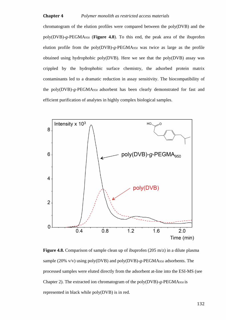
Chapter 4 Polymer monolith as restricted access materials
132
chromatogram of the elution profiles were compared between the poly(DVB) and the
poly(DVB)-g-PEGMA950 (Figure 4.8). To this end, the peak area of the ibuprofen
elution profile from the poly(DVB)-g-PEGMA950 was twice as large as the profile
obtained using hydrophobic poly(DVB). Here we see that the poly(DVB) assay was
crippled by the hydrophobic surface chemistry, the adsorbed protein matrix
contaminants led to a dramatic reduction in assay sensitivity. The biocompatibility of
the poly(DVB)-g-PEGMA950 adsorbent has been clearly demonstrated for fast and
efficient purification of analytes in highly complex biological samples.
Figure 4.8. Comparison of sample clean up of ibuprofen (205 m/z) in a dilute plasma
sample (20% v/v) using poly(DVB) and poly(DVB)-g-PEGMA950 adsorbents. The
processed samples were eluted directly from the adsorbent at-line into the ESI-MS (see
Chapter 2). The extracted ion chromatogram of the poly(DVB)-g-PEGMA950 is
represented in black while poly(DVB) is in red.

Chapter 4 Polymer monolith as restricted access materials
133
4.4. Concluding remarks
A number of essential morphological criteria are necessary for miniaturised SPE. First,
the adsorbent bed must be highly permeable to circumvent the backpressure limitations
of the miniaturised device. Second, to achieve a highly sensitive assay the adsorbent bed
must possess a large surface area for analyte interaction. The morphology of poly(DVB)
polymer monoliths makes them highly appealing for miniaturised SPE. These
adsorbents are both highly permeable and present a large surface area for analyte
interaction. Despite the desirable morphological attributes of polymer monolith
poly(DVB), use with highly complex biological samples can be problematic due to non-
specific protein adsorption to the hydrophobic surface. To improve the suitability of
poly(DVB) adsorbents for the preparation of biological samples the hydrophilic
monomers PEGMA360 and PEGMA950 were crosslinked to the hydrophobic surface to
provide a biocompatible RAMs.
It was essential to carefully evaluate the adsorbent performance for both poly(DVB)-g-
PEGMA360 and poly(DVB)-g-PEGMA950 as a hydrophilic coating can be a double-
edged sword. While the coating may prevent proteins from accessing the hydrophobic
surface, it may also diminish the adsorbent’s suitability for miniaturised SPE as longer
time is required for the analyte to penetrate for interaction with the hydrophobic surface.
Frontal analyses of anisole and ibuprofen were employed to compare the breakthrough
curves of the two grafted adsorbents with the ungrafted precursor. Poly(DVB)-g-
PEGMA360 exhibited a shallow breakthrough curve which is not conducive with rapid
miniaturised SPE workflows as it becomes necessary to optimise operational flow rates
to achieve high analyte recoveries. The presence of the PEGMA950 layer did not
dramatically affect the adsorptive properties of the poly(DVB)-g-PEGMA950, only a
small reduction in hydrophobic capacity was observed and the analyte mass transfer

Chapter 4 Polymer monolith as restricted access materials
134
was not noticeable affected. It is demonstrated here that the poly(DVB)-g-PEGMA950
adsorbent affords a high extraction performance independent to operational flow rate,
which is a highly desirable characteristic of a miniaturised SPE adsorbent. Furthermore,
protein adsorption was substantially restricted by the grafted a layer of PEGMA950
qualifying that the poly(DVB)-g-PEGMA950 is a high surface area biocompatible RAM
exceedingly suited towards miniaturised SPE. The poly(DVB)-g-PEGMA950 RAM was
demonstrated to produce cleaner extracts for a more sensitive ESI-MS assay. The
superiority of the poly(DVB)-g-PEGMA950 adsorbent morphology is extremely
beneficial for rapid and efficient miniaturised SPE purification of analytes in highly
complex biological samples.
4.5 References
[1] Westerlund, D. Chromatographia 1987, 24, 155–164.
[2] Annesley, T. M. Clin. Chem. 2003, 49, 1041–1044.
[3] Li, Y., Lee, M. L. J. Sep. Sci. 2009, 32, 3369–3378.
[4] Georgi, K., Boos, K. S. LC GC Eur. 2004, 17, 21–24.
[5] Cassiano, N. M., Lima, V. V., Oliveira, R. V., de Pietro, A. C., Cass, Q. B.
Anal. Bioanal. Chem. 2006, 384, 1462–1469.
[6] Souverain, S., Rudaz, S., Veuthey, J.-L. J. Chromatogr. B 2004, 801, 141–156.
[7] Unger, K. K. Chromatographia 1991, 31, 507–511.
[8] Hagestam, I. H., Pinkerton, T. C. Anal. Chem. 1985, 57, 1757–1763.
[9] Souverain, S., Rudaz, S., Veuthey, J.-L. J. Chromatogr. B 2004, 801, 141–156.
[10] Cook, S. E., Pinkerton, T. C. J. Chromatogr. A 1986, 368, 233–248.
[11] Boos, K.-S., Rudolphi, A., Vielhauer, S., Walfort, A., Lubda, D., Eisenbeiß, F.
Fresenius J. Anal. Chem. 1995, 352, 684–690.

Chapter 4 Polymer monolith as restricted access materials
135
[12] Gisch, D. J., Hunter, B. T., Feibush, B. J. Chromatogr. A 1988, 433, 264–268.
[13] Kanda, T., Shirota, O., Ohtsu, Y., Yamaguchi, M. J. Chromatogr. A 1996, 722,
115–121.
[14] Desilets, C. P., Rounds, M. A., Regnier, F. E. J. Chromatogr. A 1991, 544, 25–
39.
[15] Liska, I. J. Chromatogr. A 1993, 655, 163–176.
[16] Svec, F., Tennikova, T. B., Deyl, Z. Monolithic Materials; Elsevier, 2003.
[17] Jarmalavičienė, R., Kornyšova, O., Westerlund, D., Maruška, A. Anal. Bioanal.
Chem. 2003, 377, 902–908.
[18] Jarmalavičienė, R., Szumski, M., Kornyšova, O., Kłodzińska, E., Westerlund,
D., Krawczyk, S., Mickevičius, D., Buszewski, B., Maruška, A.
Electrophoresis 2008, 29, 1753–1760.
[19] Turson, M., Zhou, M., Jiang, P., Dong, X. J. Sep. Sci. 2011, 34, 127–134.
[20] Sýkora, D., Peters, E. C., Svec, F. Macromol. Mater. Eng. 2000, 275, 42–47.
[21] Lubbad, S. H., Buchmeiser, M. R. J. Chromatogr. A 2010, 1217, 3223–3230.
[22] Buchmeiser, M. R. Polymer 2007, 48, 2187–2198.
[23] Morcombe, C. R., Zilm, K. W. J. Magn. Reson. 2003, 162, 479–486.
[24] Rohr, T., Hilder, E. F., Donovan, J. J., Svec, F., Frechet, J. M. Macromolecules
2003, 36, 1677–1684.
[25] Tripp, J. A., Needham, T. P., Ripp, E. M., Konzman, B. G., Homnick, P. J.
React. Funct. Polym. 2010, 70, 414–418.
[26] Candish, E., Gooley, A., Wirth, H.-J., Dawes, P. A., Shellie, R. A., Hilder, E. F.
J. Sep. Sci. 2012, 35, 2399–2406.
[27] Smirnov, K. N., Dyatchkov, I. A., Telnov, M. V., Pirogov, A. V., Shpigun, O.
A. J. Chromatogr. A 2011, 1218, 5010–5019.
[28] Gu, B., Armenta, J. M., Lee, M. L. J. Chromatogr. A 2005, 1079, 382–391.

Chapter 4 Polymer monolith as restricted access materials
136
[29] Krenkova, J., Lacher, N. A., Svec, F. Anal. Chem. 2009, 81, 2004–2012.
[30] Stachowiak, T. B., Svec, F., Fréchet, J. M. J. Chem. Mater. 2006, 18, 5950–
5957.
[31] Barlow, K. J., Hao, X., Hughes, T. C., Hutt, O. E., Polyzos, A., Turner, K. A.,
Moad, G. Polym. Chem. 2014, 5, 722–732.
[32] Peters, E. C., Svec, F., Fréchet, J. M. J., Viklund, C., Irgum, K.
Macromolecules 1999, 32, 6377–6379.
[33] Gaborieau, M., Nebhani, L., Graf, R., Barner, L., Barner-Kowollik, C.
Macromolecules 2010, 43, 3868–3875.
[34] Hubbard, K. L., Finch, J. A., Darling, G. D. React. Funct. Polym. 1999, 42,
279–289.
[35] Jarmalavičienė, R., Kornyšova, O., Westerlund, D., Maruška, A. Anal. Bioanal.
Chem. 2003, 377, 902–908.
[36] Jarmalavičienė, R., Kornyšova, O., Bendokas, V., Westerlund, D., Buszewski,
B., Maruška, A. Anal. Bioanal. Chem. 2008, 391, 2323–2328.
[37] D’Sa, R. A., Meenan, B. J. Langmuir 2010, 26, 1894–1903.

Chapter 5 General conclusions & future perspectives
137
5. General conclusions and future perspectives
The following general conclusions summarise the main findings of this thesis exploring
improved miniaturised solid phase extraction (SPE) technologies for rapid sample
preparation assays.
5.1 Controlled directional flow miniaturised solid phase extraction
5.1.1 Conclusions
The inherent nature of the microextraction by packed sorbent (MEPS) dictates that both
sample and solvent must pass the adsorbent twice. First, upon aspiration then again
when the fluid is expelled from the syringe barrel. This bi-directional fluid flow was
shown to be problematic for two reasons; first the analyte is diluted and mixed in the
entire volume of elution solvent to yield a broad, unfocused elution band that
subsequently leads to reduced assay sensitivity. Second, to dispense, the analyte must
pass back through the adsorbent bed where normal partitioning equilibria dictates
diminished recoveries and increased sample carryover. In the example demonstrated in
this thesis the signal for MEPS carryover (second elution) of codeine was shown to be
65% of the first elution. Previous investigations into this technology have developed
rigorous adsorbent wash protocols to solve this problem. This is an unfavourable
solution as it necessitates lengthy optimisation protocols and introduces additional steps
to the workflow, increasing the time required for sample preparation.
The alternative approach is to improve the MEPS technology to circumvent the bi-
directional fluid flow path. A two-way valve was integrated into the barrel of the
syringe to control the direction that fluid is introduced to the device. Sample and solvent
could be apirated via a side arm into the syringe barrel then dispensed over the

Chapter 5 General conclusions & future perspectives
138
adsorbent bed. In the modified approach, the fluid makes a single pass of the adsorbent
bed. The advantage of controlled directional flow (CDF)-MEPS is that the sample can
be eluted without dilution, which eliminates the need to optimise the elution volumes.
The direct carryover of codeine was reduced from 65% for conventional MEPS to only
1% for CDF-MEPS, which removes the need for extensive wash cycles and aids in the
realisation of rapid miniaturised SPE workflows. The CDF-MEPS technology was
interfaced directly with electrospray ionisation mass spectrometry (ESI-MS). A rapid
and sensitive analysis was accomplished, as sharp, concentrated sample bands were
eluted directly from the adsorbent into the ESI-MS. The at-line ESI-MS analysis of a
range of opiates and urinary codeine metabolites was demonstrated using CDF-MEPS.
This demonstrated that the developed technology affords a flexible, fast and sensitive
approach to screen for target analytes (existing over a dynamic concentration range) in
biological matrices.
5.1.2 Future work
Traditional bi-directional miniaturised SPE devices necessitate a highly permeable bed
to facilitate the aspiration of fluid. While dispensing fluid over an adsorbent bed is not
generally problematic, fluid aspiration is challenging due to the restriction created by
the adsorbent bed. Thus, traditional miniaturised SPE devices’ are limited to short bed
lengths and large particulate SPE adsorbents (in the order of 40-100 µm). The CDF
valve removes these limitations as sample and solvent can be applied from above the
adsorbent bed. CDF-MEPS format reveals exciting possibilities to further develop the
device for more efficient sample preparation and at-line MS analysis. Preliminary work
with a disposable format of CDF-MEPS highlights that packing the cartridge with
smaller particles (3 - 20 µm) can deliver an increased assay sensitivity (Figure 5.1) [1].
In addition, longer bed lengths can also be investigated (2.5 mm – 100 mm). The

Chapter 5 General conclusions & future perspectives
139
procedures and benefits of a “in-syringe separation” hyphenated directly with MS can
be explored. A preliminary scoping study was undertaken for the at-line analysis of a
tryptic digest of cytochrome c using a 100 mm long adsorbent bed (10 µm C18
particulate) (Figure 5.2). A crude separation that reduces peak overlap can result in a
cleaner mass spectrum for increased assay sensitivity. However, the use of smaller
particles and longer adsorption bed lengths uncovers additional operational/engineering
challenges that must be addressed as increasing demand is placed on the syringe drivers
to dispense fluid over a well-packed adsorbent bed.
Figure 5.1. Extracted ion chromatogram of codeine-6-glucuronide in urine (10% v/v)
prepared at-line with 5 μm C18 particles (black) and 50 μm C18 particles (red). Insert:
Disposable SPE cartridge with an integrated CDF valve. The disposable format
integrates the valve into the cartridge body, the restriction created by the adsorbent bed
means fluid bypasses the bed on aspiration. During the dispense cycle the valve seals
and liquid penetrates through the packed bed.

Chapter 5 General conclusions & future perspectives
140
Figure 5.2. Right) The total ion chromatogram of an at-line elution of the tryptic
peptides of cytochrome c eluted from the 100 mm long 10 µm C18 adsorbent bed with
40:60 methanol:water v/v. Left) The extracted ion chromatogram corresponding to the
tryptic peptides of cytochrome c with zero and one missed cleavage.
Alternatively, coupling together MEPS adsorbent (2.5 mm) of complementing
chemistries for a “two-dimensional” miniaturised SPE could provide a more efficient
sample clean up. Fractions from the first extraction could be further purified on the
second adsorbent bed. Exciting possibilities exist to explore the at-line MS analysis of
drugs of abuse by coupling a strong cation exchange extraction with a hydrophobic
extraction.
Finally, the rapid analysis and reduced solvent consumption of the CDF-MEPS
approach represents a new era of sample preparation with the exciting capability of

Chapter 5 General conclusions & future perspectives
141
coupling MEPS with miniaturised portable and fieldable MS technologies for point-of-
collection analysis.
5.2 Polymer monoliths for solid phase extraction
5.2.1 Conclusions
Adsorbents for miniaturised SPE necessitate a number of essential morphological
criteria; first the adsorbent bed must be highly permeable to circumvent any
backpressure limitations of the device. Second, to achieve a highly sensitive assay the
adsorbent bed must possess a large surface area for a high capacity of analyte
interactions. Adsorbent fabricated from poly(divinyl benzene) (DVB) and
hypercrosslinked poly(4-vinylbenzyl chloride-co-styrene -co-DVB) (VBC-co-Sty-co-
DVB) displayed both a highly permeable macroporous structure with an enhanced
quantity of micro- and mesopores. The permanent highly porous morphology of large
surface area polymer monoliths made them highly appealing to investigate for SPE.
The polymeric microglobules possess non-porous highly crosslinked cores and any
interactable surface of polymer monolith adsorbents was accessible by a small diffusion
distance. Thus, the polymer monolith structure provides improved solute mass transfer
kinetics. Both frontal analysis and off-line SPE with anisole and phenol as the probe
analytes demonstrated the structural superiority of the poly(DVB) polymer monolith
over a conventional 85 µm poly(Sty-co-DVB) particulate. As a result of the polymer
monolith architecture the extraction efficiency of the poly(DVB) polymer monolith is
largely independent of the operational flow rate. This is highly attractive as lengthy
protocols to optimise operational flow rate to achieve a high analyte recovery can
largely be eliminated. In contrast, the extraction performance of the 85 µm poly(Sty-co-

Chapter 5 General conclusions & future perspectives
142
DVB) particulate adsorbent was found to be highly dependent on the flow rate of
extraction, not conducive with rapid SPE workflows.
However, an in depth probing into the structure of large surface area polymer monoliths
revealed some fundamental physical characteristics that could limit their suitability for
SPE. In aqueous environments the surface structure is almost exclusively composed of
micropores (< 2 nm). Therefore, the utility of large surface area polymer monoliths is
limited to analytes that can penetrate these small pores. While analytes such as anisole
and phenol could freely access the micropores, a size exclusion mechanism prevented
the larger probe analyte, cortisone, from penetrating the microporous structure, as
evident by the small analyte capacity.
The size exclusion mechanism of the poly(DVB) adsorbents was exploited to provide a
selective extraction of low molecular weight analytes in complex biological matrices.
Unfortunately, complex biological samples rich in proteins are problematic as proteins
adsorb non-specifically on the hydrophobic surfaces. This fouls the adsorbent and
complicates the “clean” processed sample. To improve the suitability of the poly(DVB)
adsorbents for the preparation of protein rich biological samples the hydrophilic
monomers poly(ethylene glycol) methacrylate Mn 360 (PEGMA360) and poly(ethylene
glycol) methylether methacrylate Mn 950 (PEGMA950) were crosslinked to the pendant
vinyl groups of the hydrophobic poly(DVB). These materials were investigated to
assess their suitability as large surface area biocompatible adsorbent.
Careful optimisation of the hydrophilic layer was essential, this layer must prevent
proteins fouling but must not diminish the hydrophobic capacity or reduce the
efficiency of the interaction between the analyte and the hydrophobic surface. The

Chapter 5 General conclusions & future perspectives
143
breakthrough curve of the poly(DVB)-g-PEGMA360 demonstrated that a thick
hydrophilic layer prevented rapid analyte mass transfer which is undesirable for
miniaturised SPE. In contrast, the thin layer of the PEGMA950 did not dramatically
effect the hydrophobic adsorptive properties of the poly(DVB)-g-PEGMA950, only a
small reduction in hydrophobic capacity was observed for both anisole and ibuprofen.
The poly(DVB)-g-PEGMA950 was demonstrated to produce cleaner extracts for a more
sensitive at-line ESI-MS assay. The superiority of the poly(DVB)-g-PEGMA950
adsorbent provides a powerful tool to yield cleaner sample extracts for MS analysis.
5.2.2 Future work
This work highlights that there is still a considerable need to gain a broader
understanding of polymer monolith morphologies to avoid assumptions of their
suitability for SPE applications. Further work is necessary to gain a precise insight into
the molecular weight limitations of large surface area polymer monoliths. A detailed
assessment using aqueous inverse size exclusion chromatography (ISEC) would provide
a greater understanding of these materials and assist with the further development of
adsorbents for SPE. To date, little work has been undertaken to identify and optimise
the exact characteristics of pore structure that could provide improved capacities for
SPE.
The macroporous properties of the larger surface area polymer monolith are one of the
simplest parameter that can be tuned to improve interactions between the adsorbent and
the analyte. The design of miniaturised SPE devices often necessitate that the fluid is
aspirated through the adsorbent bed. Thus the polymer monoliths investigated in this
work were specifically explored because of their large macropores (> 5 µm). However,
the adoption of the CDF syringe has removed this limitation enabling the fluid to be

Chapter 5 General conclusions & future perspectives
144
aspirated directly into the barrel of the syringe and applied to the adsorbent bed from
above. Scope now exists to explore polymer monoliths that present macropores over a
broader range (as low as 0.5 µm). Tuning the porogenic solvent to favour solubilisation
of the growing polymer chain will provide a larger number of smaller globules. This
ultimately increases the surface area producing a greater number of interaction points
for more efficient interactions between the analyte and the adsorbent as the sample is
forced through the smaller macropores.
Developing polymer monoliths with improved mesoporous structural characteristics and
a high density of functionality remains a practical challenge and innovation is required
to drive this technology forward. Recent works by Zhang et al. and Sterchele et al. has
described a refreshingly novel approach to synthesise polymer monoliths based on a
‘‘solvothermal’’ synthetic method. A polymerisation of poly(DVB) using a mixture of
tetrahydrofuran and water as the porogenic solvent was undertaken at a relatively high
polymerisation temperature (100°C) in an autoclave. The unusual polymerisation
conditions led to materials with very high specific surface areas of 700 m2 g-1 provided
by mesopores of 22 nm [3, 4]. Exciting possibilities exist to investigate these materials
and apply them as adsorbents for miniaturised SPE.
Further scope exists to investigate the thermal grafting of a variety of functional
monomers to large surface area adsorbents. The possibility to extend this work to the
thermal grafting of ion exchange monomers may facilitate the fabrication of mixed
mode polymer monolith with possess both a high hydrophobic capacity and a high ion
exchange capacity. In addition, a comprehensive study into PEGMA monomers with
increasing glycol chain length could enable the complete elimination of protein fouling
of hydrophobic surfaces.

Chapter 5 General conclusions & future perspectives
145
Finally, these biocompatible polymer monolith restricted access adsorbents could
provide an ideal structural morphology for the at-line MS analysis of drugs of abuse,
particularly the low molecular weight amphetamines, methamphetamine and their
structural analogs in biological fluids. Further investigation could assist in the
realisation of miniaturised portable analytical laboratories for confirmatory road side
drug testing.
5.3 References
[1] Candish, E., Gooley, A. A., Shellie, R. A., Hilder, E. F. G.I.T. Laboratory
Journal 2012, 18, 32–34.
[2] Candish. E. Hon, W. B., Gooley, A. A., Wirth, H-J., Shellie, R. A. & Hilder, E. F.
Microsample preparation devices for filtration, digestion and extraction of whole
blood (poster presentation). 62nd ASMS Conference on Mass Spectrometry and
Allied Topics. Baltimore. USA. 2014.
[3] Zhang, Y., Wei, S., Liu, F., Du, Y., Liu, S., Ji, Y., Yokoi, T., Tatsumi, T., Xiao,
F.-S. Nano Today 2009, 4, 135–142.
[4] Sterchele, S., Centomo, P., Zecca, M., Hanková, L., Jerabek, K. Microporous
Mesoporous Mater. 2014, 185, 26–29.

Appendix
146
Appendix A
Table 1. The program for the digital analytical syringe for the digital at-line CDF-
MEPS ESI-MSI workflow in Chapter 2
Step Aspirate
(μL)
Dispence
(μL)
Speed
(μL/min)
Valve Position
Condition
Aspirate - Methanol 50 400 1
Dispense 50 400 1
Equilibrate
Aspirate - Water 50 400 1
Dispense 50 400 1
Sample load
Aspirate - Urine (10% v/v) 50 300 1
Dispense 50 300 1
Wash
Aspirate - Water 50 400 1
Dispense 50 400 1
Elute
Aspirate - Methanol 50 400 2
Dispense 50 20 1
*Note: All solvents and samples contained 0.1% v/v formic acid

Appendix
147
Appendix B
Table 2. The program for the digital analytical syringe for SPE workflow for HPLC
analysis in Chapter 3 and 4.
Step Aspirate
(μL)
Dispence
(μL)
Speed
(μL/min)
Valve Position
Condition
Aspirate - Methanol 100 3500 2
Dispense 100 1000 or 500 1
Equilibrate
Aspirate - Water 100 3500 2
Dispense 100 1000 or 500 1
Sample load
Aspirate – Aqueous solution 100 3500 2
Dispense 100 1000 or 500 1
Wash
Aspirate - Water 100 3500 2
Dispense 100 1000 or 500 1
Elute
Aspirate - Methanol 100 3500 2
Dispense 100 1000 or 500 1

Appendix
148
Appendix C
Table 3. Characteristics of hypercrosslinked poly(VBC-co-Sty-co-DVB) polymer
monoliths in the dry state obtained from argon adsorption/desorption at 77K and
mercury intrusion porosimetry
Adsorbent SBET SMicropore
Pore
volume
Micropore
volume
Macropore
size
(m2g-1) (m2g-1) (cm3g-1) (cm3g-1) (µm)
Hypercrosslinked
16% VBC 817 810 0.381 0.368 12
12% VBC 737 719 0.353 0.324 11
8% VBC 686 675 0.327 0.308 7

Appendix
149
Appendix D
Table 4. The program for the digital analytical syringe for the digital at-line CDF-
MEPS ESI-MSI workflow in Chapter 4
Step Aspirate
(μL)
Dispence
(μL)
Speed
(μL/min)
Valve Position
Condition
Aspirate - Acetonitrile 50 3500 1
Dispense 50 1000 1
Equilibrate
Aspirate - Water 50 3500 1
Dispense 50 1000 1
Sample load
Aspirate - Plasma (20% v/v) 50 3500 1
Dispense 50 1000 1
Wash
Aspirate - Water 50 3500 1
Dispense 50 1000 1
Elute
Aspirate - Acetonitrile 50 3500 2
Dispense 50 20 1
*Note: All solvents and samples contained 0.3% v/v ammonium hydroxide