Embed Size (px)
Citation preview
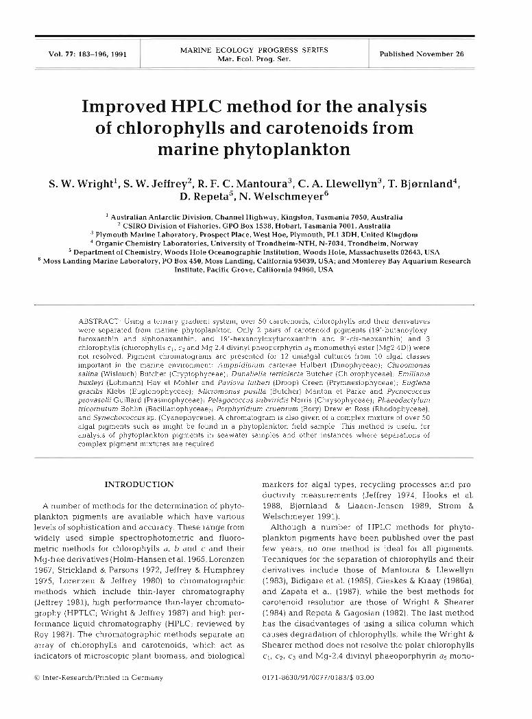
Vol. 77: 183-196, 1991 MARINE ECOLOGY PROGRESS SERIES Mar. Ecol. Prog. Ser.
- -
Published November 26
Improved HPLC method for the analysis of chlorophylls and carotenoids from
marine phytoplankton
Australian Antarctic Division, Channel Highway, Kingston, Tasmania 7050, Australia CSIRO Division of Fisheries, GPO Box 1538. Hobart. Tasmania 7001, Australia
Plymouth Marine Laboratory, Prospect Place, West Hoe, Plymouth, PL1 3DH, United Kingdom Organic Chemistry Laboratories, University of Trondheirn-NTH, N-7034, Trondheim, Norway
Department of Chemistry, Woods Hole Oceanographic Institution, Woods Hole, Massachusetts 02643. USA Moss Landing Marine Laboratory, PO Box 450, Moss Landing, California 95039, USA; and Monterey Bay Aquarium Research
Institute, Pacific Grove, California 94960, USA
ABSTRACT. Using a ternary gradlent s}istem, over 50 carotenoids, chlorophylls and their denvatives were separated from marine phytoplankton. Only 2 palrs of carotenoid pigments (19'-butanoyloxy- fucoxanthin and siphonaxanthin, and 19'-hexanoyloxyfucoxanthin and 9'-cis-neoxanthln) and 3 chlorophylls (chlorophylls c , , c2 and Mg 2.4 divinyl pheoporphyrin a5 monomethyl ester [Mg2,4D]) were not resolved. Pigment chromatograms are presented for 12 uniaigal cultures from 10 algal classes important in the marine environment: Alnphidinlum cal-terae Hulbert (Dinophyceae), Chroomonas salina (Wislouch) Butcher (Cryptophyceae), Dunallella tertrolecta Butcher (Chlorophyceae); Emil~anla huxleyi (Lohmann) Hay et Mohler and Pavlova lutheri (Droop) Green (Prymnesiophyceae); Euglena gracllis Klebs (Euglenophyceae); Micl-omonas pusllla (Butcher) Manton et Parke and Pycnococcus provasohi Guillard (Praslnophyceae); Pelagococcus subv~ridis Norris (Chrysophyceae); Phaeodactylum tncornutum Bohlln (Baclllanophyceae); Porphyndlum cruentum (Bory) Drew et Ross (Rhodophyceae), and Synechococcus sp (Cyanophyceae). A chromatogram IS also given of a complex mixture of over 50 algal pigments such as might be found in a phytoplankton field sample This method is useful for analysis of phytoplankton plgments in seawater samples and other Instances where separations of complex pigment mixtures are required
INTRODUCTION
A number of methods for the determination of phyto- plankton pigments are available which have various levels of sophistication and accuracy. These range from widely used simple spectrophotonletric and fluoro- metric methods for chlorophylls a, b and c and their Mg-free derivatives (Holm-Hansen et al. 1965, Lorenzen 1967, Strickland & Parsons 1972, Jeffrey & Humphrey 1975, Lorenzen & Jeffrey 1980) to chromatographic methods which include thin-layer chromatography (Jeffrey 1981), high performance thin-layer chromato- graphy (HPTLC; Wright & Jeffrey 1987) and high per- formance liquid chromatography (HPLC; reviewed by Roy 1987). The chromatographic methods separate an array of chlorophylls and carotenoids, which act as indicators of microscopic plant biomass, and biological
Inter-Research/Printed in Germany
markers for algal types, recycling processes and pro- ductivity measurements (Jeffrey 1974, Hooks et al. 1988, Bjarnland & Liaaen-Jensen 1989, St ron~ & Welschmeyer 1991).
Although a number of HPLC methods for phyto- plankton pigments have been published over the past few years, no one method is ideal for all pigments. Techniques for the separation of chlorophylls and their derivatives include those of Mantoura & Llewellyn (1983), Bidigare et al. (1985), Gieskes & Kraay (1986a), and Zapata et al. (1987), while the best methods for carotenoid resolution are those of Wright & Shearer (1984) and Repeta & Gagosian (1982). The last method has the disadvantages of using a silica column which causes degradation of chlorophylls, while the Wright & Shearer method does not resolve the polar chlorophylls c l , c2, c3 and Mg-2,4 divinyl phaeoporphyrin as mono-
Mar Ecol. Prog. Ser. 77: 183-196, 1991
methyl ester (Mg2,4D) which coelute as a single peak, or adequately resolve the chlorophyllides a and b. A single method, suitable for the full range of important chlorophylls, chlorophyll derivatives and taxonomically significant carotenoids found in phytoplankton was urgently needed, particularly for international oceano- graphic programs currently underway for climate change studies (e.g. the Joint Global Ocean Flux Study).
Aware of the need to evaluate and compare pigment methods, the Scientific Committee for Oceanic Research (SCOR) founded 'Woriung Group 78: Measurement of Photosynthetic Pigments in Seawater', whose objectives included: (1) comparing spectrophotometric, fluoromet- ric and HPLC methods for chlorophyll a determination; (2) recommending a simple isocratic HPLC shipboard technique for chlorophylls and derivatives; (3) evaluat- ing gradient HPLC techniques for detailed separations of chlorophylls and carotenoids.
For the third objective, the Working Group mem- bers who attended the SCOR Chlorophyll Workshop at the CSIRO Marine Laboratories, November, 1988 used the method of Wright & Shearer (1984) for carotenoids and non-polar chlorophylls, and added to it a n initial solvent system used by Welschmeyer & Hoepffner (in press) for separating polar chlorophylls. An improved method resulted which separates more than 50 chlorophylls, carotenoids, their derivatives and isomers from marine phytoplankton in one ternary gra- dient system. This paper gives details of the HPLC method and examples of chromatograms from refer- ence algae. Quantitative analyses form part of the SCOR Workshop Report, and are not duplicated here. While several carotenoids and 3 chlorophyll cpigments (Jeffrey 1989) are not resolved, this new method pro- vides oceanographers with a significantly improved system for the analysis of phytoplankton pigments In the sea.
MATERIALS AND METHODS
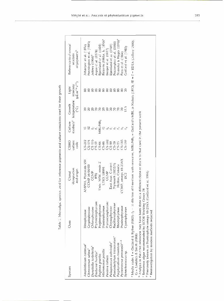
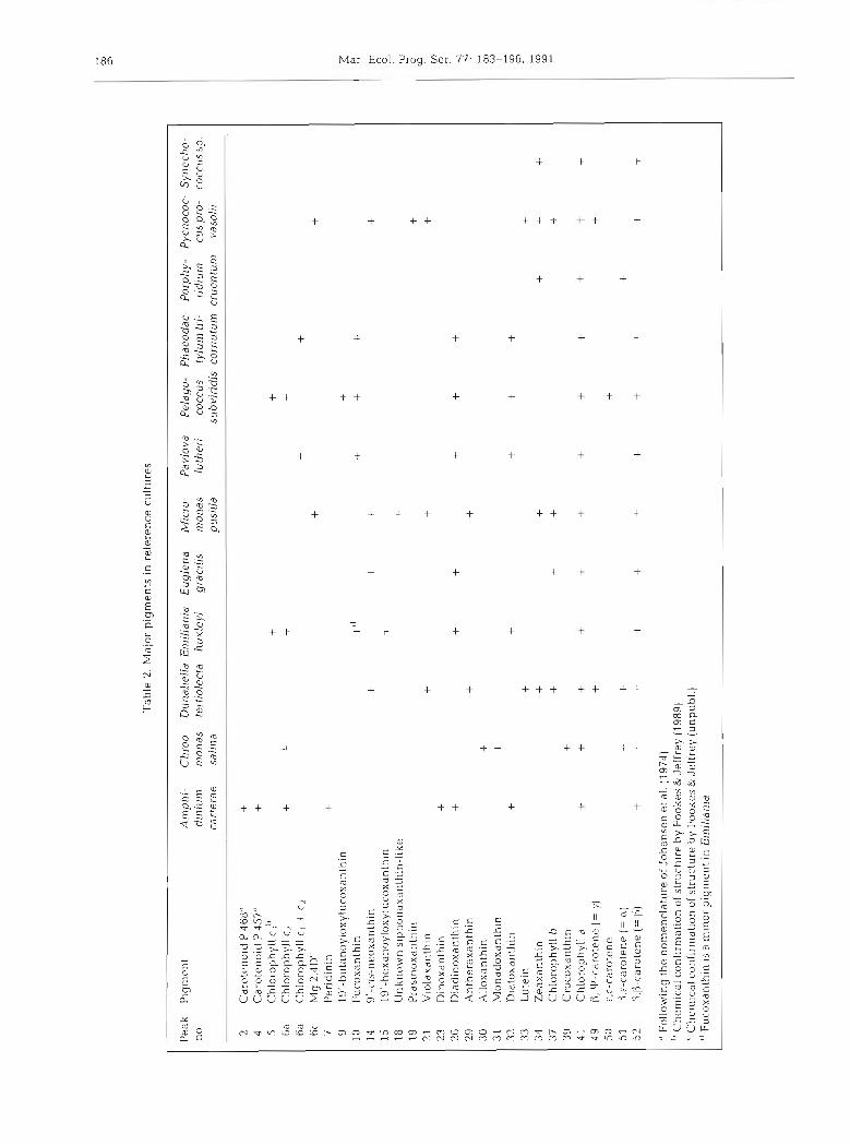
Algal cultures. Because of the instability and com- mercial non-availability of pure algal pigment stan- dards, the SCOR Working Group 78 recommended that the source of such standards should be reference algal cultures whose pigments had been fully characterised. A small number of species were selected (Table 1) which together contained most significant pigments known to be found in marine phytoplankton (Table 2). Additional cultures to those recommended by SCOR were incIuded for the present work to ensure that a full range of algal classes was represented.
The cultures were maintained in the CSIRO Algal Culture Collection (Jeffrey 1980). Table 1 llsts clonal
designation, CSIRO Culture Code number, culture medium, temperature and light intensities used for growth as well as references to the chemical identifica- tion of pigments. All cultures were grown in stationary 125 m1 Erlenmeyer flasks containing 75 m1 culture medium. Illumination was provided by banks of Philips 'daylight' fluorescent tubes, beneath glass shelves sup- porting the culture flasks. Light irradiances (12 : 12 h 1ight:dark cycles) were measured in the culture medium with a Biospherical Optics light meter. Culture stocks were transferred every 7 to 10 d.
Culture harvest. Cells were harvested before the end of log phase and 10 m1 culture aliquots were taken for pigment analysis. These were filtered through 25 mm Whatman GF/F filters under low vacuum (e.g. 5 100 mm Hg) and rinsed with filtered medium or sea- water. The filter was extracted immediately, or was folded, placed in a labelled cryotube and immediately frozen in liquid nitrogen. Several replicates of each culture were made. The time taken from filtration to liquid nitrogen was not more than 30 S, and for the experiments described in this paper, storage in liquid nitrogen was not more than 1 to 10 d.
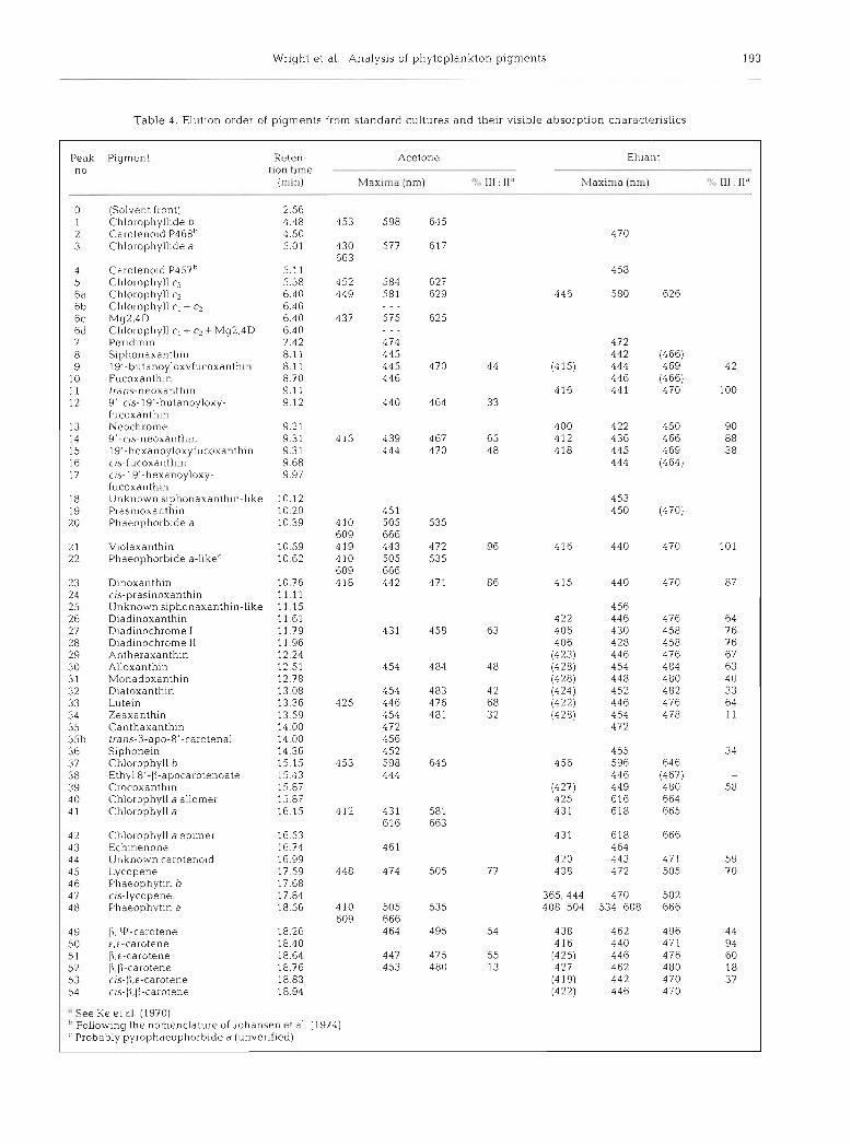
Field samples. The sample illustrated was collected from 90 m depth at 51°54' S, 73'14' E in the Southern Ocean in the vicinity of Heard Island on 9 June 1990. Seawater (1 to 5 1) was filtered through a 25 mm diameter Whatman GF/F filter. This was folded, blotted dry and stored in a cryotube under liquid nitrogen and returned to the laboratory for HPLC. The particular sample illustrated was stored for 3 mo before analysis. No discernible changes occur during this time (SCOR Working Group 78, unpubl.).
Pigment extraction: cultures. The SCOR Working Group 78 report will separately consider in detail the optimum methods for extraction of pigments from cultures and field samples. The frozen filter containing the algal sample was cut into small slices (several mm x 1 cm) and was ground for 30 s in a motorized grinder with a teflon pestle in 2.7 m1 l00 % acetone. Water (0.3 ml) was then added to make up to 90 % acetone, and the sample reground. Methanol could have been used as extraction solvent, and often is for field samples (see below). The results of comprehensive tests of sol- vents and extraction protocols will be described in the SCOR report. The filter plus acetone extract was trans- ferred to a plastic centrifuge tube cut down to 5 ml, which had been 'needle-pierced' a t the bottom (Wnght & Shearer 1984) and covered with a 5 mm circle of GF/ F filter. The cut-down centrifuge tube containing the extract was then placed in the top of a 10 m1 conical glass centrifuge tube, and both tubes centrifuged in tandem for 3 min at 700 X g. The clear green-yellow extract was collected in the lower glass centrifuge tube, while the filter debris remained in the upper tube. The
ble
1. M
icro
alga
l sp
ecie
s u
sed
for
ref
eren
ce p
igm
ents
an
d c
ultu
re c
ondi
tion
s u
sed
for
the
ir g
row
th
Sp
ecie
s C
lass
C
lona
l C
SlR
O
Cu
ltu
re
Gro
wth
L
ight
R
efer
ence
to c
hem
ical
de
sign
atio
n cu
ltur
e m
ediu
ma
tem
per
atu
re
inte
nsit
y an
alys
is
and
ori
gin
cod
e ("
C)
m-'
S-'
) of
pig
men
tsh
Am
phid
iniu
m c
arte
raeC
D
ino
ph
yce
ae
AM
PH
I. P
lym
outh
450
C
S-2
12
G
20
80
Jo
han
sen
et
al.
(197
4)
Ch
roo
mo
nas
sal
inaC
C
ryp
top
hy
ceae
C
CM
P 5
6/00
/00
CS
-174
fE
20
8
0
Pen
ning
ton
et
al.
(198
5)
Dun
alie
lla
tert
iole
ctaC
C
hlo
rop
hy
ceae
C
CM
P
CS
-175
12
20
8
0
Jeff
rey
(196
8)e
Em
ilia
nia
huxl
eyi c
P
rym
nesi
ophy
ceae
B
T6
CS
-57
12
20
80
A
rpin
et
al.
(197
6)
Eu
gle
na
grac
ilis
E
ug
len
op
hy
ceae
U
niv.
NS
W,
stra
in Z
C
S-6
6 M
BL
/NB
2 20
8
0
Bjo
rnla
nd e
t al
. (1
989)
M
icro
mon
as p
usil
la
Pra
sino
phyc
eae
UT
EX
LB
991
CS
-86
G
20
80
h
cket
ts (
1966
a, b
, 19
70)'
P
avlo
va l
uth
eri
Pry
mne
siop
hyce
ae
CCMP
CS
-182
f2
20
8
0
Ber
ger
et
al.
(197
7)
Pel
agoc
occu
s su
bvir
idis
C
Ch
ryso
ph
yce
ae
Eas
t A
ust.
Cu
rren
t C
S-9
9 12
20
80
Bjo
rnla
nd e
t al
. (1
989)
P
hae
od
acty
lum
tri
corn
utum
C
Bac
illa
riop
hyce
ae
Ply
mou
th (
1052
/1)
CS
-29
f2
20
80
Pen
ning
ton
et a
l. (
1988
) P
orph
yrid
ium
cru
entu
rn
Rho
doph
ycea
e H
alif
ax,
Can
ada
CS
-25
f2
20
80
S
tran
sky
& H
ager
(19
70)'
P
ycno
cocc
us p
rova
soli
ic+
P
rasi
noph
ycea
e C
CM
P, o
meg
a 48
-23A
X
CS
-185
f2
20
80
Fo
ss e
t al
. (1
984)
S
ynec
hoco
ccus
sp
. C
yan
op
hy
ceae
D
C2
CS
-197
12
17
.5
20
Gui
llar
d et
al.
(19
85)
" M
ed
~a
cod
e: f
= G
uill
ard
& R
ythe
r (1
962)
; f2
= %
dil
utio
n of
f
med
ium
w1t
.h s
eaw
ater
, MB
UN
Bl
= G
uill
ard'
s M
BL
in
Nic
hols
(1
97
3);
fE =
f +
ED
TA
(Je
ffre
y 19
80);
G
= L
oebl
ich
& S
mit
h (1
968)
" A
naly
sls
In t
he
cite
d re
fere
nce
may
hav
e b
een
do
ne
on a
dif
fere
nt c
lone
or
stra
~n
to t
hat
use
d I
n th
e p
rese
nt
wor
k "R
efer
ence
alg
ae r
eco
mm
end
ed b
y S
CO
R W
orki
ng G
rou
p 7
8 P
revi
ousl
y k
no
wn
as
Pra
sino
phyt
e o
meg
a 48
-23A
X (
Gui
llar
d et
al.
199
1)
' Rei
nves
tiga
tion
by
mod
ern
met
ho
ds
requ
ired
Tab
le 2
. M
ajor
pig
men
ts i
n re
fere
nce
cult
ures
Pea
k P
lgnr
c~rt
A
mp
hi-
C
hroo
- D
~rn
alie
lla E
rnil
iani
a E
ugle
na
Mic
ro-
Pav
lova
P
elag
o-
Ph
aeo
dac
- P
orph
y-
Pyc
noco
c- S
yn
ech
o-
no.
dini
um
rnon
as
ter-
trol
ecla
hu
xley
i g
rac~
lls
rnon
as
luth
eri
cocc
us
lylu
m t
ri-
r~d
iurn
cu
s p
ro-
cocc
us s
p.
carl
erae
sa
lina
pu
sill
a su
hvir
idis
cor
nutu
m
crue
ntum
va
soli
i
Car
olen
oid
P 4
68"
+ C
aro
trn
oid
P 4
57"
+ C
hlor
ophy
ll C
.;''
Ch
lol~
~p
hy
ll
CL
Ch
loro
l~h
yll
c, +
c2
My
2,4D
C
Per
idin
in
19'-butanoyloxylucoxanthin
Fuc
oxan
thin
9'
-cis
-neo
xanl
hin
19
'-h
cxan
oy
lox
yfu
cox
anth
~n
IJ
nkno
wn
siph
onax
anth
in-l
ike
Pra
sino
xant
hin
Vlo
ldxd
nlhl
n D
ino
xd
nth
~n
D
iadl
noxa
nthi
n A
nthe
raxa
nthi
n A
llox
anth
in
Mon
adox
anth
ln
Dia
toxd
nthi
n L
utei
n Z
eaxa
nthi
n C
hlor
ophy
ll b
C
roco
xdnt
hin
Chl
orop
hyll
a
13, V
J-ca
rote
ne (=
y)
r,c-
caro
tcnc
13
,~-c
arot
ene (=
m)
13,1
3-ca
rote
ne (=
P)
" F
ollo
win
g th
e no
men
clat
ure
of J
oh
anse
n e
t al.
(19
74)
" C
heni
ical
con
firm
atio
n of
str
uctu
re b
y F
ooke
s &
Jef
frey
(19
89)
" C
hcm
~cd
l conf
irm
atio
n of
str
uctu
re b
y F
ooke
s &
Jef
frey
(u
np
ub
l )
" F
ucox
anth
ln 1
s a m
inor
pig
nien
l in
Ein
l/ra
nrd
Wright et a1 : Analys~s of phytoplankton pigments 187
homogenizer and pestle were rinsed with 1 m1 90 % acetone, whlch was added to the filter debris in the upper tube, and recentrifuged. The clear comblned extracts from the lower tube were then transferred to a 5 m1 volumetric flask and made up exactly to 5 m1 with 90 "k acetone to ensure volumetric accuracy. After fil- tering through a Millex-SR 0.5 ,pm filter (Milllpore), an aliquot of the extract (about 25 p1) was injected immediately into the liquid chromatograph for pigment analysis.
Pigment extraction: field samples. The procedure for field samples was slightly different from that of the cultured samples because of the need to keep extraction volumes to a minimum and hence achieve a higher final concentration of pigments. Filters were cut into small pieces (several mm X 1 cm) and sonicated for 30 s in the cut down centrifuge tubes using 1.5 m1 methanol (or acetone) and a Braun Labsonic 1510 equipped with a 4 mm diameter probe, operated at 50 W. The bottom needle-hole in the centrifuge tube was securely covered with a single layer of Nescofilm or Paraf~lm to prevent solvent leakage durlng sonication. During centrifuga- tion (as above), the f~ lm burst allowing the extract to collect in the lower tube. The extract was filtered (Mil- lex-SR 0.5 pm) and then, immediately before injection, was diluted with water to 80 O/O MeOH (or 66 O/O acetone if acetone was used). 125 p1 of the diluted extract was injected. In general, the field samples were much less concentrated than the culture samples, hence a larger injection volun~e was required. However, if a large aliquot of strong solvent (e.g. methanol or 90 % acetone) was injected, then the pigment bands, particularly the early ones, were too broad and retention times decreased. Dilution of the extract, as mentioned above, increased the affinity of pigments for the column during the loading step, resulting in sharper peaks and allow- ing greater overall loading than possible with the undi- luted extract. It is important that the extract is not diluted until immediately before injection, since highly aqueous extracts are not stable and pigment losses (adsorption and precipitation, particularly the more hydrophobic pigments) occur on standing.
It was suggested by an anonymous reviewer that the addition of 2 % ammonium acetate buffer to the methanol extraction solvent improved the height and sharpness of the chlorophyll c peaks. This has been confirmed in our laboratories.
High performance liquid chromatography. Three HPLC instruments were used for comparison of separa- tion efficiencies: (1) a Waters Associates liquid chromatograph comprising M6000A, M45 and M501 pumps, Valco injector, Hewlett-Packard 8450A diode array spectrophotometer, and a Waters 440 absorbance detector, connected via a Waters System Interface Module to a microcomputer running Waters Maxima
software; (2) a Varlan Model 5000 l~quid chromato- graph with Varian UV50 variable wavelength detector, Varlan Fluorichrome fluorescence detector, and Varian 'Vista' 402 data module; and (3) a Spectraphysics HPLC comprising an SP8800 ternary pump, Spectra- Focus detector and a Gllson 231 autosampler (200 btl
loop) with samples refrigerated to -10 'C. Chromato- grams from the Varian 5000 were used in Figs. 1 & 2, and from the Spectraphysics instrument in Fig. 3. Reversed phase columns used were Spherisorb ODS2, 25 cm x 4.6 mm ID, 5 pm particle size, packed by Australian Government Analytical Laboratories, Mel- bourne (90000 to 100000 plates m-'). Similar resolu- tion was obtained with commercial columns such as Waters Resolve C I 8 and Activon Ultratechsphere, although some C I 8 columns tested were not as effi- cient, particularly in the ability to resolve lutein and zeaxanthln.
Pigment detection was at 436 nm (Figs. 1 to 3) for all chlorophylls and carotenoids and 405 nin for phaeo- phytin a and phaeophorbide a (not shown). The solvent systems used were as follows:
Solvent A: 80:20 methanol : 0.5 M ammonium acetate (aq . ; pH 7.2 v/v)
Solvent B: 90:lO acetonitrile (210 nm UV cut-off grade) : water (v/v)
Solvent C: ethyl acetate (HPLC grade)
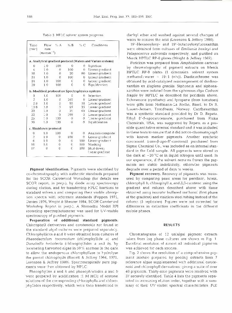
Flow rate was 1 m1 min-'. The gradient systems used are shown in Table 3. In the analytical system an initial 4 min gradient from 100 O/O A to 100 % B provided improved separation of polar compounds. The program returned to initial conditions and re-equilibrated for 5 min before the next sample injection. Table 3a shows the gradient used on the Varian system to obtaln the results presented in Figs. 1 & 2. Table 3b shows the modified procedure for the Spectraphysics system. In this case, the slope of the gradlent for Solvent C was reduced between 2.6 and 13.6 min for the region of the chromatogram between fucoxanthin and chlorophyll a (increasing the resolution in the most complex region of the chromatogram) and increased elsewhere to reduce the overall time for analysis. Table 3c shows the shut- down protocol whereby the column is washed in the strongest solvent (ethyl acetate) to remove any retained material before being shutdown overnight or for storage. The start-up protocol is the reverse of this program.
Methanol, acetonitrile and ethyl acetate were HPLC grade reagents from Waters and BDH (Hypersolve), used without further purification other than filtration and degassing by sonication. Water was purified using a Millipore Milli-Q system. Ammonium acetate was A.R. grade.
188 Mar. Ecol. Prog. Ser. 77: 183-196, 1991
Table 3 HPLC solvent system programs
Time Flow ?/o A % B % C Conditions (min) rate
(m1 min-'1
a. Analytical gradient protocol (Waters and Varian systems)
0 1.0 100 0 0 Injection 4 1 .O 0 100 0 Linear gradient
18 1.0 0 20 80 Lineargradlent 21 1.0 0 100 0 Linear gradient 24 1.0 100 0 0 Linear gradient 29 1.0 100 0 0 Equilibration
b. Modified protocol on Spectraphysics systems
Injection Linear gradlent Linear grad~ent Linear gradient Linear gradient Linear gradient Linear gradient Equilibration
c. Shutdown protocol
0 1.0 100 0 0 Analysis complete 3 1.0 0 100 0 Linear gradient 6 1.0 0 0 100 L~near gradlent
16 1.0 0 0 100 Washing 17 0 0 0 100 Shutdown;
linear gradient
Pigment identification. Pigments were identified by CO-chromatography with authentic standards prepared for the SCOR Carotenoid Workshop (for details see SCOR report, in prep.), by diode array spectroscopy during elution, and by transferring HPLC fractions to standard solvents and comparing their visible absorp- tion spectra with reference standards (Foppen 1971, Davies 1976, Wright & Shearer 1984, SCOR Carotenoid Workshop Report in prep.). A Shimadzu Model RP1 recording spectrophotometer was used for UV-visible spectroscopy of purified pigments.
Preparation of additional standard pigments. Chlorophyll derivatives and carotenoids not found in the standard dlyal cultures were prepared separately. Chlorophyllides a and b were obtained from cultures of Phaeodactylum tricornutum (chlorophyllide a ) and Dunaliella tert~olecta (chlorophyllides a and b) , by incubating harvested algae in 50 % acetone in the dark to allow the endogenous chlorophyllase to hydrolyse the parent chlorophylls (Barrett & Jeffrey 1964, 1971. Lorenzen & Jeffrey 1980). Spectroscopically pure pig- ments were then obtained by HPLC.
Phaeophytins a and b and phaeophorbides a and b were prepared by acidification (1 M HC1) of acetone solutions of the corresponding chlorophylls and chloro- phyllides respectively, which were then transferred to
diethyl ether and washed against several changes of water to remove the acid (Lorenzen & Jeffrey 1980).
19'-Hexanoyloxy- and 19'-butanoyloxyfucoxanthin were obtained from cultures of Emiliania huxleyi and Pelagococcus subviridis respectively, and purified on Merck HPTLC RP-8 plates (Wright & Jeffrey 1987).
Peridinin was prepared from Amphidinium carterae by chromatography of pigment extracts on Merck HPTLC RP-8 plates (1 dimension, solvent system methanol : water = 19 : 1 (v/v)). Diadinochrome was obtained by acid-catalyzed rearrangement of diadino- xanthin ex Euglena gracilis. Siphonein and siphona- xanthin were isolated from the siphonous alga Codium fragile by HPTLC as described for peridinin above. Echinenone (synthetic) and lycopene (from tomatoes) were gifts from Hoffmann-La Roche, Basel, to Dr S. Liaaen-Jensen, Trondheim, Norway. Canthaxanthin was a synthetic standard provided by Dr D. Repeta. Ethyl 8'-P-apocarotenoate, purchased from Fluka Chemicals, USA, was suggested by Repeta as a pos- sible quantitative internal standard and it was included in these tests to ensure that it did not CO-chromatograph with known marker pigments. Another synthetic carotenoid, trans-6-apo-8'-carotenal, purchased from Sigma Chemical Co., was included as a n internal stan- dard in the field sample. All pigments were stored in the dark at -20 'C or in liquid nitrogen until used. In our experience, if the solvent remains frozen the pig- ments are stable indefinitely, otherwise pigments degrade over a period of days to weeks.
Pigment recovery. Recovery of pigments was meas- ured by comparing peak areas for peridinin, lutein, chlorophyll b, chlorophyll a, and @,B-carotene using the gradient and column described above with those obtained using isocratic buffered methanol (first phase of the gradient) and stainless steel tubing instead of the column (5 replicates). Figures were not corrected for differences in extinction coefficients in the different mobile phases.
RESULTS
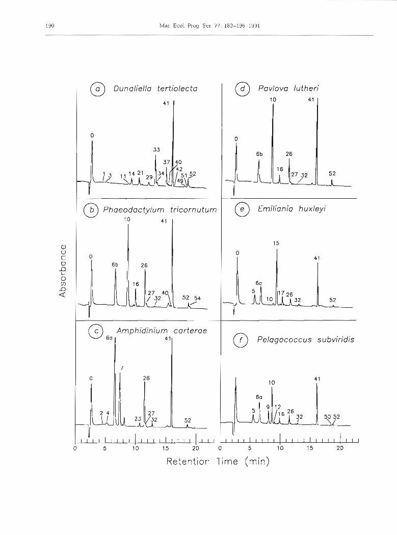
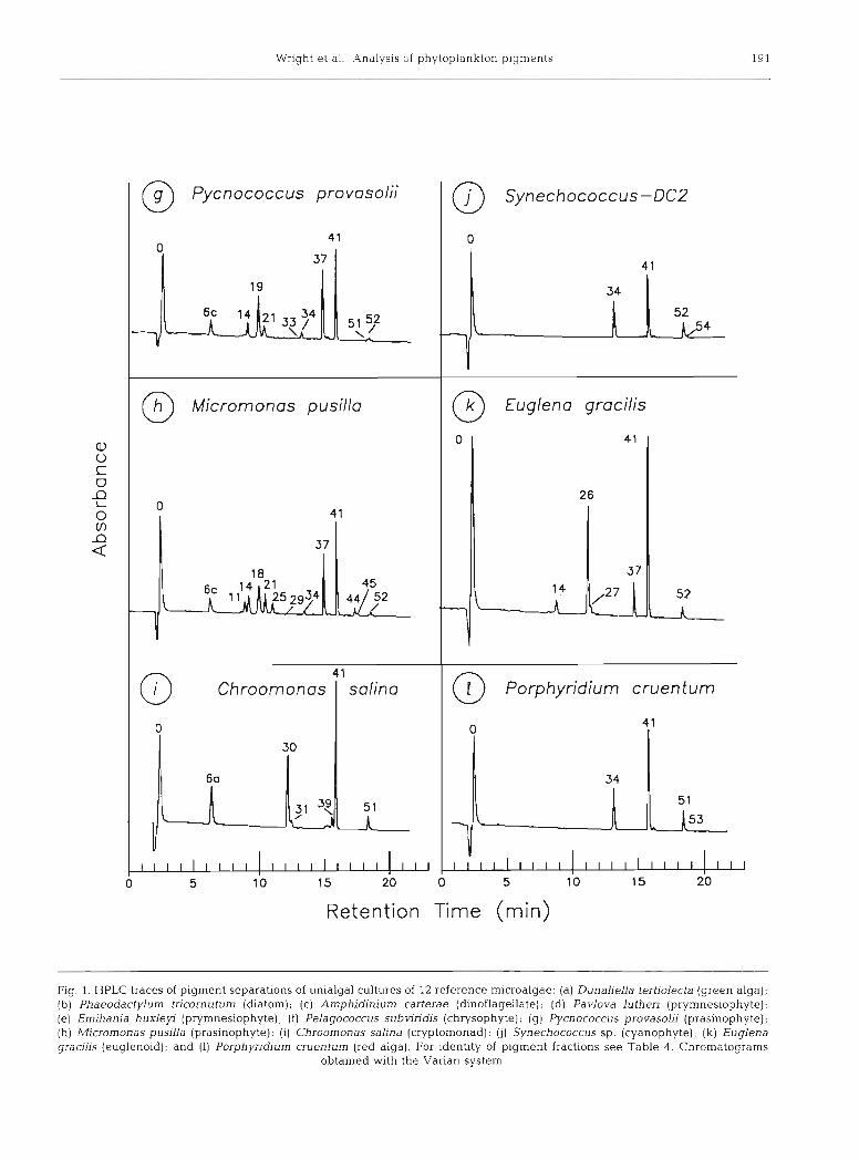
Chromatograms of 12 unialgal pigment extracts taken from log phase cultures are shown in Fig. 1 Excellent resolution of almost all individual pigments was achieved for each species.
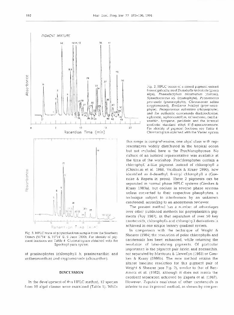
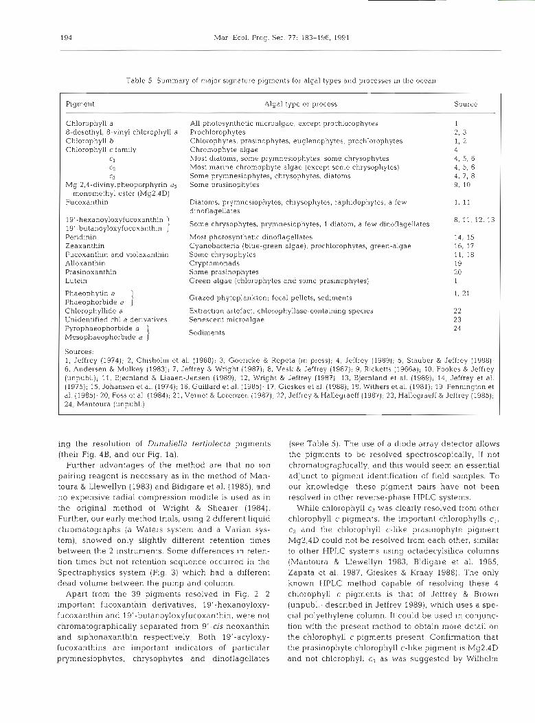
Fig. 2 shows the resolution of a comprehensive pig- ment mixture prepared by pooling extracts from 7 reference algae supplemented with additional carote- noid and chlorophyll derivatives; giving a suite of over 40 pigments. Thirty-nine pigments were resolved with 37 securely identified. Table 4 lists the pigments sepa- rated i.n increasing elution order, together with a sum- mary of their UV-visible spectral characteristics. Full
Wright et al.: Analysis of F )hytoplankton pigments 189
visible spectra in appropriate solvents (including HPLC eluant) are available in the SCOR Working Group 78 Cai-otenoid Report (in prep.).
At the polar end of the chromatogram, the phytol- free chlorophyll derivatives chlorophyllide b, chloro- phyllide a (Fig. l a ) and chlorophyll c3 (Fig, l e ) were almost completely resolved from chlorophyll c, + c2 + Mg2,4D which coeluted as a single peak (Figs. l b & 2).
In the complex central region of the chromatogram, most of the known carotenoid bio-markers (peridinin, fucoxanthin, prasinoxanthin, violaxanthin, alloxanthin, lutein, zeaxanthin, canthaxanthin and siphonein) were adequately resolved. Only 2 pairs of carotenoids were not separated: 19'-butanoyloxyfucoxanthin from sipho- naxanthin, and 19'-hexanoyloxyfucoxanthin from 9'- cis-neoxanthin, although the individual components of each pair could be distinguished spectrally by diode array detection. Many minor carotenoid components were also resolved and identified (Figs. 1 & 2 and Table 4).
At the non-polar end of the chromatogram, the phy- tolated chlorophylls a and b, the phaeophytins, and the less-polar carotenoids and carotenes were resolved. In fresh extracts, the native chlorophylls predominated with a very small proportion of chlorophyll a allomer, epimer and trace unknown chlorophyll a derivatives (less than 1 %). We cannot determine whether such trace pigments are present in situ, or are formed during the short (1 to 10 d) low temperature storage period or the short 'extraction to injection' period. The internal carotenoid standard, ethyl 8'-P-apocarotenoate, sepa- rated as a single peak between chlorophyll b and chlorophyll a (Flg. 2). The other internal standard, trans-(3-apo-8'-carotenal, CO-chromatographed with canthaxanthin, eluting before chlorophyll b (Fig. 3). The hydrocarbons, lycopene (v,zy-carotene) and p,$- carotene, were completely resolved. (3,~-Carotene eluted as the leading shoulder of the (3,(3-carotene peak. Cis-(3,(3-carotene eluted as the trailing shoulder of the (3,(3-carotene peak (see Figs. l a & 2).
Recovery of pigments from the column was excellent. Recoveries measured were: peridinin, 102 % ; lutein, 107 %; chlorophyll b, 100 %; chlorophyll a, 107 % ; and @,@-carotene, 98 %. Recovery figures greater than 100 % may reflect the different extinction coefficients in the mobile phases used.
In each of the single algal chromatograms, the native all-trans carotenoids predominated, with only small amounts of cis-isomers. Note that in Dunaliella ter- tiolecta (Fig. l a ) , and all other green algae to our knowledge, neoxanthin is present as the 9'-cis isomer in fresh extracts (Bjornland 1990). The trans isomer appears in the extract within hours of preparation.
Most of the pigments of the species examined have been characterised with modern spectrometric
methods and require no detailed discussion here (cf. Table 1). Exceptions are those of Dunaliella tertiolecta (Fig. la ) , Porphyridium cruentum (Fig. 11) and the widely-distributed prasinophyte Micromonas pusilla (Fig. lh) . The last species contains the chlorophyll c- like pigment Mg2,4D (Fookes & Jeffrey unpubl.), and a range of carotenoids differing from those of normal chlorophytes (see Dunaliella tertiolecta, Fig, l a ) , and prasinoxanthin-containing prasinophytes (Pycnococ- cus provasolii, Fig. lg) . Two significant carotenoid components in M. pusilla had siphonaxanthin-like spectra; the first one (peak 18) almost co-chromato- graphed with prasinoxanthin but differed from it spec- trally in lacking any trace of a shoulder at about 475 nm. It is possible that minor contaminants may be altering its apparent spectrum. In some cultures, a leading shoulder was observed on peak 18, having maxima at (406), 429 and 455 nm. A similar shoulder was observed on the neoxanthin peak with maxima at 447 and 470 nm. The identity of these pigments is presently being studied by Dr S Liaaen-Jensen and co-workers, Trond- heim, Norway.
Two phaeophorbide a-like peaks CO-eluted with carotenoids but did not interfere with detection of the carotenoids because they did not absorb at 436 nm. The phaeophorbides can be detected without interfer- ence from carotenoids using fluorescence (excitation 407 nm, emission 672 nm for phaeophorbide a ; excita- tion 432 and emission 659 nm for phaeophorbide b); or absorption at 405 or 665 nm.
Fig. 3 shows the resolution of chlorophylls and carotenoids from a field sample from the Southern Ocean in the vicinity of Heard Island. Note that the field sample was analysed with the Spectraphysics HPLC system and the retention times do not exactly match those of the Varian HPLC system, listed in Table 4 . The absence of chlorophyll a epimer and chloi-ophyllides indicated that little degradation had occurred. The chromatogram shows a good resolution of the major pigments. Apart from chlorophyll a, the chromatogram is dominated by fucoxanthin, its 19'- hexanoyloxy- and 19'-butanoyloxy-derivatives, chloro- phylls c3 and cl+c2, and diadinoxanthin. Smaller amounts of peridinin, chlorophyll b, prasinoxanthin, and antheraxanthin were present along with traces of alloxanthin and zeaxanthin.
Examination of the samples by light microscopy showed them to be dominated by small diatoms (con- sistent with the abundance of fucoxanthin), a numeri- cally smaller dinoflagellate population (indicated by pendinin) with many unidentifiable flagellates. The pigment composition suggests that the flagellates com- prised mainly prymnesiophytes (chlorophyll CS, fuco- xanthin, 19'-hexanoyloxyfucoxanthin and 19'-butan- oyloxyfucoxanthin) together with smaller populations
Ab
sorb
an
ce
Mar Ecol. Prog. Ser 77- 183-196. 1991
PIGMENT MIXTURE
6d 1014.15
a v C 0
9 0 V)
D Q
5 10 15 20
Retention Time (rnin)
Fig. 2. HPLC traces of a mixed pigment extract from algal cultures of Dunaliella tertiolecta (green alga), Phaeodactylum tricornutum (diatom), Synechococcus sp. (cyanophyte), Pycnococcus provasolii (pras~nophyte), Chroomonas salina (cryptomonad), Emiliania huxleyi (prymnesio- phyte), Pelagococcus subviridis (chrysophyte), and the authentic carotenoids diadinochrome, siphonein, siphonaxanthin, echinenone, cantha- xanthin, lycopene, peridinin and the internal synthetic standard ethyl 8'-0-apocarotenoate. For identity of pigment fractions see Table 4. Chromatogram obtained with the Varian system
Fig. 3. HPLC trace of phytoplankton sample from the Southern Ocean (51°54' S, 73"14' E, 9 June 1990). For identity of pig- ment fractions see Table 4 . Chromatogram obtained with the
Spectraphysics system
of prasinophytes (chlorophyll b, prasinoxanthin and antheraxanthin) and cryptomonads (alloxanthin).
DISCUSSION
In the development of this HPLC method, 12 species from 10 algal classes were examined (Table 1). While
this range is comprehensive, one algal class with rep- resentatives widely distributed in the tropical ocean but not included here is the Prochlorophyceae. No culture of an isolated representative was available at the time of the workshop. Prochlorophytes contain a chlorophyll a-like pigment instead of chlorophyll a (Chisholm et al. 1988, Veldhuis & Kraay 1990), now identified as 8-desethyl, 8-vinyl chlorophyll a (Goe- ricke & Repeta in press). These 2 pigments can be separated in normal phase HPLC systems (Gieskes &
Kraay 1983a), but coelute in reverse phase systems unless converted to their respective pheophytins, a technique subject to interference by an unknown carotenoid, according to an anonymous reviewer.
The present method has a number of advantages over other published methods for phytoplankton pig- ments (Roy 1987), in that separation of over 50 key carotenoids, chlorophylls and chlorophyll derivatives is achieved in one simple ternary gradient system.
In comparison with the technique of Wright & Shearer (1984) the resolution of polar chlorophylls and carotenoids has been enhanced, while retaining the resolution of later-eluting pigments. Of particular importance is the pigment pair lutein and zeaxanthin, not separated by Mantoura & Llewellyn (1983) or Gies- kes & Kraay (198613). The new method retains the almost baseline resolut~on for thls plgment pair of Wright & Shearer (see Fig. 2), similar to that of Ben- Amotz et al. (1982), although it does not match the excellent separation achieved by Zapata et al. (1987). However, Zapata's resolution of other carotenoids is inferior to our improved method, as shown by compar-
Wright et a l . : Analysis of phytoplankton p igments 19
T a b l e 4. Elution order of pigments from s tandard cul tures a n d their visible absorpt ion characteristics
Peak Plgment no
Reten- Acetone Eluant tion tlme
(min) Maxima (nm) 111 : ll"' Maxlrna (nm) v # % 111 11''
(Solvent front) Chlorophyll~de b Carotenold P468" Chlorophyll~de a
Carotenold P457b Chlorophyll c3 Chlorophyll c2 Chlorophyll c, + c2 My2.4D Chlorophyll c l + cL+ Mg2.4D Perldinin Slphonaxanthin 19'-butanoyloxyfucoxanth~n Fucoxanthin trans-neoxanthin 9'-cis-19'-butanoyloxy- fucoxanthln Neochrome 9 ' -cis-neoxanth~n 19'-hexanoyloxyfucoxanthi~~ cis-fucoxanthin cis-19'-hexanoyloxy- fucoxanthin Unknown slphonaxanthln-like Praslnoxanthin Phaeophorbide a
Violaxanthin Phaeophorblde a-likec
D~noxanthin as-prasinoxanth~n Unknown s~phonaxanthin-like Dladlnoxanthin Diadlnochrome I Dladlnochrome 11 Antheraxanthin Alloxanthin Monadoxanthin Diatoxanthln Luteln Zeaxanthln Canthaxanthln trans-P-apo-8'-carotenal Slphonein Chlorophyll b Ethyl 8'-P-apocarotenoate Crocoxanthin Chlorophyll a allomer Chlorophyll a
Chlorophyll a eplmer Echlnenone Unknown carotenold Lycopene Phaeophytln b c~s-lycopene Phaeophytin a
" See Ke et al (1970) h Follow~ng the nomenclature of Johansen et a1 (1974) ' Probably pyrophaeophorb~de a (unver~fied)
194 Mar Ecol. Prog. Ser. 77: 183-196. 1991
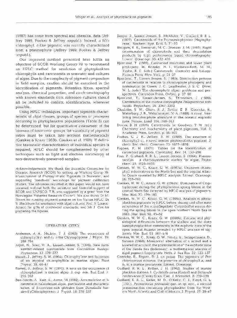
Table 5. Summary of major signature pigments for algal types and processes in the ocean
Pigment Algal type or process Source
Chlorophyll a All photosynthetic microalgae, except prochlorophytes 1 8-desethyl, 8-vinyl chlorophyll a Prochlorophytes 2, 3 Chlorophyll b Chlorophytes, prasinophytes, euglenophytes, prochlorophytes 1, 2 Chlorophyll c family Chromophyte algae 4
C1 hlost diatoms, some prymnesiophytes, some chrysophytes 4. 5. 6 C2 Most marine chromophyte algae (except some chrysophytes) 4. 5, 6 C3 Some prymnesiophytes, chrysophytes, diatoms 4. 7, 8
Mg 2,4-divinylpheoporphyrin a5 Some prasinophytes 9, 10 monomethyl ester (Mg2,4D)
Fucoxanthin Diatoms, prymnesiophytes, chrysophytes, raphidophytes, a few 1, 11 dinoflagellates
19'-hexanoyloxyfucoxanthin 1 Some chrysophytes, prymnesiophytes, 1 diatom, a few dinoflagellates 8, 11, 12, 13 19'-butanoyloxyfucoxanthin Peridinin Most photosynthetic dinoflagellates 14, 15 Zeaxanthin Cyanobacter~a (blue-green algae), prochlorophytes, green-algae 16, 17 Fucoxanthin and violaxanthin Some chrysophytes 11. 18 Alloxanthin Cryptomonads 19 Prasinoxan thin Some prasinophytes 20 Lutein Green algae (chloropbytes and some prasinophytesj 1
Phaeophytin a i Grazed phytoplankton; fecal pellets, sediments 1, 21 Phaeophorbide a Chlorophyllide a Extraction artefact, chlorophyllase-containing species 22 Unidentified chl a derivatives Senescent microalgae 23 Pyrophaeophorbide a 1 Sediments 24 Mesophaeophorbide a
Sources: 1, Jeffrey (1974); 2. Chisholm et al. (1988); 3, Goericke & Repeta (in press); 4, Jeffrey (1989); 5, Stauber & Jeffrey (1988); 6 , Andersen & Mulkey (1983); 7, Jeffrey & Wright (1987); 8, Vesk & Jeffrey (1987); 9, Ricketts (1966a); 10, Fookes & Jeffrey (unpubl.); 11, Bjornland & Liaaen-Jensen (1989), 12, Wright & Jeffrey (1987); 13, Bjarnland et al. (1989); 14, Jeffrey et al. (1975); 15, Johansen et al. (1974); 16, Guillard et al. (1985); 17, Gieskes et a1 (1988); 18, W~thers et al. (1981); 19, Pennington et al. (1985); 20, Foss et al. (1984); 21, Vernet & Lorenzen (1987); 22, Jeffrey & Hallegraeff (1987); 23, Hallegraeff & Jeffrey (1985); 24, Mantoura (unpubl.)
i n g t h e resolution of Dunaliella tertiolecta pigments (their Fig. 4B, a n d our Fig. l a ) .
Further advantages of t h e method a r e that n o ion pairing reagent is necessary a s in the method of Man- toura & Llewellyn (1983) a n d Bidigare e t al. (1985), a n d n o expensive radial compression module is used a s in t h e original method of Wright & Shearer (1984). Further, our early method trials, using 2 d ~ f f e r e n t liquid chromatographs (a Waters system a n d a Varian sys- tem), showed only slightly different retention times be tween t h e 2 instruments. Some differences in reten- tion times but not retention sequence occurred in the Spectraphysics system (Fig. 3) which h a d a different d e a d volume be tween t h e p u m p a n d column.
Apart from t h e 3 9 pigments resolved in Fig. 2, 2 important fucoxanthin derivatives, 19'-hexanoyloxy- fucoxanthin a n d 19'-butanoyloxyfucoxanthin, were not chromatographically separated from 9'-cis neoxanthin a n d s iphonaxanthin respectively. Both 19'-acyloxy- fucoxanthins a r e important indicators of particular prymnesiophytes, chrysophytes a n d dinoflagellates
( see Table 5 ) . T h e use of a diode array detector allows t h e pigments to b e resolved spectroscopically, if not chromatographically, a n d this would seem a n essential adjunct to pigment identification of field samples. To our knowledge, these pigment pairs have not b e e n resolved in other reverse-phase HPLC systems.
While chlorophyll c3 w a s clearly resolved from other chlorophyll c pigments, the important chlorophylls c l , c* and the chlorophyll c-like prasinophyte pigment Mg2,4D could not b e resolved from e a c h other, similar to other HPLC systems using octadecylsilica columns (Mantoura & Llewellyn 1983, Bidigare e t al. 1985, Zapata et al. 1987, Gieskes & Kraay 1988). The only known HPLC method capable of resolving these 4 chlorophyll c pigments is that of Jeffrey & Brown (unpubl . ; described in Jeffrey 1989), which uses a spe- cial polyethylene column. It could b e used in conjunc- tion with the present method to obtain more detail on the chlorophyll c pigments present. Confirmation that the prasinophyte chlorophyll c-like pigment is Mg2,4D and not chlorophyll c, a s w a s suggested by Wilhelm
Wright et al.: Analysis of phytoplankton pigments 195
(1987) has come from spectral a n d chemical da ta (Jef- frey 1989, Fookes & Jeffrey unpubl . ) . Indeed, a fifth
chlorophyll c-like pigment was recently characterised
from a prasinophyte (Jeffrey 1989, Fookes & Jeffrey
unpubl.).
O u r improved method presented here fulfils an objective of SCOR Working Group 78 to recommend
a n HPLC method for resolution of phytoplankton
chlorophylls a n d carotenoids in seawater a n d cultures
of a lgae. Due to the complexity of pigment composition
in field samples, caution should b e exercised in the
identification of pigments . Retention times, spectral
analysis, chemical properties, and CO-chromatography
with known standards from reference cultures should
all b e included to confirm identifications, wherever
possible. Using HPLC techniques, important pigments charac-
teristic of algal classes, groups of species or processes
occurring in phytoplankton populations (Table 5) can
b e determined, but for quantitative assessment of the
biomass of taxonomic groups the variability of pigment
ratios must b e taken into account mathematically
(Gieskes & Kraay 198313, Gieskes e t al. 1988). If defini- tive taxonomic characterization of individual species is
required, HPLC should b e complemented by other
techniques such as light a n d electron microscopy of non-destructively preserved samples.
Acknowledgements. We thank the Scientific Committee for Oceanic Research (SCOR) for setting up Work~ng Group 78: Measurement of Photosynthetic Pigments in Seawater, and supporting 'hands-on' workshops for pigment calibration studies. The advances reported in this paper would not have occurred without both the initiative and financial support of SCOR and UNESCO. T.B. was supported by a grant from the Norwegian Fisheries Research Council. We also thank Dr M. Brown for running pigment samples on the Varian HPLC, Dr S. Blackburn for assistance with algal cultures, Prof. S. Liaaen- Jensen for gifts of carotenoid standards, and Mr J . Cox for preparing the figures.
LITERATURE CITED
Andersen, R. A., Mulkey, T J. (1983). The occurrence of chlorophylls c, and c2 in the Chrysophyceae. J. Phycol. 19: 289-294
Arpin, N., Svec, \h' A., Liaaen-Jensen, S. (1976). New fuco- xanthin-related carotenoids from Coccolithus huxleyi. Phytochem. 15: 529-532
Barrett, J. , Jeffrey, S. W. (1964). Chlorophyllase and formation of an atypical chlorophyllide in nlarlne algae. Plant Physiol. 39: 44-47
Barrett, J., Jeffrey, S. W. (1971). A note on the occurrence of chlorophyllase in marine algae. J. exp. mar. Biol Ecol. 7: 255-262
Ben-Amotz, A., Katz, A., Avron, M. (1982). Accumulation of p- carotene in halotolerant algae: purification and characteri- sation of P-carotene-rich globules from Dunaliella bar- dawil (Chlorophyceae). J. Phycol. 18: 529-537
Berger, R., Liaaen-Jensen, S., McAlister, V., Guillard. R. R . L. (1977). Carotenoids of the Prymnesiophyceae (Haptophy- ceae). Biochem Syst. Ecol. 5: 71-75
Bidigare, R R . , Kennicutt, M. C., Brookes, J. M. (1985). Rapid determinat~on of chlorophylls and their degradation products by high performance liquid chromatography. Limnol. Oceanogr. 30: 432-435
Bjornland, T (1989). Carotenoid structures and lower plant phylogeny. In: Krinsky. N. I . , Mathews-Roth, M. M., Taylor, R. F. (eds.) Carotenoids: chemistry and biology. Plenum Press. New York, p. 21-37
Bjernland. T., Liaaen-Jensen, S. (1989). Distribution patterns of carotenoids in relation to chromophyte phylogeny and systematics. In: Green, J. C., Leadbeater, B. S. C., Diver, W L. (eds.) The chromophyte algae: problems and per- spectives Clarendon Press, Oxford, p. 37-60
Bjarnland, T., Liaaen-Jensen, S., Throndsen, J (1989). Carotenoids of the marine chrysophyte Pelagococcus sub- viridis. Phytochem. 28: 3347-3353
Chisholm, S. W., Olson. R. J . , Zettler, E. R., Goericke, R.. Waterbury, J B., Welschmeyer, N. A. (1988). A novel, free- living prochlorophyte abundant in the oceanic euphotic zone. Nature, Lond. 334: 340-343
Davies, B. H. (1976). Carotenoids. In: Goodwin, T W. (ed.) Chemistry and biochemistry of plant pigments, Vol. 2. Academic Press, London, p. 38-165
Fookes, C. J. R.. Jeffrey, S. W. (1989). The structure of chlorophyll c3, a novel marine photosynthetic pigment. J. chem. Soc. chem. Commun. 23: 1827-1828
Foppen, F. H. (1971). Tables for the identification of carotenoid pigments. Chromatog. Rev. 14: 133-298
Foss, P,, Guillard, R. R L., Liaaen-Jensen, S. (1984). Pras~no- xanthin - a chemosystematic marker for algae. Phyto- chem. 23- 1629-1633
Gieskes, W. W. C., Kraay, G. W. (1983a). Unknown chloro- phyll a derivatives in the North Sea and the tropical Atlan- tic Ocean revealed by HPLC analysis. Limnol. Oceanogr. 28: 757-766
Gieskes, W. W. C. , Kraay, G. W. (1983b). Dominance of Cryp- tophyceae during the phytoplankton spring bloom in the central North Sea detected by HPLC analysis of pigments. Mar. Biol. 75: 179-185
Gieskes, W. W C., Kraay, G. W. (1986a). Analysis of phyto- plankton pigments by HPLC before, during and after mass occurrence of the microflagellate Corymbellus aureus du- ring the spring bloom in the open northern North Sea in 1983. Mar Biol. 92: 45-52
Gieskes, W. W C.. Kraay, G. W. (1986b). Floristic and phy- siological differences between the shallow and the deep nanophytoplankton community in the euphotic zone of the open tropical Atlantic revealed by HPLC analysis of pig- ments. Mar Biol. 91: 567-576
Gieskes, W. W. C , Kraay, G. W., Nontji, A., Setiapermana, D . , Sutomo (1988) Monsoonal alternation of a mixed and a layered structure in the phytoplankton of the euphotic zone of the Banda Sea (Indonesia): a mathematical analysis of algal pigment fingerprints. Neth. J. Sea Res. 22: 123-137
Goericke, R., Repeta, D. J. (in press). The pigments of Pro- chlorococcus minutus: the presence of chlorophyll a , and b, in a marine procaryote. Limnol. Oceanogr.
Guillard, R. R. L., Ryther, J . H. (1962). Studies of marine plankton diatoms. I. Cyclotella nana Hustedt and Detonula confervacea (Cleve) Gran. Can. J . Microbiol. 8: 229-239
Guillard. R. R. L., Keller, M. D., O'Kelly, C. J., Floyd. G. L. (1991). Pycnococcus provasolii gen. et sp. nov., a coccoid prasinoxanthin-containing phytoplankter from the West- ern North Atlantic and Gulf of Mexico. J . Phycol. 27. 3 9 4 7
196 Mar Ecol. Prog. Ser. 77 183-196, 1991
Guillard, R. R. L.. Murphy. L. S., Foss, P., Liaaen-Jensen, S. (1985). Synechococcus spp. as likely zeaxanthin-dominant ultraphytoplankton in the North Atlantic. Limnol. Oceanogr 30: 412-4 14
Hallegraeff, G. M-, Jeffrey, S. W. (1985). Description of new chlorophyll a alteration products in marine phytoplankton. Deep Sea Res. 32: 697-705
Holm-Hansen. O., Lorenzen, C. J . , Holmes, R. W., Strickland, J. D. H. (1965). Fluorimetric determination of chlorophyll. J . Cons, perm. int. Explor. Mer 30: 3-15
Hooks, C . E., Bidigare, R R., Keller, M. D. , Guillard, R. R. L. (1988). Coccoid eukaryotic marine ultraplankters with four different HPLC pigment signatures. J . Phycol. 24: 571-580
Jeffrey, S . W. (1968). Quantitative thin-layer chromatography of chlorophylls and carotenoids from marine algae. Biochim. biophys. Acta 162: 271-285
Jeffrey, S. W. (1974). Profiles of photosynthetic pigments in the ocean using thin-layer chromatography. Mar. Biol. 26: 101-110
Jeffrey. S. W (1980). Cultivating unicellular marine plants. CSIRO (Hobart, Tasmania) Fish and Oceanogr Ann. Rep. 1977-1979, p. 22-43
Jeffrey, S. W. (1981). An improved thin-layer chromatographic technique for marine phytoplankton pigments. Llmnol. Oceanogr 26: 191-197
Jeffrey, S. W. (1989). Chlorophyll c pigments and their dis- tribution in the chrornophyte algae. In: Green, J . C., Leadbeater, B. S. C., Diver, W L. (eds.) The chromophyte algae: problems and perspectives. Clarendon Press, Oxford, p. 13-36
Jeffrey, S . W. , Hallegraeff, G. M. (1987). Chlorophyllase dis- tribution in 10 classes of phytoplankton - a problem for chlorophyll analysis. mar. Ecol. Prog. Ser. 35: 293-304
Jeffrey. S. W., Humphrey. G. F. (1975). New spectrophotomet- ric equations for determining chlorophylls a, b, cl and cl in h ~ g h e r plants, algae and natural phytoplankton. Biochem. Physiol. Pflanz. 167: 191-194
Jeffrey. S. W.. Sielicki, M.. Haxo. F. T (1975). Chloroplast p ~ g m e n t patterns in dinoflagellates J. Phycol. 1 l : 374-384
Jeffrey, S. W. , Wright, S. W. (1987) A new spectrally distinct component In preparations of chlorophyll c from the microalga Emiliania huxleyi (Prymnesiophyceae). Bio- chim. biophys. Acta 894: 180-188
Johansen, J . E., Svec, W A., Liaaen-Jensen, S. , Haxo, F T. (1974). Carotenoids of the Dinophyceae. Phytochem. 13: 2261-2271
Ke, B , Imsgard. F . , Kjosen, H., Liaaen-Jensen, S. (1970). Electronic spectra of carotenoids at 77°K Biochm bio- phys. Acta 210: 139-152
Loeblich, A. R., Smith, V E. (1968). Chloroplast pigments of the marine dinoflagellate Gyrodinium resplendens. Lipids 3 - 5-13
Lorenzen, C. J . (1967). Determination of chlorophyll and pheopigments: spectrophotometric equations. Limnol. Oceanogr 12: 343-346
Lorenzen, C. J . , Jeffrey, S. W (1980). Determinat~on of chlorophyll in seawater. UNESCO Tech. Pap. Mar. Sci. 35. p. 1-20
Mantoura, R. F. C., Llewellyn, C. A. (1983). The r a p ~ d determi- nation of algal chlorophyll and carotenoid pigments and their breakdown products in natural waters by reverse- phase high-performance liquid chromatography. Analyt. Chim. Acta 151: 297-314
Nichols, H. W (1973). Growth media-freshwater In. Stein,
This article was submitted to the editor
J . R. (ed.) Handbook of phycological methods. Cambridge University Press, Cambridge, p. 7-24
Pennington, F., Guillard. R. R. L , Liaaen-Jensen, S (1988). Carotenoid distribution patterns in the Bacillariophyceae. Biochem. Syst. Ecol. 16: 589-592
Pennington, F. C., Haxo. F. T., Borch, G., Liaaen-Jensen. S. (1985). Carotenoids of Cryptophyceae. Biochem. Syst. Ecol. 13: 215-219
Repeta, D. J., Gagosian, R. B. (1982). Carotenoid transforma- tions in coastal marine waters. Nature, Lond. 295: 51-54
Ricketts, T R. (1966a). Magnesium 2,4-divinylpheoporphyrin as monornethyl ester, a protochlorophyll-like pigment pre- sent in some unicellular flagellates. Phytochem. 5. 223-229
Ricketts, T R. (1966b). The carotenoids of the phytoflagellate, Micromonas pusilla. Phytochem. 5: 571-580
Ricketts, T. R. (1970). The pigments of the Prasinophyceae and related organisms. Phytochem. 9: 1835-1842
Roy, S. (1987). High performance liquid chromatographic analysis of chloropigments. J . Chromatog 391. 19-34
Stauber, J . L., Jeffrey. S. W (1988). Photosynthetic pigments In 51 species of marine diatoms. J. Phycol. 24: 158-172
Stransky, H., Hager, A. (1970). Das Carotinoidmuster und die Verbreitung des lichtinduzierten Xanthophyllcyclus in verschiedenen Algenklassen. IV. Cyanophyceae und Rhodophyceae. Arch Mikrobiol. 72: 84-96
Strickland, J . D. H., Parsons, T R. (1972). A practical hand- book of seawater analysis, 2nd edn. Bull. Fish. Res. Bd Can. 167
Strom, S. L., Welschmeyer, N. A. (1991). Pigment-specific rates of phytoplankton growth and microzooplankton grazing in the open subarctic Pacific Ocean. Limnol. Oceanogr. 36: 50-63
Veldhuis, M. J . W., Kraay, G . W. (1990). Vertical distribution and pigment composition of a picoplanktonic prochloro- phyte in the subtropical North Atlantic: a combined study of HPLC analysis and flow cytometry. Mar. Ecol. Prog. Ser. 68: 121-127
Vernet, M., Lorenzen, C. J . (1987). The presence of chlorophyll b and the estimation of phaeopigrnents in marine phytoplankton. J . Plankton Res 9: 255-265
Vesk, M., Jeffrey, S W. (1987) Ultrastructure and pigments of two strains of the picoplanktonic alga, Pelagococcus sub- viridis (Chrysophyceae). J . Phycol. 23: 322-336
Welschmeyer, N. A., Hoepffner, N. (in press). Xanthophyll cycling I. Dynamics of r a p ~ d , light-induced pigment changes in phytoplankton. Limnol. Oceanogr
Wilhelm, C. (1987). Purification and identification of chlorophyll c, from the green alga Mantoniella squamata Biochem. biophys. Acta 892. 23-29
Withers, N. W., Fiksdahl, A., Tuttle, R. C.. Liaaen-Jensen, S. (1981). Carotenoids of the Chrysophyceae. Comp. Biochem. Physiol. 68B: 345-349
Wright, S. W., Jeffrey, S. W (1987). Fucoxanthin plgment markers of marine phytoplankton analysed by HPLC and HPTLC. Mar Ecol. Prog. Ser. 38: 259-266
Wright, S W , Shearer, J . D. (1984). Rapid extraction and high- performance liquid chromatography of chlorophylls and carotenoids from marine phytoplankton. J . Chromatog. 294: 281-295
Zapata, M , Ayala, A. M., Franco, J . M. , Garrido, J. L (1987) Separation of chlorophylls and their degradation products in marine phytoplankton by reversed-phase high per- formance liquid chromatography. Chromatographia 23: 26-30
manuscript first received: May 29, 1991 Revised version accepted: August 15, 1991