Embed Size (px)
Citation preview
Improved Correspondence for DTI PopulationStudies via Unbiased Atlas Building
Casey Goodlett1, Brad Davis1,2, Remi Jean3, John Gilmore3, and GuidoGerig1,3
1 Department of Computer Science, University of North Carolina,[email protected]
2 Department of Radiation Oncology, University of North Carolina3 Department of Psychiatry, University of North Carolina ?
Abstract. We present a method for automatically finding correspon-dence in Diffusion Tensor Imaging (DTI) from deformable registrationto a common atlas. The registration jointly produces an unbiased av-erage DTI atlas along with diffeomorphic correspondence between eachimage. The registration image match metric uses a feature detector forthin fiber structures of white matter, and interpolation and averaging ofdiffusion tensors use the Riemannian symmetric space framework. Theanatomically significant correspondence provides a basis for comparisonof tensor features and fiber tract geometry in clinical studies and forbuilding DTI population atlases.
1 Introduction
Diffusion tensor imaging (DTI) has become increasingly important as a meansof investigating the structure and properties of neural white matter. The localdiffusion properties of water in the brain can be measured in vivo using dif-fusion tensor MRI (DT MRI). In brain tissue, water diffuses more easily alongmyelinated axons which make up the white matter fiber bundles. Acquiring mul-tiple images with different gradient sensitizing directions provides an estimatefor the local diffusion tensor at each voxel. The major eigenvector of each tensorcorresponds to the direction of the local fiber bundle, and the field of principaleigenvectors can be integrated to produce fiber tracts.
Many approaches have been proposed to analyze DTI in clinical studies. Forexample, derived scalar properties such as fractional anisotropy (FA), relativeanisotropy (RA), or mean diffusivity (MD) of the tensors are often comparedin regions of interest drawn by experts. Other methods have characterized the
? This work is part of the National Alliance for Medical Image Computing (NA-MIC),funded by the National Institutes of Health through the NIH Roadmap for MedicalResearch, Grant U54 EB005149. The authors acknowledge support from the NIMHSilvio Conte Center for Neuroscience of Mental Disorders MH064065 as well as grantsupport from the National Alliance for Autism Research (NAAR) and the Blowitz-Ridgeway Foundation.

2
Tensor and FAEstimation
Affine Alignment
DeformableRegistration
ForwardMap h(x)
InverseMap h-1(x)
DWI ImagesDWI ImagesDiffusionWeightedImages
DiffusionTensorImage I
i
LocallyRotated
Tensors Iirot
AtlasCoordinates
hi(x)
hi-1(x)
Fig. 1. Flowchart of atlas building process.
geometry of white matter through tractography, as well as quantitative analysisof tractography [1]. However, region of interest approaches require an expertto segment structures of interest, and inter-subject comparison of tractographylacks the correspondence between fiber tracts needed to make statistical compar-isons. Wakana et al. [2] have built a fiber-tract atlas in the form of voxel mapsof prior probabilities for major fiber bundles. We propose to build an atlas fortensor images to provide a basis for statistical analysis of tensors, tensor-derivedmeasures, and fiber bundle geometry.
We use the techniques of registration and atlas-building to provide inter-subject correspondence for statistical analysis of diffusion data, an overview isshown in in figure 1. Our metric for optimizing the parameters of registrationis based on a structural operator of the tensor volumes. An initial alignmentis performed by computing the affine transformation between the structuralimages, and applying the transformation to the tensor volumes. A deformableatlas-building procedure is then applied which produces mappings between eachsubject and a common atlas coordinate system using the method of Joshi et al[3]. We validate our method by showing an improvement over affine registrationalone.
2 Registration
Several image metrics have been proposed for inter-subject registration of DTIincluding metrics based on the baseline images and the full diffusion tensors [4,5]. We propose an intermediate, heuristic solution between using only baselineimages and using metrics based directly on the diffusion tensors. Our method isbased on a structural operator of the FA image that is more sensitive to majorfiber bundles than metrics based only on baseline images. Given a tensor imageI and the corresponding FA image FA, the structural operator C is defined interms of the maximum eigenvalue of the Hessian,
C = max [eigenvalues(H)], where H ≡
FAxx FAxy FAxz
FAyx FAyy FAyz
FAzx FAzy FAzz
. (1)

3
Fig. 2. The top row shows axial, sagittal, and coronal slices of the FA image from aDTI scan of a 1-year old subject. The bottom row shows the result of the structuraloperator on the FA image taken at σ = 2.0mm. Major fiber bundles such as the corpuscallosum, fornix, and corona radiata are highlighted, while the background noise ismuted.
Figure 2 shows the FA image of a tensor field and the corresponding structuralimage C. Let hi(x) be a mapping which gives the corresponding point in thesubject image Ii for all x in the domain Ω of the atlas image I. Given twoimages I1 and I2 the image match functional that is optimized in the registrationprocess is
M(I1(x), I2(h(x))) =∫
x∈Ω
[C1(x)− C2(h(x))]2 dx, (2)
the mean squared error between C1 and C2.We use C over existing methods for two main reasons. First, we observe that
C is a good detector of major fiber bundles which occur as tubular or sheet-likestructures. Callosal fibers form a thin swept U; the corona radiate is a thin fan;the cingulum is a tubular bundle, and C serves as a strong feature detector forall types of these thin structures. Consequently, C optimizes correspondence offiber tracts better than the baseline image, because C has the strongest responseat the center of major fiber bundles, while the baseline image has the strongestsignal in the cortico-spinal fluid (CSF). Secondly, we use C instead of a fulltensor metric or the FA itself in order to avoid biasing analysis by using thesame feature for registration that will be used for statistical comparison. TheHessian at a fixed scale σ is a first step towards basing the registration on thegeometry of fiber bundles rather than the values of the tensors. Future work willinvestigate a multi-scale approach to computing C to make the measurementdependent only on the local width of the structure.
Using our definition for an image match functional, registration of the im-ages proceeds in two stages. First, a template tensor image is aligned into a
4
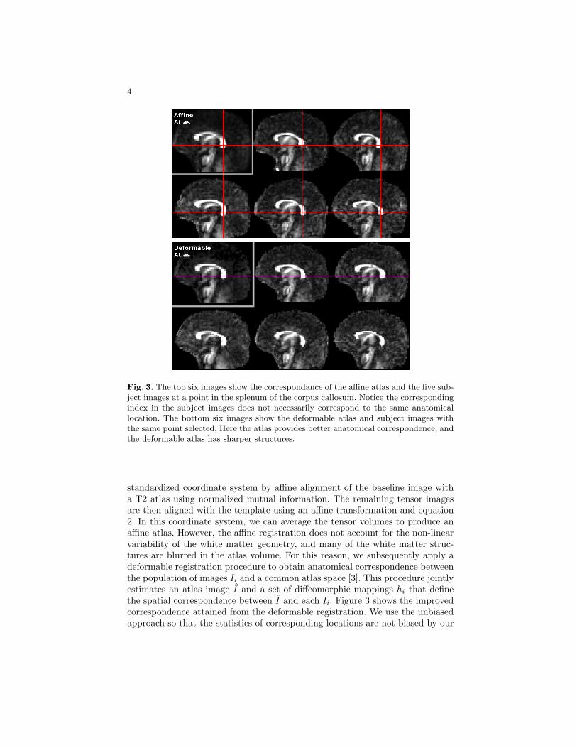
Fig. 3. The top six images show the correspondance of the affine atlas and the five sub-ject images at a point in the splenum of the corpus callosum. Notice the correspondingindex in the subject images does not necessarily correspond to the same anatomicallocation. The bottom six images show the deformable atlas and subject images withthe same point selected; Here the atlas provides better anatomical correspondence, andthe deformable atlas has sharper structures.
standardized coordinate system by affine alignment of the baseline image witha T2 atlas using normalized mutual information. The remaining tensor imagesare then aligned with the template using an affine transformation and equation2. In this coordinate system, we can average the tensor volumes to produce anaffine atlas. However, the affine registration does not account for the non-linearvariability of the white matter geometry, and many of the white matter struc-tures are blurred in the atlas volume. For this reason, we subsequently apply adeformable registration procedure to obtain anatomical correspondence betweenthe population of images Ii and a common atlas space [3]. This procedure jointlyestimates an atlas image I and a set of diffeomorphic mappings hi that definethe spatial correspondence between I and each Ii. Figure 3 shows the improvedcorrespondence attained from the deformable registration. We use the unbiasedapproach so that the statistics of corresponding locations are not biased by our

5
choice of the template image. The computed transformations are applied to eachtensor volume as described in the next section.
3 Tensor Processing
The application of high-dimensional transforms to the DTI volumes must ac-count for the space of valid tensors. The orientation of a diffusion tensor pro-vides a measurement of fiber orientation relative to the anatomical location, andspatial transformations of the tensor fields must account for the reorientation ofthe tensor. Furthermore, since diffusion tensors are symmetric positive-definitematrices, operations on the images must preserve this constraint.
3.1 Spatial Transformations of Tensor Images
When spatial transformations of diffusion images are performed to align theanatomy of different scans, the tensors must also be transformed to maintainthe relationship between anatomy and anisotropy orientation. We use the finitestrain approach of Alexander et al. to reorient tensors in a deformation field bydecomposing the local linear approximation of the transformation into a rota-tion and deformation component [6]. The rotation of each tensor is computedby performing singular value decomposition (SVD) on the local linear approxi-mation of the transformation F , where F is given by the Jacobian matrix of thedeformation field computed by finite differencing. A local linear deformation Fis decomposed into a rotation matrix R and a deformation matrix U , where
F = UR. (3)
The local transformation of a tensor D is given as
D′ = RDRT . (4)
3.2 Interpolation and Averaging of Tensor Images
The space of valid diffusion tensors does not form a vector space. Euclideanoperations on diffusion tensors such as averaging can produce averages with alarger determinant than the interpolating values, which is not physically sensible.Furthermore, operations on diffusion tensors are not guaranteed to preserve thepositive-definite nature of diffusion. The Riemannian framework has been shownas a natural method for operating on diffusion tensors, which preserves the phys-ical interpretation of the data, and constrains operations to remain in the validspace of symmetric positive-definite matrices [7, 8]. Further simplifications haveshown an efficient method for computation using the Log-Euclidean metric [9].Interpolation and averaging are treated as weighted sums in the Log-Euclideanframework defined as
D = exp
(N∑
i=1
wi log(Di)
), (5)
6
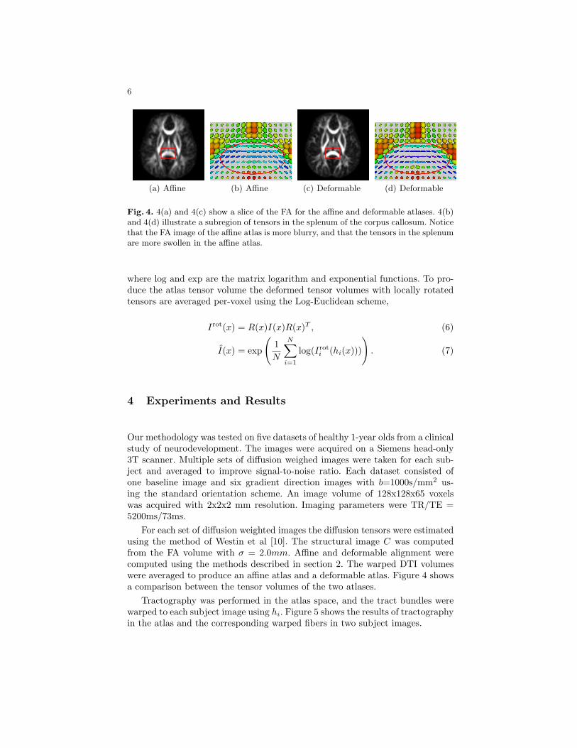
(a) Affine (b) Affine (c) Deformable (d) Deformable
Fig. 4. 4(a) and 4(c) show a slice of the FA for the affine and deformable atlases. 4(b)and 4(d) illustrate a subregion of tensors in the splenum of the corpus callosum. Noticethat the FA image of the affine atlas is more blurry, and that the tensors in the splenumare more swollen in the affine atlas.
where log and exp are the matrix logarithm and exponential functions. To pro-duce the atlas tensor volume the deformed tensor volumes with locally rotatedtensors are averaged per-voxel using the Log-Euclidean scheme,
Irot(x) = R(x)I(x)R(x)T , (6)
I(x) = exp
(1N
N∑i=1
log(Iroti (hi(x)))
). (7)
4 Experiments and Results
Our methodology was tested on five datasets of healthy 1-year olds from a clinicalstudy of neurodevelopment. The images were acquired on a Siemens head-only3T scanner. Multiple sets of diffusion weighed images were taken for each sub-ject and averaged to improve signal-to-noise ratio. Each dataset consisted ofone baseline image and six gradient direction images with b=1000s/mm2 us-ing the standard orientation scheme. An image volume of 128x128x65 voxelswas acquired with 2x2x2 mm resolution. Imaging parameters were TR/TE =5200ms/73ms.
For each set of diffusion weighted images the diffusion tensors were estimatedusing the method of Westin et al [10]. The structural image C was computedfrom the FA volume with σ = 2.0mm. Affine and deformable alignment werecomputed using the methods described in section 2. The warped DTI volumeswere averaged to produce an affine atlas and a deformable atlas. Figure 4 showsa comparison between the tensor volumes of the two atlases.
Tractography was performed in the atlas space, and the tract bundles werewarped to each subject image using hi. Figure 5 shows the results of tractographyin the atlas and the corresponding warped fibers in two subject images.

7
(a) Atlas (b) Image 1 (c) Image 2
Fig. 5. Fibers traced in the corpus callosum of the atlas (a) are mapped to correspond-ing locations in the subject images (b) (c) despite pose and shape changes.
5 Validation
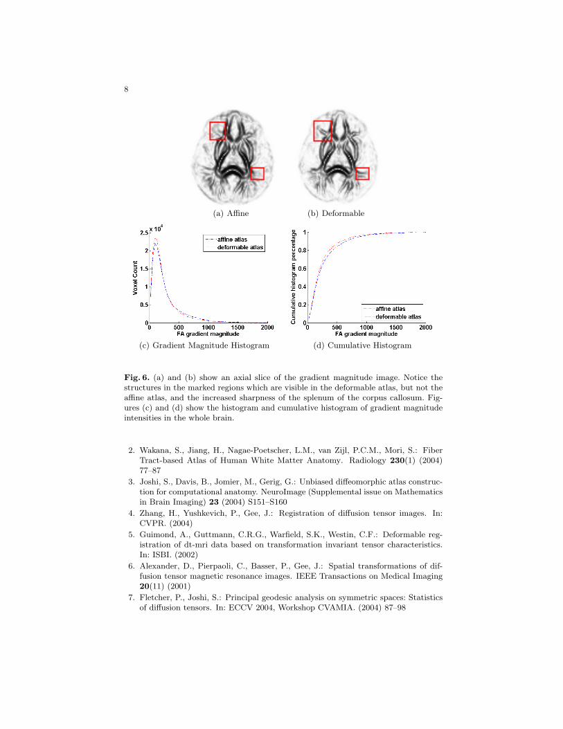
Visual inspection of tractography in the atlas volume shows an initial qualitativevalidation that the registration and averaging methods provide anatomically sen-sible results. Histogram comparisons of derived tensor measures in the affine anddiffeomorphic atlas show an initial quantitative validation of the improvementof the deformable registration over affine registration alone. The gradient mag-nitude of the FA was measured in the whole brain of the affine and deformableatlases. Figure 6 shows the gradient magnitude images and a histogram compar-ison. At the 90th percentile of the cumulative histogram, the deformable atlashas a gradient magnitude of 684 while the affine atlas is 573, an increase of20%. This shows that the deformable atlas better preserves thin structures viaimproved alignment.
6 Discussion and Future Work
We have developed an automatic method for producing correspondence in dif-fusion tensor images through deformable registration, and a novel image matchwhich alignes structural features. We apply the transformation using the finitestrain model for tensor rotation and a Riemannian framework for averagingand interpolation. Initial validation of deformable registration is performed byshowing improvement in thin structure preservation over affine alignment. Fu-ture validation work will try to quantify the quality of correspondence of fibertracts. In future work, we intend to build DTI atlases of different populations tocompare tract geometry and tensor statistics along tracts.
References
1. Corouge, I., Fletcher, P.T., Gilmore, J.H., Gerig, G.: Fiber tract-oriented statisticsfor quantitative diffusion tensor MRI analysis. In: MICCAI. Volume 3749 of LNCS.(2005) 131–139
8
(a) Affine (b) Deformable
(c) Gradient Magnitude Histogram (d) Cumulative Histogram
Fig. 6. (a) and (b) show an axial slice of the gradient magnitude image. Notice thestructures in the marked regions which are visible in the deformable atlas, but not theaffine atlas, and the increased sharpness of the splenum of the corpus callosum. Fig-ures (c) and (d) show the histogram and cumulative histogram of gradient magnitudeintensities in the whole brain.
2. Wakana, S., Jiang, H., Nagae-Poetscher, L.M., van Zijl, P.C.M., Mori, S.: FiberTract-based Atlas of Human White Matter Anatomy. Radiology 230(1) (2004)77–87
3. Joshi, S., Davis, B., Jomier, M., Gerig, G.: Unbiased diffeomorphic atlas construc-tion for computational anatomy. NeuroImage (Supplemental issue on Mathematicsin Brain Imaging) 23 (2004) S151–S160
4. Zhang, H., Yushkevich, P., Gee, J.: Registration of diffusion tensor images. In:CVPR. (2004)
5. Guimond, A., Guttmann, C.R.G., Warfield, S.K., Westin, C.F.: Deformable reg-istration of dt-mri data based on transformation invariant tensor characteristics.In: ISBI. (2002)
6. Alexander, D., Pierpaoli, C., Basser, P., Gee, J.: Spatial transformations of dif-fusion tensor magnetic resonance images. IEEE Transactions on Medical Imaging20(11) (2001)
7. Fletcher, P., Joshi, S.: Principal geodesic analysis on symmetric spaces: Statisticsof diffusion tensors. In: ECCV 2004, Workshop CVAMIA. (2004) 87–98

9
8. Pennec, X., Fillard, P., Ayache, N.: A Riemannian framework for tensor computing.International Journal of Computer Vision 66(1) (2006) 41–66
9. Arsigny, V., Fillard, P., Pennec, X., Ayache, N.: Fast and simple calculus ontensors in the Log-Euclidean framework. In: MICCAI. Volume 3749 of LNCS.(2005) 115–122
10. Westin, C.F., Maier, S.E., Mamata, H., Nabavi, A., Jolesz, F.A., Kikinis, R.: Pro-cessing and visualization of diffusion tensor MRI. Medical Image Analysis 6(2)(2002) 93–108