Embed Size (px)
Citation preview

Important Pathogenic Bacteria During Child Growth
Evita Mayasari dr MKesEvita Mayasari, dr., MKes.
Microbiology Department Medical Faculty University of Sumatera UtaraMedical Faculty, University of Sumatera Utara
1

HAEMOPHILUS INFLUENZAE TYPE B (HIB)( )First isolated by Pfeiffer during the influenza pandemic of 1890
Family: PasteurellaceaeSmall Gram (-) coccobacilli or filamentous rods, hence the ( )descriptive term pleomorphic.
Polysaccharide capsule (+) : consists of a repeating polymer of 5 carbon sugar units, ribose, and ribitolphosphate.
Envelope includes lipo-oligosaccharide (LOS) and outer-membrane proteins (OMP).
Pili fi b i t b i blPili or fimbriae presence appears to be variable
2

Case: Upper Airway Problems in Children: pp yAcute Epiglottitis
3
IDENTIFICATIONIDENTIFICATIONGrow on chocolate agar, where they have a glistening, semitransparent appearance semitransparent appearance.
Identified by the requirement for X (hemin or other porphyrins) and V (coenzyme NAD) factors for growth on blood agar and V (coenzyme NAD) factors for growth on blood agar.
A more sensitive test for the X factor requirement is to test th bilit f H i fl t t d lt i l li ithe ability of H. influenzae to convert delta aminolevulinicacid to porphyrin.
Other tests such as the production of indole from tryptophan and detection of β-galactosidase (ONPG test) activity are also useful in discriminating H. influenzae from other Haemophilusg pspecies.
4

5

6
Tissues infected by type b and nontypable strains of H. influenzae

PATHOGENESISPATHOGENESISThe major virulence factor: type b polysaccharide capsule → allows them to resist phagocytosis and capsule → allows them to resist phagocytosis and complement-mediated lysis in the nonimmune host.
E l t d i t t th ith li Encapsulated organisms can penetrate the epithelium of the nasopharynx and invade the blood capillaries directly → Bacteremia → spread to CNS and distant directly → Bacteremia → spread to CNS and distant sites such as bones and joints Attachment to respiratory epithelial cells is mediated Attachment to respiratory epithelial cells is mediated by pili and other adhesins.
7

TRANSMISSIONTRANSMISSION
Respiratory route (person to person)p y (p p )Close contact with patients suffering H. Influenzae infection presents a definite risk for nonimmune ppersons and nonimmune children <4 years old.Prophylaxis with Rifampin is recommended for such children.Vaccination with type b polysaccharide (in the form of Hib conjugate vaccines) is effective in preventing infection.
8
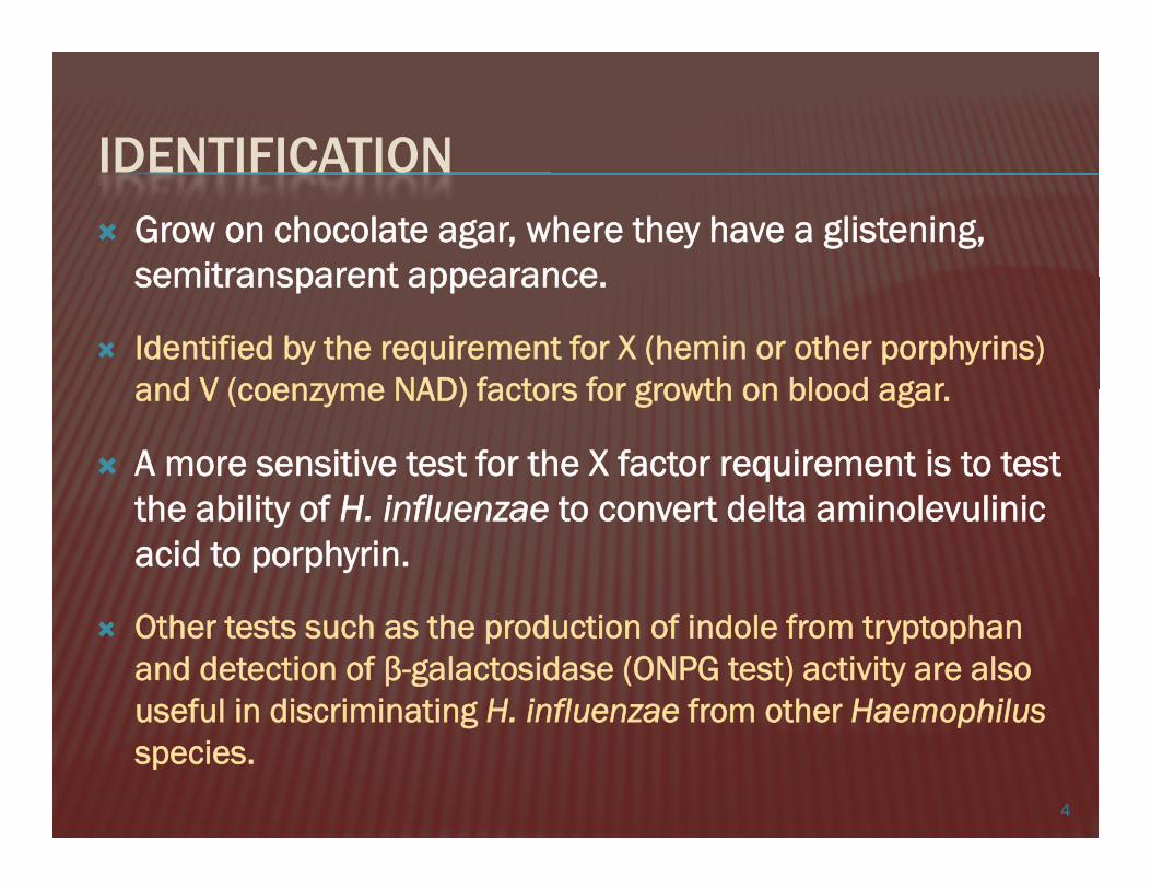
SPECIMEN COLLECTIONSPECIMEN COLLECTIONBloodCSF ( b i l fl id)CSF (cerebrospinal fluid)Prompt transport of those samples is mandatory to ensure the fastest possible diagnosis and survival of ensure the fastest possible diagnosis and survival of m.o. in the sampleOther specimens: aspirated synovial fluid pericardial Other specimens: aspirated synovial fluid, pericardial fluid, pleural fluid, pus, sputum, nasopharyngeal or throat swabs, purulent discharge from infected eyes.For samples taken from respiratory tract: avoid contamination with commensals → throat swabs must be collected from the pharynx
9
SPECIMEN TRANSPORTSPECIMEN TRANSPORT
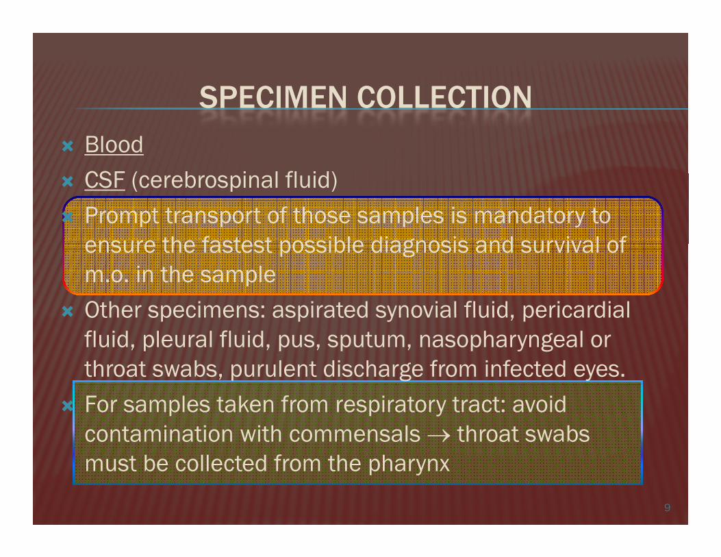
Samples must be transported to the lab. in a suitable transport medium (thioglycolate-hemin-based)Or samples should be spread directly onto appropriate agar media whenever possible app op ate aga ed a e e e poss b e(esp. conjunctival specimens)The viability of most haemophilus is readily lost The viability of most haemophilus is readily lost as a result of drying out, they do not survive more than a few days in clinical samplesmore than a few days in clinical samples.
10

BORDETELLA PERTUSSISBORDETELLA PERTUSSIS1906: first isolated in pure culture by Bordet and GengouTi (0 5 t 1 0 ) G g ti b ill Tiny (0.5 to 1.0 μm), Gram-negative coccobacillus morphologically much like Haemophilus.The surface exhibits a rod-like protein called the The surface exhibits a rod like protein called the filamentous hemagglutinin (Fha) because of its ability to bind to and agglutinate erythrocytes.Contains surface pili Capsule includes a protein called pertactin.Strict aerobe, nonmotile, non–spore formingExtracellular products: Pertussis toxin (PT), adenylate
l i h l icyclase toxin, tracheal cytotoxin
11

Bordetella pertussis, the agent of pertussis or whooping cough.
12
p , g p p g gGram stain. (CDC)
IDENTIFICATIONIDENTIFICATION
Culture media enriched with blood: Bordet-Gengou, g ,charcoal-horseblood agar (Regan-Lowe) Incubated in moist environment, slow growth (3 to 7 g (days)Identified by immunofluorescence stainingForms acid but no gas from glucose and lactoseHemolysis of blood (in medium) is associated with y ( )virulent B. pertussisBordetella DNA can also be detected by PCR.
13

"mercury drop" colonies on Bordet-Gengou is likely to be B. pertussis
14
PERTUSSIS (WHOOPING COUGH)PERTUSSIS (WHOOPING COUGH)Mortimer & Jones, 1979: in USA, ±5 of every 1,000 infants born alive died of the disease before their fifth birthdays yAfter the introduction of immunization (1940), the incidence dropped from 250,000 cases a year to below 1/100,000population (in US)Now well controlled in the US and other developed countriesMANIFESTATIONS i b ti i d (7 10 d ) t g MANIFESTATIONS: incubation period (7-10 days) → stages: (1) catarrhal; rhinorrhea that persists for 1-2 weeks, malaise, fever, sneezing, and anorexia may also be present g y p(2) paroxysmal; persistent cough up to 50 times a day for 2-4 weeks. Whoop, vomiting, apnea (infants) (3) convalescent; frequency and severity ↓ (3 4 weeks) (3) convalescent; frequency and severity ↓ (3-4 weeks)
15

TRANSMISSION
The agent of whooping cough is transmitted primarily via d l t
16
droplets.

PATHOGENESISPATHOGENESIS1. Introduced into the respiratory tract. It has a tropism for
ciliated bronchial epithelium attaching to the ciliaciliated bronchial epithelium attaching to the ciliathemselves. This adherence is mediated by Fha, pili, pertactin, and the binding subunits of PT.
2. Once attached, the bacteria immobilize the cilia then progressively destroyed the ciliated cellsThis local injury is caused primarily by the action of the tracheal cytotoxin. This prod ces an epitheli m de oid of the ciliar blanket 3. This produces an epithelium devoid of the ciliary blanket, which moves foreign matter away from the lower airways.
4 Persistent coughing is the clinical correlate of this deficit4. Persistent coughing is the clinical correlate of this deficit.
17

Synergy between pertussis toxin and the filamentous hemagglutinin in binding to ciliated respiratory epithelial cells.
18
g y

SPECIMEN COLLECTION & TRANSPORTSPECIMEN COLLECTION & TRANSPORTNasopharyngeal (NP) aspiratesP t i NP bPosterior NP swabsIf properly collected, those specimens contain the ciliated respiratory epithelial cells for which B pertussisciliated respiratory epithelial cells for which B. pertussisexhibit tropismInferior specimens: throat swabs, but may be suitable for p yPCR diagnosis of B. pertussis infectionRecommended: collect two NP swab specimens, one through each nostrilSpecimens are then directly plated or placed in suitable transport media: Casamino Acids or Amies with charcoaltransport media: Casamino Acids or Amies with charcoal
19
MYCOBACTERIUM TUBERCULOSISMYCOBACTERIUM TUBERCULOSISSlim, strongly acid-alcohol-fast rod 2-4 μm (length) and 0 2-0 5 μm (width)and 0.2 0.5 μm (width)Nonmotile, obligate aerobes,do not form sporesThe cell wall contains peptidoglycan similar to The cell wall contains peptidoglycan similar to Gram(+) organisms, except that it contains N-glycolylmuramic,rather than N-acetylmuramic, acid.g y y , y ,Attached to peptidoglycan are: polysaccharides, proteins, and lipids; long-chain fatty acids called p p g ymycolic acids (make up >60% of the total cell wall mass)Enveloped
20

MYCOBACTERIUM TUBERCULOSIS
Grows at 37°C but not at room temperature
MYCOBACTERIUM TUBERCULOSIS
pGrowth is enhanced by 5 to 10% CO2
Requires enriched or complex media for primary Requires enriched or complex media for primary growth: Lowenstein–JensenThe dry, rough, buff-colored colonies usually appear y, g , y ppafter 3-6 weeks of incubation.Due to its hydrophobic lipid surface, M. tuberculosis is y p punusually resistant to drying,to common disinfectants, and to acids and alkalis.
21
Colonies of Mycobacterium tuberculosis on Lowenstein-Jensen medium. CDC
22
CDC.

MYCOBACTERIUM TUBERCULOSIS
Sensitive to heat, including pasteurization, and
MYCOBACTERIUM TUBERCULOSIS
, g p ,individual organisms in droplet nuclei are susceptibleto inactivation by UV light.Staining characteristic: acid-fast (Ziehl-Neelsen stain method). Once stained, acid-fast bacteria will retain dyes when heated and treated with acidified organic compounds.Th b t i i f lt ti i t ll l it The bacterium is a facultative intracellular parasite, usually of macrophages, and has a slow generation time 15-20 hourstime, 15-20 hours
23
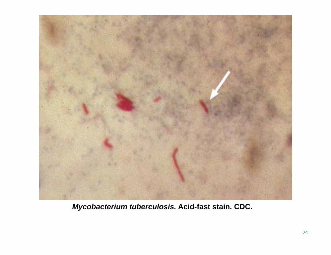
Mycobacterium tuberculosis Acid-fast stain CDC
24
Mycobacterium tuberculosis. Acid fast stain. CDC.

PATHOGENESISPATHOGENESIS
There are 5 stages of the disease (tuberculosis)Stage 1
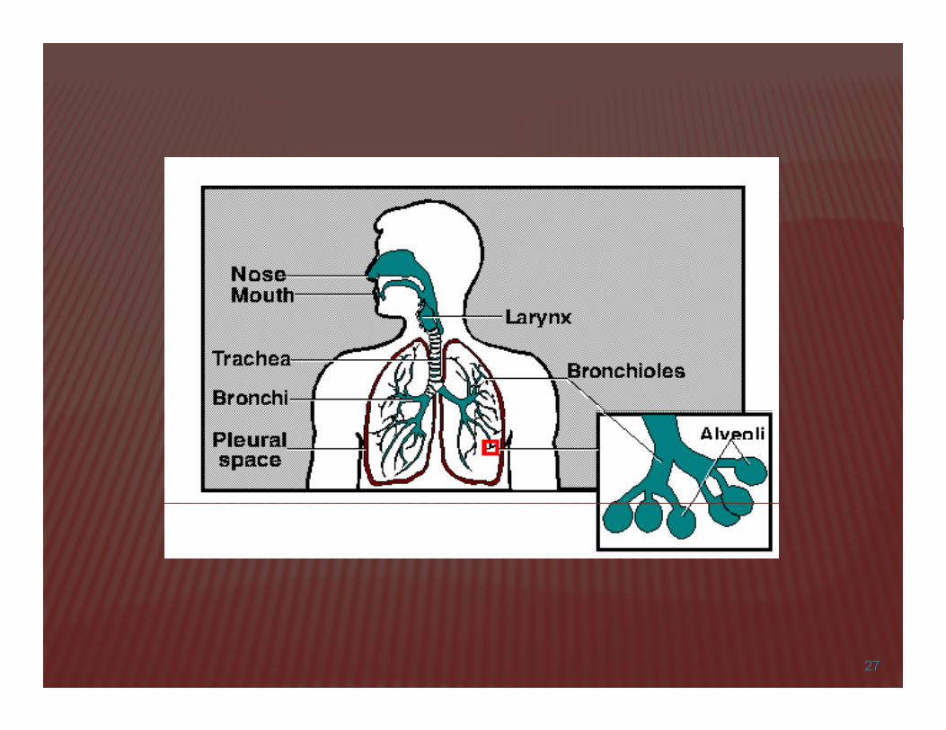
Droplet nuclei are inhaled. Droplet nuclei are inhaled. One droplet nuclei contains no > 3 bacilli. D l t ll th t th i iDroplets are so small that they can remain air-borne for extended periods of time. Droplets are generated by during talking, singing, coughing, and sneezing.
25
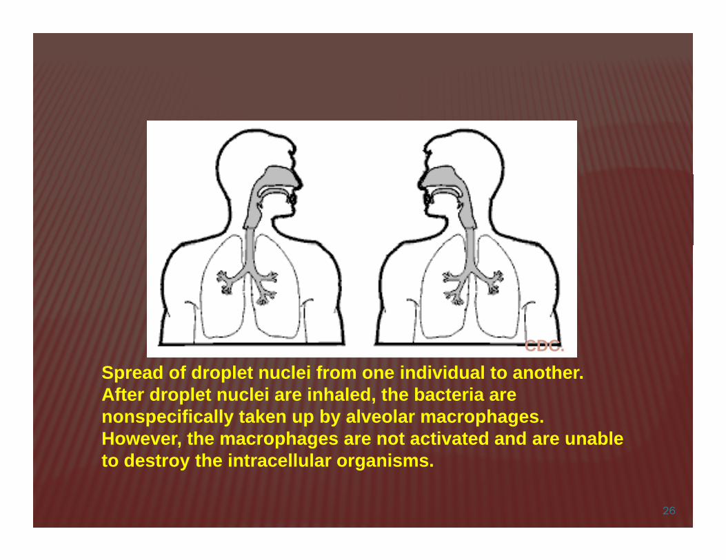
Spread of droplet nuclei from one individual to another. CDC.
After droplet nuclei are inhaled, the bacteria are nonspecifically taken up by alveolar macrophages. However, the macrophages are not activated and are unable t d t th i t ll l i
26
to destroy the intracellular organisms.
27
PATHOGENESIS
Stage 2
PATHOGENESIS
Begins 7-21 days after initial infection. MTB multiplies unrestricted within unactivated pmacrophages until the macrophages burst. Other macrophages begin to extravasate from Other macrophages begin to extravasate from peripheral blood. These macrophages also phagocytose MTB but These macrophages also phagocytose MTB, but they are also unactivated and hence can not destroy the bacteria destroy the bacteria.
28

Stage 3Lymphocytes begin to infiltrate → recognize MTB antigen → T-cell activation and the liberation of cytokines including gamma interferon (IFN) → activation of including gamma interferon (IFN) → activation of macrophages These activated macrophages are now capable of ese act ated ac op ages a e o capab e odestroying MTB.Positive tuberculin reaction Tubercle formation begins. The center is characterized by "caseation necrosis“= takes on a semi-solid or "cheesy"
i t consistency. MTB cannot multiply within tubercles because of the low pH and anoxic environment MTB can however persist
29
pH and anoxic environment. MTB can, however, persist within these tubercles for extended periods.
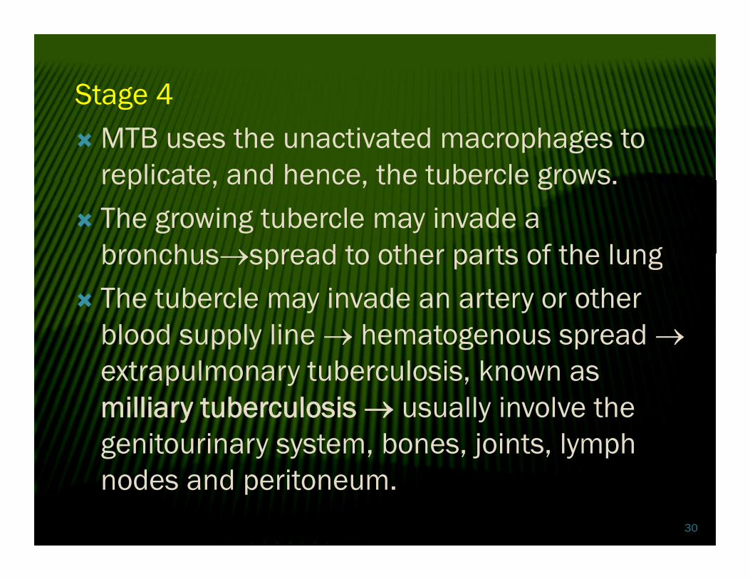
Stage 4gMTB uses the unactivated macrophages to replicate and hence the tubercle growsreplicate, and hence, the tubercle grows.The growing tubercle may invade a bronchus→spread to other parts of the lungbronchus→spread to other parts of the lungThe tubercle may invade an artery or other bl d l li h t dblood supply line → hematogenous spread →extrapulmonary tuberculosis, known as
illi t b l i ll i l th milliary tuberculosis → usually involve the genitourinary system, bones, joints, lymph
d d it30
nodes and peritoneum.

Stage 5For unknown reasons, the caseous centers of the tubercles liquefy. This liquid is very conducive to MTB growth, and the organism begins to rapidly multiply extracellularly. After time, the large antigen load causes the walls of nearby bronchi to become necrotic and rupture. This results in cavity formation. This also allows MTB to spread to other parts of the lung. Note: only a smaller percentage of MTB infections
31
progress to an advanced stage

CLOSTRIDIUM TETANICLOSTRIDIUM TETANISlim, Gram-positive rod, forms a typical round terminal spore that gives the organism a drumstick spore that gives the organism a drumstick appearanceThe spores remain viable in soil or culture for many The spores remain viable in soil or culture for many years. It is resistant to most disinfectants and withstands boiling for several minutes.Flagellate and motile, strict anaerobicThe most important product : neurotoxic exotoxin, p ptetanospasmin or tetanus toxinFound in soils and in the intestinal tracts and feces of various animals
32

C tetani Gram stain
33
C. tetani Gram stain.

PATHOGENESISPATHOGENESISTetanus bacilli multiply locally and neither damage nor invade adjacent tissues but synthesizes the tetanus toxininvade adjacent tissues, but synthesizes the tetanus toxinThe tetanus toxin initially binds to peripheral nerve terminals→ transported within the axon and across synaptic junctions until it reaches the CNS → rapidly fixed to gangliosides at the presynaptic inhibitory motor nerve endings and is taken up into the axon by endocytosis →endings, and is taken up into the axon by endocytosis →block the release of inhibitory neurotransmitters (glycine and γ-amino butyric acid), which is required to check the nervous impulse.If nervous impulses cannot be checked by normal inhibitory mechanisms it produces the generalized muscular spasms mechanisms, it produces the generalized muscular spasms characteristic of tetanus.
34

35
Pathogenesis of tetanus caused by C tetani

ISOLATIONISOLATION
Well growth on CDC anaerobe blood agar and Well growth on CDC anaerobe blood agar and phenylethyl alcohol blood agar (PEA) (1-2 days)Non selective media: Brucella agar + 5% sheep Non-selective media: Brucella agar + 5% sheep blood, columbia agar, brain heart infusion agarI l t d l i b lt d t h d t Isolated colonies: subcultured to chopped-meat medium, incubated overnightSpore selection techniques: alcohol treatment, heat treatment
36

IDENTIFICATIONIDENTIFICATION
Preliminary identification: Gram stain, colony Preliminary identification: Gram stain, colony morphology: swarming, indole reaction: variable, spore location: terminal, drumstick variable, spore location: terminal, drumstick shaped.Definitive identification: PRAS Biochemical Definitive identification: PRAS Biochemical Inoculation, Gelatin Hydrolysis : (+), Esculin Hydrolysis: ( ) Carbohydrate FermentationHydrolysis: (-), Carbohydrate Fermentation
37

CORYNEBACTERIUM DIPHTHERIAECORYNEBACTERIUM DIPHTHERIAE
Aerobic and facultative Gram-positive rods.p0.5-1 μm in diameterIrregular swellings at one end: “club shaped” appearanceGranules are irregularly distributed within the rods →aniline dyesProduce diphtheria toxin (DT) → inactivates elongation factor EF-2 → arrest of protein synthesis → necrotizing and neurotoxic effectand neurotoxic effect
38

C. diphtheriae cells stained by Albert's technique . Note the characteristic "Chinese-letter" arrangement of cells.
39

PATHOGENESISPATHOGENESIS
Susceptible persons may acquire toxigenic diphtheria b illi i th h bacilli in the nasopharynx. The organism produces DT that inhibits cellular protein s nthesis and is responsible for local tiss e protein synthesis and is responsible for local tissue destruction and membrane formation. The toxin produced at the site of the membrane is The toxin produced at the site of the membrane is absorbed into the bloodstream and then distributed to the tissues of the body→responsible for the major the tissues of the body→responsible for the major complications of myocarditis and neuritis and can also cause low platelet counts (thrombocytopenia) and protein in the urine (proteinuria).
40

LABORATORY TESTS DIAGNOSTICLABORATORY TESTS DIAGNOSTIC
Specimen: dacron swabs from the nose, throat: -Obtained before administered of Antimicrobial drugs -Collected from beneath any visible membraneThe swab then placed in semisolid transport media (e.g.: Amies)Amies)Smears stained with Gram stain On blood agar: colonies are small, granular, gray, irregular O b ood aga co o es a e s a , g a u a , g ay, egu aedges, may have small zone of hemolysisOn potassium tellurite agar: brown-black halo
41

LABORATORY TESTS DIAGNOSTICLABORATORY TESTS DIAGNOSTIC
Modification elek method described by WHOModification elek method described by WHOPCR- based methods: detect the DT gene (tax) ELISA d t t DT f li i l C di hth i ELISA : detect DT from clinical C. diphtheriae isolates Immunochromographic strip assay: detect DT in hours and highly sensitive
42


![[Micro] mycobacterium tuberculosis](https://img.pdfslide.us/doc/110x75/55d6fc67bb61ebfa2a8b47ea/micro-mycobacterium-tuberculosis.jpg)