Embed Size (px)
Citation preview
WashingtonWASHINGTON•UNIVERSITY•IN•ST•LOUIS
Mallinckrodt Instituteof RadiologyMIR
Importance and Role of Radioisotopes to the Medical
Community
Michael J. Welch
Division of Radiological SciencesWashington University School of Medicine
St Louis, MO

WashingtonWASHINGTON•UNIVERSITY•IN•ST•LOUIS
Mallinckrodt Instituteof RadiologyMIR
Radionuclides Used in Clinical Nuclear Medicine (Diagnostic)
• Single Photon99Mo/99mTc generator, 201Tl, 111In, 67Ga, *I• Positron Emitting18F 2-fluoro-2-deoxy-glucose
WashingtonWASHINGTON•UNIVERSITY•IN•ST•LOUIS
Mallinckrodt Instituteof RadiologyMIR
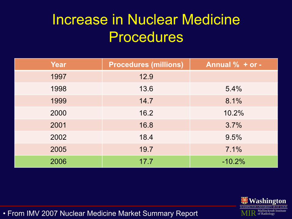
Increase in Nuclear Medicine Procedures
Year Procedures (millions) Annual % + or -1997 12.91998 13.6 5.4%1999 14.7 8.1%2000 16.2 10.2%2001 16.8 3.7%2002 18.4 9.5%2005 19.7 7.1%2006 17.7 -10.2%
• From IMV 2007 Nuclear Medicine Market Summary Report
WashingtonWASHINGTON•UNIVERSITY•IN•ST•LOUIS
Mallinckrodt Instituteof RadiologyMIR
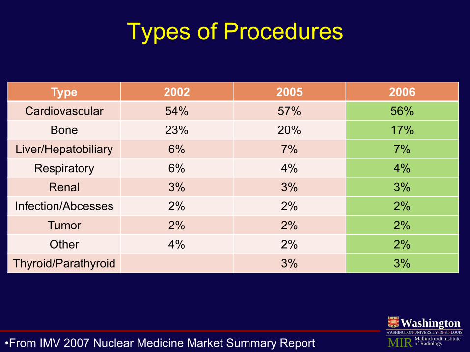
Types of Procedures
Type 2002 2005 2006Cardiovascular 54% 57% 56%
Bone 23% 20% 17%Liver/Hepatobiliary 6% 7% 7%
Respiratory 6% 4% 4%Renal 3% 3% 3%
Infection/Abcesses 2% 2% 2%Tumor 2% 2% 2%Other 4% 2% 2%
Thyroid/Parathyroid 3% 3%
•From IMV 2007 Nuclear Medicine Market Summary Report
WashingtonWASHINGTON•UNIVERSITY•IN•ST•LOUIS
Mallinckrodt Instituteof RadiologyMIR
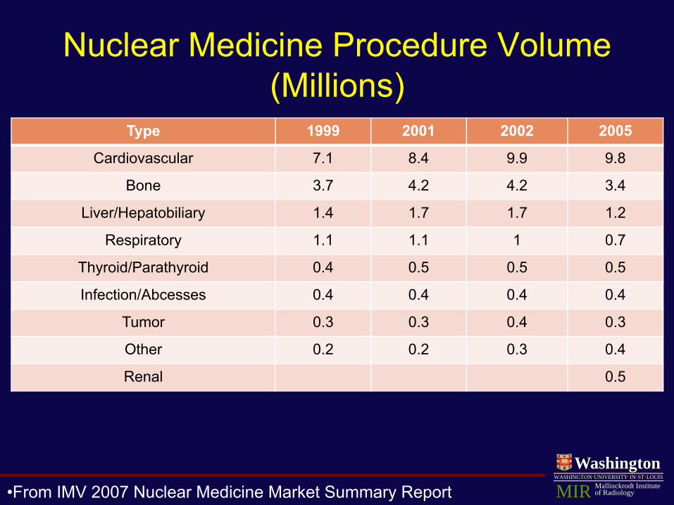
Nuclear Medicine Procedure Volume (Millions)
Type 1999 2001 2002 2005
Cardiovascular 7.1 8.4 9.9 9.8
Bone 3.7 4.2 4.2 3.4
Liver/Hepatobiliary 1.4 1.7 1.7 1.2
Respiratory 1.1 1.1 1 0.7
Thyroid/Parathyroid 0.4 0.5 0.5 0.5
Infection/Abcesses 0.4 0.4 0.4 0.4
Tumor 0.3 0.3 0.4 0.3
Other 0.2 0.2 0.3 0.4
Renal 0.5
•From IMV 2007 Nuclear Medicine Market Summary Report
WashingtonWASHINGTON•UNIVERSITY•IN•ST•LOUIS
Mallinckrodt Instituteof RadiologyMIR
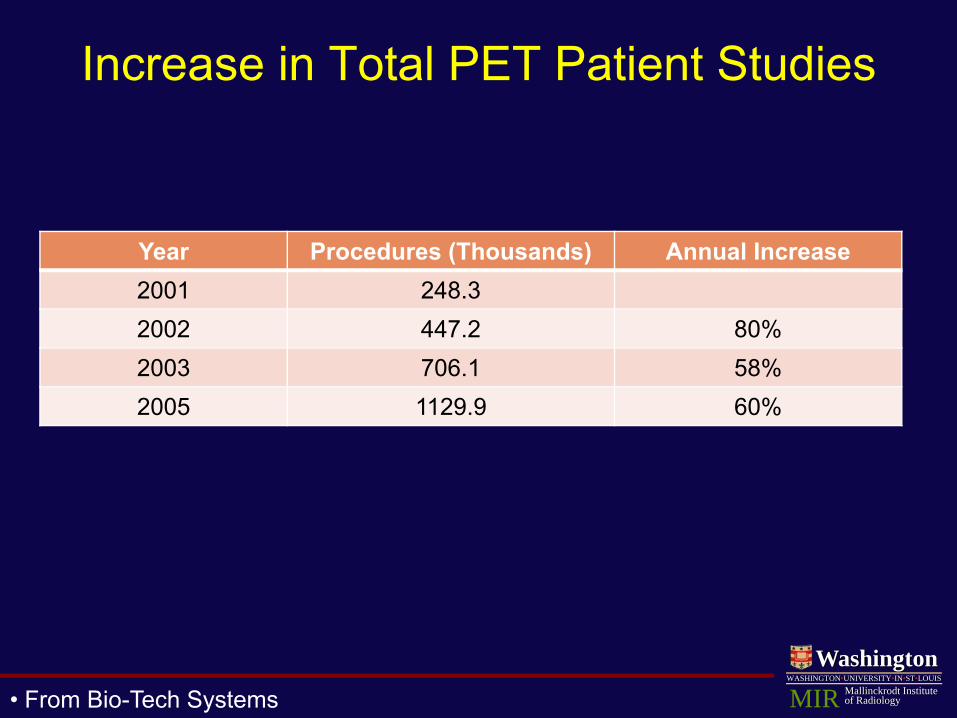
Increase in Total PET Patient Studies
Year Procedures (Thousands) Annual Increase2001 248.32002 447.2 80%2003 706.1 58%2005 1129.9 60%
• From Bio-Tech Systems
WashingtonWASHINGTON•UNIVERSITY•IN•ST•LOUIS
Mallinckrodt Instituteof RadiologyMIR
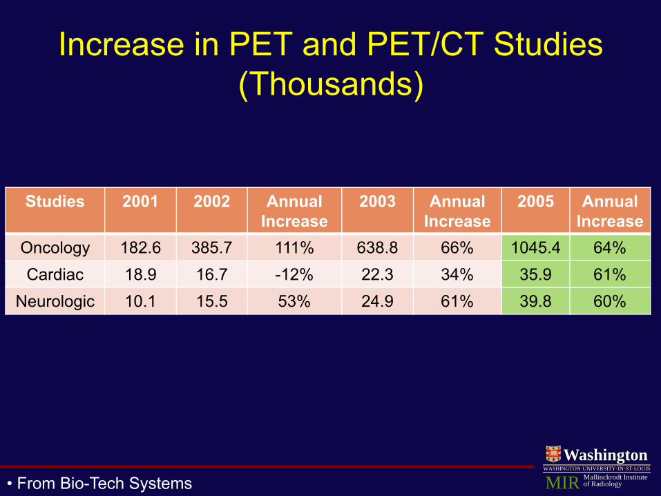
Increase in PET and PET/CT Studies (Thousands)
Studies 2001 2002 Annual Increase
2003 Annual Increase
2005 Annual Increase
Oncology 182.6 385.7 111% 638.8 66% 1045.4 64%Cardiac 18.9 16.7 -12% 22.3 34% 35.9 61%
Neurologic 10.1 15.5 53% 24.9 61% 39.8 60%
• From Bio-Tech Systems
WashingtonWASHINGTON•UNIVERSITY•IN•ST•LOUIS
Mallinckrodt Instituteof RadiologyMIR
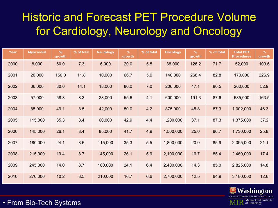
Historic and Forecast PET Procedure Volume for Cardiology, Neurology and Oncology
Year Myocardial % growth
% of total Neurology %growth
% of total Oncology % growth
% of total Total PET Procedures
% growth
2000 8,000 60.0 7.3 6,000 20.0 5.5 38,000 126.2 71.7 52,000 109.6
2001 20,000 150.0 11.8 10,000 66.7 5.9 140,000 268.4 82.8 170,000 226.9
2002 36,000 80.0 14.1 18,000 80.0 7.0 206,000 47.1 80.5 260,000 52.9
2003 57,000 58.3 8.3 28,000 55.6 4.1 600,000 191.3 87.6 685,000 163.5
2004 85,000 49.1 8.5 42,000 50.0 4.2 875,000 45.8 87.3 1,002,000 46.3
2005 115,000 35.3 8.4 60,000 42.9 4.4 1,200,000 37.1 87.3 1,375,000 37.2
2006 145,000 26.1 8.4 85,000 41.7 4.9 1,500,000 25.0 86.7 1,730,000 25.8
2007 180,000 24.1 8.6 115,000 35.3 5.5 1,800,000 20.0 85.9 2,095,000 21.1
2008 215,000 19.4 8.7 145,000 26.1 5.9 2,100,000 16.7 85.4 2,460,000 17.4
2009 245,000 14.0 8.7 180,000 24.1 6.4 2,400,000 14.3 85.0 2,825,000 14.8
2010 270,000 10.2 8.5 210,000 16.7 6.6 2,700,000 12.5 84.9 3,180,000 12.6
• From Bio-Tech Systems
WashingtonWASHINGTON•UNIVERSITY•IN•ST•LOUIS
Mallinckrodt Instituteof RadiologyMIR
Treatment Assessment with FDG-PET
• Residual mass: post-treatment effect or tumor?
• Prediction and early monitoring of treatment effectiveness
WashingtonWASHINGTON•UNIVERSITY•IN•ST•LOUIS
Mallinckrodt Instituteof RadiologyMIR
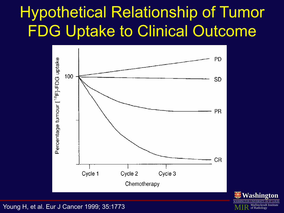
Hypothetical Relationship of Tumor FDG Uptake to Clinical Outcome
Young H, et al. Eur J Cancer 1999; 35:1773
WashingtonWASHINGTON•UNIVERSITY•IN•ST•LOUIS
Mallinckrodt Instituteof RadiologyMIR
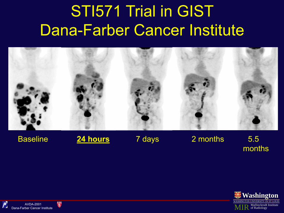
STI571 Trial in GIST Dana-Farber Cancer Institute
Baseline 24 hours 7 days 2 months 5.5 months
AVDA-2001Dana-Farber Cancer Institute
WashingtonWASHINGTON•UNIVERSITY•IN•ST•LOUIS
Mallinckrodt Instituteof RadiologyMIR
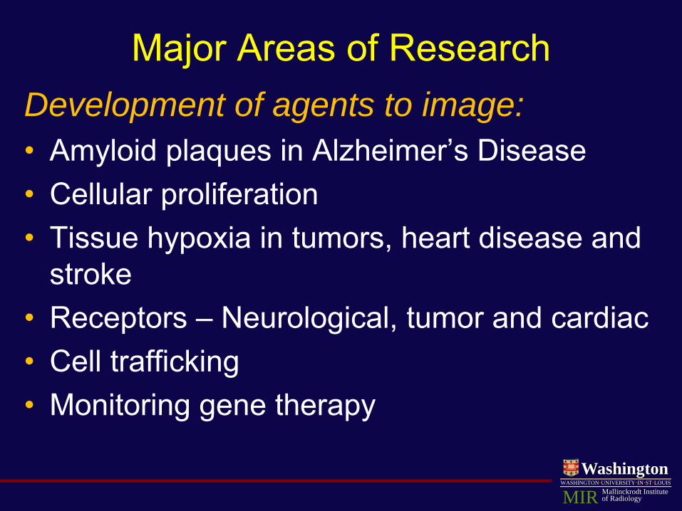
Major Areas of ResearchDevelopment of agents to image:• Amyloid plaques in Alzheimer’s Disease• Cellular proliferation• Tissue hypoxia in tumors, heart disease and
stroke• Receptors – Neurological, tumor and cardiac• Cell trafficking• Monitoring gene therapy
WashingtonWASHINGTON•UNIVERSITY•IN•ST•LOUIS
Mallinckrodt Instituteof RadiologyMIR
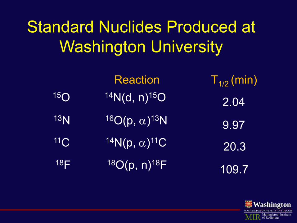
Reaction T1/2 (min)15O
13N11C18F
14N(d, n)15O 2.04 16O(p, α)13N 9.97 14N(p, α)11C 20.3 18O(p, n)18F 109.7
Standard Nuclides Produced at Washington University
WashingtonWASHINGTON•UNIVERSITY•IN•ST•LOUIS
Mallinckrodt Instituteof RadiologyMIR
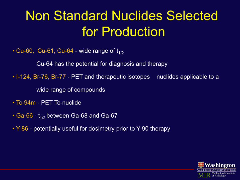
Non Standard Nuclides Selected for Production
• Cu-60, Cu-61, Cu-64 - wide range of t1/2
Cu-64 has the potential for diagnosis and therapy
• I-124, Br-76, Br-77 - PET and therapeutic isotopes nuclides applicable to a
wide range of compounds
• Tc-94m - PET Tc-nuclide
• Ga-66 - t1/2 between Ga-68 and Ga-67
• Y-86 - potentially useful for dosimetry prior to Y-90 therapy
WashingtonWASHINGTON•UNIVERSITY•IN•ST•LOUIS
Mallinckrodt Instituteof RadiologyMIR
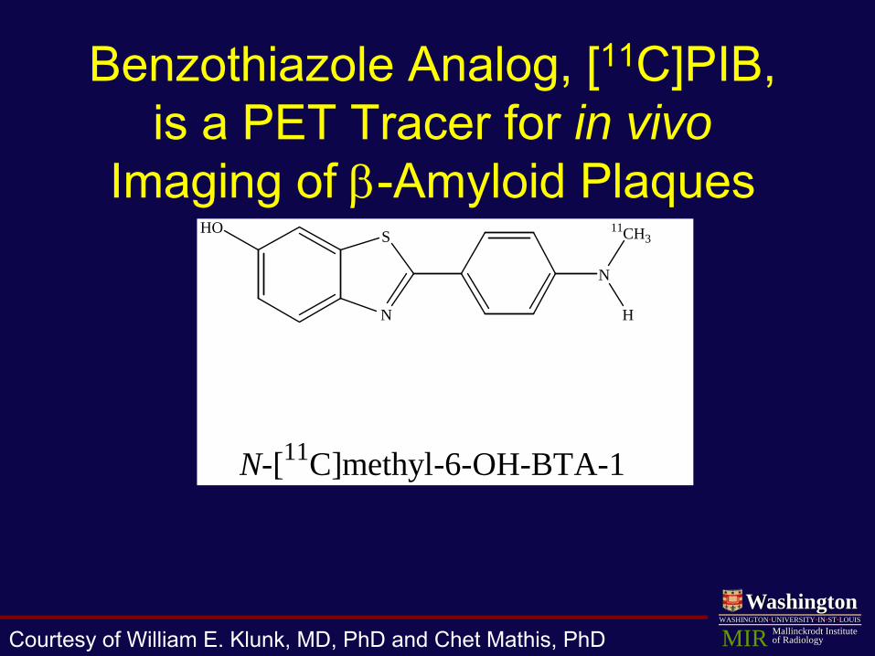
Benzothiazole Analog, [11C]PIB, is a PET Tracer for in vivo
Imaging of β-Amyloid Plaques
N
S
N
HO 11CH3
H
N-[11C]methyl-6-OH-BTA-1
Courtesy of William E. Klunk, MD, PhD and Chet Mathis, PhD
WashingtonWASHINGTON•UNIVERSITY•IN•ST•LOUIS
Mallinckrodt Instituteof RadiologyMIR
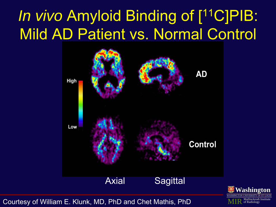
In vivo Amyloid Binding of [11C]PIB: Mild AD Patient vs. Normal Control
Axial Sagittal
Courtesy of William E. Klunk, MD, PhD and Chet Mathis, PhD
WashingtonWASHINGTON•UNIVERSITY•IN•ST•LOUIS
Mallinckrodt Instituteof RadiologyMIR
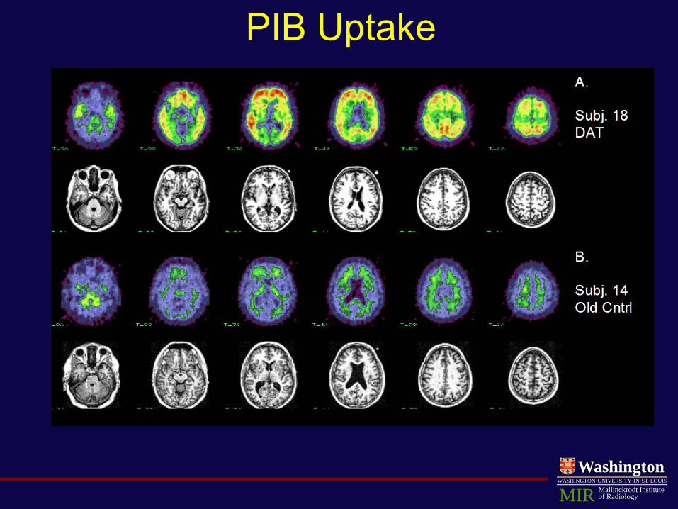
PIB Uptake
WashingtonWASHINGTON•UNIVERSITY•IN•ST•LOUIS
Mallinckrodt Instituteof RadiologyMIR
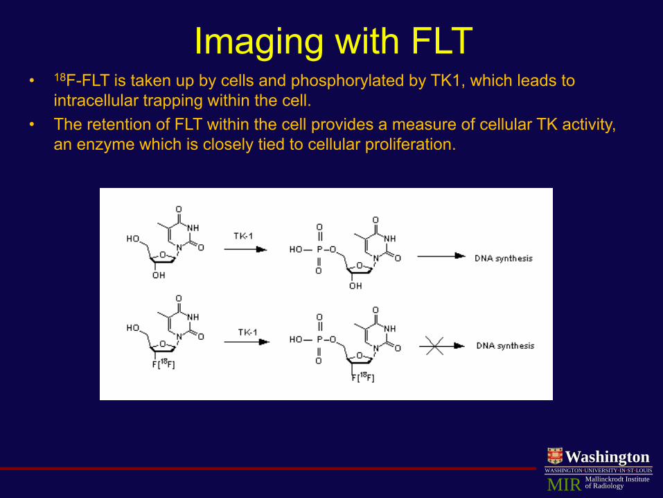
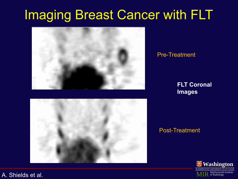
Imaging with FLT • 18F-FLT is taken up by cells and phosphorylated by TK1, which leads to
intracellular trapping within the cell.• The retention of FLT within the cell provides a measure of cellular TK activity,
an enzyme which is closely tied to cellular proliferation.
WashingtonWASHINGTON•UNIVERSITY•IN•ST•LOUIS
Mallinckrodt Instituteof RadiologyMIR
Pre-Treatment
Post-Treatment
FLT CoronalImages
A. Shields et al.
Imaging Breast Cancer with FLT
WashingtonWASHINGTON•UNIVERSITY•IN•ST•LOUIS
Mallinckrodt Instituteof RadiologyMIR
Imaging Hypoxia
WashingtonWASHINGTON•UNIVERSITY•IN•ST•LOUIS
Mallinckrodt Instituteof RadiologyMIR
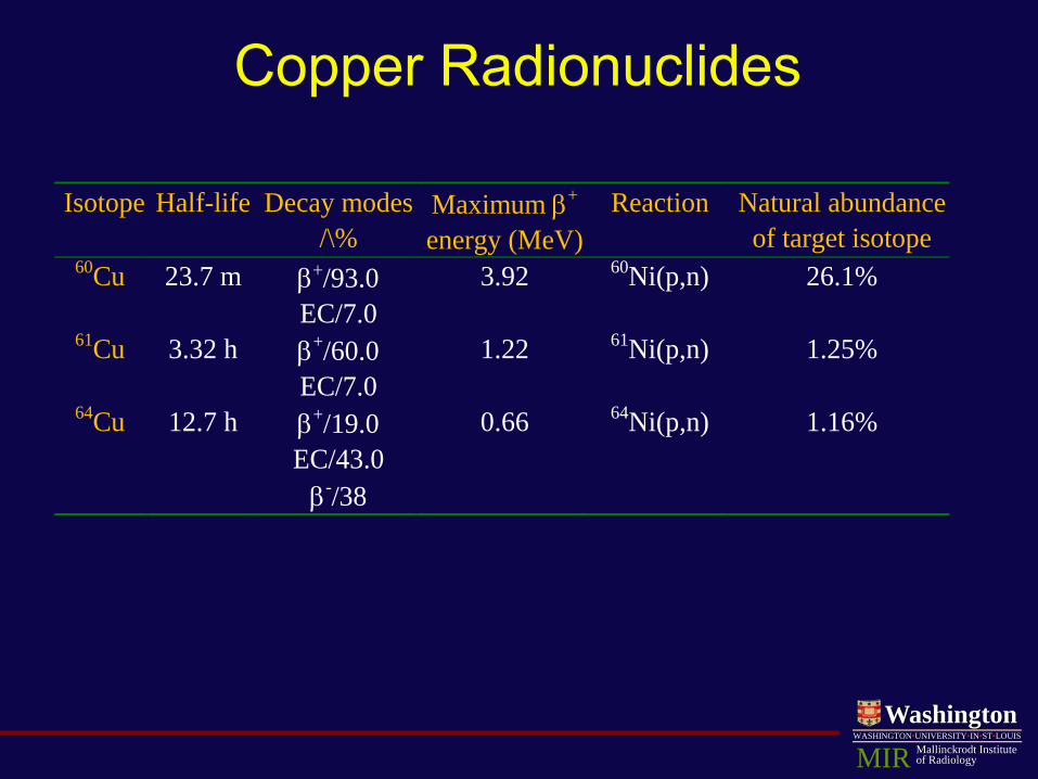
Isotope Half-life Decay modes/\%
Maximum β+
energy (MeV)Reaction Natural abundance
of target isotope 60Cu 23.7 m β+/93.0
EC/7.0 3.92 60Ni(p,n) 26.1%
61Cu 3.32 h β+/60.0 EC/7.0
1.22 61Ni(p,n) 1.25%
64Cu 12.7 h β+/19.0 EC/43.0 β-/38
0.66 64Ni(p,n) 1.16%
Copper Radionuclides
WashingtonWASHINGTON•UNIVERSITY•IN•ST•LOUIS
Mallinckrodt Instituteof RadiologyMIR
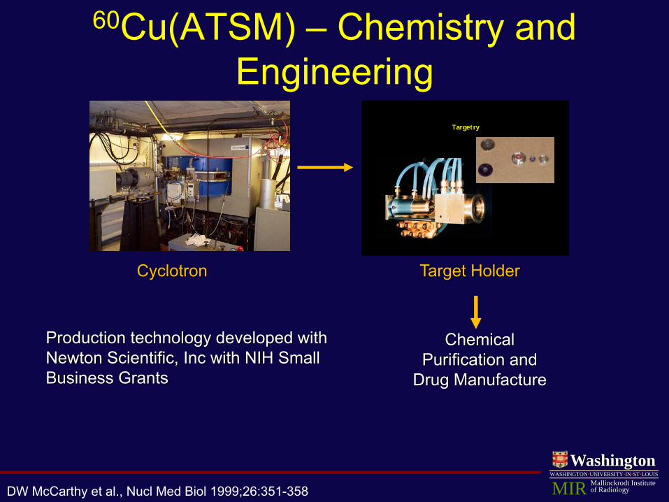
60Cu(ATSM) – Chemistry and Engineering
Target Holder
Chemical Purification and
Drug Manufacture
Production technology developed with Newton Scientific, Inc with NIH Small Business Grants
Cyclotron
DW McCarthy et al., Nucl Med Biol 1999;26:351-358
Targetry
WashingtonWASHINGTON•UNIVERSITY•IN•ST•LOUIS
Mallinckrodt Instituteof RadiologyMIR
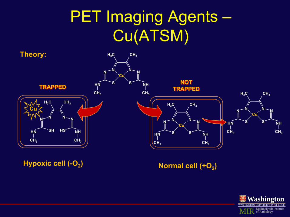
PET Imaging Agents –Cu(ATSM)
Theory:
Cu
H3C CH3
N N N N
CH3
HN
CH3
NHSS NOTTRAPPED
Normal cell (+O2)
Cu
H3C CH3
N N N N
CH3
HN
CH3
NHSS
TRAPPED
Hypoxic cell (-O2)
Cu
H3C CH3
N N N N
CH3
HN
CH3
NHSS
HN
CH3 CH3
NH
H3C
N N
SH
CH3
N N
HS
Cu
WashingtonWASHINGTON•UNIVERSITY•IN•ST•LOUIS
Mallinckrodt Instituteof RadiologyMIR
0
.2
.4
.6
.8
1C
um. S
urvi
val
0 200 400 600 800 1000 1200 1400
Time (Days)
T/M ≥ 3.0
T/M < 3.0
P = 0.02
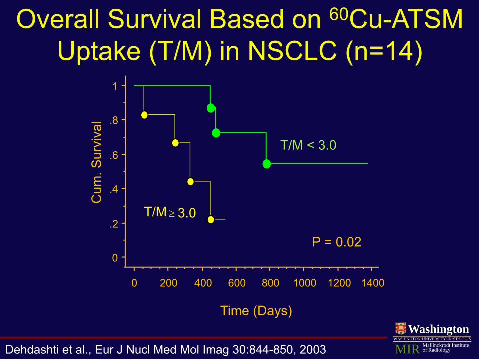
Overall Survival Based on 60Cu-ATSM Uptake (T/M) in NSCLC (n=14)
Dehdashti et al., Eur J Nucl Med Mol Imag 30:844-850, 2003
WashingtonWASHINGTON•UNIVERSITY•IN•ST•LOUIS
Mallinckrodt Instituteof RadiologyMIR
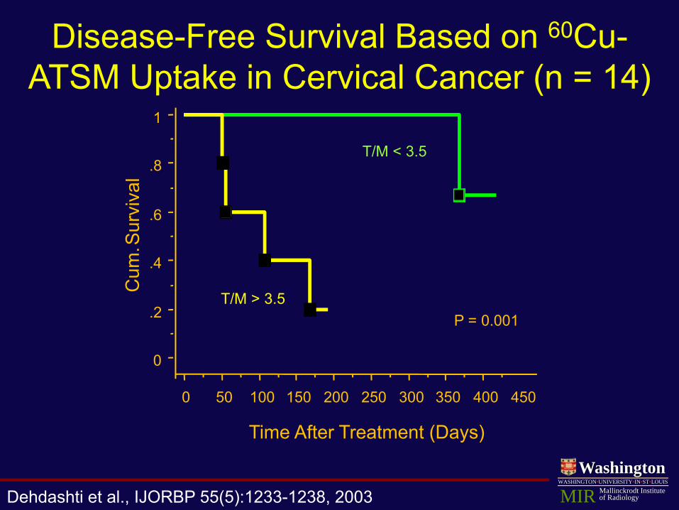
Disease-Free Survival Based on 60Cu-ATSM Uptake in Cervical Cancer (n = 14)
0
.2
.4
.6
.8
1C
um. S
urvi
val
0 50 100 150 200 250 300 350 400 450
Time After Treatment (Days)
P = 0.001T/M > 3.5
T/M < 3.5
Dehdashti et al., IJORBP 55(5):1233-1238, 2003
WashingtonWASHINGTON•UNIVERSITY•IN•ST•LOUIS
Mallinckrodt Instituteof RadiologyMIR
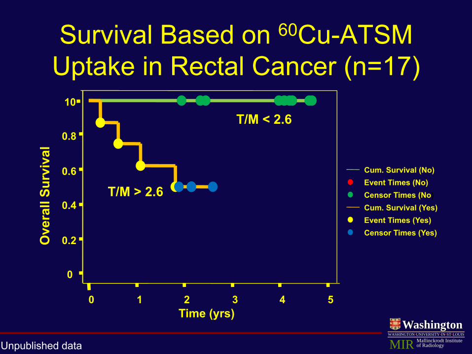
Survival Based on 60Cu-ATSM Uptake in Rectal Cancer (n=17)
Time (yrs)
0
0.2
0.4
0.6
0.8
10
0 1 2 3 4 5
Ove
rall
Surv
ival
T/M < 2.6
T/M > 2.6
Censor Times (Yes)Event Times (Yes)Cum. Survival (Yes)Censor Times (No)Event Times (No)Cum. Survival (No)
Unpublished data
WashingtonWASHINGTON•UNIVERSITY•IN•ST•LOUIS
Mallinckrodt Instituteof RadiologyMIR
Comparison of 60Cu-ATSM and 64Cu-ATSM (IND 62,675)
• Assessed quality of 60Cu- and 64Cu-ATSM PET images• Crossover study of 10 patients with Ib2-IVa cervical CA
– Subjective – comparable; but, 64Cu-ATSM images less noisy
• Similar quality in 8 patients • 64Cu-ATSM better than 60Cu-ATSM in 2 patients
– T/M evaluation• Generally better target to background ratio
(tumors seen more clearly on 64Cu-ATSM-PET in most cases)
WashingtonWASHINGTON•UNIVERSITY•IN•ST•LOUIS
Mallinckrodt Instituteof RadiologyMIR
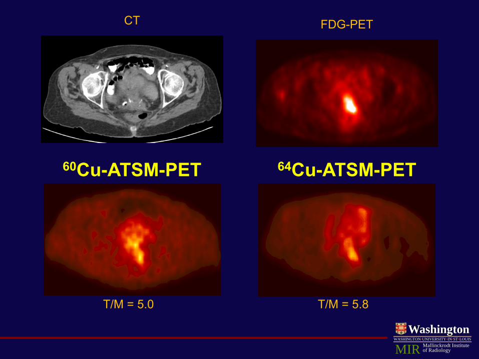
T/M = 5.0 T/M = 5.8
FDG-PET
64Cu-ATSM-PET
CT
60Cu-ATSM-PET
WashingtonWASHINGTON•UNIVERSITY•IN•ST•LOUIS
Mallinckrodt Instituteof RadiologyMIR
2
4
6
8
10
12
14
16
2 3 4 5 6 7 8 9 10 11
T/M Ratio 64Cu-ATSM
T/M
Rat
io 60
Cu-
ATSM
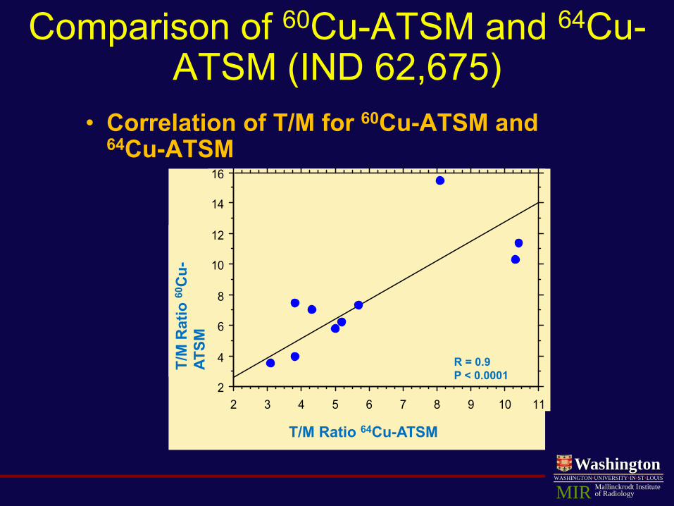
R = 0.9P < 0.0001
Comparison of 60Cu-ATSM and 64Cu-ATSM (IND 62,675)
• Correlation of T/M for 60Cu-ATSM and 64Cu-ATSM
WashingtonWASHINGTON•UNIVERSITY•IN•ST•LOUIS
Mallinckrodt Instituteof RadiologyMIR
Tumor Detection and Treatment Using Bavituximab Labeled with Arsenic
RadionuclidesGuiyang Hao, Xiankai Sun, Philip E.Thorpe, and
Ralph P. Mason
Departments of Radiology and Pharmacology University of Texas Southwestern Medical Center at
Dallas, Texas
WashingtonWASHINGTON•UNIVERSITY•IN•ST•LOUIS
Mallinckrodt Instituteof RadiologyMIR
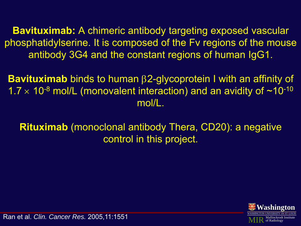
Bavituximab: A chimeric antibody targeting exposed vascularphosphatidylserine. It is composed of the Fv regions of the mouse
antibody 3G4 and the constant regions of human IgG1.
Bavituximab binds to human β2-glycoprotein I with an affinity of 1.7 × 10⁻⁸ mol/L (monovalent interaction) and an avidity of ~10-10
mol/L.
Rituximab (monoclonal antibody Thera, CD20): a negative control in this project.
Ran et al. Clin. Cancer Res. 2005,11:1551
WashingtonWASHINGTON•UNIVERSITY•IN•ST•LOUIS
Mallinckrodt Instituteof RadiologyMIR
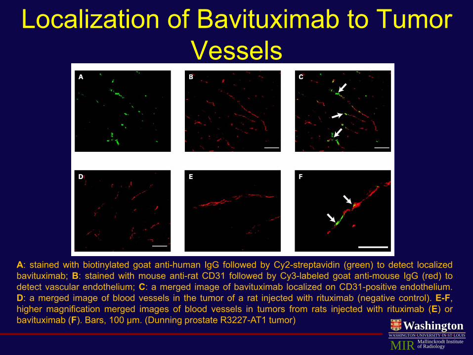
Localization of Bavituximab to Tumor Vessels
A: stained with biotinylated goat anti-human IgG followed by Cy2-streptavidin (green) to detect localizedbavituximab; B: stained with mouse anti-rat CD31 followed by Cy3-labeled goat anti-mouse IgG (red) todetect vascular endothelium; C: a merged image of bavituximab localized on CD31-positive endothelium.D: a merged image of blood vessels in the tumor of a rat injected with rituximab (negative control). E-F,higher magnification merged images of blood vessels in tumors from rats injected with rituximab (E) orbavituximab (F). Bars, 100 µm. (Dunning prostate R3227-AT1 tumor)
WashingtonWASHINGTON•UNIVERSITY•IN•ST•LOUIS
Mallinckrodt Instituteof RadiologyMIR
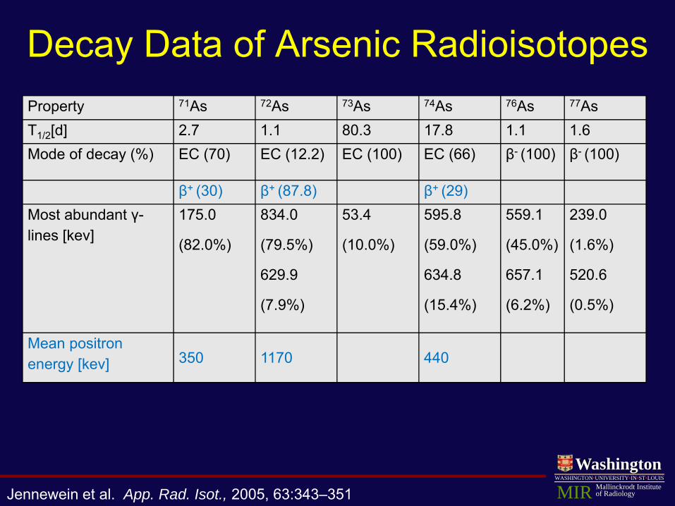
Decay Data of Arsenic RadioisotopesProperty 71As 72As 73As 74As 76As 77AsT1/2[d] 2.7 1.1 80.3 17.8 1.1 1.6Mode of decay (%) EC (70) EC (12.2) EC (100) EC (66) β- (100) β- (100)
β+ (30) β+ (87.8) β+ (29)Most abundant γ-lines [kev]
175.0
(82.0%)
834.0
(79.5%)
629.9
(7.9%)
53.4
(10.0%)
595.8
(59.0%)
634.8
(15.4%)
559.1
(45.0%)
657.1
(6.2%)
239.0
(1.6%)
520.6
(0.5%)
Mean positron energy [kev] 350 1170 440
Jennewein et al. App. Rad. Isot., 2005, 63:343–351
WashingtonWASHINGTON•UNIVERSITY•IN•ST•LOUIS
Mallinckrodt Instituteof RadiologyMIR
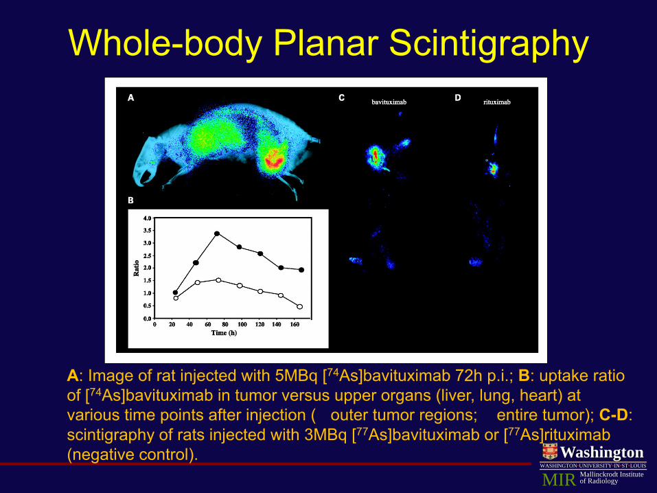
Whole-body Planar Scintigraphy
A: Image of rat injected with 5MBq [74As]bavituximab 72h p.i.; B: uptake ratio of [74As]bavituximab in tumor versus upper organs (liver, lung, heart) at various time points after injection ( outer tumor regions; entire tumor); C-D: scintigraphy of rats injected with 3MBq [77As]bavituximab or [77As]rituximab(negative control).
WashingtonWASHINGTON•UNIVERSITY•IN•ST•LOUIS
Mallinckrodt Instituteof RadiologyMIR
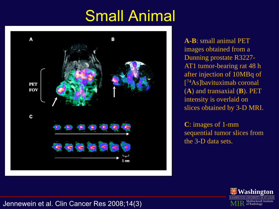
Small Animal PET image A-B: small animal PET
images obtained from a Dunning prostate R3227-AT1 tumor-bearing rat 48 h after injection of 10MBq of [74As]bavituximab coronal (A) and transaxial (B). PET intensity is overlaid on slices obtained by 3-D MRI.
C: images of 1-mm sequential tumor slices from the 3-D data sets.
Jennewein et al. Clin Cancer Res 2008;14(3)
WashingtonWASHINGTON•UNIVERSITY•IN•ST•LOUIS
Mallinckrodt Instituteof RadiologyMIR
Acknowledgements
DOD IDEA awardsW81XWH-06-1-0149 and W81XWH-06-1-0050
National Cancer Institute Pre-ICMIC P20CA086334 and SAIRP U24 CA126608
Conclusion
Radioarsenic-labeled bavituximab has shown potential as a new agent for imaging (74As) the vasculature and radiotherapy (77As) of solid tumors.
WashingtonWASHINGTON•UNIVERSITY•IN•ST•LOUIS
Mallinckrodt Instituteof RadiologyMIR
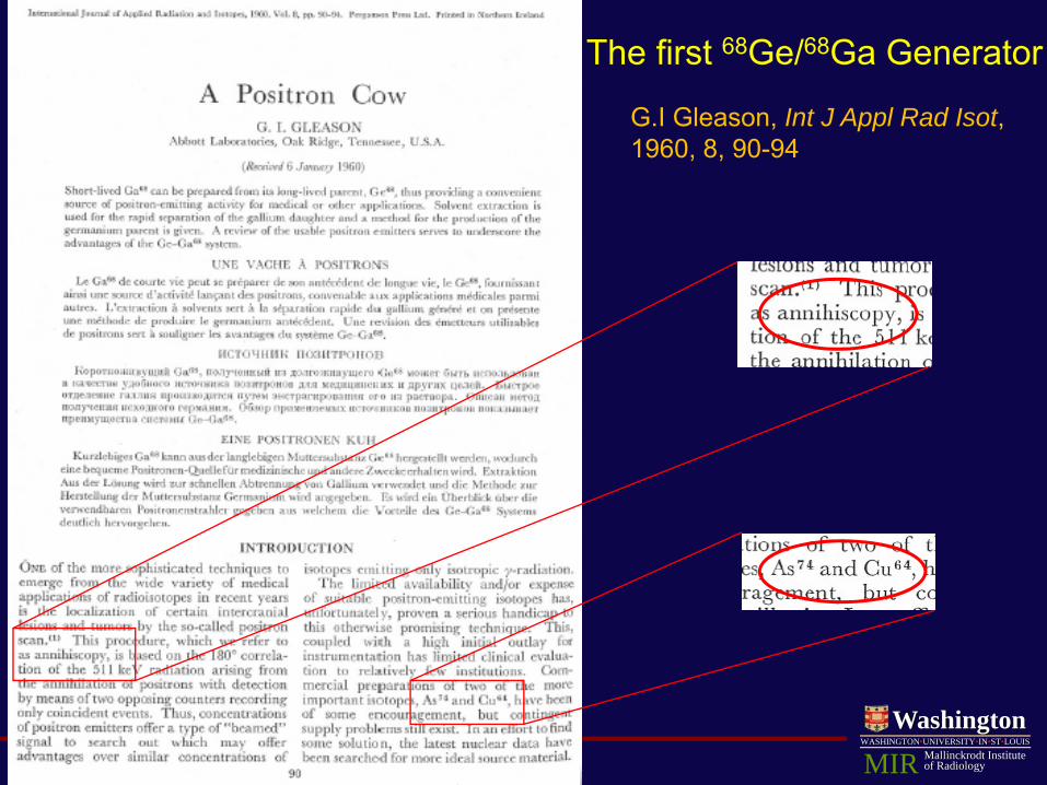
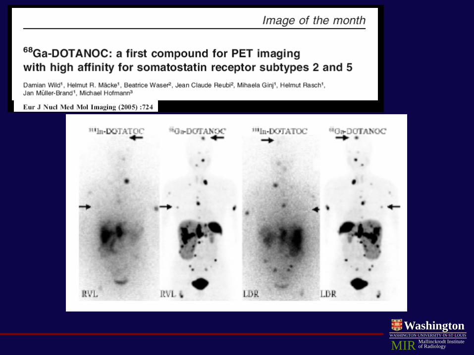
The first 68Ge/68Ga Generator
G.I Gleason, Int J Appl Rad Isot, 1960, 8, 90-94
WashingtonWASHINGTON•UNIVERSITY•IN•ST•LOUIS
Mallinckrodt Instituteof RadiologyMIR
WashingtonWASHINGTON•UNIVERSITY•IN•ST•LOUIS
Mallinckrodt Instituteof RadiologyMIR
WashingtonWASHINGTON•UNIVERSITY•IN•ST•LOUIS
Mallinckrodt Instituteof RadiologyMIR
Translation of PET agents to the Clinic
95% of studies involve FDG
WHY?
WashingtonWASHINGTON•UNIVERSITY•IN•ST•LOUIS
Mallinckrodt Instituteof RadiologyMIR
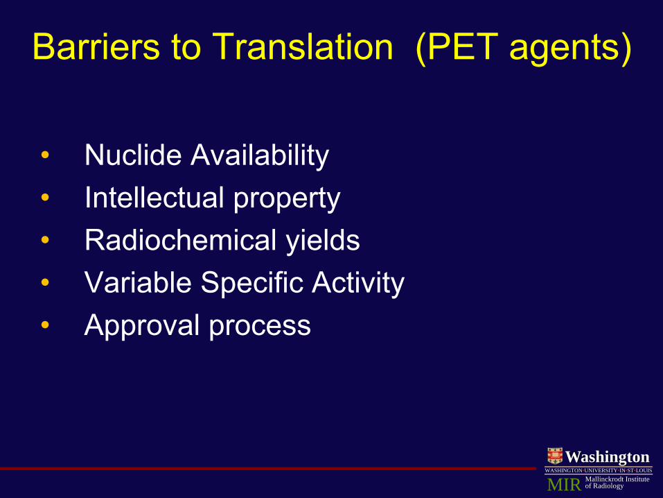
Barriers to Translation (PET agents)
• Nuclide Availability• Intellectual property• Radiochemical yields• Variable Specific Activity• Approval process
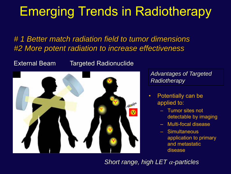
Emerging Trends in Radiotherapy
• Potentially can be applied to:
– Tumor sites not detectable by imaging
– Multi-focal disease– Simultaneous
application to primary and metastatic disease
External Beam Targeted RadionuclideAdvantages of Targeted Radiotherapy
# 1 Better match radiation field to tumor dimensions#2 More potent radiation to increase effectiveness
Short range, high LET α-particles
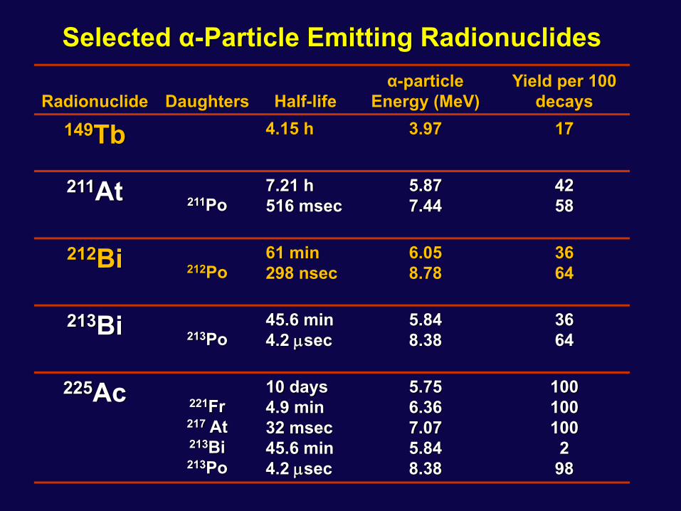
Selected α-Particle Emitting Radionuclides
Radionuclide Daughters Half-lifeα-particle
Energy (MeV)Yield per 100
decays149Tb 4.15 h 3.97 17
211At 211Po7.21 h516 msec
5.877.44
4258
212Bi 212Po61 min298 nsec
6.058.78
3664
213Bi 213Po45.6 min4.2 μsec
5.848.38
3664
225Ac 221Fr217 At213Bi213Po
10 days4.9 min32 msec45.6 min4.2 μsec
5.756.367.075.848.38
100100100
298
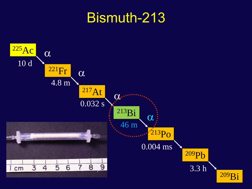
213Bi
10 d
4.8 m
0.032 s
0.004 ms
3.3 h
46 m
225Ac
217At
221Fr
213Po
209Pb
209Bi
α
α
α
α
Bismuth-213
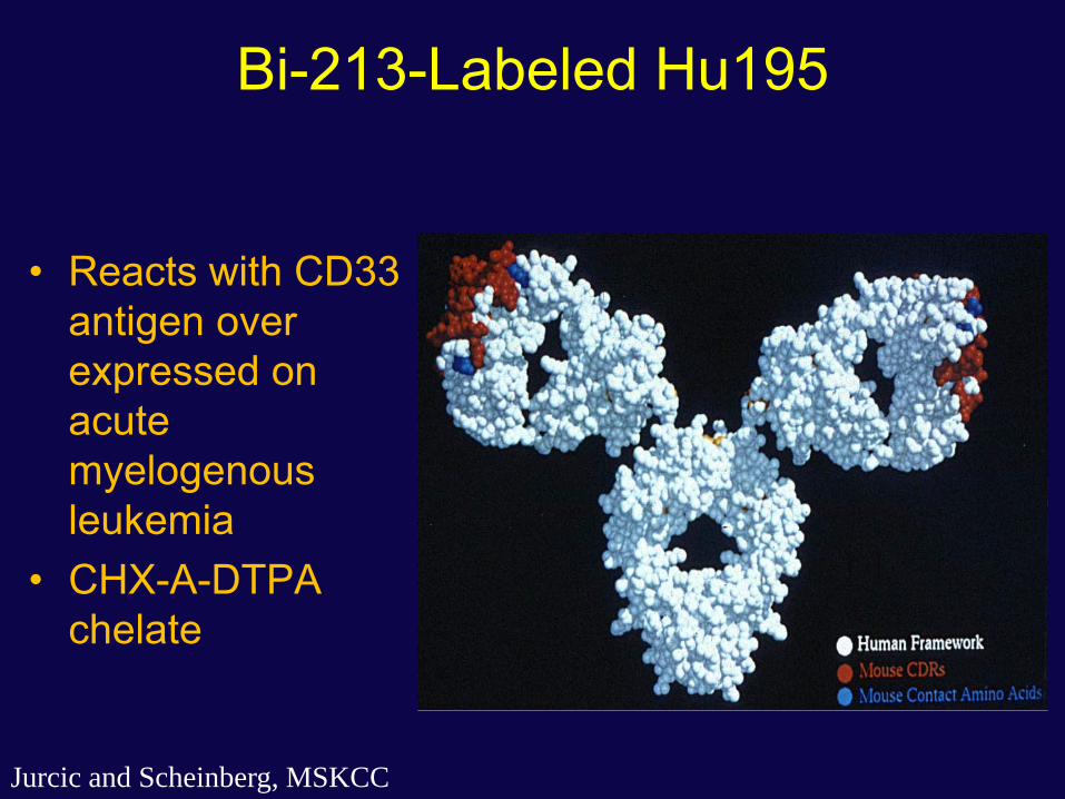
Bi-213-Labeled Hu195
• Reacts with CD33 antigen over expressed on acute myelogenous leukemia
• CHX-A-DTPA chelate
Jurcic and Scheinberg, MSKCC
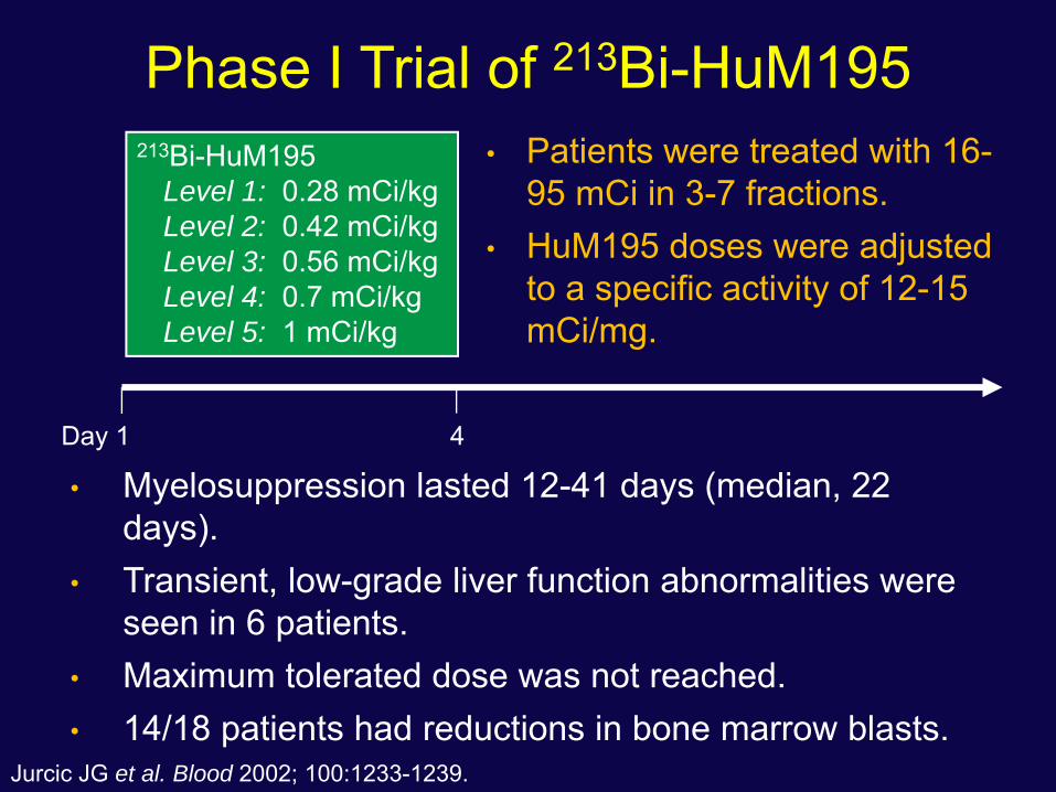
Phase I Trial of 213Bi-HuM195
• Myelosuppression lasted 12-41 days (median, 22 days).
• Transient, low-grade liver function abnormalities were seen in 6 patients.
• Maximum tolerated dose was not reached.• 14/18 patients had reductions in bone marrow blasts.
Jurcic JG et al. Blood 2002; 100:1233-1239.
213Bi-HuM195Level 1: 0.28 mCi/kgLevel 2: 0.42 mCi/kgLevel 3: 0.56 mCi/kgLevel 4: 0.7 mCi/kgLevel 5: 1 mCi/kg
• Patients were treated with 16-95 mCi in 3-7 fractions.
• HuM195 doses were adjusted to a specific activity of 12-15 mCi/mg.
Day 1 4
Jurcic JG et al. Blood 2002; 100:1233-1239
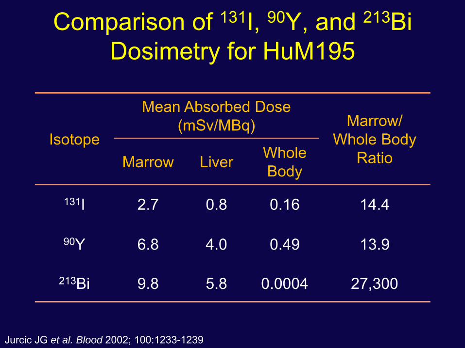
Comparison of 131I, 90Y, and 213Bi Dosimetry for HuM195
Isotope
Mean Absorbed Dose(mSv/MBq) Marrow/
Whole BodyRatioMarrow Liver Whole
Body
131I 2.7 0.8 0.16 14.4
90Y 6.8 4.0 0.49 13.9
213Bi 9.8 5.8 0.0004 27,300
Rationale for α-Particle Emitters in Cytoreduced Disease
• The short range and high LET of α-particles make them best-suited for treatment of small-volume disease.
• In patients with overt AML, there are 1016 CD33 binding sites, making it difficult to target 1-2 213Bi atoms to each leukemia cell.
• Hypothesis: Cytoreduction with cytarabine should decrease tumor burden by 1-2 logs and increase the ratio of 213Bi atoms to target cells.
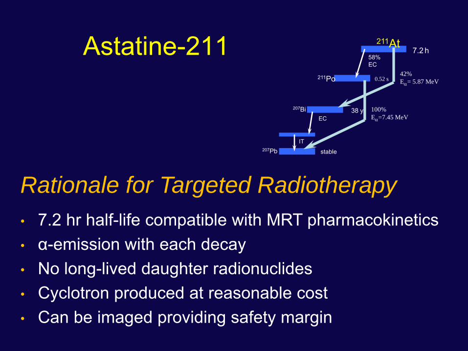
7.2 h58%EC
42%Eα= 5.87 MeV
211At
stable207PbIT
EC
207Bi 38 y
211Po 0.52 s
100%Eα=7.45 MeV
Astatine-211
• 7.2 hr half-life compatible with MRT pharmacokinetics• α-emission with each decay• No long-lived daughter radionuclides• Cyclotron produced at reasonable cost• Can be imaged providing safety margin
Rationale for Targeted Radiotherapy
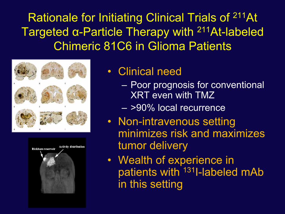
Rationale for Initiating Clinical Trials of 211At Targeted α-Particle Therapy with 211At-labeled
Chimeric 81C6 in Glioma Patients
• Clinical need– Poor prognosis for conventional
XRT even with TMZ– >90% local recurrence
• Non-intravenous setting minimizes risk and maximizes tumor delivery
• Wealth of experience in patients with 131I-labeled mAb in this setting
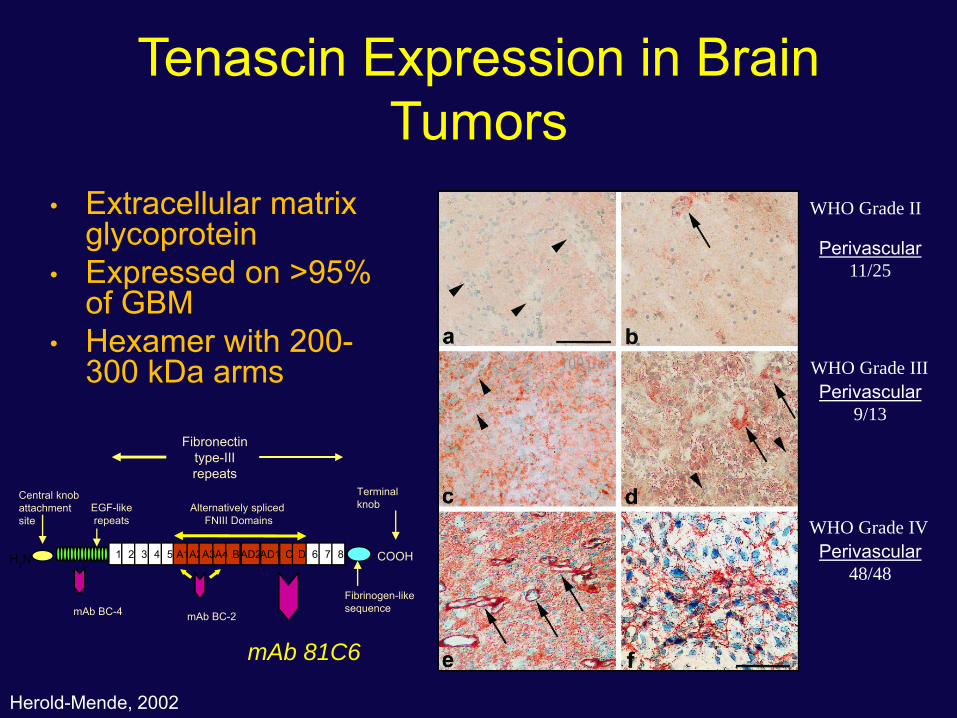
• Extracellular matrix glycoprotein
• Expressed on >95% of GBM
• Hexamer with 200-300 kDa arms
Fibronectintype-III repeats
H2N
Terminal knob
CA2 BA4A31 2 543 AD2AD1 876 COOH
EGF-likerepeats
Alternatively splicedFNIII Domains
Fibrinogen-likesequencemAb BC-4
mAb 81C6
A1 D
mAb BC-2
Central knobattachmentsite
Tenascin Expression in Brain Tumors
100 μm
Herold-Mende, 2002
WHO Grade II
WHO Grade III
WHO Grade IV
Perivascular11/25
Perivascular9/13
Perivascular48/48
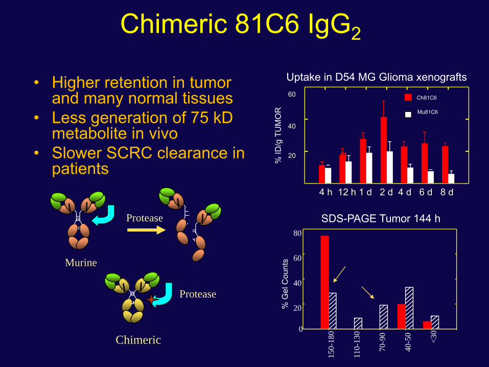
Chimeric 81C6 IgG2
• Higher retention in tumor and many normal tissues
• Less generation of 75 kD metabolite in vivo
• Slower SCRC clearance in patients
Murine
Protease
Chimeric
Protease
150-
180
110-
130
70-9
0
40-5
0
<300
20
40
60
80
SDS-PAGE Tumor 144 h
% G
el C
ount
s%
ID/g
TU
MO
R
4 h 12 h 1 d 2 d 4 d 6 d 8 d
60
40
20
Ch81C6
Mu81C6
Uptake in D54 MG Glioma xenografts
At-211 Labeled Chimeric 81C6: Clinical Protocol
• Thyroid blocking with SSKI and Cytomel beginning 48 hr prior to therapy
• Dose administration via indwelling catheter• Patients injected via the SCRC with 10 mg of
mAb labeled with 2 (n=5), 4 (n=7), 6.7 (n=5) or 10 mCi (n=1) mCi 211At
• Blood sampling at 1, 2, 4, 8, 12, 18 and 24 hr • SPECT of head and whole body imaging at 2,
4, 8, 18 and 24 hr
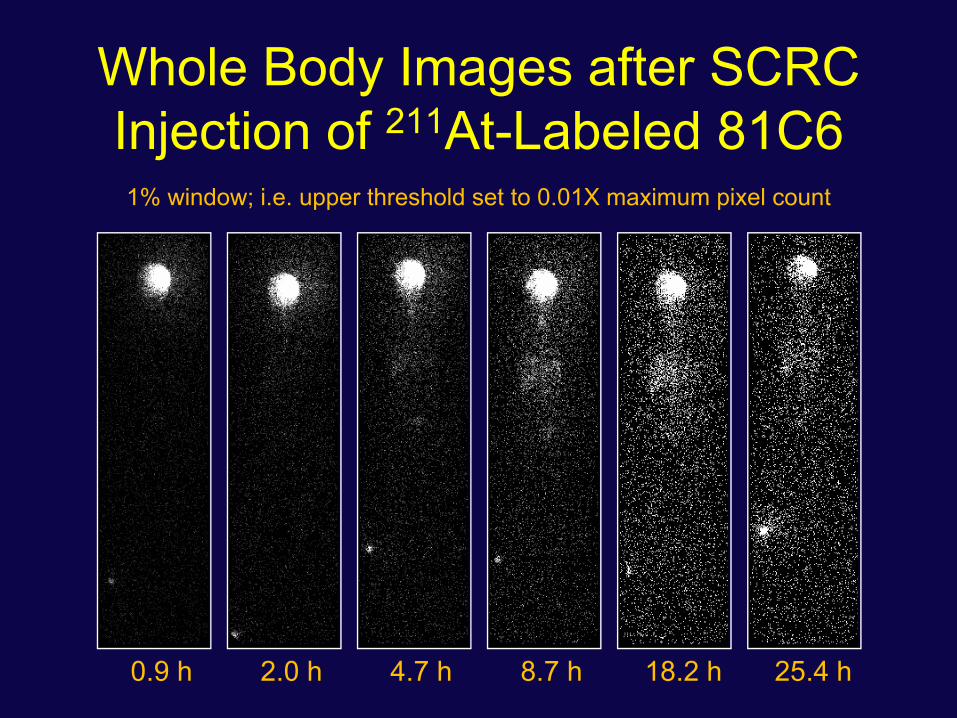
Whole Body Images after SCRC Injection of 211At-Labeled 81C6
0.9 h 2.0 h 4.7 h 8.7 h 18.2 h 25.4 h
1% window; i.e. upper threshold set to 0.01X maximum pixel count
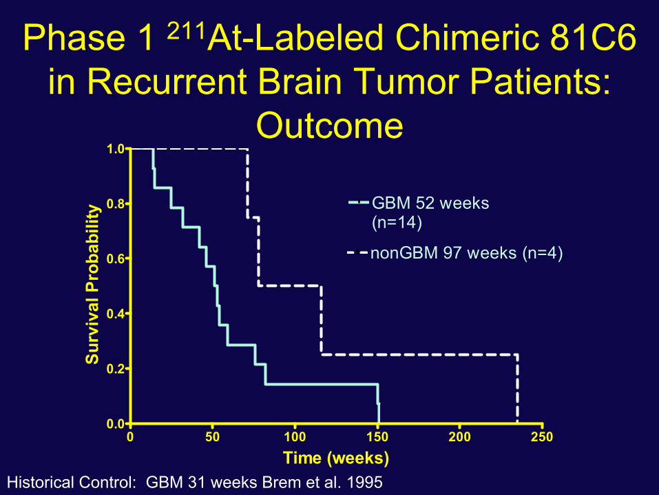
0 50 100 150 200 2500.0
0.2
0.4
0.6
0.8
1.0
GBM 52 weeks(n=14)
nonGBM 97 weeks (n=4)
Time (weeks)
Surv
ival
Pro
babi
lity
Phase 1 211At-Labeled Chimeric 81C6 in Recurrent Brain Tumor Patients:
Outcome
Historical Control: GBM 31 weeks Brem et al. 1995
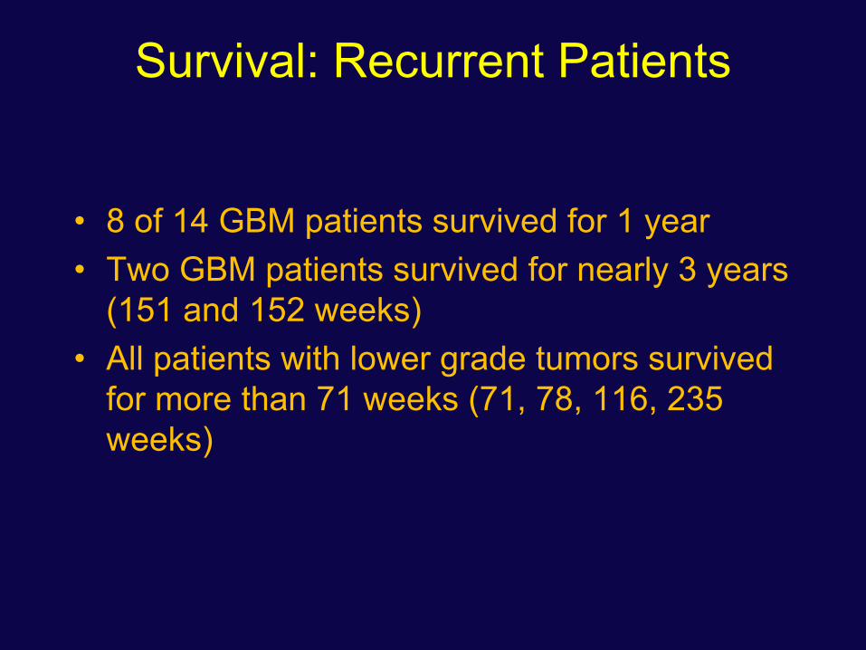
Survival: Recurrent Patients
• 8 of 14 GBM patients survived for 1 year• Two GBM patients survived for nearly 3 years
(151 and 152 weeks)• All patients with lower grade tumors survived
for more than 71 weeks (71, 78, 116, 235 weeks)
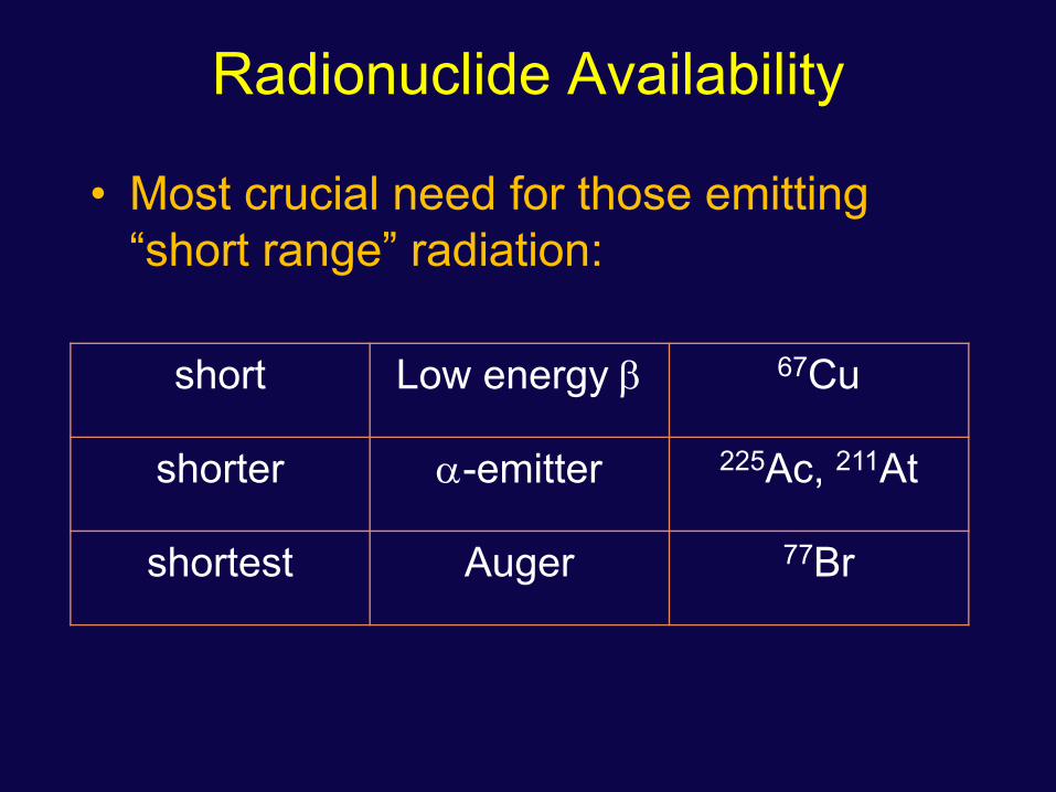
Radionuclide Availability
short Low energy β 67Cu
shorter α-emitter 225Ac, 211At
shortest Auger 77Br
• Most crucial need for those emitting “short range” radiation:
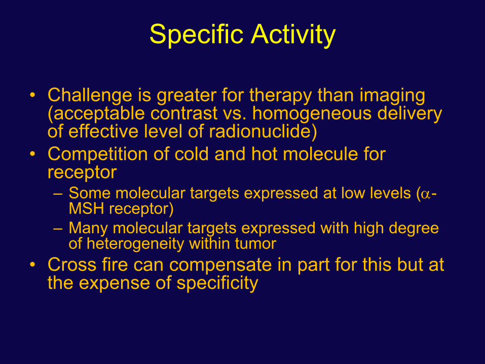
Specific Activity
• Challenge is greater for therapy than imaging (acceptable contrast vs. homogeneous delivery of effective level of radionuclide)
• Competition of cold and hot molecule for receptor– Some molecular targets expressed at low levels (α-
MSH receptor)– Many molecular targets expressed with high degree
of heterogeneity within tumor• Cross fire can compensate in part for this but at
the expense of specificity
Regulatory Affairs
• Requirement for evaluating late radiation effects without adequate guidance (endpoints, species, time frame)
• Guidelines for radiotoxicity of high-LET emitters• Handling of patient-specific treatment plans
(cocktails of radionuclides and carriers, variations in dosing schemes)
Consequences of Heterogeneity for Radionuclide Needs
• Macro: Need to administer multiple radionuclides to compensate for range of tumor sizes in a particular patient
• Micro: Need to balance advantages of longer range radiation (cross fire of receptor negative populations) with disadvantages (irradiation of normal tissue)
• Normal tissue: Need to distribute uptake of labeled catabolites among organs through use of different radionuclides and labeling methods