Embed Size (px)
Citation preview
Brett S. KlosterhoffGeorge W. Woodruff School
of Mechanical Engineering,
Georgia Institute of Technology,
Atlanta, GA 30332;
Parker H. Petit Institute for
Bioengineering and Bioscience,
Georgia Institute of Technology,
Atlanta, GA 30332
Melissa TsangSchool of Electrical and Computer Engineering,
Georgia Institute of Technology,
Atlanta, GA 30332
Didi SheDepartment of Electrical and
Systems Engineering,
University of Pennsylvania,
Philadelphia, PA 19104
Keat Ghee OngDepartment of Biomedical Engineering,
Michigan Technological University,
Houghton, MI 49931
Mark G. AllenSchool of Electrical and Computer Engineering,
Georgia Institute of Technology,
Atlanta, GA 30332;
Department of Electrical and
Systems Engineering,
University of Pennsylvania,
Philadelphia, PA 19104
Nick J. WillettParker H. Petit Institute for
Bioengineering and Bioscience,
Georgia Institute of Technology,
Atlanta, GA 30332;
Department of Orthopaedics,
Emory University,
Atlanta, GA 30303;
Atlanta Veteran’s Affairs Medical Center,
Decatur, GA 30033;
Wallace H. Coulter Department
of Biomedical Engineering,
Georgia Institute of Technology
and Emory University,
Atlanta, GA 30332
Robert E. GuldbergGeorge W. Woodruff School
of Mechanical Engineering,
Georgia Institute of Technology,
Atlanta, GA 30332;
Parker H. Petit Institute for
Bioengineering and Bioscience,
Georgia Institute of Technology,
Atlanta, GA 30332
Implantable Sensors forRegenerative MedicineThe translation of many tissue engineering/regenerative medicine (TE/RM) therapies thatdemonstrate promise in vitro are delayed or abandoned due to reduced and inconsistentefficacy when implemented in more complex and clinically relevant preclinical in vivomodels. Determining mechanistic reasons for impaired treatment efficacy is challengingafter a regenerative therapy is implanted due to technical limitations in longitudinallymeasuring the progression of key environmental cues in vivo. The ability to acquire real-time measurements of environmental parameters of interest including strain, pressure,pH, temperature, oxygen tension, and specific biomarkers within the regenerative nichein situ would significantly enhance the information available to tissue engineers to moni-tor and evaluate mechanisms of functional healing or lack thereof. Continued advance-ments in material and fabrication technologies utilized by microelectromechanicalsystems (MEMSs) and the unique physical characteristics of passive magnetoelastic sen-sor platforms have created an opportunity to implant small, flexible, low-power sensorsinto preclinical in vivo models, and quantitatively measure environmental cues through-out healing. In this perspective article, we discuss the need for longitudinal measure-ments in TE/RM research, technical progress in MEMS and magnetoelastic approachesto implantable sensors, the potential application of implantable sensors to benefit preclin-ical TE/RM research, and the future directions of collaborative efforts at the intersectionof these two important fields. [DOI: 10.1115/1.4035436]
Keywords: tissue engineering, regenerative medicine, MEMS, sensors, in vivomonitoring
Manuscript received July 6, 2016; final manuscript received November 28, 2016;published online January 19, 2017. Assoc. Editor: Victor H. Barocas.
Journal of Biomechanical Engineering FEBRUARY 2017, Vol. 139 / 021009-1Copyright VC 2017 by ASME
Downloaded From: http://biomechanical.asmedigitalcollection.asme.org/ on 05/30/2018 Terms of Use: http://www.asme.org/about-asme/terms-of-use
1 Introduction
Tissue engineering and regenerative medicine (TE/RM) com-prises a wide range of therapeutic approaches, each with the intentto mimic or augment endogenous biological mechanisms toreplace or repair injured tissues and organs [1].The notion ofharnessing innate developmental or regenerative biology to engi-neer clinical therapies dates back to 1938, and in recent decadeshas developed into an established commercial industry [2,3]. Cur-rent TE/RM developments can largely be segmented into twoapproaches which are increasingly implemented in combination:(1) stimulation of intrinsic repair and (2) replacement of theinjured tissue [4]. However, with the exception of tissue engi-neered skin products, the translation of TE/RM approaches intoviable therapies for many clinical injuries has remained elusive.In particular, the regeneration of large, vascularized, and multi-tissue injuries including those as a result of chronic degenerationor trauma represents an urgent clinical need for more effectiveTE/RM treatment options.
The translation of many TE/RM therapies that demonstratepromise in vitro are delayed or abandoned due to reduced andinconsistent efficacy when implemented in more complex andclinically relevant preclinical in vivo models. Throughout essen-tially every developmental or healing process, the organization,proliferation, and differentiation of cells follow a highly coordi-nated spatiotemporal profile that is optimized to produce orrestore functional tissue. Likewise, once a regenerative therapy isdelivered in vivo, there are dynamic, bidirectional mechanical andbiochemical interactions between the implant and the local injuryenvironment which can either promote or dysregulate the healingprocess. Determining mechanistic reasons for impaired treatmentefficacy is challenging after a regenerative therapy is implanteddue to technical limitations in longitudinally measuring the pro-gression of key environmental cues in vivo. The ability to acquirereal-time measurements of environmental parameters of interestincluding strain, pressure, force, pH, temperature, and oxygen ten-sion within the regenerative niche in situ would significantlyenhance the information available to tissue engineers to monitorand evaluate mechanisms of functional healing or lack thereof.Moreover, correlation of such longitudinal parameters with quan-titative measures of functional regeneration would provide arational approach for the design of improved regenerativestrategies.
Continued advancements in material and fabrication technolo-gies utilized by microelectromechanical systems (MEMS) and theunique physical characteristics of passive magnetoelastic sensorplatforms have created an opportunity to implant small, flexible,low-power sensors into preclinical in vivo models and quantita-tively measure environmental cues throughout healing. In particu-lar, the capabilities of implantable sensors offer three excitingapplications to TE/RM research: (1) longitudinal, minimally inva-sive measurements of environmental parameters throughoutendogenous tissue repair to define desirable biomechanical andbiochemical design criteria for new regenerative therapies; (2)in vivo monitoring of novel TE/RM therapies to evaluate theirability to mimic desired spatiotemporal healing profiles, and toelucidate potential reasons for successful versus unsuccessfulimplementations; (3) environmentally tuned TE/RM constructswith integrated sensors that can trigger specific actions (e.g., drugor growth factor depot release) at a certain environmental thresh-old in a closed-loop fashion.
2 The Need for Longitudinal Measurements
The dynamic nature of healing has important implications onmechanical and chemical environments that regenerative medi-cine therapies seek to manipulate. The structure and compositionof cells and extracellular matrix (ECM) comprising healing tissueis spatiotemporally heterogeneous. Likewise, the multiscalemechanical properties and internal stress–strain distribution
evolve rapidly, particularly in regenerating load-bearing tissues.In addition, the level of vascular infiltration delivering oxygenand nutrients and removing waste is highly transient. As a result,important environmental cues known to regulate cell migration,differentiation, proliferation, apoptosis, factor secretion, and ECMdeposition are continuously changing variables. To more explic-itly articulate this perspective, we briefly cite large bone defectregeneration throughout the text as a relevant example which is ofparticular interest to our laboratories.
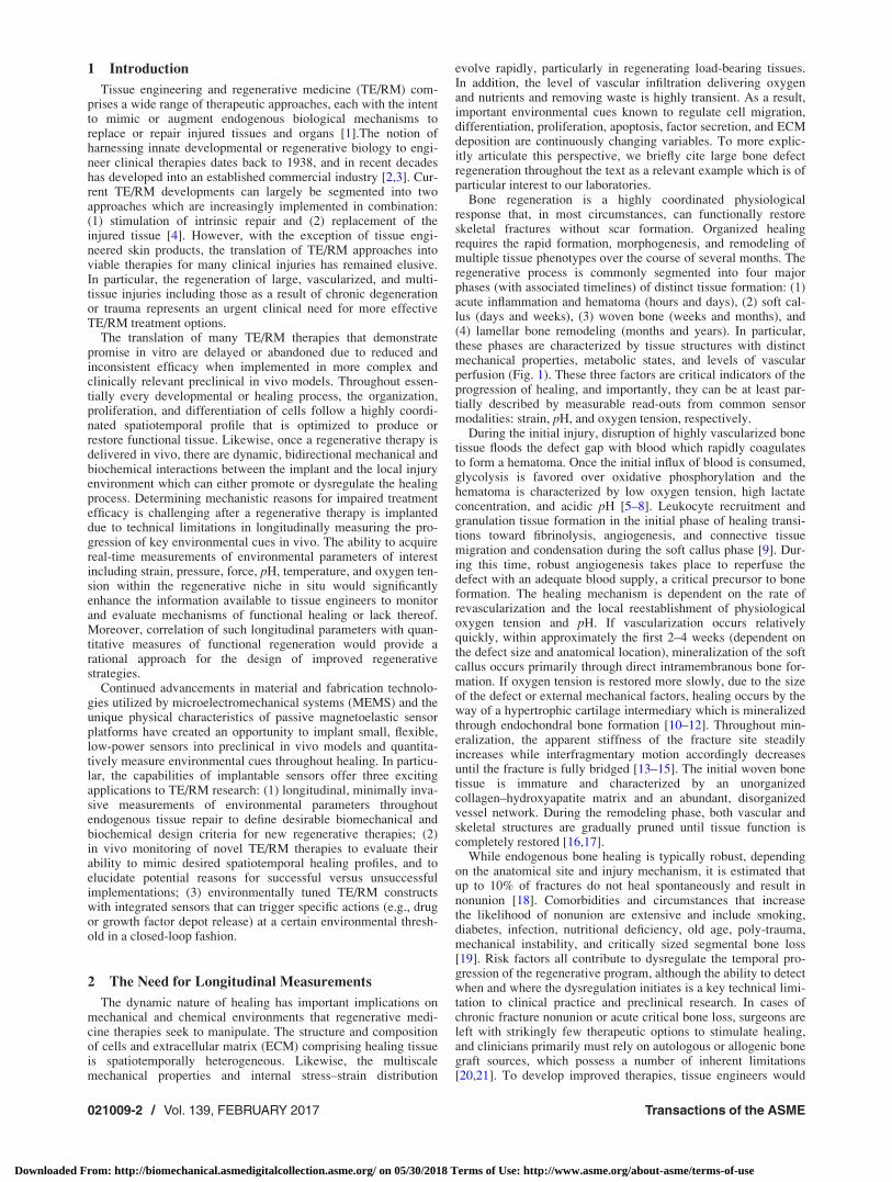
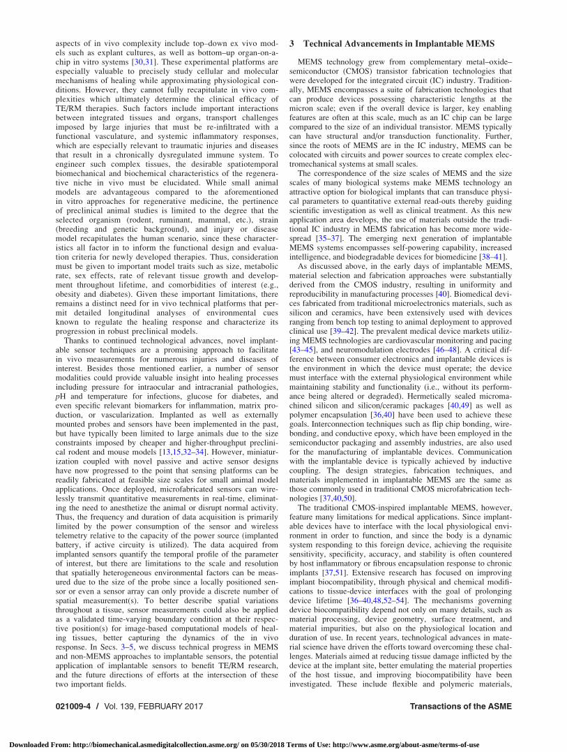
Bone regeneration is a highly coordinated physiologicalresponse that, in most circumstances, can functionally restoreskeletal fractures without scar formation. Organized healingrequires the rapid formation, morphogenesis, and remodeling ofmultiple tissue phenotypes over the course of several months. Theregenerative process is commonly segmented into four majorphases (with associated timelines) of distinct tissue formation: (1)acute inflammation and hematoma (hours and days), (2) soft cal-lus (days and weeks), (3) woven bone (weeks and months), and(4) lamellar bone remodeling (months and years). In particular,these phases are characterized by tissue structures with distinctmechanical properties, metabolic states, and levels of vascularperfusion (Fig. 1). These three factors are critical indicators of theprogression of healing, and importantly, they can be at least par-tially described by measurable read-outs from common sensormodalities: strain, pH, and oxygen tension, respectively.
During the initial injury, disruption of highly vascularized bonetissue floods the defect gap with blood which rapidly coagulatesto form a hematoma. Once the initial influx of blood is consumed,glycolysis is favored over oxidative phosphorylation and thehematoma is characterized by low oxygen tension, high lactateconcentration, and acidic pH [5–8]. Leukocyte recruitment andgranulation tissue formation in the initial phase of healing transi-tions toward fibrinolysis, angiogenesis, and connective tissuemigration and condensation during the soft callus phase [9]. Dur-ing this time, robust angiogenesis takes place to reperfuse thedefect with an adequate blood supply, a critical precursor to boneformation. The healing mechanism is dependent on the rate ofrevascularization and the local reestablishment of physiologicaloxygen tension and pH. If vascularization occurs relativelyquickly, within approximately the first 2–4 weeks (dependent onthe defect size and anatomical location), mineralization of the softcallus occurs primarily through direct intramembranous bone for-mation. If oxygen tension is restored more slowly, due to the sizeof the defect or external mechanical factors, healing occurs by theway of a hypertrophic cartilage intermediary which is mineralizedthrough endochondral bone formation [10–12]. Throughout min-eralization, the apparent stiffness of the fracture site steadilyincreases while interfragmentary motion accordingly decreasesuntil the fracture is fully bridged [13–15]. The initial woven bonetissue is immature and characterized by an unorganizedcollagen–hydroxyapatite matrix and an abundant, disorganizedvessel network. During the remodeling phase, both vascular andskeletal structures are gradually pruned until tissue function iscompletely restored [16,17].
While endogenous bone healing is typically robust, dependingon the anatomical site and injury mechanism, it is estimated thatup to 10% of fractures do not heal spontaneously and result innonunion [18]. Comorbidities and circumstances that increasethe likelihood of nonunion are extensive and include smoking,diabetes, infection, nutritional deficiency, old age, poly-trauma,mechanical instability, and critically sized segmental bone loss[19]. Risk factors all contribute to dysregulate the temporal pro-gression of the regenerative program, although the ability to detectwhen and where the dysregulation initiates is a key technical limi-tation to clinical practice and preclinical research. In cases ofchronic fracture nonunion or acute critical bone loss, surgeons areleft with strikingly few therapeutic options to stimulate healing,and clinicians primarily must rely on autologous or allogenic bonegraft sources, which possess a number of inherent limitations[20,21]. To develop improved therapies, tissue engineers would
021009-2 / Vol. 139, FEBRUARY 2017 Transactions of the ASME
Downloaded From: http://biomechanical.asmedigitalcollection.asme.org/ on 05/30/2018 Terms of Use: http://www.asme.org/about-asme/terms-of-use
benefit from nondestructive measurement platforms that can lon-gitudinally quantify mechanical and chemical parameters withinthe healing environment to aid in the identification of criticalthresholds that are detrimental to healing and to isolate time-points where dysregulation occurs. Common quantitative, nondes-tructive preclinical assays are intended to evaluate overall healing(e.g., computed tomography measurements of bone volume andarchitecture), which are highly dependent on the animal and injurymodel being investigated. They do not quantify the actualmechanical and biochemical cues regulating injury progression.Since local measurements of environmental signals are typicallynot measured directly, a meaningfully quantitative description ofan ideal versus a challenged healing environment is difficult togeneralize across multiple studies.
While longitudinal evaluation platforms improve the ability toevaluate physiological processes in their entirety, they also offeradditional benefits by maximizing data acquired from a singlestudy subject which consequently could reduce the number ofanimals required for an adequately powered study. Numerousinnovative nondestructive or minimally invasive experimentaltechniques have been developed to measure important parameterssuch as tissue mechanical properties, oxygen tension, and pHin vivo. Such techniques are often image-based and include ultra-sound strain imaging, shear wave elastography, and magnetic res-onance imaging (MRI) [22–26]. Nonimage based techniques tomeasure mechanical strains or properties of structural tissues suchas healing bones utilize external fixation hardware which are
subjected to estimated physiological loads by a mechanical testinginstrument [27]. While still nondestructive, the aforementionedmethods are typically limited to snapshot measurements acquiredrelatively infrequently due to practical limits on the number ofoccasions animals can be safely anesthetized to perform the mea-surement. Additionally, imaging methods often rely on expensiveequipment and typically must be conducted one specimen at atime. Thus, such techniques are inherently low-throughput andcostly for studies utilizing large sample numbers.
Finite element (FE) models and numerical computational fluiddynamics (CFD) simulations based on in vivo imaging or sim-plified representations of healing tissue structures offer uniqueinsights into the spatial distribution of stress, strain, and oxygentension within the tissue at the healing site. Additionally, com-putational models allow for parametric analyses to study thesensitivity of the tissue structure and the relative importance ofdistinct variables in a tissue-engineered construct (i.e., scaffoldmodulus, porosity, and initial growth factor concentration).However, the results of such computational models are affecteddrastically by the initial boundary conditions imposed during thedefinition of the model. Boundary conditions such as externalload magnitudes and oxygen concentration profiles are oftenbased on simplified assumptions lacking experimental validation,which can compromise the accuracy of conclusions obtained bysimulations [28,29].
Other approaches to overcome limitations in longitudinalin vivo measurement techniques while maintaining some
Fig. 1 Schematic outlining the temporal profile of bone regeneration, illustrating phases of healing, structural progressionin the defect, and qualitative estimates of environmental parameter profiles. Nondestructive, quantitative measurements ofthese environmental cues would significantly enhance fundamental understanding of the temporal progression of the bonehealing environment as well as many other diseases of interest, providing a better foundation to develop and evaluate effec-tive regenerative therapies. Created using images from Servier Medical Art, CC-BY 3.0.
Journal of Biomechanical Engineering FEBRUARY 2017, Vol. 139 / 021009-3
Downloaded From: http://biomechanical.asmedigitalcollection.asme.org/ on 05/30/2018 Terms of Use: http://www.asme.org/about-asme/terms-of-use
aspects of in vivo complexity include top–down ex vivo mod-els such as explant cultures, as well as bottom–up organ-on-a-chip in vitro systems [30,31]. These experimental platforms areespecially valuable to precisely study cellular and molecularmechanisms of healing while approximating physiological con-ditions. However, they cannot fully recapitulate in vivo com-plexities which ultimately determine the clinical efficacy ofTE/RM therapies. Such factors include important interactionsbetween integrated tissues and organs, transport challengesimposed by large injuries that must be re-infiltrated with afunctional vasculature, and systemic inflammatory responses,which are especially relevant to traumatic injuries and diseasesthat result in a chronically dysregulated immune system. Toengineer such complex tissues, the desirable spatiotemporalbiomechanical and biochemical characteristics of the regenera-tive niche in vivo must be elucidated. While small animalmodels are advantageous compared to the aforementionedin vitro approaches for regenerative medicine, the pertinenceof preclinical animal studies is limited to the degree that theselected organism (rodent, ruminant, mammal, etc.), strain(breeding and genetic background), and injury or diseasemodel recapitulates the human scenario, since these character-istics all factor in to inform the functional design and evalua-tion criteria for newly developed therapies. Thus, considerationmust be given to important model traits such as size, metabolicrate, sex effects, rate of relevant tissue growth and develop-ment throughout lifetime, and comorbidities of interest (e.g.,obesity and diabetes). Given these important limitations, thereremains a distinct need for in vivo technical platforms that per-mit detailed longitudinal analyses of environmental cuesknown to regulate the healing response and characterize itsprogression in robust preclinical models.
Thanks to continued technological advances, novel implant-able sensor techniques are a promising approach to facilitatein vivo measurements for numerous injuries and diseases ofinterest. Besides those mentioned earlier, a number of sensormodalities could provide valuable insight into healing processesincluding pressure for intraocular and intracranial pathologies,pH and temperature for infections, glucose for diabetes, andeven specific relevant biomarkers for inflammation, matrix pro-duction, or vascularization. Implanted as well as externallymounted probes and sensors have been implemented in the past,but have typically been limited to large animals due to the sizeconstraints imposed by cheaper and higher-throughput preclini-cal rodent and mouse models [13,15,32–34]. However, miniatur-ization coupled with novel passive and active sensor designshave now progressed to the point that sensing platforms can bereadily fabricated at feasible size scales for small animal modelapplications. Once deployed, microfabricated sensors can wire-lessly transmit quantitative measurements in real-time, eliminat-ing the need to anesthetize the animal or disrupt normal activity.Thus, the frequency and duration of data acquisition is primarilylimited by the power consumption of the sensor and wirelesstelemetry relative to the capacity of the power source (implantedbattery, if active circuity is utilized). The data acquired fromimplanted sensors quantify the temporal profile of the parameterof interest, but there are limitations to the scale and resolutionthat spatially heterogeneous environmental factors can be meas-ured due to the size of the probe since a locally positioned sen-sor or even a sensor array can only provide a discrete number ofspatial measurement(s). To better describe spatial variationsthroughout a tissue, sensor measurements could also be appliedas a validated time-varying boundary condition at their respec-tive position(s) for image-based computational models of heal-ing tissues, better capturing the dynamics of the in vivoresponse. In Secs. 3–5, we discuss technical progress in MEMSand non-MEMS approaches to implantable sensors, the potentialapplication of implantable sensors to benefit TE/RM research,and the future directions of efforts at the intersection of thesetwo important fields.
3 Technical Advancements in Implantable MEMS
MEMS technology grew from complementary metal–oxide–semiconductor (CMOS) transistor fabrication technologies thatwere developed for the integrated circuit (IC) industry. Tradition-ally, MEMS encompasses a suite of fabrication technologies thatcan produce devices possessing characteristic lengths at themicron scale; even if the overall device is larger, key enablingfeatures are often at this scale, much as an IC chip can be largecompared to the size of an individual transistor. MEMS typicallycan have structural and/or transduction functionality. Further,since the roots of MEMS are in the IC industry, MEMS can becolocated with circuits and power sources to create complex elec-tromechanical systems at small scales.
The correspondence of the size scales of MEMS and the sizescales of many biological systems make MEMS technology anattractive option for biological implants that can transduce physi-cal parameters to quantitative external read-outs thereby guidingscientific investigation as well as clinical treatment. As this newapplication area develops, the use of materials outside the tradi-tional IC industry in MEMS fabrication has become more wide-spread [35–37]. The emerging next generation of implantableMEMS systems encompasses self-powering capability, increasedintelligence, and biodegradable devices for biomedicine [38–41].
As discussed above, in the early days of implantable MEMS,material selection and fabrication approaches were substantiallyderived from the CMOS industry, resulting in uniformity andreproducibility in manufacturing processes [40]. Biomedical devi-ces fabricated from traditional microelectronics materials, such assilicon and ceramics, have been extensively used with devicesranging from bench top testing to animal deployment to approvedclinical use [39–42]. The prevalent medical device markets utiliz-ing MEMS technologies are cardiovascular monitoring and pacing[43–45], and neuromodulation electrodes [46–48]. A critical dif-ference between consumer electronics and implantable devices isthe environment in which the device must operate; the devicemust interface with the external physiological environment whilemaintaining stability and functionality (i.e., without its perform-ance being altered or degraded). Hermetically sealed microma-chined silicon and silicon/ceramic packages [40,49] as well aspolymer encapsulation [36,40] have been used to achieve thesegoals. Interconnection techniques such as flip chip bonding, wire-bonding, and conductive epoxy, which have been employed in thesemiconductor packaging and assembly industries, are also usedfor the manufacturing of implantable devices. Communicationwith the implantable device is typically achieved by inductivecoupling. The design strategies, fabrication techniques, andmaterials implemented in implantable MEMS are the same asthose commonly used in traditional CMOS microfabrication tech-nologies [37,40,50].
The traditional CMOS-inspired implantable MEMS, however,feature many limitations for medical applications. Since implant-able devices have to interface with the local physiological envi-ronment in order to function, and since the body is a dynamicsystem responding to this foreign device, achieving the requisitesensitivity, specificity, accuracy, and stability is often counteredby host inflammatory or fibrous encapsulation response to chronicimplants [37,51]. Extensive research has focused on improvingimplant biocompatibility, through physical and chemical modifi-cations to tissue-device interfaces with the goal of prolongingdevice lifetime [36–40,48,52–54]. The mechanisms governingdevice biocompatibility depend not only on many details, such asmaterial processing, device geometry, surface treatment, andmaterial impurities, but also on the physiological location andduration of use. In recent years, technological advances in mate-rial science have driven the efforts toward overcoming these chal-lenges. Materials aimed at reducing tissue damage inflicted by thedevice at the implant site, better emulating the material propertiesof the host tissue, and improving biocompatibility have beeninvestigated. These include flexible and polymeric materials,
021009-4 / Vol. 139, FEBRUARY 2017 Transactions of the ASME
Downloaded From: http://biomechanical.asmedigitalcollection.asme.org/ on 05/30/2018 Terms of Use: http://www.asme.org/about-asme/terms-of-use
hybrid composites, and biological materials, such as proteins,cells, and tissues, that may be considered for implantable applica-tions in regenerative medicine and tissue engineering [53].
Flexible implantable MEMS devices are very attractive formedical applications, as their less rigid nature may reduce localdamage and thereby improve the host foreign body response.Flexible devices that can be bent, stretched, or twisted to adapt tothe local tissue geometry can minimize irritation and improveconformal contact with the physiological environment [55]. Theavailability of low-cost manufacturing and rapid prototypingmethods with plastic materials has also contributed to the devel-opment of flexible devices. The use of flexible polymeric materi-als, such as parylene and polyimide, for clinical applicationscontinues to increase steadily due to their biocompatibility andease of processing with traditional microfabrication technologies[36]. In 2001, a polyimide-based multichannel intracortical elec-trode array was manufactured with standard, planar, photolitho-graphic, and CMOS-compatible techniques. Polyimide served asthe mechanically flexible substrate that was manipulated intounique three-dimensional designs. The array was electricallyinterfaced with an integrated polyimide cable to provide efficientcontact points for a high density of channels [56]. Another exam-ple is a wireless, passive, radio frequency pressure sensor forlong-range continuous intraocular pressure monitoring for glau-coma patients. The sensor featured parylene-C (poly-chloro-p-xylene) as the encapsulant and sensing membrane. The flexiblecoil substrate can be folded into a smaller form factor to enableminimally invasive implantation (e.g., catheter-based deployment)and, subsequently, can naturally unfold to its original state withoutdamage. Long-term and short-term device testing in a six-monthin vivo model and acute ex vivo model, respectively, verified thefeasibility and efficacy of the sensor, including robust fixation andlong-term biocompatibility in the intraocular environment [57].
Biodegradable devices are sensors and actuators that breakdown after a targeted functional lifetime into nontoxic compo-nents that may either be resorbed or expelled by the body. Thisdistinguishing feature may overcome the complications associatedwith permanent implants for applications that are transient innature, such as bone healing. Further, the resorbable natureeliminates the need for secondary surgery to extract the implant.A typical example is the passive wireless pressure sensor demon-strated by Luo et al. [58,59]. The pressure sensor comprises flexi-ble plates bearing inductor windings to form a resonant electricalcircuit with the capacitor and to magnetically couple with anexternal loop. Zinc/iron bilayers were used as the sensor conduc-tor material, and biodegradable polymers poly-L-lactide (PLLA)and polycaprolactone (PCL) were used as dielectric and structuralmaterials. The fabricated sensor demonstrated a linear frequencyresponse with external applied pressure. The functional lifetime ofthe sensors was approximately 4 days, and can be tailored by thechoice of polymer encapsulation and area ratio of the bilayergalvanic couple.
Previously reported biodegradable MEMS sensors (i.e., asdescribed above) are mostly passive, with no need for an inter-nal power source or circuitry, and by necessity limited in func-tionality [60]. It is therefore useful to consider the incorporationof active elements to achieve a full electrical system. The powerconsumption of an active device, however, demands the explora-tion of biodegradable batteries as viable energy sources [61–63].Tsang et al. [64] presented a magnesium/iron battery featuringPCL as the packaging and functional material. Compared withmedical-grade nondegradable lithium-ion batteries at similar sizescales, the PCL-coated Mg/Fe batteries showed superior per-formance of up to six times higher in energy density and 1–2orders of magnitude reduction in volume. More recently, a bio-degradable battery featuring a solid electrolyte of sodium chlo-ride and PCL was demonstrated by the same group [63]. Thisapproach harnesses the body fluid that diffuses into the cell asan element of the electrolyte, and the large excess of ionic mate-rial suspended in the PCL holds intracellular conditions constant
to achieve a constant discharge profile in the presence of vary-ing external aqueous conditions.
In order to achieve a full electrical system, active devices andpackaging for device-level integration must be addressed. Towardthese ends, Zhang et al. [65] presented the development of con-ductive polymer-based biodegradable electrical interconnectscomprising Fe microparticles and PCL as the conductor and insu-lating matrix, respectively. The electrical resistivity and themechanical and electrochemical properties of the interconnectswere investigated during physiological degradation. Tensile andadhesion tests were also performed to confirm the interconnectviability. This work demonstrates fully biodegradable MEMScomponents which are critical to ultimately achieve a physiologi-cally integrated MEMS system possessing multiple sensingmodalities. Further material advances have supported dually bio-degradable and flexible electronics. A variety of structures in theform of meshes, webs, and high-aspect-ratio nanopillars havebeen developed to form an active, functional layer to facilitatereliable and conformal interfacing. A representative example canbe found in the work by Kim et al. [66], which presented an ultra-thin electronic system featuring bioresorbable silk fibroin as thesupporting substrate. Specialized mesh designs and ultrathinforms for the electronics ensured minimal stresses on the tissueand highly conformal coverage, even for complex curvilinearsurfaces, as confirmed by experimental and theoretical studies.Future iterations of biodegradable sensors must also consider thespecific physiological compartment they will occupy and thetissues they will have direct contact with, as highly vascularizedtissues will result in much faster device breakdown compared toless vascular tissues like cartilage, significantly altering the func-tional lifespan of the sensor.
The work in biomimetic devices encompassing biological mate-rials remains relatively unexplored, and presents many new andexciting possibilities. The direct use of biological materials, suchas proteins, cells, and tissues, to attain native mechanicaland chemical properties might reduce tissue inflammation andlocal damage; these technologies would open a broad spectrum ofopportunities in regenerative medicine, such as minimallyobstructive deep brain implants, artificial organs, hybrid sensingdevices, and tools to promote tissue integration. An example pre-sented by Shen et al. [67] is the extracellular matrix-basedimplantable neural electrodes. Microfabrication strategies weredeveloped for the micropatterning and processing of collagen andimplemented to develop extracellular matrix-based intracorticalelectrodes. The design rendered the implants sufficiently rigid forpenetration into the target brain region. The device subsequentlysoftened from hydration after insertion so that the mechanicalproperties of the electrode better matched that of brain tissue thantraditional silicon-based intracortical recording devices and,thereby, reduced inflammation and device-induced mechanicalstrain in the tissue.
4 Magnetoelastic Materials as Passive
Implantable Sensors
As discussed in Sec. 3, passive (battery and circuitry-free) sen-sors [68–71] are well suited for monitoring in vivo conditions incertain medical implants [72–74] since they do not require aninternal power source and are generally more robust and reliabledue to their simple design [72,74]. A particularly intriguing exam-ple of passive sensors not necessarily fabricated utilizing MEMStechniques are those based on magnetoelastic materials [75],which are a class of magnetic materials that can efficiently convertmagnetic to mechanical energies and vice versa. A common typeof magnetoelastic sensor, typically made of a strip or wire of mag-netoelastic material, undergoes mechanical resonance when ener-gized with an AC magnetic field at its resonant frequency(kHz–MHz). Since the sensor’s resonant frequency changes withapplied stress, this type of sensor, known as a magnetoelastic reso-nance sensor [68], is commonly used to measure small mass
Journal of Biomechanical Engineering FEBRUARY 2017, Vol. 139 / 021009-5
Downloaded From: http://biomechanical.asmedigitalcollection.asme.org/ on 05/30/2018 Terms of Use: http://www.asme.org/about-asme/terms-of-use
loading or pressure which may be ideal in preclinical small animaland tissue engineering applications [72]. Due to the dampening ofthe sensor’s resonance at high mechanical loading, a different sen-sor design based on magnetic induction is commonly employedwhen the expected applied mass loading is approaching the massof the sensor. For this design, the sensor is usually adhered ordeposited to the substrate, and then exposed to a low frequencymagnetic AC field (tens to hundreds of Hz) to become magne-tized. The magnetized sensor in turn generates a secondary mag-netic field that is sensitive to applied mechanical loads. This typeof sensor has been applied to monitor force loading at bone fixa-tion plates [73] or medical sutures [74].
Essentially, magnetoelastic sensors are strain or pressure sen-sors that can wirelessly gather physical information in real-time,making them ideal for use in musculoskeletal TE/RM where themechanical environment is a critical regulator of healing out-comes. These sensors are best suited for peri- or postoperativemonitoring of orthopedic repairs where implants are already uti-lized. Due to the simple design of these sensors (typically simplestrips or wires [68]), they can be incorporated into the existingorthopedic implants without significantly affecting the implants’functionality. Furthermore, magnetoelastic sensors are not limitedto monitoring mechanical deformation. For example, magnetoe-lastic resonance sensors have been used to investigate and monitorcell adhesion on an implant [76], allowing the study of postopera-tive orthopedic regeneration behavior such as integration betweenbone/tissue and the implant. In addition, by incorporating chemi-cally responsive surface materials which alter the resonant fre-quency of the magnetoelastic construct, a biochemical sensor canbe developed to monitor concentrations of certain bioactive mole-cules (e.g., glucose [68]) at surgical sites.
Magnetoelastic materials can also be deployed as implantableactuators [77,78]. By incorporating a high-strain magnetoelasticmaterial such as Terfenol-D to orthopedic implants, it is possibleto produce a mechanically active, externally controlled fixationdevice for bone fracture repair. Furthermore, it is also found thatsmall mechanical perturbations, such as those generated by themagnetoelastic resonance sensor [77], can affect cell behavior.Thus, the incorporation of a magnetoelastic vibration layer nearthe outer surface of an orthopedic implant can mechanically stim-ulate the healing environment by means of a remotely activatedexternal magnetic field.
A challenge for magnetoelastic materials is the lack of under-standing of their biocompatibility when integrated into animplantable device. Investigations on passivating the materialwith coatings such as parylene-C have been promising [79], but anaturally biocompatible magnetoelastic material is desirable.Recent work has shown that the iron-gallium magnetoelastic alloyis noncytotoxic, although its long-term biocompatibility has yet tobe validated [75].
Geometry and material components of magnetoelastic sensorsand actuators can be tailored to quantify and produce both largeand small mechanical perturbations. The passive, electronic-freenature and simple design of magnetoelastic materials offer distinctadvantages over complex technologies to monitor certain regener-ative environments, with particular promise for orthopedicapplications. With continuing development, it is expected thatthis class of sensor/actuator technology will be instrumental infacilitating real-time monitoring and precision control of themechanical environment for a variety of TE/RM applications.
5 Implantable Sensors Applications in TE/RM
A large body of research has been demonstrated on various bio-medical applications of sensors, but most efforts have focused onclinical monitoring rather than TE/RM or preclinical applications[80,81]. Implantable sensors differ significantly in their designsand fabrication techniques, but the endpoint sensing modalitiesinclude biopotential [82], electrical impedance [83], pressure
[84,85], flow [86,87], strain [88], oxygen [89], pH [83], and glu-cose [90,91].
Clinical diagnostics have greatly benefited from implantablesensors, as it enables in situ monitoring of physiological metricsto track the progression of or recovery from a disease. Sensingmechanisms for implantable MEMS sensors include mechanical[84], optical [85], magnetic [92], and electrochemical detectionmethods [89], as well as combinations thereof, which underscorethe appeal of MEMS technology; implantable MEMS can trans-duce a physiological input into an electrical output, oftentimesrequiring only a small sample or stimulus. In spite of the formida-ble challenges of avoiding adverse tissue response to implants, thegoal of in vivo sensing has largely been achieved for a subset ofclinical applications. Pressure sensors are a wonderful example ofsuch, as they have been extensively demonstrated for arterial[93–95], intraocular [96], and intracranial [97,98] pressure moni-toring. Pressure sensors have further been presented to indirectlydetect aneurysms [99] and restenosis [100], as well as to identifyoptimal settings for pacemakers [45]. Flow and glucose sensorshave been investigated for diagnosing cardiovascular diseases[87] and continuous glucose monitoring [101], respectively. Dueto continued advancements in clinical sensing, we see an excitingopportunity to leverage and adapt implantable sensors to enhancethe preclinical development and evaluation of novel TE/RMtherapies for a number of relevant diseases.
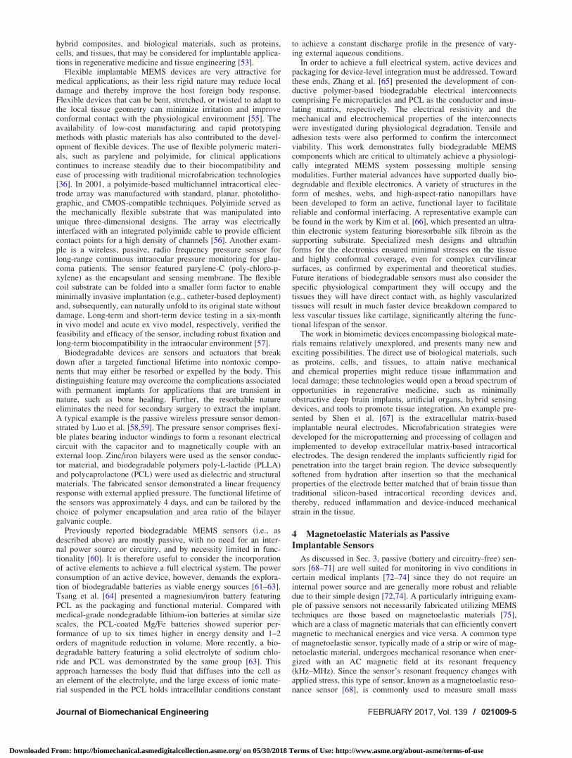
Oftentimes, multiple conditions or cues, such as physical,chemical, and biological, are relevant in evaluating how tissueengineered constructs perform in vivo. MEMS offers notableadvantages over alternative types of implantable systems for tis-sue engineering and regenerative medicine due to the spectrumof supported sensing modalities, compactness of size, and ame-nability to integration [102–104]. For example, the role the localenvironment in bone healing could be more deeply understoodby longitudinal monitoring of strain, oxygen tension, and pHwithin the defect. This could be enabled by the development ofa multimodal sensor system designed for a preclinical animalmodel. Strain sensors have previously been deployed to evaluatemechanical strains on internal and external fixation instrumenta-tion in humans and sheep, which show promise for clinical mon-itoring [13–15,105]; however, implementations have primarilybeen limited to long bones of the leg where there is a substantialhardware footprint for sensor integration. The role of local oxy-gen tension is of particular interest to fracture healing. In 1972,Brighton and Krebs measured oxygen tension in a rabbit fibularfracture using platinum microelectrodes which were notimplanted but inserted at each time point [6]. They notedmarked differences and temporal trends of the oxygen tension inthe hematoma, woven bone, and intact diaphyseal bone. Epariet al. revisited this approach with new technology in 2008, uti-lizing a percutaneously mounted, commercial multimodal cathe-ter probing the fracture gap to simultaneously measure pressure,oxygen tension, and temperature in a sheep tibial defect over a10 day period [106]. While larger canine and ovine models arepreferable to assess human-scale orthopedic and spine implantsdue to improved biomechanical similarity, small animal modelspossess important relative cost and throughput advantages,which makes them better suited for investigating newer andless-established therapeutic strategies for a wide range of dis-eases. Miniaturization of sensors and telemetry sufficient forsmall animal models, which are the primary test-bed for noveltissue engineering therapies, has not been demonstrated. Contin-ued efforts toward preclinical sensors for bone healing and ahost of other relevant injury and disease models would substan-tially inform a more quantitative understanding of the healingenvironment encountered in vivo, and also help to elucidatemechanistic reasons when different healing outcomes areobserved between preclinical models. A rendering of oneapproach to implement an implantable oxygen tension or strainsensor platform in a rodent femoral bone defect model is illus-trated and described in Fig. 2.
021009-6 / Vol. 139, FEBRUARY 2017 Transactions of the ASME
Downloaded From: http://biomechanical.asmedigitalcollection.asme.org/ on 05/30/2018 Terms of Use: http://www.asme.org/about-asme/terms-of-use
Reactive oxygen species and inflammation (nitric oxide andpH) can also be monitored by electrochemical or optical sensors[107]. All sensors mentioned above can be integrated into oneintelligent system for data collection and transmission. The use ofmultiple detection mechanisms, such as electrochemical and opti-cal, can minimize crosstalk between different types of sensors orvalidation and calibration for sensors of the same modality. Thisillustrates that certain attributes of MEMS, in this case their abil-ity to detect and transduce various mechanical and chemical cuesmediating bone healing into electrical signals, can be leveragedand applied toward a broad spectrum of tissue engineering andregenerative medicine applications. The microscale nature ofMEMS devices can be designed to meet the physical constraintsof preclinical animal models, which are oftentimes rodents orother small animals, to enable in situ, real-time sensing of physio-logical cues continuously within an animal and, thus, overcomethe limitations of ex vivo endpoint measurements. Alternatively,the trend toward more biomimetic and physiologically inspireddevices supports their integration with TE/RM constructs toenable local evaluation of the therapeutic efficacy of TE/RM con-structs in preclinical and clinical settings.
It is critical that sensors disturb the natural healing environmentas little as possible to ensure that valid measurements areacquired. To ensure that novel implantable devices do not promptfibrous encapsulation or actively irritate the tissue to a degree thatalters the course of healing, the host response should be rigorouslyvalidated by histological evaluations against “sensor-free” con-trols for increased fibrous tissue growth and for localization of
pro-inflammatory immune cells within the tissue of interest at theconclusion of the study. Additionally, sensors should be character-ized under controlled ex vivo conditions before and after implan-tation to ensure their sensitivity or functionality does not driftover time due to interactions with the surrounding tissue. Migra-tion of the sensor within the tissue is another factor that couldpotentially compromise the validity of the resultant data. Thus,TE/RM applications where a structural implant is used (e.g.,orthopedic fixation hardware, vascular stent, and tissue scaffold)are advantageous since they can act as a foundation to anchor thesensor in the healing environment (as in Fig. 2). Including specificattachment features in the device design such as loops to accom-modate sutures may be required. Additionally, longitudinal radi-ography could serve as a valuable tool to track implant migrationthroughout a study. To mitigate the risk of implant-induced infec-tion, preclinical sensors must also be able to endure sterilizationprocesses. While autoclaving is preferred and compatible withsome conventional sensor materials and designs, novel sensorshousing delicate chemical species or dissolvable materials mayhave to rely on more delicate sterilization approaches such asethylene oxide or gamma irradiation and test their efficacy. Insuch cases, consultation of GMP or ISO standards may serve as ahelpful guide.
In the design of an implantable system that facilitates tissueregeneration, a pivotal factor is the interplay between the targetedtissue and the implant. The integration of implantable sensorswith TE/RM technologies can support this endeavor by providinga closed loop system for customizing regenerative therapies. Acommon example is the incorporation of a sensor into a drugdelivery system so that the timing and rate of drug delivery can betuned by changes in certain local physiological conditions. Theintegration of sensing components into responsive polymeric sys-tems for controlled drug release has been the subject of extensiveresearch [108]. Reports in the literature include systems triggeredby the application of ultrasound [109], changes in pH [110,111],temperature [112], analyte concentrations [113,114], and electric[115] or magnetic [116] fields. The delivered molecules arediverse, including low molecular weight drugs, nucleic acids, pep-tides, and proteins, for the accelerated regeneration of tissues[52,54].
As the fields of TE/RM and implantable sensors continue togrow, emerging technologies should consider the union of thesetwo areas for smart, multifunctional regenerative therapies andpreclinical tools for better understanding and modulating the com-plex biological world.
6 Conclusions and Future Directions
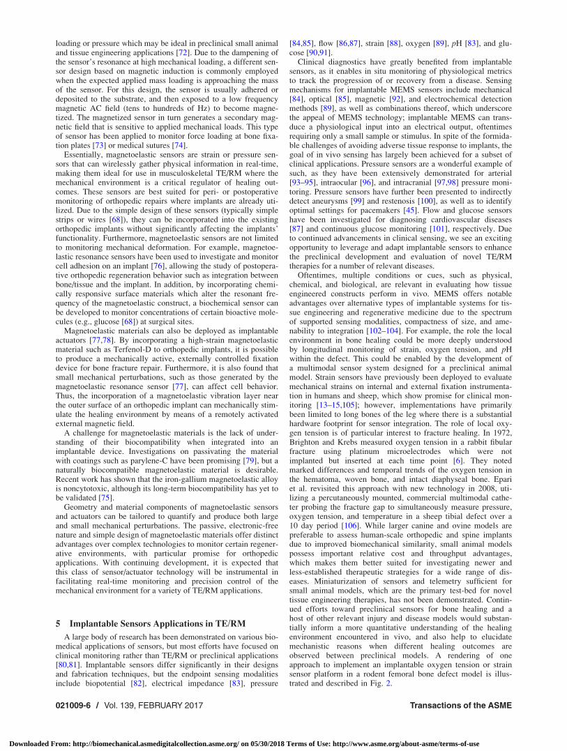
While significant strides have been made in the field ofimplantable sensors, key challenges and opportunities remain indeveloping implantable devices for continuous, in vivo monitor-ing. Whereas the early incarnations of implantable sensors weretechnology-driven devices, enabled by traditional CMOS-basedmaterials and microfabrication technologies, recent developmentsin implantable MEMS highlight a trend toward more application-driven device design. Figure 3 illustrates this progression inimplantable sensors, where direct offshoots of traditional CMOStechnology included silicon microelectrode arrays for neuromodu-lation and ceramic-based pressure sensors for cardiac monitoringthat are commercially available. In fact, most sensing modalitieshave corresponding commercial or near-commercial devices fea-turing traditional CMOS materials and processing. The implemen-tation of sensors in TE/RM applications will significantly benefitfrom the advances in implantable sensors research. These can begrossly categorized into a bottom–up versus a top–down changein the sensor design. Specifically, recent developments in flexible,biodegradable, and biomimetic sensors were mainly designedfrom a bottom–up, materials-level approach. Whether the goalwas to minimize the deleterious response to the device, toovercome the negative effects of permanent implants or to better
Fig. 2 Rendering of one approach which could be imple-mented to implant sensors in a rodent femoral defect model tomeasure oxygen tension and/or strain during bone regenera-tion. Animal injury models utilizing structural implants are par-ticularly advantageous for implantable devices because theyprovide a stable foundation to anchor the sensor. Dependingon the size constraints of the anatomical space under investiga-tion, transceiver and circuitry components could be packagedwithin a single device or subcutaneous wires could be routedto a remote transceiver pack mounted either intraperitoneallyor subcutaneously. Created using images from Servier MedicalArt, CC-BY 3.0.
Journal of Biomechanical Engineering FEBRUARY 2017, Vol. 139 / 021009-7
Downloaded From: http://biomechanical.asmedigitalcollection.asme.org/ on 05/30/2018 Terms of Use: http://www.asme.org/about-asme/terms-of-use
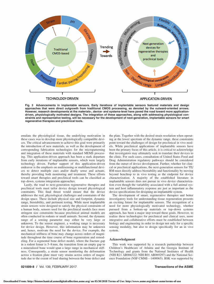
emulate the physiological tissue, the underlying motivation inthese cases was to develop more physiologically compatible devi-ces. The critical advancements to achieve this goal were primarilythe introduction of new materials, as well as the development ofcorresponding fabrication technologies for the micropatterningand integration of these materials with standard MEMS process-ing. This application-driven approach has been a stark departurefrom early iterations of implantable sensors, which were largelytechnology driven. Further support of this application-drivenendeavor is the emphasis on multisensing or multifunctional devi-ces to detect multiple cues and/or dually sense and actuate,thereby providing both monitoring and treatment. These effortstoward smart therapies and preclinical tools can be classified astop–down, systems integration approaches.
Lastly, the road to next-generation regenerative therapies andpreclinical tools must tailor device design toward physiologicalconstraints. This final thrust would ensure that the deviceaddresses the real, physiological challenges and constraints of thedesign space. These include physical size and footprint, dynamicrange, biostability, and pertinent testing. While most implantablestrain sensors were designed to satisfy the physical constraints ofa human body, sensors used for the preclinical models face morestringent size constraints because preclinical animal models areoften conducted in rodents or small animals. Second, the dynamicrange of a sensing parameter may either be very large orunknown. To the sensor engineer, a working range is necessaryfor device design. However, this information may be unknownand, hence, motivate the need for the device. For example, themechanical stiffness of bone may change across orders of magni-tude throughout the time course of bone regeneration and remod-eling. For a segmental bone defect model, where the fracture gapin a rodent femur is 5–8 mm, the transition from an empty gap toa mineralized bone would span a large range in mechanical stiff-ness. Consequently, a strain sensor monitoring the deformationacross a fixation plate must vary strains across orders of magni-tude due to the extent of load sharing between the bone defect and
the plate. Together with the desired strain resolution when operat-ing at the lower spectrum of the dynamic range, these constraintspoint toward the challenges of design for preclinical in vivo mod-els. While preclinical applications of implantable sensors havebeen the primary focus of this article, it is critical to acknowledgethat investigators may ultimately seek to translate their devices tothe clinic. For such cases, consultation of United States Food andDrug Administration regulatory pathways should be consideredfrom the outset of device development. Further, whether for clini-cal or preclinical applications, the next-generation sensors for TE/RM must directly address biostability and functionality by movingbeyond benchtop to in vivo testing as the endpoint for devicecharacterization. A majority of the established literature inimplantable sensors does not pursue in vivo device characteriza-tion even though the variability associated with a full animal sys-tem and host inflammatory response are just as important as thedevice specifications for designing an implantable system.
The development of smart, regenerative therapies and betterinvestigatory tools for understanding tissue regeneration presentsan exciting future for implantable sensors. The recognition of aneed for more physiologically motivated technology, whetherpursued from a bottom–up materials or top–down systemsapproach, has been a major step toward these goals. However, torealize these technologies for preclinical and clinical uses, moreintegrative and collaborative efforts must be established betweenbiology and engineering in order to design not only for a specificsensing modality, but also to design specifically for an in vivosystem.
Acknowledgment
This work was supported by a research partnership betweenChildren’s Healthcare of Atlanta and the Georgia Institute ofTechnology and grants from the National Institutes of Health(NIH R21 AR066322; NIH R01 AR069297) and the National Sci-ence Foundation (NSF CMMI—1400065). BSK was supported by
Fig. 3 Advancements in implantable sensors. Early iterations of implantable sensors featured materials and designapproaches that were direct outgrowth from traditional CMOS processing, as denoted by the outward-oriented arrows.However, research developments at the materials-, device- and systems-level have paved the road toward more application-driven, physiologically motivated designs. The integration of these approaches, along with addressing physiological con-straints and representative testing, will be necessary for the development of next-generation, implantable sensors for smartregenerative therapies and preclinical tools.
021009-8 / Vol. 139, FEBRUARY 2017 Transactions of the ASME
Downloaded From: http://biomechanical.asmedigitalcollection.asme.org/ on 05/30/2018 Terms of Use: http://www.asme.org/about-asme/terms-of-use
the Cell and Tissue Engineering NIH Biotechnology TrainingGrant (T32-GM008433).
References[1] Nerem, R. M., 2006, “Tissue Engineering: The Hope, the Hype, and the
Future,” Tissue Eng., 12(5), pp. 1143–1150.[2] Carrel, A., and Lindbergh, C., 1938, “The Culture of Organs,” Can. Med.
Assoc. J., 39(4), p. 416.[3] Lysaght, M. J., Jaklenec, A., and Deweerd, E., 2008, “Great Expectations:
Private Sector Activity in Tissue Engineering, Regenerative Medicine, andStem Cell Therapeutics,” Tissue Eng. Part A, 14(2), pp. 305–315.
[4] Guldberg, R. E., 2009, “Spatiotemporal Delivery Strategies for PromotingMusculoskeletal Tissue Regeneration,” J. Bone Miner. Res., 24(9),pp. 1507–1511.
[5] Wray, J. B., 1970, “The Biochemical Characteristics of the Fracture Hema-toma in Man,” Surg., Gynecol. Obstet., 130(5), pp. 847–852.
[6] Brighton, C. T., and Krebs, A. G., 1972, “Oxygen Tension of Healing Frac-tures in the Rabbit,” J. Bone Jt. Surg. Am., 54(2), pp. 323–332.
[7] Brighton, C. T., and Krebs, A. G., 1972, “Oxygen Tension of Nonunion ofFractured Femurs in the Rabbit,” Surg. Gynecol. Obstet., 135(3), pp. 379–385.
[8] Kolar, P., Gaber, T., Perka, C., Duda, G. N., and Buttgereit, F., 2011, “HumanEarly Fracture Hematoma is Characterized by Inflammation and Hypoxia,”Clin. Orthop. Relat. Res., 469(11), pp. 3118–3126.
[9] Yuasa, M., Mignemi, N. A., Nyman, J. S., Duvall, C. L., Schwartz, H. S.,Okawa, A., Yoshii, T., Bhattacharjee, G., Zhao, C., Bible, J. E., Obremskey,W. T., Flick, M. J., Degen, J. L., Barnett, J. V., Cates, J. M. M., and Schoe-necker, J. G., 2015, “Fibrinolysis is Essential for Fracture Repair and Preven-tion of Heterotopic Ossification,” J. Clin. Invest., 125(8), pp. 3117–3131.
[10] Boerckel, J. D., Uhrig, B. A., Willett, N. J., Huebsch, N., and Guldberg, R. E.,2011, “Mechanical Regulation of Vascular Growth and Tissue RegenerationIn Vivo,” Proc. Natl. Acad. Sci., 108(37), pp. E674–E680.
[11] Boerckel, J. D., Kolambkar, Y. M., Stevens, H. Y., Lin, A. S. P., Dupont, K.M., and Guldberg, R. E., 2012, “Effects of In Vivo Mechanical Loading onLarge Bone Defect Regeneration,” J. Orthop. Res., 30(7), pp. 1067–1075.
[12] Lienau, J., Schmidt-Bleek, K., Peters, A., Haschke, F., Duda, G. N., Perka, C.,Bail, H. J., Sch€utze, N., Jakob, F., and Schell, H., 2009, “Differential Regula-tion of Blood Vessel Formation Between Standard and Delayed BoneHealing,” J. Orthop. Res., 27(9), pp. 1133–1140.
[13] Claes, L. E., and Cunningham, J. L., 2009, “Monitoring the Mechanical Prop-erties of Healing Bone,” Clin. Orthop. Relat. Res., 467(8), pp. 1964–1971.
[14] Seide, K., Aljudaibi, M., Weinrich, N., Kowald, B., J€urgens, C., M€uller, J.,and Faschingbauer, M., 2012, “Telemetric Assessment of Bone Healing Withan Instrumented Internal Fixator: A Preliminary Study,” J. Bone Jt. Surg. Br.,94(3), pp. 398–404.
[15] McGilvray, K. C., Unal, E., Troyer, K. L., Santoni, B. G., Palmer, R. H.,Easley, J. T., Demir, H. V., and Puttlitz, C. M., 2015, “Implantable Microelec-tromechanical Sensors for Diagnostic Monitoring and Post-Surgical Predictionof Bone Fracture Healing,” J. Orthop. Res., 33(10), pp. 1439–1446.
[16] Claes, L., Recknagel, S., and Ignatius, A., 2012, “Fracture Healing UnderHealthy and Inflammatory Conditions,” Nat. Rev. Rheumatol., 8(3),pp. 133–143.
[17] Korn, C., and Augustin, H. G., 2015, “Mechanisms of Vessel Pruning andRegression,” Dev. Cell, 34(1), pp. 5–17.
[18] Tzioupis, C., and Giannoudis, P. V., 2007, “Prevalence of Long-Bone Non-Unions,” Injury, 38(Suppl. 2), pp. S3–S9.
[19] Hak, D. J., Fitzpatrick, D., Bishop, J. A., Marsh, J. L., Tilp, S., Schnettler, R.,Simpson, H., and Alt, V., 2014, “Delayed Union and Nonunions: Epidemiol-ogy, Clinical Issues, and Financial Aspects,” Injury, 45(Suppl. 2), pp. S3–S7.
[20] Tang, D., Tare, R. S., Yang, L.-Y., Williams, D. F., Ou, K.-L., and Oreffo, R.O. C., 2016, “Biofabrication of Bone Tissue: Approaches, Challenges andTranslation for Bone Regeneration,” Biomaterials, 83, pp. 363–382.
[21] Amini, A. R., Laurencin, C. T., and Nukavarapu, S. P., 2012, “Bone TissueEngineering: Recent Advances and Challenges,” Crit. Rev. Biomed. Eng.,40(5), pp. 363–408.
[22] Sebag, F., Vaillant-Lombard, J., Berbis, J., Griset, V., Henry, J. F., Petit, P.,and Oliver, C., 2010, “Shear Wave Elastography: A New Ultrasound ImagingMode for the Differential Diagnosis of Benign and Malignant Thyroid Nod-ules,” J. Clin. Endocrinol. Metab., 95(12), pp. 5281–5288.
[23] Weidemann, F., Eyskens, B., Jamal, F., Mertens, L., Kowalski, M., D’Hooge,J., Bijnens, B., Gewillig, M., Rademakers, F., Hatle, L., and Sutherland, G. R.,2002, “Quantification of Regional Left and Right Ventricular Radial and Lon-gitudinal Function in Healthy Children Using Ultrasound-Based Strain Rateand Strain Imaging,” J. Am. Soc. Echocardiography, 15(1), pp. 20–28.
[24] Mason, R. P., Antich, P. P., Babcock, E. E., Constantinescu, A., Peschke, P.,and Hahn, E. W., 1994, “Non-Invasive Determination of Tumor Oxygen Ten-sion and Local Variation With Growth,” Int. J. Radiat. Oncol., 29(1),pp. 95–103.
[25] Zhang, X., Lin, Y., and Gillies, R. J., 2010, “Tumor pH and Its Measurement,”J. Nucl. Med., 51(8), pp. 1167–1170.
[26] Gallagher, F. A., Kettunen, M. I, Day, S. E., Hu, D.-E., Ardenkjaer-Larsen,J. H., Zandt, R. in’t, Jensen, P. R., Karlsson, M., Golman, K., Lerche, M. H.,and Brindle, K. M., 2008, “Magnetic Resonance Imaging of pH In Vivo UsingHyperpolarized 13C-Labelled Bicarbonate,” Nature, 453(7197), pp. 940–943.
[27] Wulsten, D., Glatt, V., Ellinghaus, A., Schmidt-Bleek, K., Petersen, A., Schell,H., Lienau, J., Sebald, W., Pl€oger, F., Seemann, P., and Duda, G. N., 2011,
“Time Kinetics of Bone Defect Healing in Response to BMP-2 and GDF-5Characterised by in vivo Biomechanics,” Eur. Cell. Mater., 21, pp. 177–192.
[28] Anderson, A. E., Ellis, B. J., and Weiss, J. A., 2007, “Verification, Validationand Sensitivity Studies in Computational Biomechanics,” Comput. MethodsBiomech. Biomed. Eng., 10(3), pp. 171–184.
[29] Henninger, H. B., Reese, S. P., Anderson, A. E., and Weiss, J. A., 2010,“Validation of Computational Models in Biomechanics,” Proc. Inst. Mech.Eng., Part H, 224(7), pp. 801–812.
[30] Brown, G. N., Sattler, R. L., and Guo, X. E., 2016, “Experimental Studies ofBone Mechanoadaptation: Bridging In Vitro and In Vivo Studies WithMultiscale Systems,” Interface Focus, 6(1), p. 20150071.
[31] Bhatia, S. N., and Ingber, D. E., 2014, “Microfluidic Organs-On-Chips,” Nat.Biotechnol., 32(8), pp. 760–772.
[32] Claes, L. E., Claes, L. E., Heigele, C. A., Heigele, C. A., Neidlinger-Wilke,C., Neidlinger-Wilke, C., Kaspar, D., Kaspar, D., Seidl, W., Seidl, W., Marge-vicius, K. J., Margevicius, K. J., Augat, P., and Augat, P., 1998, “Effects ofMechanical Factors on the Fracture Healing Process,” Clin. Orthop. Relat.Res., Oct(355Suppl.), pp. S132–S147.
[33] Epari, D. R., Lienau, J., Schell, H., Witt, F., and Duda, G. N., 2008, “Pressure,Oxygen Tension and Temperature in the Periosteal Callus During BoneHealing—An In Vivo Study in Sheep,” Bone, 43(4), pp. 734–739.
[34] Szivek, J. A., Ruth, J. T., Heden, G. J., Martinez, M. A., Diggins, N. H., andWenger, K. H., 2016, “Determination of Joint Loads Using New SensateScaffolds for Regenerating Large Cartilage Defects in the Knee,” J. Biomed.Mater. Res., Part B, epub.
[35] Rebello, K. J., 2004, “Applications of MEMS in Surgery,” Proc. IEEE, 92(1),pp. 43–55.
[36] Pang, C., Lee, C., and Suh, K. Y., 2013, “Recent Advances in Flexible Sensorsfor Wearable and Implantable Devices,” J. Appl. Polym. Sci., 130(3),pp. 1429–1441.
[37] Bashir, R., 2004, “BioMEMS: State-of-the-Art in Detection, Opportunitiesand Prospects,” Adv. Drug Delivery Rev., 56(11), pp. 1565–1586.
[38] Du, H., and Bogue, R., 2007, “MEMS Sensors: Past, Present and Future,”Sens. Rev., 27(1), pp. 7–13.
[39] Grayson, A. C. R., Shawgo, R. S., Johnson, A. M., Flynn, N. T., Li, Y., Cima,M. J., and Langer, R., 2004, “A BioMEMS Review: MEMS Technology forPhysiologically Integrated Devices,” Proc. IEEE, 92(1), pp. 6–21.
[40] Receveur, R. A. M., Lindemans, F. W., and De Rooij, N. F., 2007,“Microsystem Technologies for Implantable Applications,” J. Micromech.Microeng., 17(5), pp. R50–R80.
[41] Wise, K. D., 2007, “Integrated Sensors, MEMS, and Microsystems: Reflec-tions on a Fantastic Voyage,” Sens. Actuators, A, 136(1), pp. 39–50.
[42] Allen, M. G., 2014, “Microfabricated Implantable Wireless Microsystems: Per-manent and Biodegradable Implementations,” IEEE International ConferenceMicro Electro Mechanical Systems, San Francisco, CA, Jan. 26–30, pp. 1–4.
[43] Langenfeld, H., Krein, A., Kirstein, M., and Binner, L., 1998, “Peak Endocar-dial Acceleration-Based Clinical Testing of the ‘BEST’ DDDR Pacemaker.European PEA Clinical Investigation Group,” Pacing Clin. Electrophysiol.,21(11 Pt 2), pp. 2187–2191.
[44] Dimarco, J. P., and Mower, M., 2003, “Implantable Cardi-overter–Defibrillators,” New Engl. J. Med., 349, pp. 1836–1847.
[45] Magalski, A., Adamson, P., Gadler, F., B€oehm, M., Steinhaus, D., Reynolds,D., Vlach, K., Linde, C., Cremers, B., Sparks, B., and Bennett, T., 2002,“Continuous Ambulatory Right Heart Pressure Measurements With anImplantable Hemodynamic Monitor: A Multicenter, 12-Month Follow-UpStudy of Patients With Chronic Heart Failure,” J. Card. Failure, 8(2),pp. 63–70.
[46] Kipke, D. R., Vetter, R. J., Williams, J. C., and Hetke, J. F., 2003, “Silicon-Substrate Intracortical Microelectrode Arrays for Long-Term Recording ofNeuronal Spike Activity in Cerebral Cortex,” IEEE Trans. Neural Syst. Reha-bil. Eng., 11(2), pp. 151–155.
[47] Schmidt, E. M., Bak, M. J., Hambrecht, F. T., Kufta, C. V., O’Rourke, D. K.,and Vallabhanath, P., 1996, “Feasibility of a Visual Prosthesis for the BlindBased on Intracortical Microstimulation of the Visual Cortex,” Brain, 119(5),pp. 507–522.
[48] Zeng, F. G., Rebscher, S., Harrison, W., Sun, X., and Feng, H., 2008,“Cochlear Implants: System Design, Integration, and Evaluation,” IEEE Rev.Biomed. Eng., 1, pp. 115–142.
[49] Ziaie, B., Von Arx, J. A., Dokmeci, M. R., and Najafi, K., 1996, “AHermetic Glass-Silicon Micropackage With High-Density On-Chip Feed-throughs for Sensors and Actuators,” J. Microelectromech. Syst., 5(3),pp. 166–179.
[50] Najafi, K., 2007, “Packaging of Implantable Microsystems,” Sixth IEEE Sen-sors Conference, Atlanta, Oct. 28–30, pp. 58–63.
[51] Gilleo, Ken, ET-Trends., L. L. C., and Warwick, R. I., 2005, “MEMS in Medi-cine,” Circuits Assembly, 16(8), pp. 1–10.
[52] Steichen, S. D., Caldorera-Moore, M., and Peppas, N. A., 2013, “A Review ofCurrent Nanoparticle and Targeting Moieties for the Delivery of CancerTherapeutics,” Off. J. Eur. Fed. Pharm. Sci., 48(3), pp. 416–427.
[53] Jivani, R. R., Lakhtaria, G. J., Patadiya, D. D., Patel, L. D., Jivani, N. P., andJhala, B. P., 2014, “Biomedical Microelectromechanical Systems (Bio-MEMS): Revolution in Drug Delivery and Analytical Techniques,” SaudiPharm. J., 24(1), pp. 1–20.
[54] Tng, D. J. H., Hu, R., Song, P., Roy, I., and Yong, K. T., 2012, “Approachesand Challenges of Engineering Implantable Microelectromechanical Systems(MEMS) Drug Delivery Systems for In Vitro and In Vivo Applications,”Micromachines, 3(4), pp. 615–631.
Journal of Biomechanical Engineering FEBRUARY 2017, Vol. 139 / 021009-9
Downloaded From: http://biomechanical.asmedigitalcollection.asme.org/ on 05/30/2018 Terms of Use: http://www.asme.org/about-asme/terms-of-use
[55] Viventi, J., Kim, D.-H., Vigeland, L., Frechette, E. S., Blanco, J. A., Kim,Y.-S., Avrin, A. E., Tiruvadi, V. R., Hwang, S.-W., Vanleer, A. C., Wulsin,D. F., Davis, K., Gelber, C. E., Palmer, L., Van der Spiegel, J., Wu, J., Xiao,J., Huang, Y., Contreras, D., Rogers, J. A., and Litt, B., 2011, “Flexible, Fold-able, Actively Multiplexed, High-Density Electrode Array for Mapping BrainActivity In Vivo,” Nat. Neurosci., 14(12), pp. 1599–1605.
[56] Rousche, P. J., Pellinen, D. S., Pivin, D. P., Williams, J. C., Vetter, R. J.,and Kipke, D. R., 2001, “Flexible Polyimide-Based Intracortical ElectrodeArrays With Bioactive Capability,” IEEE Trans. Biomed. Eng., 48(3),pp. 361–370.
[57] Chen, P. J., Saati, S., Varma, R., Humayun, M. S., and Tai, Y. C., 2010,“Wireless Intraocular Pressure Sensing Using Microfabricated MinimallyInvasive Flexible-Coiled LC Sensor Implant,” J. Microelectromech. Syst.,19(4), pp. 721–734.
[58] Luo, M., Song, C. J., Herrault, F., and Allen, M. G., 2014, “AMicrofabricated RF Wireless Pressure Sensor Made Completely of Biodegrad-able Materials,” Journal of Microelectromechanical Systems, 23(1), pp. 4–13.
[59] Luo, M., Martinez, A. W., Song, C., Herrault, F., and Allen, M. G., 2014, “AMicrofabricated Wireless RF Pressure Sensor Made Completely of Biodegrad-able Materials,” J. Microelectromech. Syst., 23(1), pp. 4–13.
[60] Boutry, C. M., Chandrahalim, H., Streit, P., Schinhammer, M., H€anzi, A. C.,and Hierold, C., 2013, “Characterization of Miniaturized RLC ResonatorsMade of Biodegradable Materials for Wireless Implant Applications,” Sens.Actuators, A, 189, pp. 344–355.
[61] Heller, A., 2006, “Potentially Implantable Miniature Batteries,” Anal. Bioanal.Chem., 385(3), pp. 469–473.
[62] Yin, L., Huang, X., Xu, H., Zhang, Y., Lam, J., Cheng, J., and Rogers,J. A., 2014, “Materials, Designs, and Operational Characteristicsfor Fully Biodegradable Primary Batteries,” Adv. Mater., 26(23), pp. 3879–3884.
[63] She, D., Tsang, M., Kim, J. K., and Allen, M. G., 2015, “Immobilized Electro-lyte Biodegradable Batteries for Implantable MEMS,” 18th International Con-ference on Solid-State Sensors, Actuators and Microsystems(TRANSDUCERS), Anchorage, Alaska, June 21–25, pp. 494–497.
[64] Tsang, M., Armutlulu, A., Martinez, A. W., Allen, S. A. B., and Allen, M. G.,2015, “Biodegradable Magnesium/Iron Batteries With PolycaprolactoneEncapsulation: A Microfabricated Power Source for Transient ImplantableDevices,” Microsyst. Nanoeng., 1, p. 15024.
[65] Zhang, T., Tsang, M., and Allen, M. G., 2016, “Biodegradable Electrical Inter-connects for Transient Implantable Systems,” Solid-State Sensor, Actuator,Microsystems Work, Philadelphia, PA, Oct. 24–27.
[66] Kim, D.-H., Viventi, J., Amsden, J. J., Xiao, J., Vigeland, L., Kim, Y.-S.,Blanco, J. A., Panilaitis, B., Frechette, E. S., Contreras, D., Kaplan, D. L.,Omenetto, F. G., Huang, Y., Hwang, K.-C., Zakin, M. R., Litt, B., and Rogers,J. A., 2010, “Dissolvable Films of Silk Fibroin for Ultrathin Conformal Bio-Integrated Electronics,” Nat. Mater., 9(6), pp. 511–517.
[67] Shen, W., Karumbaiah, L., Liu, X., Saxena, T., Chen, S., Patkar, R.,Bellamkonda, R. V., and Allen, M. G., 2015, “Extracellular Matrix-BasedIntracortical Microelectrodes: Toward a Microfabricated Neural InterfaceBased on Natural Materials,” Microsyst. Nanoeng., 1, p. 15010.
[68] Grimes, C. A., Roy, S. C., Rani, S., and Cai, Q., 2011, “Theory, Instrumenta-tion and Applications of Magnetoelastic Resonance Sensors: A Review,”Sensors (Basel), 11(3), pp. 2809–2844.
[69] Pereles, B. D., Dienhart, T., Sansom, T., Johnston, K., and Ong, K. G., 2012,“A Wireless, Passive Load Cell Based on Magnetoelastic Resonance,” SmartMater. Struct., 21(7), p. 075018.
[70] Pereles, B. D., DeRouin, A. J., and Ong, K. G., 2015, “Partially LoadedMagnetoelastic Sensors With Customizable Sensitivities for Large ForceMeasurements,” IEEE Sens. J., 15(1), pp. 591–597.
[71] Nakamura, T., Inoue, Y., Kim, D., Matsuhisa, N., Yokota, T., Sekitani, T.,Someya, T., and Sekino, M., 2014, “Basic Characteristics of Implantable Flex-ible Pressure Sensor for Wireless Readout Using MRI,” 36th Annual Interna-tional Conference of the Engineering in Medicine and Biology Society, IEEE,pp. 2338–2341.
[72] Green, S. R., Kwon, R. S., Elta, G. H., and Gianchandani, Y. B., 2013,“in vivo and In Situ Evaluation of a Wireless Magnetoelastic Sensor Arrayfor Plastic Biliary Stent Monitoring,” Biomed. Microdevices, 15(3),pp. 509–517.
[73] Oess, N. P., Weisse, B., and Nelson, B. J., 2009, “Magnetoelastic Strain Sen-sor for Optimized Assessment of Bone Fracture Fixation,” IEEE Sens. J., 9(8),pp. 961–968.
[74] DeRouin, A., Pacella, N., Zhao, C., An, K.-N., and Ong, K., 2015, “A WirelessSensor for Real-Time Monitoring of Tensile Force on Sutured Wound Sites,”IEEE Trans. Biomed. Eng., 63(8), pp. 1665–1671.
[75] Holmes, H. R., DeRouin, A., Wright, S., Riedemann, T. M., Lograsso, T. A.,Rajachar, R. M., and Ong, K. G., 2014, “Biodegradation and Biocompatibilityof Mechanically Active Magnetoelastic Materials,” Smart Mater. Struct.,23(9), p. 095036.
[76] Vlaisavljevich, E., Holmes, H. R., Tan, E. L., Qian, Z., Trierweiler, S., Ong,K. G., and Rajachar, R. M., 2013, “Magnetoelastic Vibrational Biomaterialsfor Real-Time Monitoring and Modulation of the Host Response,” J. Mater.Sci. Mater. Med., 24(4), pp. 1093–1104.
[77] Vlaisavljevich, E., Janka, L. P., Ong, K. G., and Rajachar, R. M., 2011,“Magnetoelastic Materials as Novel Bioactive Coatings for the Control of CellAdhesion,” IEEE Trans. Biomed. Eng., 58(3), pp. 698–704.
[78] Pepakayala, V., Stein, J., and Gianchandani, Y., 2015, “Resonant Magnetoe-lastic Microstructures for Wireless Actuation of Liquid Flow on 3D Surfacesand Use in Glaucoma Drainage Implants,” Microsyst. Nanoeng., 1, p. 15032.
[79] Trierweiler, S., Holmes, H., Pereles, B., Rajachar, R., and Ong, K. G., 2013,“Remotely Activated, Vibrational Magnetoelastic Array System for Control-ling Cell Adhesion,” J. Biomed. Sci. Eng., 06(4), pp. 478–482.
[80] Chew, D. J., Zhu, L., Delivopoulos, E., Minev, I. R., Musick, K. M.,Mosse, C. A., Craggs, M., Donaldson, N., Lacour, S. P., McMahon, S. B.,and Fawcett, J. W., 2013, “A Microchannel Neuroprosthesis for BladderControl After Spinal Cord Injury in Rat,” Sci. Transl. Med., 5(210),pp. 210–155.
[81] Chow, E. Y., Chlebowski, A. L., Chakraborty, S., Chappell, W. J., and Irazo-qui, P. P., 2010, “Fully Wireless Implantable Cardiovascular Pressure MonitorIntegrated With a Medical Stent,” IEEE Trans. Biomed. Eng., 57(6),pp. 1487–1496.
[82] Griss, P., Enoksson, P., Tolvanen-Laakso, H. K., Meril€ainen, P., Ollmar, S.,and Stemme, G., 2001, “Micromachined Electrodes for Biopotential Meas-urements,” J. Microelectromech. Syst., 10(1), pp. 10–16.
[83] Cao, H., Landge, V., Tata, U., Seo, Y. S., Rao, S., Tang, S. J., Tibbals, H. F.,Spechler, S., and Chiao, J. C., 2012, “An Implantable, Batteryless, andWireless Capsule With Integrated Impedance and pH Sensors for Gastro-esophageal Reflux Monitoring,” IEEE Trans. Biomed. Eng., 59(12 Part 2),pp. 3131–3139.
[84] Troughton, R. W., Ritzema, J., Eigler, N. L., Melton, I. C., Krum, H.,Adamson, P. B., Kar, S., Shah, P. K., Whiting, J. S., Heywood, J. T.,Rosero, S., Singh, J. P., Saxon, L., Matthews, R., Crozier, I. G., andAbraham, W. T., 2011, “Direct Left Atrial Pressure Monitoring in SevereHeart Failure: Long-Term Sensor Performance,” J. Cardiovasc. Transl. Res.,4(1), pp. 3–13.
[85] Totsu, K., Haga, Y., and Esashi, M., 2003, “Vacuum Sealed UltraMiniature Fiber-Optic Pressure Sensor Using White Light Interferometry,”12th International Conference Solid-State Sensors, Actuators Microsystems,(TRANSDUCERS), Boston, June 8–12, pp. 931–934.
[86] Lal, A., 2001, “Integrated Pressure and Flow Sensor in Silicon-Based Ultra-sonic Surgical Actuator,” IEEE Ultrasonics Symposium. An InternationalSymposium, Oct. 7–10, pp. 1373–1376.
[87] Hong, M. K., Wong, S. C., Mintz, G. S., Popma, J. J., Kent, K. M., Pichard, A.D., Satler, L. F., Leon, M. B., and Tobis, J. M., 1995, “Can Coronary FlowParameters After Stent Placement Predict Restenosis?,” Catheterization Cardi-ovasc. Diagn., 36(3), pp. 278–282.
[88] Umbrecht, F., Wendlandt, M., Juncker, D., Hierold, C., and Neuenschwander,J., 2005, “A Wireless Implantable Passive Strain Sensor System,” IEEE Sen-sors, pp. 20–23.
[89] Mahutte, C. K., 1998, “On-Line Arterial Blood Gas Analysis With Optodes:Current Status,” Clin. Biochem., 31(3), pp. 119–130.
[90] Kim, Y. T., Kim, Y.-Y., and Jun, C.-H., 1999, “Needle-Shaped Glucose Sen-sor With Multi-Cell Electrode Fabricated by Surface Micromachining,” Proc.SPIE 680, pp. 924–930.
[91] Mastrototaro, J. J., Cooper, K. W., Soundararajan, G., Sanders, J. B., andShah, R. V., 2006, “Clinical Experience With an Integrated Continuous Glu-cose Sensor/Insulin Pump Platform: A Feasibility Study,” Adv. Ther., 23(5),pp. 725–732.
[92] Ling, Y., Pong, T., Vassiliou, C. C., Huang, P. L., and Cima, M. J., 2011,“Implantable Magnetic Relaxation Sensors Measure Cumulative Exposure toCardiac Biomarkers,” Nat. Biotechnol., 29(3), pp. 273–277.
[93] DeHennis, A. D., and Wise, K. D., 2006, “A Fully Integrated Multisite Pres-sure Sensor for Wireless Arterial Flow Characterization,” J. Microelectro-mech. Syst., 15(3), pp. 678–685.
[94] Ritzema-Carter, J. L. T., Smyth, D., Troughton, R. W., Crozier, I. G., Melton,I. C., Richards, A. M., Eigler, N., Whiting, J., Kar, S., Krum, H., and Abra-ham, W. T., 2006, “Dynamic Myocardial Ischemia Caused by CircumflexArtery Stenosis Detected by a New Implantable Left Atrial Pressure Monitor-ing Device,” Circulation, 113(15), pp. 705–707.
[95] Schnakenberg, U., Kruger, C., Pfeffer, J. G., Mokwa, W., Vom Bogel, G.,Gunther, R., and Schmitz-Rode, T., 2004, “Intravascular Pressure MonitoringSystem,” Sens. Actuators, A, 110(1–3), pp. 61–67.
[96] Twa, M. D., Roberts, C. J., Karol, H. J., Mahmoud, A. M., Weber, P. A., andSmall, R. H., 2010, “Evaluation of a Contact Lens-Embedded Sensor for Intra-ocular Pressure Measurement,” J. Glaucoma, 19(6), pp. 382–390.
[97] Miyake, H., Ohta, T., Kajimoto, Y., and Matsukawa, M., 1997, “A NewVentriculoperitoneal Shunt With a Telemetric Intracranial Pressure Sensor:Clinical Experience in 94 Patients With Hydrocephalus,” Neurosurgery, 40(5),pp. 931–935.
[98] Signorini, D. F., Shad, A., Piper, I. R., and Statham, P. F., 1998, “A ClinicalEvaluation of the Codman MicroSensor for Intracranial Pressure Monitoring,”Br. J. Neurosurg., 12(3), pp. 223–227.
[99] Milner, R., 2006, “Remote Pressure Sensing for Thoracic Endografts,” Endo-vascular Today, pp. 1–3.
[100] Takahata, K., DeHennis, A., Wise, K. D., and Gianchandani, Y. B., 2004, “AWireless Microsensor for Monitoring Flow and Pressure in a Blood VesselUtilizing a Dual-Inductor Antenna Stent and Two Pressure Sensors,” 17thIEEE International Conference on Micro Electro Mechanical Systems, Maas-tricht, Germany, Jan. 25–29, pp. 216–219.
[101] Renard, 2004, “Implantable Glucose Sensors for Diabetes Monitoring,” Mini-mally Invasive Ther. Allied Technol., 13(2), pp. 78–86.
[102] Receveur, R. A. M., Marxer, C. R., Woering, R., Larik, V. C. M. H., andde Rooij, N. F., 2005, “Laterally Moving Bistable MEMS DC Switch for Bio-medical Applications,” J. Microelectromech. Syst., 14(5), pp. 1089–1098.
[103] Schwarz, M., Ewe, L., Hauschild, R., Hosticka, B. J., Huppertz, J., Kolnsberg,S., Mokwa, W., and Trieu, H. K., 2000, “Single Chip CMOS Imagers and
021009-10 / Vol. 139, FEBRUARY 2017 Transactions of the ASME
Downloaded From: http://biomechanical.asmedigitalcollection.asme.org/ on 05/30/2018 Terms of Use: http://www.asme.org/about-asme/terms-of-use
Flexible Microelectronic Stimulators for a Retina Implant System,” Sens.Actuators, A, 83(1), pp. 40–46.
[104] Siwapornsathain, E., Lal, A., and Binard, J., 2002, “A Telemetry and SensorPlatform for Ambulatory Urodynamics,” 2nd Annual International IEEE-EMBS Special Topic Conference Microtechnologies in Medicine Biology,pp. 283–287.
[105] D’Lima, D. D., Fregly, B. J., and Colwell, C. W., 2013, “Implantable SensorTechnology: Measuring Bone and Joint Biomechanics of Daily Life In Vivo,”Arthritis Res. Ther., 15(1), p. 203.
[106] Epari, D. R., Lienau, J., Schell, H., Witt, F., and Duda, G. N., 2008, “Pressure,Oxygen Tension and Temperature in the Periosteal Callus During BoneHealing-An In Vivo Study in Sheep,” Bone, 43(4), pp. 734–739.
[107] Frost, M. C., and Meyerhoff, M. E., 2002, “Implantable Chemical Sensors forReal-Time Clinical Monitoring: Progress and Challenges,” Curr. Opin. Chem.Biol., 6(5), pp. 633–641.
[108] Langer, R., 1998, “Drug Delivery and Targeting,” Nature, 392(6679),pp. 5–10.
[109] Azagury, A., Khoury, L., Enden, G., and Kost, J., 2014, “Ultrasound MediatedTransdermal Drug Delivery,” Adv. Drug Delivery Rev., 72, pp. 127–143.
[110] Gao, W., Chan, J., and Farokhzad, O. C., 2010, “pH-Responsive Nanoparticlesfor Drug Delivery,” Mol. Pharm., 7(6), pp. 1913–1920.
[111] Liu, J., Huang, Y., Kumar, A., Tan, A., Jin, S., Mozhi, A., and Liang, X. J.,2014, “PH-Sensitive Nano-Systems for Drug Delivery in Cancer Therapy,”Biotechnol. Adv., 32(4), pp. 693–710.
[112] Bikram, M., Gobin, A. M., Whitmire, R. E., and West, J. L., 2007,“Temperature-Sensitive Hydrogels With SiO2-Au Nanoshells for ControlledDrug Delivery,” J. Controlled Release, 123(3), pp. 219–227.
[113] Koo, A. N., Lee, H. J., Kim, S. E., Chang, J. H., Park, C., Kim, C., Park, J. H.,and Lee, S. C., 2008, “Disulfide-Cross-Linked PEG-Poly(Amino Acid)sCopolymer Micelles for Glutathione-Mediated Intracellular Drug Delivery,”Chem. Commun. (Cambridge)., 2008(48), pp. 6570–6572.
[114] Banerjee, J., Hanson, A. J., Gadam, B., Elegbede, A. I., Tobwala, S.,Ganguly, B., Wagh, A. V., Muhonen, W. W., Law, B., Shabb, J. B.,Srivastava, D. K., and Mallik, S., 2009, “Release of Liposomal Contents byCell-Secreted Matrix Metalloproteinase-9,” Bioconjugate Chem., 20(7),pp. 1332–1339.
[115] Ge, J., Neofytou, E., Cahill, T. J., Beygui, R. E., and Zare, R. N., 2012, “DrugRelease From Electric-Field-Responsive Nanoparticles,” ACS Nano, 6(1),pp. 227–233.
[116] Cai, K., Luo, Z., Hu, Y., Chen, X., Liao, Y., Yang, L., and Deng, L., 2009,“Magnetically Triggered Reversible Controlled Drug Delivery From Microfabri-cated Polymeric Multireservoir Devices,” Adv. Mater., 21(40), pp. 4045–4049.
Journal of Biomechanical Engineering FEBRUARY 2017, Vol. 139 / 021009-11
Downloaded From: http://biomechanical.asmedigitalcollection.asme.org/ on 05/30/2018 Terms of Use: http://www.asme.org/about-asme/terms-of-use