Embed Size (px)
Citation preview
I m p l a n t r e h a b i l i t a t i o n o f
t h e e d e n t u l o u s i r r a d i a t e d m a n d i b l e .
Sharon Liberali
A thesis submitted for the degree of Doctor of Clinical Dentistry (Special Needs Dentistry) School of Dentistry, The University of Adelaide April 2009
Principal supervisor: Professor Alastair Goss School of Dentistry The University of Adelaide Australia

2
3
TABLE OF CONTENTS.
Page No.
List of Abbreviations 11 List of Appendices 12 List of Tables 13 List of Figures 16 Abstract 18 Thesis Declaration 20 Acknowledgements 21 Thesis Format 22 CHAPTER 1: INTRODUCTION 23 1.1 Description of topic 23 1.2 Limitations of previous studies 24 1.3 Thesis rationale 25 1.4 Aims and Objectives 25 1.5 Hypothesis 26 1.6 Conceptual framework 26 1.7 Table of Comparisons for Systematic review (PICO CHART) 27 CHAPTER 2: LITERATURE REVIEW 29 2.1 Incidence and prevalence of head and neck cancer 29 2.1.1 Oral cancer 29 2.1.2 Salivary gland cancer 33 2.1.3 Laryngeal cancer 34 2.1.4 Oropharyngeal and Hypopharyngeal cancer 34 2.1.5 Nasopharyngeal cancer 35 2.2 Ablative surgery and reconstructive techniques for head and neck cancer 35
2.2.1 Ablative surgery 35 2.2.2 Neck dissection 36
4
2.2.3 Reconstructive techniques 37 2.2.3.1 Local flaps 39 2.2.3.2 Grafts 39 2.2.3.3 Regional flaps 40 2.2.3.4 Free-flaps 40
2.2.3.4.1 Radial forearm free flap 41 2.2.3.4.2 Fibulae free flap 41 2.2.3.4.3 Iliac crest free flap 42 2.2.3.4.4 Scapula free flap 43
2.2.3.5 Donor site selection 43 2.2.3.6 Reconstruction plates 44 2.2.3.7 Osseointegrated implants 45
2.3 Sequelae of head and neck ablative surgery 45
2.3.1 General sequelae 46 2.3.1.1 Psychological impact 46 2.3.1.2 Appearance and aesthetics 46 2.3.1.3 Shoulder function 46
2.3.2 Oral sequelae 47 2.3.2.1 Saliva control 48 2.3.2.2 Swallowing function 48 2.3.2.3 Speech articulation and intelligibility 48 2.3.2.4 Removable prostheses 48 2.3.2.5 Mastication and dietary impact 49
2.4 Radiotherapy to the head and neck region. 49
2.4.1 Indications 50 2.4.2 Techniques 50
2.4.2.1 Conventional fractionation 50 2.4.2.2 Hyper-fractionation 51 2.4.2.3 Accelerated fractionation 51 2.4.2.4 Computerized planning techniques 52
5
2.5 Oral sequelae of head and neck radiotherapy 52 2.5.1 Oral mucosa 55 2.5.2 Taste buds 56 2.5.3 Salivary glands 57
2.5.3.1 Hyposalivation/Xerostomia 57 2.5.3.2 Altered salivary composition 58
2.5.4 Dentition 59 2.5.5 Periodontium 59 2.5.6 Musculature and Temporomandibular joint 60
2.5.6.2 Trismus 60 2.5.6.2 Dysphagia 60
2.5.7 Bone 61 2.5.7.1 Osteoradionecrosis (ORN) 61
2.6 Prevention and treatment of consequences of Head and Neck Radiotherapy 66
2.6.1 Mucositis 66 2.6.2 Taste loss 68 2.6.3 Hyposalivation 68 2.6.4 Radiation caries 70 2.6.5 Periodontal disease 71 2.6.6 Trismus 71 2.6.7 Osteoradionecrosis (ORN) 71
2.7 Implant mandibular prostheses (overdentures) 73
2.7.1 Definitions 74 2.7.2 Prosthodontic classification system 75 2.7.3 Classification of oral implants 76 2.7.4 Standard of care for the edentulous mandible 76 2.7.5 Osseointegration 78
2.7.5.1 Definition 78 2.7.5.2 Biologic stages of osseointegration 79
2.7.6 Patient screening and treatment planning 80 2.7.7 Success criteria 84
6
2.7.8 Implant failure 85 2.7.9 Treatment of complications 93
2.8 Oncologic treatment modalities which impact on osseointegration 96
2.8.1 Radiotherapy 96 2.8.1.1 Irradiation after implant placement 96 2.8.1.2 Irradiation before implant placement 99 2.8.1.3 Irradiation before and after implant placement 100
2.8.2 Chemotherapy 100 2.9 Radiotherapy related risk factors to implant surgery 101
2.9.1 Region of placement in the craniofacial skeleton 103 2.9.2 Patient selection 104 2.9.3 Irradiation dose 105 2.9.4 Time from radiotherapy to 1st stage implant surgery 107 2.9.5 Time from 1st and 2nd stage implant surgery 108 2.9.6 Implant fixture length 109 2.9.7 Marginal bone loss 110 2.9.8 Soft tissue condition 110 2.9.9 Design and retention 110 2.9.10 Surgeon’s experience 112 2.9.11 Risk of ORN in relation to implant surgery 113
2.10 Hyperbaric Oxygen therapy 113
2.10.1 Basic effects on tissues 114 2.10.2 Therapeutic uses of Hyperbaric oxygen therapy 117
2.10.2.1 Carbon monoxide poisoning 118 2.10.2.2 Decompression sickness 118 2.10.2.3 Arterial gas embolism 118 2.10.2.4 Clostridial myonecrosis 118 2.10.2.5 Necrotising fasciitis 118 2.10.2.6 Refractory osteomyelitis 118 2.10.2.7 Acute traumatic ischaemic injury 119
7
2.10.2.8 Anaemia due to exceptional blood loss 119 2.10.2.9 Thermal burns 119 2.10.2.10 Problem wounds 119 2.10.2.11 Compromised skin grafts and flaps 119 2.10.2.12 Radiation-induced hard tissue injury (ORN) 120 2.10.2.13 Prevention of implant loss in the irradiated patient 121
2.10.3 Treatment protocols for radiation-induced hard tissue injury (ORN) 122 2.10.3.1 Prophylactic 122 2.10.3.2 Therapeutic 123
2.10.4 Contraindications to Hyperbaric oxygen therapy 125 2.10.4.1 Pneumothorax 126 2.10.4.2 Optic Neuritis 126 2.10.4.3 Acute viral infection or upper respiratory tract infection 127 2.10.4.4 Pregnancy 127 2.10.4.5 Claustrophobia 127 2.10.4.6 History of prior thoracic or middle ear surgery 127 2.10.4.7 Existing neoplasia 128 2.10.5 Complications of Hyperbaric oxygen therapy 128 2.10.5.1 Barotrauma 129 2.10.5.2 Arterial gas emboli 129 2.10.5.3 Middle ear problems 129 2.10.5.4 Oxygen toxicity 129 2.10.5.5 Tooth and sinus pain 130 2.10.5.6 Myopia 130 2.10.5.7 Other complications 130
2.11 Osseointegration in irradiated tissues 131
2.11.1 Clinical studies with primary implant provision 131 2.11.2 Clinical studies with secondary implant provision 132 2.11.3 Clinical studies related to region of placement – mandible 133 2.11.4 Clinical studies related to region of placement - reconstructed mandible 136
2.11.4.1 Vascularised graft 136 2.11.4.2 Non-vascularised graft 137
8
2.11.5 Clinical studies showing an increased rate of implant loss when placed in irradiated tissues 138
2.11.6 Clinical studies showing no increased rate of implant loss when placed in irradiated tissues 138
2.11.7 Clinical studies showing stimulation of osseointegration by hyperbaric oxygen 139
2.11.8 Clinical studies showing that hyperbaric oxygen is not necessary for osseointegration 140
2.11.9 Histological case reports 141 2.11.9.1 Animal 141 2.11.9.2 Human 142
2.12 Quality of Life 142
2.12.1 Definition 142 2.12.2 Impact of cancer on quality of life 143 2.12.3 Impact of ablative surgery on quality of life 144 2.12.4 Impact of radiotherapy on quality of life 146 2.12.5 Impact of oral rehabilitation on quality of life 148
2.13 Quality of life assessment tools 151
2.13.1 European Organization for Research and Treatment of Cancer (EORTC) questionnaires 153 2.13.1 1 EORTC QLQ-C30 154 2.13.1.2 EORTC H&N 35 156
2.13 2. Oral Health Impact Profile questionnaire 157 2.13.2.1 OHIP-49 157 2.13.2.2 OHIP-14 158 2.13.2.3 OHIP-EDENT 158
9
CHAPTER 3: METHODOLOGY 160 3.1 Study design 160 3.1.1 Sampling frame 160 3.1.1.1 Target population 160 3.1.1.2 Inclusion criteria 160 3.1.1.3 Exclusion criteria 161 3.1.1.4 Patient selection 161 3.2 Data Collection 162 3.2.1 Pre-treatment assessment 162 3.2.1.1 Clinical examination 162 3.2.1.2 Radiographic examination 163 3.2.1.3 Baseline Questionnaires (T0) 163 3.2.2 Dental Treatment provided 164 3.2.2.1 Research subjects in Group 1 164 3.2.2.2 Research subjects in Group 2 165 3.2.3 Review assessment 166 3.2.3.1 Clinical examination 166 3.2.3.2 Radiographic examination 166 3.2.3.3 Review Questionnaires (T1) 166 3.3 Data Management 167 3.3.1 Data weighting 167
3.3.1.1 EORTC quality of life questionnaires 167 3.3.1.2 Oral Health Impact Profile 167
3.3.2 Data scoring 167 3.3.2.1 EORTC quality of life questionnaires 167 3.3.2.1.1 EORTC QLQ-C30 168 3.3.2.1.2 EORTC H&N-35 168 3.3.2.2 Oral Health Impact Profile 168 3.3.2.2.1 OHIP-14 168 3.3.2.2.2 OHIP-EDENT 168 3.3.3 Data analysis 169
10
3.4 Ethic Implications and Approvals 169 CHAPTER 4: RESULTS 170 4.1 Patient Clinical Assessments 170 4.1.1 Patient characteristics 170 4.1.1.1 Group1 patients 171 4.1.1.2 Group 2 patients 177 4.2 Quality of Life questionnaires 178 4.2.1 EORTC Reference data 178
4.2.2 EORTC QLQ-C30 questionnaire results 180 4.2.3 EORTC H&N35 questionnaire results 182 4.2.4 OHIP-14 questionnaire results 185 4.2.5 OHIP-EDENT questionnaire results 187 4.2.6 Results for successful implants and mandibular overdentures 188 CHAPTER 5: DISCUSSION, CONCLUSION AND RECOMMENDATIONS 192 5.1 Discussion 192
5.1.1 Results and comparison with previous studies 194 5.1.2 Methodological strengths and limitations of this study 199 5.1.3 Implications of this study 200 5.1.4 Future research 200
5.2 Conclusions 201 5.3 Recommendations 202
CHAPTER 6: BIBLIOGRAPHY 224
11
LIST OF ABBREVIATIONS
WHO World Health Organization ICD International Classification of Diseases SCC Squamous Cell Carcinoma H&N Head and Neck Gy Gray ORN Osteoradionecrosis PEG Per Endoscopic Gastrostomy HBO Hyperbaric Oxygen ATA Atmospheres Absolute QOL Quality of Life HR-QOL Health-related Quality of Life
12
LIST OF APPENDICES:
Page No
Appendix 1 – Letter of introduction 205 Appendix 2 – Appointment letter 206 Appendix 3 - Consent form 207 Appendix 4 – Information sheet for Research subjects (Group 1) 208 Appendix 5 – Information sheet for Research subjects (Group 2) 210 Appendix 6 – EORTC QLQ-C30 questionnaire 212 Appendix 7 – EORTC H&N 35 questionnaire 214 Appendix 8 – OHIP-14 questionnaire 216 Appendix 9 – Clinical Assessment form 218 Appendix 10 – OHIP-EDENT questionnaire 219 Appendix 11 – Case Report form 220 Appendix 12 – Royal Adelaide Hospital Human Ethics Committee Approval letter 221 Appendix 13 – South Australian Dental Service Research Approval letter 222 Appendix 14 - EORTC QOL C30 User’s agreement 223
13
LIST OF TABLES:
Page No
Table 1: 28 PICO chart Table 2: 32 Oral cancer cases reported between1977-2001 in South Australia (excluding lip cancer and salivary gland malignancy) Table 3: 43 Patient factors influencing donor site selection. Table 4: 44 Oro-mandibular defect analysis Table 5: 76 Classification of oral implants. Table 6: 79 Brǻnemark’s definitions of osseointegration. Table 7: 86 Classification of oral implant failures according to the osseointegration concept. Table 8: 87 Summary of the main clinical, radiographic and histologic characteristics of late implant failures. Table 9: 90 Factors associated with increased failure rates. Table 10: 106 Failure rates of Brǻnemark implants in irradiated jaws with regard to location and total irradiation dose. Table 11: 107 Failure rates of Brǻnemark implants in irradiated jaws with regard to location and hyperbaric oxygen therapy. Table 12: 117 Diseases for which hyperbaric oxygen is currently used. Table 13: 124 Staging for Osteoradionecrosis Table 14: 126 Contraindications to hyperbaric oxygen therapy.
14
LIST OF TABLES:
Table 15: 128 Complications of hyperbaric oxygen therapy. Table 16: 135 Literature 1993-2003: Secondary implant provision Table 17: 159 Comparison of questions asked in OHIP-49, OHIP-14 and OHIP-EDENT. Table 18: 170 Patient characteristics – Groups 1 & 2 Table 19: 172 Patient details – Group 1 Table 20: 172 Smoking history – Group 1 Table 21: 174 Post-operative complications – Group 1 Table 22: 176 Peri-implant parameters – Group 1 Table 23: 177 Functional assessment results – Group 1 Table 24: 178 Patient details – Group 2 Table 25: 179 Comparison of baseline data to EORTC reference data Table 26: 180 EORTC QLQ-C30 results. Table 27: 181 EORTC QLQ-C30 results for global quality of life domain Table 28: 181 Independent samples t-test for EORTC QLQ-C30 Table 29: 182 EORTC H&N-35 results – continuous outcomes
15
LIST OF TABLES:
Table 30: 183 Independent samples t-test for EORTC H&N-35 Table 31: 184 EORTC H&N-35 results – categorical outcomes. Table 32: 184 GEE regression model for EORTC H&N-35 Table 33: 185 OHIP-14 results Table 34: 186 Independent samples t-test for OHIP-14 Table 35: 186 OHIP-14 results using weighted scores Table 36: 187 Independent samples t-test for OHIP-14 using weighted scores Table 37: 188 Descriptive statistics – continuous outcomes for modified Group 1 Table 38: 189 Descriptive statistics – categorical outcomes for modified Group 1 Table 39: 190 Paired samples t-test for modified Group 1 Table 40: 191 GEE regression model for EORTC H&N-35 for modified Group 1
16
LIST OF FIGURES:
Page No.
Figure 1: 27 Conceptual framework Figure 2: 30 Annual incidence of cancers of the mouth per 100,000. Figure 3: 31 Relative risk of mouth/oral cancers among males, as related to the number of cigarettes smoked per day for 20 years. Figure 4: 31 Relative risk of mouth/oral cancers among males, as related to the number of alcohol drinks per week. Figure 5: 33 Annual incidence of cancers of major salivary glands per 100,000. Figure 6: 34 Annual incidence of Laryngeal cancer per 100,000. Figure 7: 35 Annual incidence of cancers of the oropharynx and hypopharynx per 100,000. Figure 8: 36 Distribution of cervical Lymph nodes. Figure 9: 53 Direct and indirect consequences of head and neck radiotherapy Figure 10: 54 Schematic diagram of time, onset and duration of radiation induced oral sequelae. Figure 11: 64 Radiation tissue injury versus time. Figure 12: 96 Treatment of peri-implant infections Figure 13: 97 The decision making process for implant insertion in the mandible during ablative surgery.
17
LIST OF FIGURES:
Figure 14: 100 The decision making process for implant insertion in the mandible after radiotherapy. Figure 15: 125 Staging and treatment algorithm for osteoradionecrosis. Figure 16: 162 Study Design Figure 17: 173 CT scan showing implant placement adjacent incisive nerve in patient no. 11 Figure 18: 175 Patient no. 14 – Osteoradionecrosis on lingual aspect of left mandibular alveolar ridge. Figure 19: 175 Patient no. 11 – Osteoradionecrosis in the interforaminal area of mandible
18
ABSTRACT
Background: The successful oral rehabilitation of edentulous head and neck cancer patients following oncologic treatment continues to be a difficult area to address. Ablative surgery combined with the adjunctive effects of radiotherapy, results in a patient who requires structural, functional and aesthetic rehabilitation, but for whom few treatment options exist.
Prosthesis stabilization through the use of endosseous implants has greatly improved the reconstructive options available. The ability of the irradiated mandible to accept implants has been extensively evaluated, with radiotherapy no longer considered to be an absolute contraindication. Adjuvant hyperbaric oxygen therapy has been advocated as a method of potentially maximizing implant osseointegration, and reducing the risk of osteoradionecrosis. Implant overdentures have the potential to enhance quality of life by improving oral function as well as overall self image through enhanced aesthetics. Objectives: The purpose of this study is to evaluate the success of implant overdentures in the irradiated and edentulous head and neck cancer patient. In particular changes related to appearance, masticatory ability, speech legibility and quality of life will be assessed. Methods: From July 2006 all edentulous patients who attended the Special Needs Unit of the Adelaide Dental Hospital and who had been treated for head and neck cancer with radiotherapy, either alone or in combination with surgery, chemotherapy or both were approached to be included in the study. In total 32 patients were included, with 14 patients electing to receive an implant mandibular overdenture (Group 1). Eighteen patients were placed in the control group (Group 2), either because they declined implant treatment or they had a history of osteoradionecrosis. Research participants in both groups completed the quality of life questionnaires [EORTC QLQ-C30, EORTC H&N35 and OHIP-14] at commencement of the study (T0). A total of 28 cylindrical thread type endosseous implants were placed in 14 patients. Prior to stage 1 implant surgery each patient received 20 sessions of hyperbaric oxygen therapy at 2.4 atmospheres
19
absolute for a 90 minute interval. Antibiotic prophylaxis was provided 1 hour prior to stage I implant surgery, followed by an additional 10 hyperbaric oxygen sessions. Stage II implant surgery was performed 6 months later. Implant overdentures were inserted approximately one month after stage II surgery. A standardized clinical examination of all participants in Group 1 was conducted in August 2008 (T1 - range 1 month to 15 months post overdenture insertion). In addition research participants in both groups again completed the quality of life questionnaires [EORTC QLQ-C30, EORTC H&N35 and OHIP-14]. Results: Implant survival is calculated at 92.9% while implant success is calculated at 57.1%. Eight of the 14 participants in Group 1 were able to successfully achieve oral rehabilitation. In Group 1 at time T1, four implants in two patients were put to sleep; two implants in one patient did not progress past stage I implant surgery due to the subsequent diagnosis of a second cancer; two implants failed in one patient due to insufficient osseointegration with early signs of osteoradionecrosis (ORN), while another two patients developed ORN. One patient developed ORN adjacent to the implants while the other patient developed spontaneous ORN unrelated to the implants. A greater risk of implant failure and ORN was identified in patients who had a significant past and current history of smoking and alcohol. In patients who achieved successful oral rehabilitation, statistically significant results suggested an improvement in some aspects of quality of life. Conclusions: This study shows that most patients are able to achieve successful oral rehabilitation with implant overdentures, resulting in improvements in eating ability, aesthetics and quality of life. Future research in this area would benefit from the development of a randomised, longitudinal study with a larger participant cohort, and preferably involving multi-centre clinics.

20
THESIS DECLARATION
This work contains no material which has been accepted for the award of any other degree or diploma in any university or other tertiary institution and, to the best of my knowledge and belief, contains no material previously published or written by another person, except where due reference has been made in the text. I give consent to this copy of my thesis, when deposited in the university library, being made available for loan and photocopying, subject to the provisions of the Copyright Act 1968. ___________________________ Sharon Liberali ____________________________ Dated
21
ACKNOWLEDGMENTS
There are many who have contributed in making this thesis possible. I would therefore like to express my sincere gratitude to the following individuals and organizations:
• Professor Alastair Goss, for his support and guidance throughout my Clinical Doctorate program.
• Dr. Elizabeth Coates and Dr. Mark Gryst for their daily guidance, supervision and encouragement.
• Ms. Lynnear Fagan for her wonderful “dental assisting skills’ as well as her computer wizardry.
• The staff of the Special Needs Unit of the Adelaide Dental Hospital for their support and friendship, especially during the last three years.
• To the staff of the Oral and Maxillofacial Surgery Unit for their support and assistance, in particular Dr Paul Sambrook, Oral and Maxillofacial Surgeon for the provision of the surgical stages of the implant placements, Ms. Cathy Byrne, Senior Dental Assistant for co-ordinating the implant components, and Ms. Judy Currie, Registered Nurse for co-ordinating patients who required Inpatient surgery.
• To the staff of the Hyperbaric Medicine Unit of the Royal Adelaide Hospital for their assistance with the research project, in particular the Director, Dr David Wilkinson and Ms. Lorna Mirabelli for co-ordinating the patient appointments.
• To Mr. Thomas Sullivan from the Data Management & Analysis Centre, Discipline of Public Health, University of Adelaide for Statistical support.
• To Dr Martin Dooland, Ms Anne Pak-Poy and Dr Ed Gorkic of the SA Dental Service for providing me with study assistance during the Clinical Doctorate program in Special Needs Dentistry.
• To the Astra-Tec Implant company for generously providing all of the implant components.
• To the patients and staff of the Oncology Units of the Royal Adelaide Hospital.
Last but not least, a special thanks to my two precious daughters Sofia and Tessa for helping mum ‘study’ and my husband David, for his encouragement and support.
22
THESIS FORMAT
This thesis presents an introductory chapter that provides background information on oral cancer and the impact of oncology treatments. Ablative surgery and radiotherapy are discussed, together with an outline of oral rehabilitation using an implant prosthesis in the edentulous head and neck cancer population. It also includes a conceptual framework, thesis rationale, aims and hypothesis. The second chapter reviews the literature on head and neck cancer including current statistical data, oncology treatment options and their sequelae. Current knowledge and requirements for oral rehabilitation through the use of mandibular implant overdentures to restore function, in particular mastication, speech legibility, and aesthetics are outlined. In particular osseointegration in the context of the irradiated mandible is discussed, and the impact of head and neck cancer on quality of life. Quality of life assessment tools are also briefly discussed. The third chapter describes the study design, sampling frame and data collection methods including details of quality of life questionnaires utilised. Data management includes data weighting and analytical approaches. The fourth chapter outlines results from the study including quality of life questionnaires and oral assessments of treatment provided. The final chapter discusses the major findings of the study, where possible, comparing them with previous studies. It also includes the strengths and limitations of this study and the significance and implications of findings. It concludes with recommendations for future research and/or directions based on the findings of this study. Tables and figures are presented together with their corresponding text, where possible. References to published work are in the text numbered in parenthesis. The complete list of references is listed in the bibliography at the end. Relevant background data is included in the Appendices.
23
CHAPTER 1: INTRODUCTION
1.1 Oral cancer is amongst the ten most common cancers worldwide, and accounts for approximately 3% of total cancers in Australia and other western countries. While its incidence is relatively low in western countries, it poses a major health problem on the Indian subcontinent and in parts of Asia where its incidence is nearly 10%. Without treatment, head and neck cancer is invariably fatal, often slowly, painfully and with a marked loss of quality of life.
DESCRIPTION OF TOPIC
Surgery, radiotherapy and chemotherapy, either alone or in combination are the main treatment modalities with improved survival. However, they are all not without significant morbidity impacting on the patient’s quality of life, both in the long and short-term. The diagnosis of head and neck cancer at an early stage reduces morbidity and mortality, as the prognosis and degree of morbidity will largely depend on the stage of disease at presentation. Initially, for a patient diagnosed with head and neck cancer, the desire for cure and survival is paramount. However, while survival may be the patient’s initial concern, once oncological treatment is completed the cancer free patient’s focus shifts towards restoration of their pre-treatment state. The return of oral function and appearance is important, with a strong relationship existing between it and quality of life. The purpose of oral rehabilitation is to restore function, in particular mastication, speech legibility, and appearance. For the completely edentulous patient this may be achieved through either the provision of a conventional removable prosthesis or an implant overdenture. Following ablative surgery, it can be difficult to achieve prosthodontic rehabilitation in an edentulous patient due to the significant alteration in the oral anatomy. This combined with the oral sequelae of radiotherapy can make the wearing of a conventional removable denture an almost impossible task. The development of the osseointegration concept and endosseous implants has proven to be a significant contribution to dental treatment in the 20th century. Their utilisation enables the predictable restoration of oral function and aesthetics, with tangible improvements in quality of life for edentulous patients. However, the success of their application is highly dependent on appropriate case selection.
24
Initially the hypoxic, hypocellular and hypovascular changes which occur in the mandible following radiotherapy were considered to be an absolute contraindication to endosseous implant placement, due to the not insignificant risks of ORN following surgical placement. With continued research came a greater understanding of both the physiologic and pathologic changes which occurred in the irradiated mandible, together with the potential benefits associated with hyperbaric oxygen therapy as a preventive and therapeutic measure for ORN. Today, the placement of endosseous implants in the irradiated mandible is no longer considered to be absolute contraindication, as reflected by the numerous articles published annually. While the success rate of osseointegration in irradiated patients is not as high as in non-irradiated patients, reasonable success has been achieved. There are also negligible rates of ORN reported in the literature associated with implant placement. There has been, and continues to be many case study articles published outlining the success rates associated with implant placement in irradiated patients. The issue of the requirement for, and benefits of, adjuvant hyperbaric oxygen in osseointegration continue to be debated, as well as the impact of implant overdentures on quality of life. 1.2 There are now over 100 publications available in the literature discussing osseointegration in irradiated tissues following head and neck ablative cancer surgery, and the impact on quality of life. However, it is very difficult to make a comparison of these studies as there is a general lack of agreement on how to evaluate implant survival or implant success. There are many different types and lengths of implants used, a variety of prosthodontic appliances provided, and also many different methods of evaluation applied.
LIMITATIONS OF PREVIOUS STUDIES
While there is sufficient scientific evidence to show relatively good success of implant osseointegration in irradiated tissues in general, there is still a higher failure rate associated with the placement of implants into irradiated tissue compared with non-irradiated tissue. There has also been some concern raised about the long-term survival of implants in irradiated tissue, with some authors finding increased implant failure or loss with longer follow-up times. However, much of the research in this area is limited by small cohort size with short follow-up periods. This could be overcome by the use of multi-centre randomly controlled trials.
25
1.3 The premise motivating this research is that traditionally, head and neck cancer patients who have had radiotherapy to their edentulous mandibles may be left without a lower denture by some clinicians. This is to minimize the potential risk of ORN caused by trauma to underlying tissues from the mobile denture. Most patients are unhappy with this advice and wish to have denture reconstruction. The recent literature indicates that this is best achieved by having an implant overdenture. The rationale for studying this research is broadly based on two issues:
THESIS RATIONALE
• That the successful provision of a lower implant overdenture improves oral health-related quality of life, in particular related to mastication, speech legibility and appearance.
• That the provision of hyperbaric oxygen, based on therapeutic protocols and oral antibiotic prophylaxis prior to implant surgery should assist osseointegration and reduce the risk of ORN.
1.4 The primary purpose of this study is to evaluate the success and improvement in quality of life by the provision of a two implant mandibular overdenture in a head and neck cancer patient with an irradiated anterior mandible. In addition, the study will investigate the merit of hyperbaric oxygen (HBO) therapy in implant osseointegration and in the prevention of ORN if induced by implant placement in the irradiated anterior mandible.
AIMS AND OBJECTIVES
The aims of the study are:
1. To test whether hyperbaric oxygen treatment with prophylactic antibiotics assists implant osseointegration, and prevents ORN if induced by implant placement.
2. To test the effectiveness and morbidity of a two implant mandibular overdenture in edentulous patients with irradiated mandibles.
3. To compare patient satisfaction and impact on quality of life with a successful implant mandibular overdenture against no denture provision and non successful implant treatment.
A successful outcome is considered to have occurred if there is
• osseointegration of implants with absence of o mobility, o persistent pain and/or infection, o peri-implant radiolucency on radiographic examination, and o ORN.
26
• successful provision of a functional implant overdenture.
• an improvement in oral health-related quality of life, between patients provided with an implant overdenture compared to those with no denture provision.
1.5 The principal hypothesis of this thesis was that the provision of an implant mandibular overdenture in patients who had undergone head and neck radiotherapy would improve their oral health-related quality of life, in particular mastication, speech legibility and appearance, while not causing any complications, such as ORN, when compared to remaining edentulous in the mandible.
HYPOTHESIS
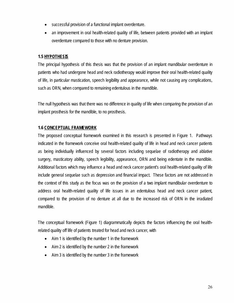
The null hypothesis was that there was no difference in quality of life when comparing the provision of an implant prosthesis for the mandible, to no prosthesis. 1.6 The proposed conceptual framework examined in this research is presented in Figure 1. Pathways indicated in the framework conceive oral health-related quality of life in head and neck cancer patients as being individually influenced by several factors including sequelae of radiotherapy and ablative surgery, masticatory ability, speech legibility, appearance, ORN and being edentate in the mandible. Additional factors which may influence a head and neck cancer patient’s oral health-related quality of life include general sequelae such as depression and financial impact. These factors are not addressed in the context of this study as the focus was on the provision of a two implant mandibular overdenture to address oral health-related quality of life issues in an edentulous head and neck cancer patient, compared to the provision of no denture at all due to the increased risk of ORN in the irradiated mandible.
CONCEPTUAL FRAMEWORK
The conceptual framework (Figure 1) diagrammatically depicts the factors influencing the oral health-related quality off life of patients treated for head and neck cancer, with
• Aim 1 is identified by the number 1 in the framework
• Aim 2 is identified by the number 2 in the framework
• Aim 3 is identified by the number 3 in the framework
27
Figure 1:
Conceptual framework
1.7 In order to obtain an overview of the current status for the use of implant mandibular prostheses in the oral rehabilitation of head and neck cancer patients and their impact on the patients quality of life, a comprehensive literature review was performed. Data for this review was identified by searches of PubMed and Scopus with the terms Head and Neck cancer, Radiotherapy, ORN, Osseointegration, Hyperbaric Oxygen, Irradiated Mandible, Edentulous Mandible and Quality of Life. Papers were limited to those published in English, to September 2008. Cross-referencing of important papers identified additionally relevant articles and those of historical value.
TABLE OF COMPARISON FOR REVIEW (PICO CHART)
Appearance Speech legibility Masticatory ability
ORAL HEALTH RELATED
QUALITY OF LIFE (3)
Ablative surgery Radiotherapy
Implant mandibular overdentures
(1, 2)
Edentate in the mandible (3)
Hyperbaric oxygen and
prophylactic antibiotics (1)
Osteoradionecrosis (1)
Implant placement (1) No lower denture provided (3)
Improved oral function (2)
28
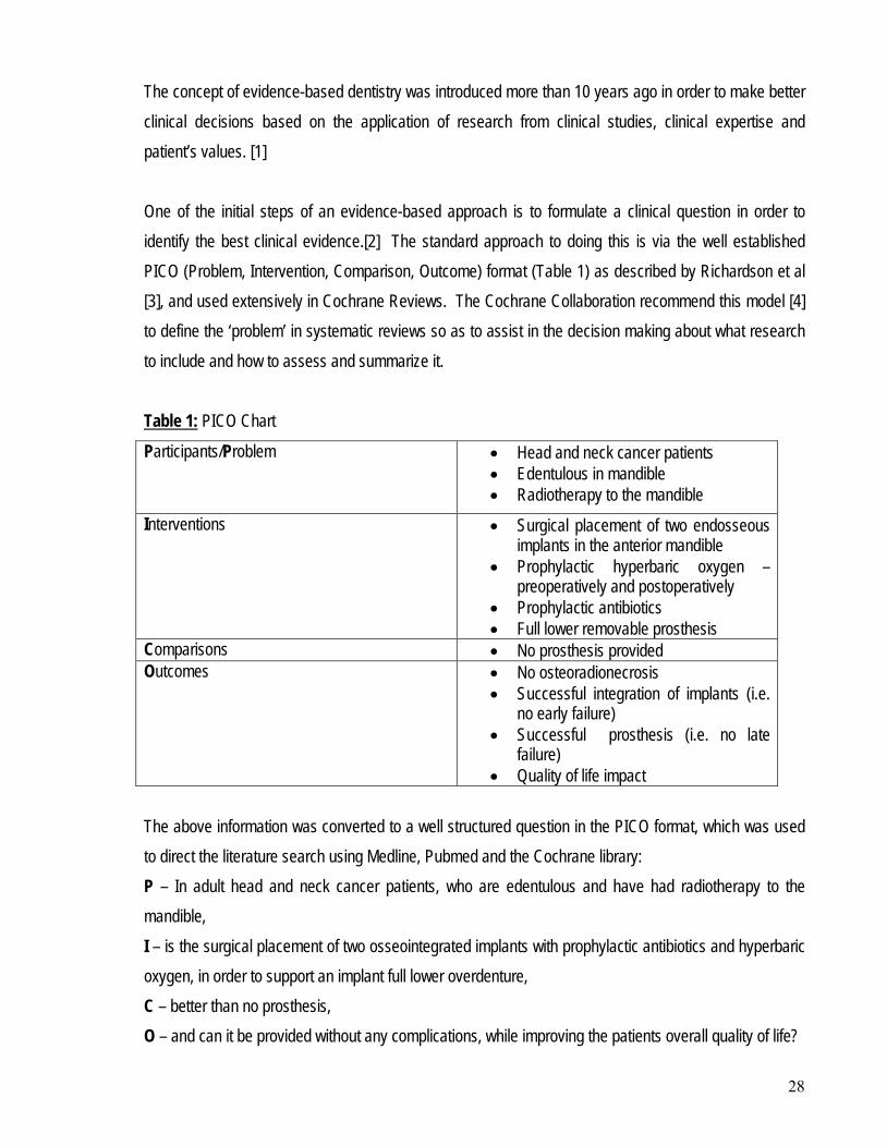
The concept of evidence-based dentistry was introduced more than 10 years ago in order to make better clinical decisions based on the application of research from clinical studies, clinical expertise and patient’s values. [1] One of the initial steps of an evidence-based approach is to formulate a clinical question in order to identify the best clinical evidence.[2] The standard approach to doing this is via the well established PICO (Problem, Intervention, Comparison, Outcome) format (Table 1) as described by Richardson et al [3], and used extensively in Cochrane Reviews. The Cochrane Collaboration recommend this model [4] to define the ‘problem’ in systematic reviews so as to assist in the decision making about what research to include and how to assess and summarize it. Table 1:Participants/Problem
PICO Chart
• Head and neck cancer patients • Edentulous in mandible • Radiotherapy to the mandible
Interventions • Surgical placement of two endosseous implants in the anterior mandible
• Prophylactic hyperbaric oxygen – preoperatively and postoperatively
• Prophylactic antibiotics • Full lower removable prosthesis
Comparisons • No prosthesis provided Outcomes • No osteoradionecrosis
• Successful integration of implants (i.e. no early failure)
• Successful prosthesis (i.e. no late failure)
• Quality of life impact The above information was converted to a well structured question in the PICO format, which was used to direct the literature search using Medline, Pubmed and the Cochrane library: P – In adult head and neck cancer patients, who are edentulous and have had radiotherapy to the mandible, I – is the surgical placement of two osseointegrated implants with prophylactic antibiotics and hyperbaric oxygen, in order to support an implant full lower overdenture, C – better than no prosthesis, O – and can it be provided without any complications, while improving the patients overall quality of life?
29
This chapter reviews the available literature on head and neck cancer including current statistical data, oncology treatment options and their sequelae. This is considered together with current knowledge and requirements for the provision of an implant prosthesis in an irradiated mandible, and the impact on quality of life.
CHAPTER 2: LITERATURE REVIEW
2.1 The anatomical location of malignancies as coded by the World Health Organization in the “International Classification of Diseases” 10th Revision, Australian Modification (ICD-10-AM) classifies malignant neoplasm’s of the lip, oral cavity and pharynx together (C00-C14). [5]
INCIDENCE AND PREVALENCE OF HEAD & NECK CANCER
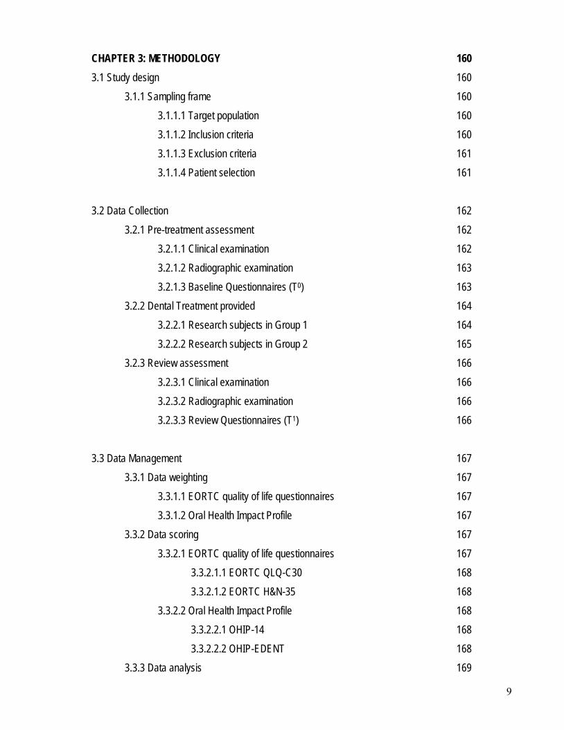
In all Australian States and Territories, cancer is a legally notifiable disease, with each operating its own cancer registry. These registries have operational guidelines which fulfil the requirements of both the Australasian and International Associations of cancer registries. The majority of population based data available from cancer registries on the incidence of oral cancers comes from western countries. Minimal data is available from developing countries where oral cancer is more common. [6, 7] Oral squamous cell carcinoma is the fifth most common cancer and is a major health problem in many countries. Annually about 500,000 new cases are diagnosed worldwide with about three quarters of these from developing countries. [8] 2.1.1 Oral cancer Australian and South Australian statistics available on mouth/oral cancer in general (tongue-C02, floor of mouth-C04, gums-C03, inner surfaces of the cheek-C06 and palate-C05) identifies an incidence similar to that of New Zealand, Northern Europe and Eastern Europe, with no evidence of any change in trend from 1977-2000, either in total or for individual areas.[9] In Australia, oral cancer accounts for approximately 2-3% of all cancers and approximately 1% of all deaths from cancer.[10] Figure 2 provides an overview of the incidence of mouth/oral cancer world-wide.[9] India has the highest incidence, with an important contributor the habitual chewing of tobacco and betel nut.
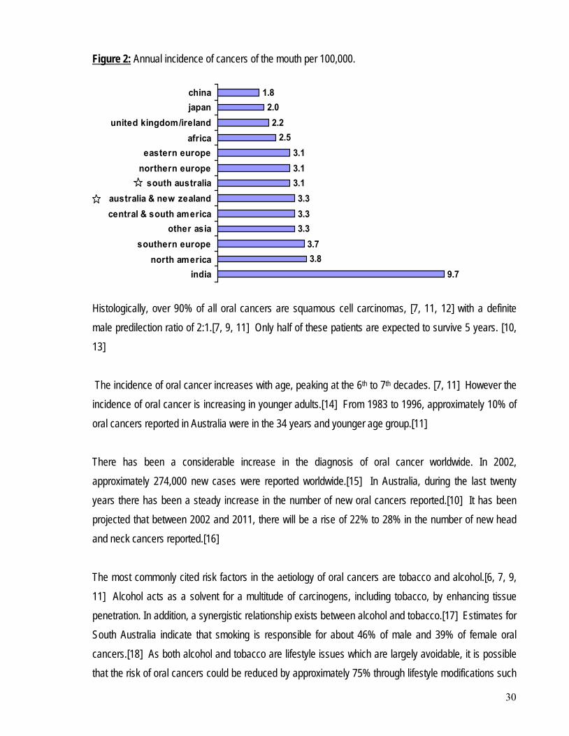
30
Figure 2:
Annual incidence of cancers of the mouth per 100,000.
9.73.83.7
3.33.33.3
3.13.13.1
2.52.2
2.01.8
indianorth america
southern europeother asia
central & south americaaustralia & new zealand
south australianorthern europeeastern europe
africaunited kingdom/ireland
japanchina
Histologically, over 90% of all oral cancers are squamous cell carcinomas, [7, 11, 12] with a definite male predilection ratio of 2:1.[7, 9, 11] Only half of these patients are expected to survive 5 years. [10, 13] The incidence of oral cancer increases with age, peaking at the 6th to 7th decades. [7, 11] However the incidence of oral cancer is increasing in younger adults.[14] From 1983 to 1996, approximately 10% of oral cancers reported in Australia were in the 34 years and younger age group.[11] There has been a considerable increase in the diagnosis of oral cancer worldwide. In 2002, approximately 274,000 new cases were reported worldwide.[15] In Australia, during the last twenty years there has been a steady increase in the number of new oral cancers reported.[10] It has been projected that between 2002 and 2011, there will be a rise of 22% to 28% in the number of new head and neck cancers reported.[16] The most commonly cited risk factors in the aetiology of oral cancers are tobacco and alcohol.[6, 7, 9, 11] Alcohol acts as a solvent for a multitude of carcinogens, including tobacco, by enhancing tissue penetration. In addition, a synergistic relationship exists between alcohol and tobacco.[17] Estimates for South Australia indicate that smoking is responsible for about 46% of male and 39% of female oral cancers.[18] As both alcohol and tobacco are lifestyle issues which are largely avoidable, it is possible that the risk of oral cancers could be reduced by approximately 75% through lifestyle modifications such
31
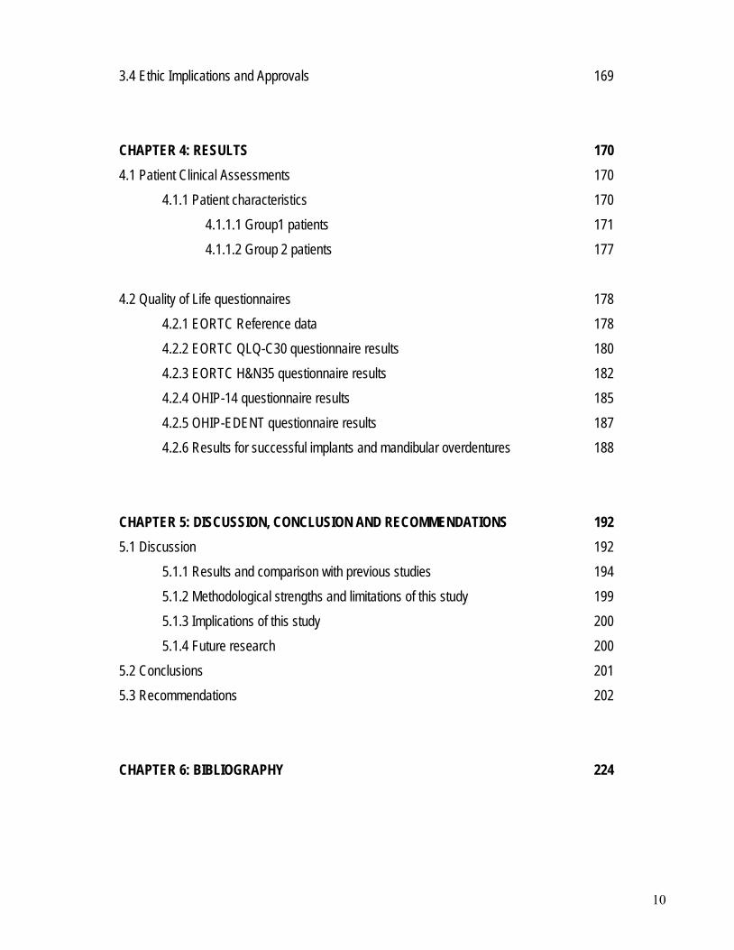
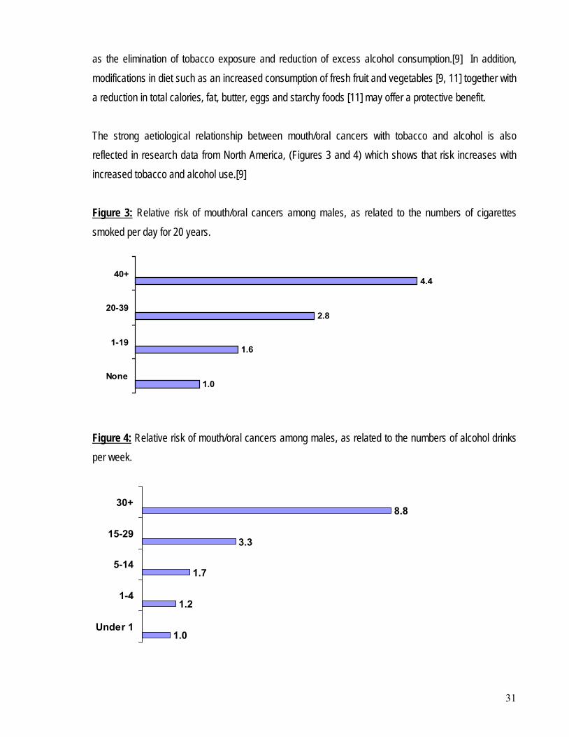
as the elimination of tobacco exposure and reduction of excess alcohol consumption.[9] In addition, modifications in diet such as an increased consumption of fresh fruit and vegetables [9, 11] together with a reduction in total calories, fat, butter, eggs and starchy foods [11] may offer a protective benefit. The strong aetiological relationship between mouth/oral cancers with tobacco and alcohol is also reflected in research data from North America, (Figures 3 and 4) which shows that risk increases with increased tobacco and alcohol use.[9] Figure 3:
1.0
1.6
2.8
4.4
None
1-19
20-39
40+
Relative risk of mouth/oral cancers among males, as related to the numbers of cigarettes smoked per day for 20 years.
Figure 4:
Relative risk of mouth/oral cancers among males, as related to the numbers of alcohol drinks per week.
1.0
1.2
1.7
3.3
8.8
Under 1
1-4
5-14
15-29
30+
32
An additional risk factor for mouth/oral cancers includes a history of precancerous conditions.[9, 14] These can include dysplasias, leukoplakias or erythroplakias. Other potential risk factors of uncertain importance include Lichen planus, Pemphigus vulgaris, Verrucous hyperplasia, and viral infections including Human papilloma virus, Herpes simplex virus and Epstein Barr virus. There are also recognized genetic predispositions to oral cancer in some syndromes. Mortality rates from oral cancers remain relatively high. Five year survival is estimated at 80% when diagnosis is made early, compared to 40% with local metastasis and approximately 10% when diagnosis is made with distant metastasis.[15] Early detection ensures not only an increased prognosis, but also a better post-treatment quality of life as a result of less extensive oncologic treatment requirements.[13] The proportion of affected individuals surviving mouth/oral cancer 5 years or more from diagnosis was 53% between 1977-1998, in South Australia. Hospital data during this period identified that surgery and radiotherapy were the most common primary treatment modalities utilised for mouth/oral cancer. [19] The tongue and floor of mouth are the most commonly reported intra-oral sites for oral cancer world-wide. They have the potential to cause serious health problems and significant morbidity.[19] Lederman’s hypothesis, suggests that the predilection for tongue and floor of mouth cancers is related to the pooling of carcinogens in saliva, with sites most at risk being the tongue, floor of mouth, anterior tonsillar pillar and the lingual aspect of the retromolar trigone.[6] In India, the incidence of tongue cancer in males is up to 6.5/100,000 per annum, and in France the male incidence is up to 8/100,000. By comparison the incidence of tongue cancer in Australia is relatively low, with the highest rates recorded amongst Northern Territory and Queensland males with an incidence of 4/100,000 in 1996.[6] Table 2 provides a synopsis of oral cancer cases in South Australia over a 24 year period. Table 2:
Oral cancer cases reported between1977-2001 in South Australia (excluding lip cancer and salivary gland malignancy) [6]
Primary cancer site Number of cases (%) Tongue 611 (44.9%) Gum 84 (6.2%) Floor of mouth 296 (21.8%) Other mouth 369 (27.1%)
33
The most common site for oral cancer in Australia during 1996 was the lips (C00) with an incidence of 9.2/100,000 for males and 3/100,000 for females.[11] The relatively high incidence of lip cancer in Australia is probably related to sun exposure, i.e. solar radiation.[10, 20] 2.1.2 Salivary gland cancer Cancers of the salivary glands predominantly involve the parotid gland (C07), although the other salivary glands (C08) may also be involved. Australia and New Zealand has a high incidence of salivary gland tumours by world standards. (Figure 5) [9] Figure 5: Annual incidence of cancers of major salivary glands per 100,000. [9]
The aetiology of salivary gland cancers are unknown, although it is possible that ionizing radiation may play a contributory role. During 1977-1998 the proportion of affected South Australians surviving these types of cancers for 5 years or more was 68%. Hospital data during this period indicates that 89% of salivary glands were treated by surgery and 77% by radiotherapy. [9] Other head and neck cancers include those of the larynx, oropharynx, hypopharynx and nasopharynx. These are all predominantly squamous cell carcinomas, and have a relatively low incidence in Australia. The aetiology of these cancers are the predominantly the same as for oral cancer i.e. tobacco smoking and excess intake of alcohol. [9, 20]
34
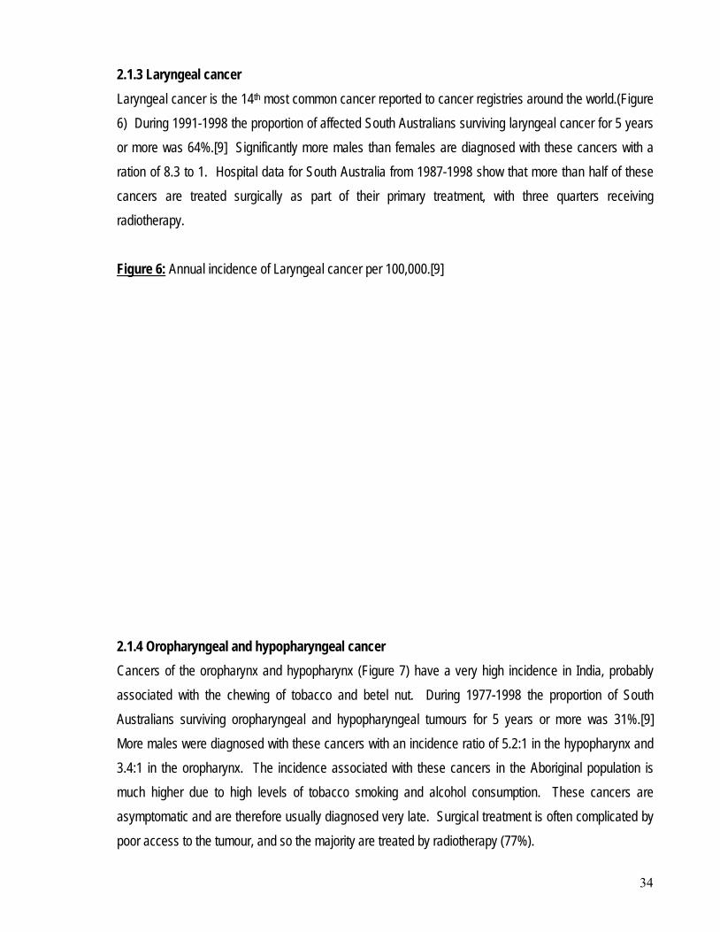
2.1.3 Laryngeal cancer Laryngeal cancer is the 14th most common cancer reported to cancer registries around the world.(Figure 6) During 1991-1998 the proportion of affected South Australians surviving laryngeal cancer for 5 years or more was 64%.[9] Significantly more males than females are diagnosed with these cancers with a ration of 8.3 to 1. Hospital data for South Australia from 1987-1998 show that more than half of these cancers are treated surgically as part of their primary treatment, with three quarters receiving radiotherapy. Figure 6:
Annual incidence of Laryngeal cancer per 100,000.[9]
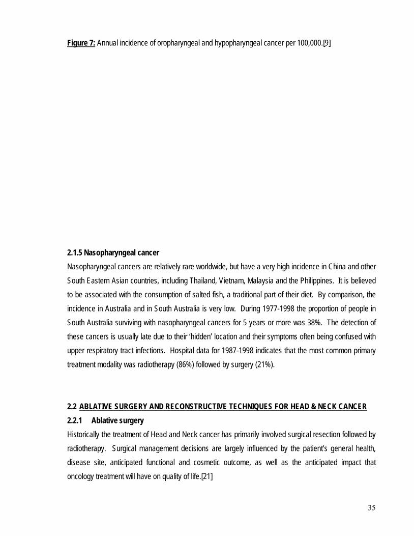
2.1.4 Oropharyngeal and hypopharyngeal cancer Cancers of the oropharynx and hypopharynx (Figure 7) have a very high incidence in India, probably associated with the chewing of tobacco and betel nut. During 1977-1998 the proportion of South Australians surviving oropharyngeal and hypopharyngeal tumours for 5 years or more was 31%.[9] More males were diagnosed with these cancers with an incidence ratio of 5.2:1 in the hypopharynx and 3.4:1 in the oropharynx. The incidence associated with these cancers in the Aboriginal population is much higher due to high levels of tobacco smoking and alcohol consumption. These cancers are asymptomatic and are therefore usually diagnosed very late. Surgical treatment is often complicated by poor access to the tumour, and so the majority are treated by radiotherapy (77%).
35
Figure 7:
Annual incidence of oropharyngeal and hypopharyngeal cancer per 100,000.[9]
2.1.5 Nasopharyngeal cancer Nasopharyngeal cancers are relatively rare worldwide, but have a very high incidence in China and other South Eastern Asian countries, including Thailand, Vietnam, Malaysia and the Philippines. It is believed to be associated with the consumption of salted fish, a traditional part of their diet. By comparison, the incidence in Australia and in South Australia is very low. During 1977-1998 the proportion of people in South Australia surviving with nasopharyngeal cancers for 5 years or more was 38%. The detection of these cancers is usually late due to their ‘hidden’ location and their symptoms often being confused with upper respiratory tract infections. Hospital data for 1987-1998 indicates that the most common primary treatment modality was radiotherapy (86%) followed by surgery (21%). 2.2 2.2.1 Ablative surgery
ABLATIVE SURGERY AND RECONSTRUCTIVE TECHNIQUES FOR HEAD & NECK CANCER
Historically the treatment of Head and Neck cancer has primarily involved surgical resection followed by radiotherapy. Surgical management decisions are largely influenced by the patient’s general health, disease site, anticipated functional and cosmetic outcome, as well as the anticipated impact that oncology treatment will have on quality of life.[21]
36
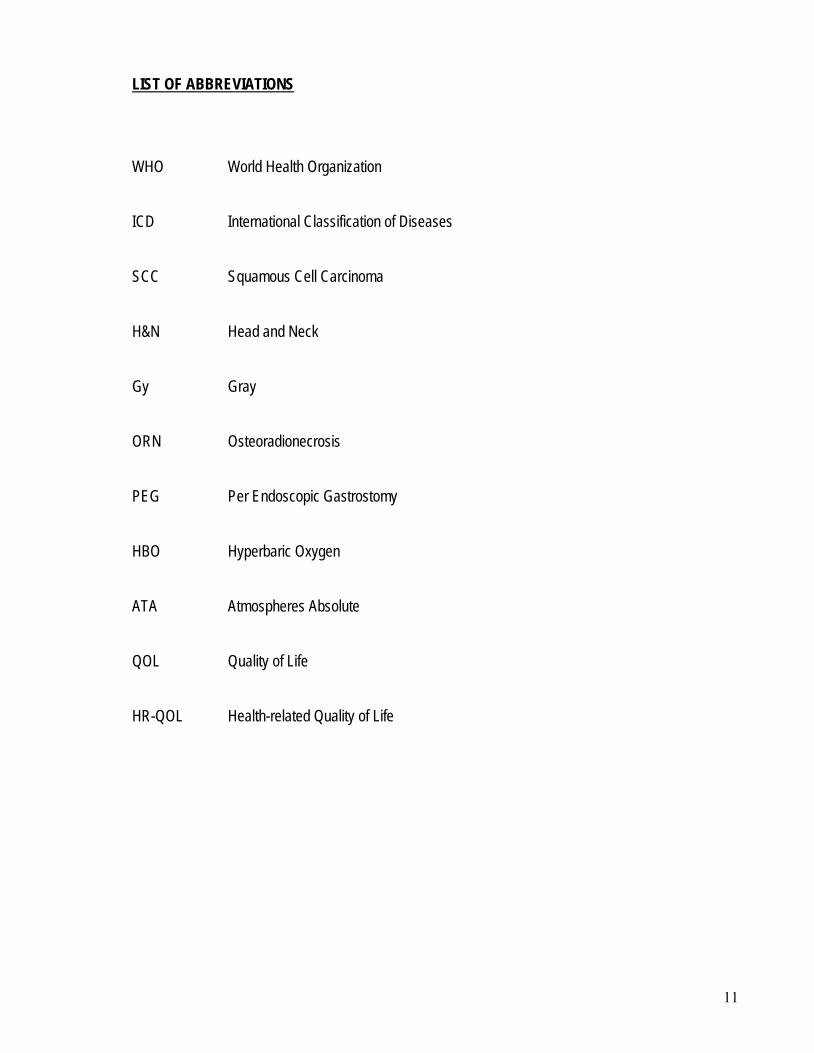
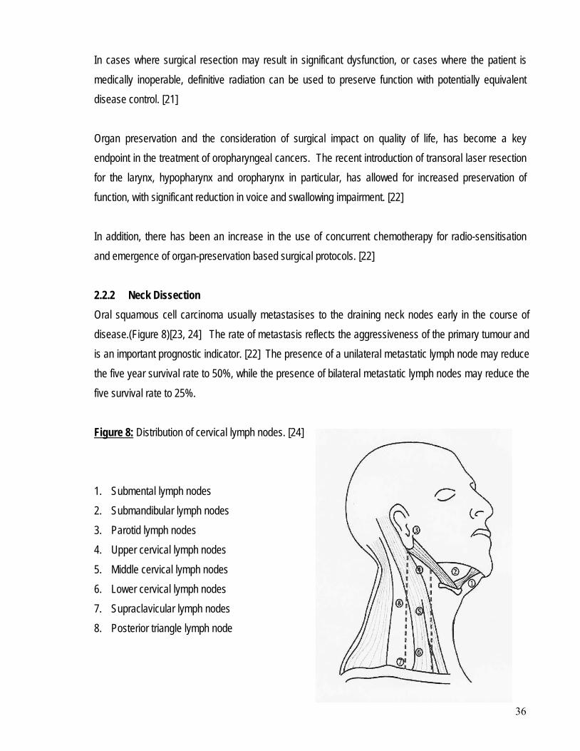
In cases where surgical resection may result in significant dysfunction, or cases where the patient is medically inoperable, definitive radiation can be used to preserve function with potentially equivalent disease control. [21] Organ preservation and the consideration of surgical impact on quality of life, has become a key endpoint in the treatment of oropharyngeal cancers. The recent introduction of transoral laser resection for the larynx, hypopharynx and oropharynx in particular, has allowed for increased preservation of function, with significant reduction in voice and swallowing impairment. [22] In addition, there has been an increase in the use of concurrent chemotherapy for radio-sensitisation and emergence of organ-preservation based surgical protocols. [22] 2.2.2 Neck Dissection Oral squamous cell carcinoma usually metastasises to the draining neck nodes early in the course of disease.(Figure 8)[23, 24] The rate of metastasis reflects the aggressiveness of the primary tumour and is an important prognostic indicator. [22] The presence of a unilateral metastatic lymph node may reduce the five year survival rate to 50%, while the presence of bilateral metastatic lymph nodes may reduce the five survival rate to 25%. Figure 8:
Distribution of cervical lymph nodes. [24]
1. Submental lymph nodes 2. Submandibular lymph nodes 3. Parotid lymph nodes 4. Upper cervical lymph nodes 5. Middle cervical lymph nodes 6. Lower cervical lymph nodes 7. Supraclavicular lymph nodes 8. Posterior triangle lymph node
Lymph nodes in the submandibular triangle are considered level 1. Levels 2, 3 and 4 are the upper, middle and lower cervical lymph nodes which traverse along the internal jugular chain. Level 5 includes the spinal accessory and posterior triangle lymph nodes. Metastatic lymph nodes are site-specific. In patients with a known primary tumour, the distribution of metastasis assists tumour staging. If the primary tumour is not identified, the distribution of proven metastatic lymph nodes may assist in its identification.[24] Classic radical neck dissection removes all five levels of cervical lymph nodes en bloc down to the deep muscular fascia, and includes the removal of the sternocleidomastoid muscle, submandibular salivary gland, jugular vein and spinal accessory nerves. This radical surgical approach is associated with a significant loss of both function and aesthetics, but was considered necessary due to the increased mortality risk associated with neck node metastasis. It was first described in 1906, and has until recently remained the gold standard treatment for cancer in the regional neck nodes. [23] The resultant deleterious impact on both function and aesthetics of the classic radical neck dissection, led to the development of modified surgical procedures in which the non-lymphatic structures of the neck were preserved, but which was still oncologically safe.[22] The modified radical neck dissection removes all five levels of cervical lymph nodes but preserves one or all of the spinal accessory nerves, the sternocleidomastoid muscle, together with the submandibular gland. The selective neck dissection removes either levels 1, 2, 3 [supraomohyoid neck dissection], levels 2, 3, 4 [anterior neck dissection] or levels 2, 3, 4, 5 [antero-lateral neck dissection] cervical lymph nodes. [21, 25] 2.2.3 Reconstructive Techniques Patients who undergo ablative surgery for oral cancer are left debilitated, both functionally and aesthetically, unless the tumour resection includes a reconstructive procedure(s). This is based on the principle that anatomical tissues which are removed as part of the ablative surgical process should either be repaired or replaced. Currently, replacement options for hard tissue include free bone grafts, vascularised bone grafts or reconstruction plates. Soft tissue replacement is via the use of local flaps, regional flaps, grafts and vascularised free tissue transfer. Tooth replacement options are limited to a removable prosthesis, or fixed prostheses. [26] The objectives of reconstruction involving the mandible include: [27]
• Restoration of bone continuity
38
• Provision of adequate bone volume
• Restoration of satisfactory bone height and width of the alveolus
• Prevention of graft resorption
• Restoration of soft tissue defects
• Restoration of oral continence [28]
• Establishment of facial contour [28-30]
• Re-establishment of masticatory function [29, 30] and occlusal relationships [28] If the above are successfully achieved the patient is often able to recover from the ablative surgical procedure both physically and psychologically. However, the outcomes of surgical reconstruction of patients who had undergone extensive tumour resection of the mandible and soft tissues had been less successful prior to the utilisation of microvascular soft tissue free-flaps and implants. With these procedures even the largest defects created during ablative surgery can be restored with predictable outcomes. [21, 30-32] With the use of microvascular soft tissue free-flaps and dental implants the optimal and potentially achievable goals of ablative surgery and oro-mandibular reconstruction include: [33]
• The re-establishment of mandibular continuity with vascularised bone rigidly fixed to the remaining mandible
• Restoration of sensation to the lower lip along with restoration of normal height so as to preserve or restore labial competence
• Restoration of sensation to the lining of the oral cavity through the use of nerve grafts or sensate flaps
• Introduction of thin pliable soft tissue to the tongue and floor of mouth following partial glossectomy to maintain the mobility of the residual tongue and to maximise oral function.
The ability to achieve these optimal reconstructive goals in any head and neck cancer patient is determined by the anatomic limitations related to the extent of the surgical resection. Despite continued improvements in surgical reconstructive techniques, the patient’s post reconstructive condition approaches, but never achieves, their pre-surgical status.
39
The two most important decisions the surgical team must make when planning the surgical management of advanced oral cancer is related to the management of the tongue and mandible. Advanced tumours of the tongue and floor of mouth often require extensive resection including the mandible, resulting in significant functional disability and cosmetic disfigurement.[32] The amount of resection and the decision of how to reconstruct in order to restore function and aesthetics will impact not only on the patient’s prognosis but also on their post-operative quality of life.[31] The most common methods of oro-facial defect reconstruction following ablative head and neck surgery include:
• Local flaps
• Grafts
• Regional flaps
• Free-flaps
• Reconstruction plates
• Osseointegrated implants 2.2.3.1 Local flaps These are segments of tissue which are sourced from the immediate area of resection and then either advanced, transposed or rotated to the recipient site while retaining some blood supply. The most common types of local flaps used are the buccal pad of fat flap, the naso-labial flap and the facial artery musculo-mucosal flap. The buccal pad of fat flap is the most versatile flap for repair of small to medium oral defects and has the additional benefit of being able to be used in conjunction with other flaps including free-flaps for the repair of larger defects.[26] 2.2.3.2. Grafts A ‘split skin graft’ is still a common method of surgically repairing a small to medium sized defect on the lateral border of the tongue, buccal mucosa and floor of mouth. Grafts do not have an intact blood supply nor drainage and therefore this needs to be re-established from the recipient bed.[26]

40
2.2.3.3 Regional flaps These types of flaps are now used less commonly. They originate from a distant donor site and are positioned with an intact vascular pedicle connected to the flap via a bridging segment. The two most common regional flaps used are the pectoralis major (myocutaneous) flap and the temporalis (myofacial) flap.[26] They remain useful as a source of well vascularised tissue in the medically morbid patient and in salvage procedures. 2.2.3.4 Free-flaps These are also referred to as free vascularised tissue transfer flaps, or microvascular free tissue flaps as they are harvested from a distant site by dividing the vascular pedicle. This pedicle is subsequently re-anastomosed to the recipient blood supply and drainage. Tissues which can be transferred include the skin, fascia and bone. Most commonly combinations of these different tissues are harvested and then are referred to as a ‘composite free flap’.[26] The microvascular free tissue flaps are the best method currently available to reconstruct mandibular defects by re-establishing mandibular continuity as well as improving cosmetic appearance and function. [28, 34] Through the use of these flaps it is possible to achieve functional success with respect to deglutition and speech intelligibility, a reasonable aesthetic result and mastication via dental rehabilitation.[21] They can achieve a high rate of success, with flap survival reported to be greater than 95%. [32, 33] Prior to the use of these flaps, conventional and maxillofacial prosthetic rehabilitation offered only very limited success after ablative surgery due to the failure to re-establish the bony and soft tissue anatomy and physiology.[35] The advantage of vascularised bone containing free-flaps is that they allow the transfer of both soft and hard tissue with a rich vascular supply that permits these tissues to withstand both the potentially detrimental effects of the normal oral flora and post-operative radiotherapy.[21, 31, 33] It must be noted that if a free flap fails, which occasionally they do, the resulting deficit is major. The four most common free-flaps used are the: [26, 28, 29, 31, 33]
• Radial forearm free flap
• Fibula free flap
• Iliac crest free flap
• Scapula free flap
41
2.2.3.4.1 Radial forearm free flap This is the most commonly used free flap in oral reconstruction due to its versatility, reliability and flexibility. It can be used to reconstruct virtually any missing oral structure with minimal donor site morbidity.[26, 30, 36] It has the ability to be harvested as a fascio-cutaneous free flap, an osseo-fascio-cutaneous composite free flap or fascial flap. When bone is harvested in an osseo-fascio-cutaneous composite free flap, only one third of the radius is involved either as a segment or vertically split. This is because there is a significant risk of radial fracture, which is the most commonly associated morbidity with this type of free flap.[33] The bone height will limit the length of osseointegrated implants which can be used in this site, which may have ramifications on the implants loading potential. Another issue associated with the use of this free flap is that the harvested dermal component of the cutaneous flap may contain hair follicles which will continue to grow intra-orally. This is not a problem if the site is later irradiated, as the hair follicles will be irreversibly damaged preventing further hair growth. 2.2.3.4.2 Fibula free flap This is the free flap of choice [26, 29, 31, 35] for the reconstruction of bony continuity defects of the mandible, due to the length of the fibula bone available for reconstruction, the low morbidity of the donor site, the long vascular pedicle available and the potential for incorporation of a skin paddle.[26] The indications for the use of the fibula osseocutaneous free flap include[33]:
• Total or subtotal mandibular reconstruction
• Reconstruction of bone only defects
• Reconstruction of an atrophic mandible
• Secondary reconstruction of the subcondylar-condylar process
• Paediatric mandibular reconstruction The height of the neo-mandible achievable with this free flap is similar to that of an edentulous atrophic mandible.[31, 33]
42
The fibula bone presents favourable conditions for implant placement and the subsequent implant prosthesis due to its good diameter and good quality of cortical bone [29] which can favourably withstand the biomechanical loading during masticatory function.[27, 31, 35] Implants placed in these reconstructed sites have obtained survival rates comparable to that of native bone,[29] and mandibles reconstructed with fibula free-flaps have documented implant survival rates at 94.6%.[37] The main limitation of this type of free flap is that the low bone height available may create problems from a prosthodontic viewpoint in dentate patients treated by partial mandibular resection, who have residual dentition on the healthy side.[29] 2.2.3.4.3 Iliac crest free flap This osseo-cutaneous flap uses the natural curvature of the Ilium for reconstruction of the mandible.[26] Unfortunately, there are often significant problems associated with donor site morbidity such as mobilisation problems due to pain, gait problems, abdominal problems and frank hernias which have limited its use. It is however, the second site of choice when vascular supply to the fibula and lower legs is inadequate.[31] The major advantage of the Iliac crest free flap is that it offers the best stock of bone available from any donor site currently available for oro-mandibular reconstruction, as the height of the neo-mandible achievable matches up favourably with that of a dentate native mandible.[30, 31, 33] The height advantage assists in successful long-term implant stability as well as the avoidance of any mismatch in bone height with the native mandible, which can make prosthetic reconstruction much more difficult.[33] The location of the vascular pedicle means that cancellous bone of the iliac crest forms the neo-alveolar crest when transplanted. This is not a major concern as the bone does re-corticate at the alveolus, allowing for excellent osseointegration.[31] The major disadvantage of this free flap is that there is often a large amount of soft tissue associated with the vascular pedicle making it a bulky flap. This often means that prosthodontic rehabilitation can be very difficult without the use of a subsequent de-bulking surgical procedure.[26, 33, 36]
43
2.2.3.4.4 Scapula free flap This is a useful flap for large soft tissue defects as the soft tissue flap is mobile relative to the bone. The major disadvantage of this flap which limits its use is that the quality of bone harvested is usually unsuitable for dental implant provision.[33] 2.2.4 Donor site selection The choice of bone free flap to be used in the reconstruction of mandibular continuity defects following ablative surgery should be made with consideration given to the need to attempt to duplicate the height and width of the resected bone as closely as possible, while also achieving an overall strength capable of withstanding masticatory forces.[28] Creation of normal alveolar bone height and width of the neo-mandible is important for:
• Structural integrity of the mandible
• The provision of stable conventional removable tissue borne prosthesis
• The placement of osseointegrated implants. The selection of an appropriate donor site is dependant upon both patient specific factors (Table 3) [33] and a critical assessment of the important components of the post-surgical defect (Table 4). [33] Table 3: Patient factors influencing donor site selection [33]
44
Table 4Volume and neurological status of residual tongue
: Oro-mandibular defect analysis [33]
Extent of remaining mucosal defects Extent of oropharyngeal defect contributing to velopharyngeal incompetence Extent and location of external cutaneous defect Extent and location of upper and lower lip defect(s) Extent of anticipated sensory deficits in the oral cavity and lips Mandibular bony defect variables:
• Length and location of segmental defect • Height of remaining native mandible • Volume of bone in the remaining mandible for placement of implants • Status of dentition in the remaining mandible and maxillae • Radiation status of native mandible
The major factors determining the selection of donor site for vascularised bone are: [28]
• The ability to harvest adequate bone length to reconstruct the mandibular defect
• The ability to maintain the natural contour of mandible with the bone graft
• Consideration of the size, length and consistency of the vascular pedicle
• The quality, vascularity and mobility of the accompanying soft tissue for resurfacing of mucosal cutaneous defects
• The accessibility of the donor site for a simultaneous team approach (only if considered necessary)
• The potential for donor site morbidity
• The individual patient factors which may mitigate against the selection of a particular donor site. 2.2.5 Reconstruction plates These were commonly used to restore mandibular continuity prior to the availability of free-flaps with bone. They may be used for primary reconstruction or as a staged procedure with subsequent bony reconstruction.[26] If used, the plates need to be contoured sufficiently to allow for correct anatomical placement of the new alveolar segment, as well as for subsequent placement of implants in an appropriate position for prosthetic rehabilitation.[31] They are also used in situations when a free flap is not suitable or when reconstruction is deferred while awaiting histopathological results. At present there are limited indications for the use of reconstruction plates as free tissue transfer is now considered the evidence-based treatment of choice for the repair of mandibular continuity defects.
45
2.2.6 Osseointegrated implants The advent of osseointegrated implants have allowed for significant improvements in the oral rehabilitation of patients who have undergone bone resection as part of ablative tumour surgery.[26, 30, 31, 33, 36] The use of overlay prostheses have allowed for replacement of missing teeth and resected alveolar structures, as well as improved lip support and facial contour.[32] Even with the use of composite free-flaps containing bone, the post-surgical anatomy following reconstruction will often not allow for successful function of a conventional removable prosthesis.[30, 31] Implants can be successfully placed in both native bone as well as free-flaps incorporating bone from the fibula, iliac crest or radius.[26] In fact, through the successful placement of vascularised bone containing free-flaps for mandibular reconstruction, the osteogenic potential of the transferred bone is maintained therefore allowing it to take an active role in osteosynthesis with the native mandibular bone, and allowing osseointegration of dental implants.[33] A study by Urken et al [38] investigated numerous functional parameters that contribute to the masticatory process in an oral cancer patient. Their conclusions were that the use of microvascular bony reconstruction and osseointegrated implants could provide excellent quality of life, bringing the patient as close as possible to their pre-disease condition.
Oral cancer and its treatment modalities carry a high level of functional morbidity, both in the short and long-term. The degree of functional morbidity is influenced by the site and size of the tumour, the site and size of the post-surgical defect and also the use of adjuvant radiotherapy.[25]
2.3 SEQUELAE OF HEAD AND NECK ABLATIVE SURGERY
The introduction of free tissue transfer reconstructive surgical procedures and microsurgical techniques have had a greater impact on oral cancer surgical treatments than on any other cancers at any other site. Many of the major functional deficits encountered following ablative resections have now been either reduced or significantly alleviated.[21, 25, 39] However, despite these advances, patients still experience post-surgical difficulties. There are a number of factors which affect patient’s status after surgical resection: [40]
46
• Impairment of sensory and/or motor control, especially with respect to neuromuscular balance between the tongue, lip and cheek
• Loss of tongue bulk and/or immobility of residual tongue tissue
• Presence and size of mandibular continuity defects and associated soft tissue defects
• Deviation of the mandible and pathway of closure of the mandible, and its impact on the maxillo-mandibular relationship
• Presence of a functional dentition or prosthesis. 2.3.1 General sequelae The general sequelae of oral and oro-pharyngeal ablative surgery may include: [25, 29, 30, 35, 39-41]
• Psychological impact, i.e. acute and chronic depression [30, 41]
• Social adaptation and employment [41]
• Aesthetics, appearance and cosmetic disfigurement [25, 29, 30, 35, 39-41]
• Shoulder function limitations [25, 39] 2.3.1.1 Psychological impact Acute depression is the most common psychological symptom of post-surgical cancer patients.[41] If the patient is left unreconstructed, there can potentially be serious problems with social adaptation, an inability to return to their previous employment, and ultimately social isolation and unwillingness to face society.[41] Oral cancer surgery has an impact on health-related quality of life as it influences psycho-social activity.[39] 2.3.1.2 Appearance and aesthetics The psychological impact of disfigurement may add to the level of resulting morbidity. Maxillofacial deformity has the potential to produce a negative impact on social functionality, including employability, honesty and trustworthiness. Even minor facial alterations can potentially have an impact.[42] A patient’s ability to adapt to the associated cosmetic disfigurement and functional morbidity following surgery is dependant on time but also their psychological health.[39, 41] 2.3.1.3 Shoulder function The surgical removal of oral cancers usually also involves the dissection of involved and potentially involved lymph nodes in the neck. Shoulder complaints following neck dissection can include shoulder
47
pain, a restricted range of motion, shoulder droop and scapular wings. [43] This shoulder dysfunction occurs as a result of loss of the sternocleidomastoid muscle and/or innervation by the spinal accessory nerve. [44] Modified or radical neck dissection may impact on the shoulder function by creating either an inability or limited ability to abduct the arm of the affected side to 90º, or to place the arm behind the head.[21, 25] The reported prevalence of shoulder complaints range from 47-100% following a radical neck dissection, 18-61% following a modified radical neck dissection, and 29-52% following a selective neck dissection. [43] Intensive physiotherapy exercise programs may assist in improving shoulder complaints and shoulder disability following neck dissection. 2.3.2 Oral sequelae The oral sequelae of oral and oro-pharyngeal ablative surgery may include: [25, 29, 30, 35, 39-41, 45]
• Drooling/salivary control [25, 29, 35, 40, 41] as a result of altered lip competence and tongue mobility [39]
• Mastication [29, 30, 35, 39-41] and its impact on nutrition and diet [25, 39, 41]
• Temporomandibular joint function [41] with trismus[39] and mandibular deviation [30, 35, 40]
• Swallowing function [29, 30, 35, 39, 41]
• Speech [25, 29, 30, 35, 39-41]
• Tactile sensation [30, 39] and loss of proprioception [35, 40]
• Taste [41] Oral function, both sensory and motor, is significantly affected following surgery especially if mandibular resection is required. Post-surgical mandibular defects can range from limited resections of the alveolar ridge and adjacent soft tissues, to extensive resections resulting in discontinuity of the mandible and associated resection of the tongue and/or floor of mouth.[40] Disabilities associated with these extensive resections if not completely reconstructed, can include impaired speech articulation, alteration to masticatory ability, deviation of the mandible during functional movements, maxillo-mandibular malocclusion, difficulty swallowing with associated reduced control of salivary secretions, and cosmetic disfigurement due to lack of bony support for facial soft tissues.[30, 34, 35, 40]
48
2.3.2.1 Saliva control Salivary drooling is a common sequelae to head and neck cancer surgery, particularly if the patient does not have adjuvant radiotherapy. Drooling is debilitating and is a consequence of reduced swallowing ability and lip incompetence. Other contributory factors include: [41]
• Restricted tongue movement
• Loss of labial/buccal and lingual sulci
• Scarring of the orbicularis muscle of the lip
• Incision scarring of the lower lip following lip split procedures
• Paralysis of the mandibular branch of the VII cranial nerve
• Loss of sensory awareness. 2.3.2.2 Swallowing function Postoperatively, swallowing may be temporarily or permanently affected. As swallowing is a primary function, it is not easily disrupted and therefore the ability to swallow will return in some form. However, approximately 26% of patients who undergo ablative surgery for oral and/or oropharyngeal surgery will experience considerable difficulty with the voluntary component of swallowing.[41] 2.3.2.3 Speech articulation and intelligibility The impairment of speech and/or intelligibility of speech following ablative surgery is a common occurrence.[25, 41] The combination of a misshapen oral cavity and restricted tongue movement together with a reduction in lip competence and motor control, can lead to a severe deterioration in speech. The tongue is the main organ of speech or articulation and consequently a reduction in its size or mobility can lead to a deterioration of the pronunciation of consonants. Vowels generally are unaffected. Speech defects are often considerable and have the potential to socially isolate the patient even further.[41] 2.3.2.4 Removable prostheses When the resection only involves the alveolar portion of the mandible, or is confined to the soft tissues, mandibular continuity is maintained. Although there is less facial or cosmetic disfigurement, edentulous patients will still experience great difficulty in successfully wearing conventional removable prostheses. This is often due to the altered oral anatomy, obliteration of the sulcus, and reduction or loss in the sensory and motor innervation. In addition, the tongue function may be affected therefore making
49
control of removable dentures even more difficult.[30] Dentate patients will also experience some difficulty with mastication due to loss of multiple teeth, usually unilaterally, with associated loss of the proprioceptive sense of occlusion leading to unco-ordinated and less precise mandibular movements.[35] 2.3.2.5 Mastication and dietary impact The alteration to the patient’s post-surgical ability to masticate food adequately, if at all, can have a profound affect on their dietary choices. This has the potential to impact on their general health as a consequence of poor and reduced nutritional intake, weight loss due to the reduced masticatory ability, as well as loss of appetite (anorexia), difficulty swallowing and possibly also alteration to taste perception. This is often further compounded if the patient undergoes adjunctive radiotherapy, with its associated mucositis and xerostomia.[25, 39, 41]
Radiotherapy plays an important role in the management of head and neck cancer and is provided with either a curative intent or as part of palliative management. Curative radiotherapy can be applied either as the primary treatment modality, pre-operatively or post-operatively as an adjunct to surgical management, or in combination with chemotherapy.
2.4 RADIOTHERAPY TO THE HEAD AND NECK REGION
The objective of effective cancer therapy includes the preservation of normal tissue function as much as possible. The head and neck region is a complex area, composed of several dissimilar anatomical structures which respond differently to radiation exposure.[46] These include:
• Mucosal linings
• Skin
• Muscle
• Salivary glands
• Teeth
• Bone and cartilage.
50
2.4.1 Indications The aim of radiotherapy is to eradicate the tumour cells by exposing them to high doses of ionizing radiation, while minimising damage to normal cells.[47] Indications for post-operative radiotherapy include: [48]
• Incomplete or non-radical resection of a tumour
• The combination of close resection margins (<5mm) and a tumour with an aggressive growth pattern with perineural growth invasion
• Multi-nodal metastases or metastases with extracapsular extension. 2.4.2 Techniques 2.4.2.1 Conventional fractionation The radiation dose received will be dependant on the location and type of malignancy, and whether it is used in isolation or in combination with other treatment modalities. Most patients with head and neck cancer where radiotherapy is provided with curative intent undergo conventional fractionation. With this technique a total radiotherapy dose of between 60 to 70 Gy, usually given over a 5 to 7 week period, once a day, 5 days per week, with 2 Gy per fraction prescribed. [46] The conventional fractionated application of radiation works on the scientific principle that there is a difference in the response between tumour tissue and normal tissue to radiotherapy. In general, normal tissue is capable of repairing sublethal radiation induced DNA damage, far better than tumour tissue. This is especially so when radiation is applied in the lower dose range, therefore the application of radiation in 2Gy fractions magnifies this difference in repair response of the two tissues. The attempt at sparing damage to normal tissues by conventional fractionation achieves the greatest impact on late responding tissues. Unfortunately, tissues which respond early to radiation are damaged to a similar extent as the tumour tissues. These can include oral mucosa, salivary glands and taste buds.[46] In addition, the conventional fractionation irradiation technique allows for the repopulation or regrowth of tissues between the application of fractions. This is especially so during the weekend when no radiotherapy treatment is provided, therefore reducing the damage done to early responding tissues. Unfortunately, this also applies to the rapidly proliferating malignant tumour tissue.
51
Another advantage of conventional fractionated radiotherapy is that it allows for the re-oxygenation of radio-resistant hypoxic tissue between fractions, therefore leading to an increase in the amount of radio-sensitive oxygenated tissue.[46] Alternative strategies aimed at increasing the tumour control while not increasing and potentially even reducing the normal tissue complications continue to be developed. These include: [46]
• Alternate fractionation schemes o Hyper-fractionation o Accelerated fractionation
• Methods to increase the oxygenation of tumour tissue and therefore increase the tissue’s radiosensitivity
• Techniques which reduce the radiation volume of tissues o 3-dimensional conformal radiation therapy o Intensity modulated tadiation therapy
2.4.2.2 Hyper-fractionation Hyper-fractionation makes use of the difference between the repair capabilities of normal tissue and tumour tissue by further fractionation of the dose to approximately 1.15Gy. Usually the overall treatment time is maintained and therefore the patients receive two fraction treatments per day. The advantage of this technique is that the total absorbed radiotherapy tumour dose can be increased without adding to the late toxicity. 2.4.2.3 Accelerated fractionation Accelerated fractionation makes use of the principle that radiation injury leads to an accelerated proliferation of tumour tissue. It is believed that by reducing the overall treatment time this accelerated proliferation should be overcome. This regime consists of 2 Gy fractions given twice per day over a shorter period of time. Combinations of hyper-fractionation and accelerated fractionation have also been used and proven to be especially beneficial for rapidly dividing tissues.[46] The main disadvantage of all of these radiation techniques is that they carry a high rate of acute toxicity, i.e. oral mucositis. Oral mucositis is the “erythematous, erosive and ulcerative lesions of the oral mucosa.”[49] A technique aimed at increasing oxygenation and reducing both acute and chronic tumour hypoxia, involves the use of an accelerated fractionation schedule of radiotherapy together with
52
Carbogen breathing and Nicotinamide. This is capable of achieving a very high local and regional tumour control. While there is some increase in both acute toxicity and late morbidity, it is considered that these are within acceptable levels, despite late bone complications which occur.[46] 2.4.2.4 Computerised planning techniques Computerised planning techniques such as the 3-dimensional conformal technique and the intensity modulated radiotherapy technique attempt to further reduce the irradiated volume of normal tissue. The 3-dimensional conformal technique is designed to focus the spatial distribution of high radiation dose to the target tissues therefore reducing the dose delivered to normal tissues. The intensity modulated radiotherapy technique is an even more conformal technique. It works by optimally assigning weights to each individual ray of the radiotherapy beam, not just a single weight to the whole beam, which is what routinely occurs. This allows for the development of a dose distribution pattern which minimizes the radiation dose to normal tissues as much as possible. Both of these treatment techniques attempt to ensure that a small volume of tissue receives the high dose radiation and a large volume of tissue receives only a low dose.
Tumoricidal ionizing radiation provided during radiotherapy causes damage to normal tissues located within the radiation field(s). This damage is especially evident in the head and neck region where several dissimilar anatomical and physiological structures are located.
2.5 ORAL SEQUELAE OF HEAD AND NECK RADIOTHERAPY
During and after radiotherapy, some degree of transient and permanent damage will occur to normal oral tissues. This damage to the healthy tissues surrounding the malignancy usually results in multiple complex oral complications ranging from mild post-treatment damage, to life threatening necrosis. (Figure 9)
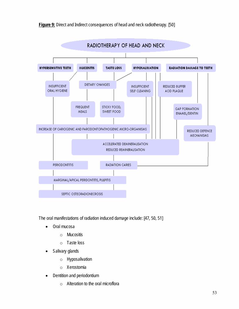
53
Figure 9:
Direct and Indirect consequences of head and neck radiotherapy. [50]
The oral manifestations of radiation induced damage include: [47, 50, 51]
• Oral mucosa o Mucositis o Taste loss
• Salivary glands o Hyposalivation o Xerostomia
• Dentition and periodontium o Alteration to the oral microflora
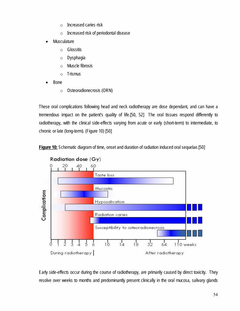
54
o Increased caries risk o Increased risk of periodontal disease
• Musculature o Glossitis o Dysphagia o Muscle fibrosis o Trismus
• Bone o Osteoradionecrosis (ORN)
These oral complications following head and neck radiotherapy are dose dependant, and can have a tremendous impact on the patient’s quality of life.[50, 52] The oral tissues respond differently to radiotherapy, with the clinical side-effects varying from acute or early (short-term) to intermediate, to chronic or late (long-term). (Figure 10) [50] Figure 10: Schematic diagram of time, onset and duration of radiation induced oral sequelae.[50]
Early side-effects occur during the course of radiotherapy, are primarily caused by direct toxicity. They resolve over weeks to months and predominantly present clinically in the oral mucosa, salivary glands
55
and taste buds.[34] The mucosa may have patches of erythema with pseudomembraneous covered ulceration. Salivary gland production is reduced with an alteration in the saliva composition, and there is a reduced acuity of taste perception.[51, 52] Late side-effects occur in a number of oral tissues, months to years after treatment. The early side-effects may also remain or increase in severity.[34] In addition there is an alteration to the connective tissues resulting in fibrosis, trismus, oedema or soft tissue necrosis, as well as the changes to bone which may lead to ORN.[51] The degree of severity of late oral sequelae is determined by the radiotherapy fractionation regimen. The total duration of treatment, the dose per fraction, the total number of fractions, the number of fractions per day, the interval between fractions and planned interventions during the course of radiotherapy treatment all may impact on the relative and absolute incidence of late complications. The severity of these complications may be further increased if adjunctive chemotherapy is provided.[51, 53] 2.5.1 Oral mucosa Damage to the oral mucosa is closely related to the radiation dose, fraction size, volume of irradiated tissue, fractionation scheme and type of ionizing radiation applied.[34, 52] The oral mucosa is relatively resistant to irradiation, unless doses of greater than 50Gy are applied.[51] In addition, some aspects of mucositis risk are genetically determined.[54] Mucositis induced by radiotherapy is defined as:
“the reactive inflammation of the oral and oro-pharyngeal mucous membrane during radiotherapy in the head and neck region. It is characterised by atrophy of squamous epithelial tissues, absence of vascular damage and an inflammatory infiltrate concentrated at the basement membrane.” [52]
Radiation induced mucositis is an inevitable but early side-effect which effects more than 80% of patients undergoing curative radiotherapy.[50, 52] The early reaction may cause significant local discomfort, as well as adding to the post-operative difficulties experienced with speech, drinking, eating and swallowing.[45] Some patients may even require enteral feeding through a nasogastric tube or per
56
endoscopic gastrostomy (PEG). In fact, if severe, mucositis may necessitate an interruption in the course of radiotherapy and is therefore considered to be a dose limiting factor.[34, 52] Mucositis predominantly affects the soft palate, followed by the hypopharynx, floor of mouth, cheek, base of tongue, lips and dorsum of tongue. Patients who have a previous history of alcoholism or heavy smoking often have a pre-existing compromised oral mucosa and therefore exhibit the most severe mucosal changes during radiotherapy.[52] During radiotherapy, after cumulative doses of between 20Gy to 30Gy, the oral mucosa becomes erythematous. After 40Gy mucositis patches commence and as treatment progresses these may coalesce.[51] Mucositis generally persists throughout the course of radiotherapy, reaching a peak at the end of the irradiation period, but then continuing for between 1-3 weeks post-treatment.[52] The late effects of radiotherapy on mucosal linings include: [51]
• Paleness and thinning of the epithelium
• Loss of mucosal pliability
• Submucosal induration
• Potentially chronic ulceration and necrosis underlying bone or soft tissue
• Compromised repair mechanisms. [34] 2.5.2 Taste buds The alteration in taste occurs early in response to ionizing radiation and often precedes mucositis. Taste perception or acuity usually decreases after a cumulative radiotherapy dose of 30Gy, and becomes completely absent when a therapeutic dose of 60Gy is reached.[34] This reduction in taste acuity is a result of the irradiation of the taste buds or their innervation nerve fibres, as well as the concomitant reduction in salivary flow rate due to the action of the radiation on the salivary glands.[53] A reduced salivary flow rate inhibits the movement and solubilisation of gustatory stimulants therefore leading to a reduction in gustatory stimulants to excite the taste buds.[50] Reduced smell acuity (hyposmia) and an altered recognition of odour often develop in association with radiation induced taste alteration, particularly when the cribriform plate is irradiated. As certain odours
57
have taste-like qualities when sniffed, impairments in perception of odour-induced tastes is often accompanied by taste impairment and vice versa. [55] The loss of taste perception of all flavours very rarely occurs. Usually a reduction in the acuity of bitter and acid is affected, with patients still being able to taste salt and sweet.[50-52] This alteration of taste perception is usually transient with some return to normal or near normal levels within one year of treatment. However, some patients retain either a reduced taste sensitivity (hypogeusia), an absence of taste sensation (aguesia) or a distortion of taste perception (dysgeusia).[34, 51, 52] Taste impairment has the potential to cause profound effects on the nutritional status of affected patients, as it is associated with weight loss through reduced appetite and altered patterns of food intake. This may only compound the difficulties patients experience maintaining an adequate nutritional intake following the post-surgical alteration to the oral anatomy.[45, 50] 2.5.3 Salivary glands The major salivary glands (parotid, submandibular and sublingual) produce about 90% of salivary secretions. The parotid glands produce a significant saliva flow at rest which is predominantly serous saliva. The submandibular glands produce both serous and mucinous saliva, while the sublingual glands secrete are predominantly mucinous.[45, 51] The major impact of radiotherapy to the salivary glands is on the salivary flow rate and composition of saliva. 2.5.3.1 Hyposalivation/Xerostomia Salivary glands exhibit both early and late responses to radiation. In particular the serous cells of both the Parotid (93% serous) and Submandibular (50% serous) salivary glands are acutely responsive to the effects of ionizing radiation. A reduction in salivary flow with a concomitant increase in viscosity occurs shortly after commencing radiotherapy and is dependent on the localisation of the primary radiotherapy beam, the total volume of the salivary glands irradiated and the total radiation dose delivered. In the lower dose range (less than 30Gy) the damage to the salivary glands is probably reversible to some extent however traditional curative doses of around 60Gy can cause irreversible damage.[34, 51, 53, 56, 57] If the radiation fields are unilateral only, patients usually experience only slight dryness. If
58
bilateral fields incorporating the Parotid salivary glands are irradiated, most patients will experience severe and persistent dryness.[51] The salivary flow rate reduces from the first week of radiotherapy, and continues to decrease such that by the completion of radiotherapy treatment it is reduced to 10% of the original flow rate.[34, 45, 51, 58] The final degree of hyposalivation is also dependant on individual patient characteristics such as pre-irradiation salivary gland activity, age and gender. Compensatory hypertrophy of the non-irradiated salivary gland tissue is possible up to 12 months post-radiotherapy, which has the potential to decrease symptoms of oral dryness. However, after this period very little improvement has been identified.[57, 58] These alterations predispose the patient to a variety of oral problems which develop either directly or indirectly. The consequences of radiation induced hyposalivation include: [34, 52, 53, 59]
• Dryness of the mouth (xerostomia)
• Thirst
• Difficulties in oral functioning, in particular mastication, manipulation of food and deglutition
• Difficulties in wearing dentures
• Nocturnal oral discomfort
• Mucus accumulation
• Burning sensation
• Altered taste perception (dysgeusia)
• Altered vocal function
• Alteration of soft tissues
• Radiation caries
• Periodontal disease
• Alteration of diet (no dry, hard or spicy foods). 2.5.3.2 Altered salivary composition Radiotherapy has also been found to alter the composition of saliva produced by the salivary glands. In particular, there is a reduction in the pH from 7.0 to 5.0 which reduces its buffering capacity, as well as altered salivary electrolyte levels and non-immune and immune anti-bacterial systems.[50, 51, 60]
59
While there is an increase in the concentration of immunoproteins, lysozyme and lactoferrin, the decreased salivary flow rate means that there is an overall significant immunoprotein deficiency.[60] The oral clearance and immunologic mechanisms which are important for host protection are reduced, allowing for an alteration to the oral microflora of irradiated patients. This presents as a significant increase in the acidogenic and cariogenic micro-organisms at the expense of the non-cariogenic micro-organisms, such as Streptococci mutans, Lactobaccilus species and Candida species. This change in the oral microflora occurs from the onset of radiotherapy until 3 months post-treatment. From six months on, the composition of the microflora remains constant and may only partially return to its baseline composition.[34, 52] 2.5.4 Dentition The effects of radiation on the teeth are predominantly indirect and occur as a result of the alteration in the composition and volume of saliva produced by the salivary glands.[34] Early changes to the teeth are reflected by an increase in dentinal hypersensitivity to temperature changes, as well as sweet and sour foods. This is probably due to the loss of the protective salivary coating on the teeth. Late changes to the teeth occur as a result of the increased cariogenic potential of the oral microflora, as well as the reduced mechanical cleansing of smooth tooth surfaces. These alterations allow for the development of ‘radiation caries’. Radiation caries has a rapid onset and progression. It is most commonly found on the smooth tooth surfaces, i.e. buccal, labial, lingual, and/or palatal, which are usually the sites most resistant to dental caries in non-irradiated patients.[34, 50, 52] 2.5.5 Periodontium The direct and indirect effects of high dose radiotherapy on the periodontium may result in increased risk of periodontal attachment loss leading to an increased tooth loss and even an increased risk for the development of ORN. These effects include:
• A decreased vascularity and cellularity of the periodontal membrane
• A widening of the periodontal ligament space
• A decreased cellularity of the cementum therefore reducing its capacity for repair and regeneration.
60
The radiation induced effects to the periodontium together with the alterations in the composition and volume of saliva produced, and altered oral microflora increase the risk of periodontal infection. However, the prevalence of advanced periodontal disease in irradiated patients is not high due to the potentially early and rapid tooth loss as a result of radiation caries.[34, 50, 52] 2.5.6 Musculature and Temporomandibular joint 2.5.6.1 Trismus Trismus is a well known complication of head and neck oncology treatment with prevalence reported in the literature ranging from 5% to 38%.[61] It may develop due to tumour invasion of the masticatory muscles and/or temporomandibular joint, or be the result of radiotherapy induced fibrosis. This fibrosis or scarring of the masticatory muscles, especially the medial pterygoid muscle occurs if they are included within the radiation field.[34, 52, 53, 61] Generally trismus develops 3-6 months post-radiotherapy and has the potential to become a lifelong problem. Jaw exercises may limit the degree of trismus but will not mobilise scarring or fibrosis once it has occurred.[62] This limitation in opening is associated with increased morbidity as it has the potential to interfere with oral hygiene, dental treatment, speech and nutritional intake.[34, 53] In severe cases, surgery, mainly coronoidectomy, may be indicated. 2.5.6.2 Dysphagia Dysphagia is a common, debilitating and potentially life threatening sequelae of head and neck radiotherapy. [63] The critical structures involved in normal swallowing (deglutition) include the tongue, larynx and pharyngeal muscles which may be initially involved in the surgical resection of the malignant tissue in the head and neck region. Following radiotherapy, the fibrosis and scarring may be increased leading to an immobility of the deglutition muscles, which are made worse by radiation induced hyposalivation.[64] These patients often suffer from a such severe disability that it compromises nutritional management and pulmonary function.[45] Swallowing food is difficult because of a generalised decrease in pharyngeal muscle mobility leading to prolonged pharyngeal transit and delay of laryngeal closure.[58] Severely dysphagic patients have a depressed cough reflex and as a result are at risk of aspiration pneumonia.[53, 58, 64]
61
2.5.7 Bone 2.5.7.1 Osteoradionecrosis The most potentially severe bone complication following therapeutic radiation therapy is osteoradionecrosis (ORN). Osteoradionecrosis is defined as “bone death secondary to radiotherapy………an exposure of non viable, irradiated bone which fails to heal without intervention.”[65] It has a varied clinical and radiographic presentation and is described as “debilitating, painful and often refractory to treatment.” [66] ORN is characterised clinically by “persistent pain, infection and a non-healing wound, with fracture and fistulae occurring in severe cases. [53] It particularly favours the mandible, due to its poor vascularity and increased bone density. [62, 67] Bone necrosis secondary to radiation damage was reported in the literature as early as 1926. When irradiation of oral malignancies became established as a common therapeutic intervention in the 1950’s, the number of case reports in the literature increased significantly. [68] Initially, ORN was thought to be an infection. This concept was later refuted by Marx when his research confirmed that it was primarily a non-healing wound secondary to endarteritis. [68] The risk and severity of ORN following radiotherapy is influenced by: [34, 47, 52, 53, 65]
• The cumulative radiation dose i.e. greater than 60Gy
• The dose fraction and number of fractions provided
• Oral trauma e.g. extractions, surgical procedures or periosteal stripping
• The patient’s age, immune or nutrition status
• The type of radiation delivered
• The total radiation dose delivered to a defined area
• The existence of concomitant therapy(s)
• The dose rate (the time rate at which the dose is administered).
In the first six months after the completion of radiotherapy, a period of tissue repair and healing occurs prior to the onset of bone changes.[65] The segment of bone which is exposed to high levels of radiation undergoes irreversible biological and physiological changes which impact deleteriously on the bone’s metabolic capacity and homeostasis. The gross changes in the bone matrix occur gradually, and include the narrowing of the vascular
62
channels within the bone (endarteritis) with progressive fibrosis and a reduction in osteocytes. [51, 62, 67] Radiation injury to the fine vasculature of the bone leads to hyperaemia, endarteritis and thrombosis of the vessels. Eventually there is obliteration of the small arteries in bone which leads to the further reduction of cell numbers and progressive fibrosis.[67] An alteration in osteocyte activity occurs initially with resultant injury caused to the bone remodelling system, involving the osteoblasts and osteoclasts. As osteoblasts are more radiosensitive, a relative increase in lytic activity may occur.[52] The bone essentially becomes non-vital, exhibiting a marked accellularity and hypovascularity, significantly limiting its remodelling and healing potential to both trauma and infection.[51, 52, 62, 67] The sequence of development of ORN as identified by Marx in 1983 is: [68]
1. Radiation therapy 2. The development of hypoxic-hypovascular-hypocellular bone tissue, where the ability of bone to
replace normal collagen loss or normal cellular loss is severely compromised or non-existent. 3. Bone tissue breakdown, where the collagen lysis and cell death in bone is greater than the
collagen synthesis and cellular replication. 4. The development of a chronic non-healing wound, due to the energy, oxygen and metabolic
requirements being in excess of what the bone is able to provide. Bone is generally resistant to high radiation doses and will not sustain any damage as long as the overlying soft tissue remains intact, and the bone itself is not subjected to any trauma.[69] The radiobiological and pathophysiological changes to the bone in trauma induced ORN have been defined by Marx in 1987 to: [65]
“represent more of a mixture of cell death and cell injury. There is a small amount of outright normal cellular kill and a greater amount of non lethal cellular injury to normal cells. If wounded by surgery, the tissue which is barely maintained homeostasis is suddenly required to meet the demands of wound healing. If it has only been a short time since irradiation, there may be
63
sufficient remaining healing capacity. If a longer period of time since irradiation has passed, the lessened healing capacity may result in ORN.”
ORN has three separate pathophysiologic conditions as identified by Marx in 1987: [65]
• Osteoradionecrosis induced by early trauma (type 1)
• Osteoradionecrosis induced by late trauma (type 2)
• Spontaneous osteoradionecrosis. Trauma induced ORN occurs most commonly following dental extractions.[46, 50-52, 62, 65-68, 70] In edentulous patients, the trauma is more commonly induced by the wearing of removable prosthetic appliances (dentures), especially if the patient has a tendency towards certain masticatory and parafunctional habits. However, the use of fixed or removable implant prosthetic appliances can minimize prosthesis related trauma. Spontaneous ORN can occur in any patient, but occurs most commonly in dentate patients. It usually occurs within the first two years following radiotherapy [52, 65, 71] and accounts for approximately 1/3rd of all ORN.[52, 62, 65, 71] The radiobiological and pathophysiological changes to the bone in spontaneous ORN have been defined by Marx in 1987 to: [65]
“represent a greater outright cellular kill of all normal tissue elements. The initial recovery and attempts at repair cannot meet the demands for replacement of cellular and structural elements rendered non viable by radiation. Tissues breakdown as they pass through the hyperaemia and inflammation stages, past hypovascularity and directly into necrosis, usually within the first two years.”
Spontaneous ORN has been identified as being an increased risk factor when: [46, 52]
• Patients are of increased age
• The radiation dose exceeds 65 Gy
• The field of irradiation includes the mandible, which receives close to the maximum radiation dosage
• The hyper-fractionation technique is used
• Radiotherapy implant sources are placed too close to the bone
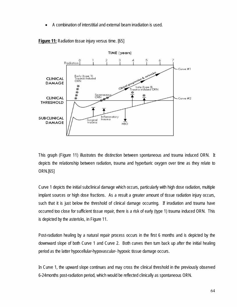
64
• A combination of interstitial and external beam irradiation is used. Figure 11: Radiation tissue injury versus time. [65]
This graph (Figure 11) illustrates the distinction between spontaneous and trauma induced ORN. It depicts the relationship between radiation, trauma and hyperbaric oxygen over time as they relate to ORN.[65] Curve 1 depicts the initial subclinical damage which occurs, particularly with high dose radiation, multiple implant sources or high dose fractions. As a result a greater amount of tissue radiation injury occurs, such that it is just below the threshold of clinical damage occurring. If irradiation and trauma have occurred too close for sufficient tissue repair, there is a risk of early (type 1) trauma induced ORN. This is depicted by the asterisks, in Figure 11. Post-radiation healing by a natural repair process occurs in the first 6 months and is depicted by the downward slope of both Curve 1 and Curve 2. Both curves then turn back up after the initial healing period as the latter hypocellular-hypovascular- hypoxic tissue damage occurs. In Curve 1, the upward slope continues and may cross the clinical threshold in the previously observed 6-24months post-radiation period, which would be reflected clinically as spontaneous ORN.
65
In Curve 2, the initial subclinical damage was far less to commence with, such that the upward slope will take many years to cross over the clinical threshold line. However, with each passing year the tissues become more fibrosed with increasing hypoxia-hypocellularity-hypovascularity placing the patient closer to the clinical threshold line for ORN should surgical trauma occur. The impact of hyperbaric oxygen (HBO) on Curve 2 is seen as a vertical downward arrow. The fibroplasia and angiogenesis induced by hyperbaric oxygen allows for the repair capability of the previously irradiated tissue to be increased. Hyperbaric oxygen is the intermittent daily inhalation of 100% oxygen at a pressure greater than 1 atmosphere absolute (ATA). ORN initially manifests clinically as an erythematous change of the overlying mucosa which eventually ulcerates to reveal necrotic bone underneath. These small asymptomatic bone exposures can remain stable in the long-term and heal with conservative management. Occasionally there is progression to severe necrosis with the potential for devitalised bone fragments to sequestrate or for a fistula to occur, which may lead to a spontaneous bone fracture. This may then necessitate surgical intervention and reconstruction. [34, 67, 69] The stages and management of established ORN is discussed in section 2.10.3. As there is no single diagnostic test available, the diagnosis of ORN is based on patient history and presenting clinical signs and symptoms. These may include: [46, 62, 72]
• Severe pain
• Ulceration and necrosis of the overlying mucosa
• The presence of non-healing and exposed necrotic bone in the irradiated area
• Orofacial fistulae
• Suppuration
• Pathologic fracture
• Repeated infections. A study by Reuther et al in 2003 reviewed 830 head and neck tumour patients who had received radiotherapy between 1969 and 1999. They identified an incidence of ORN of 8.2% with a more than three fold higher rate in males compared to females.[67] Vudiniabola et al in 2000, identified 17 cases of facial bone ORN (15 in the mandible, and 2 in the temporal bone) during a nine year period from 1987 to
66
1996. This was 1.2% of all cases of head and neck cancer treated with radiotherapy. This study showed different patterns of ORN with the different facial bones, with the mandible identified as the most commonly involved facial bone. [73] A study by Epstein et al in 1997 reinforced the view that ORN can occur at any period of time after radiation therapy, and that patients remained at risk for extended periods of time, often indefinitely.[72]
The oral sequelae of head and neck radiotherapy may cause substantial problems during and after radiation treatment, and may be one of the major factors influencing the patient’s quality of life.
2.6 PREVENTION AND TREATMENT OF THE CONSEQUENCES OF HEAD & NECK RADIOTHERAPY
Radiation induced oral side-effects can often be reduced to some degree with appropriate prevention and/or treatment. To successfully achieve this, the head and neck oncology team work with the dental practitioner to ensure that patients who undergo head and neck radiotherapy receive specific oral care, education and dental treatment with respect to preventive protocols. The early involvement of the dental practitioner in the development of preventive and therapeutic strategies, together with education and post-treatment rehabilitation or reconstruction of the patient is vital in addressing quality of life issues, especially with respect to the oral consequences of head and neck radiotherapy. Most treatment protocols for the management of these oral sequelae are based on clinical experience and anecdotal evidence only, with very few evidence-based clinical practice guidelines available.[74] As a result there is a great diversity in the supportive care policies and preventive approaches available. However, a crucial factor to the success of any preventive and treatment regime used by the dental practitioner is the compliance of the patient. 2.6.1 Mucositis At present the management of mucositis is limited to: [46]
• The reduction of severity through symptomatic relief via the application of oral care management programs
67
• The relief of pain and discomfort
• The identification of strategies to eliminate micro-organisms involved in the development and promotion of radiation mucositis.
Oral care management programs do provide some symptomatic relief to patients experiencing oral mucositis. There are currently no evidence-based preventive or therapeutic protocols based on randomly controlled trials published in the literature. Most of these oral care management programs are based on anecdotal clinical experience, and include: [46, 50, 74]
• Removal of mucosal irritating factors
• The cleansing of the oral mucosa
• Maintenance of mouth and lip moisture
• Relief of mucosal pain and inflammation
• Prevention and treatment of oral infection
• Discouraging of use of removable prosthetic appliances during radiotherapy treatment
• Discouraging smoking or alcohol consumption both during and after radiotherapy treatment
• Limitation of ingestion of spicy or acidic foods.
Oral bactericidal or bacteriostatic chemotherapeutic agents applied topically are recommended for maintenance of oral hygiene during radiotherapy. Chlorhexidine gluconate is commonly recommended for use during radiotherapy to maintain oral hygiene, but has no proven ability to control radiation mucositis.[50, 75] Benzydamine hydrochloride (‘Difflam’) is the only oral chemotherapeutic agent available which has been shown in multi-centre double blind trials to reduce mucositis and pain in patients with head and neck cancer.[62] Other management strategies recommended for the symptomatic relief of mucositis are dependant on the use of : [46]
• Mucosal coating agents
• Topical anaesthetics
• Systemic analgesics.
68
However, the treatment of established oral mucositis remains a challenge. Due to the significant clinical and economic impact, there has been a substantial increase in clinical research for interventions to reduce or prevent oral mucositis during the past decade. These include: [46]
• Growth factors e.g. recombinant keratinocyte growth factor, granulocyte macrophage colony stimulating growth factor
• Anti-inflammatory agents e.g. prostaglandins E1 and E2
• Topical coating agents or mucosal barriers
• Nutritional supplements and antioxidants.
2.6.2 Taste Loss The prevention of loss of taste is possible to achieve through the use of either direct shielding of the healthy taste buds during radiation treatment, or by placement of the taste buds outside of the radiation field by the use of repositioning devices. Most taste loss experienced as a result of head and neck radiotherapy will return to near normal levels within 12 months of the completion of treatment. The use of zinc containing nutritional supplements may assist with the acceleration of taste improvement during the post-radiotherapy period for those patients who are left with some degree of hypogeusia post-radiation.[46] 2.6.3 Hyposalivation The prevention or limitation of salivary gland damage as a result of radiotherapy should be considered when developing the treatment plan for radiation beam arrangement and radiation fields. Exclusion of both major and minor salivary glands from irradiation fields, especially the parotid and submandibular glands, will avoid the significant direct and indirect oral sequelae associated with hyposalivation and xerostomia.[46, 50, 51] Newer fractionation regimes such as hyper-fractionation and accelerated fractionation, together with the potential impact of newer radiation technology including 3-dimensional conformal and intensity modulated radiotherapy, plus the availability of functional imaging techniques (PET-positive emission tomography) may assist in the reduction of radiation exposure to the salivary glands.[34, 46, 50] Direct radioprotection is also possible through the use of drugs such as Amifostine, when administered systemically during treatment.[34, 46]
69
Often it is not possible to avoid radiation exposure to the salivary glands, and hyposalivation is the inevitable outcome. When this does occur current management options are limited to the: [46]
• Stimulation of any residual secretory capacity of the affected salivary gland(s) through the use of o Gustatory sialagogues o Masticatory sialagogues o Pharmacologic sialagogues
• Use of saliva substitutes
• Use of preventive regimes to limit potential damage to the dentition and mucosa as a result of the altered salivary flow and composition.
The stimulation of any residual secretory capacity of affected salivary glands may be possible through the use of systemic cholinergic sialagogues such as Pilocarpine. It’s success is dependant on the patient taking the drug continuously for a minimum period of 90 days, and the salivary gland having some residual functional ability. The side-effects of Pilocarpine may include reduced visual acuity, cardiovascular effects and gastro-intestinal discomfort. Gustatory or masticatory stimuli such as sugar-free chewing gum have been used successfully in a limited capacity to increase saliva secretion. Unfortunately the success of sialagogues is usually minimal, as the resultant increase in the saliva secretion is insufficient to address the significant oral dryness and the associated mucosal and dental problems.[46, 50, 56, 62] Saliva substitutes such as artificial saliva based on either carboxymethylcellulose or mucin are available. Mucin based saliva substitutes are the most commonly preferred by patients due to their: [46, 50]
• Rheological and wetting properties
• Potential to restore healthy oral microflora
• Prevention of demineralisation
• Remineralisation potential in the presence of fluoride and calcium.
However, these saliva replacements are usually a poor substitute in the long-term. The simplest and most common solution commonly chosen by xerostomic patients is the frequent moistening of the mouth with water. The main disadvantage of this is that there is a necessity for frequent application due to water’s poor retention properties.
70
Based on a review of the literature by Vissink et al in 2003 [46] the following recommendations have been made for the treatment of hyposalivation:
• Severe hyposalivation: o Saliva substitutes with gel like properties (mucin based)
• Moderate hyposalivation: o Gustatory or pharmacologic stimulation of residual salivary secretion, if this does not
work then the use of saliva substitute
• Slight hyposalivation o Gustatory or pharmacologic stimulation.
2.6.4 Radiation caries Vissink et al have recommended that sound or adequately restored teeth should not be extracted prior to radiotherapy in a patient who understands the importance of, and has the capacity to perform, good oral hygiene in the presence of post-radiotherapy hyposalivation. The exception to this is if the teeth which will be within the high dose radiation field: [46]
• have a need for substantial restorative, periodontal or endodontic treatment
• have moderate to severe periodontal disease with pocket depths greater than 5mm
• have root tips in situ which are not fully covered by alveolar bone
• are impacted or incompletely erupted and are in contact with the oral environment
• are in close proximity to the tumour. Radiation caries is mainly the indirect effect of irradiation induced changes to the salivary gland tissues that result in hyposalivation, altered salivary composition, a shift in the oral microflora towards cariogenic bacteria and dietary changes to compensate for the altered oral condition. (Figure 8) [50] Based on clinical experience and empirical evidence, it is possible to achieve caries prevention despite hyposalivation/xerostomia if patients comply with the daily application of 1% neutral fluoride in conjunction with meticulous oral hygiene. In addition the restriction of cariogenic foods and the use of remineralisation mouth rinses assist in the caries prevention process. Radiation caries is a lifelong risk to these patients, and as such a lifelong commitment is required by the patient to maintain the preventive regime.[46, 50]
71
2.6.5 Periodontal disease Optimal oral and periodontal hygiene must be maintained indefinitely post-radiotherapy due to the decreased healing ability of the periodontium following radiotherapy. The risk for the development of periodontal disease and possibly ORN as a consequence, is reduced in patients who maintain good oral hygiene, and regularly seek professional dental hygiene/periodontal treatment.[34] 2.6.6 Trismus Patients at risk of the development of trismus should be educated on the application of mobilisation exercises to maintain maximum oral opening and jaw mobility. It is imperative that these exercises commence as soon as possible, as the prevention of trismus is paramount. Despite this, some patients still develop trismus, and in these cases the exercise program needs to be intensified and if necessary combined with physiotherapy in an attempt to regain lost inter-arch distance. The use of dynamic bite opener appliances such as the Therabite® [Atos, Medical AB, Sweden] may improve trismus through repetitive passive stretching of affected masticatory muscles. Patient compliance and perseverance is a critical component of the exercise program’s success because dramatic results are usually not achieved in the short-term.[46, 53] Established trismus with a mouth opening of less than 20mm which remains refractory to sustained stretching exercises can be addressed surgically. This requires the use of fibre-optic intubation for safe general anaesthesia, a stepwise muscle detachment, intra-operative stretching and mobilisation, and usually a coronoidectomy. This must be followed by immediate and prolonged muscle exercise in the post-operative period. There is the risk that this surgical approach may make the condition worse if inexpertly provided or if the patient does not comply with the post-surgical exercise program. It also increases the risk of ORN in irradiated patients. 2.6.7 Osteoradionecrosis Osteoradionecrosis is a potentially serious complication of head and neck irradiation and is difficult and time consuming to manage. It is a lifelong threat, and therefore patients require a comprehensive dental examination prior to radiotherapy, with close monitoring and regular review subsequently. It is imperative that the prevention of ORN is achieved, with prophylactic measures put in place should dental extractions or oral surgical procedures be required.
72
The risk of ORN is increased if the patient has had:[76, 77]
• Internal radiotherapy
• A radiotherapy dose greater than 50Gy to 55Gy
• Radiotherapy which includes the mandible
• No hyperbaric oxygen therapy prior to surgical intervention
• Radiotherapy completed more than 6 months ago
• Trauma
• Chemotherapy
• Malnutrition
• A history of tobacco smoking or alcohol abuse which remains unreformed. In view of the significant risks associated with ORN, pre-treatment dental assessment is critical, with consideration in particular given to: [46, 51]
• The pre-radiotherapy dental status
• The radiotherapy treatment plan, including radiation dose, fractionation schedule, field plan, and any adjunctive therapy(s)
• Extraction(s) required, in particular to o ensure an atraumatic procedure is provided, with careful tissue handling and primary
closure o allow sufficient healing time prior to the commencement of radiotherapy, a minimum of
10-14 days, preferably 21 days. In addition, the following should be considered as part of the pre-treatment assessment: [76, 77]
• Total radiation dose greater than 66Gy increases the risk of ORN
• Hyperbaric oxygen treatment should be given if radiotherapy doses greater than 50Gy are used and future dental treatment(s) necessitating trauma to the bone is required post irradiation
• Surgery should not be carried out during radiotherapy, and avoided during mucositis
• Implant placement should be deferred until 8 months post irradiation
• Pressure on mucosal surfaces should be minimized if a prosthesis is provided
• Immediate loading techniques should not be used
• Strict asepsis must be maintained
• Antimicrobial cover should be considered.
73
A clinical practice guideline for the preventive management of irradiated patients was developed by Marx et al in 1987. [65] This identified that:
• The removal of teeth or any sort of surgical wounding during radiotherapy is not recommended
• Any surgical procedures in irradiated tissue should be only be undertaken after 20 sessions of pre-surgical Hyperbaric oxygen therapy, and 10 sessions of post-surgical Hyperbaric oxygen therapy.
This clinical practice guideline was based on his previous work on the pathogenesis of ORN [68] and also the role of hyperbaric oxygen in the prevention of ORN following tooth extraction.[78] Marx identified that ORN was a radiation induced non-healing and hypoxic wound, and not osteomyelitis of irradiated bone, as was previously thought. Micro-organisms were proven to play a very minor role in the pathophysiology of ORN, acting only as surface contaminants and not as infective agents. The 1985 [78] article built upon this work by clearly demonstrating that the incidence of ORN following tooth extraction if prophylactic hyperbaric oxygen was given was reduced to 5.4% from 29.9% if antibiotics only were provided. These significant pieces of work are the scientific basis on which the treatment for ORN using adjunctive hyperbaric oxygen to reverse the hypoxic-hypocellular-hypovascular tissue is based.[68, 70, 78] This will be discussed in detail in section 2.10.3.
The prosthodontic tools to restore an edentulous mandible consist of either: 2.7 IMPLANT MANDIBULAR PROSTHESES
• A conventional tissue supported removable complete denture, or
• An implant and tissue supported removable complete overdenture, or
• A retained fixed prosthesis. Clinical trials have been published in the dental and medical literature which has demonstrated the viability, safety, superior functional performance and increased patient satisfaction with implant supported and tissue supported overdentures, particularly when compared with traditional removable complete dentures.[79-82]
74
The restoration of edentulous spaces with endosseous implants has evolved dramatically from 1965, when the first patient received implants in Brǻnemark’s Gothenburg clinic. The success of this original work was based on the restoration of the edentulous mandible, with implant placement in the inter-foramina area. The rationale for implant placement in this site was that:
• implants of substantial length could be used, or bicortical implants
• the implant positioning anterior to the mental foramen ensured that there was no risk of trauma to the inferior alveolar nerve
• it would reduce the risk of complications as a result of flexure of the posterior mandible, therefore increasing the chance of successful osseointegration. [83] 2.7.1 Definitions An endosseous dental implant as defined in the Glossary of Prosthodontic Terms is: [84]
“a device placed into the alveolar and/or dental bone of the mandible or maxillae, and transecting only one (1) cortical plate” or, “that portion of the dental implant that provides anchorage to the bone through the process of tissue integration.”
The endosseous dental implant is comprised of the:
• endosseous dental implant body
• endosseous dental implant abutment
• endosseous dental implant abutment element The Glossary of Prosthodontic Terms provides the following definitions for the implant componentry: [84] The endosseous dental implant abutment-
“connects to the dental implant (by means of screws, thread/screw interfacing, compression/luting agent)….passes through the oral mucosa and serves to support and/or retain the dental prosthesis or maxillofacial prosthesis” and is “that portion of the dental implant that passes through the oral mucosa and provides connection between the endosteal dental implant body and the prosthesis.”
75
The endosseous dental implant abutment element is- “any component which is used to secure either the dental implant abutment to the dental implant or the prosthesis to the dental implant abutment.”
2.7.2 Prosthodontic classification system (American College of Prosthodontists) The American College of Prosthodontists has developed a classification system for both partial edentulism [85] and complete edentulism [86] both of which are based on diagnostic findings. This framework was designed to identify increasing levels of diagnostic and treatment complexity. In the Classification system for partial edentulism, four categories are defined, with Class IV representing the most complex clinical situation(s). Patients are assigned to the classification by specific diagnostic criteria. Patients assigned to Class IV, include those with: [85]
• edentulous areas with acquired/congenital maxillofacial defects and/or
• severe oral manifestations of systemic disease, including sequelae from oncologic treatment and/or
• an edentulous mandible opposing a partially edentulous or dentate maxillae. In the Classification system for complete edentulism, four categories are defined ranging from Class I to Class IV. Class IV represents the most complex and high risk situations which each class differentiates by specific diagnostic criteria. Patients assigned to class IV represent those with the most debilitating edentulous oral conditions, including those with: [86]
• major conditions requiring pre-prosthetic surgery
• complex implant placement
• surgical correction of dentofacial abnormalities
• hard tissue augmentation required
• soft tissue augmentations required
• acquired or congenital maxillofacial defects
• severe oral manifestations of systemic disease or conditions. Both of these classification systems have been defined to identify those more complex patients who would most likely require treatment by a specialist or a practitioner with additional training and
76
experienced in these advanced techniques. Post-treatment oral cancer patients clearly fit the Class IV definition for complex patients. 2.7.3 Classification of oral implants There are a large number of oral implants utilised in dental practice. The classification of these implants can be based on either the placement modality or implant designs. Table 5 provides an overview of the classification of implants available for use in dentistry. Table 5Subperiosteal implants
: Classification of oral implants [87]
A metal framework which fits over the atrophied bone, providing the equivalent of multiple tooth roots.
Ramus frame implants A ‘hybrid’ between subperiosteal and endosseous implants. Endosseous/endosteal implants An implant placed directly into the bone like tooth roots.
These can include: • pins and needles • blades and disks • root formed analogues
Transosteal implants Mandibular staple bone plate and transmandibular implants 2.7.4 Standard of care for the edentulous mandible Edentulous patients with severely resorbed mandibles often experience ongoing problems with their conventional dentures, because of an impaired load bearing capacity which presents as problems with pain during mastication, or insufficient stability and retention of the dentures.[79] In 2003 it was controversially concluded that the evidence available at the time suggested that the restoration of the edentulous mandible with a conventional removable denture should no longer be considered to be the treatment of choice. An implant overdenture was proposed as the standard of care.[82, 88] According to the Glossary of Prosthodontic Terms the definition of an overdenture is: [84]
“a removable partial or complete denture that covers and rests on one or more remaining natural teeth, roots and/or dental implants” or
77
“a prosthesis that covers and is partially supported by natural teeth, tooth roots and/or dental implants.”
The ideal number of implants required for a lower denture remains controversial. In a two implant system the retention, support and stability of the overdenture is provided by both the implants and mucosa. In a four implant system the retention, support and stability is primarily provided by the implants.[89, 90] To date, there is no study which confirms “the superiority of 4 compared to 2 implants for mandibular overdentures with regard to implant survival”.[90] A comprehensive literature review by Allen et al in 2005 [91] identified that numerous studies have documented the efficacy of restoring the edentulous mandible with two (2) implants in the anterior mandible as retention for a complete overdenture. This was considered to be the treatment of choice for the edentulous mandible because:
• the overall success of the osseointegration of dental implants is high and as a result implant restoration of edentulous spaces has become the treatment of choice
• mandibular overdentures with two (2) ball implants demonstrated the greatest retention after 10 years
• at 10 years the overdentures were functioning well but the required maintenance of implant overdentures is substantial and should not be underestimated.
Ball abutments have been found to be the ideal attachments for overdentures in a two implant system. This is primarily because their placement are less technique sensitive and they are easier to clean, reducing the potential for mucosal hyperplasia.[90, 92] For acquired intra-oral defects such as following ablative surgery for oral cancer, the anatomic conditions may lead to an inability to successfully wear a conventional removable complete denture. Implant overdentures have ensured that in these situations, the patient is able to be reconstructed so as to achieve structural, functional and aesthetic rehabilitation. In addition the following criteria may be achieved: [80]
• the removable prosthesis allows for inspection of the surgical site
• implants are ideally placed where adequate bone is present
• the denture base replaces both soft and hard tissue
78
• the lip and facial support can be provided by a labial denture flange
• oral hygiene is facilitated, and
• alteration of the denture is possible, if required. The clinical requirements for an implant mandibular overdenture is the same for ‘healthy’ patients as for those with acquired intra-oral defects, however the predictability of success is often reduced due to: [80]
• inadequate bone quality or quantity
• presence of skin grafts
• primary implant placement i.e. immediate implant placement during grafting procedure(s)
• poor alignment of implants due to reduced bone quality
• radiation therapy, and/or
• reduced salivary flow. Minor complications associated with implant mandibular overdentures are common, and may be associated with a high burden of maintenance.[89, 92] The most common complications include:
• breakage of retentive clips on bar attachments
• peri-implant mucosal problems
• implant and acrylic resin component fractures. It is important to recognise that the provision of a stable implant prosthesis does not predictably result in an improvement in either subjective or objective oral function. This is particularly so if the patient has undergone either radiotherapy, ablative surgery or both resulting in an altered oral anatomy, reduced mandibular and tongue movements, as well as motor and/or sensory deficits. [93] 2.7.5 Osseointegration 2.7.5.1 Definition There are many definitions of osseointegration available in the literature, however the most quoted definition regarding the direct relationship between bone and implant is that by Brǻnemark and Albrektsson:
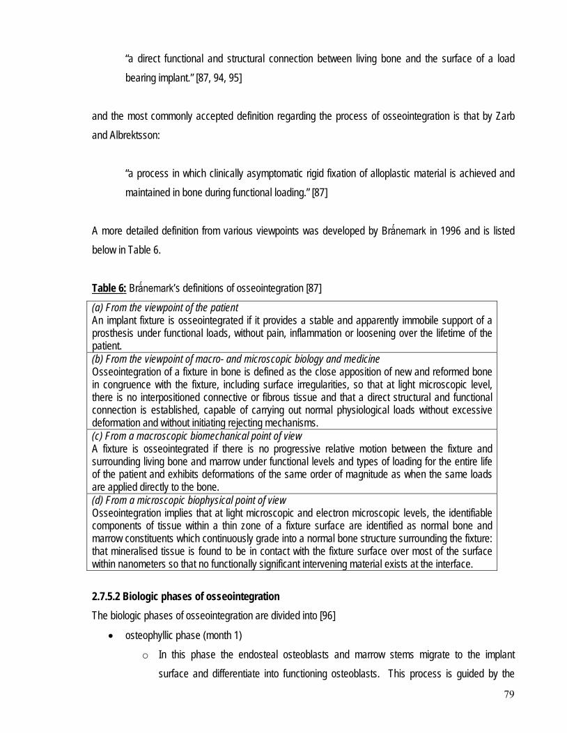
79
“a direct functional and structural connection between living bone and the surface of a load bearing implant.” [87, 94, 95]
and the most commonly accepted definition regarding the process of osseointegration is that by Zarb and Albrektsson:
“a process in which clinically asymptomatic rigid fixation of alloplastic material is achieved and maintained in bone during functional loading.” [87]
A more detailed definition from various viewpoints was developed by Brǻnemark in 1996 and is listed below in Table 6. Table 6(a) From the viewpoint of the patient
: Brǻnemark’s definitions of osseointegration [87]
An implant fixture is osseointegrated if it provides a stable and apparently immobile support of a prosthesis under functional loads, without pain, inflammation or loosening over the lifetime of the patient. (b) From the viewpoint of macro- and microscopic biology and medicine Osseointegration of a fixture in bone is defined as the close apposition of new and reformed bone in congruence with the fixture, including surface irregularities, so that at light microscopic level, there is no interpositioned connective or fibrous tissue and that a direct structural and functional connection is established, capable of carrying out normal physiological loads without excessive deformation and without initiating rejecting mechanisms. (c) From a macroscopic biomechanical point of view A fixture is osseointegrated if there is no progressive relative motion between the fixture and surrounding living bone and marrow under functional levels and types of loading for the entire life of the patient and exhibits deformations of the same order of magnitude as when the same loads are applied directly to the bone. (d) From a microscopic biophysical point of view Osseointegration implies that at light microscopic and electron microscopic levels, the identifiable components of tissue within a thin zone of a fixture surface are identified as normal bone and marrow constituents which continuously grade into a normal bone structure surrounding the fixture: that mineralised tissue is found to be in contact with the fixture surface over most of the surface within nanometers so that no functionally significant intervening material exists at the interface. 2.7.5.2 Biologic phases of osseointegration The biologic phases of osseointegration are divided into [96]
• osteophyllic phase (month 1) o In this phase the endosteal osteoblasts and marrow stems migrate to the implant
surface and differentiate into functioning osteoblasts. This process is guided by the
80
bone morphogenetic protein, which is released during the surgical preparation of the bone to receive the implant. Growth factors such as platelet derived growth factor and transforming growth factor-β as incorporated into the blood clot.
• Osteoconductive phase (month 2-4) o In this phase the osteoblasts migrate along the metal surface of the implant. This is
termed bone conduction.
• Osteoadaptive phase (month 4 onwards) o After 4 months the bone-to-metal interface is maintained and matured by a continual
bone resorption-bone apposition remodelling cycle. There is no net gain or loss of bone about the metal surface, unless disease or trauma develops.
Clinical studies which have made comparisons between the degree of bone-to-metal contact with implants have ascertained that:
• Normal bone achieves a 47% bone-to-metal contact
• Irradiated bone achieves a 39% bone-to-metal contact
• Grafted bone achieves a 72% bone-to-metal contact. [96] 2.7.6 Patient screening and treatment planning The use of an osseointegrated implant mandibular overdenture after surgical intervention and mandibular reconstruction is an opportunity for functional rehabilitation. However, the decision making process with respect to patient selection criteria can be complex, and should include consideration of: [93]
• Tumour prognosis
• Method of surgical reconstruction
• Secondary effects of radiotherapy
• Decreased mandibular opening (trismus)
• The amount and quality of bone available
• Alcohol, tobacco or other substance abuse
• Patient motivation
• Neuromuscular, sensory and mobility status of tongue and circumoral soft tissues
• Inter-arch space and mandibulo-maxillary discrepancies
• Bulk and quantity of peri-implant soft tissues
81
• Bulk and quantity of denture bearing soft tissues. There are some systemic health problems which may be major risk factors for implant placement. It is important that attempts are made to explore the possibility of improving or stabilising these medical conditions if possible, so as to not unnecessarily exclude the provision of implant to patients with relative contraindications to implant placement.[97, 98] An absolute contraindication to implant placement is defined as [99]
“health conditions that have the potential to jeopardise the patients overall health and safety, and seriously compromise the survival of implanted systems, causing residual chronic complications.”
These include:[77, 99, 100]
• Recent myocardial infarction
• Recent valvular prosthesis
• Severe renal disease
• Treatment resistant diabetes mellitus
• Generalised secondary osteoporosis
• Intravenous bisphosphonates [101]
• Treatment resistant osteomalacia
• Active cancer therapy i.e. radiotherapy or chemotherapy in progress
• Severe hormone deficiency(s)
• Neuro-psychiatric disorders
• Drug addiction
• Chronic or severe alcoholism
• A current heavy smoking habit (>20 cigarettes/day) Smoking cessation should be recommended to all potential implant candidates. It is considered to be one of the most severe limitations to implant success because it damages the angiogenic mechanisms for the formation and maintenance of peri-implant and periodontal soft tissues. [99] A retrospective report identified that smoking increased the risk of implant failure by as much as 2.5 times. In particular, persistent tobacco use was found to decrease the ability of bone and other periodontal tissues to adapt
82
over time therefore compromising all stages of implant treatment after fixture uncovering.[100, 102] In fact, rather that affecting the initial process of osseointegration, the negative impact of smoking seemed to occur predominantly after 2nd stage implant surgery.[103] A retrospective cohort study by Moy et al in 2005 assessed dental implant failure rates with associated risk factors by investigating 4680 implant placed in 1140 patients between January 1982 and January 2003. They identified that smoking, diabetes, head and neck radiotherapy and post-menopausal oestrogen therapy were associated with a significantly increased failure rate. In the mandible this failure rate was reported as 4.93%. They concluded that there were no absolute contraindications to implant placement, and that risk factors should be considered during the treatment planning process and factored into the informed consent process.[104] Relative contraindications to implant placement are determined by their [99]
“direct relation with the nature and severity of the systemic disorder, and whether or not they can be satisfactorily corrected prior to surgery.”
These include:[77, 99, 102]
• AIDS and HIV seropositivity
• Prolonged use of corticosteroids
• Osteoporosis and oral bisphosphonates [101]
• Immunocompromised states
• Diabetes
• Cardiac disease
• Disorders of the phosphocalcic metabolism i.e. bone disease, hypothyroidism
• Haematopoietic disorder i.e. bleeding disorders
• Buccopharyngeal disorders
• History of cancer therapy i.e. radiotherapy or chemotherapy
• Mild renal disorder
• Multiple endocrine disorder
• Psychological disorders and psychoses
• Unhealthy life style
• Smoking history
83
• Lack of understanding and motivation
• Interleukin-1 genotype associated periodontal disease A comprehensive review article by Wood et al [103] which reported on English language articles published between 1969 and 2003, looked at evidence-based treatment planning for dental implants and impact on osseointegration based on:
• Systemic host factors o age o gender o various medical conditions, including cancer modalities o patient habits, in particular tobacco smoking
• Local factors o quantity and quality of bone and soft tissue o presence of past or present infection o occlusal forces
• Prosthetic design factors o number and arrangement of implants o size and coatings of implants o cantilevers and connectors to natural teeth.
This review concluded that there was no systemic factor or habit which was an absolute contraindication to the placement of osseointegrated implants in an adult patient, but that the cessation of smoking could improve the outcome significantly. The most important local patient factor for successful treatment was found to be the quality and quantity of bone available at the implant site. The article did highlight that for head and neck oncology patients, there was concern regarding
• the ability of irradiated tissues to support osseointegration
• the effects of systemic chemotherapy on the bone quality. A more recent review by Scully et al [77], in 2007 concurred with Wood’s conclusion. Scully believed that there were a number of medical problems which had the potential to increase the risk of failure and/or complications. He felt that it was imperative for an individualised patient assessment, prior to implant provision. This was supported by Zitzmann et al in 2008.[98]
84
A survey conducted in the United Kingdom in 1999 with respect to the provision of osseointegrated implants in the National Health Service Hospital Services, identified that the majority of Consultants felt that smoking, psychoses and previous irradiation were the most important medical factors which contraindicated patient selection for implant prostheses.[105] 2.7.7 Success criteria The success criteria for osseointegration as determined by the 1st European workshop on Periodontology in 1994 were [87]
• An absence of mobility
• An average radiographic bone loss of less than1.5mm during the first year of function, and less than 0.2mm annually thereafter
• An absence of pain or paraesthesia. The now commonly quoted success criteria for the evaluation of bone loss associated with osseointegrated implants was originally proposed by Albrektson in 1986. However, it is difficult to apply from a technical perspective due to limitations in measuring annual progression of 0.2mm on radiographs.[106] The definition of a successful implant is “an osseointegrated dental implant that is successfully restored and contributing to the functional success of a dental restorative treatment or one that could be used for such purposes.” [103] There are six factors which impact on the ability to obtain osseointegration: [107]
• Material biocompatibility
• Macrostructure of the implant fixture
• Microstructure of the implant fixture
• A gentle and atraumatic surgical technique employed in implant fixture placement
• The status of the implant bed, preferably one which is well vascularised
• Loading conditions. In order for an implant to be considered successful it needs to meet certain criteria. If the following criteria are not achieved, then the implant is considered to be surviving: [87]
85
• Function (an ability to chew)
• Tissue physiology (presence and maintenance of osseointegration)
• Absence of pain and other pathological processes
• User satisfaction (aesthetics and absence of discomfort) 2.7.8 Implant failure An implant failure is essentially any implant which is not fulfilling the success criteria for osseointegration as determined by the 1st European workshop on Periodontology in 1994. It may be defined as:
“the first instance at which the performance of the implant measured in some quantitative way, falls below a specified acceptable level.” [87]
As identified in Table 7, failures can be divided into: [87, 108]
• Biological failure – failures related to biological processes to establish or maintain osseointegration.
o early biological failure failure due to an interference in the healing process refers to an implant that fails to osseointegrate prior to the 2nd stage of surgery
or uncovering of the implant [103] o late biological failure
failure due to a breakdown in osseointegration refers to the loss of osseointegration or mechanical failure of an implant, after
2nd stage surgery [103]
• Mechanical failure – failures of implant componentry, including failures of implants, coatings, connecting screws and prostheses
• Iatrogenic failure – failure which occurs due to an implant not being used due to poor positioning or nerve damage
• Inadequate patient adaptation failure – failures due to psychological, aesthetic or phonetic problems.
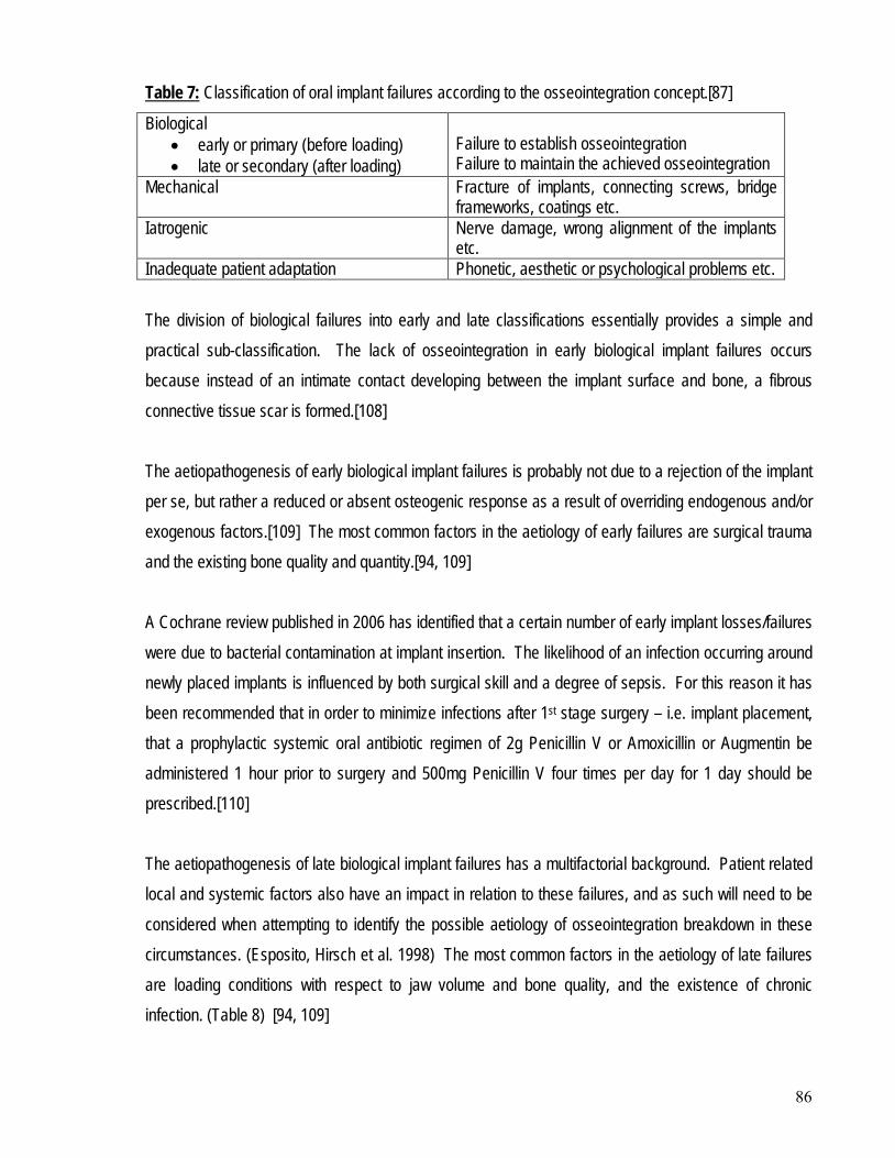
86
Table 7Biological
: Classification of oral implant failures according to the osseointegration concept.[87]
• early or primary (before loading) • late or secondary (after loading)
Failure to establish osseointegration Failure to maintain the achieved osseointegration
Mechanical Fracture of implants, connecting screws, bridge frameworks, coatings etc.
Iatrogenic Nerve damage, wrong alignment of the implants etc.
Inadequate patient adaptation Phonetic, aesthetic or psychological problems etc. The division of biological failures into early and late classifications essentially provides a simple and practical sub-classification. The lack of osseointegration in early biological implant failures occurs because instead of an intimate contact developing between the implant surface and bone, a fibrous connective tissue scar is formed.[108] The aetiopathogenesis of early biological implant failures is probably not due to a rejection of the implant per se, but rather a reduced or absent osteogenic response as a result of overriding endogenous and/or exogenous factors.[109] The most common factors in the aetiology of early failures are surgical trauma and the existing bone quality and quantity.[94, 109] A Cochrane review published in 2006 has identified that a certain number of early implant losses/failures were due to bacterial contamination at implant insertion. The likelihood of an infection occurring around newly placed implants is influenced by both surgical skill and a degree of sepsis. For this reason it has been recommended that in order to minimize infections after 1st stage surgery – i.e. implant placement, that a prophylactic systemic oral antibiotic regimen of 2g Penicillin V or Amoxicillin or Augmentin be administered 1 hour prior to surgery and 500mg Penicillin V four times per day for 1 day should be prescribed.[110]
The aetiopathogenesis of late biological implant failures has a multifactorial background. Patient related local and systemic factors also have an impact in relation to these failures, and as such will need to be considered when attempting to identify the possible aetiology of osseointegration breakdown in these circumstances. (Esposito, Hirsch et al. 1998) The most common factors in the aetiology of late failures are loading conditions with respect to jaw volume and bone quality, and the existence of chronic infection. (Table 8) [94, 109]
87
Table 8: Summary of the main clinical, radiographic and histologic characteristics of late implant failures.[109]
A review of the literature by Quirynen et al in 2002 on late implant failures related to infectious processes identified that the possible sources of direct bacterial contamination during surgery included: [94]
• Surgical instruments
• Gloves
• Saliva
• Peri-oral skin He concluded that the salivary load could be reduced by 95% by a pre-operative chlorhexidine rinse. This concept was supported in a comprehensive literature review by Wood et al in 2004. [103] They identified that optimum oral hygiene should be maintained around implants, and the use of pre-surgical chlorhexidine gluconate 0.12% has been recommended prior to both 1st and 2nd stage twice daily for 2 days. It has been found to:
• reduce the incidence of post-surgical infection
• reduce the incidence of early implant loss
• reduce bacterial contamination of collected bone debris for augmentation procedures. [110]
88
The aetiopathogenesis of all biological failures can be divided into endogenous factors and/or exogenous factors. Endogenous factors are essentially systemic or local factors or conditions which may impair the bone healing process in early failures, or interfere with the maintenance of osseointegration in late failures. Endogenous systemic factors include: [103, 109, 111]
• age
• genetics
• general health
• smoking
• intravenous bisphosphonate medications [101] Occasionally a consistent failure of several fixtures in one patient will occur. This has been described in the literature as the ‘cluster phenomenon’ and probably indicates that some genetic or systemic factor(s) are impacting on the osseointegration process.[103, 108, 109] Endogenous local factors include: [103, 108, 109, 111]
• bone quality, bone quantity and anatomical location
• bone grafting
• parafunctional habits
• local immune response and previous history of periodontal disease
• presence of an adequate band of attached keratinised soft tissue
• previous history of head and neck radiation therapy. Bone quality and quantity as well as the anatomical location of implant placement are the major influencing endogenous factors related to implant failure, independent of whether the implant is loaded or not. In general, higher implant failure rates have been reported in the literature for implants placed in the maxillae and also implants placed in the posterior segments of the mandible.[109]
This may in part be explained by the different type of bone located in these regions. A classification of bone has been made based on radiographic appearance and the resistance to drilling: [109]
• Type I bone
•
: the entire jaw is composed of homogenous compact bone
Type II bone: a thick layer of compact bone surrounds a core of dense trabecular bone
89
• Type III bone
•
: a thin layer of cortical bone surrounds a core of dense trabecular bone of favourable strength
Type IV bone
: a thick layer of cortical bone surrounds a core of low density trabecular bone of poor strength. Higher failure rates have been associated with this type of bone.[108]
A study was conducted on early implant failures to assess the influence of endogenous local factors on the occurrence of implant failure on 399 patients with 1263 implants placed between 1995 and 1999. The results of this study identified that: [108]
• Chemotherapy and radiotherapy of the oral tissues were significantly related to implant failure (P<0.01)
• A slightly higher success rate was noticed in the anterior mandible (97.9%) compared to the posterior mandible (96.3%)
• Heavy smoking predisposes to implant failure. A retrospective cohort analysis by Moy et al in 2005 highlighted the influence of bone type and smoking on implant failure. In this study a total of 4680 implants were placed in 1140 patients in a consistent manner by a single surgeon. The study came to the following conclusions: [104]
• In general low failure rates documented in the study reflected the predictability of dental implants
• Patients with a history of smoking had a failure rate of 20% with a 1.56 relative risk of failure compared to non smokers (P<0.05). From a life table analysis the majority of failures occurred in the first 12 months.
• Implant failures were higher in the maxillae compared to the mandible: o anterior mandible - 2.89% failure rate o posterior mandible – 5.89% failure rate o anterior maxillae – 6.75% failure rate o posterior maxillae – 9.26% failure rate.
Exogenous factors are either operator related or biomaterial related factors which also may impair or impact upon the bone healing process in early failures, or interfere with the maintenance of osseointegration in late failures. Most exogenous factors are strongly related to host factors as a result of reciprocal influences. Exogenous operator related factors include: [109]
• Operator experience and the surgical technique utilised with the risk of
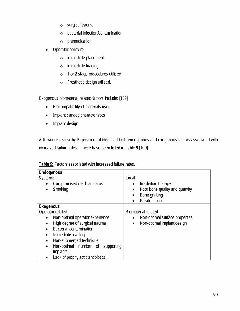
90
o surgical trauma o bacterial infection/contamination o premedication
• Operator policy re o immediate placement o immediate loading o 1 or 2 stage procedures utilised o Prosthetic design utilised.
Exogenous biomaterial related factors include: [109]
• Biocompatibility of materials used
• Implant surface characteristics
• Implant design A literature review by Esposito et al identified both endogenous and exogenous factors associated with increased failure rates. These have been listed in Table 9.[109] Table 9Endogenous
: Factors associated with increased failure rates.
• Compromised medical status Systemic
• Smoking
• Irradiation therapy Local
• Poor bone quality and quantity • Bone grafting • Parafunctions
Exogenous
• Non-optimal operator experience Operator related
• High degree of surgical trauma • Bacterial contamination • Immediate loading • Non-submerged technique • Non-optimal number of supporting
implants • Lack of prophylactic antibiotics
• Non-optimal surface properties Biomaterial related
• Non-optimal implant design
91
Little is known about the use of prophylactic antibiotic administration for reduction of bacterial contamination leading to implant failure. A study by Dent et al in 1997 examined 2641 implants. Their conclusion was that a higher failure rate was experienced in patients who did not have preoperative antibiotics, and that the provision of preoperative antibiotic prophylaxis significantly increased the survival rate of endoseous dental implants up to and including stage II implant surgery.[112] However, a study by Gynther et al in 1998 found the converse, with no significant difference identified with respect to early and late postoperative infections nor implant survival in patients who did not have preoperative antibiotics. They concluded that antibiotic prophylaxis for routine dental implant surgery offers no advantage for the patient.[113] The Cochrane Review on the use of antibiotics to prevent complications following dental implant treatment published in 2003 found that there were no randomly controlled trials which met its criteria. This review concluded that there was no systematic evidence to recommend either the continuation or cessation of antibiotic prophylaxis to prevent complications and failures of dental implants. [110] Even though endogenous factors are associated with a higher incidence of implant failure, this does not mean that patients considered being at higher risk should not receive the benefit of implant therapy.[109] The most common clinical parameters used for the evaluation of established implant failures include: [109]
• Clinical signs of early infection o swelling, fistulae formation, suppuration, mucosal dehiscence, osteomyelitis
• Pain or sensitivity
• Clinically discernible mobility. This is a cardinal sign of implant failure and includes o rotational mobility o lateral or horizontal mobility o axial or vertical mobility
• Dull percussion sound which is indicative of soft tissue encapsulation.
• Radiographic signs of failure e.g. peri-implant radiolucency. It is imperative that a standardised periapical radiograph is taken at regular intervals to assess the peri-fixtural bone height and detect for any radiolucency or progressive bone loss. Orthopantomographs have a limited value in monitoring implant conditions due to the inferior quality of image resolution and
92
an inability to modify the film angulation. Radiographic signs of failure are the primary tool for the non clinical detection of implant failure. While it is relatively simple to differentiate between successful and failed implants it is often quite difficult to identify failing implants. The most common clinical parameters used for the evaluation of failing implants include: [109, 114]
• Clinical signs of late infection o Hyperplastic soft tissue o Suppuration o Swelling, fistulation o Colour changes to marginal peri-implant tissues
• Bleeding on probing
• Sulcular bleeding index
• Pocket probing depth
• Mucosal recession
• Probing attachment levels
• Crevicular fluid analysis (Interleukin β)
• Microbial testing for periodontal pathogens. These periodontal parameters were introduced so as to better describe the state of the peri-implant tissues. The epidemiological data available in the literature on implant failure and success is primarily based on the Brǻnemark system because of the availability of long-term data on the performance of these implants. A summary of published articles which provided data on early and late implant failures of Brǻnemark implants used in various anatomical locations and clinical situations were analysed as part of a Literature review by Esposito et al in 1998, using a meta-analytical approach.[109] They concluded that:
• In a fully edentulous patient higher failure rates were seen in the overdentures for both early (5.9%) and total number of implant losses (12.8%). This was felt to be due to the fact that overdentures were favoured compared to fixed prosthesis in more critical clinical situations.
• In complicated situations including bone graft sites: o higher failure rates were seen (14.9%) than in other situations
93
o implants in the maxillae did not perform as well as mandible, except in the partially edentulous situation were they performed equally well
o the average failure incidence for both early and late failures was 3.6%. 2.7.9 Treatment of Complications Complications associated with dental implants can include: [115]
• Prosthesis instability
• Implant mobility
• Occlusal trauma
• Fractured implant componentry
• Pain
• Inflammation
• Infection
• Neuropathy Complications associated with implant provision may even include involvement of the implant in oral cancer. A case report published by Schache et al in 2008 identified macroscopic and microscopic evidence of the implant/mucosa interface being the likely route of entry for a bony [mandibular] extension of a squamous cell carcinoma.[116] A literature review encompassing articles from 1981 to 2001 by Goodacre et al identified the types of complications encountered with endosseous root form implants and associated implant prosthesis. Complications were divided into six (6) categories:[117]
• Surgical o neurosensory disturbances [118] o haemorrhage related complications o mandibular fractures
• Implant loss
• Bone loss
• Peri-implant soft tissue complications o fenestration and/or dehiscences o gingival inflammation/proliferation
94
o fistulaes
• Mechanical failures related to overdentures
• Aesthetic and/or phonetic complications. In this article by Goodacre et al, the most common complications related to implant overdentures were identified as: [117]
• Loosening of overdenture retentive mechanisms (33%)
• Reline of overdentures (19%)
• Overdenture clip/attachment fracture (16%). It is important that a clear differentiation be made between a failed implant(s) and the complications associated with failing implants. “Complications might indicate an increased risk of failure, but are either of temporary significance or amenable to treatment.” [109] Successfully osseointegrated implants are susceptible to complications which may lead to the loss of the implant(s), i.e. late biological failures. Common soft tissue complications include: [109, 115, 119]
• Peri-implant disease o this is the collective term for any inflammatory reaction(s) in the soft tissue surrounding
a functioning implant.
• Peri-implant mucositis o this is a reversible inflammation in the soft tissue surrounding a functioning implant
without any bone loss.
• Peri-implantitis o this is an inflammatory reaction with loss of supporting bone in the tissues surrounding a
functioning implant. Cochrane reports by Esposito et al in 2004 and 2006 identified that it is important to institute an effective preventive regimen or supportive therapy in order to maintain healthy tissues around oral implants. However, the report found that there was very little reliable evidence to identify the most effective intervention(s) for maintaining health around oral implants.[120, 121] These reports were subsequently updated in 2008 by Grusovin et al. In this last review it was found that there was very little reliable
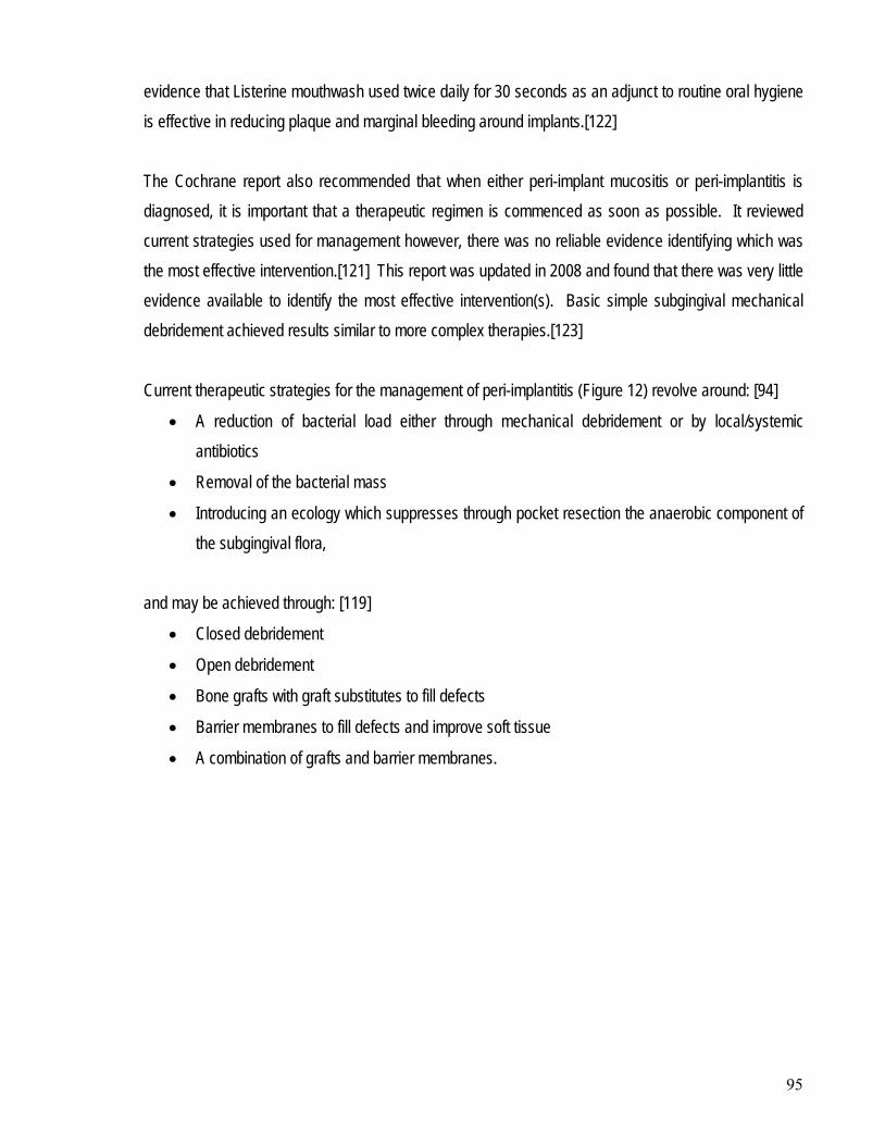
95
evidence that Listerine mouthwash used twice daily for 30 seconds as an adjunct to routine oral hygiene is effective in reducing plaque and marginal bleeding around implants.[122] The Cochrane report also recommended that when either peri-implant mucositis or peri-implantitis is diagnosed, it is important that a therapeutic regimen is commenced as soon as possible. It reviewed current strategies used for management however, there was no reliable evidence identifying which was the most effective intervention.[121] This report was updated in 2008 and found that there was very little evidence available to identify the most effective intervention(s). Basic simple subgingival mechanical debridement achieved results similar to more complex therapies.[123] Current therapeutic strategies for the management of peri-implantitis (Figure 12) revolve around: [94]
• A reduction of bacterial load either through mechanical debridement or by local/systemic antibiotics
• Removal of the bacterial mass
• Introducing an ecology which suppresses through pocket resection the anaerobic component of the subgingival flora,
and may be achieved through: [119]
• Closed debridement
• Open debridement
• Bone grafts with graft substitutes to fill defects
• Barrier membranes to fill defects and improve soft tissue
• A combination of grafts and barrier membranes.
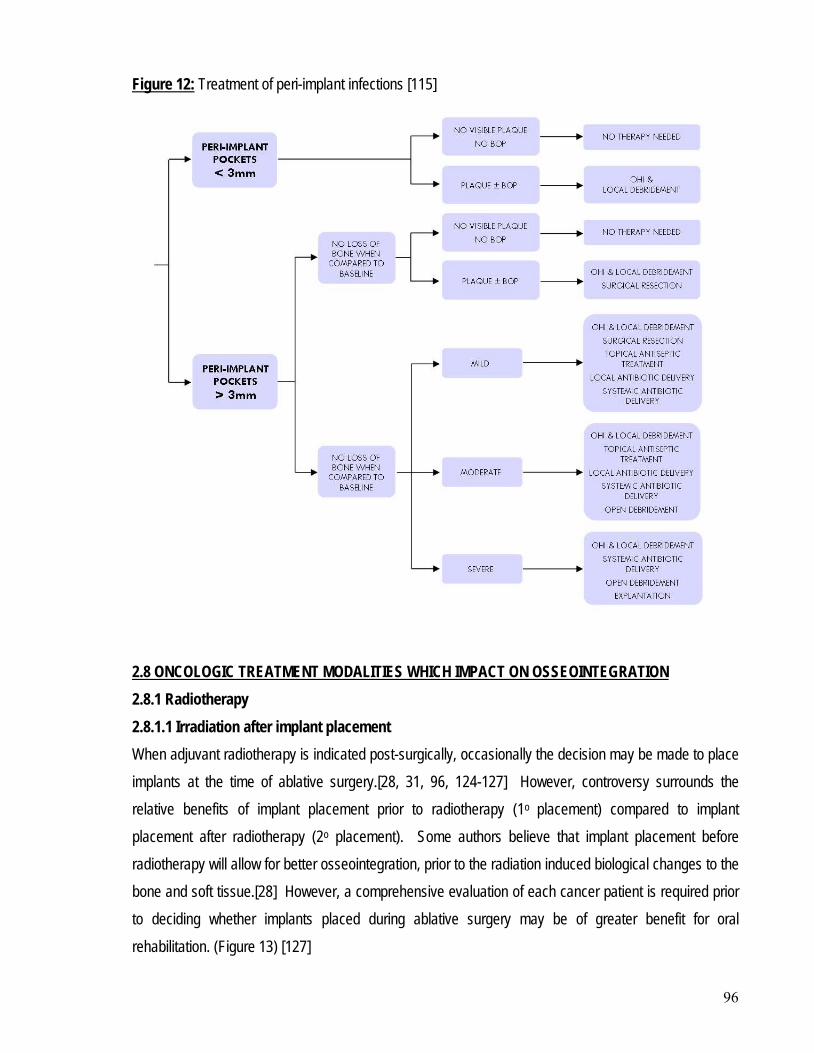
96
Figure 12: Treatment of peri-implant infections [115]
2.8.1 Radiotherapy 2.8 ONCOLOGIC TREATMENT MODALITIES WHICH IMPACT ON OSSEOINTEGRATION
2.8.1.1 Irradiation after implant placement When adjuvant radiotherapy is indicated post-surgically, occasionally the decision may be made to place implants at the time of ablative surgery.[28, 31, 96, 124-127] However, controversy surrounds the relative benefits of implant placement prior to radiotherapy (1o placement) compared to implant placement after radiotherapy (2o placement). Some authors believe that implant placement before radiotherapy will allow for better osseointegration, prior to the radiation induced biological changes to the bone and soft tissue.[28] However, a comprehensive evaluation of each cancer patient is required prior to deciding whether implants placed during ablative surgery may be of greater benefit for oral rehabilitation. (Figure 13) [127]
97
Figure 13: The decision making process for implant insertion in the mandible during ablative surgery. [127]
The indications for primary implant placement have been identified when: [96]
• Cancer surgery involves soft tissue excision only
• Partial jaw resection is immediately reconstructed with a titanium plate but no bone placed
• Bone resection retains 10mm or more of the inferior border and subapical bone i.e. a peripheral resection
• When a free microvascular bone transfer is used with a reconstruction plate. The advantages of primary implant placement during ablative surgery include: [31, 127, 128]
• Improved osseointegration. Research by Marx et al has shown that when radiotherapy commences approximately 6 weeks post implant placement, the osseointegration process has completed the osteophyllic phase and is well into the osteoconductive phase before radiation induced damage to both the soft tissues and bone accumulates. [96]
• A reduction in the number of surgical procedures required.
• The provision of implant prosthesis earlier [28]
• The avoidance of the need for hyperbaric oxygen therapy. This is because of implant surgery into irradiated tissues is avoided reducing late complications.
98
The disadvantages of primary implant placement have been identified as: [127, 128]
• An additional surgical procedure may still be required for soft tissue recontouring or debulking, prior to 2nd stage implant surgery [36, 129]
• An increased risk of interference or delay of oncological treatment
• An increased risk of development of post-treatment complications including o soft tissue striping off a free vascular graft increasing the potential for devascularisation
of the graft. [28] o dehiscence [124]
• An increased risk of implants fixtures not being used due to o improper implant placement or positioning due to the gross alteration of anatomy and
interproximal relationship [31, 36, 130] o early tumour recurrence.
A study by Schepers et al identified that 24.5% of implants placed at time of ablative surgery, never became functional as a result of cancer related or psychological reasons. [48] The placement of implants at the time of ablative surgery together with the large number of patients in the general population currently having endosseous implants placed, will likely lead to a number of patients requiring irradiation with implants already in situ. There is a general concern by head and neck surgeons and radiotherapists that titanium implants present in the field of irradiation may have the potential to cause backscatter during the course of radiotherapy, with the region in front of the implant considered to be the ‘overdose’ region and the region behind the implant the ‘underdose’ region.[131] Research has shown that tissue within 1mm of the implants may receive as much as a 10% -15% dose enhancement.[132] While the possible effects of backscatter radiation is not completely understood, [133] it has the potential to: [124, 126]
• Increase the risk of ORN in the bone adjacent to the implant
• Increase the risk of soft tissue dehiscences around implants [109]
• Lead to a possible loss of osseointegration and implant failure due to an increase in radiation dose
• Provide a reduced dose of irradiation to the tumour, if it is situated behind the implants.
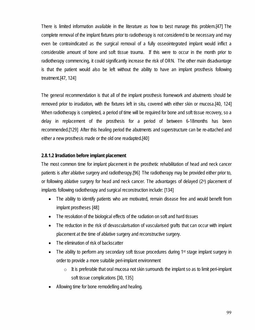
99
There is limited information available in the literature as how to best manage this problem.[47] The complete removal of the implant fixtures prior to radiotherapy is not considered to be necessary and may even be contraindicated as the surgical removal of a fully osseointegrated implant would inflict a considerable amount of bone and soft tissue trauma. If this were to occur in the month prior to radiotherapy commencing, it could significantly increase the risk of ORN. The other main disadvantage is that the patient would also be left without the ability to have an implant prosthesis following treatment.[47, 124] The general recommendation is that all of the implant prosthesis framework and abutments should be removed prior to irradiation, with the fixtures left in situ, covered with either skin or mucosa.[40, 124] When radiotherapy is completed, a period of time will be required for bone and soft tissue recovery, so a delay in replacement of the prosthesis for a period of between 6-18months has been recommended.[129] After this healing period the abutments and superstructure can be re-attached and either a new prosthesis made or the old one readapted.[40]
2.8.1.2 Irradiation before implant placement The most common time for implant placement in the prosthetic rehabilitation of head and neck cancer patients is after ablative surgery and radiotherapy.[96] The radiotherapy may be provided either prior to, or following ablative surgery for head and neck cancer. The advantages of delayed (2o) placement of implants following radiotherapy and surgical reconstruction include: [134]
• The ability to identify patients who are motivated, remain disease free and would benefit from implant prostheses [48]
• The resolution of the biological effects of the radiation on soft and hard tissues
• The reduction in the risk of devascularisation of vascularised grafts that can occur with implant placement at the time of ablative surgery and reconstructive surgery.
• The elimination of risk of backscatter
• The ability to perform any secondary soft tissue procedures during 1st stage implant surgery in order to provide a more suitable peri-implant environment
o It is preferable that oral mucosa not skin surrounds the implant so as to limit peri-implant soft tissue complications [30, 135]
• Allowing time for bone remodelling and healing.
100
A schematic representation of the decision making process for insertion of implants in the mandible following radiotherapy is outlined in Figure 14. [127] Figure 14: The decision making process for implant insertion in the mandible after radiotherapy [127]
2.8.1.3 Irradiation before and after implant placement Occasionally a patient may get a recurrence necessitating further radiotherapy to an already irradiated field. Tissues which are exposed to very high radiation doses with implants in situ challenge the limits of osseointegration with resultant high implant failures.[133] It is not known how much radiation tissues can withstand and still be able to be capable of osseointegrating implants. A study by Granstrom et al investigated 3 patients who were irradiated before and after implant treatment, with 14 implants directly in the radiation field.[136] The results were that:
• All 3 patients developed ORN
• 9 failures (64.2%) of the 14 implants occurred
• The radiation dosage which lead to implant failure was 100-145Gy 2.8.2 Chemotherapy The management of head and neck cancer can include chemotherapy as an adjunct to either ablative surgery, and/or radiotherapy or as a monotherapy. Research by Granstom et al successfully established
101
that chemotherapy before implant surgery had no deleterious effects on osseointegration, but he was not able to verify if chemotherapy after implant surgery had an impact on osseointegration. [133] While chemotherapy is reported to be a risk factor for osseointegration, [77, 99, 137] a study by Kovacs in 2000 was unable to find evidence that it has a negative influence on implant prognosis.[138] In a later retrospective study by Kovacs, two groups of oral cancer patients were compared over 10 years. Neither group underwent radiotherapy, but one group of 30 patients with 106 implants completed chemotherapy with a regime of Cisplatin or Carboplatin with 5-Fluorouracil. A lifetable analysis demonstrated that there was no significant difference between implant survival in either group. He concluded that this chemotherapy regimen was not detrimental to the survival and success of implants in the mandible. [139] Chemotherapy given near to the time of 1st stage implant surgery has been shown to have a negative impact on implant survival and osseointegration. Implant survival was less affected when the chemotherapy was administered some time before or within 1 month after 1st stage implant surgery.[140] In a review article by Wood et al it was stated that “implant integration during
active chemotherapy cannot be supported”. [103]
A review by Hwang et al in 2006 [100], found that there had only been a limited number of investigations conducted on the effect of chemotherapy on implant survival. They concluded that patients who underwent chemotherapy after implant placement showed conflicting results, although mostly implants were adversely affected. A more recent article by Granstrom has concluded that chemotherapy over a longer time perspective has a similar negative effect on osseointegration as radiotherapy.[95]
Patients, who have undergone ablative surgery for head and neck cancer with adjunctive radiotherapy, are some of the most difficult patients to restore prosthetically. They are also those who would most benefit from implant prostheses to restore both function and aesthetics. Due to their often reduced life expectancy it is difficult to delay treatment. Implant prostheses are increasingly being provided as part
2.9 RADIOTHERAPY RELATED RISK FACTORS TO IMPLANT SURGERY
102
of the reconstruction process despite the well documented adverse biological effects that occur when soft tissue and bone have been exposed to ionising radiation. The capacity for irradiated bone to successfully integrate endosseous implants continues to be evaluated in the literature. In a retrospective study, August et al concluded that past tumoricidal irradiation should not be considered an absolute contraindication to implant placement, but reduced success rates are to be expected.[141] In particular, implants should not be considered an absolute contraindication when the total irradiation dose is less than 55Gy.[47, 109] Both extra-oral and intra-oral implant survival is significantly lower in irradiated bone compared to non-irradiated bone when radiation doses of at least 50Gy have been provided.[34] In the planning process for implant prostheses in irradiated tissues of the head and neck, the following issues need to be considered: [47]
• The field irradiated
• The dosage of radiation provided
• The possibility that the implants will not osseointegrate
• The risk of severe complications due to the implant surgery. There are a number of radiotherapy related factors which impact on the success of implant placement and have been reported in the literature.[142] These include the:
• Region of placement in the craniofacial skeleton
• Patient selection
• Radiation dosage
• Time from radiotherapy to 1st stage implant surgery
• Time from 1st to 2nd stage implant surgery
• Implant fixture length
• Marginal bone loss
• Soft tissue condition
• Prosthesis design and retention
• Surgeon’s experience
• Risk for ORN in relation to implant surgery.
103
2.9.1 Region of placement in the craniofacial skeleton The first endosseous implant was placed in the craniofacial skeleton in 1977.[95] The main reason for placement of implants in the craniofacial skeleton was to rehabilitate defects involving the ear, nose, orbit, or midface following ablative cancer surgery. A study by Tolman in 1996, reported a higher loss of implants in irradiated tissues of the craniofacial skeleton (85% success) compared to non-irradiated tissues (97% success), but there was significant variation which was site specific, with 99% success rate in the auricular bone compared to 81% success rate in the nasal bone. [143] It has been recommended that irradiated patients receive hyperbaric oxygen prior to implant placement in the craniofacial region. [144] In the literature there are several studies available which detail information on extra-oral implant placement in the irradiated craniofacial skeleton:
• Jacobsson et al reported 14% loss of implants placed in 9 patients with a follow-up period of 44 months. [145]
• Parel and Tjellstrom reported a 39% loss of implants compared to 5% loss in non-irradiated patients in the United States and Sweden. [146]
• Granstrom reported a 35% implant loss over 11 years. He reported a significant relationship between implant failure and site with frontal bone (50%), zygoma (46%) and mandible (33%) carrying higher failure rates compared to the maxillae (14%) and temporal bone (9%). [147]
• Granstrom in 1994 looked at 258 extra-oral implants, of which 88 implants were placed in non-irradiated bone (17% loss), 125 in irradiated bone (38.4% loss) and 45 in irradiated bone with adjunctive hyperbaric oxygen (no loss) [144]
• Granstrom in 2007 found that during the last twenty years, failure rates in the craniofacial region had been approximately 10%, with the temporal bone having the lowest failure rate at 8%.[95]
The greatest radiobiological effect on the craniofacial skeleton tends to be seen clinically in the mandible as it is commonly involved in the radiation field with exposure to high dose radiation. [148] It is the bone most susceptible to ORN. [147] From the 6th decade on, there is a significant reduction in the ability of the Inferior alveolar artery to provide an adequate supply of blood to the mandible, resulting in the subperiosteal plexus becoming the major blood supply to the posterior mandible. The vessels of this plexus are very sensitive to the effects of radiation.[96, 148] The blood supply to the mainly cancellous bone of the symphysis interforaminal
104
region of the anterior mandible is less affected by radiation.[145, 149] As the anterior mandible is often not within the radiation field, it usually maintains viable bone even when the posterior regions are highly irradiated. As a result endosseous implants can be placed in this region with a reasonable degree of success.[148] 2.9.2 Patient selection Head and neck cancer patients experience significant post-operative side-effects following ablative surgery, but with implant prostheses the potential to achieve successful functional rehabilitation has increased. Dental implant rehabilitation should not be routinely provided to all head and neck cancer patients, as patient selection is still an important factor, particularly for those who have had radiotherapy.[47] In fact, Keller believed that through careful patient selection and diligent surgical technique, successful osseointegration could be achieved without the need for adjunctive hyperbaric oxygen. [150] The criteria for patient selection for rehabilitation involving an implant prosthesis after head and neck cancer treatment includes: [151]
• Adequate patient motivation, expectations and resources
• Reasonable oncologic prognosis. It is imperative that the cancer is in remission and the prognosis relatively favourable.
• Good oral hygiene
• Bone of adequate quality and volume with a healthy soft tissue and a suitable inter-arch relationship [109]
• No medical contraindications to further surgery. It has been recommended that smoking cessation and alcohol withdrawal programmes be part of the rehabilitation process. [131]
It is important that the patient fully understands the significant risks [47] and the extensive commitment involved in the rehabilitation process. He/she must be willing to: [129]
• Undertake further surgical procedures and possibly hyperbaric oxygen treatment
• Attend multiple appointments for implant placement and prosthesis construction
• Be motivated to achieve and maintain meticulous oral hygiene
• Be motivated to achieve and maintain the cessation of smoking and alcohol intake.
105
A retrospective review by Kwakman et al in 1997 investigated the patient selection process for implant prostheses by reviewing the patient records of attendees to the Oral and Maxillofacial Surgery Department University Hospital Nijmegen, The Netherlands from January 1989 to December 1990. Of the 95 patients identified: [125]
• 25 patients required no prosthetic treatment
• 32 patients were successfully provided with a conventional prosthesis
• 28 patients were deceased or had a poor prognosis
• 23 patients could benefit from implants, but of these o 15 patients refused treatment or implants o 5 patients had local or general contraindications to implant treatment o only 3 patients underwent implant treatment.
2.9.3 Irradiation dose The exact dose of radiotherapy delivered to the potential implant site is of fundamental importance in the treatment planning process, as the extent of cellular damage to both the soft and hard tissues in the head and neck region is dose dependant, and influenced by the delivery protocol. A review of the literature by Esposito in 1998 concluded that: [109]
• A radiation dose less than 48 Gy very rarely created soft and hard tissue complications
• A radiation dose greater than 64Gy resulted in increased soft and hard tissue complications. This lead Granstrom in 2003 to conclude that a while a full course of therapeutic radiotherapy, i.e. 50-65Gy is no contraindication to implant therapy, implant survival and success in higher doses is reduced, and the patient must be informed of the possible risks.[47] However, consideration of the dose of radiation provided without taking into account the fractionation schedule can be misleading. In a study by Granstom et al in 1992 it was found that there was an increase in the number of late complications with hyper-fractionation techniques, compared to conventional fractionation techniques.[131] The ‘cumulative radiation effect’ is considered to be a more reliable estimation of the irradiation dose provided to tissues within the radiation field.[109, 131, 152] In some situations it may be necessary to calculate the irradiation dose to each potential implant site to determine the optimum implant installation sites.[153]
106
Research in beagle dogs by Asikainen et al in 1998 compared the bone osseointegration response to radiation doses of 40Gy, 50Gy and 60Gy. His results suggested that with fractionated doses of 40-50Gy, titanium implants may still become osseointegrated in the mandible of beagle dogs.[154] This result was confirmed clinically, when doses of 40-50 Gy were found to impair the healing capacity of bone with an increase in the inherent risk of complications, when performing implant surgery.[40] An attempt to evaluate the relationship between total irradiation dose and implant success was performed by Esposito et al.[109] They reviewed several articles which investigated implant failure rates in Brǻnemark implants in irradiated jaws with regard to location and total irradiation dose.[135, 150, 152, 155-157] A summary is provided below in Table 10. Table 10: Failure rates of Brǻnemark implants in irradiated jaws with regard to location and total irradiation dose. [109]
Esposito et al concluded that the total irradiation dose does seem to influence the clinical outcome with regard to osseointegration.[109] Other clinical studies investigated the impact of the radiation dose on osseointegration. Shaw et al also found that implant losses were similar in doses less than 50Gy compared to doses greater than 50Gy. [151], while Schliephake et al found that implant survival in patients who had received 60Gy was 84.6% compared to implant survival of 43% with 32 Gy.[158] A similar finding on high implant losses with a lower radiation dosage was also reported in two articles by Granstrom et al.[133, 144] He has suggested that these unexpected implant losses at lower radiation doses are a result of either:
The use of low energy radiation, which is known to be tissue damaging [47, 153] or
An extended time delay between irradiation and implant surgery [133] or
The hyper-fractionation schedule. [144]
107
As a result of these studies identifying the impact of radiation doses greater than 55 Gy on implant osseointegration and survival, there has been much discussion in the literature on the benefit of using adjunctive hyperbaric oxygen.[40, 135, 148, 152] Esposito reviewed several articles which investigated implant failure rates in Brǻnemark implants placed in irradiated bone with regard to location and hyperbaric oxygen therapy.[135, 150, 155-157, 159, 160] A summary is provided below in Table 11. [109] Table 11: Failure rates of Brǻnemark implants in irradiated jaws with regard to location and hyperbaric oxygen therapy. [109]
Esposito et al concluded that adjunctive hyperbaric oxygen treatment does not significantly improve the performance of mandibular implants.[109] 2.9.4 Time from radiotherapy to 1st stage implant surgery In the literature to date there is no definitive data to identify the ideal time frame between the completion of radiotherapy and the surgical placement of the implant fixture(s). The optimal time of implant placement post-radiotherapy remains controversial and anecdotal, and despite the many studies conducted, the issue remains inconclusive with no evidence-based recommendations available.[127] The time frame from the end of radiotherapy to implant surgery is considered to be important to implant survival. When considered from: [144, 147]
The patient’s point of view, implant surgery should be provided as soon as possible to enable prosthetic rehabilitation to be completed.
From a tumour-biology point of view, a reasonable time delay is required for the detection of possible tumour recurrences. i.e.1-3 years after tumour surgery
From a radiobiologic point of view, reasonable time is required for acute tissue reactions to have subsided and the healing phase established. i.e. 2-4 months after irradiation
From a surgical point of view, reasonable time is required for resolution of surgical tissue reactions i.e.6-18months post-irradiation.
108
There are essentially two schools of thought regarding the timing of implant placement. There are those who believe that 1st stage implant surgery should be performed as soon as possible after completion of radiotherapy. This is based on the belief that following radiation therapy there is no reduction in the perfusion to bone until after 6 months.[144] This was observed in a human study by Marx and Johnson where fibrosis and loss of vascularity began 6 months after irradiation and then progressively worsened [65] while Jacobsson et al reported an improvement in the bone’s healing capacity over the 12 month period following irradiation.[149] Initially it was believed that patients who were rehabilitated early after radiotherapy showed the highest implant survival.[27, 109, 133, 159] There are even those who believe that implant placement should be provided during the time of ablative surgery, so that patients can be rehabilitated earlier.[133] However, the problem with placing implants too soon after ablative surgery and radiotherapy is that there is:
• an increased risk of ORN [47]
• an increased risk of surgical complications [65]
• the potential for tumour recurrence, as most recur within 12 months of initial oncology treatment. [127]
In particular, there is an increased risk that some of the implants placed may never be used.
Despite these risks, early rehabilitation is often recommended. This is justified as the improvement in the quality of life is significant following reconstruction and prosthetic rehabilitation.[47] It has been demonstrated by Jacobsson et al that irradiation has a negative impact on the ability of bone to regenerate.[149] In their study there was a significant depression of osteogenesis when implants were surgically placed immediately after irradiation. Based on the studies by Jacobsson on bone behaviour after irradiation, many researchers have recommended waiting at least 6-12 months prior to implant rehabilitation [34, 47, 76, 104, 129, 134, 144, 145, 149, 161-164], some even suggesting a proposed delay of up to 2 years.[135, 155] 2.9.5 Time from 1st to 2nd stage implant surgery After implant placement in an irradiated site, it is generally recommended to wait at least 4-6 months before abutment connection to allow the implants extra time for osseointegration.[30, 76, 96, 104, 127, 133-135, 152, 157, 165]
109
The extended integration time from the conventional 3 months in normal bone, to 4-6 months in irradiated bone, prior to uncovering and loading the implant fixtures is recommended based on:
• Experimental studies which show that the osseointegration process in irradiated tissues takes place at a reduced speed [47]
• A clinical study which found that there was a significantly higher failure rate when the time from 1st to 2nd stage implant surgery was less than 4 months. [76]
• Taylor et al reported a 100% implant success rate in irradiated bone using a 6 month osseointegration period in the mandible. [135]
A shorter osseointegration period has been advocated when adjunctive hyperbaric oxygen is used however the exact relationship between integration times and the application of hyperbaric oxygen is unknown.[129] There is also some controversy surrounding the loading of the mucosa with removable dentures during the osseointegration period. Anecdotally:
• Some authors prefer no loading of the irradiated mucosa during this period [135],
• While others do allow loading with a removable prosthesis during this period. While there was an increase in mucosal soft tissue screw perforations noted, there was no increase in implant failures.[152]
In contrast, there is consensus that the prosthodontic rehabilitation can start two weeks after abutment connection is completed.[128]
2.9.6 Implant fixture length As with implant placement in non-irradiated bone, fixture length plays an important role in the success and survival of osseointegrated implants. Studies have found that a higher proportion of short fixtures compared to long fixtures failed or were lost in irradiated bone.[133, 138, 144, 160, 166] Granstrom et al concluded that this is probably related to the short fixtures being exposed to high loading forces [144] however a more recent study by Yerit et al concluded that implant brand, length or diameter did not correlate significantly with implant survival.[167]
110
It has also been recommended that the bi-cortical engagement of implants in bone is associated with a higher success rate compared to mono-cortical engagement.[47, 109] 2.9.7 Marginal bone loss As with non-irradiated bone, marginal bone loss is an important factor in evaluating implant success. A study by Kovacs found that a very high proportion of horizontal bone loss, between 73-84% occurred in the peri-implant area.[138, 168] An increase in marginal bone loss in irradiated patients has also been reported by other researchers.[47, 164] 2.9.8 Soft tissue condition The most significant problems for irradiated implant patients are related to soft tissue inflammation. [141, 156] These include:
• Early problems o Soft tissue overgrowth o Cover screw perforations [152] o Tongue ulceration o Intra-oral wound dehiscence
• Late problems o Fistulae complications o Gingivitis, mainly due to poor oral hygiene. [164]
An article by Kovacs [168] looked at 90 patients who received 320 implants after oral cancer resection and immediate soft tissue reconstruction from June 1990 to December 1997. The survival rate of the 320 implants after 12 months was 93% and after 6 years was 83.5%. He concluded that “adaptive rebuilding takes place in an operated area with transplanted soft tissue despite constant moderate plaque accumulation. This rebuilding leads to a decrease in the peri-implant inflammation over time which is contrary to healthy or at least normal gingiva.” 2.9.9 Design and retention The use of osseointegrated implants has increased the success of functional rehabilitation. Prior to implant prostheses, rehabilitation was limited to the use of conventional removable appliances, which were usually not well tolerated. For patients who were not irradiated, there were a greater variety of reconstructive options available, with post-surgical rehabilitation proceeding relatively quickly. However,
111
for patients who required radiotherapy, reconstruction was often delayed and far more technically challenging.[129] There is insufficient evidence in the literature to suggest that one type of prosthesis is superior to another. Fixed prostheses in the irradiated head and neck cancer patient are often recommended as the preferred option since the prostheses are completely implant supported and retained, and therefore do not put any contact pressure on the irradiated soft tissues, preventing mucosal ulceration. Irradiated mucosa is often fibrosed, telangiectatic and atrophic and is at greater risk of ulceration with the likelihood that bone exposure may lead to ORN.[169] However, the risk of ORN related to denture trauma is small.[51] The superiority of the implant fixed prosthesis in the mandible to the implant removable prosthesis has been highlighted by several authors[28, 141, 170] A study by Weischer et al demonstrated that while both can be used to provide a functional, stable and aesthetically satisfactory prosthesis, only the fixed prosthesis prevented any mucosal soft tissue lesions, therefore reducing the risk of ORN. He recommended that irradiated patients should only be restored with implant fixed prostheses.[169] The following definitions are provided for implant prostheses: [28]
•
o “a removable prosthesis supported by 2 endosseous implants in the symphyseal area. The load of the denture is distributed between implants and the tissue, with the tissue taking most of the load. The purpose of the implants is to assist in the primary retention of the denture and to help stabilise the denture by removing the lateral forces that dislodge it in function.”
Implant assisted removable prosthesis/implant assisted denture:
o
•
o “a fixed retrievable denture connected to abutments via screws or provisional soft cement. These dentures are supported by at least 4 fixtures placed around the arch to distribute the forces transmitted by mastication.”
Implant borne fixed prosthesis/implant borne denture:
Weischer concluded that although implant removable prostheses were not contraindicated, the highest implant survival occurs in fixed prostheses.[142] Removable overdentures were associated with
112
increased implant failures [133, 134, 144, 159, 169, 171] and should only be used in exceptional circumstances.[169] In contrast, Shaw et al identified that the benefit of a removable prosthesis is that it can be easily removed to examine the mucosa for recurrence, as a secondary primary malignancy or recurrence can present clinically similar to that of a benign peri-implant complication. Shaw et al recommended that care should be taken in providing a fixed prosthesis in patients with a history of previous dysplastic changes in the mucosa.[172] 2.9.10 Surgeon’s experience Following an extensive review of the literature in 1998 Esposito concluded that the influence of the operator with respect to surgery, patient selection and site selection has an impact on the outcome of implant treatment. [109] He found that for surgeons who
• had placed less than 50 implants, the failure rate was twice that of surgeons who had placed more than 50 implants.
• were inexperienced, the failure rate for 2nd stage surgery was higher compared to surgeons who had more than two years experience.
Esposito concluded that while the higher failure rates were essentially due to operator technique with respect to surgical trauma and bacterial contamination, there was no quantitative data available to support this hypothesis.[109] Granstrom’s research also supported this proposal. [95, 133, 144] There is general agreement in the literature that implant surgery requires the use of a gentle surgical technique with minimum reflection of the periosteum and use of pre-operative antibiotics to prevent wound healing disturbances.[95, 127, 134, 135, 157] The surgical protocol for implant placement in irradiated tissues requires: [135, 173]
• meticulous soft tissue handling
• minimal periosteum handling
• atraumatic bone removal, with low heat production and copious irrigation
• primary wound closure
• broad antibiotic coverage
• avoidance of oral prosthesis tissue trauma
113
• aggressive preventive dental and oral hygiene care. However Granstrom’s later work found that the experience of the surgeon alone did not affect implant survival.[142], as surgical technique was more important. He surmised that the possible reasons for this were that:
• the surgeons used in this subsequent study were all experienced enough, or
• the irradiation factor is more important for implant survival than the surgeons experience, or
• experienced surgeons tended to manage more complex cases. 2.9.11 Risk of osteoradionecrosis in relation to implant surgery The mandible is considered to be very susceptible to ORN due to its compact bone and blood supply from the periosteum. As a result, implant prostheses are occasionally not offered to irradiated head and neck cancer patients due to the perceived risk of ORN following implant surgery.[174] The incidence of ORN in different studies has ranged from 4% to 35% and is dependant on: [131]
• the dose of irradiation
• the source of irradiation, as the incidence of ORN is reduced with the use of high energy radiation sources.
ORN has been reported in the literature following implant surgery [151, 173] as well as incidental findings.[76, 147, 157, 164] However some researchers have raised concerns that it is being under-reported.[47, 153] Epstein et al reviewed the literature regarding the incidence of ORN in the mandible after head and neck irradiation and found the incidence varied from 5.8 - 44.1% in 4000 non-implanted subjects.[175] The incidence of ORN after endosseous implant placement reveals only
• 3 cases among 170 (1.8%) [150]
• 2 cases among 34 (5.8%) [151]
Hyperbaric oxygen therapy involves the intermittent, systemic administration of 100% oxygen under pressure greater than 1 atmosphere absolute (ATA).[131, 176-178]
2.10 HYPERBARIC OXYGEN THERAPY
114
There is strong support in the literature for the benefits of hyperbaric oxygen on soft and hard tissues compromised by irradiation.[131, 176, 177] There are also numerous references for its application in the prevention and treatment of ORN.[66, 73, 78, 179] Hyperbaric oxygen has been used in clinical practice since the 1930’s, and is delivered either in a monoplace chamber, which is completely pressurised with oxygen, or in a multiplace chamber, which is pressurised with air and the oxygen delivered via a facemask.[133] The therapeutic uses, mechanisms of action, contraindications and complications of hyperbaric oxygen therapy are all well documented in the literature. While hyperbaric oxygen is the only known technique which can be used to counteract the deleterious effects of radiation on tissues [131], potential alternative therapies in the management of radiation compromised tissue which are mentioned in the literature include: [131, 133]
• Pharmacologic factors e.g. calcium, phosphorous, vitamin D
• Supplements e.g. calcitonin, oestrogen in postmenopausal women
• Anabolic steroids in cachetic cancer patients
• Synthetic oxygen transporters
• Various growth factors 2.10.1 Basic effects on tissues The therapeutic action of hyperbaric oxygen is related to an elevation of the partial pressure of oxygen (oxygen tension) in the irradiated tissues. The pressure assists in increasing the solubility of oxygen in the tissue fluids.[47] The daily elevation of oxygen tension in the hypoxic irradiated hard (bone) and soft tissue leads to: [131]
• Fibroplastic proliferation
• Collagen synthesis
• Capillary angiogenesis
• Ingrowth of capillaries
115
Irradiated tissues have been found to have a partial pressure of oxygen (Po2) between 5-15mmHg. During hyperbaric oxygen therapy the arterial Po2 can be increased to between 1000-1300mmHg, with sustained raised Po2 levels following therapy in tissue of between 100-250mmHg. [131, 176, 180] These increased Po2 levels achieved with hyperbaric oxygen therapy can be retained for several years.[78] It is also significant that while the haemoglobin saturation of oxygen cannot be improved more than 100% by hyperbaric oxygen, the oxygen saturation in plasma and interstitial fluid does increase sharply.[176, 181] A study by Thorn et al in 1997 [179] looked at the effect that hyperbaric oxygen therapy had on the transmucosal oxygen tension in irradiated human mucosa. During hyperbaric oxygen therapy, the transmucosal oxygen tension increased significantly after 30 treatments to a mean of 86% of normal healthy gingiva. Other research has shown that a steep oxygen gradient with a tissue Po2 of between 30-40mmHg, is necessary for fibroblastic synthesis of collagen and the subsequent development of the collagen matrix which is necessary for the ingrowth of capillaries into avascular areas and wounds. A tissue oxygen tension below this level causes a suppression of the cellular proliferation of fibroblasts [177] i.e. collagen synthesis is oxygen dependant.[180, 181] Irradiated tissue is characterised by a low or no oxygen gradient.[179, 182] Low oxygen tension also prevents the differentiation of mesenchymal cells into either chondroblasts or osteoblasts. Anoxic or hypoxic tissue favours the production of more chondroblasts, with hyperoxic tissue producing more osteoblasts.[107] One of the initial effects of hyperbaric oxygen is the fibroplasia which occurs in the hypocellular-hypovascular-hypoxic irradiated tissue.[65, 68, 182] This fibroblastic proliferation results in the synthesis of new collagen. Once the new collagen has been laid down in the hypoxic irradiated tissues, it acts as a framework for endothelial proliferation (neoangiogenesis) and new capillary ingrowth (neovascularisation) occurs, eventually leading to the reversal of the local hypoxia.[107, 176, 177, 181, 182] Therefore, the major benefit of hyperbaric oxygen therapy is improved cellularity, vascularity and oxygenation of the once hypocellular-hypovascular-hypoxic tissue, through the combination of fibroplasia, collagen synthesis and deposition, neoangiogenesis and neovascularisation.
116
A study by Marx et al in 1990 [182] looked at the relationship between the oxygen dose to angiogenesis induction in irradiated tissue. He compared the angiogenic properties of normobaric oxygen, hyperbaric oxygen and air (control) in the rabbit model. His results indicated that while normobaric oxygen had no angiogenic properties in irradiated tissue, the hyperbaric oxygen demonstrated an 8-9 fold increase in the vascularity of tissues compared to both the normobaric oxygen and air breathing controls. (p=0.001) The angiogenic properties of hyperbaric oxygen have also been demonstrated in both animal and human tissue biopsies with therapeutic hyperbaric oxygen protocols.[65, 68, 78, 181, 182] Additional effects on tissue with hyperbaric oxygen therapy include: [177]
• Vasoconstriction
• Antimicrobial activity
• Improved osteoclastic function
• Increased erythrocyte deformability
• Increased in hard tissue formation and mineralisation (bone) Research has shown that hyperbaric oxygen causes vasoconstriction.[176] It is considered to be a potent vasoconstrictor which doesn’t significantly reduce the oxygenation of the tissues. This can be potentially useful in reducing the oedema associated with skin flaps and bone grafts.[177] Hyperbaric oxygen has also been found to be bactericidal for certain anaerobes, bacteriostatic for microaerophilic organisms and some species of escherischia, and responsible for increasing the rate of bacterial death by macrophage induced phagocytosis.[183] This is particularly useful as local hypoxia in irradiated tissues predisposes wounds to infection. Hyperbaric oxygen restores the defence mechanisms of neutrophils and macrophages against infection.[177, 180] Improvement in the osteoclastic function has also been reported to be a benefit of improved oxygen tension by the application of hyperbaric oxygen. This function is paramount in the removal of necrotic bone associated with ORN, and infected bone with osteomyelitis.[177] An increase in erythrocyte deformability is also a reported benefit of hyperbaric oxygen therapy. These cells are the principal oxygen transport cells of the body, and their ability to pass easily through peripheral tissues is important.[177]
117
Experimentally it has been found that hyperbaric oxygen has the ability to increase the formation and mineralisation of bone, affecting bone turnover.[133, 177] While the exact mechanism of action at a cellular level is unclear, it is believed that there is a synergistic mechanism of action between the hyperbaric oxygen and fibroblast growth factor, which enhances the level of insulin like growth factor. This growth factor is known to promote the proliferation and differentiation of bone, by affecting the bone progenitor cells to: [47]
• Produce more osteoblasts
• Promote DNA synthesis
• Stimulate enzymes involved in bone formation
• Affect involved membrane receptors.
2.10.2 Therapeutic uses of hyperbaric oxygen therapy There is only very limited evidence-based information obtained from randomly controlled trials in the literature regarding the application and efficacy of hyperbaric oxygen in most diseases.[180] The Hyperbaric Oxygen Committee of the Undersea and Hyperbaric Society currently recommends hyperbaric oxygen for several uses, as detailed in Table 12. [180] Table 12Diseases for which the weight of scientific evidence supports hyperbaric oxygen as effective therapy
: Diseases for which Hyperbaric Oxygen is currently used.
Primary therapy: • Arterial gas embolism • Decompression sickness • Exceptional blood-loss anaemia • Severe carbon monoxide poisoning
Adjunctive therapy: • Clostridial myonecrosis • Compromised skin grafts and flaps • Osteoradionecrosis prevention
Diseases for which the weight of scientific evidence suggests hyperbaric oxygen may be helpful Primary therapy:
• Less severe carbon monoxide poisoning Adjunctive therapy:
• Acute traumatic ischaemic injury • Osteoradionecrosis • Refractory osteomyelitis • Selected problem wounds • Radiation-induced soft tissue injury
118
Diseases for which the weight of scientific evidence does not support the use of hyperbaric oxygen but for which it may be helpful Adjunctive therapy:
• Necrotising fasciitis • Thermal burns
2.10.2.1 Carbon monoxide poisoning While hyperbaric oxygen is able to reverse both the acute and chronic effects of carbon monoxide poisoning, it is currently the fastest method of reversing the potentially life threatening acute effects.[180, 184] 2.10.2.2 Decompression sickness This usually occurs as a result of uncontrolled ascent to the surface in diving. Hyperbaric oxygen works in decompression sickness by reducing the bubble size and correcting the hypoxia.[180, 184] 2.10.2.3 Arterial gas embolism This can also occur as a consequence of uncontrolled ascent to the surface in diving or more commonly during mechanical ventilation. The mechanism of action with the use of hyperbaric oxygen is the same as with decompression sickness.[180, 184] 2.10.2.4 Clostridial myonecrosis Clostridia are commonly involved in the contamination of traumatic wounds, and can lead to rapidly progressive muscle tissue death. Life-threatening infection is rare, particularly if hyperbaric oxygen together with debridement surgery and antibiotics is provided within a reasonable time frame.[180, 184] 2.10.2.5 Necrotising fasciitis This is a rapidly progressive infection of the skin and underlying tissue without muscle involvement, and has a similar presentation to clostridial myonecrosis. As a result the treatment is very similar, and involves the provision of hyperbaric oxygen, debridement surgery and antibiotic provision. [180, 184] Mortality is relatively high particularly if treatment is not instituted early.[185] 2.10.2.6 Refractory osteomyelitis Hyperbaric oxygen therapy greatly improves the outcome in patients with chronic osteomyelitis which is unresponsive to the standard management of surgery and antibiotic treatment.[180, 184]
119
2.10.2.7 Acute traumatic ischaemic injury Crush injuries, and other similar severe traumatic injuries to the extremities can result in tears of major blood vessels and damage to the microcirculation, with resultant ischaemia and problems with tissue necrosis. Hyperbaric oxygen therapy is a recommended adjunct treatment to surgery in order to achieve advanced wound healing.[180] Hyperbaric oxygen has the ability to increase healing rates in these injuries and limit wound infection and dehiscence.[184, 185] 2.10.2.8 Anaemia due to exceptional blood loss Hyperbaric oxygen therapy can assist in the management of haemorrhagic shock following exceptional blood loss in patients for whom suitable blood is not available, or for patients who refuse transfusions for religious reasons.[180, 184] 2.10.2.9 Thermal burns Hyperbaric oxygen therapy assists in the management of thermal burns by reducing the oedema which can lead to hypoxic vasoconstriction.[180, 184] 2.10.2.10 Problem wounds Hyperbaric oxygen can assist in healing of chronic non-healing wounds, caused by arterial insufficiency, such as diabetic foot infections and leg ulcers.[180, 184] 2.10.2.11 Compromised skin grafts and flaps Often skin grafts and flaps fail as a result of inadequate perfusion and hypoxia in otherwise compromised tissues. Hyperbaric oxygen therapy has been able to achieve a reversal of the flap ischaemia and increase the success associated with grafting in poorly vascularised tissues.[180, 184] A review of the literature by Wang et al in 2003 [185] evaluated the scientific evidence regarding the benefits and risks of hyperbaric oxygen therapy as an adjunctive therapy to standard wound care. While they found that the literature provided no guidance as to when hyperbaric oxygen therapy should be commenced, they concluded that it is able to successfully assist wound healing in compromised skin grafts, ORN, soft tissue necrosis and chronic non-healing diabetic wounds when provided as an adjunct to surgical wound debridement and antibiotics.
120
2.10.2.12 Radiation-induced hard tissue injury (ORN) The effect of radiation on soft tissue and bone is a progressive obliterative endarteritis that leads to a reduction of the vascularity and cellularity of the tissues. As a result the tissues become progressively more hypoxic and if damaged are unable to meet the metabolic demands of tissue repair.[65, 68] Hyperbaric oxygen therapy has gained strong international support for its application in the management of irradiated tissue, following its introduction in the early 1970’s.[70, 177, 178, 186] Prior to the availability of hyperbaric oxygen, the reconstruction of irradiated patients with oropharyngeal and other head and neck tumours was often unsuccessful, with complications including ORN and soft tissue necrosis developing in at least 50% of patients.[180, 184] A study by Marx et al in 1982 [181] identified that adjunctive hyperbaric oxygen produced excellent results with reduced morbidity in the bony reconstruction of hard and soft tissue in irradiated and tissue deficient patients. The mechanism of action of hyperbaric oxygen on non-healing wounds in irradiated tissue is complex, but essentially involves: [73]
• An enhancement of the phagocytic ability of leucocytes
• An inhibition of both aerobic and anaerobic bacteria
• An inhibition of bacterial toxin formation
• A stimulation of fibroblasts
• An increase in collagen formation
• Promotion of growth of new capillaries
• Reversal of the oxygen tension of tissues in favour of wound healing. A review of the literature by Feldmeier et al in 2002 [187] reported on the results of hyperbaric oxygen therapy in the treatment and/or prophylaxis of delayed radiation injuries. They concluded that “hyperbaric oxygen therapy is recommended for delayed radiation injuries for soft tissue and bony injuries of most sites.” Pasquier et al in 2004 reviewed the literature from 1960 for a consensus conference organised by the European Society for Therapeutic Radiotherapy and Oncology, and the European Committee for Hyperbaric Medicine, dealing with the hyperbaric oxygen implications on radiotherapy for the treatment and prevention of late complications. This review concluded that despite the small number of randomly
121
controlled trials, hyperbaric oxygen therapy may be indicated for the treatment of mandibular ORN in combination with surgery, and for the prevention of ORN after dental extractions. [183] 2.10.2.13 Prevention of implant loss in the irradiated patient As hyperbaric oxygen had been used successfully for the prevention and management of delayed radiation injuries in head and neck cancer patients, Granstrom proposed that it would be a useful adjunct to improve the success of osseointegration in irradiated bone [177] and developed a protocol based on the earlier work of Marx.[70, 78] The principle actions of hyperbaric oxygen in osseointegration, which act to support healing and incorporate the implants into the irradiated bone include: [107, 177]
• Hard tissue forming capacity – it stimulates bone growth in and around the implants [165, 188]
• Angioinductive effect [182]
• Stimulation of osteoclasts to assist with bone remodelling
• Healing of soft tissue therefore preventing dehiscence and infections [165, 189]
• Reduction of the risk of ORN. [65] A study by Johnsson et al in 1993 investigated the possible effects of hyperbaric oxygen in relation to osseointegration, [188] where standardised titanium screws were used to measure the extrusion force necessary to unscrew the implants. They showed that the force required to unscrew the implants after radiotherapy was reduced by 60%, but following hyperbaric oxygen therapy the force required improved by 40%. The conclusion they made from this experiment was that hyperbaric oxygen actively improved osseointegration. Despite a growing body of evidence supporting the benefits of hyperbaric oxygen therapy for osseointegration in irradiated tissues, it continues to remain a controversial issue. A review by Coulthard et al for the Cochrane Collaboration in 2002, and subsequent articles in 2003 [178, 190] identified that while there were extensive experimental and human clinical reports in the literature, the use of adjunctive hyperbaric oxygen therapy for ossseointegrated implants in the craniofacial region continues to create controversy regarding its effectiveness. Following the review, Coulthard recommended that as there were no randomised controlled trials available, clinicians should make patients aware that there is
122
a lack of clinical evidence regarding the effectiveness of hyperbaric oxygen in irradiated patients requiring implants. The Cochrane review concluded that: [178]
“not only is there a need for randomly controlled trials to determine the effectiveness of hyperbaric oxygen, but it is likely that these trials will need to be multi-centred as each centre may have a limited number of patients. Only with that will clinicians receive the evidence they need to make the best treatment decisions possible.”
In 2007 the first randomly controlled trial comparing the effects of hyperbaric oxygen on osseointegration was published by Shoen et al.[191] The results of this study of 26 patients was that there was no benefit of hyperbaric oxygen treatment with respect to implant survival and prevention of ORN when compared to non treated patients who received only prophylactic antibiotics. The updated Cochrane Review on this subject published in 2008 [192] identified the work by Schoen et al as being the only randomly controlled trial available in the literature. They concluded that “readers should be aware that the ‘evidence’ on this matter remains highly controversial.” 2.10.3 Treatment protocols for radiation induced hard tissue injuries (ORN) Numerous studies are available in the literature, which attest to the benefits of hyperbaric oxygen in the management and prevention of osteoradionecrosis.[65, 66, 70, 73, 78, 180, 181, 185, 193] The protocols for hyperbaric oxygen currently in use today are those based on the work of Marx.[70, 78, 181, 193] Hyperbaric oxygen can be provided either through the use of a monoplace chamber, or a multiplace chamber. In a monoplace chamber, the patient breathes 100% oxygen at 2.4 ATA (atmosphere absolute) in the pressurised compartment. In a multiplace chamber, the compartment is pressurised with air, and individual patients breathe 100% oxygen at 2.5 ATA through masks. The slight increase in the atmosphere absolute which the patients breathe in through the masks in the multiplace chamber is required so as to compensate for possible leakage through the masks.[107]
2.10.3.1 Prophylactic protocol The most important risk factor identified associated with the development of ORN in a patient who has had irradiation to the mandible is tooth extraction. In a randomised trial by Marx et al [78] the efficacy of hyperbaric oxygen in the prevention of ORN following tooth extraction was proven. The prophylactic treatment protocol for the prevention of ORN as developed by Marx involves: [78]
123
• The patient breathing 100% oxygen at 2.4 ATA or 240kPa for 90 minutes daily, for 20 sessions,
• Extraction of identified teeth, followed by
• The patient breathing 100% oxygen at 2.4 ATA or 240kPa for 90 minutes daily, for 10 sessions.
This prophylactic protocol is based on the results of Marx which showed that angiogenesis became measurable following 8 sessions of hyperbaric oxygen, rapidly progressed to a plateau of between 80-85% of that of non-irradiated tissue by 20 sessions and remained at that level without further improvement with additional hyperbaric oxygen. [78] The rationale behind the 10 sessions of post-operative hyperbaric oxygen is the reduction for the potential of wound dehiscence. Further research by Marx showed that up to 3 years after the original application of hyperbaric oxygen, the oxygen levels in tissue where within 90% of the values recorded just after treatment, indicating that induced angiogenesis with hyperbaric oxygen does not undergo regression with time.[65] The same prophylactic protocol has been adopted for the surgical placement of implants in irradiated tissues of the craniofacial skeleton. [177] This protocol has been shown to improve osseointegration and increase implant survival with the following results: [133]
• Radiated patients following hyperbaric oxygen therapy having a significantly lower implant failure rate compared to radiated patients (P<0.001), and
• Non-irradiated patients having a significantly lower failure rate compared to the radiated patients following hyperbaric oxygen therapy (P<0.005)
The use of this protocol to reduce implant loss has been reported in a number of studies.[96, 135, 147, 152, 157, 160, 162, 166, 174] 2.10.3.2 Therapeutic protocol In 1983 Marx [70] developed a staged protocol based on clinical presentation and progression, for the management of established ORN combining surgical debridement and adjunctive hyperbaric oxygen. [73] In 2004, on completion of a review of the literature, Pasquier concluded that hyperbaric oxygen could be effective in the treatment of mandibular ORN when more conservative measures were not successful.[183]
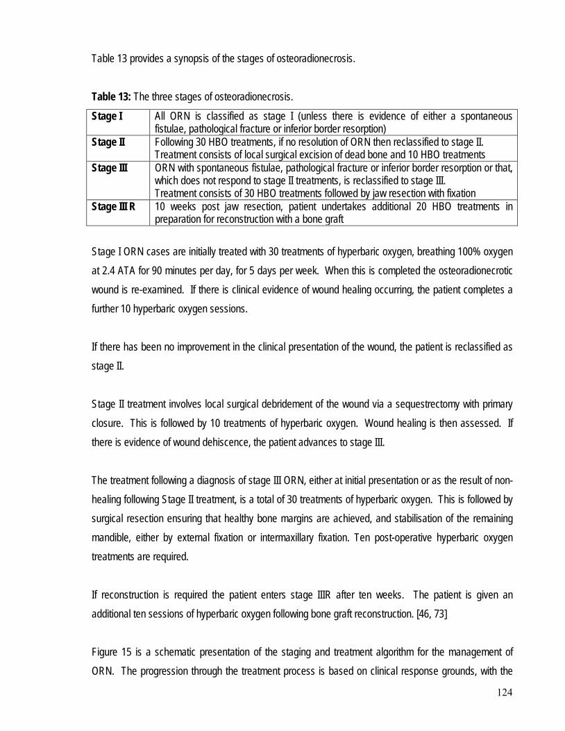
124
Table 13 provides a synopsis of the stages of osteoradionecrosis. Table 13: The three stages of osteoradionecrosis.
Stage I All ORN is classified as stage I (unless there is evidence of either a spontaneous fistulae, pathological fracture or inferior border resorption)
Stage II Following 30 HBO treatments, if no resolution of ORN then reclassified to stage II. Treatment consists of local surgical excision of dead bone and 10 HBO treatments
Stage III ORN with spontaneous fistulae, pathological fracture or inferior border resorption or that, which does not respond to stage II treatments, is reclassified to stage III. Treatment consists of 30 HBO treatments followed by jaw resection with fixation
Stage III R 10 weeks post jaw resection, patient undertakes additional 20 HBO treatments in preparation for reconstruction with a bone graft
Stage I ORN cases are initially treated with 30 treatments of hyperbaric oxygen, breathing 100% oxygen at 2.4 ATA for 90 minutes per day, for 5 days per week. When this is completed the osteoradionecrotic wound is re-examined. If there is clinical evidence of wound healing occurring, the patient completes a further 10 hyperbaric oxygen sessions. If there has been no improvement in the clinical presentation of the wound, the patient is reclassified as stage II. Stage II treatment involves local surgical debridement of the wound via a sequestrectomy with primary closure. This is followed by 10 treatments of hyperbaric oxygen. Wound healing is then assessed. If there is evidence of wound dehiscence, the patient advances to stage III. The treatment following a diagnosis of stage III ORN, either at initial presentation or as the result of non-healing following Stage II treatment, is a total of 30 treatments of hyperbaric oxygen. This is followed by surgical resection ensuring that healthy bone margins are achieved, and stabilisation of the remaining mandible, either by external fixation or intermaxillary fixation. Ten post-operative hyperbaric oxygen treatments are required. If reconstruction is required the patient enters stage IIIR after ten weeks. The patient is given an additional ten sessions of hyperbaric oxygen following bone graft reconstruction. [46, 73] Figure 15 is a schematic presentation of the staging and treatment algorithm for the management of ORN. The progression through the treatment process is based on clinical response grounds, with the
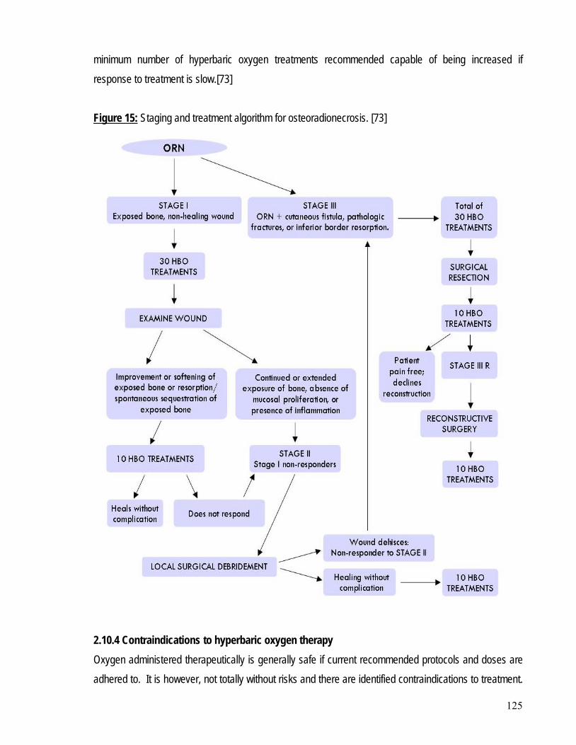
125
minimum number of hyperbaric oxygen treatments recommended capable of being increased if response to treatment is slow.[73] Figure 15: Staging and treatment algorithm for osteoradionecrosis. [73]
2.10.4 Contraindications to hyperbaric oxygen therapy Oxygen administered therapeutically is generally safe if current recommended protocols and doses are adhered to. It is however, not totally without risks and there are identified contraindications to treatment.
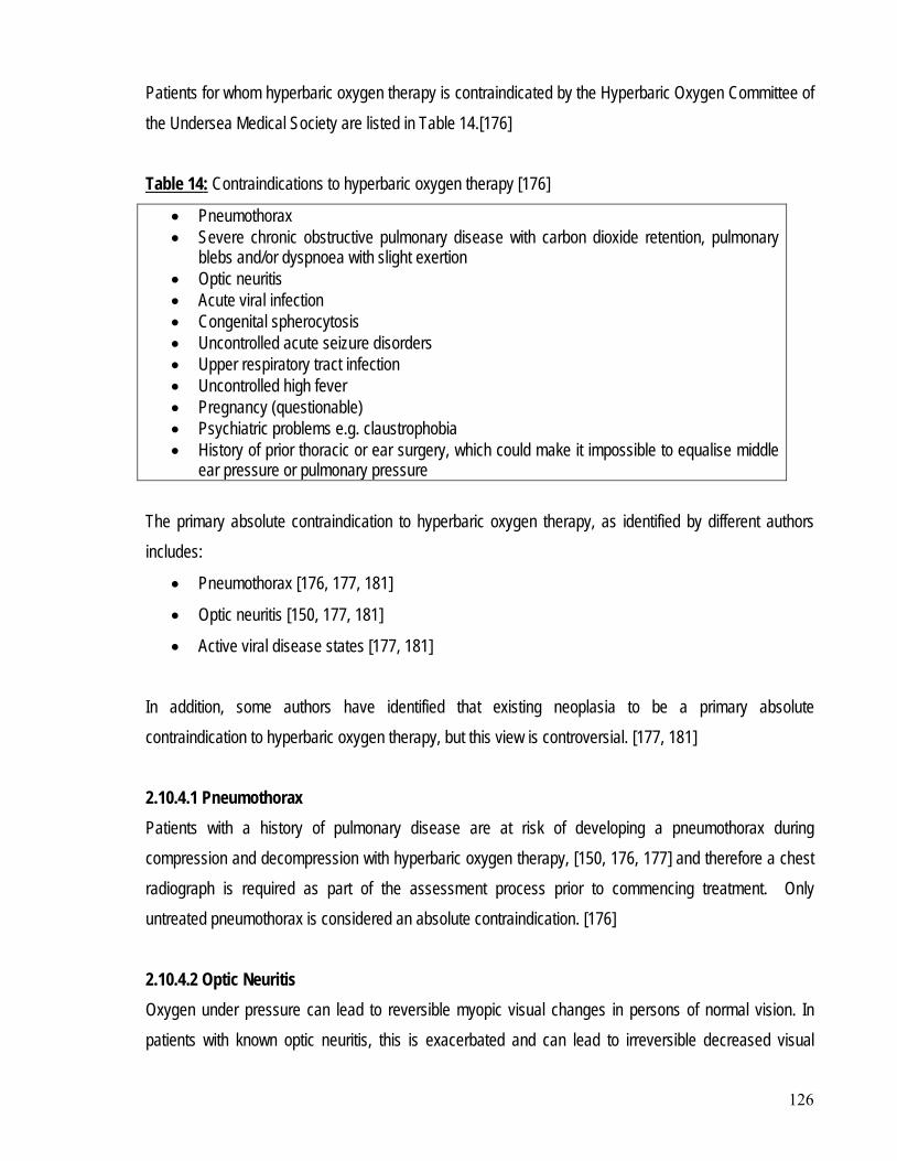
126
Patients for whom hyperbaric oxygen therapy is contraindicated by the Hyperbaric Oxygen Committee of the Undersea Medical Society are listed in Table 14.[176] Table 14
• Pneumothorax
: Contraindications to hyperbaric oxygen therapy [176]
• Severe chronic obstructive pulmonary disease with carbon dioxide retention, pulmonary blebs and/or dyspnoea with slight exertion
• Optic neuritis • Acute viral infection • Congenital spherocytosis • Uncontrolled acute seizure disorders • Upper respiratory tract infection • Uncontrolled high fever • Pregnancy (questionable) • Psychiatric problems e.g. claustrophobia • History of prior thoracic or ear surgery, which could make it impossible to equalise middle
ear pressure or pulmonary pressure The primary absolute contraindication to hyperbaric oxygen therapy, as identified by different authors includes:
• Pneumothorax [176, 177, 181]
• Optic neuritis [150, 177, 181]
• Active viral disease states [177, 181] In addition, some authors have identified that existing neoplasia to be a primary absolute contraindication to hyperbaric oxygen therapy, but this view is controversial. [177, 181] 2.10.4.1 Pneumothorax Patients with a history of pulmonary disease are at risk of developing a pneumothorax during compression and decompression with hyperbaric oxygen therapy, [150, 176, 177] and therefore a chest radiograph is required as part of the assessment process prior to commencing treatment. Only untreated pneumothorax is considered an absolute contraindication. [176] 2.10.4.2 Optic Neuritis Oxygen under pressure can lead to reversible myopic visual changes in persons of normal vision. In patients with known optic neuritis, this is exacerbated and can lead to irreversible decreased visual
127
acuity. It is therefore imperative that all patients being considered for hyperbaric oxygen therapy undergo a thorough ophthalmologic examination prior to treatment.[177] Despite this concern hyperbaric oxygen therapy has been used in some countries for the treatment of multiple sclerosis, which carries a 25% incidence of optic neuritis. This has been done without reported incident, and therefore the contraindication may be only relative.[176] 2.10.4.3 Acute viral infection or upper respiratory tract infection In the literature it has been identified that hyperbaric oxygen has the potential to exacerbate existing viral disease and immunosuppressive disorders [176, 181], and also cause an increase in the haemolysis of red blood cells in patients with congenital spherocytosis.[176] It has been recommended that hyperbaric oxygen therapy should be delayed until symptoms of decongestion and rhinitis subside in patients with upper respiratory tract infections, as congestion may make pressure equalisation in the sinus’ and middle ear difficult, if not impossible.[176, 177] 2.10.4.4 Pregnancy While pregnancy is considered to be a relative risk due to potential adverse effects that oxygen at high partial pressure may have on the foetus, it is a controversial issue internationally as it has been used in the former Soviet Union and in Great Britain with no reported complications.[176] However as a rule, a risk benefit analysis should be applied for each individual case. 2.10.4.5 Claustrophobia Patients who have a history of claustrophobia or other related psychiatric illness are potentially not suitable for hyperbaric oxygen therapy. This is primarily because a significant problem will exist if there is a need for a quick exit from a chamber. The reported safe decompression time in a monoplace chamber is less than 1 minute, while in a multiplace chamber it can be as much as 20 minutes. [176, 177] 2.10.4.6 History of prior thoracic or middle ear surgery If a patient has a history of previous surgery in either the thoracic chamber or middle ear, this may present a relative contraindication to hyperbaric oxygen therapy. This is because either may lead to difficulties in pressure equalisation, due to a differential in atmospheric pressure created between the internal body cavity and the external environment. There is also a significant risk that prior thoracic
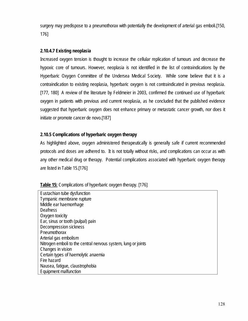
128
surgery may predispose to a pneumothorax with potentially the development of arterial gas emboli.[150, 176] 2.10.4.7 Existing neoplasia Increased oxygen tension is thought to increase the cellular replication of tumours and decrease the hypoxic core of tumours. However, neoplasia is not identified in the list of contraindications by the Hyperbaric Oxygen Committee of the Undersea Medical Society. While some believe that it is a contraindication to existing neoplasia, hyperbaric oxygen is not contraindicated in previous neoplasia. [177, 180] A review of the literature by Feldmeier in 2003, confirmed the continued use of hyperbaric oxygen in patients with previous and current neoplasia, as he concluded that the published evidence suggested that hyperbaric oxygen does not enhance primary or metastatic cancer growth, nor does it initiate or promote cancer de novo.[187] 2.10.5 Complications of hyperbaric oxygen therapy As highlighted above, oxygen administered therapeutically is generally safe if current recommended protocols and doses are adhered to. It is not totally without risks, and complications can occur as with any other medical drug or therapy. Potential complications associated with hyperbaric oxygen therapy are listed in Table 15.[176] Table 15Eustachian tube dysfunction
: Complications of hyperbaric oxygen therapy. [176]
Tympanic membrane rupture Middle ear haemorrhage Deafness Oxygen toxicity Ear, sinus or tooth (pulpal) pain Decompression sickness Pneumothorax Arterial gas embolism Nitrogen emboli to the central nervous system, lung or joints Changes in vision Certain types of haemolytic anaemia Fire hazard Nausea, fatigue, claustrophobia Equipment malfunction
129
In order to prevent the above mentioned complications, a complete medical and physical examination is required prior to commencing treatment. This should include a chest radiograph, ophthalmologic investigations, hearing test and blood chemistry pathology.[177] 2.10.5.1 Barotrauma Inequalities between internal and external pressures (barotrauma) can lead to pneumothorax, decompression sickness and high pressure nervous syndrome. If pressures significantly greater than those recommended are used, the resultant increases in barometric pressure and subsequent decompression can lead to patients presenting with complex neurological and neuropsychological signs of high-pressure nervous syndrome.[176] Decompression sickness associated with hyperbaric oxygen therapy is rare, but is classified as either type I or type II. Type I manifests as either skin bends or joint pains (classical bends) as a result of bubble formation outside the central nervous system. Type II is the result of an intravascular bubble causing a neurological or pulmonary impact which can be potentially fatal.[176] Very rarely, patients develop a pneumothorax from too rapid decompression, leading to rupture of the air spaces within the lungs or expansion of the air in the pleural cavity.[177] 2.10.5.2 Arterial gas emboli An arterial gas emboli is a sudden and potentially lethal phenomenon involving air or other gases passing directly into the blood stream. It can be associated with hyperbaric oxygen therapy usually occurring as a result of either vessel perforation following pneumothorax or mediastinal emphysema. This is a true medical emergency and must be diagnosed and managed immediately with 10 hyperbaric oxygen treatments.[176, 180] 2.10.5.3 Middle ear problems Eustachian tube dysfunction, tympanic membrane rupture, middle ear haemorrhage, pain and deafness are all potential complications of middle ear dysfunction which can occur if the patient is unable to equalise middle ear pressure with external environment pressure.[176, 180, 184] 2.10.5.4 Oxygen toxicity Oxygen toxicity involving either the pulmonary or central nervous system can potentially occur. If the pulmonary system is affected, then the patient develops oedema in the lungs leading to alveolar collapse and pulmonary stenosis. If the central nervous system is affected it leads to seizures in about 0.03% of
130
patients undergoing hyperbaric oxygen therapy, which if treated promptly will cause no permanent damage.[176, 177, 184] 2.10.5.5 Tooth or sinus pain Sinus or toothache can occur as a result of hyperbaric oxygen treatment. Toothache is a risk, particularly in endodontically compromised teeth. Patients may occasionally experience mild to severe pain in restored teeth as a result of rapid pressure changes to compromised pulpal tissues. Toothache with no identified cause is rare, and is often confused with sinus pain which is far more common. The frontal sinus is predominantly involved due to inflammation of the membranes impeding drainage and causing pain from an inability to equalise pressures. This pain is often managed through the provision of antihistamines or decongestants.[176, 180, 184] A study by Ambiru et al in 2008 of 1609 patients undertaking hyperbaric oxygen treatments identified that pressure equalisation problems expressed as pain or discomfort, such as cranial sinus pain and teeth pain was observed in 156 patients (9.7%).[194] 2.10.5.6 Myopia Occasionally, patients report deterioration in vision (myopia) following hyperbaric oxygen treatment. It is a known side-effect which is reversible.[176, 177, 180, 184] Myopia is thought to be caused by refractive index changes to the eye’s lens resulting from oxidative damage to the lens proteins. It is not possible to predict which patients may be at risk of myopia, but there is an increased risk when more than twenty consecutive hyperbaric oxygen treatments are provided and the treatment is provided by hood rather than an oro-nasal mask.[195, 196] 2.10.5.7 Other complications Equipment malfunction is also a possible complication of hyperbaric oxygen treatment. Occasionally patients receiving hyperbaric oxygen treatment are either intubated or have an intravenous line in situ. It is important that in these situations no glass bottles are taken into the hyperbaric chamber and any intravenous bags present have more than 100ml of solution available prior to ascent. If not there is a risk of air from the bag entering the intravenous line, with a risk of concomitant venous or arterial air embolism.[176] The increased concentrations of oxygen present within the chamber may also present a
131
fire risk. Safety precautions should be in instituted that are designed to prevent sparks and other ignition sources, so as to minimize the risk of fire.[177]
There are now more than 100 publications available in the literature discussing osseointegration in irradiated tissues following head and neck ablative cancer surgery. Only publications pertaining to the area of interest i.e. the mandible have been identified and segregated as outlined below:
2.11 OSSEOINTEGRATION IN IRRADIATED TISSUES
• Clinical studies with primary implant provision
• Clinical studies with secondary implant provision
• Clinical studies related to region of placement mandible reconstructed grafted mandible
• Vascularised graft
• Non-vascularised graft
• Clinical studies showing an increased rate of implant loss when placed in irradiated tissues
• Clinical studies showing no increased rate of implant loss when placed in irradiated tissues
• Clinical studies showing stimulation of osseointegration by hyperbaric oxygen
• Clinical studies showing that hyperbaric oxygen is not necessary for osseointegration
• Histological case reports Animal Human.
2.11.1 Clinical studies with primary implant provision A study by Granstrom et al looked at 11 patients who had radiotherapy provided, following the primary provision of implants. The radiation dose varied from 50-80Gy. Of the 32 implant fixtures: [124]
• 2 were removed as part of extensive tumour surgery
• 2 were lost in conjunction with chemotherapy
• 5 patients exhibited skin dehiscence’s around 9 implants after irradiation
• ORN developed in 3 patients after radiotherapy
• 12 implants were placed in the mandible and no failures were reported.
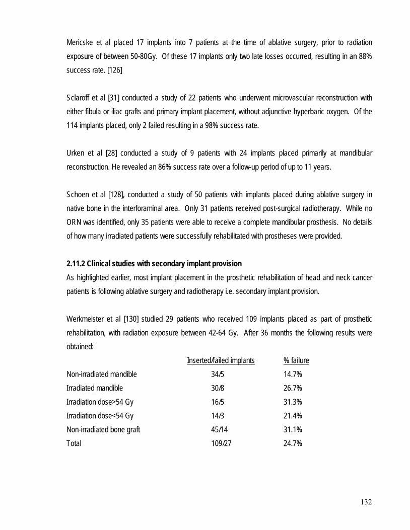
132
Mericske et al placed 17 implants into 7 patients at the time of ablative surgery, prior to radiation exposure of between 50-80Gy. Of these 17 implants only two late losses occurred, resulting in an 88% success rate. [126] Sclaroff et al [31] conducted a study of 22 patients who underwent microvascular reconstruction with either fibula or iliac grafts and primary implant placement, without adjunctive hyperbaric oxygen. Of the 114 implants placed, only 2 failed resulting in a 98% success rate. Urken et al [28] conducted a study of 9 patients with 24 implants placed primarily at mandibular reconstruction. He revealed an 86% success rate over a follow-up period of up to 11 years. Schoen et al [128], conducted a study of 50 patients with implants placed during ablative surgery in native bone in the interforaminal area. Only 31 patients received post-surgical radiotherapy. While no ORN was identified, only 35 patients were able to receive a complete mandibular prosthesis. No details of how many irradiated patients were successfully rehabilitated with prostheses were provided. 2.11.2 Clinical studies with secondary implant provision As highlighted earlier, most implant placement in the prosthetic rehabilitation of head and neck cancer patients is following ablative surgery and radiotherapy i.e. secondary implant provision. Werkmeister et al [130] studied 29 patients who received 109 implants placed as part of prosthetic rehabilitation, with radiation exposure between 42-64 Gy. After 36 months the following results were obtained: Inserted/failed implants Non-irradiated mandible 34/5 14.7%
% failure
Irradiated mandible 30/8 26.7% Irradiation dose>54 Gy 16/5 31.3% Irradiation dose<54 Gy 14/3 21.4% Non-irradiated bone graft 45/14 31.1% Total 109/27 24.7%
133
Schliephake et al [158] investigated the provision of primary versus secondary implant placement in patients who had received bone grafts as part of their reconstructive surgery. The success of the implant survival was significantly (P= 0.0197) related to the timing of implant placement, with
• 1o implant placement having a survival of 36.2%
• 2o implant placement having a survival of 67.1% More recently in 2007, Brandt et al published a case report on the immediate placement of 5 implants placed between the mandibular mental foramina with a fixed appliance using hyperbaric oxygen, on a 45 year old male with a history of squamous cell carcinoma of the floor of the mouth, five years post-surgery and radiotherapy. After 45 months the patient remains free of ORN and has had no complications of implant treatment.[197] 2.11.3 Clinical studies related to region of placement – mandible The first report of osseointegration in irradiated tissues was published by Jacobsson et al in 1988. In this study 9 patients were followed for 44 months during which time the implant failure rate was identified as being 14%.[145] The ability for the irradiated mandible to integrate endosseous implants has continued to be researched. This is supported by the extensive number of clinical reports available in the literature.[135, 150, 155, 156, 163, 198, 199] Most of the patients in these studies were edentulous and received implants in the anterior mandible.[35, 36, 135, 144, 148, 155, 157, 159, 163, 164, 199, 200] In all studies, varying amounts of radiotherapy are included. Franzen et al [155] reported on 5 patients irradiated with a mean dose of 40.3Gy [range 20 to 50Gy] in which 20 implants were inserted into the mandible. Only 1 implant was lost during the 3-6 year follow-up period. Eckert et al [156] reported on 20 irradiated patients [range 20 to 60Gy] with 89 implants in the mandible. Only 1 implant was lost during follow-up. Ali et al [160] reviewed 10 patients with 32 mandibular implants placed in bone irradiated to between 25 to 57.5 Gy. Follow-up at 52 months showed no implant loss.
134
Esser et al [157] placed 71 IMZ and 150 Brǻnemark implants into mandibles irradiated to 60Gy. The 5 year survival rate was 77.5% for the IMZ implants and 83.6% for the Brǻnemark implants. The control group showed a 5.6% loss during the same period. Anderson et al [137] followed 15 patients with 90 Brǻnemark implants installed in the irradiated mandible [range 44 to 68Gy]. Follow-up at 8 years showed a success rate of 97.8%. Wagner et al [76] reported on 275 Brǻnemark implants placed in the mandible of 63 cancer patients. The five (5) year survival rate was 97.9%. Only 35 patients were irradiated as part of their oncology treatment, with doses of 60Gy. No significant difference was noted between non-irradiated and irradiated patients with respect to osseointegration results. Brogniez et al [161] reported on 19 patients who had 38 implants installed early after radiation [range 45 to 74Gy]. Two implants were lost from the mandible over 38 months. Werkmeister et al [130] reported on the implant survival in 29 patients with oral cancers. After 36 months follow-up, 85% of implants in non-irradiated mandibles were still functioning compared to 73% in irradiated mandibles [range 42 to 64Gy]. Moy et al [104] investigated 4680 implants placed in 1140 patients with risk factors including radiotherapy, between January 1982 and January 2003. In this study the success rate of 68.18% was lower than other studies, and significantly lower than that observed in healthy patients. While most of the implants that failed did so in the first two years, Moy concluded that the benefit of oral rehabilitation to quality of life was greater than the risk of failure. A multi-centre study reported by Albrektsson [163] presents a total overview of implants inserted by 11 teams worldwide. No mention of whether hyperbaric oxygen was used nor the amount and type of irradiation. The 5 year success rate was
• 92.82% for 196 mandibular implants in non-irradiated patients.
• 100% for 21 mandibular implants in irradiated patient’s mandibles
• 94.74% for 19 implants in grafted mandibles. However, Albrektsson did comment that a 100% success rate for implant placement in irradiated mandibles was ‘unrealistic’.
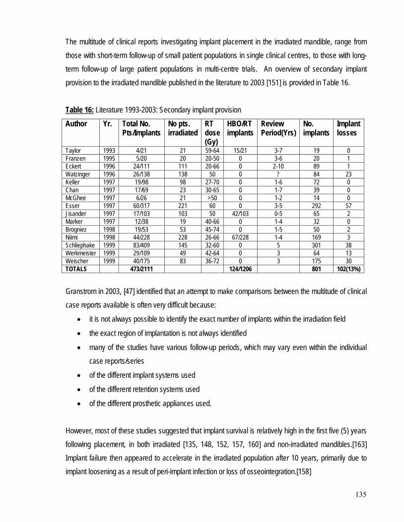
135
The multitude of clinical reports investigating implant placement in the irradiated mandible, range from those with short-term follow-up of small patient populations in single clinical centres, to those with long-term follow-up of large patient populations in multi-centre trials. An overview of secondary implant provision to the irradiated mandible published in the literature to 2003 [151] is provided in Table 16. Table 16:Author
Literature 1993-2003: Secondary implant provision
Yr. Total No. Pts/Implants
No pts. irradiated
RT dose (Gy)
HBO/RT implants
Review Period(Yrs)
No. implants
Implant losses
Taylor 1993 4/21 21 59-64 15/21 3-7 19 0 Franzen 1995 5/20 20 20-50 0 3-6 20 1 Eckert 1996 24/111 111 20-66 0 2-10 89 1 Watzinger 1996 26/138 138 50 0 ? 84 23 Keller 1997 19/98 98 27-70 0 1-6 72 0 Chan 1997 17/69 23 30-65 0 1-7 39 0 McGhee 1997 6/26 21 >50 0 1-2 14 0 Esser 1997 60/317 221 60 0 3-5 292 57 Jisander 1997 17/103 103 50 42/103 0-5 65 2 Marker 1997 12/38 19 40-66 0 1-4 32 0 Brogniez 1998 19/53 53 45-74 0 1-5 50 2 Niimi 1998 44/228 228 26-66 67/228 1-4 169 3 Schliephake 1999 83/409 145 32-60 0 5 301 38 Werkmeister 1999 29/109 49 42-64 0 3 64 13 Weischer 1999 40/175 83 36-72 0 3 175 30 TOTALS 473/2111 124/1206 801 102(13%) Granstrom in 2003, [47] identified that an attempt to make comparisons between the multitude of clinical case reports available is often very difficult because:
• it is not always possible to identify the exact number of implants within the irradiation field
• the exact region of implantation is not always identified
• many of the studies have various follow-up periods, which may vary even within the individual case reports/series
• of the different implant systems used
• of the different retention systems used
• of the different prosthetic appliances used. However, most of these studies suggested that implant survival is relatively high in the first five (5) years following placement, in both irradiated [135, 148, 152, 157, 160] and non-irradiated mandibles.[163] Implant failure then appeared to accelerate in the irradiated population after 10 years, primarily due to implant loosening as a result of peri-implant infection or loss of osseointegration.[158]
136
In 1999, Schliephake et al [158] analysed the long-term survival rates of implants placed in 83 patients. Life table analysis was used to determine the survival rate of 145 implants placed in irradiated bone over 13 years. He concluded that there was no statistical difference found between irradiated and non-irradiated patients. He also identified that the cumulative survival of these implants had a:
• 1 year probability of implant survival of 93.7%
• 5 year probability of implant survival of 86.2%
• 10 year probability of implant survival of 56.5% 2.11.4 Clinical studies related to region of placement - reconstructed mandible 2.11.4.1 Vascularised graft Following ablative surgery many patients are left with a defect in the mandible which is reconstructed with a vascular graft. [47] The benefit of vascularised grafts is that they tend to maintain their shape and form compared to non-vascularised grafts. [129] Grafted vascularised bone has been found to behave more like non-irradiated bone when placed in situ, which is of benefit since the quality of the bone is of the utmost importance for implant survival and success.[27, 31, 150, 158] While long-term data on the use of bone grafts is scarce, particularly in studies with larger sample sizes, implants placed in vascularised bone grafts have been found to be less successful than implants placed in native bone [47], significance (P=0.04) [201] but more successful than implants placed into non-vascularised grafts. [127, 151] This is predominantly because the perfusion of the vascularised bone may be poor. Schliephake et al [158] reported a 74.5% success rate of implants placed in vascularised bone grafts after a 13 year follow-up period. Although there is a reduced success rate of implants into mandibles reconstructed with a vascularised graft, there is a significant functional improvement to be achieved through the provision of implant prostheses in the rehabilitation of these oral cancer patients.[167] Watzinger et al [164] reported on implant survival in 26 patients who underwent radiotherapy with a total dose of 50Gy. The patients were divided into three groups based on the type of bone the implant was placed in. Life table analysis identified the following success rates after three years:
• Implant socket consisted of irradiated local bone (87.8% success)
137
• Implant socket consisted of irradiated local bone following marginal mandibulectomy, with implant surrounded by transplanted soft tissue (69.1% success)
• Implant socket consisted of transplanted bone and soft tissue (58.3% success) Weischer et al [171] studied 7 patients, 2 of which received microvascular anastomosed iliac bone grafts, of which 4 implants were placed in native mandible and 6 in graft. Five patients received open iliac bone grafts, of which 8 implants were placed in native mandible and 15 implants in graft. Patients received radiation doses of between 36-75Gy. Twenty one implants were placed in grafted bone and 12 in native bone, in the two sets of grafted patients. Three implant failures were recorded in the iliac grafts and had to be removed one month after placement due to spontaneous mandibular fracture. McGhee et al [202] reconstructed 6 patients with microvascular fibula or radial grafts. Twenty six implants were placed with 100% survival of implants placed in grafts (14/14) compared to 83% survival of implants in native mandible (10/12). The two implants which failed occurred in the same patient who was a smoker. 2.11.4.2 Non-vascularised graft The long-term function of osseointegrated implants is dependant on viable bone which is capable of remodelling as the implant is subjected to the stresses associated with supporting, retaining and stabilising the prosthesis. The viability of an irradiated and non-vascularised graft may be compromised and not be sufficient to ensure a predictable outcome. Remodelling and resorption can lead to early implant failure in non-vascularised grafts.[129] Wekmeister et al [130] found that the implant survival in non-vascularised grafts was 68% at 36 months follow-up. During the same period implant survival was 73% in the irradiated native mandible and 85% in the non-irradiated mandible. He therefore advocated that non-vascularised grafts not be used when implant placement was planned in irradiated areas. Watzinger et al [164] reported a similarly low success rate of 58.3% when the implant socket consisted of transplanted bone and soft tissue, as did Wagner et al [76] albeit with a slightly higher success rate of 77.5%.
138
Beumer IIIrd et al [40] reported that in a reconstructed non-irradiated mandible a 90% success rate is achieved for implants placed in a free non-vascularised graft, with higher results achievable in a free vascularised graft. They did believe however that lower success rates would be seen in irradiated bone sites. 2.11.5 Clinical studies showing an increased rate of implant loss when placed in irradiated tissues While the use of oral implants in irradiated tissues is no longer contraindicated, it is recognised that the predictability of implant success is altered with implant losses noted in the literature of up to 35%.[30, 33, 96, 126, 127, 130, 134, 135, 137, 143, 146-148, 150, 152, 155-157, 160-162, 164-166, 173, 201-204] It is now well understood that when implants are placed in irradiated bone the failure rate increases because the healing and remodelling capacity of the bone is reduced, impairing the osseointegration process.[124, 144, 145, 147, 148] The first clinical report outlining the possible reduced osseointegration ability of the irradiated bone was published in 1988.[145] The failure rate in Jacobsson’s report was 14% in 9 patients after 44 months, but later studies have shown that the failure rate increases with time.[146] Some studies have seen high implant osseointegration success rates of between 94-100% when implants have been placed into the irradiated anterior mandible.[135, 148, 155] In comparison, in non-irradiated mandibles the implant survival rate in most studies is at least 90% [79, 127, 163, 168] and doesn’t decrease over time. 2.11.6 Clinical studies showing no increased rate of implant loss when placed in irradiated tissues In a comprehensive review Granstrom stated that [47] there is
“no general agreement that osseointegrated implants should fail to a higher degree due to radiation.”
However, the continuous study of the outcome of irradiated patients has shown that with time implant failure has been higher than originally considered. [47, 107, 142, 204]
139
2.11.7 Clinical studies showing stimulation of osseointegration by hyperbaric oxygen Hyperbaric oxygen therapy was introduced as part of the treatment protocol for the installation of osseointegrated implants in Granstrom’s institution in 1998, because at the time it was the only therapy available clinically which could potentially be used to counteract the negative impact of radiotherapy to the soft tissue and bone.[142] However, it cannot completely overcome the progressive obliterative endarteritis induced by radiotherapy.[107] As hyperbaric oxygen had been used successfully for the treatment of ORN [68, 70, 135], it was advocated as a potential adjunct to implant therapy to reduce implant failure in irradiated bone.[177] This was supported by both animal [165] and clinical studies.[27, 135, 147, 162, 165, 205] There was also potential for it to be used as a preventive measure in patients who had received more than 50Gy irradiation to the implant site.[135] Several studies have highlighted the advantages of hyperbaric oxygen for soft tissue wound healing.[126, 134, 157, 167, 204] As a result some investigators have started using hyperbaric oxygen as adjunct therapy to diminish healing disturbances and soft tissue complications. Hyperbaric therapy has been proven beneficial to patients when used prior to 1st stage implant surgery on bone and soft tissue which has been exposed to therapeutic radiation doses.[27, 70, 165, 177, 181] It positively affects the local conditions of the bone and soft tissue, improving the healing capacity and enhancing the process of osseointegration.[145, 147, 159, 162, 165, 177, 206, 207] However, while the healing process is improved it is still slow, so additional time is required for osseointegration in irradiated bone.[135] As stated earlier, the loss of implants in irradiated bone has been as high as 35%. There have been a number of clinical studies published in the literature which have highlighted the benefits of hyperbaric oxygen for osseointegration, and have produced significant reductions in implant loss or failure to less than 10%.[96, 131, 134, 135, 147, 152, 162, 174, 204, 205] Hyperbaric oxygen therapy has also been shown to reduce the failure of implants to less than 15% after 5 years.[134, 142, 174]
140
2.11.8 Clinical studies showing that hyperbaric oxygen is not necessary for osseointegration Several case reports have been published in the literature with acceptable osseointegration results in irradiated bone without hyperbaric oxygen.[155, 156, 159, 161, 163, 166, 191] Some researchers have been able to achieve less than 10% implant failure without using hyperbaric oxygen.[76, 137, 150, 155, 157, 159, 160, 167, 171, 191] Some have been able to sustain this level of failure at less than 10% for 5 years [76, 126, 150, 156, 161] while others have achieved a 10 year failure rate of less than15%.[201] The conclusion which some authors have come to is that hyperbaric oxygen therapy can be an adjunct to osseointegration in irradiated bone, but it is not always required for every patient.[150, 156, 161] Careful patient selection and a diligent atraumatic surgical technique can assist in achieving successful osseointegration without the use of hyperbaric oxygen.[137, 150] Some clinical studies have shown that there is no benefit in the use of hyperbaric oxygen for osseointegration in the mandible, as similar results are achievable with and without its use.[109, 159, 166] Due to the heterogeneity of the number of clinical studies available in the literature, Rosenquist continued to question: [208]
“when is hyperbaric oxygen treatment a necessary part of the treatment plan and when is it not?”
This question was answered by Donoff et al who concluded that currently there is only scant evidence that hyperbaric oxygen therapy is necessary for the majority of patients receiving intra-oral implants.[209] He believed that continued bone biology research and new methods in identifying bone quality and healing capacity will be the long-term solutions to improving osseointegration in irradiated bone.
141
2.11.9 Histology case reports 2.11.9.1 Animal Similar to the clinical case reports, the literature reveals contradictory opinions and experiences pertaining to the use of hyperbaric oxygen in animal histology studies. Animal experiments have shown that while a longer integration time is necessary for implants placed in irradiated bone, the exact relationship between the radiation dose and the integration period is as yet unknown.[129] Larsen et al studied the osseointegration of implants in irradiated rabbit tibias, both with and without the use of hyperbaric oxygen.[189] It was the information from this study which Keller [150] later used as the basis of his debate with Larsen [134] in the Journal of Oral and Maxillofacial Surgery in 1997, to conclude that hyperbaric oxygen is not necessary to attain successful implant osseointegration. Keller concluded that implant placement in the animal model resulted in predictable osseointegration with adjunctive hyperbaric oxygen, which could also be achieved by increasing the healing time alone.[150] Larsen later evaluated the effect of hyperbaric oxygen on osseointegration in rabbit tibias irradiated to 45 Gy. He results of this study were that: [165]
• Osseointegration was successful in both the irradiated bone and the control
• A significant decrease in the percentage of histologic bone to metal contact was noted in the animals irradiated
• This decrease in histologic bone to metal contact was found even though clinically and radiographically there appeared to be successful osseointegration
• Hyperbaric oxygen pre-treatment achieved a bone to metal contact in the irradiated rabbit tibias nearing that of the non-irradiated controls.
Johnson et al studied the long bones of rabbits which were irradiated with a single dose of 15Gy, with half of the cohort receiving hyperbaric oxygen. When the bone to metal contact was evaluated, it was concluded that: [207]
• Irradiation decreased the capacity for osseointegration
• Hyperbaric oxygen improved the bone formation and maturation.
142
2.11.9.2 Human Human histologic data concerning irradiated bone that supports osseointegration of implants is limited to studies of:
• 6 implants radiated to 25-86Gy taken from the maxillae, orbit and temporal bone [145]
• 4 implants radiated to 92Gy taken from the temporal bone at autopsy [147]
• 3 implants radiated to 48Gy taken from the temporal bone at biopsy [203]
• 2 implants radiated to 72Gy taken from the mandible at autopsy [210]
• 23 implants, radiated to 50- 90Gy, with 16 from the oral cavity with11 from mandible, 4 maxillae and1 unknown taken as a result of autopsy and biopsy [211]
All of these studies provided histologic evidence either following autopsy or biopsy, of direct bone to metal contact, despite some bone having received very high radiation doses. In the most recent research from Bolind et al [211] they found “no correlation between high irradiation dose and reduced bone-metal contact or bone in threads”. In fact what they did find was that a correlation existed between time after insertion and bone metal contact, with oral implants in situ for a short period only demonstrating only mainly dense connective tissue at the implant interface.
2.12.1 Definition 2.12 QUALITY OF LIFE
Quality of life is defined as:
“a person’s sense of well-being that stems from satisfaction or dissatisfaction with areas of life that are important.” [212]
Quality of life is usually accepted as being a multidimensional concept, with health-related quality of life an important part. It is generally identified as having two separate components :[212, 213]
• The ability to perform everyday activities. This has a direct impact on the individual’s physical, psychological and social well being.
• The patient’s satisfaction with their levels of function, control of the disease and treatment related symptoms.
143
In head and neck cancer, quality of life is an essential consideration within the treatment planning process. It is an important retrospective tool for clinicians to evaluate the outcome of treatment(s) provided, both success and failure, with respect to mortality, morbidity, survival and recurrence rates.[212] 2.12.2 Impact of cancer on quality of life A diagnosis of head and neck cancer will always have a significant impact on a patient’s quality of life. The malignancy itself as well as associated treatment(s) causes a disturbance to basic functions including speech, mastication, and swallowing, while also having a potential to cause an alteration in appearance together with significant pain. All of these functional impacts can impact deleteriously on the patient’s psychological well being.[213-216] In fact, oral symptoms have been reported as being one of the most distressing aspects in patients being treated for head and neck cancer.[217] Prospective studies have demonstrated that quality of life decreases during treatment, starts improving 3-6 months after completion of treatment and approaches pre-treatment levels by 12 months post-treatment. After this time, quality of life continues to improve slightly for the next 2-3 years.[218, 219] A study by Mehanna et al in 2006 was able to show that there is a late deterioration in quality at life at 10 years post-treatment.[219] The goal of therapy in the treatment of locally advanced head and neck cancer is for: [21]
• Cure
• Organ preservation
• Reduction of morbidity associated with therapy
• Reasonable quality of life. Traditionally, assessments of head and neck cancer patient management have focused on the cure and control of cancer. That is statistical rather than quality of life assessments.[213] Quality of life assessments have the potential to add another dimension to the decision making process, particularly when alternative treatment plans may have similar outcomes with respect to tumour response, but different outcomes with respect to the impact on post-treatment quality of life for the patient(s). Results of a study by Epstein et al in 2001 [220] recommended that there is a need to assess the potential oral dysfunction associated with any cancer treatment(s), so that a treatment plan which
144
provides the best cure or best palliation of the malignancy is achieved with the least impact on oral function and quality of life. In addition, oral health-related quality of life surveys have concluded that achieving a reduction in the side-effects of cancer therapy is the best way to reduce the associated psychological morbidity.[213] Currently, the influence of age on quality of life for patients being treated for head and neck cancer is not clear, with some reporting worse outcomes for younger patients (less than 60 years old)[221], and others reporting worse outcomes in older patients (greater than 75 years).[222] A study by Rogers et al in 2006 identified that younger patients fare much worse compared to older patients, especially with respect to mobility, pain/discomfort and anxiety/depression. The authors concluded that this potentially occurs because “a younger patient experiences more ‘functional’ losses in comparison to others and they may have higher hopes and expectations of returning to a ‘normal’ outcome state.”[221] In comparison, Khafif et al identified that several quality of life domains are decreased in elderly patients (greater than 75 years of age) which could be mainly attributed to a more pronounced effect of pain on their daily living activities, an inability to resume a normal life and a sense of burden on caregivers.[222] The importance of pain management in head and neck cancer patients is well recognized and has significant implications on quality of life. There is also some evidence to suggest that head and neck cancer patients who do not survive in the long-term have substantially different health-related quality of life profiles than long-term survivors, both quantitatively and qualitatively. A study by Goldstein et al in 2007 found that short-term survivors (less than one year post-treatment) had a lower health-related quality of life compared to long-term survivors (greater than three years).[218] 2.12.3 Impact of ablative surgery on quality of life There have been significant advances on the treatments and successes associated with the management of head and neck cancers, especially with respect to functional rehabilitation, but there continues to be a considerable continuing psychological and social impact of head and neck cancer.[214]
145
One of the most psychological disturbing outcomes of treatment prior to the availability of microvascular free-flaps was the cosmetic deformity associated with ablative surgery. The surgical resection of head and neck and oropharyngeal cancers can potentially have a significant functional impact with alterations to the individual’s: [45, 215, 223]
• Clarity of articulation
• Functional swallow
• Ability to control saliva secretions
• Mouth opening
• Oral sensation
• Muscular control All of these functional alterations have an impact on the individual’s quality of life.[45, 224] While there have been general assessments of quality of life in head and neck cancer patients, there has been limited assessment of the oral/dental complications of head and neck cancer treatment(s) and the quality of post-treatment oral function.[213, 225] A study by Rogers et al in 1998 assessed the quality of life of 48 patients who were undergoing primary ablative surgery for oral and oropharyngeal squamous cell carcinomas.[214] Quality of life was assessed at the time of presentation, 3 months post-operatively and again at 12 months post-operatively. At the time of presentation, patients obtained scores lower than the norm especially for limitations in physical, mental and social functioning. At the 3 month post-operative assessment, there was a considerable deterioration in the patient’s physical functioning, energy levels and general health perception. However, at the 12 month post-operative assessment, patients approached their pre-treatment results. The conclusion made by the authors was that during this 12 month period, there was a continued need for both psychological and physical support following ablative surgery for head and neck cancer.[214] A study by Epstein et al in 1999 [213] assessed the quality of life, oral function and oral symptoms following treatment for oral cancer. Results of this study indicated that quality of life was reported to be higher in non-surgically managed head and neck cancer patients compared to those who underwent
146
ablative surgery. This study also identified that the most common side-effects associated with ablative surgery were:
• Difficulty eating (82% of patients)
• Difficulty swallowing (81% of patients)
• Difficulty speaking (77% of patients)
• Difficulty chewing (68% of patients)
• Disfigurement (55% of patients). These results were supported by work of Zuydam et al in 2005 which identified that issues surrounding post-surgical speech and swallowing were rated as amongst the most important health-related quality of life factors in survivors at 12 and 18 months, along with reduced saliva and decreased chewing ability.[226] Prospective studies that quantify quality of life related to purely surgical measures are lacking.[227, 228] A study by Schliephake et al in 2002 identified that surgical therapy of oral cancer in the floor of mouth led to a temporary deterioration of physical function and role function three months after surgery. In addition, a significant decrease in oral function, body image and social contact was also identified. In this clinical scenario, no quality of life measures reflected pre-treatment levels after 12 months.[227] 2.12.4 Impact of radiotherapy on quality of life Radiotherapy is a common adjunctive therapy in the management of head and neck cancer. Oral complications during and after radiotherapy are common. General oral discomfort, together with eating and speaking difficulties has been demonstrated to have a significant impact on the patient’s quality of life.[229] In fact, radiotherapy seems to be a dominating factor influencing oral function and quality of life. Many problems with oral function are a result of post-radiotherapy sequelae.[45] A study by Ohrn in 2002 found that of 18 patients treated with radiotherapy, 56% reported that their post-radiotherapy oral condition had a profound influence on their health-related quality of life, and an additional 33% reported some influence on their health-related quality of life. Only 11% reported no influence on their health-related quality of life.[217]
147
Bansal et al assessed the impact of radiation related morbidities on quality of life in 2004.[229] They identified that an increase in symptom scores of appetite loss, fatigue and pain led to a significant decline in physical, social and emotional functioning, as well as a reduction in global health status. A study by Epstein et al in 1999 [213] assessed the quality of life, oral function and oral symptoms using the European Organization for Research and Treatment of Cancer (EORTC) QLQ-C30 questionnaire following treatment for oral cancer. This study identified that the most common side-effects associated with radiotherapy were:
• Oral dryness (84% of patients)
• Pain (70% of patients)
• Reduced taste sensation (70% of patients)
• Reduced appetite (70% of patients), and
• Reduced smell (45% of patients). These results indicated that oral dryness or xerostomia was a significant side-effect of radiotherapy. The authors concluded that managing the oral dryness associated with radiotherapy is central to maintaining oral comfort and function, and therefore impacted significantly on the patient’s quality of life. Epstein et al built on this work in 2001 [220] by assessing the quality of life, oral function and oral symptoms in a cohort of 65 patients during and after radiotherapy. While it is known that patients experience significant oral affects following radiotherapy to the head and neck particularly associated with xerostomia, the frequency and severity of these affects are not well understood. This study used the EORTC QLQ-C30 questionnaire and reported that symptoms involving oral function were:
• Xerostomia (95% of patients)
• Change in taste (90% of patients)
• Chewing/eating difficulties (70% of patients)
• Moderate to severe dysphagia (65% of patients)
• Speech difficulties (65% of patients)
• Mouth pain (55% of patients) While xerostomia is known to be a common sequelae of radiotherapy with a significant impact on quality of life, the relationship between an individual’s perception of oral dryness and alteration to the actual salivary flow rate has not been clearly defined. A study by Logemann et al in 2001[230] examined the
148
quantity of saliva produced prior to and following radiotherapy (40Gy) for oral, pharyngeal and laryngeal cancers. The conclusion reached was that:
• Post-radiotherapy xerostomia led to a significant increase in the patient’s perception of swallowing difficulties despite the fact that it did not affect the physiologic aspect of swallowing, and
• Xerostomia affected the sensory process and comfort of eating more than the physiologic process of bolus transport.
Logemann expanded on this research in 2003 [231] and found that while the reduction in salivary weight associated with xerostomia did not affect the physiologic aspect of bolus transport, it did alter the patient’s perception of their swallowing ability and level of comfort, which in turn impacted on their dietary choices. 2.12.5 Impact of oral rehabilitation on quality of life An important consideration in the oral rehabilitation of the head and neck cancer patient is their current residual functional capacity and prognosis. A long-term study by Duke et al identified that patients who were edentulous due to cancer had a decreased quality of life.[232] Functional oral rehabilitation is a long and complex process in which some patients: [224]
• Choose to have no involvement following completion of cancer treatment(s), while others
• Do not survive long enough or remain well enough to gain benefit from rehabilitation, as the majority of recurrences occur within the first year of treatment.
The median time frame ascertained for rehabilitation by Rogers et al [224] was:
• 12 months from ablative surgery to commencement of rehabilitation
• 14 months from commencement to completion of rehabilitation. Patients requiring functional rehabilitation following cancer treatment(s) for head and neck surgery are primarily those who tend to: [224]
• Have larger more extensive tumours
• Be edentulous in the mandible with a denture in the maxillae
• Have scored lower in quality of life assessments
149
• Have undergone adjunctive radiotherapy.
If patients are not able achieve oral rehabilitation after cancer treatment(s) they experience significantly increased psychological as well as functional morbidity.[215] In order to achieve successful functional and aesthetic rehabilitation, and improved oral health quality of life, patients need to be satisfied with respect to: [233]
• Comfort
• Aesthetics
• Prosthesis stability and retention
• Phonetics or speech intelligibility
• Masticatory performance
• Chewing comfort
• General oral health.
The selection of a patient for functional oral rehabilitation is dependant upon: [215, 224]
• The patient’s desire and consent to treatment
• The patient’s expectations
• Adequate access to oral cavity (no trismus limitations)
• No contraindications to the provision of treatment. The purpose of oral rehabilitation following cancer surgery is to restore lost function and anatomic form while concomitantly improving the patients well being and quality of life. From a dental perspective this may encompass the provision of:
• No dental prosthesis
• A conventional prosthesis (denture), or
• An implant prosthesis.
A study by Kwakman et al in 1997 examined the case notes of 95 consecutive head and neck cancer patients to identify the number or proportion of these patients who required an implant prosthesis as part of their oral rehabilitation. Their results were that 45% of patients did not need any specific prosthodontic rehabilitation, 25% had a clinical indication for the use of implant prostheses, but only 3% of these patients were actually provided with an implant prosthesis. [125]
150
The oral complications of cancer treatment(s) often have implications for dental health and the patient’s ability to successfully wear or even tolerate conventional dentures. Surgical alterations in the oral musculature, tongue immobility, compromised jaw opening, reduced sensation together with radiotherapy related xerostomia and mucosal fragility all contribute to a reduced ability to manage conventional dentures.[215] This has the potential to impact on the patient’s food choices and ability to maintain an adequate nutritional intake.[213] The functional success of prostheses in edentulous patients is believed to have an impact on their diet. Studies have found that patients who are edentulous have generally a: [234]
• Reduced consumption of fruits, vegetables an fibre, particularly in males
• Increased consumption of fat, particularly for females
• Increased consumption of processed foods which may include increased consumption of saturated fats and cholesterol.
A strong co-dependant relationship between oral health and nutrition exists, particularly in patients with head and neck cancer.[235] However, a recent review of the literature by Al-Omiri et al [233] has identified that while implant prostheses substantially enhanced quality of life and the self confidence of patients by enhancing their masticatory ability, it did not necessary lead to an improved diet. While it has been identified that many patients cannot tolerate conventional dentures, there have been significant and predictable improvements in patient function, patient satisfaction and oral health-related quality of life, when complete lower dentures are supported by a minimum of two (2) implants. [25, 39, 215, 233, 236] A study by Heydecke et al [236] in 2003 looked at the impact of implant overdentures in general and with respect to the oral health quality of life using the Oral Health Impact Profile questionnaire before treatment, and then at two (2) and six (6) months post-insertion. Their results were that:
• Overdentures were superior, with respect to functional limitations, physical pain and disability, and psychological disability
• Patients’ obtained higher scores of satisfaction with respect to chewing ability, comfort, ability to speak and aesthetics. Overall there was an improvement in the patient’s psychological well being.
151
Similar results with respect to comfort, stability and aesthetics were obtained by Allen and McMillan in 2003 [237], and Abu-Hantash et al in 2006 [238] when they compared prostheses with conventional prostheses. More recent research conducted by Schoen et al in 2007 [191, 225] and in 2008 [128] has concluded that while surgical treatment and subsequent radiotherapy in head and neck cancer patients often results in an anatomic and physiological condition which is unfavourable for prosthodontic rehabilitation, the provision of implant prostheses can likely improve these patients' quality of life with respect to oral function and denture satisfaction.
In the past, clinical research mainly focussed on the course of disease and treatment results.[239] Results of treatment strategies for oral cancer were mostly identified as an expression of the patients’ disease-free state or overall survival. Although this data is important for the purpose of comparison of different treatment regimes, it provides no information about treatment specific problems or long-term sequelae for patients. The need for additional instruments in order to provide more detailed outcome research has led to the development of quality of life assessment tools.
2.13 QUALITY OF LIFE ASSESSMENT TOOLS
Progressively more clinical trials are incorporating the use of quality of life assessment tools as part of the review process to identify patient satisfaction and/or oral health quality of life as outcome variables. These two variables complement the clinical outcomes which clinicians use as markers of success.[236] The measurement of patient perception outcomes as data endpoints in clinical research was initially viewed with considerable scepticism, primarily because clinicians were trained to analyse and value objective data. Eventually the concept was embraced, and now patient perceptions and health-related quality of life measures are considered to be an accurate and candid reflection of the patients’ well-being, and ability to carrying out daily living activities. The importance of quality of life measurements in head and neck cancer patients is acknowledged due to the significant effect(s) of the disease and its treatment. The focus of quality of life measurements is on the emotional, social and physical aspect of the disease and its treatments.[240]
152
In health, we focus on the patient’s health-related quality of life. This is a “multi-dimensional concept dealing with quality of life related specifically to health and disease”.[217] Health-related quality of life (HR-QOL) refers to “the physical, psychological and social functioning of patients and the impact of disease of their abilities and daily functioning.”[217] The World Health Organization (WHO) has defined health-related quality of life as “an individual’s perception of their position in life in the context of the culture and value systems in which they live and in relation to their goals, standards and concerns. It is a broad ranging concept affected in a complex way by the person’s physical health, psychosocial state, level of independence, social relationships and their relationships to salient features of their environment.”[241] Any instrument designed to assess health-related quality of life must not only reflect the definitions described above, but also suit the patient population, clinical setting and practical needs of any given study parameter, including the timing of assessment, disease sub-site and treatment. Often there is no single quality of life measurement tool that ideally measures health-related quality of life in any particular clinical situation, and therefore several different questionnaires or modules may be required.[241] As a result, in the assessment of health status and quality of life it has been recommended that both specific and generic tools/questionnaires be used. [213] During the past few years a wide variety of instruments have been developed, assessed, critically reviewed and validated in cancer patients for assessment of quality of life.[241] The spectrum of these measures or instruments has encompassed general health-related questionnaires and general cancer questionnaires, as well as specific head and neck modules which have been designed and used in both cross-sectional and longitudinal studies. The choice of both generic and specific measurement tools used in the form of questionnaires depends on the purpose of the study and the resources available. Generic questionnaire tools provide information on the impact of the cancer on the patient’s general health, well being and function. Specific questionnaire tools provide information on alterations to the body parts and activities which are directly affected by the cancer. [236] Previously the successes and failures associated with head and neck surgery were measured and recorded by the treating clinician(s). Currently detailed outcome research via the use of quality of life assessment tools are being undertaken through the use of self-administered patient questionnaires [227], as it has been recognised that clinicians and patients may have substantially different opinions on
153
what each considers important for overall quality of life.[242, 243] It is important that these questionnaires are easily understandable, and able to be completed within a reasonable time frame (10 minutes).[212] A study by Bjordal et al concluded that patient self-administered questionnaires were more accurate and sensitive than clinician-rated questionnaires. One of the benefits of these types of questionnaires is that the results can be used as a tool for improved communication between clinicians and patients, to assist in the differentiation and choice between different treatment modalities which may have similar survival rates but different functional outcomes.[242] While the results of these quality of life assessment tools do add value, it often remains difficult to define and identify at what level of change this data is clinically relevant. The ‘minimally important difference’ (MID), refers to the smallest difference identified in quality of life assessment tool(s) data that reflect a clinically important change. A study by Ringash et al in 2007 concluded that a positive MID is about 5% of the maximal instrument score, while a negative MID is significantly higher, at about 10% of the maximal instrument score.[244] The other important aspect of these tools is that the ability to obtain statistically significant changes in head and neck cancer patients is difficult, especially for short-term research within a single institution setting in which quality of life is the measured variable.[245] 2.13.1 European Organization for Research and Treatment of Cancer (EORTC) questionnaire(s) While there is an overwhelming volume of literature published pertaining to quality of life assessment, there is minimal specific to head and neck cancers. An article by Rogers et al in 1999 [212], reviewed the English language literature from 1980 to 1997, and provided a summary of 65 articles on quality of life assessment in oral cancer. They found that one of the most commonly used generic cancer questionnaires used to identify quality of life was the European Organization for Research and Treatment of Cancer, Quality of life C30 questionnaire (EORTC QLQ-C30), and its addendum, the Head and Neck specific questionnaire EORTC H&N35.[212] In 2007 Pusic et al completed a systematic review of patient reported outcome measures in head and neck cancer surgery. The goal of this review was to identify and compare site specific health-related quality of life questionnaires in head and neck cancer surgery. They concluded that the EORTC questionnaires were particularly robust, and had undergone thorough development and validation.[223]
154
The European Organization for Research and Treatment of Cancer (EORTC) QLQ-C30 in combination with the EORTC H&N35 constitutes a set of 65 questions that have been developed to identify disease-, site- and treatment-specific effects as well as general measures including emotional, mental and social aspects of cancer. Comparison of the EORTC questionnaires with both general cancer questionnaires and global health-related measurement tools has identified that both the EORTC QLQ-C30 and the H&N35 addendum are able to identify subtle changes in many areas. Both of these tools have been validated extensively, both in cross-section and longitudinal studies in large populations.[227] The two questionnaires are specifically designed vehicles for assessing the quality of life of cancer patients participating in clinical trials and other types of research in which patient-reported outcomes are collected. For both questionnaires there is international reference data available [246] specific to head and neck cancer. This reference data can provide information about the distribution of quality of life scores, which may assist when assessing clinical endpoints, and it may also be of use as a prognostic factor for clinical outcomes and response to treatment. 2.13.1.1 EORTC QLQ-C30 In 1986 the EORTC brought together a study group to develop an integrated, modular approach for the evaluation of quality of life of patients participating in international cancer trials.[247, 248] This resulted in the development of the EORTC QLQ-C30 questionnaire to assess the general aspects of cancer. It was trialled during development on patients of many cultures and languages, to ensure cross cultural validity [223, 241, 247, 248], and is a patient-based self-administered instrument. During trials the average time to complete the questionnaire was 11 minutes.[248] It is now a copyrighted instrument which has been translated and validated in 63 languages and has been used in more than 3000 studies worldwide.[242, 249, 250] The EORTC QLQ-C30 is a, general standardised measurement tool which can be applied to any malignant process. It provides information on the symptoms and side-effects, both physical and psychological, of treatment(s) used in the management of malignancies.[212, 220, 223] The current version of the QLQ-C30 (version 3) questionnaire consists of 30 questions which assesses several domains including 5 functional scales, 2 global scales, 3 symptom scales, and 6 single items:[241]
• Functional scales o physical function (5 questions) o role function (2 questions)
155
o social function (2 questions) o emotional function (4 questions) o cognitive function (2 questions), and
• Global scales o global health status (1 question) o quality of life (1 question)
• Symptoms scales o pain (2 questions) o fatigue (3 questions) o nausea and vomiting (2 questions)
• Single items o diarrhoea o constipation o breathing function – dyspnoea o appetite loss o sleep disturbance/insomnia o economic sequelae – financial impact.
Each scale is linearly transformed to a score from 0-100 with no summary score. Higher scores in the functional and global scales reflect a better quality of life. In the symptom and single item scales, higher scores represent a higher degree of problems or symptoms, and therefore a worse quality of life. One of the major differences between the EORTC quality of life questionnaires and other quality of life measures is that there is no composite score generated.[227, 241] A composite quality of life score, or sum of the domain score, is at risk of an internal cancellation effect, and therefore may not be as sensitive. Individual analysis of the quality of life domains provides for a more accurate picture of the complex functional changes which occur as a result of cancer or its treatment. [245] The EORTC QLQ-C30 is designed to be used together with diagnosis-specific modules to increase the sensitivity and specificity of the assessments in various patients and treatment groups.[251] A modular approach ensures that the assessments are tailored to specific patients groups, through the use of disease specific modules, while still allowing comparability across studies for patients with different cancers through the use of the general measure (EORTC QLQ-C30).[250] For patients with head and neck cancer, the disease specific module is the EORTC H&N35.
156
2.13.1.2 EORTC H&N35 The EORTC core generic questionnaire (QLQ – C30), and the head and neck specific addendum (H&N35), have both been rigorously tested and evaluated for cross-cultural applicability. When used together, they provide the clinician(s) with a measure of the quality of life and oral function capacity in head and neck cancer patients in all stages of the disease - acute, chronic, treated and untreated. This oral outcome measure is then available to use for comparative purposes, as well as for the assessment of oral care prevention and management strategies in head and neck cancer patients.[220] The EORTC H&N35 is a carefully constructed head and neck specific disease module which has undergone extensive international testing and it appears psychometrically robust.[251-253] It was developed following a literature review, expert opinion, focus groups, patient interviews, item reduction with factor analysis and psychometric testing.[252] It is designed to be patient administered, with a mean completion time of 18 minutes.[251] The EORTC head and neck addendum (EORTC H&N 35) is a specific standardised measurement tool, which has been designed to be used across a range of patients with head and neck cancer of varying stages. It comprises of 35 questions which assess the specific complications, symptoms and side-effects of treatment.[212, 220, 252] It is comprised of seven domains or multi-item scales and 11 single items:
• Pain (4 questions)
• Swallowing (4 questions)
• Senses (2 questions)
• Speech (3 questions)
• Social eating (4 questions)
• Social contact (5 questions)
• Sexuality (2 questions)
• Single items o problems with teeth o problems with mouth opening o dry mouth o sticky saliva o coughed
157
o felt ill o painkillers o nutritional supplements o feeding tube o weight loss o weight gain
The scores of this module also range form 0-100 with a higher score representing higher degrees of problems and good results showing a low score. A review by Sherman et al in 2000 identified one of the major advantages of the EORTC H&N35 was that it provided this subscale of scores for different domains of functioning, instead of a composite quality of life score, therefore preventing problem areas from being obscured, as is the risk with general total scores.[250] 2.13.2 Oral Health Impact Profile (OHIP) questionnaire The Oral Health Impact Profile is a “scaled index of the social impact of oral disorders”. It is an instrument that was specifically designed to measure the impact of oral health on quality of life, and was developed following interviews of 64 patients in which 535 statements were recorded and subsequently reduced to 49 statements. These statements described the consequences of oral disorders, and perceptions of the impact of oral conditions on the patient’s wellbeing.[254] 2.13.2.1 OHIP-49 The Oral Health Impact Profile (OHIP) tool is a 49 item profile that comprehensively measures the impact of the oral health condition on aspects of function, daily living and social interactions including: [236, 254]
• Functional limitations (9 questions)
• Physical pain (9 questions)
• Psychological discomfort (5 questions)
• Physical disability (9 questions)
• Psychological disability (6 questions)
• Social disability (5 questions)
• Handicap (6 questions)
158
It is a reliable, valid and responsive instrument which is able to obtain a detailed measurement of the social impact of oral disease, and has potential benefits for both clinical decision making and research.[254, 255] Some of the benefits of the OHIP have been identified as:[254]
• Identification of priorities for dental care
• An understanding of oral health-related patient behaviours
• Advocacy for oral health care
• Improved evaluation of dental treatment. A study by Lee et al in 2007 used the Oral Health Impact Profile to identify if an individual’s subjective perceptions of oral health status had a greater impact than their actual clinical status on their health-related quality of life. They concluded that it did, and that the OHIP was “slightly more focussed on the measurement of the mental aspect” of quality of life.[240] While the OHIP development focussed as a population based measure, it has the potential to be used to assess the impact of disease on individuals.[254] However, it is a relatively lengthy questionnaire, and in 1997 a short form OHIP was developed and validated.(OHIP-14).[256] 2.13.2.2 OHIP-14 The OHIP-14 contains fourteen questions which are a subset of the original 49 items, and therefore has retained the conceptual dimensions of the original OHIP. The instrument has good reliability, validity and precision and so the modified instrument should be useful for quantifying levels of impact on well-being in situations where only a limited number of questions can be administered.[256] 2.13.2.3 OHIP-EDENT In 2002, Allen and Locker developed a modified short version (19 questions) of the OHIP for assessing health-related quality of life in edentulous adults (OHIP-EDENT). They identified that a number of questions relating to denture wearing were excluded in the OHIP-14, which had the potential to affect results when evaluating prosthodontic treatment outcomes in edentulous patients. They believed that a portion of the statements in the OHIP-14 may not be suitable for identifying a clinically meaningful change following prosthodontic procedures. In particular, their research identified that the OHIP-14 had a poorer responsiveness to change in edentulous patients, particularly when comparing conventional prostheses to implant prostheses. In comparison, the OHIP-EDENT was able to detect change in the edentulous patients’ ratings of their new prostheses.[257]
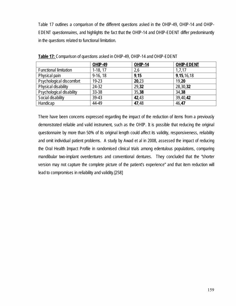
159
Table 17 outlines a comparison of the different questions asked in the OHIP-49, OHIP-14 and OHIP-EDENT questionnaires, and highlights the fact that the OHIP-14 and OHIP-EDENT differ predominantly in the questions related to functional limitation. Table 17:
Comparison of questions asked in OHIP-49, OHIP-14 and OHIP-EDENT
OHIP-49 OHIP-14 OHIP-EDENT Functional limitation 1-18, 17 2,6 1,7,17 Physical pain 9-16, 18 9,15 9,15,16,18 Psychological discomfort 19-23 20,23 19,20 Physical disability 24-32 29,32 28,30,32 Psychological disability 33-38 35,38 34,38 Social disability 39-43 42,43 39,40,42 Handicap 44-49 47,48 46,47 There have been concerns expressed regarding the impact of the reduction of items from a previously demonstrated reliable and valid instrument, such as the OHIP. It is possible that reducing the original questionnaire by more than 50% of its original length could affect its validity, responsiveness, reliability and omit individual patient problems. A study by Awad et al in 2008, assessed the impact of reducing the Oral Health Impact Profile in randomised clinical trials among edentulous populations, comparing mandibular two-implant overdentures and conventional dentures. They concluded that the “shorter version may not capture the complete picture of the patient’s experience” and that item reduction will lead to compromises in reliability and validity.[258]
160
This chapter explains the methods used to conduct the study. It describes the study design, sampling frame and data collection methods, provides details of the quality of life questionnaires, clinical and radiographic examinations undertaken, dental treatment provided, as well as a summary of the analytical approaches undertaken. Data management includes data weighting, data scoring, recording of patient oncology details and response formats. Ethical implications and approvals are also reported.
CHAPTER 3: METHODOLOGY
The study was a prospective, longitudinal, non-random design. It evaluated the treatment outcome (condition of peri-implant tissues, implant survival, implant related complications) and impact on quality of life, following prosthodontic rehabilitation with an implant overdenture compared to no implant overdenture. All participants were edentulous head and neck cancer patients who have had radiotherapy to the mandible. All participants resided in South Australia at the commencement of the research period.
3.1 STUDY DESIGN
3.1.1 Sampling frame 3.1.1.1 Target population The target population comprised of edentulous adults greater than 18 years of age, who had received radiotherapy to the mandible as part of oncology treatment for head and neck cancer, and who currently have no lower complete denture. 3.1.1.2 Inclusion criteria The inclusion criteria included:
• A head and neck cancer patient
• who has received at least 55 Gy of radiotherapy
• in which the external beam included the mandible
• who has also possibly had ablative surgery for the head and neck cancer, and
• who has also possible had chemotherapy for the head and neck cancer,
• who is currently attending clinics at the Royal Adelaide Hospital
• is free of active disease
• living in South Australia
• has signed a consent form.
161
3.1.1.3 Exclusion criteria If a potential research subject meets all of the inclusion criteria, they may still be excluded if they
• are dentate in the mandible
• have declined involvement in the research
• have a previous history of ORN
• have a high likelihood of being lost to follow-up, either as a result of planning to move interstate or they are a long distance rural patient
• have experienced claustrophobia in the hyperbaric oxygen chamber
• have experienced ear barotrauma after two treatments, or
• there is a contraindication to hyperbaric oxygen treatment as determined following assessment by the hyperbaric medicine consultant. (assessment includes Ophthalmological and ENT examinations)
3.1.1.4 Patient selection From July 2006, all edentulous patients who attended the Special Needs Unit of the Adelaide Dental Hospital and who had been diagnosed and treated for a malignancy in the head and neck region (including squamous cell carcinoma and others) with radiotherapy alone or in combination with either chemotherapy or surgery or both, were approached to be included in the study. All potential research subjects received a letter of introduction (Appendix 1). Following the initial assessment and screening appointment with the Special Needs Dentist, subjects were provided with a consultation appointment with the Oral and Maxillofacial Surgeon and the Hyperbaric Oxygen Consultant (Appendix 2) if they were interested in an implant overdenture. Once the subject had agreed to be involved in the research in either Group 1(intervention group) or Group 2 (control group), a consent form was completed (Appendix 3), and the appropriate information sheet provided (Appendix 4 and 5) Medical records of all enrolled research subjects were reviewed for the following data:
• Tumour diagnosis and location
• TNM classification
• Specific oncological treatments provided
• Irradiation dose and field
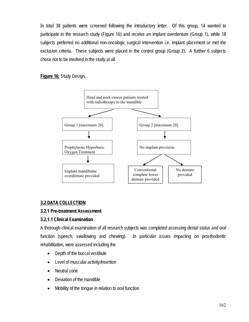
162
In total 38 patients were screened following the introductory letter. Of this group, 14 wanted to participate in the research study (Figure 16) and receive an implant overdenture (Group 1), while 18 subjects preferred no additional non-oncologic surgical intervention i.e. implant placement or met the exclusion criteria. These subjects were placed in the control group (Group 2). A further 6 subjects chose not to be involved in the study at all. Figure 16:
Study Design.
Head and neck cancer patients treated with radiotherapy to the mandible
Group 1 [maximum 20]
Group 2 [maximum 20]
Prophylactic Hyperbaric Oxygen Treatment
No implant provision
Implant mandibular overdenture provided
3.2.1 Pre-treatment Assessment 3.2 DATA COLLECTION
3.2.1.1 Clinical Examination A thorough clinical examination of all research subjects was completed assessing dental status and oral function (speech, swallowing and chewing). In particular issues impacting on prosthodontic rehabilitation, were assessed including the
• Depth of the buccal vestibule
• Level of muscular activity/insertion
• Neutral zone
• Deviation of the mandible
• Mobility of the tongue in relation to oral function
Conventional complete lower
denture provided
No denture provided
163
• Sensibility of the lip and chin
• Wetness of the oral mucosa. 3.2.1.2 Radiographic Examination An orthopantomograph was taken as part of the initial examination process to assist with the detection of potential pathologies, as well as to provide a two dimensional assessment of the bone quality in the mandible for all patients. For research patients in Group 1 this assisted with the treatment planning for the Oral and Maxillofacial Surgeon with respect to implant length and positioning in the mandibular interforaminal region. If further radiographic information was required, a CT scan was obtained. 3.2.1.3 Baseline Questionnaires (T0) Research subjects in both groups were asked to complete the self-administered European Organization for Research and Treatment of Cancer quality of life questionnaires (EORTC QLQ-C30 and EORTC H&N35), as well as the shortened Oral Health Impact Profile questionnaire (OHIP-14). The European Organization for Research and Treatment of Cancer core questionnaire (EORTC QLQ-C30) (Appendix 6) consists of 30 questions or items exploring 6 multi-item function scales (physical function, role function, social function, emotional function, cognitive function and overall health status or quality of life), as well as 3 multi-item symptom scales (pain, fatigue and emesis) and 6 single items (bowel function, breathing, appetite, sleeping disorders and economic sequelae). The European Organization for Research and Treatment of Cancer head and neck module (EORTC H&N 35) (Appendix 7) consists of 35 questions exploring symptoms and side-effects of treatment. It comprises of 6 multi-item scales (pain, swallowing, senses, speech, social eating, social contact and sexuality) and 7 single items (problems with teeth, problems with mouth opening, dry mouth, sticky saliva, coughed, felt ill, painkillers, nutritional supplements, feeding tube, weight loss and weight gain). The physical, psychological and social impact of oral disorders was assessed using the shortened version of the Oral Health Impact Profile (OHIP-14) questionnaire (Appendix 8). This comprises of 7 multi-item scales including functional limitation, physical pain, physical disability, psychological discomfort, psychological disability, social disability and handicap.
164
3.2.2 Dental Treatment provided 3.2.2.1 Research subjects in Group 1 Prior to undergoing 1st stage implant surgery for implant fixture placement, research subjects in Group 1 received hyperbaric oxygen treatment at the Hyperbaric Medicine Unit of the Royal Adelaide Hospital. This involved the provision of 20 treatments of 100% oxygen at 2.4 Atmospheres for 80 minutes, in a multi-place hyperbaric chamber. Four patients from Group 1 [Patients no. 1,6,8,12] had received hyperbaric oxygen treatment in the last 12 months, and therefore their treatment protocol was modified such that they only received 10 treatments prior to 1st stage implants surgery. Research subjects were able to nominate if the 1st stage implant surgery was to be provided under local anaesthesia or general anaesthesia. All patients who were treated under local anaesthesia received antibiotic prophylaxis one hour prior to surgery. The antibiotic of choice was 2g Amoxicillin orally, followed by 1g Amoxicillin orally six hours later, or for those patients who are Penicillin allergic, 600mg oral Clindamycin. For the two patients who were treated under general anaesthesia, 2g Amoxicillin was provided intravenously at the time of surgery. In all research subjects, two endosseous 3.5/4.0 implants [Osseospeed, Astratech, Sweden] were placed by one experienced Oral and Maxillofacial Surgeon in the interforaminal region of the mandible. Immediately after 1st stage surgery, all research subjects received an additional 10 hyperbaric treatments of 100% oxygen at 2.4 Atmospheres for 80 minutes, in a multi-place hyperbaric chamber. Following a healing period of four weeks, all research subjects had conventional complete upper and lower dentures constructed by the author, according to standard clinical and laboratory procedures, with the lower denture incorporating a permanent silicone based soft liner [Molloplast B, Detax Gmbh & Co, KG Germany]. After an osseointegration period of six months, 2nd stage implant surgery was provided. This consisted of uncovering of the implant body under local anaesthesia with minimal soft tissue trauma. The osseointegration of each implant was assessed by placing manual force on the implant to assess if there was any mobility. If the implant was immobile, the transmucosal healing abutment was connected and
165
then mucosal tissues abutted with no suture placement. Research subjects were instructed to not wear the complete lower denture for a period of two weeks, and to rinse after each meal with 0.2% chlorhexidine [Curasept, Curaden Swiss]. A minimum of two weeks after 2nd stage implant surgery, the transmucosal healing abutments were removed and ball implants placed. The height of the ball abutments were determined by the use of an abutment depth gauge, to ensure that the gingival margin was placed at the tapered end of the ball abutment. The ball abutment was then torqued to 25Ncm. The conventional complete lower denture was then eased to allow the denture to seat properly over the ball abutments, and an impression taken with the rubber-based impression material, Impregum [3M Espe]. The female implant component was then incorporated into the relined lower complete denture using standard laboratory procedures. The implant overdenture was inserted one week later and the occlusion balanced bi-laterally, with special care taken to ensure freedom of movement during functional dynamic occlusion. The fitting surface of the overdenture was also checked to ensure minimal soft tissue pressure. Home care instructions were provided with regard to the daily hygiene of both the prosthesis and peri-implant tissues, as well the provision of Curasept an alcohol free 0.5% chlorhexidine gel. [Curaden Swiss] Research subjects were reviewed on a regular basis following insertion of the overdenture, to provide adjustments of the prosthesis as required thereby minimising denture related soft tissue trauma and ulceration during the initial post-insertion period. 3.2.2.2 Research subjects in Group 2 In the control group, research subjects were provided with a conventional denture if requested. In total, twelve research subjects received a conventional full lower prosthesis with a soft liner Molloplast B [Detax Gmbh & Co, KG Germany]. These subjects were counselled regarding the risk of ORN associated with denture related ulceration, and reviewed regularly during the post-insertion period. Research subjects were also advised prior to denture construction on the likely success associated with their wearing of conventional removable dentures.
166
3.2.3 Review Assessment 3.2.3.1 Clinical Examination A standardised clinical examination (Appendix 9) of all Group 1 research subjects was conducted at time T1 [range 1 month to 15 months post overdenture insertion]. This included an assessment of
• The peri-implant tissue condition using standard periodontal indices [259, 260] o plaque index (score 0-3) o calculus index (score 0-1) o bleeding index (score 0-3) o gingival index (score 0-3) o probing depth (mm) mesially, labially, lingually and distally, using a plastic periodontal
probe [PDT Sensor probe Type US Williams. Markings 2.3.4.5.7.9]
• Post-operative complications o implant mobility (yes/no) o signs or symptoms of ORN (yes/no) o pain (yes/no) o infection (yes/no)
• Functional assessment (subjective) of o eating/chewing ability (better/same/worse) o appearance (better/same/worse) o speech (better/same/worse) o saliva (better/same/worse)
3.2.3.2 Radiographic Examination An orthopantomograph was taken of all Group 1 research subjects at time T1 [range 1 month to 15 months post overdenture insertion], to assess for peri-implant radiolucency. 3.2.3.3 Review Questionnaires (T1) Research subjects in both groups were asked to complete the self-administered European Organization for Research and Treatment of Cancer quality of life questionnaires (EORTC QLQ-C30, EORTC H&N35), as well as the shortened Oral Health Impact Profile questionnaire (OHIP-14) in August 2008 (T1). In addition, the Oral Health Impact Profile questionnaire for the edentulous population (OHIP-EDENT) was also completed by all Group 1 research subjects. (Appendix 10)
167
Data from all questionnaires, the clinical assessment form and case report form (Appendix 11) were entered into a Microsoft Excel database. The data was evaluated using SAS Version 1 [SAS Institute Inc., Cary NC, USA]. Changes were stated as significant if P<0.05. As the data was not normally distributed, non-parametric tests were used, medians and interquartile [IQR] ranges, as these were more appropriate.
3.3 DATA MANAGEMENT
3.3.1 Data weighting 3.3.1.1 European Organization for research and treatment of cancer quality of life questionnaires At present, it is recommended that the European Organization for Research and Treatment of Cancer quality of life questionnaire (EORTC QLQ-C30, EORTC H&N35) scales are calculated using the EORTC QLQ-C30 scoring manual [261] and results are based on unweighted summated scores. In addition, it is recommended that a total global score based on the sum of items is not used. The global health status or quality of life scale based on Q29 and Q30 in the EORTC QLQ-C30 should be used as the overall summary score. 3.3.1.2 Oral Health Impact Profile The shortened version of the Oral Health Impact Profile [OHIP-14] is weighted to account for variations in the impact of different events assessed in the questionnaire. These weights reflect population judgements about the unpleasantness of each pair of items within each dimension. Coded responses to each question were multiplied by the weights and the product added to produce seven subscale scores. There have been no weights allocated to the OHIP-EDENT. 3.3.2 Data scoring 3.3.2.1 European Organization for research and treatment of cancer quality of life questionnaires Response options of both the EORTC QLQ-C30 and EORTC H&N-35 are on a 4 point scale with each item ranging from ‘very much’ (score 4), ‘quite a bit’ (score 3), ‘a little’ (score 2) to ‘not at all’ (score 1), except for the Global health status/Quality of life questions which are rated from ‘very poor’ (score 1) to ‘excellent’ (score 7). A linear transformation is then used to standardise the raw score.
168
3.3.2.1.1 EORTC QLQ-C30 All scores in each scale of the EORTC QLQ-C30 range from 0-100. With regard to the functional scales of the EORTC QLQ-C30, higher scores mean a higher quality of life and better results. In the symptom scales and the single-item scales, higher scores represent higher degrees of problems caused by the symptoms, and so the best result of these scales is a score of zero. 3.3.2.1.2 EORTC H&N-35 The score of the head and neck module also has a range from 0-100, with higher scores representing higher degrees of problems, and good results reflected in low scores. 3.3.2.2 Oral Health Impact Profile 3.3.2.2.1 OHIP-14 All scores in each scale of the OHIP-14 ranged from 0-8, with a total OHIP score ranging from 0-56. Response options of the OHIP-14 were on a 4 point Likert scale, with each item ranging from ‘very often’ (score 4), ‘fairly often’ (score 3), ‘sometimes’ (score 2), ‘rarely’ (score 1) to ‘never’ (score 0). Adding the scores resulted in a total score per scale, with a high score representing a high impact on the aspect concerned. Personal communication with the developer of the OHIP-14, ‘don’t know’ responses were given a mean score among the group if there were less than or equal to two ‘don’t know’ responses per questionnaire. It was recommended that if there were more than three ‘don’t know’ responses per questionnaire, then that particular questionnaire was not included in the analysis. Fortunately, this did not occur in the questionnaires completed to date. 3.3.2.2.2 OHIP-EDENT Scores in each scale of the OHIP-EDENT varied with the physical pain score ranging from 0-16, functional limitation, physical disability and social disability scores ranging from 0-12, and psychological discomfort, psychological disability and handicap ranging from 0-8. The total OHIP-EDENT score ranged from 0-76. Response options of the OHIP-EDENT were on a 4 point Likert scale, with each item ranging from ‘very often’ (score 4), ‘fairly often’ (score 3), ‘sometimes’ (score 2), ‘rarely’ (score 1) to ‘never’ (score 0). Adding the scores resulted in a total score per scale, with a high score representing a high impact on the aspect concerned.
169
3.3.3 Data Analysis Each of the study aims was analysed. For Aim 1, in order to test the effectiveness and morbidity of a two implant overdenture in edentulous patients with irradiated mandibles, clinical assessment of post-operative complications and peri-implant parameters, as well as functional assessment of chewing ability, aesthetics, speech legibility and wetness were recorded and analyzed. For Aim 2, in order to compare patient satisfaction and impact on quality of life with implant overdentures against no denture provision; the EORTC QLQ-C30, EORTC H&N-35, and OHIP-14 questionnaires were utilized and analyzed. For Aim 3, in order to test whether hyperbaric oxygen treatment with prophylactic antibiotics assists implant osseointegration, and prevents ORN if induced by implant placement, clinical assessment regarding success of osseointegration and presence of ORN were measured.
The Human Research Committee of the Royal Adelaide Hospital approved the study on 23rd June 2006 (Appendix 12) conducted from July 2006 and currently ongoing. The Royal Adelaide Hospital Human Research Committee deliberations are guided by the National Health and Medical Research Council (NHMRC) national statement on ethical conduct in research involving humans.
3.4 ETHICAL IMPLICATIONS AND APPROVALS
A research impact statement was submitted and approved electronically by the South Australian Dental Service Executive on the 19th July 2006 (Appendix 13) prior to commencement of the study. This additional approval was necessary because the study used South Australian Dental Service patients and staff as well as the physical and human resources of the Special Needs Unit of the Adelaide Dental Hospital for treatment provision. All research subjects signed a consent form for participation (Appendix 3). An EORTC QLQ-C30 User’s agreement was signed and confirmed on the 7th July 2006. (Appendix 14).
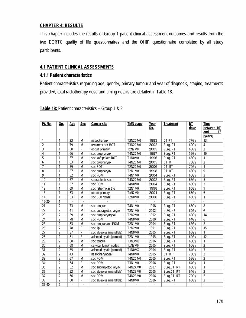
170
This chapter includes the results of Group 1 patient clinical assessment outcomes and results from the two EORTC quality of life questionnaires and the OHIP questionnaire completed by all study participants.
CHAPTER 4: RESULTS
4.1.1 Patient characteristics 4.1 PATIENT CLINICAL ASSESSMENTS
Patient characteristics regarding age, gender, primary tumour and year of diagnosis, staging, treatments provided, total radiotherapy dose and timing details are detailed in Table 18. Table 18: Patient characteristics – Group 1 & 2
Pt. No. Gp. Age Sex Cancer site TMN stage Year Dx.
Treatment RT dose
Time between RT and T0 [years]
1 1 23 M nasopharynx T3N2CM0 1993 CT,RT 77Gy 13 2 1 79 M recurrent scc BOT T3N2CM0 2002 Surg, RT 60Gy 4 3 1 50 F occult primary TxN?M0 2005 Surg, RT 66Gy 2 4 1 66 M scc oropharynx T4N2CM0 1997 Surg, RT 55Gy 10 5 1 67 M scc soft palate BOT T1N0M0 1996 Surg, RT 66Gy 11 6 1 63 M scc oropharynx T4N2CM0 2005 CT, RT 70Gy 2 7 1 59 M scc BOT T2N2CM0 2006 CT, RT 70Gy 1 8 1 67 M scc oropharynx T2N1M0 1998 CT, RT 68Gy 9 9 1 52 M scc FOM T4N1M0 2004 Surg, RT 66Gy 3 10 1 67 M supraglottic scc T4N2CM0 2002 Surg, RT 66Gy 5 11 1 57 M scc FOM T4N0M0 2004 Surg, RT 66Gy 3 12 1 69 M scc retromolar trig T2N1M0 1998 Surg, RT 60Gy 9 13 1 63 M occult primary TxN2M0 2001 Surg, RT 66Gy 6 14 1 53 M scc BOT/tonsil T2N0M0 2006 Surg, RT 66Gy 1 15-20 1 - - - - - - - - 21 2 73 M scc tongue T4N1M0 1998 Surg, RT 66Gy 8 22 2 61 M scc supraglottic larynx T2N1M0 2002 Surg, RT 60Gy 4 23 2 59 M scc oropharyngeal T2N2M0 1992 Surg, RT 60Gy 14 24 2 78 M scc FOM T4N0M0 2000 Surg, RT 64Gy 6 25 2 83 M scc tongue and FOM T2N1M0 2004 Surg, RT 66Gy 2 26 2 78 F scc lip T2N2M0 1991 Surg, RT 60Gy 15 27 2 57 F scc alveolus (mandible) T4N0M0 2005 Surg, RT 60Gy 1 28 2 81 F adenoid cystic (parotid) T2N1M0 1995 Surg, RT 60Gy 12 29 2 68 M scc tongue T3N3MX 2006 Surg, RT 66Gy 1 30 2 68 M cervical lymph nodes TxN3M0 2005 Surg, RT 60Gy 2 31 2 55 M adenoid cystic (parotid) T1N0MX 2004 Surg, RT 64Gy 3 32 2 43 F nasopharyngeal T4N0M0 2005 CT, RT 70Gy 2 33 2 67 M scc FOM T4N2CM0 2005 Surg, RT 55Gy 2 34 2 64 F scc FOM T3N1M0 2006 Surg, RT 64Gy 1 35 2 52 M scc supraglottic larynx T4N2AM0 2007 Surg,CT, RT 66Gy 1 36 2 52 M scc alveolus (mandible) T4N2BM0 2005 Surg,CT, RT 64Gy 3 37 2 66 M scc FOM T4N2AM0 2006 Surg,CT, RT 70Gy 2 38 2 60 F scc alveolus (mandible) T4N0M0 2006 Surg, RT 60Gy 2 39-40 2 - - - - - - - -
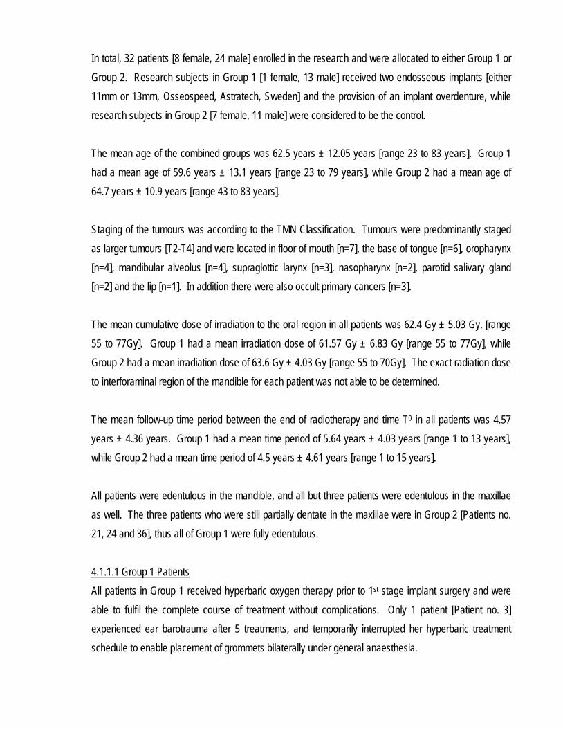
In total, 32 patients [8 female, 24 male] enrolled in the research and were allocated to either Group 1 or Group 2. Research subjects in Group 1 [1 female, 13 male] received two endosseous implants [either 11mm or 13mm, Osseospeed, Astratech, Sweden] and the provision of an implant overdenture, while research subjects in Group 2 [7 female, 11 male] were considered to be the control. The mean age of the combined groups was 62.5 years ± 12.05 years [range 23 to 83 years]. Group 1 had a mean age of 59.6 years ± 13.1 years [range 23 to 79 years], while Group 2 had a mean age of 64.7 years ± 10.9 years [range 43 to 83 years]. Staging of the tumours was according to the TMN Classification. Tumours were predominantly staged as larger tumours [T2-T4] and were located in floor of mouth [n=7], the base of tongue [n=6], oropharynx [n=4], mandibular alveolus [n=4], supraglottic larynx [n=3], nasopharynx [n=2], parotid salivary gland [n=2] and the lip [n=1]. In addition there were also occult primary cancers [n=3]. The mean cumulative dose of irradiation to the oral region in all patients was 62.4 Gy ± 5.03 Gy. [range 55 to 77Gy]. Group 1 had a mean irradiation dose of 61.57 Gy ± 6.83 Gy [range 55 to 77Gy], while Group 2 had a mean irradiation dose of 63.6 Gy ± 4.03 Gy [range 55 to 70Gy]. The exact radiation dose to interforaminal region of the mandible for each patient was not able to be determined. The mean follow-up time period between the end of radiotherapy and time T0 in all patients was 4.57 years ± 4.36 years. Group 1 had a mean time period of 5.64 years ± 4.03 years [range 1 to 13 years], while Group 2 had a mean time period of 4.5 years ± 4.61 years [range 1 to 15 years]. All patients were edentulous in the mandible, and all but three patients were edentulous in the maxillae as well. The three patients who were still partially dentate in the maxillae were in Group 2 [Patients no. 21, 24 and 36], thus all of Group 1 were fully edentulous.
All patients in Group 1 received hyperbaric oxygen therapy prior to 1st stage implant surgery and were able to fulfil the complete course of treatment without complications. Only 1 patient [Patient no. 3] experienced ear barotrauma after 5 treatments, and temporarily interrupted her hyperbaric treatment schedule to enable placement of grommets bilaterally under general anaesthesia.
4.1.1.1 Group 1 Patients
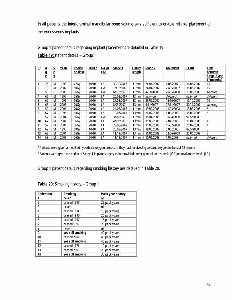
172
In all patients the interforaminal mandibular bone volume was sufficient to enable reliable placement of the endosseous implants. Group 1 patient details regarding implant placement are detailed in Table 19. Table 19:
Patient details – Group 1
* Patients were given a modified hyperbaric oxygen protocol if they had received hyperbaric oxygen in the last 12 months ^Patients were given the option of Stage 1 implant surgery to be provided under general anaesthesia [GA] or local anaesthesia [LA]
Group 1 patient details regarding smoking history are detailed in Table 20. Table 20:
Smoking history – Group 1
Pt A g e
Sex
Yr Dx Radiation dose
HBO * GA or LA^
Stage 1 Fixture length
Stage 2 Abutment FLOD Time between Stage 2 and T1 (months)
1 23 M 1993 77Gy 10/10 LA 30/10/2006 11mm 24/04/2007 8/05/2007 16/05/2007 15 2 79 M 2002 60Gy 20/10 GA 1/11/2006 11mm 24/04/2007 29/05/2007 15/06/2007 15 3 50 F 2005 66Gy 20/10 GA 6/07/2007 11mm 4/03/2008 20/05/2008 23/05/2008 sleeping 4 66 M 1997 55Gy 20/10 LA 20/03/2007 13mm deferred deferred deferred deferred 5 67 M 1996 66Gy 20/10 LA 27/03/2007 13mm 25/09/2007 15/10/2007 19/10/2007 10 6 63 M 2005 70Gy 10/10 LA 8/05/2007 13mm 6/11/2007 27/11/2007 30/11/2007 sleeping 7 59 M 2006 70Gy 20/10 LA 24/07/2007 11mm 19/02/2008 11/03/2008 13/03/2008 5 8 67 M 1998 68Gy 10/10 LA 10/07/2007 13mm 26/02/2008 6/03/2008 18/03/2008 5 9 52 M 2004 66Gy 20/10 GA 3/08/2007 11mm 15/04/2008 30/04/2008 9/05/2008 3 10 67 M 2002 66Gy 20/10 LA 4/09/2007 13mm 11/03/2008 7/04/2008 11/04/2008 4 11 57 M 2004 66Gy 20/10 LA 28/08/2007 11mm 11/03/2008 15/07/2008 21/07/2008 1 12 69 M 1998 60Gy 10/10 LA 28/08/2007 13mm 18/03/2007 5/05/2008 8/05/2008 3 13 63 M 2001 66Gy 20/10 LA 11/12/2007 13mm 10/06/2008 24/06/2008 27/06/2008 2 14 53 M 2006 66Gy 20/10 LA 11/12/2007 11mm 10/06/2008 1/07/2008 deferred deferred
Patient no. Smoking Pack year history 1 never nil 2 ceased 1998 25 pack years 3 never nil 4 ceased 2005 10 pack years 5 ceased 1990 20 pack years 6 ceased 1997 15 pack years 7 ceased 1997 25 pack years 8 never nil 9 yes still smoking 40 pack years 10 ceased 2002 40 pack years 11 yes still smoking 40 pack years 12 ceased 1973 10 pack years 13 ceased 2001 20 pack years 14 yes still smoking 35 pack years
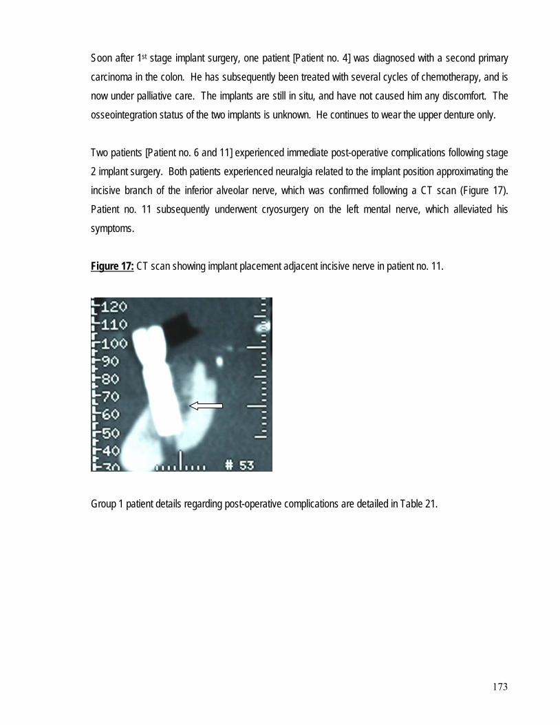
173
Soon after 1st stage implant surgery, one patient [Patient no. 4] was diagnosed with a second primary carcinoma in the colon. He has subsequently been treated with several cycles of chemotherapy, and is now under palliative care. The implants are still in situ, and have not caused him any discomfort. The osseointegration status of the two implants is unknown. He continues to wear the upper denture only. Two patients [Patient no. 6 and 11] experienced immediate post-operative complications following stage 2 implant surgery. Both patients experienced neuralgia related to the implant position approximating the incisive branch of the inferior alveolar nerve, which was confirmed following a CT scan (Figure 17). Patient no. 11 subsequently underwent cryosurgery on the left mental nerve, which alleviated his symptoms. Figure 17:
CT scan showing implant placement adjacent incisive nerve in patient no. 11.
Group 1 patient details regarding post-operative complications are detailed in Table 21.
174
Table 21: Post-operative complications – Group 1 Pt No Mobility ORN Pain Infection Implant Outcome 1 no no no no functional 2 no no no no functional 3 no no yes no sleeping 4 deferred deferred deferred deferred deferred 5 no no no no functional 6 no no yes no sleeping 7 no no no no functional 8 no no no no functional 9 yes no yes yes non function - loose 10 no no no no functional 11 no yes no no non functional - ORN 12* DNA DNA DNA DNA functional 13 no no no no functional 14 no yes no no non functional - ORN
* did not attend (DNA) clinical review appointments
Implant overdentures were constructed for all of the patients in Group 1, except for patient number 4. Patient number 3 found that wearing a lower prosthesis worsened her already significant xerostomia to the point that she could not tolerate the prosthesis in her mouth. Three months following the insertion of the implant overdenture she requested removal of the implant abutments, and the implants were put to sleep. She is currently not wearing any lower prosthesis. Problems with neuralgia worsened for patient no. 6 upon wearing of the overdenture, and he requested removal of the implant abutments. The implants were put to sleep and the lower denture was subsequently converted back to a conventional denture with a permanent silicone based soft liner [Molloplast B, Detax Gmbh & Co, KG Germany]. Soon after implant loading and overdenture insertion, patient no. 9 experienced problems with loosening of both implants and subsequent implant loss. The lower right implant was avulsed during overdenture removal and the lower left implant was removed. There appeared to be delayed healing of the right implant site with possible early signs of ORN developing. Patient number 12 moved interstate soon after insertion of the implant overdenture. He has failed to attend review appointments, but did complete post-treatment questionnaires. He reports no problems with the overdentures and is happy with the current outcome of his treatment.
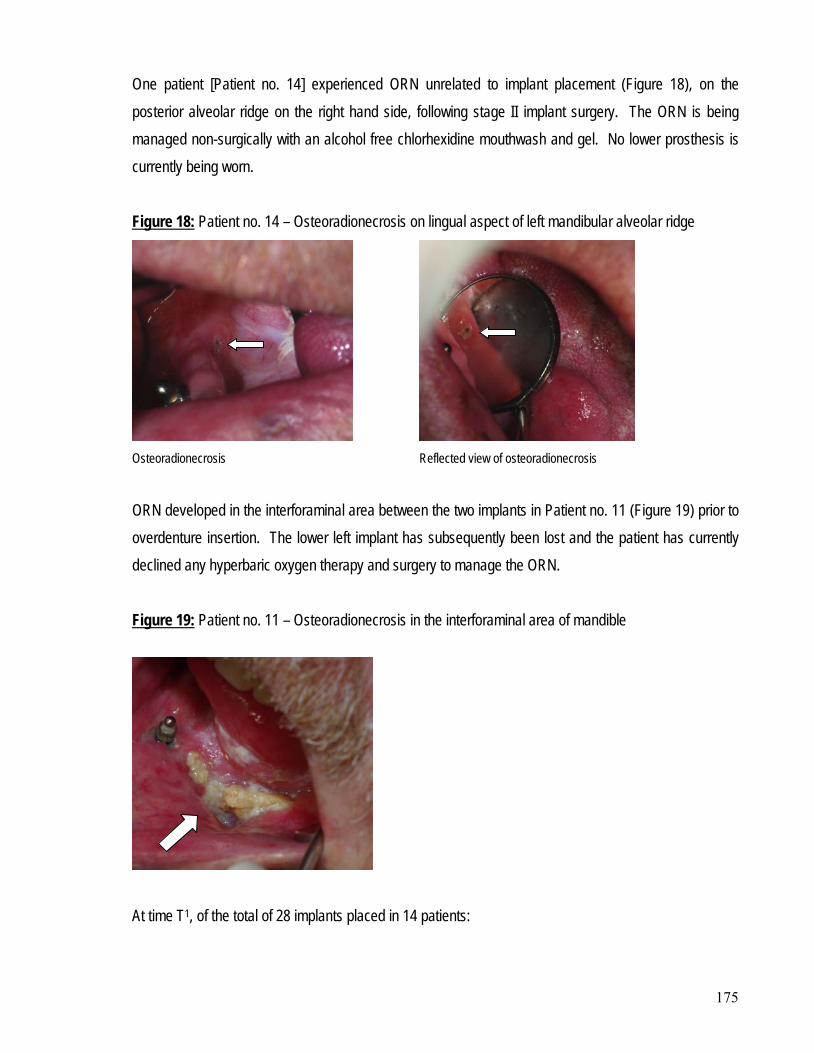
175
One patient [Patient no. 14] experienced ORN unrelated to implant placement (Figure 18), on the posterior alveolar ridge on the right hand side, following stage II implant surgery. The ORN is being managed non-surgically with an alcohol free chlorhexidine mouthwash and gel. No lower prosthesis is currently being worn. Figure 18: Patient no. 14 – Osteoradionecrosis on lingual aspect of left mandibular alveolar ridge
Osteoradionecrosis Reflected view of osteoradionecrosis
ORN developed in the interforaminal area between the two implants in Patient no. 11 (Figure 19) prior to overdenture insertion. The lower left implant has subsequently been lost and the patient has currently declined any hyperbaric oxygen therapy and surgery to manage the ORN. Figure 19:
Patient no. 11 – Osteoradionecrosis in the interforaminal area of mandible
At time T1, of the total of 28 implants placed in 14 patients:
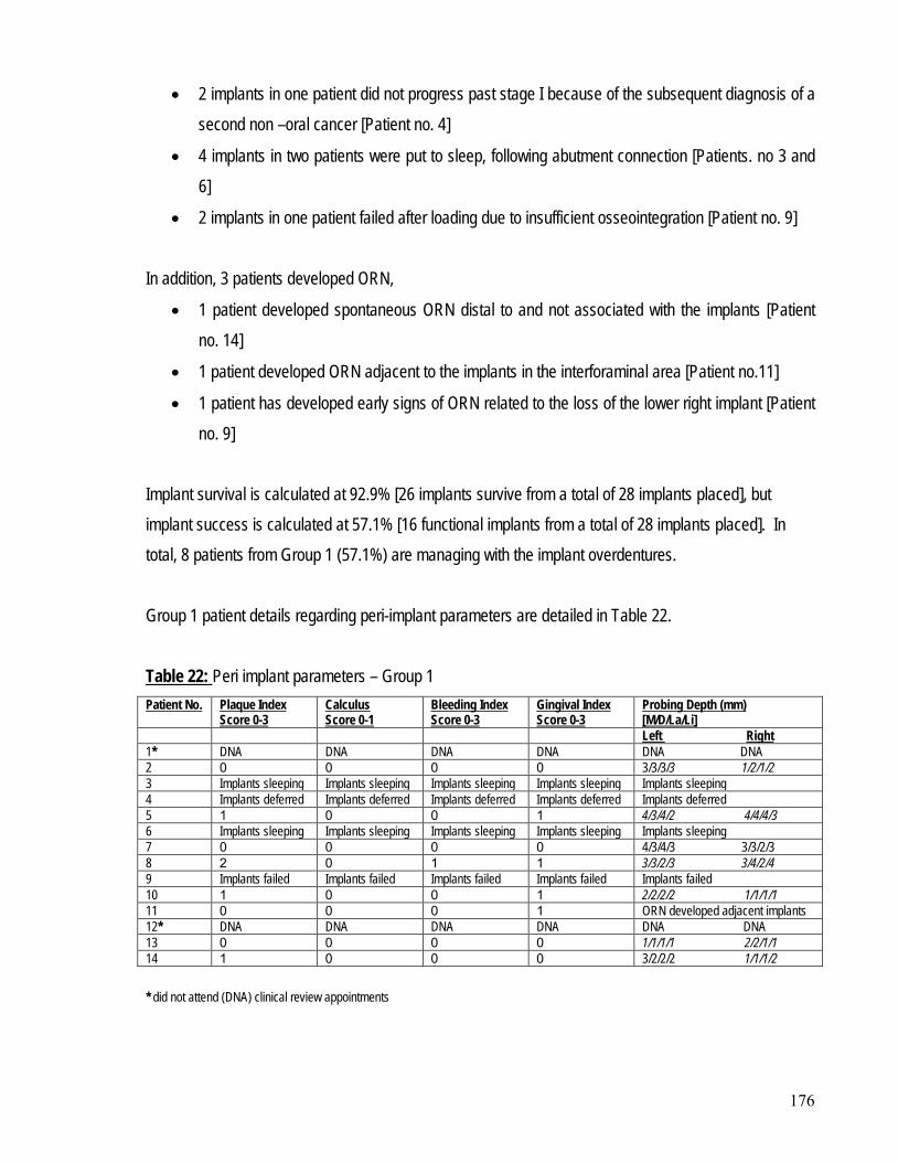
176
• 2 implants in one patient did not progress past stage I because of the subsequent diagnosis of a second non –oral cancer [Patient no. 4]
• 4 implants in two patients were put to sleep, following abutment connection [Patients. no 3 and 6]
• 2 implants in one patient failed after loading due to insufficient osseointegration [Patient no. 9]
In addition, 3 patients developed ORN,
• 1 patient developed spontaneous ORN distal to and not associated with the implants [Patient no. 14]
• 1 patient developed ORN adjacent to the implants in the interforaminal area [Patient no.11]
• 1 patient has developed early signs of ORN related to the loss of the lower right implant [Patient no. 9]
Implant survival is calculated at 92.9% [26 implants survive from a total of 28 implants placed], but implant success is calculated at 57.1% [16 functional implants from a total of 28 implants placed]. In total, 8 patients from Group 1 (57.1%) are managing with the implant overdentures. Group 1 patient details regarding peri-implant parameters are detailed in Table 22. Table 22: Peri implant parameters – Group 1 Patient No. Plaque Index
Score 0-3 Calculus Score 0-1
Bleeding Index Score 0-3
Gingival Index Score 0-3
Probing Depth (mm) [M/D/La/Li]
Left Right 1* DNA DNA DNA DNA DNA DNA 2 0 0 0 0 3/3/3/3 1/2/1/2 3 Implants sleeping Implants sleeping Implants sleeping Implants sleeping Implants sleeping 4 Implants deferred Implants deferred Implants deferred Implants deferred Implants deferred 5 1 0 0 1 4/3/4/2 4/4/4/3 6 Implants sleeping Implants sleeping Implants sleeping Implants sleeping Implants sleeping 7 0 0 0 0 4/3/4/3 3/3/2/3 8 2 0 1 1 3/3/2/3 3/4/2/4 9 Implants failed Implants failed Implants failed Implants failed Implants failed 10 1 0 0 1 2/2/2/2 1/1/1/1 11 0 0 0 1 ORN developed adjacent implants 12* DNA DNA DNA DNA DNA DNA 13 0 0 0 0 1/1/1/1 2/2/1/1 14 1 0 0 0 3/2/2/2 1/1/1/2 * did not attend (DNA) clinical review appointments
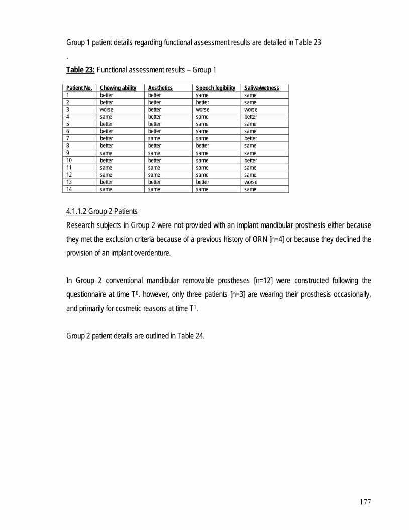
177
Group 1 patient details regarding functional assessment results are detailed in Table 23 . Table 23:
Functional assessment results – Group 1
Patient No. Chewing ability Aesthetics Speech legibility Saliva/wetness 1 better better same same 2 better better better same 3 worse better worse worse 4 same better same better 5 better better same same 6 better better same same 7 better same same better 8 better better better same 9 same same same same 10 better better same better 11 same same same same 12 same same same same 13 better better better worse 14 same same same same
Research subjects in Group 2 were not provided with an implant mandibular prosthesis either because they met the exclusion criteria because of a previous history of ORN [n=4] or because they declined the provision of an implant overdenture.
4.1.1.2 Group 2 Patients
In Group 2 conventional mandibular removable prostheses [n=12] were constructed following the questionnaire at time T0, however, only three patients [n=3] are wearing their prosthesis occasionally, and primarily for cosmetic reasons at time T1. Group 2 patient details are outlined in Table 24.
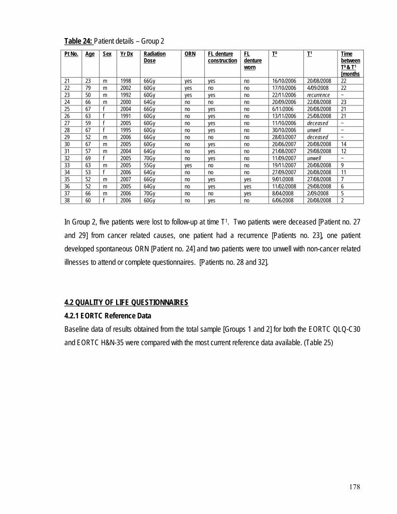
178
Table 24: Patient details – Group 2 Pt No. Age Sex Yr Dx Radiation
Dose ORN FL denture
construction FL denture worn
T0 T1 Time between T0 & T1
[months 21 23 m 1998 66Gy yes yes no 16/10/2006 20/08/2008 22 22 79 m 2002 60Gy yes no no 17/10/2006 4/09/2008 22 23 50 m 1992 60Gy yes yes no 22/11/2006 recurrence ~ 24 66 m 2000 64Gy no no no 20/09/2006 22/08/2008 23 25 67 f 2004 66Gy no yes no 6/11/2006 20/08/2008 21 26 63 f 1991 60Gy no yes no 13/11/2006 25/08/2008 21 27 59 f 2005 60Gy no yes no 11/10/2006 deceased ~ 28 67 f 1995 60Gy no yes no 30/10/2006 unwell ~ 29 52 m 2006 66Gy no no no 28/03/2007 deceased ~ 30 67 m 2005 60Gy no yes no 20/06/2007 20/08/2008 14 31 57 m 2004 64Gy no yes no 21/08/2007 29/08/2008 12 32 69 f 2005 70Gy no yes no 11/09/2007 unwell ~ 33 63 m 2005 55Gy yes no no 19/11/2007 20/08/2008 9 34 53 f 2006 64Gy no no no 27/09/2007 20/08/2008 11 35 52 m 2007 66Gy no yes yes 9/01/2008 27/08/2008 7 36 52 m 2005 64Gy no yes yes 11/02/2008 29/08/2008 6 37 66 m 2006 70Gy no no yes 8/04/2008 2/09/2008 5 38 60 f 2006 60Gy no yes no 6/06/2008 20/08/2008 2 In Group 2, five patients were lost to follow-up at time T1. Two patients were deceased [Patient no. 27 and 29] from cancer related causes, one patient had a recurrence [Patients no. 23], one patient developed spontaneous ORN [Patient no. 24] and two patients were too unwell with non-cancer related illnesses to attend or complete questionnaires. [Patients no. 28 and 32].
4.2.1 EORTC Reference Data 4.2 QUALITY OF LIFE QUESTIONNAIRES
Baseline data of results obtained from the total sample [Groups 1 and 2] for both the EORTC QLQ-C30 and EORTC H&N-35 were compared with the most current reference data available. (Table 25)
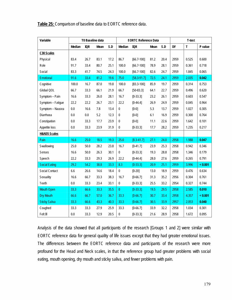
179
Table 25:
Comparison of baseline data to EORTC reference data.
Variable T0 Baseline data EORTC Reference Data T-test
Median IQR Mean S.D Median IQR Mean S.D DF T P value
C30 Scales
Physical 83.4 26.7 83.1 17.2 86.7 [66.7-100] 81.2 20.4 2959 0.525 0.600
Role 91.7 33.4 80.7 25.1 100.0 [66.7-100] 78.9 28.1 2959 0.361 0.718
Social 83.3 41.7 74.5 24.3 100.0 [66.7-100] 82.6 24.7 2959 1.845 0.065
Emotional 91.6 33.4 81.2 19.6 75.0 [58.3-91.7] 72.5 24.1 2959 2.035 0.042
Cognitive 100.0 16.7 87.0 19.8 100.0 [83.3-100] 85.9 19.7 2959 0.314 0.753
Global QOL 66.7 33.3 66.1 21.9 66.7 [50-83.3] 64.1 22.7 2959 0.496 0.620
Symptom – Pain 16.6 33.3 26.0 28.1 16.7 [0-33.3] 23.2 26.1 2959 0.603 0.547
Symptom – Fatigue 22.2 22.2 26.7 23.1 22.2 [0-44.4] 26.9 24.9 2959 0.045 0.964
Symptom – Nausea 0.0 16.6 7.8 13.4 0 [0-0] 5.3 13.7 2959 1.027 0.305
Diarrhoea 0.0 0.0 5.2 12.3 0 [0-0] 6.1 16.9 2959 0.300 0.764
Constipation 0.0 33.3 17.7 23.9 0 [0-0] 11.1 22.6 2959 1.642 0.101
Appetite loss 0.0 33.3 23.9 31.9 0 [0-33.3] 17.7 28.2 2959 1.235 0.217
H&N35 Scales
Pain 16.6 25.0 18.5 19.3 25.0 [8.3-41.7] 27.1 24.0 2958 1.988 0.047
Swallowing 25.0 50.0 28.2 23.8 16.7 [0-41.7] 23.9 25.3 2958 0.942 0.346
Senses 16.6 50.0 26.3 30.1 0 [0-33.3] 19.3 28.8 2958 1.346 0.179
Speech 22.2 33.3 29.3 26.9 22.2 [0-44.4] 28.0 27.6 2959 0.265 0.791
Social Eating 29.2 54.2 38.8 33.3 8.3 [0-33.3] 20.9 25.1 2959 3.996 < 0.001
Social Contact 6.6 26.6 14.6 18.4 0 [0-20] 13.0 18.9 2959 0.476 0.634
Sexuality 16.6 66.7 33.3 38.3 16.7 [0-66.7] 31.3 35.2 2956 0.304 0.761
Teeth 0.0 33.3 23.4 33.1 0 [0-33.3] 25.5 33.2 2954 0.327 0.744
Mouth Open 33.3 66.6 33.3 35.5 0 [0-33.3] 19.5 29.5 2958 2.585 0.010
Dry Mouth 66.6 66.7 57.0 36.7 33.3 [0-66.7] 30.7 33.4 2958 4.357 < 0.001
Sticky Saliva 33.3 66.6 43.3 40.3 33.3 [0-66.7] 30.5 33.9 2957 2.053 0.040
Coughed 33.3 33.3 27.9 25.9 33.3 [0-66.7] 33.9 32.2 2958 1.034 0.301
Felt Ill 0.0 33.3 12.9 20.5 0 [0-33.3] 21.6 28.9 2958 1.672 0.095
Analysis of the data showed that all participants of the research [Groups 1 and 2] were similar with EORTC reference data for general quality of life issues except that they had greater emotional issues. The differences between the EORTC reference data and participants of the research were more profound for the Head and Neck scales, in that the reference group had greater problems with social eating, mouth opening, dry mouth and sticky saliva, and fewer problems with pain.
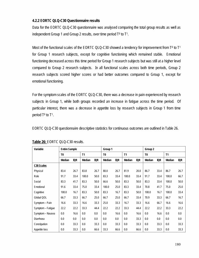
180
4.2.2 EORTC QLQ-C30 Questionnaire results Data for the EORTC QLQ-C30 questionnaire was analysed comparing the total group results as well as independent Group 1 and Group 2 results, over time period T0 to T1. Most of the functional scales of the EORTC QLQ-C30 showed a tendency for improvement from T0 to T1 for Group 1 research subjects, except for cognitive functioning which remained stable. Emotional functioning decreased across this time period for Group 1 research subjects but was still at a higher level compared to Group 2 research subjects. In all functional scales across both time periods, Group 2 research subjects scored higher scores or had better outcomes compared to Group 1, except for emotional functioning. For the symptom scales of the EORTC QLQ-C30, there was a decrease in pain experienced by research subjects in Group 1, while both groups recorded an increase in fatigue across the time period. Of particular interest, there was a decrease in appetite loss by research subjects in Group 1 from time period T0 to T1. EORTC QLQ-C30 questionnaire descriptive statistics for continuous outcomes are outlined in Table 26. Table 26:
Variable
EORTC QLQ-C30 results.
Entire Sample Group 1 Group 2
T0 T1 T0 T1 T0 T1
Median IQR Median IQR Median IQR Median IQR Median IQR Median IQR
C30 Scales
Physical 83.4 26.7 83.8 26.7 80.0 26.7 81.9 20.0 86.7 33.4 86.7 26.7
Role 91.7 33.4 100.0 50.0 83.3 33.4 100.0 33.4 91.7 33.4 100.0 66.7
Social 83.3 41.7 83.3 50.0 66.6 50.0 83.3 50.0 83.3 33.4 100.0 50.0
Emotional 91.6 33.4 75.0 33.4 100.0 25.0 83.3 33.4 70.8 41.7 75.0 25.0
Cognitive 100.0 16.7 83.3 50.0 83.3 16.7 83.3 50.0 100.0 16.7 100.0 33.4
Global QOL 66.7 33.3 66.7 25.0 66.7 25.0 66.7 33.4 70.9 33.3 66.7 16.7
Symptom – Pain 16.6 33.3 16.6 33.3 25.0 33.3 16.7 33.3 16.6 66.7 16.6 16.6
Symptom – Fatigue 22.2 22.2 33.3 44.4 22.2 22.2 33.3 44.4 22.2 22.2 33.3 22.2
Symptom – Nausea 0.0 16.6 0.0 0.0 0.0 16.6 0.0 16.6 0.0 16.6 0.0 0.0
Diarrhoea 0.0 0.0 0.0 0.0 0.0 0.0 0.0 33.3 0.0 0.0 0.0 0.0
Constipation 0.0 33.3 0.0 33.3 0.0 33.3 0.0 33.3 0.0 33.3 0.0 33.3
Appetite loss 0.0 33.3 0.0 66.6 33.3 66.6 0.0 66.6 0.0 33.3 0.0 33.3
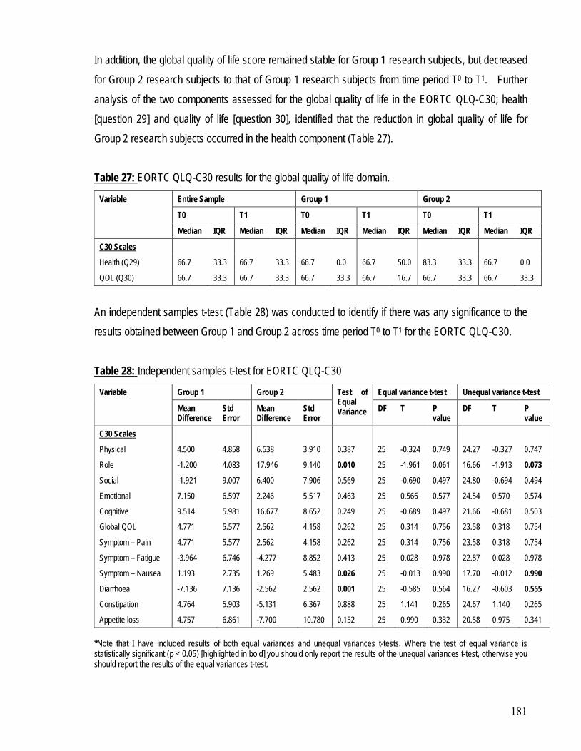
181
In addition, the global quality of life score remained stable for Group 1 research subjects, but decreased for Group 2 research subjects to that of Group 1 research subjects from time period T0 to T1. Further analysis of the two components assessed for the global quality of life in the EORTC QLQ-C30; health [question 29] and quality of life [question 30], identified that the reduction in global quality of life for Group 2 research subjects occurred in the health component (Table 27). Table 27:
Variable
EORTC QLQ-C30 results for the global quality of life domain.
Entire Sample Group 1 Group 2
T0 T1 T0 T1 T0 T1
Median IQR Median IQR Median IQR Median IQR Median IQR Median IQR
C30 Scales
Health (Q29) 66.7 33.3 66.7 33.3 66.7 0.0 66.7 50.0 83.3 33.3 66.7 0.0
QOL (Q30) 66.7 33.3 66.7 33.3 66.7 33.3 66.7 16.7 66.7 33.3 66.7 33.3
An independent samples t-test (Table 28) was conducted to identify if there was any significance to the results obtained between Group 1 and Group 2 across time period T0 to T1 for the EORTC QLQ-C30. Table 28:
Variable
Independent samples t-test for EORTC QLQ-C30
Group 1 Group 2 Test of Equal Variance
Equal variance t-test Unequal variance t-test
Mean Difference
Std Error
Mean Difference
Std Error
DF T P value
DF T P value
C30 Scales
Physical 4.500 4.858 6.538 3.910 0.387 25 -0.324 0.749 24.27 -0.327 0.747
Role -1.200 4.083 17.946 9.140 0.010 25 -1.961 0.061 16.66 -1.913 0.073
Social -1.921 9.007 6.400 7.906 0.569 25 -0.690 0.497 24.80 -0.694 0.494
Emotional 7.150 6.597 2.246 5.517 0.463 25 0.566 0.577 24.54 0.570 0.574
Cognitive 9.514 5.981 16.677 8.652 0.249 25 -0.689 0.497 21.66 -0.681 0.503
Global QOL 4.771 5.577 2.562 4.158 0.262 25 0.314 0.756 23.58 0.318 0.754
Symptom – Pain 4.771 5.577 2.562 4.158 0.262 25 0.314 0.756 23.58 0.318 0.754
Symptom – Fatigue -3.964 6.746 -4.277 8.852 0.413 25 0.028 0.978 22.87 0.028 0.978
Symptom – Nausea 1.193 2.735 1.269 5.483 0.026 25 -0.013 0.990 17.70 -0.012 0.990
Diarrhoea -7.136 7.136 -2.562 2.562 0.001 25 -0.585 0.564 16.27 -0.603 0.555
Constipation 4.764 5.903 -5.131 6.367 0.888 25 1.141 0.265 24.67 1.140 0.265
Appetite loss 4.757 6.861 -7.700 10.780 0.152 25 0.990 0.332 20.58 0.975 0.341 *Note that I have included results of both equal variances and unequal variances t-tests. Where the test of equal variance is statistically significant (p < 0.05) [highlighted in bold] you should only report the results of the unequal variances t-test, otherwise you should report the results of the equal variances t-test.
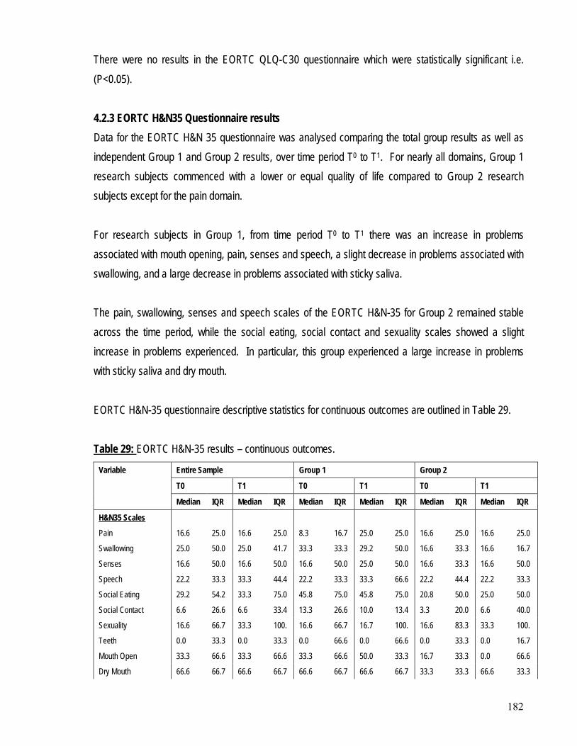
182
There were no results in the EORTC QLQ-C30 questionnaire which were statistically significant i.e. (P<0.05). 4.2.3 EORTC H&N35 Questionnaire results Data for the EORTC H&N 35 questionnaire was analysed comparing the total group results as well as independent Group 1 and Group 2 results, over time period T0 to T1. For nearly all domains, Group 1 research subjects commenced with a lower or equal quality of life compared to Group 2 research subjects except for the pain domain. For research subjects in Group 1, from time period T0 to T1 there was an increase in problems associated with mouth opening, pain, senses and speech, a slight decrease in problems associated with swallowing, and a large decrease in problems associated with sticky saliva. The pain, swallowing, senses and speech scales of the EORTC H&N-35 for Group 2 remained stable across the time period, while the social eating, social contact and sexuality scales showed a slight increase in problems experienced. In particular, this group experienced a large increase in problems with sticky saliva and dry mouth. EORTC H&N-35 questionnaire descriptive statistics for continuous outcomes are outlined in Table 29. Table 29:
Variable
EORTC H&N-35 results – continuous outcomes.
Entire Sample Group 1 Group 2
T0 T1 T0 T1 T0 T1
Median IQR Median IQR Median IQR Median IQR Median IQR Median IQR
H&N35 Scales
Pain 16.6 25.0 16.6 25.0 8.3 16.7 25.0 25.0 16.6 25.0 16.6 25.0
Swallowing 25.0 50.0 25.0 41.7 33.3 33.3 29.2 50.0 16.6 33.3 16.6 16.7
Senses 16.6 50.0 16.6 50.0 16.6 50.0 25.0 50.0 16.6 33.3 16.6 50.0
Speech 22.2 33.3 33.3 44.4 22.2 33.3 33.3 66.6 22.2 44.4 22.2 33.3
Social Eating 29.2 54.2 33.3 75.0 45.8 75.0 45.8 75.0 20.8 50.0 25.0 50.0
Social Contact 6.6 26.6 6.6 33.4 13.3 26.6 10.0 13.4 3.3 20.0 6.6 40.0
Sexuality 16.6 66.7 33.3 100. 16.6 66.7 16.7 100. 16.6 83.3 33.3 100.
Teeth 0.0 33.3 0.0 33.3 0.0 66.6 0.0 66.6 0.0 33.3 0.0 16.7
Mouth Open 33.3 66.6 33.3 66.6 33.3 66.6 50.0 33.3 16.7 33.3 0.0 66.6
Dry Mouth 66.6 66.7 66.6 66.7 66.6 66.7 66.6 66.7 33.3 33.3 66.6 33.3
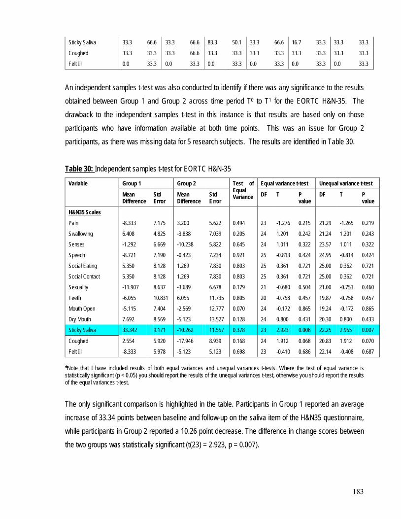
183
Sticky Saliva 33.3 66.6 33.3 66.6 83.3 50.1 33.3 66.6 16.7 33.3 33.3 33.3
Coughed 33.3 33.3 33.3 66.6 33.3 33.3 33.3 33.3 33.3 33.3 33.3 33.3
Felt Ill 0.0 33.3 0.0 33.3 0.0 33.3 0.0 33.3 0.0 33.3 0.0 33.3
An independent samples t-test was also conducted to identify if there was any significance to the results obtained between Group 1 and Group 2 across time period T0 to T1 for the EORTC H&N-35. The drawback to the independent samples t-test in this instance is that results are based only on those participants who have information available at both time points. This was an issue for Group 2 participants, as there was missing data for 5 research subjects. The results are identified in Table 30. Table 30:
Variable
Independent samples t-test for EORTC H&N-35
Group 1 Group 2 Test of Equal Variance
Equal variance t-test Unequal variance t-test
Mean Difference
Std Error
Mean Difference
Std Error
DF T P value
DF T P value
H&N35 Scales
Pain -8.333 7.175 3.200 5.622 0.494 23 -1.276 0.215 21.29 -1.265 0.219
Swallowing 6.408 4.825 -3.838 7.039 0.205 24 1.201 0.242 21.24 1.201 0.243
Senses -1.292 6.669 -10.238 5.822 0.645 24 1.011 0.322 23.57 1.011 0.322
Speech -8.721 7.190 -0.423 7.234 0.921 25 -0.813 0.424 24.95 -0.814 0.424
Social Eating 5.350 8.128 1.269 7.830 0.803 25 0.361 0.721 25.00 0.362 0.721
Social Contact 5.350 8.128 1.269 7.830 0.803 25 0.361 0.721 25.00 0.362 0.721
Sexuality -11.907 8.637 -3.689 6.678 0.179 21 -0.680 0.504 21.00 -0.753 0.460
Teeth -6.055 10.831 6.055 11.735 0.805 20 -0.758 0.457 19.87 -0.758 0.457
Mouth Open -5.115 7.404 -2.569 12.777 0.070 24 -0.172 0.865 19.24 -0.172 0.865
Dry Mouth 7.692 8.569 -5.123 13.527 0.128 24 0.800 0.431 20.30 0.800 0.433
Sticky Saliva 33.342 9.171 -10.262 11.557 0.378 23 2.923 0.008 22.25 2.955 0.007
Coughed 2.554 5.920 -17.946 8.939 0.168 24 1.912 0.068 20.83 1.912 0.070
Felt Ill -8.333 5.978 -5.123 5.123 0.698 23 -0.410 0.686 22.14 -0.408 0.687 *Note that I have included results of both equal variances and unequal variances t-tests. Where the test of equal variance is statistically significant (p < 0.05) you should report the results of the unequal variances t-test, otherwise you should report the results of the equal variances t-test. The only significant comparison is highlighted in the table. Participants in Group 1 reported an average increase of 33.34 points between baseline and follow-up on the saliva item of the H&N35 questionnaire, while participants in Group 2 reported a 10.26 point decrease. The difference in change scores between the two groups was statistically significant (t(23) = 2.923, p = 0.007).
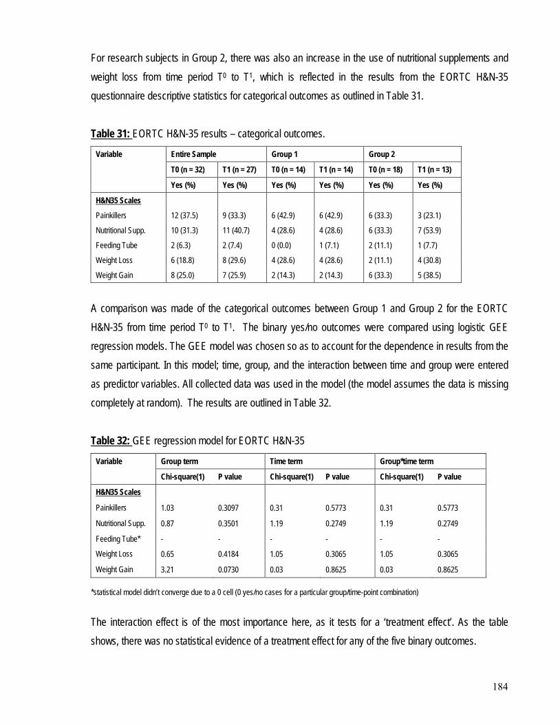
184
For research subjects in Group 2, there was also an increase in the use of nutritional supplements and weight loss from time period T0 to T1, which is reflected in the results from the EORTC H&N-35 questionnaire descriptive statistics for categorical outcomes as outlined in Table 31. Table 31:
Variable
EORTC H&N-35 results – categorical outcomes.
Entire Sample Group 1 Group 2
T0 (n = 32) T1 (n = 27) T0 (n = 14) T1 (n = 14) T0 (n = 18) T1 (n = 13)
Yes (%) Yes (%) Yes (%) Yes (%) Yes (%) Yes (%)
H&N35 Scales
Painkillers 12 (37.5) 9 (33.3) 6 (42.9) 6 (42.9) 6 (33.3) 3 (23.1)
Nutritional Supp. 10 (31.3) 11 (40.7) 4 (28.6) 4 (28.6) 6 (33.3) 7 (53.9)
Feeding Tube 2 (6.3) 2 (7.4) 0 (0.0) 1 (7.1) 2 (11.1) 1 (7.7)
Weight Loss 6 (18.8) 8 (29.6) 4 (28.6) 4 (28.6) 2 (11.1) 4 (30.8)
Weight Gain 8 (25.0) 7 (25.9) 2 (14.3) 2 (14.3) 6 (33.3) 5 (38.5)
A comparison was made of the categorical outcomes between Group 1 and Group 2 for the EORTC H&N-35 from time period T0 to T1. The binary yes/no outcomes were compared using logistic GEE regression models. The GEE model was chosen so as to account for the dependence in results from the same participant. In this model; time, group, and the interaction between time and group were entered as predictor variables. All collected data was used in the model (the model assumes the data is missing completely at random). The results are outlined in Table 32. Table 32:
Variable
GEE regression model for EORTC H&N-35
Group term Time term Group*time term
Chi-square(1) P value Chi-square(1) P value Chi-square(1) P value
H&N35 Scales
Painkillers 1.03 0.3097 0.31 0.5773 0.31 0.5773
Nutritional Supp. 0.87 0.3501 1.19 0.2749 1.19 0.2749
Feeding Tube* - - - - - -
Weight Loss 0.65 0.4184 1.05 0.3065 1.05 0.3065
Weight Gain 3.21 0.0730 0.03 0.8625 0.03 0.8625 *statistical model didn’t converge due to a 0 cell (0 yes/no cases for a particular group/time-point combination) The interaction effect is of the most importance here, as it tests for a ‘treatment effect’. As the table shows, there was no statistical evidence of a treatment effect for any of the five binary outcomes.
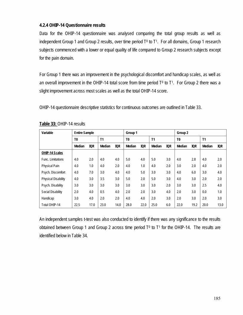
185
4.2.4 OHIP-14 Questionnaire results Data for the OHIP-14 questionnaire was analysed comparing the total group results as well as independent Group 1 and Group 2 results, over time period T0 to T1. For all domains, Group 1 research subjects commenced with a lower or equal quality of life compared to Group 2 research subjects except for the pain domain. For Group 1 there was an improvement in the psychological discomfort and handicap scales, as well as an overall improvement in the OHIP-14 total score from time period T0 to T1. For Group 2 there was a slight improvement across most scales as well as the total OHIP-14 score. OHIP-14 questionnaire descriptive statistics for continuous outcomes are outlined in Table 33. Table 33:
Variable
OHIP-14 results
Entire Sample Group 1 Group 2
T0 T1 T0 T1 T0 T1
Median IQR Median IQR Median IQR Median IQR Median IQR Median IQR
OHIP-14 Scales
Func. Limitations 4.0 2.0 4.0 4.0 5.0 4.0 5.0 3.0 4.0 2.8 4.0 2.0
Physical Pain 4.0 1.0 4.0 2.0 4.0 1.0 4.0 2.0 3.0 2.0 4.0 2.0
Psych. Discomfort 4.0 7.0 3.0 4.0 4.0 5.0 3.0 3.0 4.0 6.0 3.0 4.0
Physical Disability 4.0 3.0 3.5 3.0 5.0 2.0 5.0 3.0 4.0 3.0 2.0 2.0
Psych. Disability 3.0 3.0 3.0 3.0 3.0 3.0 3.0 2.0 3.0 3.0 2.5 4.0
Social Disability 2.0 4.0 0.5 4.0 2.0 2.0 3.0 4.0 2.0 3.0 0.0 1.0
Handicap 3.0 4.0 2.0 2.0 4.0 4.0 2.0 3.0 2.0 3.0 2.0 3.0
Total OHIP-14 22.5 17.0 23.0 14.0 28.0 22.0 25.0 6.0 22.0 19.2 20.0 13.0
An independent samples t-test was also conducted to identify if there was any significance to the results obtained between Group 1 and Group 2 across time period T0 to T1 for the OHIP-14. The results are identified below in Table 34.
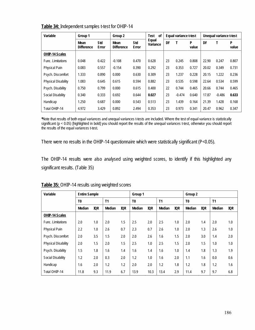
186
Table 34:
Variable
Independent samples t-test for OHIP-14
Group 1 Group 2 Test of Equal Variance
Equal variance t-test Unequal variance t-test
Mean Difference
Std Error
Mean Difference
Std Error
DF T P value
DF T P value
OHIP-14 Scales
Func. Limitations 0.048 0.422 -0.108 0.470 0.628 23 0.245 0.808 22.90 0.247 0.807
Physical Pain 0.083 0.557 -0.154 0.390 0.292 23 0.353 0.727 20.02 0.349 0.731
Psych. Discomfort 1.333 0.890 0.000 0.630 0.309 23 1.237 0.228 20.15 1.222 0.236
Physical Disability 1.083 0.645 0.615 0.594 0.882 23 0.535 0.598 22.64 0.534 0.599
Psych. Disability 0.750 0.799 0.000 0.615 0.400 22 0.744 0.465 20.66 0.744 0.465
Social Disability 0.340 0.333 0.692 0.644 0.027 23 -0.474 0.640 17.87 -0.486 0.633
Handicap 1.250 0.687 0.000 0.543 0.513 23 1.439 0.164 21.39 1.428 0.168
Total OHIP-14 4.972 3.429 0.892 2.494 0.353 23 0.973 0.341 20.47 0.962 0.347 *Note that results of both equal variances and unequal variances t-tests are included. Where the test of equal variance is statistically significant (p < 0.05) [highlighted in bold] you should report the results of the unequal variances t-test, otherwise you should report the results of the equal variances t-test. There were no results in the OHIP-14 questionnaire which were statistically significant (P<0.05). The OHIP-14 results were also analysed using weighted scores, to identify if this highlighted any significant results. (Table 35) Table 35:
Variable
OHIP-14 results using weighted scores
Entire Sample Group 1 Group 2
T0 T1 T0 T1 T0 T1
Median IQR Median IQR Median IQR Median IQR Median IQR Median IQR
OHIP-14 Scales
Func. Limitations 2.0 1.0 2.0 1.5 2.5 2.0 2.5 1.0 2.0 1.4 2.0 1.0
Physical Pain 2.2 1.0 2.6 0.7 2.3 0.7 2.6 1.0 2.0 1.3 2.6 1.0
Psych. Discomfort 2.0 3.5 1.5 2.0 2.0 2.6 1.6 1.5 2.0 3.0 1.4 2.0
Physical Disability 2.0 1.5 2.0 1.5 2.5 1.0 2.5 1.5 2.0 1.5 1.0 1.0
Psych. Disability 1.5 1.8 1.6 1.4 1.6 1.4 1.6 1.0 1.4 1.8 1.3 1.9
Social Disability 1.2 2.0 0.3 2.0 1.2 1.0 1.6 2.0 1.1 1.6 0.0 0.6
Handicap 1.6 2.0 1.2 1.2 2.0 2.0 1.2 1.8 1.2 1.8 1.2 1.6
Total OHIP-14 11.8 9.3 11.9 6.7 13.9 10.3 13.4 2.9 11.4 9.7 9.7 6.8
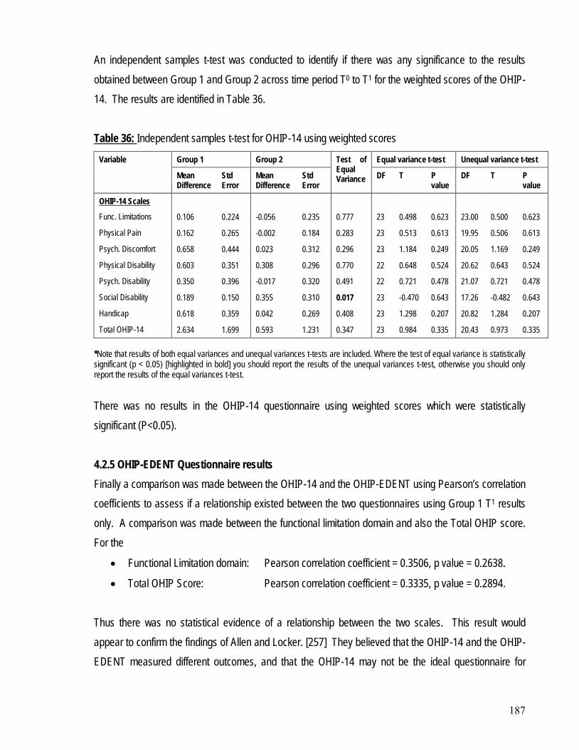
187
An independent samples t-test was conducted to identify if there was any significance to the results obtained between Group 1 and Group 2 across time period T0 to T1 for the weighted scores of the OHIP-14. The results are identified in Table 36. Table 36:
Variable
Independent samples t-test for OHIP-14 using weighted scores
Group 1 Group 2 Test of Equal Variance
Equal variance t-test Unequal variance t-test
Mean Difference
Std Error
Mean Difference
Std Error
DF T P value
DF T P value
OHIP-14 Scales
Func. Limitations 0.106 0.224 -0.056 0.235 0.777 23 0.498 0.623 23.00 0.500 0.623
Physical Pain 0.162 0.265 -0.002 0.184 0.283 23 0.513 0.613 19.95 0.506 0.613
Psych. Discomfort 0.658 0.444 0.023 0.312 0.296 23 1.184 0.249 20.05 1.169 0.249
Physical Disability 0.603 0.351 0.308 0.296 0.770 22 0.648 0.524 20.62 0.643 0.524
Psych. Disability 0.350 0.396 -0.017 0.320 0.491 22 0.721 0.478 21.07 0.721 0.478
Social Disability 0.189 0.150 0.355 0.310 0.017 23 -0.470 0.643 17.26 -0.482 0.643
Handicap 0.618 0.359 0.042 0.269 0.408 23 1.298 0.207 20.82 1.284 0.207
Total OHIP-14 2.634 1.699 0.593 1.231 0.347 23 0.984 0.335 20.43 0.973 0.335 *Note that results of both equal variances and unequal variances t-tests are included. Where the test of equal variance is statistically significant (p < 0.05) [highlighted in bold] you should report the results of the unequal variances t-test, otherwise you should only report the results of the equal variances t-test. There was no results in the OHIP-14 questionnaire using weighted scores which were statistically significant (P<0.05). 4.2.5 OHIP-EDENT Questionnaire results Finally a comparison was made between the OHIP-14 and the OHIP-EDENT using Pearson’s correlation coefficients to assess if a relationship existed between the two questionnaires using Group 1 T1 results only. A comparison was made between the functional limitation domain and also the Total OHIP score. For the
• Functional Limitation domain: Pearson correlation coefficient = 0.3506, p value = 0.2638.
• Total OHIP Score: Pearson correlation coefficient = 0.3335, p value = 0.2894. Thus there was no statistical evidence of a relationship between the two scales. This result would appear to confirm the findings of Allen and Locker. [257] They believed that the OHIP-14 and the OHIP-EDENT measured different outcomes, and that the OHIP-14 may not be the ideal questionnaire for
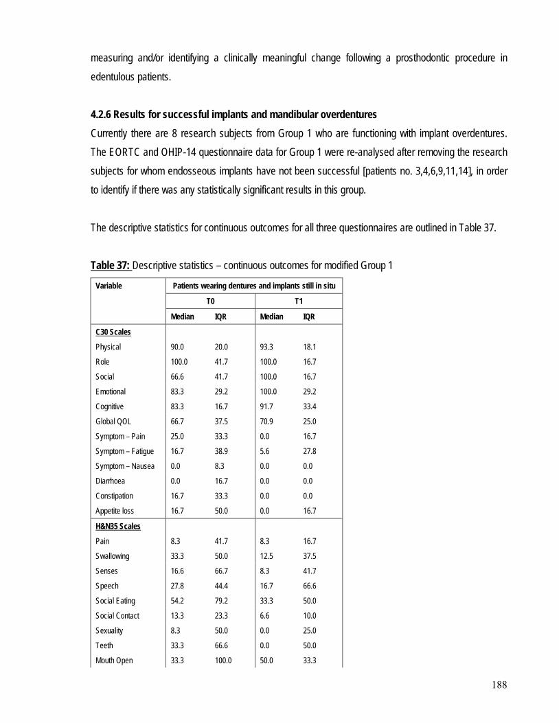
188
measuring and/or identifying a clinically meaningful change following a prosthodontic procedure in edentulous patients. 4.2.6 Results for successful implants and mandibular overdentures Currently there are 8 research subjects from Group 1 who are functioning with implant overdentures. The EORTC and OHIP-14 questionnaire data for Group 1 were re-analysed after removing the research subjects for whom endosseous implants have not been successful [patients no. 3,4,6,9,11,14], in order to identify if there was any statistically significant results in this group. The descriptive statistics for continuous outcomes for all three questionnaires are outlined in Table 37. Table 37:
Variable
Descriptive statistics – continuous outcomes for modified Group 1
Patients wearing dentures and implants still in situ
T0 T1
Median IQR Median IQR
C30 Scales
Physical 90.0 20.0 93.3 18.1
Role 100.0 41.7 100.0 16.7
Social 66.6 41.7 100.0 16.7
Emotional 83.3 29.2 100.0 29.2
Cognitive 83.3 16.7 91.7 33.4
Global QOL 66.7 37.5 70.9 25.0
Symptom – Pain 25.0 33.3 0.0 16.7
Symptom – Fatigue 16.7 38.9 5.6 27.8
Symptom – Nausea 0.0 8.3 0.0 0.0
Diarrhoea 0.0 16.7 0.0 0.0
Constipation 16.7 33.3 0.0 0.0
Appetite loss 16.7 50.0 0.0 16.7
H&N35 Scales
Pain 8.3 41.7 8.3 16.7
Swallowing 33.3 50.0 12.5 37.5
Senses 16.6 66.7 8.3 41.7
Speech 27.8 44.4 16.7 66.6
Social Eating 54.2 79.2 33.3 50.0
Social Contact 13.3 23.3 6.6 10.0
Sexuality 8.3 50.0 0.0 25.0
Teeth 33.3 66.6 0.0 50.0
Mouth Open 33.3 100.0 50.0 33.3
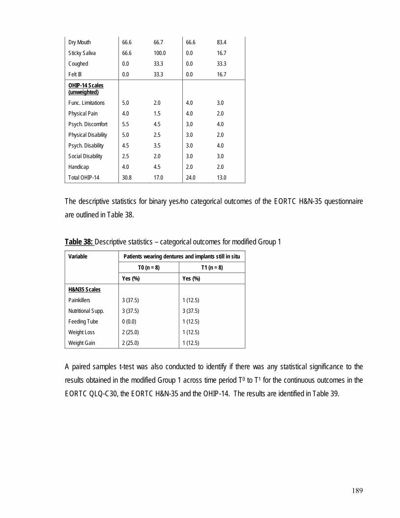
189
Dry Mouth 66.6 66.7 66.6 83.4
Sticky Saliva 66.6 100.0 0.0 16.7
Coughed 0.0 33.3 0.0 33.3
Felt Ill 0.0 33.3 0.0 16.7
OHIP-14 Scales (unweighted)
Func. Limitations 5.0 2.0 4.0 3.0
Physical Pain 4.0 1.5 4.0 2.0
Psych. Discomfort 5.5 4.5 3.0 4.0
Physical Disability 5.0 2.5 3.0 2.0
Psych. Disability 4.5 3.5 3.0 4.0
Social Disability 2.5 2.0 3.0 3.0
Handicap 4.0 4.5 2.0 2.0
Total OHIP-14 30.8 17.0 24.0 13.0
The descriptive statistics for binary yes/no categorical outcomes of the EORTC H&N-35 questionnaire are outlined in Table 38. Table 38:
Variable
Descriptive statistics – categorical outcomes for modified Group 1
Patients wearing dentures and implants still in situ
T0 (n = 8) T1 (n = 8)
Yes (%) Yes (%)
H&N35 Scales
Painkillers 3 (37.5) 1 (12.5)
Nutritional Supp. 3 (37.5) 3 (37.5)
Feeding Tube 0 (0.0) 1 (12.5)
Weight Loss 2 (25.0) 1 (12.5)
Weight Gain 2 (25.0) 1 (12.5)
A paired samples t-test was also conducted to identify if there was any statistical significance to the results obtained in the modified Group 1 across time period T0 to T1 for the continuous outcomes in the EORTC QLQ-C30, the EORTC H&N-35 and the OHIP-14. The results are identified in Table 39.
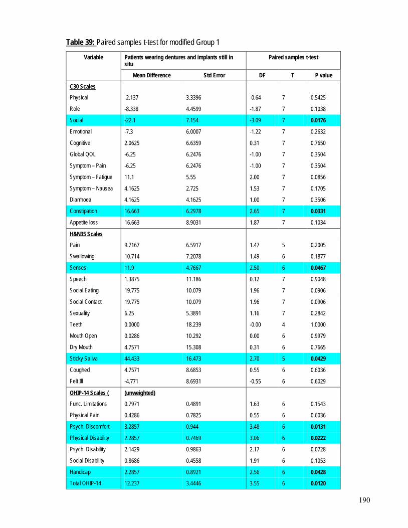
190
Table 39:
Variable
Paired samples t-test for modified Group 1
Patients wearing dentures and implants still in situ
Paired samples t-test
Mean Difference Std Error DF T P value
C30 Scales
Physical -2.137 3.3396 -0.64 7 0.5425
Role -8.338 4.4599 -1.87 7 0.1038
Social -22.1 7.154 -3.09 7 0.0176
Emotional -7.3 6.0007 -1.22 7 0.2632
Cognitive 2.0625 6.6359 0.31 7 0.7650
Global QOL -6.25 6.2476 -1.00 7 0.3504
Symptom – Pain -6.25 6.2476 -1.00 7 0.3504
Symptom – Fatigue 11.1 5.55 2.00 7 0.0856
Symptom – Nausea 4.1625 2.725 1.53 7 0.1705
Diarrhoea 4.1625 4.1625 1.00 7 0.3506
Constipation 16.663 6.2978 2.65 7 0.0331
Appetite loss 16.663 8.9031 1.87 7 0.1034
H&N35 Scales
Pain 9.7167 6.5917 1.47 5 0.2005
Swallowing 10.714 7.2078 1.49 6 0.1877
Senses 11.9 4.7667 2.50 6 0.0467
Speech 1.3875 11.186 0.12 7 0.9048
Social Eating 19.775 10.079 1.96 7 0.0906
Social Contact 19.775 10.079 1.96 7 0.0906
Sexuality 6.25 5.3891 1.16 7 0.2842
Teeth 0.0000 18.239 -0.00 4 1.0000
Mouth Open 0.0286 10.292 0.00 6 0.9979
Dry Mouth 4.7571 15.308 0.31 6 0.7665
Sticky Saliva 44.433 16.473 2.70 5 0.0429
Coughed 4.7571 8.6853 0.55 6 0.6036
Felt Ill -4.771 8.6931 -0.55 6 0.6029
OHIP-14 Scales ( (unweighted)
Func. Limitations 0.7971 0.4891 1.63 6 0.1543
Physical Pain 0.4286 0.7825 0.55 6 0.6036
Psych. Discomfort 3.2857 0.944 3.48 6 0.0131
Physical Disability 2.2857 0.7469 3.06 6 0.0222
Psych. Disability 2.1429 0.9863 2.17 6 0.0728
Social Disability 0.8686 0.4558 1.91 6 0.1053
Handicap 2.2857 0.8921 2.56 6 0.0428
Total OHIP-14 12.237 3.4446 3.55 6 0.0120
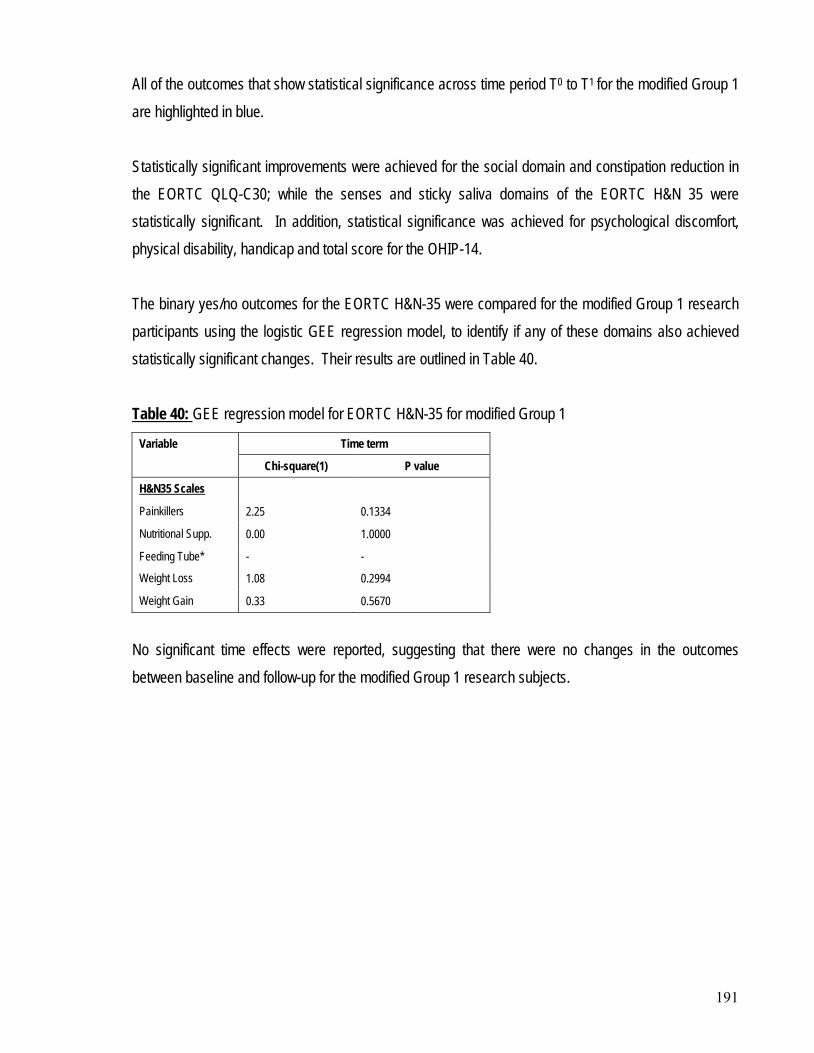
191
All of the outcomes that show statistical significance across time period T0 to T1 for the modified Group 1 are highlighted in blue. Statistically significant improvements were achieved for the social domain and constipation reduction in the EORTC QLQ-C30; while the senses and sticky saliva domains of the EORTC H&N 35 were statistically significant. In addition, statistical significance was achieved for psychological discomfort, physical disability, handicap and total score for the OHIP-14. The binary yes/no outcomes for the EORTC H&N-35 were compared for the modified Group 1 research participants using the logistic GEE regression model, to identify if any of these domains also achieved statistically significant changes. Their results are outlined in Table 40. Table 40:
Variable
GEE regression model for EORTC H&N-35 for modified Group 1
Time term
Chi-square(1) P value
H&N35 Scales
Painkillers 2.25 0.1334
Nutritional Supp. 0.00 1.0000
Feeding Tube* - -
Weight Loss 1.08 0.2994
Weight Gain 0.33 0.5670
No significant time effects were reported, suggesting that there were no changes in the outcomes between baseline and follow-up for the modified Group 1 research subjects.
192
This chapter gives an overview of the major findings of the study, with particular reference to the hypotheses in relevant sections. Whenever possible, comparisons have been made to previous studies and possible explanations for the differences discussed. It also includes the strengths and limitations, the significance, and implications of findings of this study. Finally, conclusions from this study are drawn and recommendations based on them.
CHAPTER 5: DISCUSSION, CONCLUSIONS AND RECOMMENDATIONS
This study shows that successful oral rehabilitation was achieved for 8 of the 14 Group 1 participants who were provided with an implant overdenture. These eight patients showed improvement in well-being, with statistically significant improvements in quality of life scales pertaining to the social, constipation, senses and sticky saliva domains of the EORTC questionnaires. Statistically significant improvements were also achieved for psychological comfort, physical disability, handicap and total oral health domains of the OHIP-14 questionnaire.
5.1 DISCUSSION
The remaining 6 patients in group 1 who did not achieve successful oral rehabilitation, only showed a statistically significant improvement with respect to the sticky saliva domain. A tendency for improvement between T0 and T1 was observed for all of the Group 1 participants in most of the functional scales of the EORTC except for the cognitive function domain and global quality of life which remained stable. Study participants were placed in Group 2, either because they elected not to proceed with an implant overdenture, or were declined implant provision due to a previous history of ORN. These participants exhibited a tendency for reduction in global quality of life, social eating and social contact between T0 and T1. In addition, there was an increase in the need for nutritional supplements in this group. Participants for both Group 1 and Group 2 showed an increase in fatigue between T0 and T1. Aim 1 of this study was to test whether hyperbaric oxygen treatment with prophylactic antibiotics assisted implant osseointegration and prevented ORN if induced by implant placement. All participants in Group 1 were able to achieve osseointegration of the implants in the short-term except for patient no. 9 who experienced early implant failure soon after the provision of the implant overdenture. ORN was diagnosed in two patients [patient no. 9 and 11] related to implant placement but subsequent to the
193
provision of the overdenture, while another developed ORN unrelated to implant placement [patient no. 14]. It is important to note that all three of these patients maintain a high alcohol intake and are currently smoking, with at least 35 pack year smoking histories. Aim 2 assessed the effectiveness and morbidity or a two implant mandibular overdenture in edentulous patients with an irradiated mandible. Clinical assessments of the successfully restored 8 participants in Group 1 all showed good peri-implant parameters, as well as good plaque index scores, calculus scores, bleeding and gingival index scores. This suggests that they were maintaining excellent oral, implant and denture hygiene. In addition, the subjective assessments by participants with respect to chewing ability, aesthetics and speech legibility were reported as either unchanged or improved. None of these eight participants reported a worsened result following treatment in any of these subjective assessments. From the remaining 6 participants in Group 1, only one person [patient no. 3] reported a worse subjective assessment following implant placement despite a technically successful result. This participant subsequently had the implants buried, and wears no lower prosthesis. Issues related to implant provision were experienced, and impacted on participant morbidity. Two patients experienced neuralgic symptoms [patients no. 6 and 11], and one experienced increased xerostomic symptoms related to prosthesis provision [patient no. 3]. Issues related to hyperbaric oxygen were minimal, with only one patient reporting middle ear barotrauma resulting in the need for grommet placement. No myopia was reported. Aim 3 was to compare patient’s satisfaction and quality of life following the successful provision of an implant mandibular overdenture, against no denture provision and non successful implant treatment. Although the fabrication of an implant overdenture did not result in a statistically significant overall quality of life, it did have a positive effect on some domains, in particular those related to social, psychological discomfort and physical disability. It is possible that only these minor improvements were able to be achieved, as oncological treatment in particular radiotherapy, is associated with significant morbidity including xerostomia, trismus, dysphagia and speech problems. The hypothesis of this thesis was that the provision of an implant mandibular overdenture in patients who had undergone radiotherapy for head and neck cancer, would improve their oral health-related quality of life, in particular chewing ability, speech legibility and appearance, while not causing any
194
complications, such as ORN. This hypothesis has been partially upheld, as minor improvements in quality of life, chewing ability, speech legibility and appearance were identified at time T1. While complications did occur, these have been identified as occurring in participants of increased risk due to continuation of smoking and heavy alcohol consumption. 5.1.1 Results and comparison with previous studies There have been a number of clinical studies published in the literature which have highlighted the benefits of hyperbaric oxygen for osseointegration in irradiated tissues, and have produced significant reductions in implant loss or failure to less than 10%.[96, 131, 134, 135, 147, 152, 162, 174, 204, 205] In this study at time T1 implant survival was calculated at 92.9%, but implant success was calculated at 57.1%
• An inability to identify the exact number of implants within the radiation field
i.e. 16 functional implants from a total of 28 implants placed. However, there are difficulties in making comparisons between the implant research available in the literature and the results of this study. This is predominantly due to:
• The exact region of implantation not always being identified
• Varying dosages of radiation applied to the implanted site, some as little as 20Gy
• Many different types, lengths and diameters of implants used
• Many different types of prosthetic appliances used
• Many of the studies having various follow-up periods (which may even vary within the individual reports)
• Many of the studies limited by too small a cohort size with short follow-up periods (an issue with this study also)
• Many different methods of evaluation applied
• Varying applications of the definition and criteria used for implant survival and implant success. Schliephake et al [158] commented that the lack of standardisation in the literature with respect to the statistical methods and criteria for success restricted the current evidence base. They identified a 12 month probability of implant survival of 93.7%. Shaw et al [151] were able to find 18 articles published between 1993 and 2003 reporting on the use of secondary implants after oral rehabilitation. They identified that 102 mandibular implants were lost in 801 patients (13% loss or 87% survival). Radiation doses in these articles ranged from 20Gy to 74Gy. Both of these studies show comparable results to
195
that achieved in this study, despite some patients having had much lower radiation dosages to the implanted area. Schoen et al published a series of articles looking at the provision of implant mandibular overdentures [127, 191, 225] Their results revealed similar results to that achieved in this study, where 13 patients who were treated with hyperbaric oxygen prior to implant placement had an implant survival 85.2% at 12 months.[191] The peri-implant parameters achieved by Schoen et al in their series [128, 191, 225] were similar to that achieved in this research. Implant survival in irradiated head and neck cancer patients within the first year following placement of a prosthesis has been found to be almost equivalent to that achieved with healthy subjects. It is important to note however that implant survival to 12 months is considered very early follow-up. Most studies have suggested that implant survival remains relatively high for the first five years.[135, 148, 152, 157, 160] However, implant loss appears to be progressive, with implant failure accelerating in the irradiated population after this time. [158] The criteria for patient selection for implant based oral rehabilitation following cancer treatments have been identified in the literature to include [93]:
• Adequate patient motivation, expectations and resources
• Reasonable oncologic prognosis
• Good oral hygiene
• Bone of adequate quality, volume and with a suitable maxillo-mandibular relationship
• Adequate oral function, in particular related to tongue and swallowing function
• No medical co-morbidities which contraindicate further surgery
• Cessation of smoking and high alcohol consumption. While these criteria have been applied where possible to patient selection in this study, this list was felt to represent a somewhat idealistic position and therefore the issue of smoking cessation in particular was not strenuously applied. At time T1, early complications associated with implant loss and ORN occurred in three patients from Group 1. All three of these patients are still current smokers, with at least 35 pack year smoking
196
histories. A current smoking habit has been identified by a number of authors as a significant exogenous factor influencing the successful integration of endosseous implants [103, 104, 108, 109, 111] with the recommendation that the cessation of smoking could improve osseointegration significantly. The results of this study would appear to support the literature, as ignoring previous and current heavy smoking habits and high alcohol consumption resulted in less favourable results, and increased risk of complication(s). The four cases of ORN identified in this research and are worthy of further discussion. In Group 2, one participant [Patient no.24] developed spontaneous ORN in the anterior mandible five years post-radiotherapy. In Group 1, three patients (21%) developed ORN [Patients no. 9, 11 and 14]. In two patients [Patient no. 9 and 11] the ORN could be directly attributable to implant placement (14%), and in the third patient [Patient no. 14] the ORN occurred spontaneously. A review of the literature pertaining to ORN in the mandible identified that the incidence to be:
• Between 5.8% and 44.1% in 4000 non-implanted subjects by Epstein et al in 1986[175]
• Three out of 170 cases (1.8%), subsequent to implant placement. This was attributed to a background incidence rather than any specific affect of implant placement by Keller et al in 1997 [150]
• Seventeen cases of ORN from a possible 1500 patients (1.1%) over a ten year period by Vudiniabola et al in 2000 in South Australia [73], and
• Two cases among 34 (5.8%) by Shaw et al in 2005 [151] In all of these articles, except for Vudiniabola et al [73], there is limited information regarding the therapeutic doses of radiotherapy delivered to the mandible, and most included ionising radiation of less than 55Gy. This is an important fact, as the literature has identified that the risk of ORN is increased if the patient has been exposed to a radiation dose of greater than 60Gy. [76, 77] This may explain the higher rate of ORN experienced in our study, as all participants, both Group 1 and 2, had been treated with a minimum of 55Gy. Another complication experienced associated with stage II implant surgery was neuralgia. Two patients (14%) developed post-implantation neuralgia, with CT scans confirming the implant body approximating the incisive branch of the inferior alveolar nerve. An article by Walton published in 2000 [118] identified that between one quarter to one third of patients with two implants placed into the anterior mandible,
197
would be expected to experience post-operative nerve sensation, which is consistent with the results obtained in this study. The provision of a successful functioning implant prosthesis which improved oral health-related quality of life was the goal of treatment in this research. The overall benefit of oral rehabilitation was assessed using quality of life assessments which were prospective, longitudinal and validated, with comparison to a control group. Initial analysis of the research cohort [Groups 1 and 2] to the EORTC-C30 reference data [246] found that the research cohort was very similar to the reference group with only the emotional domain showing statistical significance. When comparisons were made to the EORTC-H&N35 reference data [246], greater differences were identified. In particular, the research subjects experienced less pain but had greater difficulties with social eating, mouth opening, dry mouth and sticky saliva. These results are not unexpected given that the research participants in both Group 1 and 2 are all edentulous in the mandible and had received a minimum of 55Gy. It would be unlikely that all participants in the EORTC reference data group would be edentulous, and many may have received lower radiotherapy doses resulting in less xerostomia. Analysis of the global health domain of all research participants for the EORTC-C30 resulted in a score of 66.7, with Group 1 scoring 66.7, and Group 2 scoring 70.9. This is consistent with results reported in the literature, with scores between 60 to 70 commonly reported.[31] Schoen et al in their series [128, 191, 225] obtained slightly higher global health domain results with scores above 70. In this study only minor statistically significant differences were observed regarding quality of life between the two groups assessed at time T1. When comparisons were made between Group 1 and Group 2 the only statistical significance identified was in the sticky saliva domain of the EORTC H&N35. Group 1 participants reported an average increase of 33.34 points between T0 to T1 compared to participants in Group 2 who had an average decrease of 10.26 points. This result would suggest that while participants in Group 1 reported an increase in problems with sticky saliva, which may possibly be attributed to the impact of hyperbaric oxygen treatment, the increase in saliva production was not clinically sufficient enough to obtain a statistically significant difference for the dry mouth domain of the EORTC H&N35.
198
It has often been suggested that hyperbaric oxygen may exert a positive effect on radiotherapy induced oral dryness, but there is limited evidence available in the literature to support this to date. Bui et al also reported a low response rate of salivary symptoms following hyperbaric oxygen. [262] Schoen et al found that patients who had received hyperbaric oxygen reported a comparable level of oral dryness as patients who had not received hyperbaric oxygen. [191] Two recently published articles have obtained results which would appear to suggest that hyperbaric oxygen therapy may positively influence saliva quantity.[263, 264] Assessment of quality of life using the Oral Health Impact Profile [OHIP-14] across time period T0 to T1
for both weighted and unweighted results showed no statistical difference between the groups. Statistical significance was only able to be achieved when the 8 successful patients from Group 1 were analysed. It had been suggested by Allen and Locker [257] that the OHIP-14 may not be a suitable tool for assessment of quality of life in the edentulous population, as a number of questions pertinent to denture wearing from the OHIP-49 were excluded in the OHIP-14 questionnaire. Their research identified that the OHIP-14 had a poorer responsiveness to change in the edentulous population particularly when comparing conventional prostheses to implant prostheses. They developed a different modified version of the OHIP-49 specific for the edentulous population which they termed the OHIP-EDENT. A comparison was made between the OHIP-14 and the OHIP-EDENT for T1 between Group 1 participants. No statistical evidence was found of a relationship between the two scales, suggesting in fact that they may measure different outcomes. As only 8 of the 14 participants in Group 1 were successfully wearing the implant mandibular prostheses, analysis of only these 8 participants EORTC and OHIP-14 questionnaires were made to identify if they achieved any additional improvements in quality of life as a result of successfully wearing the implant prostheses. Statistically significant results suggesting improvement in quality of life were achieved for the:
• Social domain and constipation domains of the EORTC QLQ-C30
• Senses and sticky saliva domains of the EORTC H&N-35
• Psychological discomfort, physical disability, handicap and total domains of the OHIP-14. This result is consistent with the work of Schoen et al [128, 191, 225] in which they identified that the provision of a successfully functioning implant prosthesis can likely improve quality of life with respect to oral function and denture satisfaction.
199
The results of this study showed predominantly tendencies of association in quality of life. This is probably due to the fact that the cohort size was too small, limiting the ability to obtain many statistically significant results. The issue of small cohort size impacting on results has also been identified in the literature.[47, 245] Weymuller et al [245] identified that achieving any statistically significant results in patient orientated studies of head and neck cancer patients can be challenging, especially in a single institution setting. He believed that when quality of life is a secondary endpoint, statistically significant changes to quality of life may be difficult to demonstrate as survivors tend to accept and adjust to their disability. 5.1.2 Methodological strengths and limitations of this study The present study featured a number of methodological shortcomings:
• There was no randomisation, as patients who declined implant and overdenture provision or who had a past history of ORN were excluded from Group 1
• An increased risk of a skewed sample, as the participants were largely drawn from patients currently involved in the Special Needs Unit of the Adelaide Dental Hospital oncology review program, and these are predominantly more complex patients or those with a higher risk of recurrence or new primary oral lesions.
• Only a limited review period [maximum of 14 months] has been achieved, resulting only in short-term results analysed
• Small sample sizes in both Group 1 and Group 2 may limit the scope and ability to obtain statistically significance, resulting in an inability to generalise the outcome of the research.
Despite these limitations, statistically significant results were able to be achieved in some of the quality of life domains of the eight participants in Group 1 who successfully managed the mandibular implant prosthesis. In addition, improvements in these patients’ subjective outcomes provided additional important information to the researcher and clinician. The principal strengths of the research included:
• The multidisciplinary involvement in treatment planning for all Group 1 patients. This included consultations with an Oral and Maxillofacial Surgeon, Hyperbaric Oxygen physician, Special Needs Dentist plus an ENT surgeon and Ophthalmologist as required.
200
• The fact that it was a prospective study, with a limited variety of tumour sites, treatment regimes and reconstructive techniques used
• The participants were relatively homogenous, therefore limiting most compounding variables
• All clinical dental treatment was provided by only one Oral and Maxillofacial Surgeon, and one Special Needs Dentist thus minimising operator variance.
5.1.3 Implications of the study Surgical and radiotherapy treatment of malignancies in the oral cavity often results in an altered anatomical and physiological oral condition. The resultant compromised oral function may in part be addressed following the provision of an implant mandibular prosthesis but there is no guarantee of success. The results of this study would suggest that careful patient selection is imperative. In particular, the exclusion of patients with high current smoking and alcohol habits and the provision of hyperbaric oxygen may increase the success of implant osseointegration and the reduction of implant related complications, in particular ORN. In addition, hyperbaric oxygen treatment may also assist in an improvement in sticky saliva. Following implant placement, the successful provision of an implant overdenture can likely improve quality of life with respect to oral function, particularly with respect to eating/chewing ability, speech legibility and appearance. In addition, the assessment of oral health-related quality of life through the application of either the OHIP-EDENT or OHIP-49 and not the OHIP-14 may be necessary to ensure that an adequate responsiveness to change in the edentulous population is able to be measured, especially when comparing conventional prostheses to implant prostheses. 5.1.4 Future research Future research in this area would benefit from the development of an international randomised, longitudinal study with a larger participant cohort, and preferably involving multi-centre clinics. It is imperative that a standardised definition and criteria is established and applied for implant survival and implant success. In addition, the following variables need to be documented in any future research:
201
• The exact number of implants within the radiation field
• The exact region of implantation
• The dosages of radiation applied to the implanted site
• Different types, lengths and diameters of implants used
• Different types of prosthetic appliances used
• Method(s) of evaluation applied
• Significant follow-up periods
• Method(s) of evaluation applied
The successful oral rehabilitation of head and neck cancer patients following oncologic treatment has continued to be a difficult area to address. Surgical ablation, although an essential and effective component of tumour management, often results in an altered external appearance as well as an altered oral anatomy which compromises or prevents denture use. This combined with the physiologic effects of radiotherapy, results in a patient who requires structural, functional and aesthetic rehabilitation, but for whom few effective treatment options exist. Despite our best efforts, the side-effects of oncologic treatment for some oral and oropharyngeal tumours of the head and neck often defy current conventional aesthetic and functional oral rehabilitation prosthodontic techniques.
5.2 CONCLUSIONS
However, post-cancer aesthetics and function remains of great significance to the individual. Many of the oral reconstructive problems can be in part resolved by use of endosseous implants, used to support and retain either fixed or removable prostheses. While there is no absolute guarantee that this will resolve the compromised oral function, the use of endosseous implants may provide significantly improved retention of prostheses where oral anatomy is grossly altered following surgical treatment. There are now more than 100 publications available in the literature discussing osseointegration in irradiated tissues following head and neck ablative cancer surgery. It is very difficult to make a comparison of these studies as there is a general lack of agreement on how to evaluate implant survival or implant success, there are many different types, lengths and diameters of implants used, and there are also many different methods of evaluation applied.
202
While there is sufficient scientific evidence to show relatively good success of implant osseointegration in irradiated tissues in general, there is still a higher failure rate associated with the placement of implants into irradiated tissue compared with non-irradiated tissue. In addition, patients with a current smoking habit will experience an increased risk of complications, in particular ORN and/or implant failure. There has also been some concern raised about the long-term survival of implants in irradiated tissue, with some authors finding increased implant failure or loss with longer follow-up times. However, much of the research in this area is limited by too small a cohort size with short follow-up periods. While in this study the provision of an implant mandibular overdenture did not result in a significant improvement in the overall quality of life at T1, it did have a positive effect on subjective oral function in terms of chewing function, aesthetics and speech legibility, as well as some improvement in oral health quality of life. It is important to highlight that implant survival to 12 months is considered very early follow-up. It is envisaged that this cohort of patients will continue to be followed beyond the scope of this thesis, and the results presented and published. This study has shown that past head and neck radiotherapy should not be considered an absolute contraindication to implant placement in the mandible.
It is important as with any medical or dental procedure, to assess each patient on his or her individual merits, taking into consideration established patient selection criteria for implant based oral rehabilitation. In patients who may potentially benefit from an implant prosthesis as part of their functional and aesthetic rehabilitation following oncologic treatment for head or neck cancer, it is important that a review of all potential risks, complications and contra-indications to the proposed treatment be completed and then discussed with the patient. There is no indication for a blanket use of implants in all patients who require reconstruction and/or functional rehabilitation following oncologic treatment for head and neck cancer.
5.3 RECOMMENDATIONS
The number of patients who have undergone irradiation to the head and neck and are likely to benefit from an implant prosthesis is limited. It is important that for this patient cohort there is:
203
• A thorough evaluation of their medical history, including a smoking and alcohol use history
• Consideration given to their needs and concerns, with particular assessment of the patient’s current quality of life
• A thorough evaluation of their oncologic history to determine if the patient has received irradiation in the specific area of concern. If the implants are to be placed into an irradiated area then additional information is required such as the:
o irradiation dose o radiation quality, type and fractionation schedule o radiation source o potential need for hyperbaric oxygen treatment
As part of the planning process for implant provision in this group of patients it is important that the following issues are addressed:
• What is the long-term prognosis for this patient?
• What is the risk of recurrence for this patient?
• Would the patient be able to tolerate and manage a conventional removable appliance, acknowledging the risk of ORN associated with denture trauma?
• Will there be a tangible benefit(s) to the patient’s quality of life as a result of the provision of an implant prosthesis for this patient?
• Are there any definitive risks associated with implant placement in this patient?
• Can the implant procedure(s) be undertaken without any side-effects or complications?
• Will the success of osseointegration be deleteriously affected as a result of altered bone quality or vascularity in the surgical area?
• Will the patient be at risk of ORN or soft tissue necrosis as a result of implant surgery in an irradiated field?
• Is the patient a current smoker? If so, will she/he be able to cease their smoking habit? Hyperbaric oxygen is often advocated as a useful adjunct to improving the success and limiting the complications potentially associated with the placement of osseointegrated implants in the irradiated mandible. Currently there is no evidence-based guideline or protocol supporting the use of hyperbaric oxygen for implant placement, with the Cochrane Collaboration identifying only one double blind,
204
controlled clinical study. There are however, many articles published in the literature which support its continued use, with good success achieved, at least in the short-term. If the clinicians can predict that there are no significant anticipated problems based on the best evidence currently available, and there is scope for an improvement in the patient’s oral health-related quality of life, then an implant prosthesis should be considered. In addition, it is important that the provision of an implant prosthesis to patients who have undergone ablative surgery and/or radiotherapy to the head and neck region for cancer, is managed by a multidisciplinary team and only in clinics that have the capacity and skill to assess and manage post-radiotherapy and surgical related problems in cancer patients.

205
Appendix 1 – Letter of Introduction
name address address date Dear Re: Research pr oject o n t he pr ovision o f dental i mplants to patients who hav e
undergone head and neck radiotherapy. Traditionally head an d neck cancer patients who have had r adiation therapy to their edentulous lower j aws are l eft w ithout a l ower dent ure. This is to minimise t he potential risk of osteoradionecrosis caused by trauma to underlying oral tissues from the mobile denture. The Oral and Maxillofacial Surgery Unit and the Special Needs Unit of the Adelaide Dental H ospital ar e currently und ertaking r esearch i nto t he pr ovision o f i mplant retained l ower dentures for t his group o f pat ients and have obtained approval from the Royal Adelaide Hospital to undertake this research project. If you would l ike to be asse ssed to determine your suitability for involvement in this research and further information regarding the research, please contact Dr Sharon Liberali on 8222-8350. Yours sincerely Dr. Sharon Liberali BDS (Adel.); Grad.Dip.Clin.Dent (Adel.) Registrar, Special Needs Unit Adelaide Dental Hospital
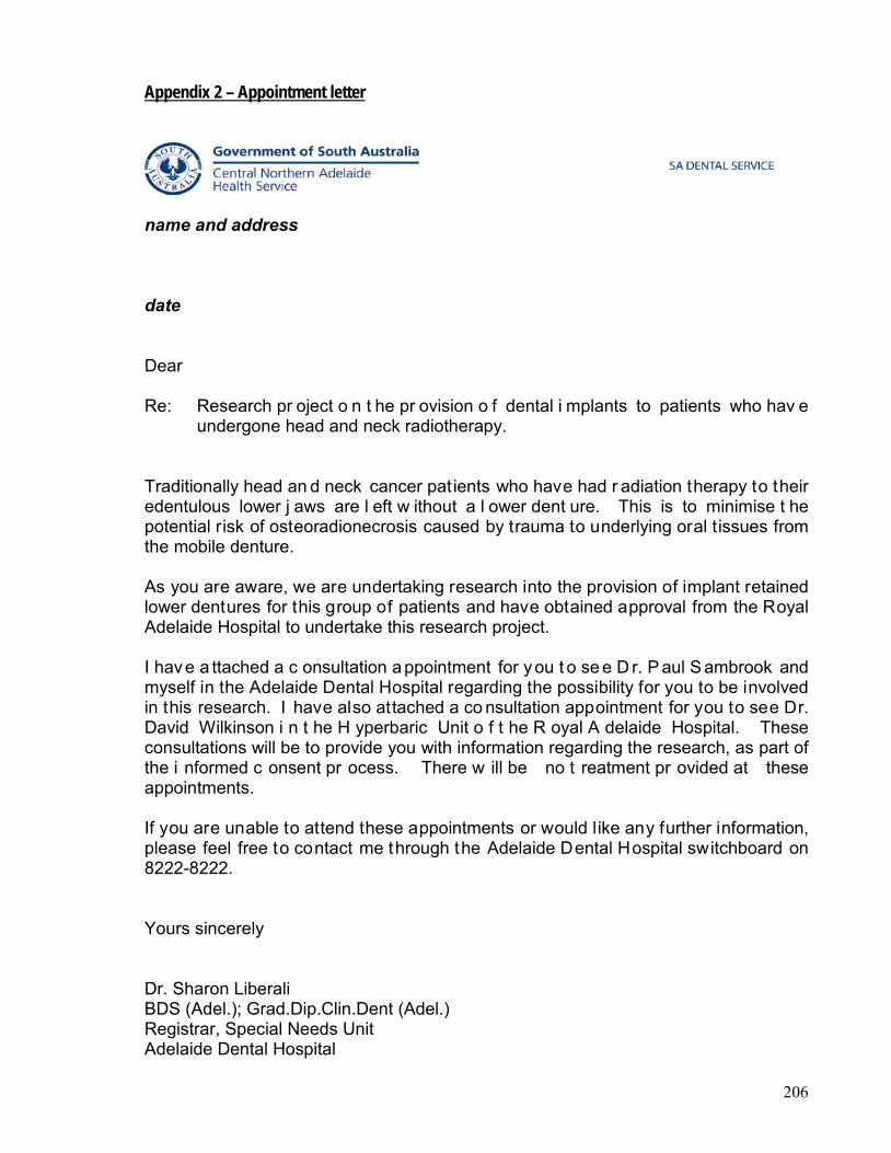
206
Appendix 2 – Appointment letter
name and address date Dear Re: Research pr oject o n t he pr ovision o f dental i mplants to patients who hav e
undergone head and neck radiotherapy. Traditionally head an d neck cancer patients who have had r adiation therapy to their edentulous lower j aws are l eft w ithout a l ower dent ure. This is to minimise t he potential risk of osteoradionecrosis caused by trauma to underlying oral tissues from the mobile denture. As you are aware, we are undertaking research into the provision of implant retained lower dentures for this group of patients and have obtained approval from the Royal Adelaide Hospital to undertake this research project. I have a ttached a c onsultation a ppointment for you t o se e D r. P aul S ambrook and myself in the Adelaide Dental Hospital regarding the possibility for you to be involved in this research. I have also attached a co nsultation appointment for you to see Dr. David Wilkinson i n t he H yperbaric Unit o f t he R oyal A delaide Hospital. These consultations will be to provide you with information regarding the research, as part of the i nformed c onsent pr ocess. There w ill be no t reatment pr ovided at these appointments. If you are unable to attend these appointments or would like any further information, please feel free to contact me through the Adelaide Dental Hospital switchboard on 8222-8222. Yours sincerely Dr. Sharon Liberali BDS (Adel.); Grad.Dip.Clin.Dent (Adel.) Registrar, Special Needs Unit Adelaide Dental Hospital
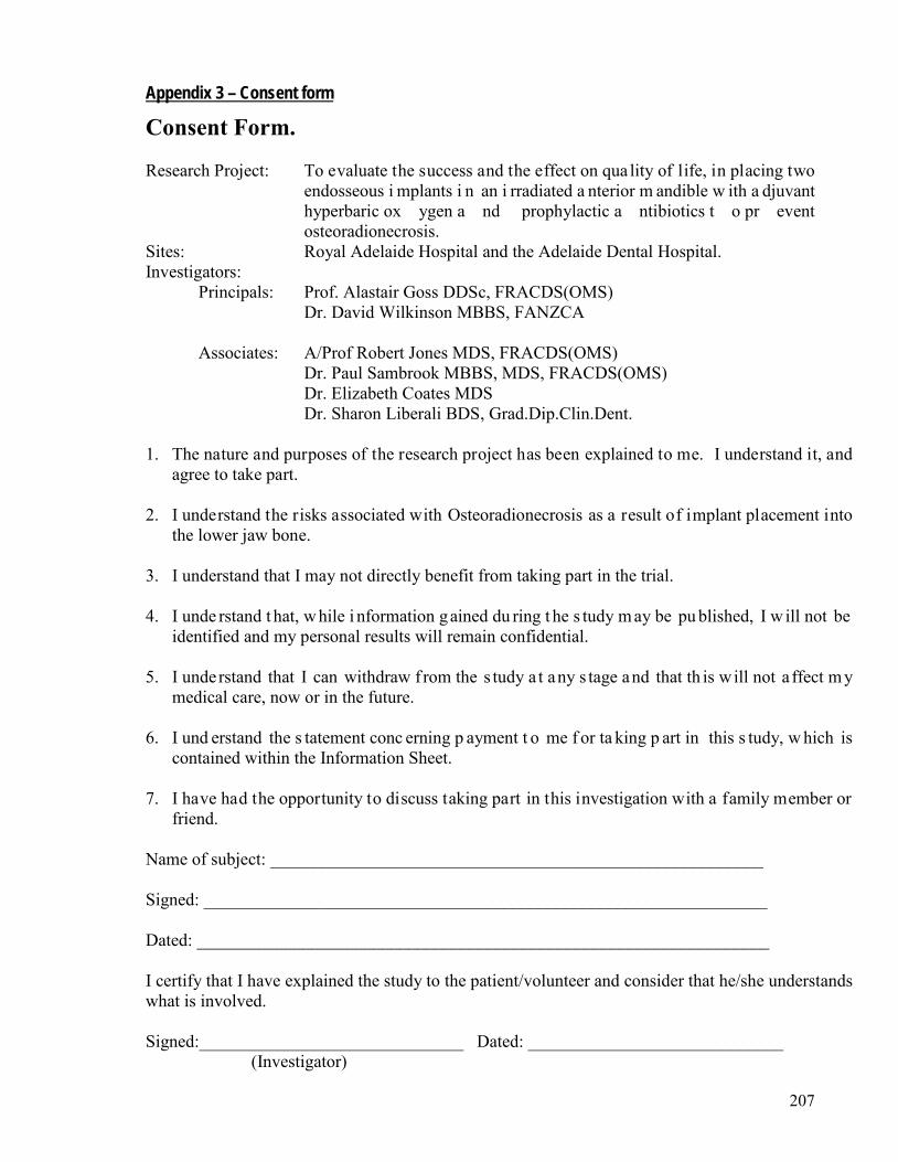
207
Consent Form. Appendix 3 – Consent form
Research Project: To evaluate the success and the effect on qua lity of life, in placing two
endosseous i mplants i n an i rradiated a nterior m andible w ith a djuvant hyperbaric ox ygen a nd prophylactic a ntibiotics t o pr event osteoradionecrosis.
Sites: Royal Adelaide Hospital and the Adelaide Dental Hospital. Investigators:
Principals: Prof. Alastair Goss DDSc, FRACDS(OMS) Dr. David Wilkinson MBBS, FANZCA
Associates: A/Prof Robert Jones MDS, FRACDS(OMS) Dr. Paul Sambrook MBBS, MDS, FRACDS(OMS) Dr. Elizabeth Coates MDS Dr. Sharon Liberali BDS, Grad.Dip.Clin.Dent.
1. The nature and purposes of the research project has been explained to me. I understand it, and
agree to take part. 2. I understand the risks associated with Osteoradionecrosis as a result of implant placement into
the lower jaw bone. 3. I understand that I may not directly benefit from taking part in the trial. 4. I unde rstand t hat, w hile i nformation gained du ring t he s tudy m ay be pu blished, I w ill not be
identified and my personal results will remain confidential. 5. I understand that I can withdraw f rom the s tudy a t any s tage and that th is will not a ffect m y
medical care, now or in the future. 6. I und erstand the s tatement conc erning p ayment t o me f or ta king p art in this s tudy, w hich is
contained within the Information Sheet. 7. I have had the opportunity to discuss taking part in this investigation with a family member or
friend. Name of subject: ________________________________________________________ Signed: ________________________________________________________________ Dated: _________________________________________________________________ I certify that I have explained the study to the patient/volunteer and consider that he/she understands what is involved. Signed:______________________________ Dated: _____________________________ (Investigator)
208
Information sheet for research subjects (Group 1) Appendix 4 – Information sheet for Research subjects (Group 1)
Research: To e valuate t he s uccess a nd t he e ffect on qua lity of l ife, i n pl acing t wo
endosseous i mplants i n a n i rradiated a nterior m andible w ith a djuvant hyperbaric oxygen and prophylactic antibiotics to prevent osteoradionecrosis.
Sites: Royal Adelaide Hospital and the Adelaide Dental Hospital.
You a re b eing a sked t o t ake pa rt i n a s tudy. This i s r esearch a nd your pa rticipation i s voluntary. If you do not wish to take part in the study, your medical and dental care will not be affected in any way. Even if you agree to participate, you may withdraw at any time.
YOUR PARTICIPATION IS VOLUNTARY
The purpose of this study is to document the risks and successes of providing a stable lower denture f or pa tients l ike yourself w hose j aw ha s r eceived hi gh dos e r adiation. I n or der t o achieve this, two implants will need to be placed in your lower jaw. For successful implants it is i mportant f or t he bone t o be he althy. Based on c urrent r esearch, h yperbaric ox ygen treatment ma y impr ove the bl ood circulation in bone, and t herefore r educe t he cha nces o f developing a bone infection called Osteoradionecrosis due the placement of the implants.
WHAT IS THE PURPOSE OF THE TRIAL?
1. You will receive 20 sessions of hyperbaric oxygen therapy, five (5) days a week for four (4) weeks. In each session you will breathe 100% oxygen for 120 m inutes (at 2.0 a tmospheric pressure).
WHAT WOULD YOU HAVE TO DO?
2. Y ou w ill be g iven 2g A moxicillin or ally 1 hour pr ior t o i mplant placement, a nd 1 g Amoxicillin orally 6 hours later. In the case of allergy to Penicillin you will be given 600mg Clindamycin orally as a single dose 1 hour prior to implant placement. 3. After finishing the 20 sessions, two (2) implants will be placed in the front area of the lower jaw. A n implant is l ike a s crew, tightened in t he j aw bone . T he i mplant i s pl aced b y experienced surgeons in the Royal Adelaide Hospital. An incision is made to expose the bone. Then a hol e i s dr illed i nto w hich t he i mplant i s s crewed. S titches a re pl aced t o close t he wound. 4. Following implant placement you will receive a further 10 s essions of hyperbaric oxygen therapy, f ive ( 5) da ys a w eek f or t wo ( 2) w eeks. In each s ession you will br eathe 100 % oxygen for 120 minutes (at 2.0 atmospheric pressure). 5. A four (4) month period is necessary for healing. A review appointment will then occur to assess the implants status. If the implants are firm, not painful and not causing severe bone resorption, the denture (false teeth) construction will be started.
If you are pregnant or likely to become so then discuss this with your treating doctors involved in the trial.
WOMEN OF CHILDBEARING AGE
209
1. It is possible to have a complication to Hyperbaric Oxygen therapy which may include ear pain i n up to 5% of cases, or an adverse r eaction to Oxygen which may include an oxygen toxic seizure in approximately 1 in 10,000 cases. Proper assessment can minimise the chances of such complications. You will be seen by the Doctor in the Hyperbaric Medicine Unit and consented separately for this treatment prior to commencing Hyperbaric Oxygen treatment.
WHAT PROBLEMS MIGHT OCCUR DURING AND/OR AFTER THE TRIAL?
2. There is a chance of implant failure if they do not attach very well to the jaw bone. 3. There is a risk that an infection in the lower jaw bone may develop. In that case you will receive m ore s essions of h yperbaric ox ygen t herapy o r r emoval of t he i nfected bon e i f necessary.
Patients w ho have ha d their j aws i rradiated as pa rt of m anagement of he ad and neck malignancy a re at r isk of O steoradionecrosis ( ORN). T his i s a known a nd s erious complication of therapeutic radiotherapy for head and neck cancer. As a result of the surgical placement of the implants there is a potential to induce osteoradionecrosis. ORN can be both painful and debilitating, and most patients commonly feel that it is a worse challenge to their well being than their original cancer which required the radiotherapy.
RESEARCH RELATED INJURY
By participating in this research, an implant retained lower denture may substantially enhance your qua lity of lif e b y improving or al f unction, especially with respect to eating ability, aesthetics and speech.
IS THERE ANYTHING TO GAIN FROM PARTICIPATING?
Traditionally head and neck cancer patients who have had radiotherapy to their bottom jaws are le ft w ithout a low er de nture to minimise th e pot ential r isk of os teoradionecrosis in the lower jaw caused by a loose lower denture rubbing on the gums. M ost patients are unhappy without a lower denture and wish to have one to improve their appearance and for eating and speaking.
WHAT ARE THE ALTERNATIVES?
The inf ormation collected as pa rt of thi s r esearch will be us ed for de scriptive s tatistical analysis and r esearch pu rposes onl y. P articipants w ill onl y b e a ble t o b e i dentified b y the Investigators involved in the research.
CONFIDENTIALITY.
If you have any questions please contact the investigators: NAMES AND CONTACT NUMBERS OF INVESTIGATORS.
• Prof. Alastair Goss telephone 8303-5103 (regarding surgical treatment) • Dr. David Wilkinson telephone 8222-5116 (regarding hyperbaric treatment) • Dr. Sharon Liberali telephone 8222-8222 (regarding denture treatment)
If you wish to discuss aspects of the study with someone not directly involved, you may also contact t he C hairman, Research and E thics C ommittee, R oyal A delaide H ospital on 8222 -4139.
INDEPENDENT CONTACT
Appendix 5 – Information sheet for Research subjects (Group 2)
210
Information sheet for research subjects (Group 2) Appendix 5 – Information sheet for Research subjects (Group 2)
Research: To evaluate t he s uccess a nd t he e ffect on qua lity of l ife, i n pl acing t wo
endosseous i mplants i n a n i rradiated a nterior m andible w ith a djuvant hyperbaric oxygen and prophylactic antibiotics to prevent osteoradionecrosis.
Sites: Royal Adelaide Hospital and the Adelaide Dental Hospital.
You a re b eing a sked t o t ake pa rt i n a s tudy. This i s r esearch a nd your pa rticipation i s voluntary. If you do not wish to take part in the study, your medical and dental care will not be affected in any way. Even if you agree to participate, you may withdraw at any time.
YOUR PARTICIPATION IS VOLUNTARY
The purpose of this study is to document the risks and successes of providing a stable lower denture for patients like yourself whose jaw has received high dose radiation.
WHAT IS THE PURPOSE OF THE TRIAL?
During your r outine r eview a ppointments you w ill be pa rticipating i n t he r esearch b y completing a survey assessing the impact of your cancer treatment(s) on quality of life from an oral function perspective, especially with respect to eating ability, aesthetics and speech. This information w ill be compared w ith t hat f rom a group of p atients w ho a re h aving i mplants placed in their lower jaw in order to have a stable lower denture.
WHAT WOULD YOU HAVE TO DO?
By participating in this research, an implant retained lower denture may become the standard treatment for patients who have no teeth and who have received high dose radiotherapy to the lower jaw.
IS THERE ANYTHING TO GAIN FROM PARTICIPATING?
You are receiving the standard treatment, which is to be without a lower denture in order to minimise t he r isk of bone death i n t he l ower j aw caused b y a l oose de nture rubbing on t he gums.
WHAT ARE THE ALTERNATIVES?
The i nformation collected as pa rt of t his r esearch will be us ed for de scriptive s tatistical analysis and r esearch pu rposes onl y. P articipants w ill onl y b e a ble t o b e i dentified b y t he Investigators involved in the research.
CONFIDENTIALITY.
If you have any questions please contact the investigators: NAMES AND CONTACT NUMBERS OF INVESTIGATORS.
• Prof. Alastair Goss telephone 8303-5103 (regarding surgical treatment) • Dr. David Wilkinson telephone 8222-5116 (regarding hyperbaric treatment) • Dr. Sharon Liberali telephone 8222-8222 (regarding denture treatment)

211
If you wish to discuss aspects of the study with someone not directly involved, you may also contact t he C hairman, R esearch and E thics C ommittee, R oyal A delaide H ospital on 8222 -4139.
INDEPENDENT CONTACT

212
Appendix 6 – EORTC QLQ-C30 questionnaire

214
Appendix 7 – EORTC H&N35 questionnaire
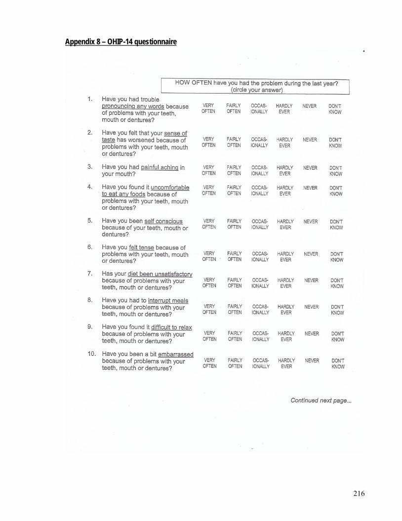
216
Appendix 8 – OHIP-14 questionnaire
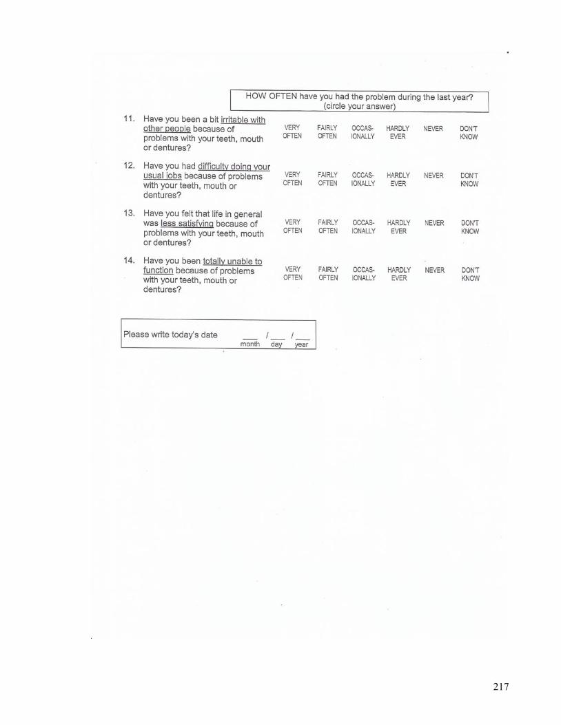
217
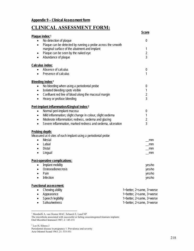
218
Appendix 9 – Clinical Assessment form
CLINICAL ASSESSMENT FORM:
Score Plaque index:1
• No detection of plaque 0
• Plaque can be detected by running a probe across the smooth marginal surface of the abutment and implant 1
• Plaque can be seen by the naked eye 2 • Abundance of plaque 3
• Absence of calculus 0 Calculus index:
• Presence of calculus 1 Bleeding index:
• No bleeding when using a periodontal probe 0 1
• Isolated bleeding spots visible 1 • Confluent red line of blood along the mucosal margin 2 • Heavy or profuse bleeding 3
Peri-implant inflammation/Gingival index:2
• Normal peri-implant mucosa 0
• Mild inflammation; slight change in colour, slight oedema 1 • Moderate inflammation; redness, oedema and glazing 2 • Severe inflammation, marked redness and oedema, ulceration 3
Measured at 4 sites of each implant using a periodontal probe Probing depth:
• Mesial __mm • Labial __mm • Distal __mm • Lingual __mm
• Implant mobility yes/no Post-operative complications:
• Osteoradionecrosis yes/no • Pain yes/no • Infection yes/no
• Chewing ability 1=better, 2=same, 3=worse Functional assessment:
• Appearance 1=better, 2=same, 3=worse • Speech legibility 1=better, 2=same, 3=worse • Saliva/wetness 1=better, 2=same, 3=worse
1 Mombelli A, van Oosten MAC, Schurch E, Land NP The microbiota associated with successful or failing osseointegrated titanium implants Oral Microbiol Immunol 1987; 2: 145-151 2 Loe H, Silness J Periodontal disease in pregnancy 1: Prevalence and severity Acta Odontol Scand 1963; 21: 533-551
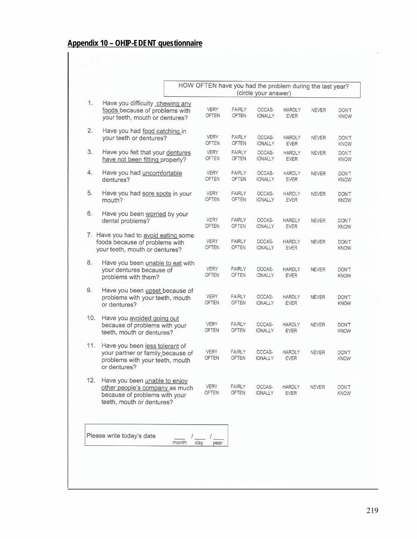
219
Appendix 10 – OHIP-EDENT questionnaire
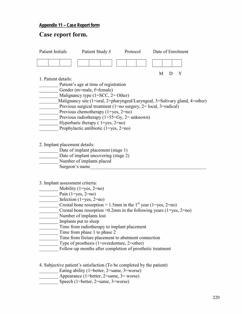
220
Appendix 11 – Case Report form
Case report form. Patient Initials Patient Study # Protocol Date of Enrolment
M D Y 1. Patient details: ________ Patient’s age at time of registration ________ Gender (m=male, f=female) ________ Malignancy type (1=SCC, 2= Other) ________Malignancy site (1=oral, 2=pharyngeal/Laryngeal, 3=Salivary gland, 4=other) ________ Previous surgical treatment (1=no surgery, 2= local, 3=radical) ________ Previous chemotherapy (1=yes, 2=no) ________ Previous radiotherapy (1=55+Gy, 2= unknown) ________ Hyperbaric therapy ( 1=yes, 2=no) ________ Prophylactic antibiotic (1=yes, 2=no) 2. Implant placement details: ________ Date of implant placement (stage 1) ________ Date of implant uncovering (stage 2) ________ Number of implants placed Surgeon’s name_________________________________________________ 3. Implant assessment criteria: ________ Mobility (1=yes, 2=no) ________ Pain (1=yes, 2=no) ________ Infection (1=yes, 2=no) ________ Crestal bone resorption > 1.5mm in the 1st year (1=yes, 2=no) ________ Crestal bone resorption >0.2mm in the following years (1=yes, 2=no) ________ Number of implants lost ________ Implants put to sleep ________ Time from radiotherapy to implant placement ________ Time from phase 1 to phase 2 ________ Time from fixture placement to abutment connection ________ Type of prosthesis (1=overdenture, 2=other) ________ Follow-up months after completion of prosthetic treatment 4. Subjective patient’s satisfaction (To be completed by the patient) ________ Eating ability (1=better, 2=same, 3=worse) ________ Appearance (1=better, 2=same, 3= worse) ________ Speech (1=better, 2=same, 3=worse)
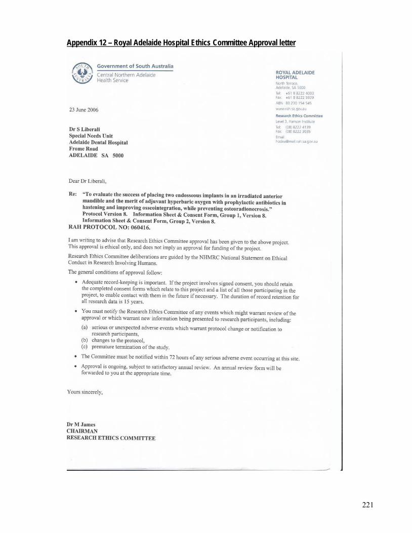
221
Appendix 12 – Royal Adelaide Hospital Ethics Committee Approval letter
222
Appendix 13 – South Australian Dental Service Research Approval letter
223
Appendix 14 – EORTC QOL C30 User’s agreement
224
CHAPTER 6: BIBLIOGRAPHY 1. Faggion, C.M. and Y.-K. Tu, Evidence-based Dentistry: A model for clinical practice.
Journal of Dental Education, 2007. 71(6): p. 825-831. 2. Forrest, J.L. and S.A. Miller, Evidence-based decision making in action: Part 1 - Finding
the best clinical evidence. The Journal of Contemporary Dental Practice, 2002. 3(3): p. 10-26.
3. Richardson, W.S., et al., The well-built clinical question: A key to evidence based decisions. ACP Journal Club, 1995. 123: p. A12-A13.
4. Cochrane, C., http://www.cochrane-net.org/openlearning/, Editor. 5. World Health Organization, Tabular List of Disease. Volume 1 of the International
Statistical Classification of Diseases and Health-related Problems, Tenth Revision, Australian Modification. (ICD-10-AM). 5th Edition ed. 2006.
6. Moore, S.R., et al., The epidemiology of tongue cancer: a review of global incidence. Oral Diseases, 2000. 6: p. 75-84.
7. Moore, S.R., et al., The epidemiology of mouth cancer: a review of global incidence. Oral Diseases, 2000. 6: p. 65-74.
8. Mignogna, M.D., S. Fedele, and L. Russo, The World cancer report and the burden of oral cancer. European Journal of Cancer Prevention, 2004. 13: p. 139-142.
9. Roder, D., South Australian Cancer Statistics. Monograph No 3. Cancers of the Respiratory Organs, Throat and Mouth, in Centre for Cancer Control Research. Monograph Series. 2002, Anti-Cancer Foundation, South Australia: Adelaide.
10. Farah, C.S. and M.J. McCullough, Oral cancer awareness for the general practitioner: A new approach to patient care. Australian Dental Journal, 2008. 53(1): p. 2-10.
11. Sugerman, P.B. and N.W. Savage, Oral Cancer in Australia:1983-1996. Australian Dental Journal, 2002. 47(1): p. 45-56.
12. Neville, B.W. and T.A. Day, Oral Cancer and Precancerous lesions. CA: A Cancer Journal for Clinicians, 2002. 52: p195-215.
13. Lopez-Jornet, P. and F. Comacho-Alonso, New barriers in oral cancer: Patient accessibility to dental examination - a pilot study. Oral Oncology, 2006. 42: p. 1022-1005.
14. Scully, C. and D.H. Felix, Oral Medicine - Update for the Dental Practitioner. Oral Cancer. British Dental Journal, 2006. 200(1): p. 13-17.
15. McLeod, N.M.H., N.R. Saeed, and E.A. Ali, Oral Cancer: Delays in referral and diagnosis persist. British Dental Journal, 2005. 198(11): p. 681-684.
16. Welfare, A.I.o.H.a., A.A.o.C. Registries, and N.C.S. Group, Cancer Incidence projections: Australia 2002-2011, A.C.N.C. 25., Editor. 2005: Canberra.
17. Subapriya, R., et al., Assessment of risk factors for oral squamous cell carcinoma in Chidambaran, Southern India: A case-control study. European Journal of Cancer Prevention, 2007. 16: p. 251-256.
18. Roder, D., South Australian Cancer Statistics. Monograph no. 8. Time trends in cancer mortality in South Australia between 1990 and 2011., C.f.C.c.r.M. series, Editor. 2005, Anti-cancer foundation, South Australia: Adelaide.
19. Lam, L., R.M. Logan, and C. Luke, Epidemiological analysis of tongue cancer in South Australia for the 24-year period, 1977-2001. Australian Dental Journal, 2006. 51(1): p. 16-21.
20. Parkin, D.M., et al., Global Cancer Statistics 2002. CA: A Cancer Journal for Clinicians, 2002. 55(2): p. 74-108.
225
21. Yao, M., et al., Current surgical treatment of squamous cell carcinoma of the head and neck. Oral Oncology, 2007. 43: p. 213-223.
22. Higgins, K.M. and J.R. Wang, State of Head and Neck surgical oncology research - a review and critical appraisal of landmark studies. Head and Neck, 2008. 30: p. 1636-1642.
23. Jones, A.S., et al., Survival of patients with neck recurrence following radical neck dissection: utility of a second neck dissection? Head and Neck, 2008. 30: p. 1514-1522.
24. Patel, S.G. and J.P. Shah, TMN staging of cancers of the head and neck: Striving for uniformity among Diversity. CA: A Cancer Journal for Clinicians, 2005. 55: p. 242-258.
25. Rogers, S.N., et al., Clinical Function after primary surgery for oral and oropharyngeal cancer: an 11-item examination. British Journal of Oral and Maxillofacial Surgery, 2002. 40: p. 1-10.
26. Chandu, A., et al., Reconstructive techniques for the repair of oral and maxillofacial oncological procedures: What are they, how do they work and what do they look like? Australian Dental Journal, 2002. 47(2): p. 99-105.
27. Barber, H.D., et al., Evaluation of Osseointegration of Endosseous Implants in Radiated Vascularised Fibula flaps to the Mandible: A Pilot Study. Journal of Oral and Maxillofacial Surgery, 1995. 53: p. 640-644.
28. Urken, M.L., et al., Primary placement of osseointegrated implants in microvascular mandibular reconstruction. Otolaryngology, Head and Neck Surgery, 1989. 101: p. 56-73.
29. Chiapasco, M., et al., Clinical outcome of dental implants placed in fibula-free-flaps used for the reconstruction of maxillo-mandibular defects following ablation for tumours or osteoradionecrosis. Clinical Oral Implants Research, 2006. 17(2): p. 220-228.
30. Chan, M.F.W.-Y., J.P. Hayter, and R.A. Howell, Oral rehabilitation with Implant-retained Prostheses following Ablative Surgery and Reconstruction with Free-flaps. International Journal of Oral and Maxillofacial Implants, 1997. 12: p. 820-827.
31. Sclaroff, A., et al., Immediate mandibular reconstruction and placement of dental implants. At the time of ablative surgery. Oral Surgery, Oral Medicine, Oral Pathology, 1994. 78: p. 711-717.
32. Won-Suck, O., Roumanas E, and Beumer, J , Maxillofacial reconstruction after head and neck tumour therapy. Compendium, 2007. 28(2): p. 28-32.
33. Urken, M.L., et al., Oromandibular reconstruction using microvascular composite flaps: report of 210 cases. Archives of Otolaryngology, Head and Neck Surgery, 1998. 124(1): p. 46-55.
34. Fischer, D.J. and J.B. Epstein, Management of patients who have undergone head and neck cancer therapy. Dental Clinics of North America, 2008. 52: p. 39-60.
35. Zlotolow, I.M., et al., Osseointegrated Implants and Functional Prosthetic Rehabilitation in Microvascular Fibula Free Flap Reconstructed Mandibles. The American Journal of Surgery, 1992. 165: p. 677-681.
36. Martin, I.C., et al., Endosseous implants in the irradiated composite radial forearm free flap. International Journal of Oral and Maxillofacial Surgery, 1992. 21: p. 266-270.
37. Roumanas E., Chang T L, and Beumer J , Use of osseo-integrated implants in the restoration of head and neck defects. Journal of the Californian Dental Association, 2006. 34(9): p. 711-718.
38. Urken, M.L., D. Buchbinder, and H. Weinberg, Functional evaluation following microvascular oromandibular reconstruction of the oral cancer patient: a comparative study of reconstructed and no reconstructed patients. Laryngoscope, 1991. 101: p. 935.
39. Rogers, S.N., et al., Health-related quality of life and clinical function after primary surgery for oral cancer. British Journal of Oral and Maxillofacial Surgery, 2002. 40: p. 11-18.
226
40. Beumer, J., E. Roumanas, and R. Nishimura, Advances in Osseointegrated Implants for Dental and Facial Rehabilitation Following Major Head and Neck Surgery. Seminars in Surgical Oncology, 1995. 11: p. 200-207.
41. Vaughan, E.D., An analysis of Morbidity following Major Head and Neck Surgery with Particular Reference to Mouth Function. Journal of Maxillofacial Surgery, 1982. 10: p. 129-134.
42. Rankin, M. and G.L. Borah, Perceived functional impact of abnormal facial appearance. Plastic and Reconstructive Surgery, 2003. 111: p. 2140-2146.
43. Stuiver, M.M., et al., Impact of shoulder complains after neck dissection on shoulder disability and quality of life. Otolaryngology, Head and Neck Surgery, 2008. 139: p. 32-39.
44. Inoue, H., et al., Quality of life after neck dissection. Arch. Otolaryngol. Head Neck Surg, 2006. 132: p. 662-666.
45. Murphy, B.A., et al., Symptom control issues and supportive care of patients with head and neck cancers. Clinical advances in Hematology & Oncology, 2007. 5(10): p. 807-822.
46. Vissink, A., et al., Prevention and Treatment of the consequences of Head and Neck Radiotherapy. Critical Review of Oral Biology and Medicine, 2003. 14(3): p. 213-225.
47. Granstrom, G., Radiotherapy, osseointegration and hyperbaric oxygen therapy. Periodontology 2000, 2003. 33: p. 145-162.
48. Schepers, R.H., et al., Effect of postoperative radiotherapy on the functional result of implant placed during ablative surgery for oral cancer. International Journal of Oral and Maxillofacial Surgery, 2006. 35: p. 803-808.
49. Lalla, R. and D. Peterson, Oral Mucositis. Dental Clinics of North America, 2005. 49(1): p. 167-184.
50. Kielbassa, A.M., et al., Radiation-related damage to dentition. The Lancet Oncology, 2006. 7: p. 326-335.
51. Cooper, J.S., et al., Late effects of Radiation therapy in the Head and Neck region. International Journal of Oncology Biology Physics, 1995. 31(5): p. 1141-1164.
52. Vissink, A., et al., Oral Sequelae of Head and Neck Radiotherapy. Critical Review of Oral Biology and Medicine, 2003. 14(3): p. 199-212.
53. Chua, D.T.T., Y. Tian, and W.I. Wei, Late oral complications following radiotherapy for head and neck cancer. Expert rev anticancer ther, 2007. 7(9): p. 1215-1244.
54. Sonis, S.T., et al., Perspectives on Cancer therapy - induced mucosal injury. Cancer, 2004. 100 (9 suppl)(9): p. 1995-2025.
55. Stevenson, R.J., L.A. Miller, and Z.C. Thayer, Impairments in the perception of odor-induced tastes and their relationship to impairments in taste perception. Journal of Experimental psychology : Human perception and performance, 2008. 34(5): p. 1183-1197.
56. Shiboski, C.H., et al., Management of salivary hypofunction during and after radiotherapy. Oral Surgery, Oral Medicine, Oral Pathology, Oral Radiology, Endodontics, 2007. 103(Supplement 1): p. S66e1-S66e19.
57. Porter, S.R., C. Scully, and A.M. Hegarty, An update on the aetiology and management of xerostomia. Oral Surgery, Oral Medicine, Oral Pathology, Oral Radiology, Endodontics, 2004. 97(28-46).
58. Dirix, P., S. Nutts, and W. van den Bogaert, Radiation induced xerostomia in patients with head and neck cancer: A literature review. Cancer, 2006. 107: p. 2525-2534.
59. Murphy, B.A., A critical component of supportive care. Supportive Oncology, 2007. 5(5): p. 228-229.
60. Brosky, M.E., The role of saliva in oral health: Strategies for prevention and management of xerostomia. Supportive Oncology, 2007. 5(5): p. 215-225.
227
61. Dijkstra, P., W.W.I. Kalk, and J.L.N. Roodenburg, Trismus in head and neck oncology: A systematic review. Oral Oncology, 2004. 40: p. 879-889.
62. Hancock, P.J., J.B. Epstein, and G.R. Sadler, Oral and Dental Management related to Radiation therapy for Head and Neck cancer. Journal of the Canadian Dental Association, 2003. 69(9): p. 585-590.
63. Nguyen, N.P., et al., Dysphagia following chemo radiation for locally advanced Head and Neck cancer. Annals of Oncology, 2004. 15: p. 383-388.
64. Kendall, K.A., et al., Structural motility in deglutition after single modality treatment in head and neck carcinoma with radiotherapy. Head and Neck, 1998. 20: p. 720-725.
65. Marx, R.E. and R.P. Johnson, Studies in the radiobiology of osteoradionecrosis and their clinical significance. Oral Surgery, Oral Medicine, Oral Pathology, 1987. 64(4): p. 379-390.
66. Vudiniabola, S., et al., Hyperbaric oxygen in the prevention of osteoradionecrosis of the jaws. Australian Dental Journal, 1999. 44(4): p. 243-247.
67. Reuther, T., et al., Osteoradionecrosis of the jaws as a side-effect of radiotherapy of head and neck tumour patients - a report of a thirty year retrospective review. International Journal of Oral and Maxillofacial Surgery, 2003. 32: p. 289-295.
68. Marx, R.E., Osteoradionecrosis: A new concept of its Pathophysiology. Journal of Maxillofacial Surgery, 1983. 41: p. 283-288.
69. Ben-David, M.A., et al., Lack of osteoradionecrosis of the mandible after intensity modulated radiotherapy for head and neck cancer: likely contributions of both dental care and improved dose distributions. International Journal of Oncology Biology Physics, 2007. 68(2): p. 396-402.
70. Marx, R.E., A new concept in the treatment of Osteoradionecrosis. Journal of Maxillofacial Surgery, 1983. 41: p. 351-357.
71. Widmark, G., S. Sagne, and P. Heikel, Osteoradionecrosis of the jaws. International Journal of Oral and Maxillofacial Surgery, 1989. 18: p. 302-306.
72. Epstein, J.B., et al., Postradiation osteonecrosis of the mandible. A long-term follow-up study. Oral Surgery, Oral Medicine, Oral Pathology, Oral Radiology, Endodontics, 1997. 83: p. 657-662.
73. Vudiniabola, S., et al., Hyperbaric Oxygen in the therapeutic management of osteoradionecrosis of the facial bones. International Journal of Oral and Maxillofacial Surgery, 2000. 29: p. 435-438.
74. Rubenstein, E.B., et al., Clinical Practice Guidelines for the Prevention and Treatment of Cancer therapy-induced oral and gastrointestinal mucositis. Cancer, 2004. 100 (9 suppl)(9): p. 2026-2046.
75. Clarkson, J.E., H. Worthington, and O. Eden, Interventions for preventing oral mucositis for patients with cancer receiving treatment. Cochrane database systematic review, 2004. 2.
76. Wagner, W., E. Esser, and K. Ostkamp, Osseointegration of Dental Implants in Patients with and without Radiotherapy. Acta Oncologica, 1998. 37(7/8): p. 693-696.
77. Scully, C., J. Hobkirk, and P.D. Dios, Dental osseo implants in the medically compromised patient. Journal of Oral Rehabilitation, 2007. 34: p. 590-599.
78. Marx, R.E., R.P. Johnson, and S.N. Kline, Prevention of Osteoradionecrosis: a randomised prospective clinical trial of hyperbaric oxygen versus penicillin. Journal of the American Dental Association, 1985. 111: p. 49-54.
79. Batenburg, R.H.K., et al., Treatment concept for Mandibular Overdentures supported by endosseous implants: A Literature review. International Journal of Oral and Maxillofacial Implants, 1998. 13(4): p. 539-545.
228
80. Mericske-Stern, R., Treatment outcomes with implant overdentures: Clinical considerations. Journal of Prosthetic Dentistry, 1998. 79(1): p. 66-73.
81. Raghoebar, G.M., et al., Effectiveness of three treatment modalities for the edentulous mandible. Clinical Oral Implants Research, 2000. 11: p. 195-201.
82. Fitzpatrick, B., Standard of care for the edentulous mandible: A systematic review. Journal of Prosthetic Dentistry, 2006. 95(1): p. 71-78.
83. Taylor, T.D. and J.R. Agar, Twenty years of progress in implant prosthodontics. Journal of Prosthetic Dentistry, 2002. 88(1): p. 89-95.
84. The Glossary of Prosthodontic terms. Journal of Prosthetic Dentistry, 2005. 94(1): p. 30,35.
85. McGarry, T.J., et al., Classification system for Partial Edentulism. Journal of Prothodontics, 2002. 11(3): p. 181-193.
86. McGarry, T.J., et al., Classification system for Complete Edentulism. Journal of Prosthodontics, 1999. 8(1): p. 27-39.
87. Esposito, M., et al., Biological factors contributing to failures of osseointegrated oral implants (i) Success criteria and epidemiology. European Journal of Oral Sciences, 1998. 106: p. 527-551.
88. Feine, J.S. and G.E. Carlsson, Implant overdentures. The standards of care for edentulous patients. 2003: Quintessence Publishing Company.
89. Burns, D.R., The mandibular complete overdenture. Dental Clinics of North America, 2004. 48: p. 603-623.
90. Mericske-Stern, R., Prosthetic considerations. Australian Dental Journal, 2008. 53 (1 Suppl): p. S49-S59.
91. Allen, E.P., et al., Annual review of selected dental literature: Report of the Committee on Scientific Investigation of the American Academy of Restorative Dentistry. Journal of Prosthetic Dentistry, 2005. 94(2): p. 146-176.
92. Sadowsky, S.J., Mandibular implant retained overdentures: a literature review. Journal of Prosthetic Dentistry, 2001. 86(5): p. 468-473.
93. Marunick, M.T. and E. Roumanas, Functional criteria for mandibular implant placement post resection and reconstruction. Journal of Prosthetic Dentistry, 1999. 82: p. 107-113.
94. Quirynen, M., M. De Soete, and D. van Steenberghe, Infectious risks for oral implants: a review of the literature. Clinical Oral Implants Research, 2002. 13: p. 1-19.
95. Granstrom, G., Craniofacial Osseointegration. Oral Diseases, 2007. 13(3): p. 261-269. 96. Marx, R.E. and M.J. Morales, The use of implants in the reconstruction of oral cancer
patients. Dental Clinics of North America, 1998. 42(1): p. 177-202. 97. Blanchaert, R.H., Implants in the Medically Challenged Patient. Dental Clinics of North
America, 1998. 42(1): p. 35-45. 98. Zitzmann, N.U., et al., Patient assessment and diagnosis in implant treatment. Australian
Dental Journal, 2008. 53 (1 Suppl): p. S3-S10. 99. Chanavaz, M., Patient screening and Medical Evaluation for Implant and Preprosthetic
Surgery. Journal of Oral Implantology, 1998. 24(4): p. 222-229. 100. Hwang, D. and H.-L. Wang, Medical contraindications to implant therapy. Part 1: Absolute
Contraindications. Implant Dentistry, 2006. 15(4): p. 353-360. 101. Cheng, A., et al., The dental implications of bisphosphonates and bone disease.
Australian Dental Journal, 2005. Medications supplement; 50(4): p. S4-S13. 102. Hwang, D. and H.-L. Wang, Medical contraindicaitons to implant therapy. Part II: Relative
contraindications. Implant Dentistry, 2007. 16(1): p. 13-23.
229
103. Wood, M.R. and S.G. Vermilyea, A review of selected dental literature on evidence-based treatment planning for dental implants: Report of the Committee on Research in Fixed Prosthodontics of the Academy of Fixed Prosthodontics. Journal of Prosthetic Dentistry, 2004. 92: p. 447-462.
104. Moy, P.K., et al., Dental Implant Failure Rates and Associated Risk Factors. International Journal of Oral and Maxillofacial Implants, 2005. 20(4): p. 569-577.
105. Butterworth, C.J., et al., The provision of dental implants in the National Health Service Hospital dental services - a national questionnaire. British Dental Journal, 2001. 190(2): p. 93-96.
106. Stellingsma, K., et al., The Oblique radiographic technique for bone height measurements on edentulous mandibles. A preclinical study and an introduction to the clinical use. Oral Surgery, oral Medicine, Oral Pathology, Oral Radiology, Endodontics, 2000. 89: p. 522-9.
107. Granstrom, G., Hyperbaric Oxygen Therapy as a Stimulator of Osseointegration. Advances in Otorhinolaryngology, 1998. 54: p. 33-49.
108. van Steenberghe, D., et al., The relative impact of local and endogenous patient-related factors on implant failure up to the abutment stage. Clinical Oral Implants Research, 2002. 13: p. 617-622.
109. Esposito, M., et al., Biological factors contributing to failures of osseointegrated oral implants (ii) Etiopathogenesis. European Journal of Oral Sciences, 1998. 106: p. 721-764.
110. Esposito, M., et al., Antibiotics to prevent complications following dental implant treatment (Review). The Cochrane Database Syst Review, 2003. CD004152.
111. Myshin, H.L. and J.P. Wiens, Factors affecting soft tissue around dental implants: A review of the literature. Journal of Prosthetic Dentistry, 2005. 94(5): p. 440-444.
112. Dent, C.D., et al., the influence of pre-operative antibiotics on the success of endosseous implants up to and including stage II surgery: a study of 2641implants. Journal of Oral and Maxillofacial Surgery, 1997. 55: p. 19-24.
113. Gynther, G.W., et al., Dental implant installation without antibiotic prophylaxis. Oral Surgery, Oral Medicine, Oral Pathology, 1998. 85: p. 509-511.
114. Ashley, E.T., et al., Ailing and Failing Endosseous Dental Implants: A Literature Review. Journal of Contemporary Dental Practice, 2003. 4(2): p. 35-50.
115. Chen, S. and I. Darby, Dental Implants: Maintenance, care and treatment of peri-implant infection. Australian Dental Journal, 2003. 48(4): p. 212-220.
116. Schache, A., S. Thavaraj, and N. Kalavrezoa, Osseointegrated implants: A potential route of entry for squamous cell carcinoma of the mandible. British Journal of Oral and Maxillofacial Surgery, 2008. 46: p. 397-9.
117. Goodacre, C.J., et al., Clinical complications with implants and implant prostheses. Journal of Prosthetic Dentistry, 2003. 90(2): p. 121-132.
118. Walton, J.N., Altered sensation associated with implants in the anterior mandible: A prospective study. Journal of Prosthetic Dentistry, 2000. 83(4): p. 443-449.
119. Roos-Jansaker, A-M, S. Renvert, and J. Egelberg, Treatment of peri-implant infections: a literature review. Journal of Clinical Periodontology, 2003. 30: p. 467-485.
120. Esposito, M., et al., Interventions for replacing missing teeth: maintaining health around dental implants (Review), in The Cochrane Collaboration. 2004, John Wiley & Sons Ltd.
121. Esposito, M., et al., Interventions for replacing missing teeth: treatment of periimplantitis (Review), in The Cochrane Collaboration. 2006, John Wiley & Sons Ltd.
122. Grusovin, M.G., et al., Interventions for replacing missing teeth: maintaining and recovering soft tissue health around implants. Cochrane database systematic review, 2008. 23(1): p. CD003069.
230
123. Esposito, M., et al., Interventions for replacing missing teeth: treatment of periimplantitis. Cochrane database systematic review, 2008. 16(2): p. CD004970.
124. Granstrom, G., A. Tjellstrom, and T.O. Albrektsson, Postimplantation irradiation for Head and Neck cancer treatment. International Journal of Oral and Maxillofacial Implants, 1993. 8(5): p. 495-501.
125. Kwakman, J.M., H.-P.M. Freihofer, and M.A.J. van Waas, Osseointegrated Oral Implants in Head and Neck Cancer patients. Laryngoscope, 1997. 107(4): p. 519-522.
126. Mericske-Stern, R., R. Perren, and J. Raveh, Life table analysis and Clinical Evaluation of Oral Implants supporting prostheses after resection of Malignant tumours. International Journal of Oral and Maxillofacial Implants, 1999. 14(5): p. 673-680.
127. Schoen, P.J., et al., The use of implant retained mandibular prostheses in the oral rehabilitation of head and neck cancer patients. A review and rationale for treatment planning. Oral Oncology, 2004. 40: p. 862-871.
128. Schoen, P.J., et al., Prosthodontic rehabilitation of oral function in head-neck cancer patients with dental implants placed simultaneously during ablative tumour: an assessment of treatment outcomes and quality of life. International Journal of Oral and Maxillofacial Surgery, 2008. 37(1): p. 8-16.
129. Misiek, D.J. and A.K. Chang, Implant reconstruction following removal of tumours of the head and neck. Otolaryngologic Clinics of North America, 1998. 31(4): p. 689-725.
130. Werkmeister, R., D. Szulczewski, and P. Walteros-Benz, Rehabilitation with dental implants or oral cancer patients. Journal of Cranio-Maxillofacial Surgery, 1999. 27: p. 38-41.
131. Granstrom, G., M.G. Jacobsson, and A. Tjellstrom, Titanium implants in irradiated tissue: Benefits from Hyperbaric Oxygen. International Journal of Oral and Maxillofacial Implants, 1992. 7(1): p. 15-25.
132. Allal, A.S., M. Richter, and M. Russo, Dose variation at the bone titanium interfaces using hollow screw osseointegrating reconstruction plates. International Journal of Radiation, Oncology and Biological Physics, 1998. 40: p. 215-219.
133. Granstrom, G., Osseointegration in the Irradiated Patient, in Osseointegration in Cranio-facial Reconstruction, P.-I. Brǻnemark and D.E. Tolman, Editors. 1998, Quintessence Publishing Company Inc: Illinois. p. 95-108.
134. Larsen, P.E., Placement of dental implants in the irradiated mandible: A protocol involving adjunctive hyperbaric oxygen. Journal of Oral and Maxillofacial Surgery, 1997. 55: p. 967-971.
135. Taylor, P.F. and P. Worthington, Osseointegrated implant rehabilitation of the previously irradiated mandible: Results of a limited trial at 3 to 7 years. Journal of Prosthetic Dentistry, 1993. 69(1): p. 60-69.
136. Granstrom, G. and A. Tjellstrom, Effects of irradiation on osseointegration before and after implant placement: A Report of three cases. International Journal of Oral and Maxillofacial Implants, 1997. 12(4): p. 547-551.
137. Andersson, G., L. Andreasson, and G. Bjelkengren, Oral implant rehabilitation in irradiated patients without adjunctive hyperbaric oxygen. International Journal of Oral and Maxillofacial Implants, 1998. 13(5): p. 647-654.
138. Kovacs, A.F., Clinical analysis of implant losses in oral tumour and defect patients. Clinical Oral Implants Research, 2000. 11: p. 494-504.
139. Kovacs, A.F., Influence of chemotherapy on endosteal implant survival and success in oral cancer patients. International Journal of Oral and Maxillofacial Surgery, 2001. 30: p. 144-147.
231
140. Wolfaardt, j., G. Granstrom, and B. Friberg, A retrospective study on the effects of chemotherapy on osseointegration. Journal of facial stomatology prosthodontics, 1996. 2: p. 99.
141. August, M., et al., Use of the fixed mandibular implant in oral cancer patients: a retrospective study. Journal of Oral and Maxillofacial Surgery, 1998. 56(3): p. 297-301.
142. Granstrom, G., Osseointegration in irradiated cancer patients: An analysis with respect to implant failures. Journal of Oral and Maxillofacial Surgery, 2005. 63: p. 579-585.
143. Tolman, D.E. and P.F. Taylor, Bone-anchored craniofacial prosthesis study. International Journal of Oral and Maxillofacial Implants, 1996. 11(2): p. 159-168.
144. Granstrom, G., K. Bergstrom, and P.-I. Brǻnemark, A detailed analysis of titanium implants lost in irradiated tissues. International Journal of Oral and Maxillofacial Implants, 1994. 9(6): p. 653-662.
145. Jacobsson, M.G., et al., Integration of Titanium Implants in irradiated bone Histologic and clinical study. Annals of Otology, Rhinology and Laryngology, 1988. 97: p. 337-340.
146. Parel, S.M. and A. Tjellstrom, The United States and Swedish experience with osseointegration and facial prostheses. International Journal of Oral and Maxillofacial Implants, 1991. 6(1): p. 75-79.
147. Granstrom, G., A. Tjellstrom, and P.-I. Brǻnemark, Bone-anchored reconstruction of the irradiated head and neck cancer patient. Otolaryngology, Head and Neck Surgery, 1993. 108(4): p. 334-343.
148. Nishimura, R., et al., Restoration of irradiated patients using osseointegrated implants: current perspectives. Journal of Prosthetic Dentistry, 1998. 79(6): p. 641-647.
149. Jacobsson, M.G., et al., Short- and Long-term effects of Irradiation on bone regeneration. Plastic and Reconstructive Surgery, 1985. 76(6): p. 841-847.
150. Keller, E.E., Placement of dental implants in the irradiated mandible: A protocol without adjunctive hyperbaric oxygen. Journal of Oral and Maxillofacial Surgery, 1997. 55: p. 972-980.
151. Shaw, R.J., et al., Oral Rehabilitation after treatment for Head and Neck malignancy. Head and Neck, 2005. 27: p. 459-470.
152. Jisander, S., B. Grenthe, and P. Alberius, Dental implant survival in the irradiated jaw: a preliminary report. International Journal of Oral and Maxillofacial Implants, 1997. 12(5): p. 643-648.
153. Granstrom, G., Placement of dental implants in irradiated bone: The case for using Hyperbaric Oxygen. Journal of Oral and Maxillofacial Surgery, 2006. 64: p. 812-818.
154. Asikainen, P., et al., Osseointegration of dental implants in bone irradiated with 40 50 or 60 Gy doses. Clinical Oral Implants Research, 1998. 9: p. 20-25.
155. Franzen, L., et al., Oral implant rehabilitation of patients with oral malignancies treated with radiotherapy and surgery without adjunctive hyperbaric oxygen. International Journal of Oral and Maxillofacial Implants, 1995. 10(2): p. 183-187.
156. Eckert, S.E., et al., Endosseous implants in an irradiated tissue bed. Journal of Prosthetic Dentistry, 1996. 76(1): p. 45-49.
157. Esser, E. and W. Wagner, Dental implants following radical oral cancer surgery and adjuvant radiotherapy. International Journal of Oral and Maxillofacial Implants, 1997. 12(4): p. 552-557.
158. Schliephake, H., et al., Long-term results of endosteal implants used for restoration of oral function after oncologic surgery. International Journal of Oral and Maxillofacial Surgery, 1999. 28: p. 260-265.
232
159. Niimi, A., et al., A Japanese multicenter study of osseointegrated implants placed in irradiated tissues: A preliminary report. International Journal of Oral and Maxillofacial Implants, 1997. 12(2): p. 259-264.
160. Ali, A., et al., Implant rehabilitation of irradiated jaws: A preliminary report. International Journal of Oral and Maxillofacial Implants, 1997. 12(4): p. 523-526.
161. Brogniez, V., et al., Dental Prosthetic Reconstruction of Osseointegrated Implants placed in irradiated bone. International Journal of Oral and Maxillofacial Implants, 1998. 13(4): p. 506-512.
162. Ueda, M., T. Kaneda, and H. Takahashi, Effect of hyperbaric oxygen therapy on Osseointegration of Titanium implants in irradiated bone: A preliminary report. International Journal of Oral and Maxillofacial Implants, 1993. 8(1): p. 41-44.
163. Albrektsson, T.O., A multicenter report on osseointegrated oral implants. Journal of Prosthetic Dentistry, 1988. 60(1): p. 75-84.
164. Watzinger, F., et al., Endosteal implants in the irradiated lower jaw. Journal of Cranio-Maxillofacial Surgery, 1996. 24: p. 237-244.
165. Larsen, P.E., et al., Osseointegration of implants in radiated bone with and without adjunctive hyperbaric oxygen. Journal of Oral and Maxillofacial Surgery, 1993. 51: p. 280-287.
166. Niimi, A., et al., Experience with osseointegrated implants placed in irradiated tissues in Japan and the United States. International Journal of Oral and Maxillofacial Implants, 1998. 13(3): p. 407-411.
167. Yerit, K.C., et al., Implant survival in mandibles of irradiated oral cancer patients. Clinical Oral Implants Research, 2006. 17: p. 337-344.
168. Kovacs, A.F., The fate of osseointegrated implants in patients following oral cancer surgery and mandibular reconstruction. Head and Neck, 2000. 22: p. 111-119.
169. Weischer, T. and C. Mohr, Ten-year experience in oral implant rehabilitation of cancer patients: treatment concept and proposed criteria for success. International Journal of Oral and Maxillofacial Implants, 1999. 14: p. 521-528.
170. Mirza, F.D. and J.V. Dikshit, Use of implant prostheses following radiation therapy. Journal of Prosthetic Dentistry, 1978. 40(6): p. 663-667.
171. Weischer, T., D. Schettler, and C. Mohr, Concept of surgical and implant-supported prostheses in the rehabilitation of patients with oral cancer. International Journal of Oral and Maxillofacial Implants, 1996. 11(6): p. 775-781.
172. Shaw, R., et al., Further malignancy in field change adjacent to osseointegrated implants. International Journal of Oral and Maxillofacial Surgery, 2004. 33: p. 353-355.
173. Keller, E.E., et al., Mandibular endosseous implants and autogenous bone grafting in irradiated tissue: a 10-year retrospective study. International Journal of Oral and Maxillofacial Implants, 1997. 12(6): p. 800-813.
174. Arcuri, M.R., et al., Titanium osseointegrated implants combined with hyperbaric oxygen therapy in previously irradiated mandibles. Journal of Prosthetic Dentistry, 1997. 77(2): p. 177-183.
175. Epstein, J.B., F. Wong, and P. Stevenson-Moore, Osteoradionecrosis: Clinical experience and a proposal for classification. Journal of Oral and Maxillofacial Surgery, 1987. 45: p. 104-110.
176. Foster, J.H., Hyperbaric Oxygen therapy: contraindications and complications. Journal of Oral and Maxillofacial Surgery, 1992. 50(10): p. 1081-1086.
177. Granstrom, G., The use of Hyperbaric Oxygen to prevent implant loss in the irradiated patient, in Advanced Osseointegration surgery - applications in the maxillofacial region,
233
P. Worthington and P.-I. Brǻnemark, Editors. 1992, Quintessence Publishing Company Inc: Illinois. p. 336-345.
178. Coulthard, P., et al., Interventions for replacing missing teeth:hyperbaric oxygen therapy for irradiated patients who require dental implants (Review), in The Cochrane Collaboration. 2002, John Wiley & Sons Ltd.
179. Thorn, J.J., et al., The effect of Hyperbaric Oxygen on Irradiation Oral Tissues: Transmucosal Oxygen Tension Measurements. Journal of Maxillofacial Surgery, 1997. 55: p. 1103-1107.
180. Tibbles, P.M. and J.S. Edelsberg, Hyperbaric Oxygen Therapy. The New England Journal of Medicine, 1996. 334(25): p. 1642-1648.
181. Marx, R.E. and J.R. Ames, The use of hyperbaric oxygen therapy in bony reconstruction of the irradiated and tissue deficient patient. Journal of Oral and Maxillofacial Surgery, 1982. 40: p. 412-420.
182. Marx, R.E., et al., Relationship of Oxygen dose to Angiogenesis Induction in Irradiation Tissue. The American Journal of Surgery, 1990. 160: p. 519-524.
183. Pasquier, D., et al., Hyperbaric oxygen therapy in the treatment of radio-induced lesions in normal tissues: a literature review. Radiotherapy and Oncology, 2004. 72(1): p. 1-13.
184. Gill, A.L. and C.N.A. Bell, Hyperbaric Oxygen: its uses, mechanisms of action and outcomes. Quarterly Journal of Medicine, 2004. 97: p. 385-395.
185. Wang, C., et al., Hyperbaric Oxygen for Treating Wounds. A Systematic Review of the Literature. Archives of Surgery, 2003. 138: p. 272-279.
186. Feldmeier, J.J. and N.B. Hampson, A systematic review of the literature reporting the application of hyperbaric oxygen prevention and treatment of delayed radiation injuries: An evidence based approach. Undersea Hyperbaric Medicine, 2002. 29(1): p. 4-30.
187. Feldmeier, J.J., et al., Hyperbaric Oxygen: Does it promote growth or recurrence of malignancy? Undersea Hyperbaric Medicine, 2003. 30(1): p. 1-18.
188. Johnsson, K., et al., The effect of HBO on bone titanium interface strength with and without preceding irradiation. International Journal of Oral and Maxillofacial Implants, 1993. 8: p. 415-419.
189. Larsen, P.E., M.J. Stronczek, and T. Liston, Implant osseointegration in irradiated rabbit tibias with and without hyperbaric oxygen. International Journal of Oral and Maxillofacial Implants, 1992. 7: p. 125.
190. Coulthard, P., et al., Therapeutic use of hyperbaric oxygen for irradiated dental implant patients: a systematic review. Journal of Dental Education, 2003. 67(1): p. 64-8.
191. Schoen, P.J., et al., Rehabilitation of oral function in head and neck cancer patients after radiotherapy with implant-retained dentures:Effects of hyperbaric oxygen therapy. Oral Oncology, 2007. 43: p. 379-388.
192. Esposito, M., et al., Interventions for replacing missing teeth: hyperbaric oxygen therapy for irradiated patients who require dental implants (Review). Cochrane database systematic review, 2008. 1: p. CD003603.
193. Marx, R.E., Osteoradionecrosis of the jaws: review and update. Hyperbaric oxygen review, 1984. 5: p. 78.
194. Ambiru, S., et al., Analysis of risk factors associated with complications of hyperbaric oxygen therapy. Journal of Critical Care, 2008. 23: p. 295-300.
195. Ross, M.E., D.P. Yolton, and R.L. Yolton, Myopia associated with hyperbaric oxygen therapy. Optometry and vision science, 1996. 73(7): p. 487-494.
196. Evanger, K., et al., Ocular refractive changes in patients receiving hyperbaric oxygen administered by oronasal mask or hood. Acta Ophthalmol. Scand, 2004. 82: p. 449-453.
234
197. Brandt, R.L., Immediate placement of implants and appliance in an irradiated patient: A case study. Clinical implant dentistry and related research, 2007. 9(2): p. 116-119.
198. Oelgiesser, D., et al., Rehabilitation of an irradiated mandible after mandibular resection using implant/tooth-supported fixed prosthesis: A clinical report. Journal of Prosthetic Dentistry, 2004. 91(4): p. 310-314.
199. Taira, Y., et al., Implant-retained overdenture following hemiglossectomy: a 10-year clinical case report. Journal of Oral Rehabilitation, 2006. 33: p. 313-315.
200. Schmelzeisen, R., et al., Combination of microsurgical tissue reconstruction with osseointegrated dental implants. International Journal of Oral and Maxillofacial Surgery, 1990. 19: p. 209-211.
201. Visch, L.L., et al., A Clinical evaluation of implants in irradiated oral cancer patients. Journal of Dental Research, 2002. 81(12): p. 856-859.
202. McGhee, M.A., et al., Osseointegrated implants in the head and neck cancer patient. Head and Neck, 1997. 19(8): p. 659-665.
203. Nishimura, R., et al., Auricular prostheses and osseointegrated implants; UCLA experience. Journal of Prosthetic Dentistry, 1995. 73(6): p. 553-558.
204. Granstrom, G., A. Tjellstrom, and P.-I. Brǻnemark, Osseointegrated implants in irradiated bone: a case-controlled study using adjunctive hyperbaric oxygen therapy. Journal of Oral and Maxillofacial Surgery, 1999. 57(5): p. 493-499.
205. Barber, H.D., Evaluation of osseointegration of implants placed in radiated bone (poster 8). Journal of Oral and Maxillofacial Surgery, 1994. 52(supplement 2): p. 149.
206. Coppola, D., G. Andaloro, and L. Levrini, Rehabilitation using prosthetic implants in patients with cancer of the oral cavity (abstract only). Minerva Stomatologica, 1999. 48(6 Supplement 1): p. 43-46.
207. Johnsson, A.A., et al., A Histomorphometric study of bone reactions to titanium implants in irradiated bone and the effect of hyperbaric oxygen treatment. International Journal of Oral and Maxillofacial Implants, 1999. 14(5): p. 699-706.
208. Rosenquist, J.B., Osseointegrated implants in irradiated bone: A case controlled study using adjunctive hyperbaric oxygen. Journal of Oral and Maxillofacial Surgery, 1999. 57: p. 499.
209. Donoff, R.B., Treatment of the irradiated patient with dental implants: The case against hyperbaric oxygen treatment. Journal of Oral and Maxillofacial Surgery, 2006. 64(5): p. 819-822.
210. Oechslin, C.K., et al., Histologic evidence of osseointegration in the irradiated and reconstructed mandible: A case report. International Journal of Oral and Maxillofacial Implants, 1999. 14(1): p. 113-117.
211. Bolind, P., et al., Retrieved implants from irradiated sites in humans: A histological/histomorphometric investigation of oral and craniofacial implants. Clinical implant dentistry and related research, 2006. 8(3): p. 142-150.
212. Rogers, S.N., S.E. Fisher, and J.A. Woolgar, A review of quality of life assessment in oral cancer. International Journal of Oral and Maxillofacial Surgery, 1999. 28: p. 99-117.
213. Epstein, J.B., et al., Quality of life and oral function following radiotherapy for head and neck cancer. Head and Neck, 1999. 21(1): p. 1-11.
214. Rogers, S.N., et al., The impact of surgery for oral cancer in quality of life as measured by the Medical Outcomes Short Form 36. Oral Oncology, 1998. 34: p. 171-179.
215. Rogers, S.N., et al., Psychologic response of the edentulous patient after primary surgery for oral cancer: A cross sectional study. Journal of Prosthetic Dentistry, 1999. 82(3): p. 317-321.
235
216. Leao, A. and A. Sheimam, Relation between Clinical Dental Status and subjective impacts on daily living. Journal of Dental Research, 1995. 74(7): p. 1408-1413.
217. Ohrn, K., The role of oral sequelae in the health-related quality of life of cancer patients. Supportive Care Cancer, 2002. 10: p. 656-658.
218. Goldstein, D.P., et al., Health-related quality of life profiles based on survivorship for head and neck cancer patients. Head and Neck, 2007. 29: p. 221-229.
219. Mehanna, H.M. and R.P. Morton, Deterioration in quality of life of late (10 years) survivors of head and neck cancer. Clinical Otolaryngology, 2006. 31: p. 204-211.
220. Epstein, J.B., et al., Quality of life and oral function in patients treated with radiation therapy for head and neck cancer. Head and Neck, 2001. 23(5): p. 389-398.
221. Rogers, S.N., et al., Patients' perceived health status following primary surgery for oral and oropharyngeal cancer. International Journal of Oral and Maxillofacial Surgery, 2006. 35: p. 913-919.
222. Khafif, A., et al., Quality of life in patients older than 75 years following major head and neck surgery. Head and Neck, 2007. 29: p. 932-939.
223. Pusic, A., et al., A systematic review of patient reported outcome measures in head and neck cancer surgery. Otolaryngology, Head and Neck Surgery, 2007. 136: p. 525-535.
224. Rogers, S.N., et al., Survey of oral rehabilitation in a consecutive series of 130 patients treated by primary resection for oral and oropharyngeal squamous cell carcinoma. British Journal of Oral and Maxillofacial Surgery, 2005. 43: p. 23-30.
225. Schoen, P.J., et al., Quality of life related to oral function in edentulous head and neck cancer patients post-treatment. International Journal of Prosthodontics, 2007. 20: p. 469-477.
226. Zuydam, A.C., et al., Predictors of speech and swallowing function following primary surgery for oral and oropharyngeal cancer. Clinical Otolaryngology, 2005. 30: p. 428-437.
227. Schliephake, H. and M.U. Jamil, Prospective evaluation of quality of life after oncologic surgery for oral cancer. International Journal of Oral and Maxillofacial Surgery, 2002. 31: p. 427-433.
228. Cawood, J.I. and P.J.N. Stoelinga, International academy for oral and facial rehabilitation - Consensus Report. International Journal of Oral and Maxillofacial Surgery, 2006. 35: p. 195-198.
229. Bansal, M., et al., Radiation related morbidities and their impact on quality of life in head and neck cancer patients receiving radical radiotherapy. Quality of life research, 2004. 13: p. 481-488.
230. Logemann, J.A., et al., Effects of xerostomia on perception and performance of swallow function. Head and Neck, 2001. 23: p. 317-321.
231. Logemann, J.A., et al., Xerostomia: 12 month changes in saliva production and its relationship to perception and performance of swallow function, oral intake and diet after chemoradioation. Head and Neck, 2003. 25 p(432-437).
232. Duke, R.L., et al., Dental status and quality of life in long-term head and neck cancer survivors. Laryngoscope, 2005. 115: p. 678-683.
233. AL-Omiri, M.K., R.O. Abu Hantash, and A.M. AL-Wahadni, Satisfaction with dental implants: A literature review. Implant Dentistry, 2005. 14(4): p. 399-408.
234. Joshipura, K.J., W.C. Willett, and C.W. Douglass, The impact of edentulousness on food and nutrient intake. Journal of the American Dental Association, 1996. 127: p. 459-467.
235. Position of the American Dietetic Association: Oral health and nutrition. Journal of the American Dietetic Association, 2003. 103(5): p. 615-625.
236. Heydecke, G., et al., Oral and general health-related quality of life with conventional and implant dentures. Community Dentistry and Oral Epidemiology, 2003. 31(3): p. 161-168.
236
237. Allen, P.F. and A.S. McMillan, A longitudinal study of quality of life outcomes in older adults requesting implant prostheses and complete removable dentures. Clinical Oral Implants Research, 2003. 14: p. 173-179.
238. Abu Hantash, R.O., M.K. AL-Omiri, and A.M. AL-Wahadni, Psychological impact on implant patients' oral health-related quality of life. Clinical Oral Implants Research, 2006. 17: p. 116-123.
239. Goodwin, W.J., Editorial footnote. Arch. Otolaryngol. Head Neck Surg, 2000. 126: p. 335-6. 240. Lee, I.C., et al., Individual's perception of oral health and its impact on the health-related
quality of life. Journal of Oral Rehabilitation, 2007. 34(2): p. 79-87. 241. Ringash, J. and A. Bezjak, A structured review of quality of life instruments for head and
neck cancer patients. Head and Neck, 2001. 23: p. 201-213. 242. Bjordal, K., et al., Self reported and clinician rated quality of life in head and neck cancer
patients: a cross-sectional study. European Journal of Cancer, 1995. 31: p. 235-241. 243. Karnell, L.H., G.F. Funk, and H.T. Hoffman, Assessing head and neck cancer patient
domains. Head and Neck, 2000. 22: p. 6-11. 244. Ringash, J., et al., Interpreting clinically significant changes in patient-reported
outcomes. Cancer, 2007. 110: p. 196-202. 245. Weymuller, E.A., et al., Quality of life in patients with head and neck cancer. Lessons
learned from 549 prospectively evaluated patients. Arch. Otolaryngol. Head Neck Surg, 2000. 126: p. 329-335.
246. Scott, N.W., et al., EORTC reference values manual. 2008, Brussels, Belgium. 247. Bjordal, K. and S. Kassa, Psychometric validation of the EORTC core quality of life
questionnaire, 30 item version and a diagnosis specific module for head and neck cancer patients. Acta Oncologica, 1992. 31(3): p. 311-321.
248. Aaronson, N.K., et al., The European Organization for research and treatment of cancer QLQ-C30: A quality of life instrument for use in international clinical trials in oncology. Journal of the National Cancer Institute, 1993. 85(5): p. 365-376.
249. http://www.eortc.be/home/qol/explQLQ-C30.htm. 250. Sherman, A.C., et al., Assessing quality of life in patients with head and neck cancer:
cross validation of the European Organization for research and treatment of cancer (EORTC) quality of life head and neck module (QLQ-H&N35). Arch. Otolaryngol. Head Neck Surg, 2000. 126(3).
251. Bjordal, K., et al., Quality of life in head and neck cancer patients:validation of the European Organization for research and treatment of cancer quality of life questionnaire: H&N35. Journal of Clinical Oncology, 1999. 17(3): p. 1008-1019.
252. Bjordal, K., et al., Development of a European Organization for research and treatment of cancer (EORTC) questionnaire module to be used in quality of life assessments in head and neck cancer patients. EORTC quality of life study group. Acta Oncologica, 1994. 33(8): p. 879-885.
253. Bjordal, K., et al., A 12 country field study of the EORTC QLQ-C30 and the head and neck cancer specific module (EORTC QLQ-H&N35) in head and neck cancer patients. EORTC Quality of life study group. European Journal of Cancer, 2000. 36(14): p. 1796-1807.
254. Slade, G. and A. Spencer, Development and evaluation of the Oral Health Impact Profile. Community Dent Health, 1994. 11: p. 3-11.
255. Slade, G.D., Assessing quality of life using the Oral Health Impact Profile. Community Dent Oral Epidemiol, 1998. 26: p. 52-61.
256. Slade, G., Derivation and validation of a short-form Oral Health Impact Profile. Community Dent Oral Epidemiol, 1997. 25: p. 284-90.
237
257. Allen, F. and D. Locker, A modified short version of the oral health impact profile for assessing health-related quality of life in edentulous adults. International Journal of Prosthodontics, 2002. 15(5): p. 446-450.
258. Awad, M.A., et al., Effect of reducing the number of items of the Oral Health Impact Profile on responsiveness, validity and reliability in edentulous populations. Community Dent Oral Epidemiol, 2008. 36(1): p. 12-20.
259. Mombelli, A., et al., The microbiota associated with successful or failing osseointegrated titanium implants. Oral Microbiol Immunol, 1987. 2: p. 145-151.
260. Visser, A., et al., Mandibular overdentures supported by two or four endosseous implants. A 5-year prospective study. Clinical Oral Implant Research, 2005. 16: p. 19-25.
261. Fayers, P.M., et al., The EORTC-C30 scoring manual [3rd Edition]. 2001, Brussels: European Organization for Research and Treatment of Cancer.
262. Bui, Q. and M. Lieber, The efficacy of hyperbaric oxygen therapy in the treatment of radiation induced late side-effects. International Journal of Radiation, Oncology and Biological Physics, 2004. 60: p. 871-878.
263. Gerlach, N.L., et al., The effect of hyperbaric oxygen therapy on quality of life in oral and oropharyngeal cancer patients treated with radiotherapy. International Journal of Oral and Maxillofacial Surgery, 2008. 37: p. 255-259.
264. Harding, S.A., et al., Impact of perioperative hyperbaric oxygen therapy on the quality of life of maxillofacial patients who undergo surgery in irradiated fields. International Journal of Oral and Maxillofacial Surgery, 2008. 37: p. 617-624.