Embed Size (px)
Citation preview

Impingement in the Hip – Cam, Pincer or is it a Mixed Bag?SCOTT BISSELL, MD
CONNECTICUT ORTHOPEDIC ASSOCIATES
AUGUST 4, 2015

Overview
Anatomy
Terminology
Cam impingement
Pincer impingement
Pathophysiology
FAI and OA
Prevalence of FAI
Diagnosis
Treatment Options
Questions

Basic Hip Anatomy

Background
Femoroacetabular Impingement (FAI) first described in the early 1990’s
Increasingly recognized as a source of hip pain and dysfunction

Pathomechanics
Work by Ganz et al FAI caused by repetitive abutment of a morphologically abnormal
proximal femur and/or acetabulum during terminal range of motion of the hip
This process eventually leads to damage of the acetabular labrum and cartilage dependent on the location of the osseous abnormality
Two most common osseous abnormalities Abnormal femoral head-neck offset
Acetabular over-coverage
Third described type as “mixed” or “combined”

Cam Impingement
Most common form of isolated FAI
Typically seen in young adult males (age 20-30)
Loss of normal femoral head-neck contour may be due to:
Abnormal extension of the proximal femoral epiphysis
Short or long femoral neck
Varus femoral neck
Perthes
Slipped capital femoral epiphysis (SCFE)

Cam Impingement
The non-spherical portion of the anterolateral femoral head produces a shear force on the chondrolabral junction as it enters the acetabulum in hip flexion
Over time, repetitive shear force results in
Chondrolabral separation
Acetabular chondral delamination
Labral detachment

Pincer Impingement
Most commonly seen in women ages 30-40
Acetabular overcoverage
Focal overcoverage at the anterior superior rim
Relative anterior overcoverage (acetabular retroversion)
Global overcoverage
Protrusio acetabuli
Coxa profunda

Pincer Impingement
Acetabular overcoverage results in crushing of the labrum against the normal femoral neck in hip flexion and internal rotation
Continued abutment results in
Labral degeneration
Chondral injury
Possible ossification of the rim
Contracoup mechanism can result in damage to the posterior femoral head and acetabulum as the femoral head levers anteriorly

Combined Impingement
Most common type of FAI (72% - 86%)
Components of both cam and pincer impingement although typically one is the dominant component

FAI and its Role in the Development of Osteoarthritis
Morphological abnormalities of the femoral head and/or acetabulum
Abnormal contact between the femoral head and acetabular
margin
Supraphysiologic stress resulting in
tearing of the labrum and avulsion of
underlying cartilage
Further deterioration and wear –
eventual onset of OA

FAI and OA Questions
Is FAI the cause or the effect of OA?
Are the deformities seen in FAI developmental or congenital or possibly a reaction to the OA (analogy osteophyte formation)?
Future directions
Identify which patients with FAI-related morphologic abnormalities are at greatest risk for developing hip OA, especially at a young age
Should we intervene in the asymptomatic hip with FAI to hopefully prevent OA in the future?

Prevalence of FAI
Ochoa et al reviewed x-rays of 155 patients (age 18-50) presenting with hip pain
87% had one findings consistent with FAI
81% had two findings consistent with FAI
Reichenbach et al reported on 1080 symptomatic military recruits in Switzerland
430 selected randomly and 244 had MRI scans
Mean age 19 years
Prevalence of CAM deformity was 24%

Prevalence of FAI
Hack et al studied 200 asymptomatic volunteers using MRI
Mean age 29.4 years
α- angle measured at two positions
53% had evidence of CAM morphology (α- angle >50.5°)
What does this mean?
Highlights the importance of clinical correlation during the diagnostic work-up for FAI

Diagnosis of FAI
Patient history
Physical exam
Selective intra-articular injections
Radiology Plain radiographs
CT scan
MRI and MRA (magnetic resonance arthrography)

Patient History
Trauma
Childhood hip disease
SCFE
Perthes
Developmental dysplasia
Symptoms
Pain
What positions?
What activities?
Clicking, popping, catching
Stiffness
Distribution of pain
Typically groin pain (83%)
May occasionally radiate to L/S spine, lateral hip
Typical patient is young and active participating in activities requiring repetitive hip flexion

Physical Exam
Often patients indicate deep interior hip pain with the “C-sign”

Radiographic Evaluation
Plain radiographs
AP pelvis
45 degree Dunn view
Lateral
False profile
CT scan
MRI or MRA

Plain Radiographs
AP pelvis 45 degree Dunn view

Plain Radiographs
Frog Lateral View False Profile View

Alpha angleMeasure of the degree of asphericity and cam impingement at the anterior head-neck junction
Noltzi et al: FAI patients (74°) and normal controls (42°)

Findings Associated with CAM Impingement
Fibrocystic change at the head-neck junction
CAM lesion

Moving to the Acetabular Side

Acetabular Version

Acetabular Version
Anteverted AcetabulumRetroverted Acetabulum

Findings Associated with Pincer Impingement
Coxa Profunda Acetabular Protrusio

Lateral Center Edge Angle
Above 40° may indicate overcoverage
Below 25° may indicate dysplasia – structural instability

Acetabular Dysplasia
Dysplasia or undercoverage Normal for comparison

MRI/MRA Imaging of the Labrum
Normal Hip Labrum Chondrolabral Separation

Clinical Summary

Management Options for FAI
Non-operative
Operative
Arthroscopic
Open
Combined Arthroscopic and Open

Non-Operative Management
Activity modification
Anti-inflammatory medication
Injections
Intra-articular
Extra-articular
Iliopsoas
Trochanteric
Physical therapy
Core strength
Flexibility (though increased hip ROM SHOULD NOT be the goal of treatment)
Data suggests that symptomatic patients with mild deformity may improve with nonsurgical management
Emara et al reported on 37 patients with α- angle <60° treated with PT and activity modification followed at 2 years
11% chose surgical management
16% experienced recurrent symptoms
89% had significant improvement in mean Harris hip score

Surgical Management
Goals Improve pain
Improve range of motion
Improve function
Perhaps decrease the risk of future progression to the OA – concept of “hip preservation”
Address the pathology Reshape the acetabular rim
Recontour the femoral head-neck junction
Debride, repair, or reconstruct the labrum
Address articular cartilage lesions
Surgical Planning – Open vs Arthroscopic Approaches
Patient characteristics
Disease pattern
Location and extent of CAM
Complex proximal femoral deformities
Surgeon preference and comfort
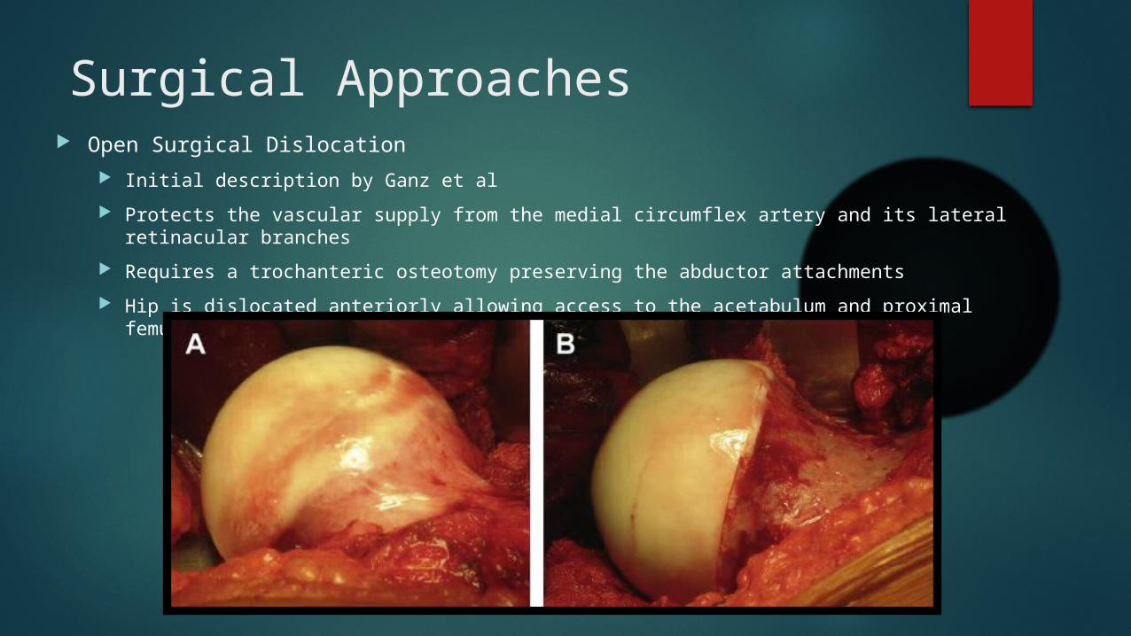
Surgical Approaches Open Surgical Dislocation
Initial description by Ganz et al
Protects the vascular supply from the medial circumflex artery and its lateral retinacular branches
Requires a trochanteric osteotomy preserving the abductor attachments
Hip is dislocated anteriorly allowing access to the acetabulum and proximal femur
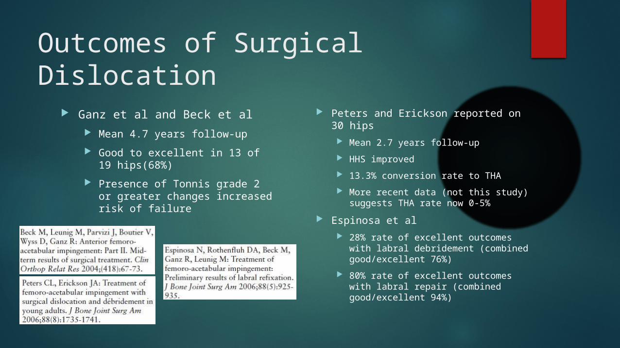
Outcomes of Surgical Dislocation
Ganz et al and Beck et al
Mean 4.7 years follow-up
Good to excellent in 13 of 19 hips(68%)
Presence of Tonnis grade 2 or greater changes increased risk of failure
Peters and Erickson reported on 30 hips
Mean 2.7 years follow-up
HHS improved
13.3% conversion rate to THA
More recent data (not this study) suggests THA rate now 0-5%
Espinosa et al
28% rate of excellent outcomes with labral debridement (combined good/excellent 76%)
80% rate of excellent outcomes with labral repair (combined good/excellent 94%)

Complications of Surgical Dislocation
Osteonecrosis (although reports are lacking to support this)
Nonunion of trochanteric osteotomy (0-3%)
Trochanteric pain (46% of all patients and 74% of female patients in one study)
Intra-articular adhesions (up to 6% of cases)
Sink et al
Multicenter cohort of 334 hips undergoing surgical hip dislocation
Overall complication rate of 9%

Hip Arthroscopy
Introduced in the late 1970s and initially was used to manage labral tears and loose bodies
Specialized equipment
Distraction table
Fluoroscopy
Long instrumentation
Access central and peripheral compartments via small “portal” incisions

Hip Arthroscopy

Hip Arthroscopy

Outcomes – Hip Arthroscopy
Multiple studies
Success rate of 67% to 90%
Rates of conversion to THA 0-9%
Retrospective studies
Larson et al compared outcomes of rim trimming with labral debridement (LD) vs labral repair (LR)
67% good/excellent with LD
90% good/excellent with LR
Nepple et al found that treatment of the bony deformity was associated with significantly greater improvement and decreased failure rates

Complications of Arthroscopic Hip Surgery
Complication rate 1% to 6%
Iatrogenic labral and articular cartilage damage
Heterotopic ossification (may be up to 8% in untreated patients)
Fracture
Nerve damage
Adhesions
Avascular necrosis
Persistent pain
Instability
Extravasation of fluid into adjacent spaces (ex. retroperitoneal)
Combined Arthroscopic and Open Approach
Can allow for address of complex deformities that may not be completely accessible via arthroscopy
Complex deformities or structural instability may require open procedures
Dysplasia
Abnormal femoral anteversion
Trochanteric impingement

Summary
CAM impingement on the femoral side
Pincer impingement on the acetabular side
Most cases of FAI are combined CAM and pincer
Radiographic findings of CAM and pincer impingement exist in the normal asymptomatic population
Some patients may be treated without surgery
Surgical options include:
Arthroscopic intervention
Open surgical dislocation
Combined approach
Surgery - regardless of approach - offers reliable good/excellent outcomes in properly selected patients with a low complication rate

Thank You
Journal of the AAOS 2013; Vol 21, Supplement 1