Embed Size (px)
Citation preview
Imperforate Anus in Feingold Syndrome
Vera Buttiker,1 Julian Wojtulewicz,1 and Meredith Wilson2*1Department of Neonatology, Royal Alexandra Hospital for Children, Westmead, New South Wales, Australia2Department of Clinical Genetics, Royal Alexandra Hospital for Children, Westmead, New South Wales, Australia
A father and daughter had the characteris-tic findings of Feingold syndrome includingmicrocephaly, short palpebral fissures,brachydactyly with clinodactyly of fifth fin-gers, and bilateral syndactyly of second tothird and fourth to fifth toes. The infant pre-sented with long-gap esophageal atresiawithout fistula (type A). Her father, who hadshort stature and learning disabilities, hadcongenital imperforate anus with a recto-vesical fistula. This is the first report of dis-tal intestinal atresia in Feingold syndrome.Am. J. Med. Genet. 92:166–169, 2000.© 2000 Wiley-Liss, Inc.
KEY WORDS: esophageal atresia; duodenalatresia; syndactyly; micro-cephaly; brachydactyly
INTRODUCTION
The clinical phenotype of Feingold syndrome, firstreported in 1975 [Feingold, 1975] was more completelydelineated in recent publications [Courtans et al.,1997; Feingold et al., 1997]. Feingold syndrome, alsoknown as MODED (microcephaly-oculo-digito-esophageal-duodenal) syndrome [Frydman et al., 1997]is a clinically variable autosomal dominant disordercomprising hand and foot abnormalities, microcephaly,short palpebral fissures, learning disabilities, andesophageal and/or duodenal atresia. Esophageal atre-sia with or without tracheo-esophageal fistula occurs inaround 30% of described individuals, sometimes incombination with duodenal atresia or obstruction.
We describe a family with Feingold syndrome, ascer-tained through an infant with esophageal atresia andminor anomalies. The infant’s affected father was bornwith imperforate anus and recto-vesical fistula, a pre-viously unreported anomaly in individuals with Fein-gold syndrome.
CLINICAL REPORTPatient 1 (III-3)
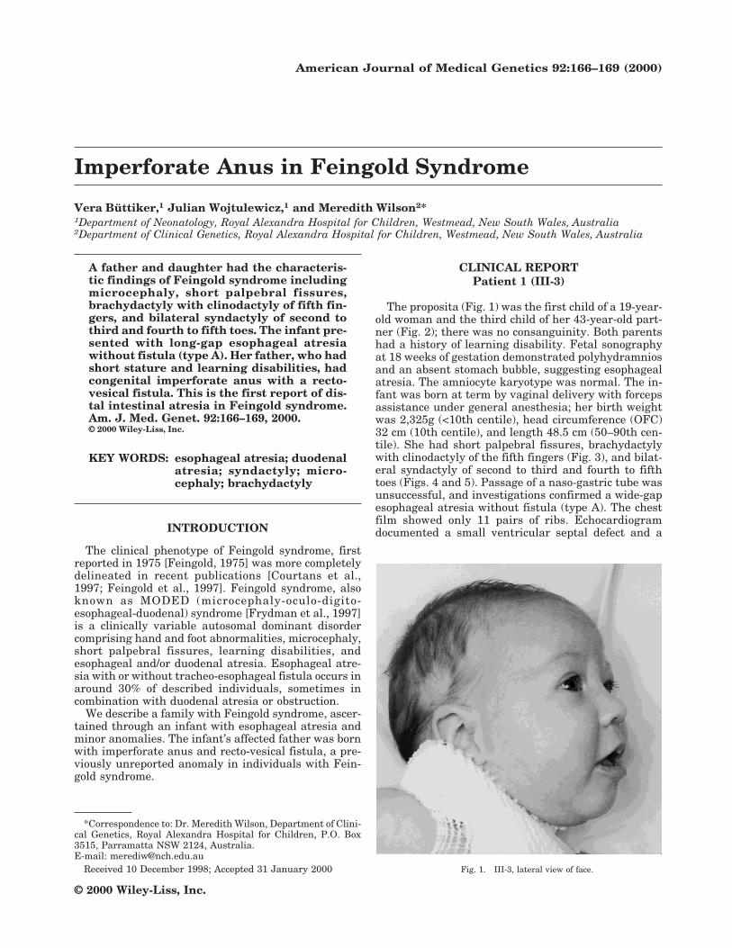
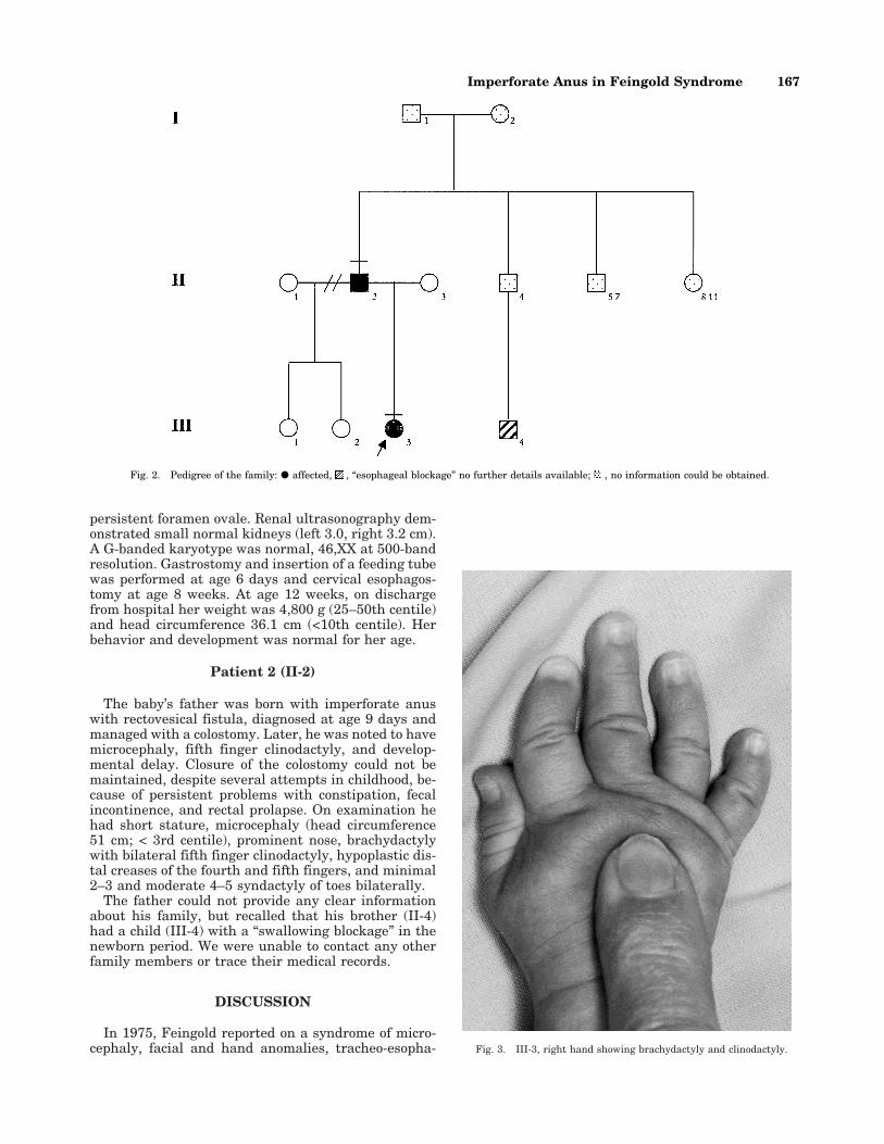
The proposita (Fig. 1) was the first child of a 19-year-old woman and the third child of her 43-year-old part-ner (Fig. 2); there was no consanguinity. Both parentshad a history of learning disability. Fetal sonographyat 18 weeks of gestation demonstrated polyhydramniosand an absent stomach bubble, suggesting esophagealatresia. The amniocyte karyotype was normal. The in-fant was born at term by vaginal delivery with forcepsassistance under general anesthesia; her birth weightwas 2,325g (<10th centile), head circumference (OFC)32 cm (10th centile), and length 48.5 cm (50–90th cen-tile). She had short palpebral fissures, brachydactylywith clinodactyly of the fifth fingers (Fig. 3), and bilat-eral syndactyly of second to third and fourth to fifthtoes (Figs. 4 and 5). Passage of a naso-gastric tube wasunsuccessful, and investigations confirmed a wide-gapesophageal atresia without fistula (type A). The chestfilm showed only 11 pairs of ribs. Echocardiogramdocumented a small ventricular septal defect and a
*Correspondence to: Dr. Meredith Wilson, Department of Clini-cal Genetics, Royal Alexandra Hospital for Children, P.O. Box3515, Parramatta NSW 2124, Australia.E-mail: [email protected]
Received 10 December 1998; Accepted 31 January 2000 Fig. 1. III-3, lateral view of face.
American Journal of Medical Genetics 92:166–169 (2000)
© 2000 Wiley-Liss, Inc.
persistent foramen ovale. Renal ultrasonography dem-onstrated small normal kidneys (left 3.0, right 3.2 cm).A G-banded karyotype was normal, 46,XX at 500-bandresolution. Gastrostomy and insertion of a feeding tubewas performed at age 6 days and cervical esophagos-tomy at age 8 weeks. At age 12 weeks, on dischargefrom hospital her weight was 4,800 g (25–50th centile)and head circumference 36.1 cm (<10th centile). Herbehavior and development was normal for her age.
Patient 2 (II-2)
The baby’s father was born with imperforate anuswith rectovesical fistula, diagnosed at age 9 days andmanaged with a colostomy. Later, he was noted to havemicrocephaly, fifth finger clinodactyly, and develop-mental delay. Closure of the colostomy could not bemaintained, despite several attempts in childhood, be-cause of persistent problems with constipation, fecalincontinence, and rectal prolapse. On examination hehad short stature, microcephaly (head circumference51 cm; < 3rd centile), prominent nose, brachydactylywith bilateral fifth finger clinodactyly, hypoplastic dis-tal creases of the fourth and fifth fingers, and minimal2–3 and moderate 4–5 syndactyly of toes bilaterally.
The father could not provide any clear informationabout his family, but recalled that his brother (II-4)had a child (III-4) with a “swallowing blockage” in thenewborn period. We were unable to contact any otherfamily members or trace their medical records.
DISCUSSION
In 1975, Feingold reported on a syndrome of micro-cephaly, facial and hand anomalies, tracheo-esopha- Fig. 3. III-3, right hand showing brachydactyly and clinodactyly.
Fig. 2. Pedigree of the family: d affected, , “esophageal blockage” no further details available; , no information could be obtained.
Imperforate Anus in Feingold Syndrome 167
geal fistula, duodenal atresia, and developmental delay[Feingold, 1975]; later he reported on a second family[Feingold, 1978]. Since then 15 families with Feingoldsyndrome have been reported [Konig et al., 1990; Brun-ner and Winter, 1991; Hall, 1994; Courtans et al., 1997;Feingold et al., 1997; Frydman et al., 1997]. This au-tosomal dominant disorder is also known as MODED(microcephaly-oculo-digito-esophageal-duodenal) syn-drome [Frydman et al., 1997]. The disorder exhibitsmarked intrafamilial variability, particularly with re-gard to gastrointestinal manifestations, but a review[Feingold et al., 1997] suggested that all have handanomalies, 80–90% have foot abnormalities, 87.5–100% have microcephaly, and 52–90% have learningdisability.
The hand and foot anomalies are distinctive. Fryd-man et al. [1997] concluded that they best resemblebrachydactyly type A4 (MIM *112800; short or absentmiddle phalanges of the second and fifth fingers, lack ofmiddle phalanges of the 2nd-5th toes) [Frydman et al.,1997] with addition of varying degrees of cutaneoussyndactyly of the 2nd–3rd and 4th–5th toes, and a widegap between the first and second toes [Brunner andWinter, 1991]. In addition to microcephaly there arefacial similarities including narrow palpebral fissures,relatively prominent nose, and mild retrognathia. Up-
per gastrointestinal anomalies are present in up to one-half of reported cases [Courtans et al., 1997; Feingoldet al., 1997]. The commonest gastrointestinal anomalyis tracheo-esophageal fistula or tracheal atresia, pre-sent in at least 25% of patients, and duodenal atresia orobstruction, present in over 20% [Feingold et al., 1997].This may represent a biased estimate as many of thepublished families were ascertained through an indexcase with intestinal atresia.
The family we report includes at least two individu-als with Feingold syndrome, the proposita and her fa-ther, both with microcephaly, short palpebral fissures,brachydactyly with clinodactyly of the fifth fingers, andbilateral partial 2–3 and 4–5 syndactyly of the toes; thefather also has learning disability and short stature.The daughter had esophageal atresia without fistula(esophageal atresia type A) while the father had imper-forate anus with recto-vesical fistula.
We were unable to find any other previously reportedpatient with Feingold syndrome and imperforate anus.Although the combination could be coincidental, itseems more likely to be an uncommon manifestation ofthe disorder. There is a non-random association of ano-
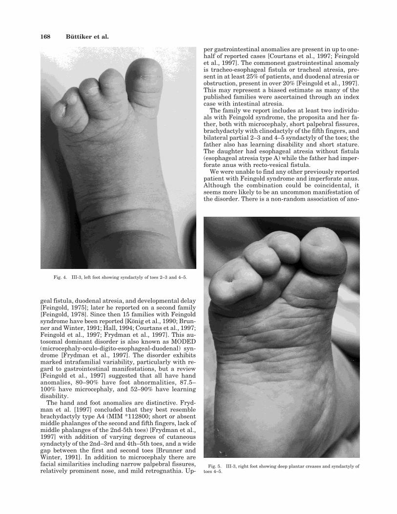
Fig. 4. III-3, left foot showing syndactyly of toes 2–3 and 4–5.
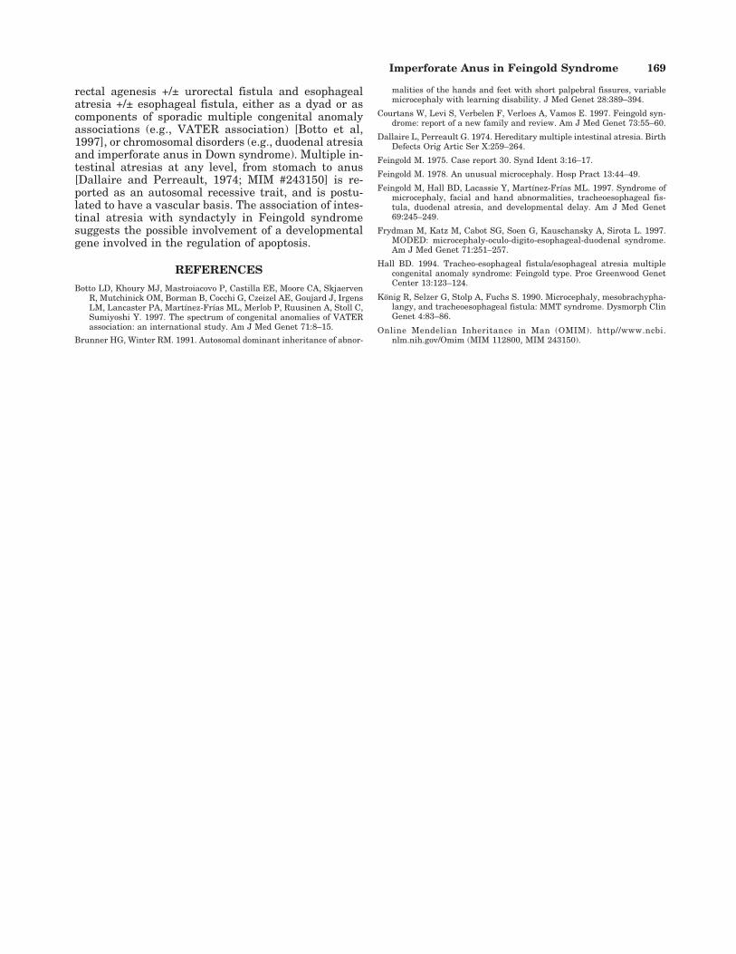
Fig. 5. III-3, right foot showing deep plantar creases and syndactyly oftoes 4–5.
168 Buttiker et al.
rectal agenesis +/± urorectal fistula and esophagealatresia +/± esophageal fistula, either as a dyad or ascomponents of sporadic multiple congenital anomalyassociations (e.g., VATER association) [Botto et al,1997], or chromosomal disorders (e.g., duodenal atresiaand imperforate anus in Down syndrome). Multiple in-testinal atresias at any level, from stomach to anus[Dallaire and Perreault, 1974; MIM #243150] is re-ported as an autosomal recessive trait, and is postu-lated to have a vascular basis. The association of intes-tinal atresia with syndactyly in Feingold syndromesuggests the possible involvement of a developmentalgene involved in the regulation of apoptosis.
REFERENCESBotto LD, Khoury MJ, Mastroiacovo P, Castilla EE, Moore CA, Skjaerven
R, Mutchinick OM, Borman B, Cocchi G, Czeizel AE, Goujard J, IrgensLM, Lancaster PA, Martınez-Frıas ML, Merlob P, Ruusinen A, Stoll C,Sumiyoshi Y. 1997. The spectrum of congenital anomalies of VATERassociation: an international study. Am J Med Genet 71:8–15.
Brunner HG, Winter RM. 1991. Autosomal dominant inheritance of abnor-
malities of the hands and feet with short palpebral fissures, variablemicrocephaly with learning disability. J Med Genet 28:389–394.
Courtans W, Levi S, Verbelen F, Verloes A, Vamos E. 1997. Feingold syn-drome: report of a new family and review. Am J Med Genet 73:55–60.
Dallaire L, Perreault G. 1974. Hereditary multiple intestinal atresia. BirthDefects Orig Artic Ser X:259–264.
Feingold M. 1975. Case report 30. Synd Ident 3:16–17.
Feingold M. 1978. An unusual microcephaly. Hosp Pract 13:44–49.
Feingold M, Hall BD, Lacassie Y, Martınez-Frıas ML. 1997. Syndrome ofmicrocephaly, facial and hand abnormalities, tracheoesophageal fis-tula, duodenal atresia, and developmental delay. Am J Med Genet69:245–249.
Frydman M, Katz M, Cabot SG, Soen G, Kauschansky A, Sirota L. 1997.MODED: microcephaly-oculo-digito-esophageal-duodenal syndrome.Am J Med Genet 71:251–257.
Hall BD. 1994. Tracheo-esophageal fistula/esophageal atresia multiplecongenital anomaly syndrome: Feingold type. Proc Greenwood GenetCenter 13:123–124.
Konig R, Selzer G, Stolp A, Fuchs S. 1990. Microcephaly, mesobrachypha-langy, and tracheoesophageal fistula: MMT syndrome. Dysmorph ClinGenet 4:83–86.
Online Mendelian Inheritance in Man (OMIM). http//www.ncbi.nlm.nih.gov/Omim (MIM 112800, MIM 243150).
Imperforate Anus in Feingold Syndrome 169


![Omphalocele, exstrophy of cloaca, imperforate anus and …oaji.net/pdf.html?n=2015/1334-1433441479.pdf · sonography may indicate an OEIS complex [3,7]. Many pregnancies electively](https://img.pdfslide.us/doc/110x75/5ae31d867f8b9a5d648d7b24/omphalocele-exstrophy-of-cloaca-imperforate-anus-and-oajinetpdfhtmln20151334-.jpg)