Embed Size (px)
Citation preview
IMPAIRMENT RATING
5TH EDITION
MODULE II
THE SPINE AND ALTERATION OF MOTION SEGMENT INTEGRITY (AOMSI)
PRESENTED BY: RONALD J. WELLIKOFF, D.C., FACC, FICC
In conjuction with:
The chapter on the spine includes how impairments impact on an individual’s activities of daily living.
Impairments of the spine include:1. Cervical2. Thoracic3. Lumbar4. Spinal Cord5. Pelvis
The changes made for the 5th edition of the “Guides” include:1. Modification of the ROM and DRE methods2. Impairment is rated only at MMI3. Impairments within a DRE category encompass a range, with
adjustments of up to 3%4. Spinal cord injuries are evaluated according to the functional
approach in the nervous system chapter
5. The “differentiators” used in the 4th edition are replaced by “objective findings”
6. Alteration of motion segment integrity (AOMSI) have been redefined
•“The DRE method is the primary method used to evaluate individual’s with an injury.”
•“Use the ROM method when the impairment is not caused by an injury or when an individual’s condition is not well represented by a DREcategory.”
•“The ROM method is also now used to evaluate individual’s with an injury at more than one level in the same spinal region and in certain individual’s with recurrent pathology.”
The 5th edition of the “Guides” has given the evaluator more latitude in the evaluation of a physical impairment.
Opinion, in this edition, plays a much larger role that previous editions.
DOCUMENTATION AND RECORDKEEPING ARE STRESSED!
“Certain conditions that may be controlled by medications or treatment and may be in total remission, may have the final impairment estimate or value increased by a small percentage (1%-3%) combining that percent with all other impairments.” (pg. 20)
“If for any reason the patient declines therapy, treatment, or surgery for a permanent impairment, that decision should not affect the estimated percentage of his or her impairment”.
Since pain is usually included in the rating percentage, do not include it a second time.
Diagnostic tests such as x-rays, MRI’s, CT’s, etc. should only be done if necessary and relevant.
The 5th edition of the “Guides” continues to use two methods to evaluate spinal impairments:
1. “Injury Model” (Diagnosis Related Estimates) (DRE)a. Cervicalb. Thoracicc. Lumbosacral
2. “Range of Motion Model” (Functional Method)a. Cervicalb. Thoracicc. Lumbosacral
NOTE: Do not use both methods in making the final impairment estimate.
SPECIAL NOTE:
REPEAT: The method of choice for evaluating the spine is the Injury Model (DRE).
As an example, if a patient has a herniated disc with nerve root irritation, the DRE is the way to determine the impairment.
If the patient does not fit into any of the categories within the DRE’s, then you would move to the Range of Motion Model.
NOTE: The Range of Motion Model is the court of last resort.
NOTE: Although there are eight categories in the DRE method, almost all individuals will fall into one of the first three DRE categories
There are two approaches to assigning a patient to a DRE category:
1. Based on symptoms, signs, and appropriate diagnostic test results.2. Based on the presence of fractures and/or disc dislocations with or
without clinical symptoms.
If a fracture is present that places the individual into a DRE category, no other verification is required (Box 15-1, pg. 382)
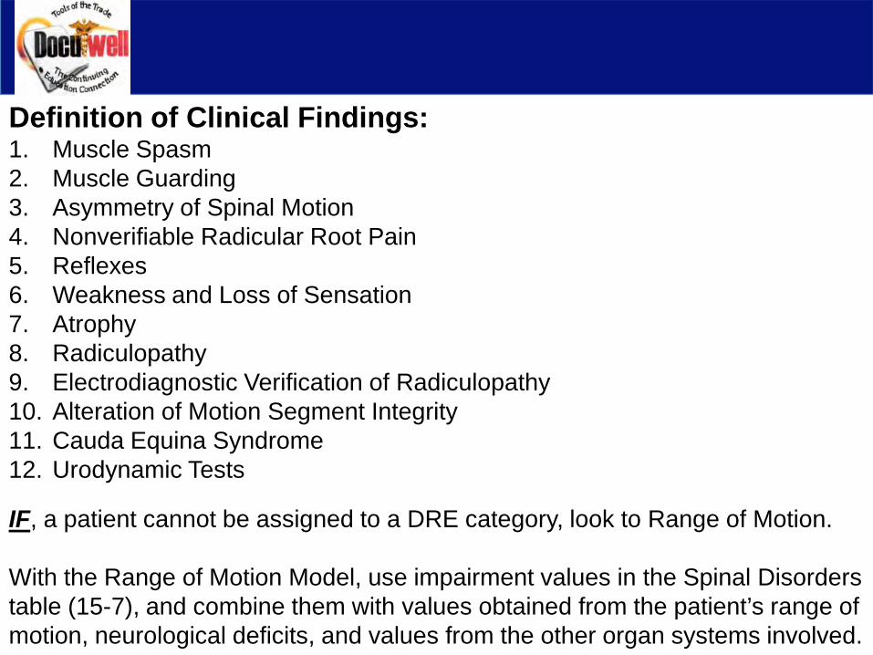
Definition of Clinical Findings:1. Muscle Spasm2. Muscle Guarding3. Asymmetry of Spinal Motion4. Nonverifiable Radicular Root Pain5. Reflexes6. Weakness and Loss of Sensation7. Atrophy8. Radiculopathy9. Electrodiagnostic Verification of Radiculopathy10. Alteration of Motion Segment Integrity11. Cauda Equina Syndrome12. Urodynamic Tests
IF, a patient cannot be assigned to a DRE category, look to Range of Motion.
With the Range of Motion Model, use impairment values in the Spinal Disorders table (15-7), and combine them with values obtained from the patient’s range of motion, neurological deficits, and values from the other organ systems involved.
Dr. Stanley KaplanDr. Ronald Wellikoff
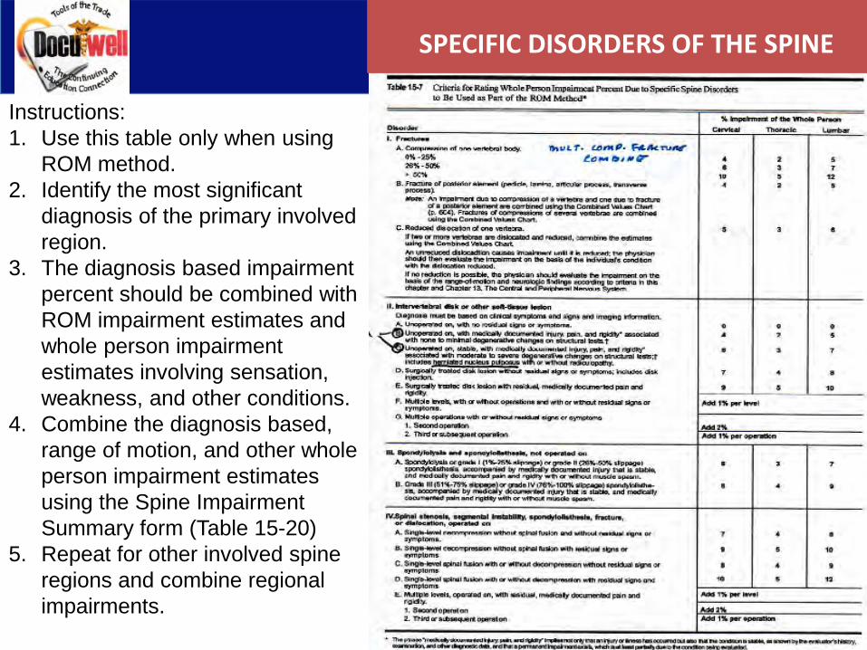
SPECIFIC DISORDERS OF THE SPINE
Instructions:1. Use this table only when using
ROM method.2. Identify the most significant
diagnosis of the primary involved region.
3. The diagnosis based impairment percent should be combined with ROM impairment estimates and whole person impairment estimates involving sensation, weakness, and other conditions.
4. Combine the diagnosis based, range of motion, and other whole person impairment estimates using the Spine Impairment Summary form (Table 15-20)
5. Repeat for other involved spine regions and combine regional impairments.
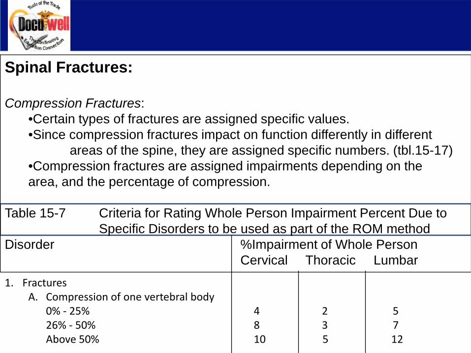
Spinal Fractures:
Compression Fractures:•Certain types of fractures are assigned specific values.•Since compression fractures impact on function differently in different
areas of the spine, they are assigned specific numbers. (tbl.15-17)•Compression fractures are assigned impairments depending on the area, and the percentage of compression.
Table 15-7 Criteria for Rating Whole Person Impairment Percent Due to Specific Disorders to be used as part of the ROM method
Disorder %Impairment of Whole PersonCervical Thoracic Lumbar
1. FracturesA. Compression of one vertebral body
0% - 25% 4 2 526% - 50% 8 3 7Above 50% 10 5 12
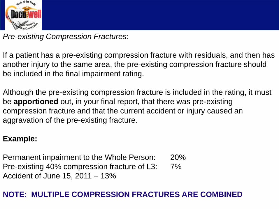
Pre-existing Compression Fractures:
If a patient has a pre-existing compression fracture with residuals, and then has another injury to the same area, the pre-existing compression fracture should be included in the final impairment rating.
Although the pre-existing compression fracture is included in the rating, it must be apportioned out, in your final report, that there was pre-existing compression fracture and that the current accident or injury caused an aggravation of the pre-existing fracture.
Example:
Permanent impairment to the Whole Person: 20%Pre-existing 40% compression fracture of L3: 7%Accident of June 15, 2011 = 13%
NOTE: MULTIPLE COMPRESSION FRACTURES ARE COMBINED
Posterior Element Fractures:
For the purposes of impairment ratings, the posterior elements are defined as:PediclesLaminaeArticular ProcessesTransverse Processes
NOTE: The spinous process is not included because a fracture at that site does not usually impede function. If pain or muscle weakness are present, and impair function, then it should be evaluated by sensory or motor impairment ratings.
Several fractures of the posterior elements of one vertebra are rated as a single fracture.
Fractures of the posterior elements in multiple vertebrae are combined.
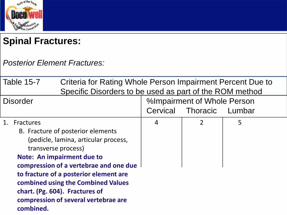
Spinal Fractures:
Posterior Element Fractures:
Table 15-7 Criteria for Rating Whole Person Impairment Percent Due to Specific Disorders to be used as part of the ROM method
Disorder %Impairment of Whole PersonCervical Thoracic Lumbar
1. Fractures 4 2 5B. Fracture of posterior elements
(pedicle, lamina, articular process, transverse process)
Note: An impairment due to compression of a vertebrae and one due to fracture of a posterior element are combined using the Combined Valueschart. (Pg. 604). Fractures of compression of several vertebrae are combined.
As with other chapters in the “Guides”, an evaluation should include:1. An accurate medical history2. A review of all pertinent records3. A comprehensive description of the individual’s current symptoms
and their relationship to activities of daily living4. Results of a physical examination5. All diagnostic test results (x-ray, MRI, CT, EMG, Labs, etc.)6. All calculations used to determine the impairment
Spinal Measurement:
•Although the goniometer has been used for spinal measurement, it is no longer being used.
•The appropriate instrument for spinal measurement is the Inclinometer.
•Factors such as angle of kyphosis of the thoracic spine and the sacral (hip) angle now are important in obtaining exact measurements.
•Repetition of measurements must be performed three times in order to be valid.
•All measurements must fall between 10% or 5 degrees of each other.
NOTE:•Calculate the mean or average of the three.
•If the average is less than 50 degree, three consecutive measurements must fall within 5 degrees of the mean: if the average is greater than 50 degrees, three consecutive measurements must fall within 10% of the mean.” (pg. 399)
In some cases such as lumbosacral flexion/extension, additional measurement of the straight leg raising test must also correlate with the findings.
Using the Inclinometer:
The inclinometer is used for each plane of movement in the spine.
3030
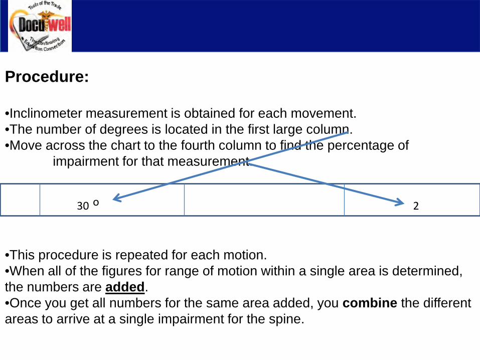
Procedure:
•Inclinometer measurement is obtained for each movement.•The number of degrees is located in the first large column.•Move across the chart to the fourth column to find the percentage of
impairment for that measurement.
30 o 2
•This procedure is repeated for each motion.•When all of the figures for range of motion within a single area is determined, the numbers are added.•Once you get all numbers for the same area added, you combine the different areas to arrive at a single impairment for the spine.
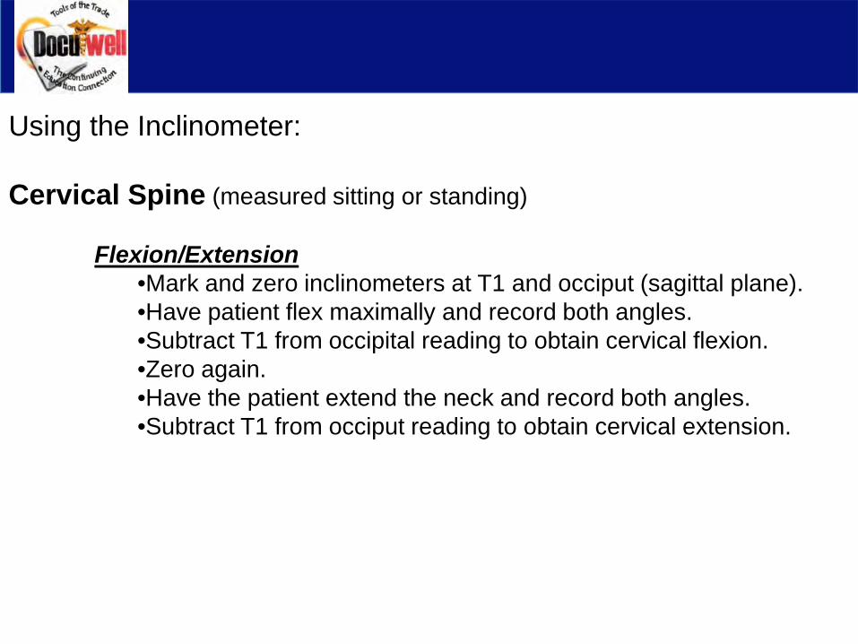
Using the Inclinometer:
Cervical Spine (measured sitting or standing)
Flexion/Extension•Mark and zero inclinometers at T1 and occiput (sagittal plane).•Have patient flex maximally and record both angles.•Subtract T1 from occipital reading to obtain cervical flexion.•Zero again.•Have the patient extend the neck and record both angles.•Subtract T1 from occiput reading to obtain cervical extension.
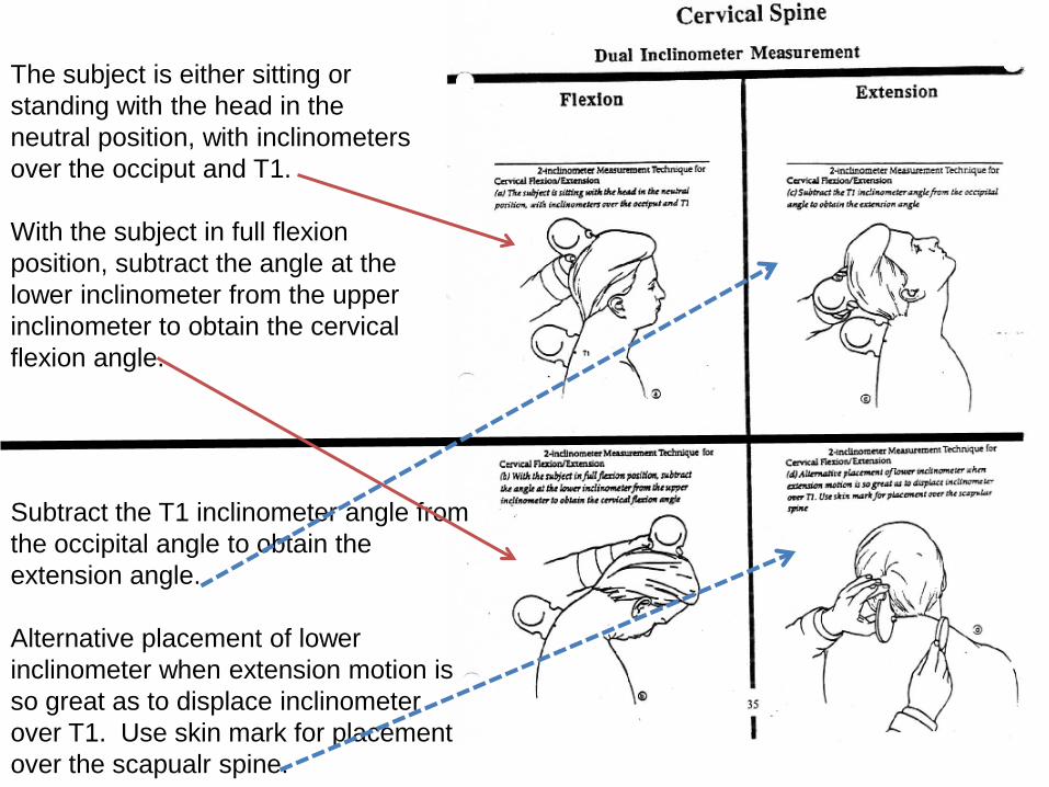
The subject is either sitting or standing with the head in the neutral position, with inclinometers over the occiput and T1.
With the subject in full flexion position, subtract the angle at the lower inclinometer from the upper inclinometer to obtain the cervical flexion angle.
Subtract the T1 inclinometer angle from the occipital angle to obtain the extension angle.
Alternative placement of lower inclinometer when extension motion is so great as to displace inclinometer over T1. Use skin mark for placement over the scapualr spine.
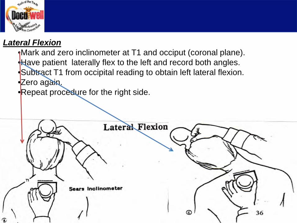
Lateral Flexion•Mark and zero inclinometer at T1 and occiput (coronal plane).•Have patient laterally flex to the left and record both angles.•Subtract T1 from occipital reading to obtain left lateral flexion.•Zero again.•Repeat procedure for the right side.
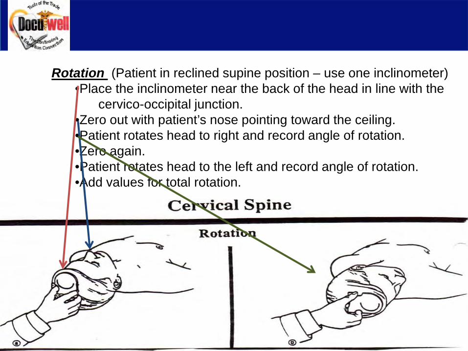
Rotation (Patient in reclined supine position – use one inclinometer)•Place the inclinometer near the back of the head in line with the
cervico-occipital junction.•Zero out with patient’s nose pointing toward the ceiling.•Patient rotates head to right and record angle of rotation.•Zero again.•Patient rotates head to the left and record angle of rotation.•Add values for total rotation.
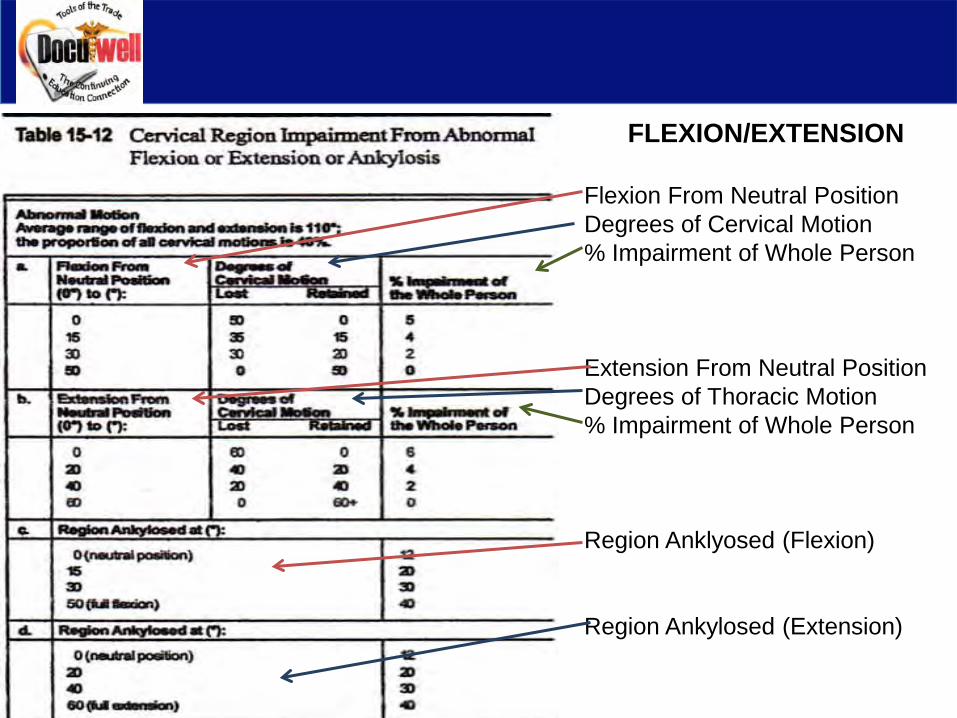
FLEXION/EXTENSION
Flexion From Neutral PositionDegrees of Cervical Motion% Impairment of Whole Person
Extension From Neutral PositionDegrees of Thoracic Motion% Impairment of Whole Person
Region Anklyosed (Flexion)
Region Ankylosed (Extension)
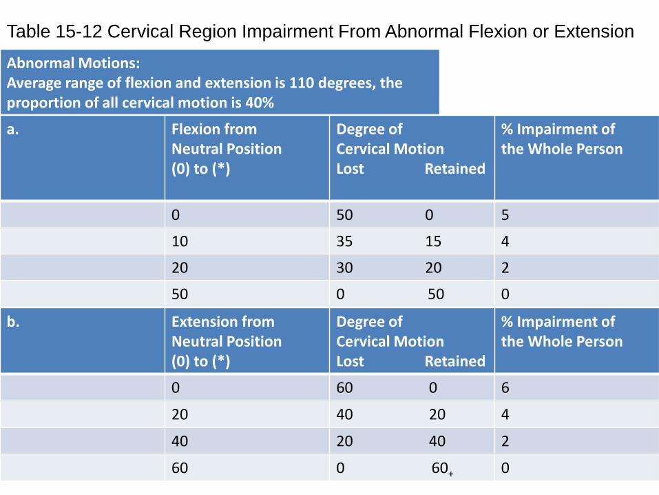
Table 15-12 Cervical Region Impairment From Abnormal Flexion or Extension
Abnormal Motions:Average range of flexion and extension is 110 degrees, the proportion of all cervical motion is 40%
a. Flexion from Neutral Position(0) to (*)
Degree of Cervical MotionLost Retained
% Impairment ofthe Whole Person
0 50 0 5
10 35 15 4
20 30 20 2
50 0 50 0
b. Extension fromNeutral Position(0) to (*)
Degree of Cervical Motion Lost Retained
% Impairment of the Whole Person
0 60 0 6
20 40 20 4
40 20 40 2
60 0 60+ 0
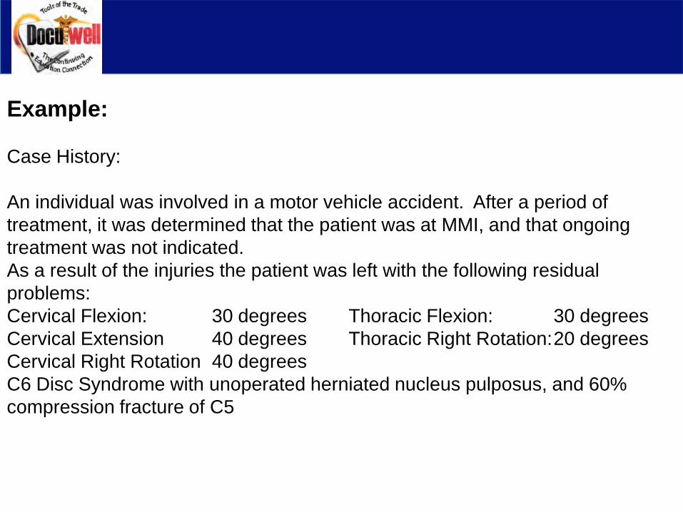
Example:
Case History:
An individual was involved in a motor vehicle accident. After a period of treatment, it was determined that the patient was at MMI, and that ongoing treatment was not indicated. As a result of the injuries the patient was left with the following residual problems:Cervical Flexion: 30 degrees Thoracic Flexion: 30 degreesCervical Extension 40 degrees Thoracic Right Rotation:20 degreesCervical Right Rotation 40 degreesC6 Disc Syndrome with unoperated herniated nucleus pulposus, and 60% compression fracture of C5
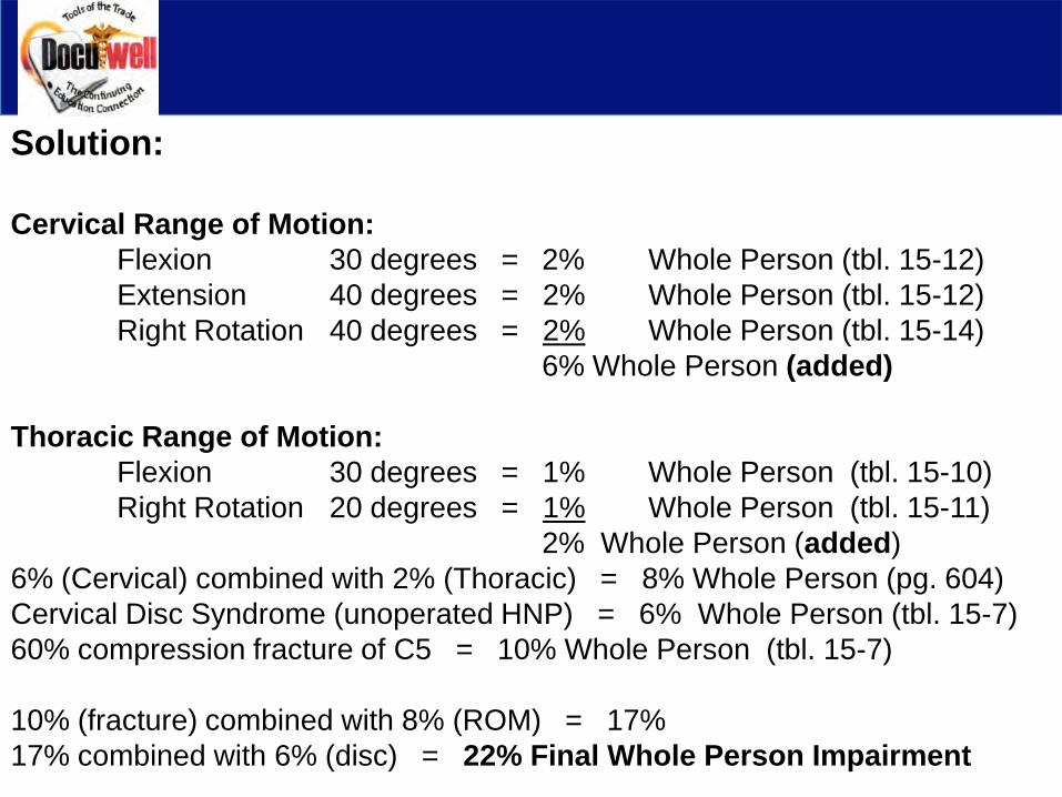
Solution:
Cervical Range of Motion:Flexion 30 degrees = 2% Whole Person (tbl. 15-12)Extension 40 degrees = 2% Whole Person (tbl. 15-12)Right Rotation 40 degrees = 2% Whole Person (tbl. 15-14)
6% Whole Person (added)
Thoracic Range of Motion:Flexion 30 degrees = 1% Whole Person (tbl. 15-10)Right Rotation 20 degrees = 1% Whole Person (tbl. 15-11)
2% Whole Person (added)6% (Cervical) combined with 2% (Thoracic) = 8% Whole Person (pg. 604)Cervical Disc Syndrome (unoperated HNP) = 6% Whole Person (tbl. 15-7)60% compression fracture of C5 = 10% Whole Person (tbl. 15-7)
10% (fracture) combined with 8% (ROM) = 17% 17% combined with 6% (disc) = 22% Final Whole Person Impairment
Ankylosis:
•Ankylosis is defined as the complete absence of joint motion or as a fixed position.
•Although ankylosis is rare in the spine, those patients who cannot reach the neutral or 0 degree position, the position or angle of restriction closest to the neutral position is considered to be the position of ankylosis.
•If a region has several range of motion impairments, and an ankylosisimpairment, the range of motion impairments are added and the total is combined with the ankylosis impairment.
•The examiner should add the ankylosis impairments in several planes within a single region and then combine with ankylosis impairment of two or more regions.
Example:
An individual who can flex the cervical region from 30 degrees to 50 degrees and who lacks 30 degrees of motion in reaching the neutral 0 degrees position has the same estimated impairment as if he or she had ankylosis at 30 degrees of cervical flexion.
According to table 15-12, the patient’s impairment is 30% of the whole person.
Thoracic Spine (may be measured sitting or standing)
Flexion/Extension
Note: Flexion and extension is relatively limited with a degree of extension significantly determined by the subject’s posture and the degree of fixed “kyphosis”. Subject is measured in a “military brace” posture to obtain angle of minimum kyphosis (found in ankylosis section of table 79). Following this, the angle of thoracic flexion is obtained by flexing the thoracic spine.
1. Zero inclinometers against a true vertical surface such as a wall.2. Mark T1 and T12 and read the inclinometers in “military brace”
position.3. Subtract T12 from T1 to get angle of minimum kyphosis.4. Zero again.5. Patient then flexes and the T1 and T12 angles are recorded.6. Subtract T12 from T1 to obtain the angle of Thoracic Flexion.
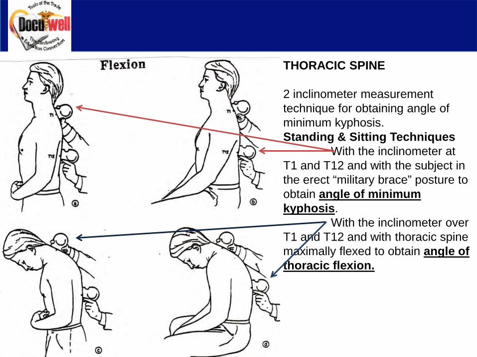
THORACIC SPINE
2 inclinometer measurement technique for obtaining angle of minimum kyphosis.Standing & Sitting Techniques
With the inclinometer at T1 and T12 and with the subject in the erect “military brace” posture to obtain angle of minimum kyphosis.
With the inclinometer over T1 and T12 and with thoracic spine maximally flexed to obtain angle of thoracic flexion.
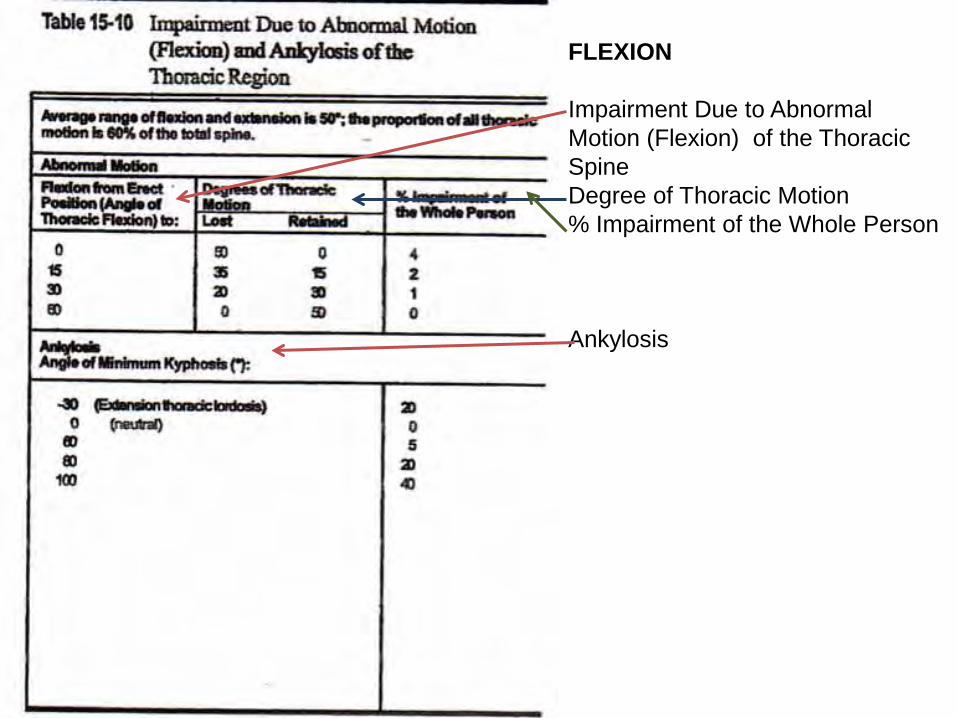
FLEXION
Impairment Due to Abnormal Motion (Flexion) of the Thoracic SpineDegree of Thoracic Motion% Impairment of the Whole Person
Ankylosis
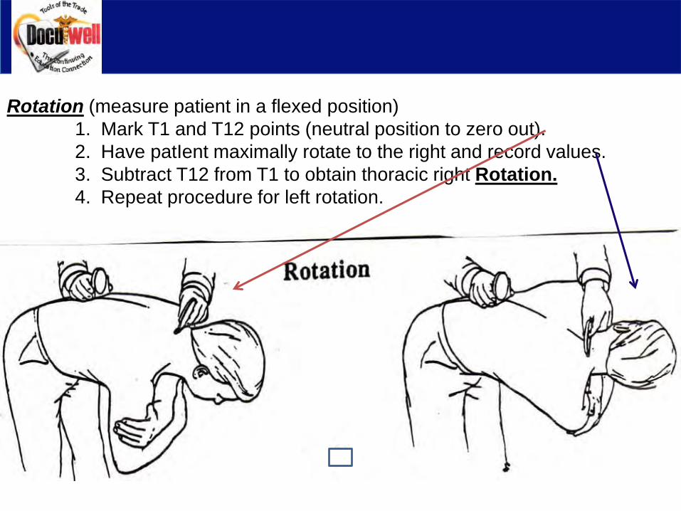
Rotation (measure patient in a flexed position)1. Mark T1 and T12 points (neutral position to zero out).2. Have patIent maximally rotate to the right and record values.3. Subtract T12 from T1 to obtain thoracic right Rotation.4. Repeat procedure for left rotation.
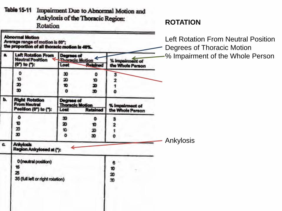
ROTATION
Left Rotation From Neutral PositionDegrees of Thoracic Motion% Impairment of the Whole Person
Ankylosis
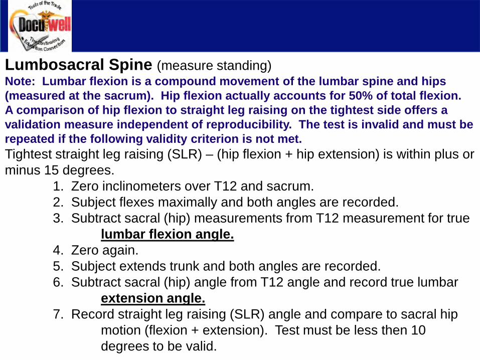
Lumbosacral Spine (measure standing)Note: Lumbar flexion is a compound movement of the lumbar spine and hips (measured at the sacrum). Hip flexion actually accounts for 50% of total flexion. A comparison of hip flexion to straight leg raising on the tightest side offers a validation measure independent of reproducibility. The test is invalid and must be repeated if the following validity criterion is not met.Tightest straight leg raising (SLR) – (hip flexion + hip extension) is within plus or minus 15 degrees.
1. Zero inclinometers over T12 and sacrum.2. Subject flexes maximally and both angles are recorded.3. Subtract sacral (hip) measurements from T12 measurement for true
lumbar flexion angle.4. Zero again.5. Subject extends trunk and both angles are recorded.6. Subtract sacral (hip) angle from T12 angle and record true lumbar
extension angle.7. Record straight leg raising (SLR) angle and compare to sacral hip
motion (flexion + extension). Test must be less then 10 degrees to be valid.
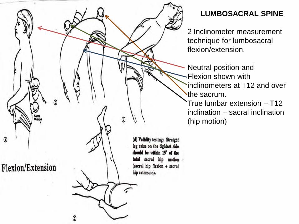
LUMBOSACRAL SPINE
2 Inclinometer measurement technique for lumbosacralflexion/extension.
Neutral position andFlexion shown with inclinometers at T12 and over the sacrum.True lumbar extension – T12inclination – sacral inclination (hip motion)
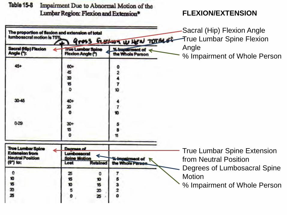
FLEXION/EXTENSION
Sacral (Hip) Flexion AngleTrue Lumbar Spine Flexion Angle% Impairment of Whole Person
True Lumbar Spine Extension from Neutral PositionDegrees of Lumbosacral Spine Motion% Impairment of Whole Person
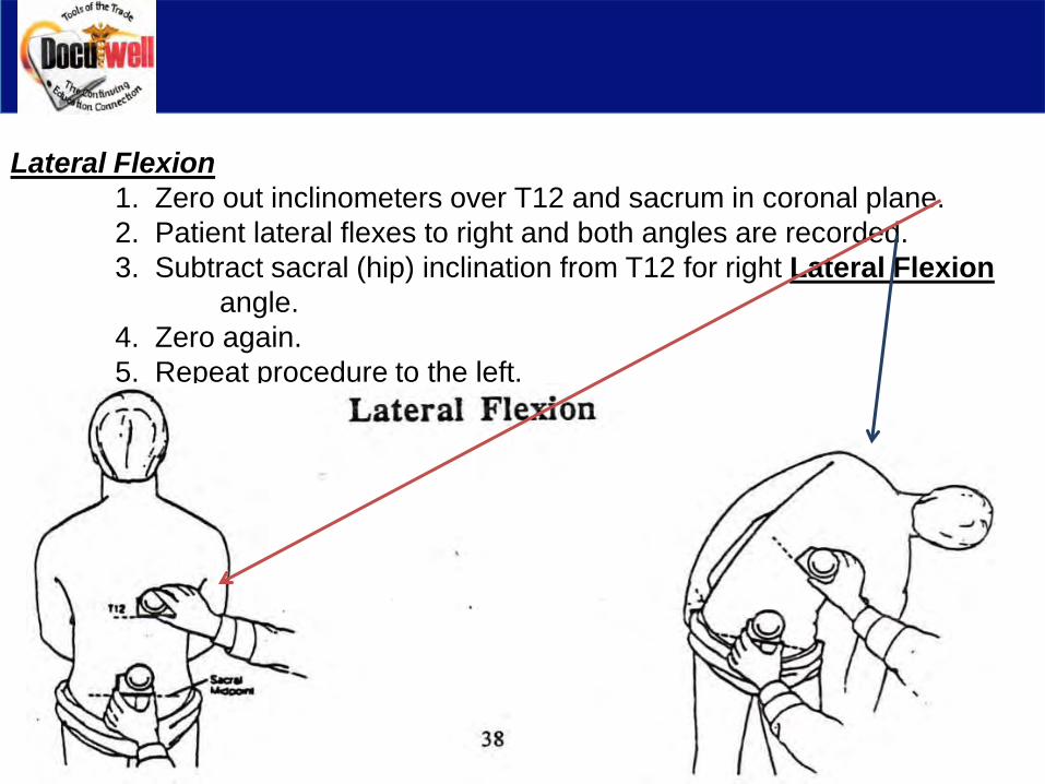
Lateral Flexion1. Zero out inclinometers over T12 and sacrum in coronal plane.2. Patient lateral flexes to right and both angles are recorded.3. Subtract sacral (hip) inclination from T12 for right Lateral Flexion
angle.4. Zero again.5. Repeat procedure to the left.
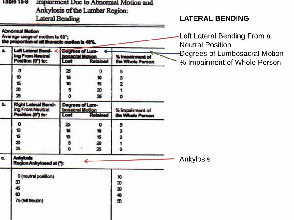
LATERAL BENDING
Left Lateral Bending From a Neutral PositionDegrees of Lumbosacral Motion% Impairment of Whole Person
Ankylosis
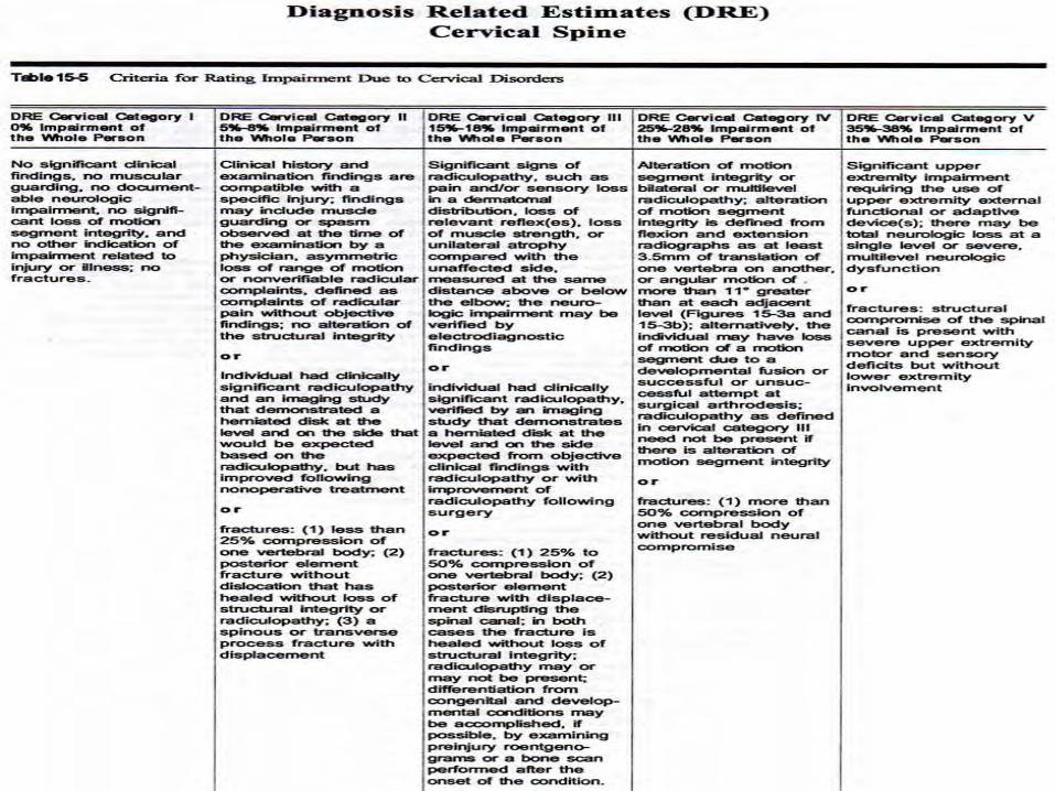
DIAGNOSIS RELATED ESTIMATES (DRE)
Within this method of rating, there are eight diagnosis related categories.
The two methods of assigning a rating are:1. Based on symptoms, signs, and appropriate diagnostic test results.2. Based on the presence fractures and/or dislocations with or without
clinical symptoms.
While there are eight categories listed in the DRE’s, for the most part, we will be addressing categories I, II, III.
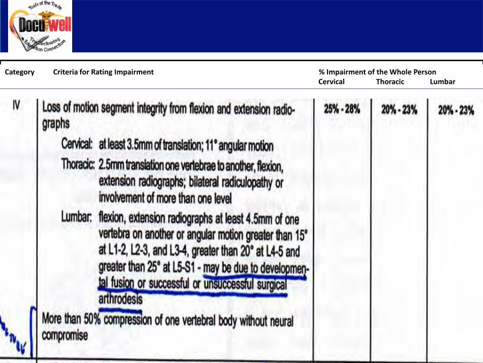
Categories IV and V address Alteration of Motion Segment Integrity and Fractures.
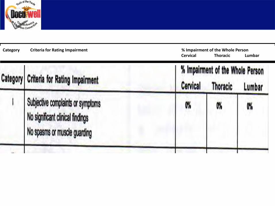
Category Criteria for Rating Impairment % Impairment of the Whole PersonCervical Thoracic Lumbar
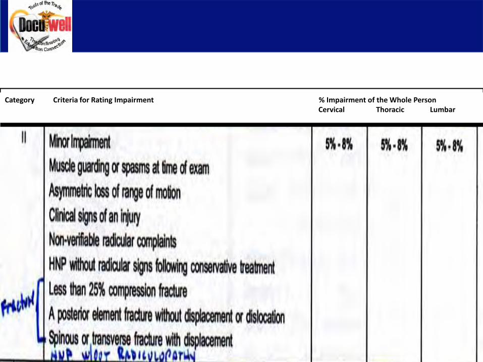
Category Criteria for Rating Impairment % Impairment of the Whole PersonCervical Thoracic Lumbar
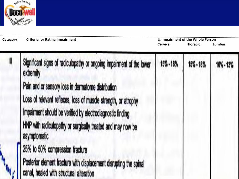
Category Criteria for Rating Impairment % Impairment of the Whole PersonCervical Thoracic Lumbar
Category Criteria for Rating Impairment % Impairment of the Whole PersonCervical Thoracic Lumbar
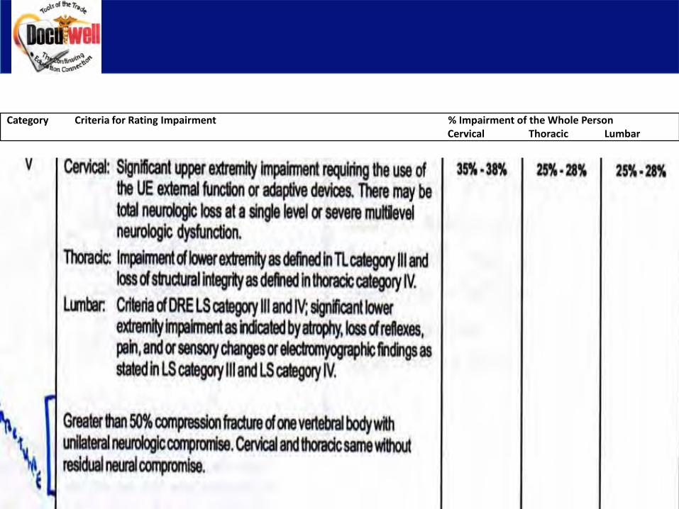
Category Criteria for Rating Impairment % Impairment of the Whole PersonCervical Thoracic Lumbar
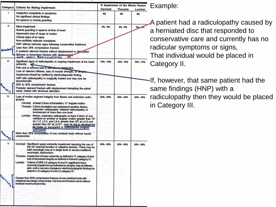
Example:
A patient had a radiculopathy caused by a herniated disc that responded to conservative care and currently has no radicular symptoms or signs, That individual would be placed in Category II.
If, however, that same patient had the same findings (HNP) with a radiculopathy then they would be placed in Category III.
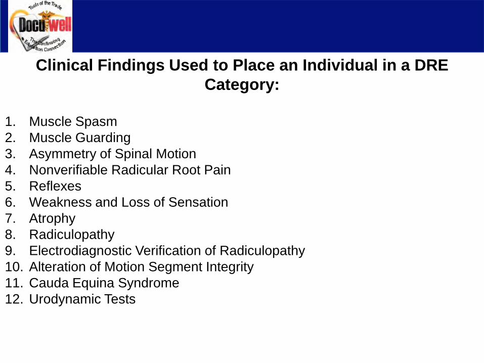
Clinical Findings Used to Place an Individual in a DRECategory:
1. Muscle Spasm2. Muscle Guarding3. Asymmetry of Spinal Motion4. Nonverifiable Radicular Root Pain5. Reflexes6. Weakness and Loss of Sensation7. Atrophy8. Radiculopathy9. Electrodiagnostic Verification of Radiculopathy10. Alteration of Motion Segment Integrity11. Cauda Equina Syndrome12. Urodynamic Tests
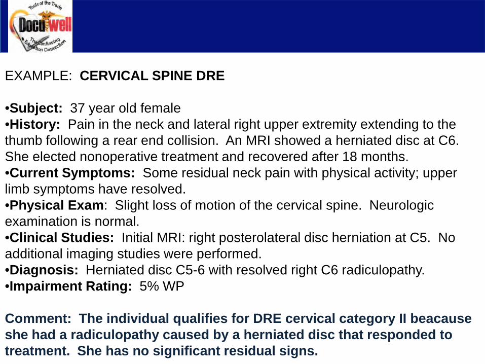
EXAMPLE: CERVICAL SPINE DRE
•Subject: 37 year old female•History: Pain in the neck and lateral right upper extremity extending to the thumb following a rear end collision. An MRI showed a herniated disc at C6. She elected nonoperative treatment and recovered after 18 months.•Current Symptoms: Some residual neck pain with physical activity; upper limb symptoms have resolved.•Physical Exam: Slight loss of motion of the cervical spine. Neurologic examination is normal.•Clinical Studies: Initial MRI: right posterolateral disc herniation at C5. No additional imaging studies were performed.•Diagnosis: Herniated disc C5-6 with resolved right C6 radiculopathy.•Impairment Rating: 5% WP
Comment: The individual qualifies for DRE cervical category II beacauseshe had a radiculopathy caused by a herniated disc that responded to treatment. She has no significant residual signs.
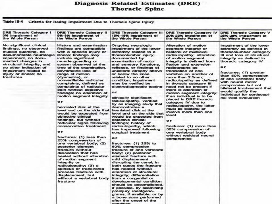
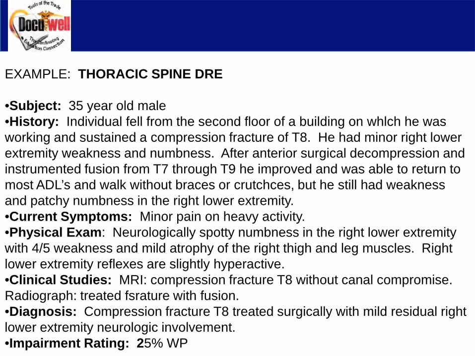
EXAMPLE: THORACIC SPINE DRE
•Subject: 35 year old male•History: Individual fell from the second floor of a building on whlch he was working and sustained a compression fracture of T8. He had minor right lower extremity weakness and numbness. After anterior surgical decompression and instrumented fusion from T7 through T9 he improved and was able to return to most ADL’s and walk without braces or crutchces, but he still had weakness and patchy numbness in the right lower extremity.•Current Symptoms: Minor pain on heavy activity.•Physical Exam: Neurologically spotty numbness in the right lower extremity with 4/5 weakness and mild atrophy of the right thigh and leg muscles. Right lower extremity reflexes are slightly hyperactive.•Clinical Studies: MRI: compression fracture T8 without canal compromise. Radiograph: treated fsrature with fusion.•Diagnosis: Compression fracture T8 treated surgically with mild residual right lower extremity neurologic involvement.•Impairment Rating: 25% WP
EXAMPLE: THORACIC SPINE DRE
Comment: This individual qualifies for DRE thoracic category V because he has mild right lower extremity neurologic deficits (category III) and alteration of motion segment integrity given fusion (category IV). A combination of categories III and IV in the thoracic region means that the individual qualifies for category V.
NOTE: Because he has alteration of motion segment integrity of more than one level (multilevel fusion), he could also be rated by the ROM method.
The best approach would be to rate the individual by both methods and award the higher rating.

EXAMPLE: LUMBAR SPINE DRE
Subject: 25 year old manHistory: Onset of back and left leg pain after a fall on a concrete surface while carrying a box. Initially presented with muscle spasm, an SLR on the left side at 60 degrees, a positive crossed SLR at 70 degrees, and an absent left Achilles tendon reflex. Treated with physical therapy but did not improve. Underwent surgical discectomy and arthodesis of L5-S1 three months after the injury. After 9 months of rehabilitation, leg and back symptoms were diminished but present.Current Symptoms: Back and thigh pain at rest and persistent numbness along the lateral side of the foot 1 year after the onset of symptoms. Pain and numbness prevent individual from maintaining a constant position, prolonged standing or walking, or performing his prior work, recreational, and some household activities.Physical Exam: Severely restricted range of motion. Loss of the Achilles reflex. Numbness in the S1 nerve root distribution and dermatomal pain I the leg on SLR.
Clinical Studies: Original MRI: a severely degenerated L5-S1 disc with a herniation on the left side. Post-operative MRI with gadolinium: fibrosis, but no residual or recurrent herniation. Fusion appears solid.Diagnosis: Left posterolateral disc herniation L5-S1 with S1 radiculopathy and severe disc degeneration, unresolved status post-discectomy and L5-S1 fusion.Impairment Rating: 28% WP
Comment: Symptoms, physical findings, and imaging studies are all consistent with a symptomatic herniated disc. Excision of he offending disc and a single level fusion did not relieve all symptoms, which are supported by signs of a persistent radiculopathy. Individual qualifies for lumbar DRE category V because he has persistent radiculopathy as well as single level alteration of motion segment integrity.
QUESTIONS:
1. Spinal impairment impacts on:a. HPIb. ROMc. ROSd. ADL
2. What method of evaluation is advised by the “Guides”a. ROM methodb. Injury methodc. Disability methodd. Handicap method
3. When should you do an impairment rating?a. On a personal injury patientb. On a workers’ compensation patientc. When asked by a patientd. When questioned by an insurance company
4. The final calculated whole person percentage should be rounded off to what?a. Nearest whole numberb. Nearest 5%c. Nearest 10%d. Not at all
5. When ever dealing with range of motion of the same joint, the figures are what?a. Combinedb. Addedc. Dividedd. Multiplied
6. In combining the following numbers, what order should they be placed?21,6,11,14,3,12a. 21,14,12,11,6,3b. 3,6,12,11,21,14c. 3,6,11,14,12, 21d. 3,6,11,12,14,21
7. Define impairment.a. Used in both legal and policy context to describe individuals living with physical problems.b. An alteration of an individual’s capacity to meet personal, social, or occupational demands.c. A loss, loss of use, or derangement of any body part, organ system or function.d. None of the above.
8. What are the two approaches to assigning a patient to a DRE category?a. Impairment, disabilityb. Symptoms, diagnosisc. Signs, fracturesd. None of the above
9. A compression fracture rating is based on what?a. Locationb. Percentagec. Positiond. Opinion
10. Multiple fractures of the posterior elements of a vertebra are rated:a. As a single fractureb. Separatelyc. Depends on which element is fracturedd. Depends on whether the spinous process is fractured
11. What instrument is used to measure spinal range of motion.a. Goniometerb. Protractorc. Compassd. Inclinometer
12. What are the landmarks for measuring cervical range of motion?a. T1 and Atlasb. C7 and Occiputc. Occiput and T1d. Atlas and Occiput
13. In measuring the Thoracic spine in flexion/extension, the first step is what?a. Zero the inclinometer against C7b. Zero the inclinometer against T1c. Zero the inclinometer against the occiputd. Zero the inclinometer against the wall
14. In measuring the Thoracic spine for flexion/extension the subject is placed in what position? a. Flexedb. Extendedc. Militaryd. Natural
15. In the L/S region, hip flexion accounts for what percentage of total flexion?a. 25%b. 50%c. 75%d. Not involved
16. In the DRE method of evaluation, the most commonly used categories are what?a. I, IV, Vb. II, III, IVc. I, II, IIId. They are all used about the same
17. What category would a HNP without radiculopathy be placed?a. Ib. IIc. IIId. IV
18. What category would a 25% compression fracture or posterior element fracture beplaced?a. Ib. IIc. IIId. IV
19. According to the 5th edition of the Guides, which is true?a. Use the version that was used at the time of the injuryb. Use the first edition whenever possiblec. Use the most current editiond. Use any edition
20. What orthopedic test is used for comparing the validity of hip flexion?a. Lasegueb. Fabere Patrickc. SLRd. Braggard
Introduction to the AMA’s 6th edition of the Guides
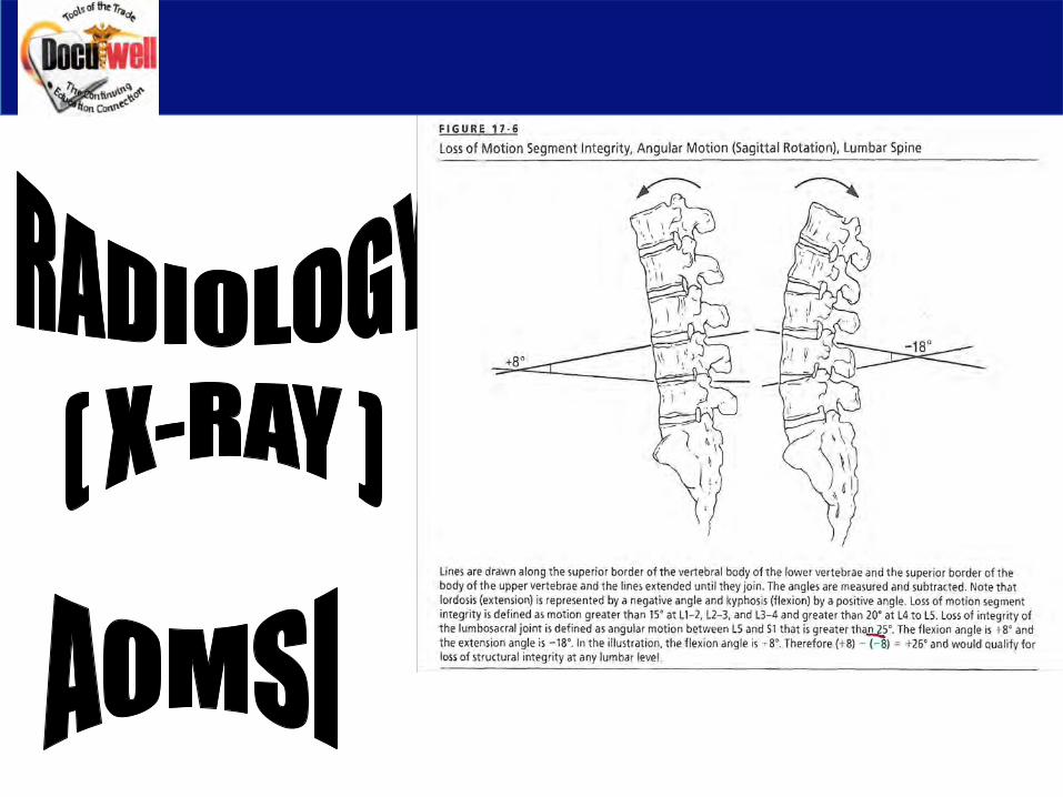
AOMSI
The purpose of our discussion, regarding AOMSI, is not to make you into a radiologist. In fact, it is my opinion that a referral to a chiropractic radiologist would make the most
sense when using this method.
Remember, you have to defend your opinion!
AOMSI
• The term AOMSI (Alteration Of Motion Segment Integrity) was first used in the Fourth Edition of the Guides to describe the loss of motion segment integrity, identified on FLEXION/EXTENSIONx-rays.
• There are several technical considerations in obtaining and interpreting radiographic studies to evaluate AOMSI, including:
•Magnification
•Film quality
•Normal thresholds for translation and angular motion
• “AOMSI as described in the Guides is assessed by plain film flexion-extension radiographs ONLY.”
• “Source to image distance (SID) for flexion-extension x-rays of the thoracic and lumbar spine is not defined in the literature or medical practice; however, 40” SID imaging parameters are most commonly used in the lumbar spine.”
• If a diagnosis of AOMSI, pseudoarthrosis, fracture, or spondylolisthesis is made, imaging studies should be excluded as a grade modifier.
With an understanding that we are not radiologists, the following are some of the figures described in the 6th Edition of the Guides:
• Cervical Spine:
•A diagnosis of AOMSI in the cervical spine by translation measurements requires greater than 20% anterior or posterior relative translation of one vertebra on another or angular motion of more than 11 degrees greater than each adjacent level on the flexion x-ray.
• Thoracic Spine:
•A diagnosis of AOMSI in the thoracic spine by translation measurements requires at least 2.5 mm anterior or posterior translation of one vertebra on another.
• Lumbar Spine:
•A diagnosis of AOMSI in the lumbar spine by translation measurements requires greater than 8% anterior or 9% posterior relative translation of one vertebra on another.
• Lumbosacral Spine:
•A diagnosis of AOMSI in the lumbosacral spine by translation measurements requires greater than 6% anterior or greater than 9% posterior relative translation at L5-S1 of L5 on S1.
NOTE: “The identification of degenerative disk disease at one or multiple levels, similar to the finding of arthrosis in an extremity joint, is not diagnostic of an injury, or disease, related pathology because such degenertative changes can be a natural consequence of the aging process.”

1. What does AOMSI mean?
Alteration of Motion Segment Integrity
2. When using the AOMSI what specific views are used?
Anterior and Posterior
3. What are the technical considerations used when obtaining or interpreting x-rays for an AOMSI?
Magnification
Film quality
Normal thresholds for translation and angular motion.
4. What is the SID for a lumbar x-ray when evaluating for an AOMSI?
40”