Embed Size (px)
Citation preview
Jackie Boultwood
LLR Molecular Haematology Unit
John Radcliffe Hospital
Oxford
Impaired ribosome function and the
molecular biology of the 5q- syndrome
The following presentation has been modified from its
original version. It has been formatted to fit this screen
and edited for content, to run in the time allotted
Ribosome dysfunction in the 5q-
syndrome
• Introduction and mapping of the CDR
• Role of haploinsufficiency of RPS14 and p53 activation
in molecular pathogenesis
• Defective translation and treatment with L-leucine
• Cooperating genetic events in disease pathogenesis
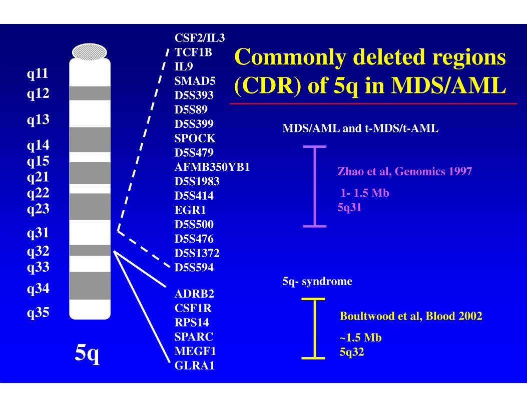
5q
CSF2/IL3
TCF1B
IL9
SMAD5
D5S393
D5S89
D5S399
SPOCK
D5S479
AFMB350YB1
D5S1983
D5S414
EGR1
D5S500
D5S476
D5S1372
D5S594
ADRB2
CSF1R
RPS14
SPARC
MEGF1
GLRA1
q11
q23
q13
q14q15q21q22
q12
q31
q32q33
q34
q35
Zhao et al, Genomics 1997
1- 1.5 Mb
5q31
5q- syndrome
Boultwood et al, Blood 2002
~1.5 Mb
5q32
Commonly deleted regions
(CDR) of 5q in MDS/AML
MDS/AML and t-MDS/t-AML
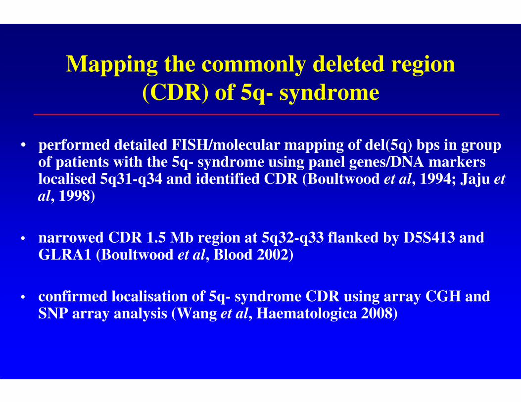
Mapping the commonly deleted region
(CDR) of 5q- syndrome
• performed detailed FISH/molecular mapping of del(5q) bps in group of patients with the 5q- syndrome using panel genes/DNA markers localised 5q31-q34 and identified CDR (Boultwood et al, 1994; Jaju et al, 1998)
• narrowed CDR 1.5 Mb region at 5q32-q33 flanked by D5S413 and GLRA1 (Boultwood et al, Blood 2002)
• confirmed localisation of 5q- syndrome CDR using array CGH and SNP array analysis (Wang et al, Haematologica 2008)
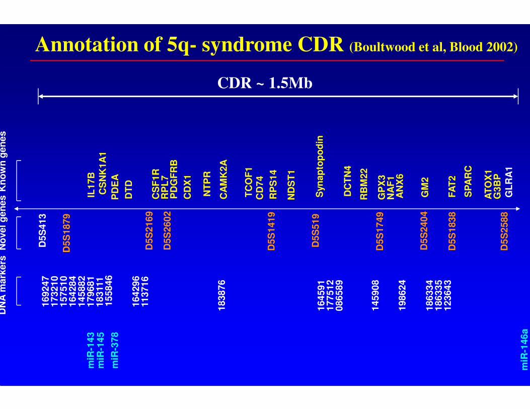
169247
173210
157510
164284
145882
179681
183111
155846
164296
113716
164591
177512
086589
145908
198624
186334
186335
123643
183876
IL17B
CS
NK
1A
1
PD
EA
DT
D
CS
F1R
PD
GF
RB
RP
L7
CD
X1
NT
PR
CA
MK
2A
TC
OF
1C
D74
ND
ST
1
RP
S14
Syn
ap
top
od
in
DC
TN
4
NA
F1
AN
X6
GM
2
FA
T2
SP
AR
C
G3B
PA
TO
X1
GL
RA
1
GP
X3
RB
M22
D5S
413
D5S
1879
D5S
2169
D5S
2602
D5S
1419
D5S
519
D5S
1749
D5S
2404
D5S
1838
D5S
2588
Kn
ow
n g
en
es
DN
A m
ark
ers
No
vel g
en
es
CDR ~ 1.5Mb
Annotation of 5q- syndrome CDR (Boultwood et al, Blood 2002)
miR
-143
miR
-145
miR
-146a
miR
-378
Ribosome dysfunction in the 5q-
syndrome
• Introduction and mapping of the CDR
• Role of haploinsufficiency of RPS14 and p53 activation
in molecular pathogenesis
• Defective translation and treatment with L-leucine
• Cooperating genetic events in disease pathogenesis
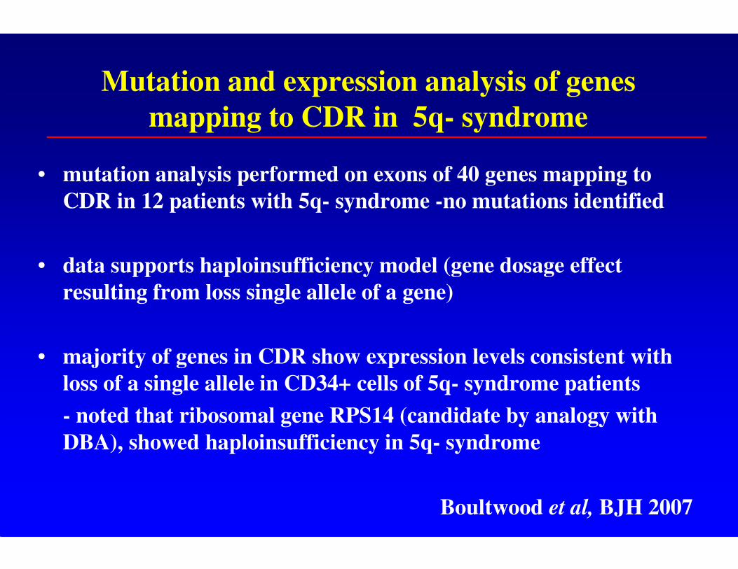
Mutation and expression analysis of genes
mapping to CDR in 5q- syndrome
• mutation analysis performed on exons of 40 genes mapping to
CDR in 12 patients with 5q- syndrome -no mutations identified
• data supports haploinsufficiency model (gene dosage effect
resulting from loss single allele of a gene)
• majority of genes in CDR show expression levels consistent with
loss of a single allele in CD34+ cells of 5q- syndrome patients
- noted that ribosomal gene RPS14 (candidate by analogy with
DBA), showed haploinsufficiency in 5q- syndrome
Boultwood et al, BJH 2007
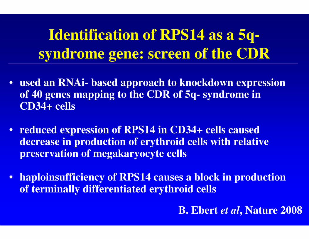
Identification of RPS14 as a 5q-
syndrome gene: screen of the CDR
• used an RNAi- based approach to knockdown expression of 40 genes mapping to the CDR of 5q- syndrome in CD34+ cells
• reduced expression of RPS14 in CD34+ cells caused decrease in production of erythroid cells with relative preservation of megakaryocyte cells
• haploinsufficiency of RPS14 causes a block in production of terminally differentiated erythroid cells
B. Ebert et al, Nature 2008
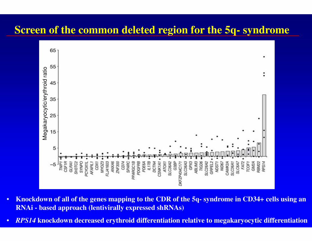
Screen of the common deleted region for the 5q- syndrome
• Knockdown of all of the genes mapping to the CDR of the 5q- syndrome in CD34+ cells using an
RNAi - based approach (lentivirally expressed shRNAs)
• RPS14 knockdown decreased erythroid differentiation relative to megakaryocytic differentiation
Megakary
ocytic/e
ryth
roid
ratio
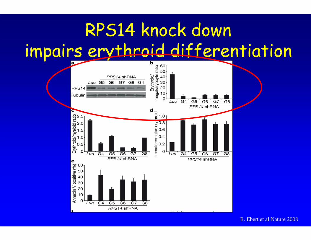
RPS14 knock downimpairs erythroid differentiation
B. Ebert et al Nature 2008
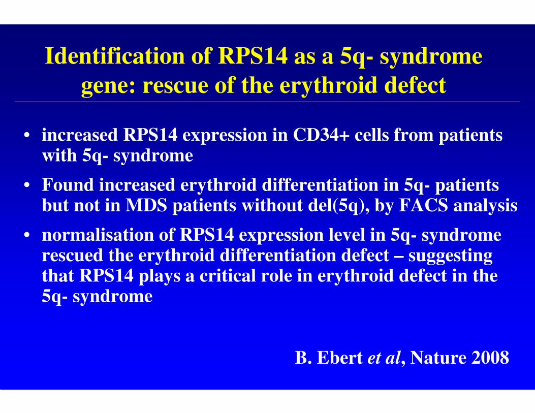
Identification of RPS14 as a 5q- syndrome
gene: rescue of the erythroid defect
• increased RPS14 expression in CD34+ cells from patients with 5q- syndrome
• Found increased erythroid differentiation in 5q- patients but not in MDS patients without del(5q), by FACS analysis
• normalisation of RPS14 expression level in 5q- syndrome rescued the erythroid differentiation defect – suggesting that RPS14 plays a critical role in erythroid defect in the 5q- syndrome
B. Ebert et al, Nature 2008
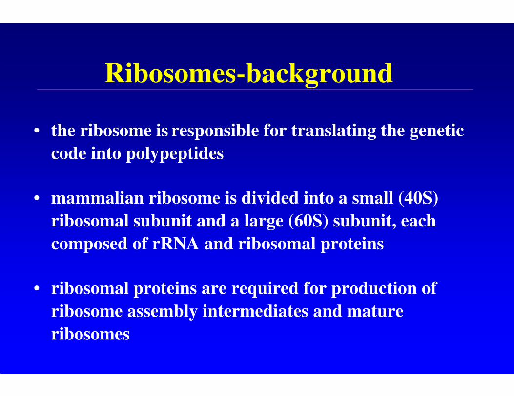
Ribosomes-background
• the ribosome is responsible for translating the genetic
code into polypeptides
• mammalian ribosome is divided into a small (40S)
ribosomal subunit and a large (60S) subunit, each
composed of rRNA and ribosomal proteins
• ribosomal proteins are required for production of
ribosome assembly intermediates and mature
ribosomes
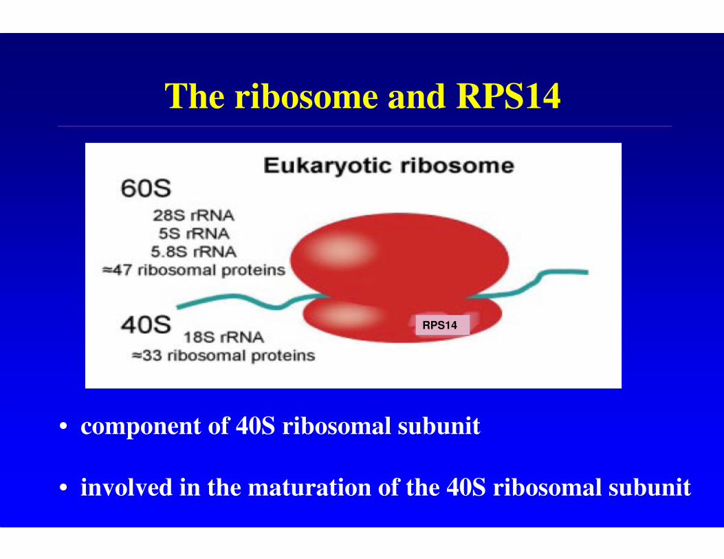
The ribosome and RPS14
RPS14
• component of 40S ribosomal subunit
• involved in the maturation of the 40S ribosomal subunit
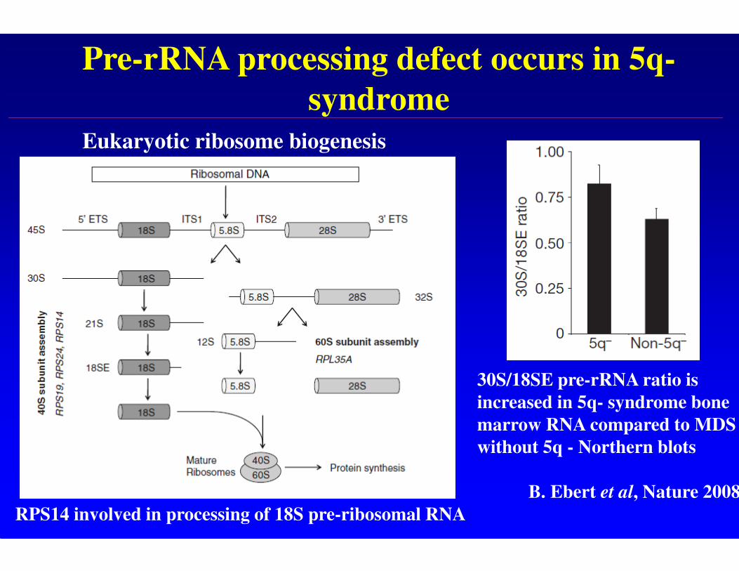
Pre-rRNA processing defect occurs in 5q-
syndrome
30S/18SE pre-rRNA ratio is
increased in 5q- syndrome bone
marrow RNA compared to MDS
without 5q - Northern blots
B. Ebert et al, Nature 2008
RPS14 involved in processing of 18S pre-ribosomal RNA
Eukaryotic ribosome biogenesis
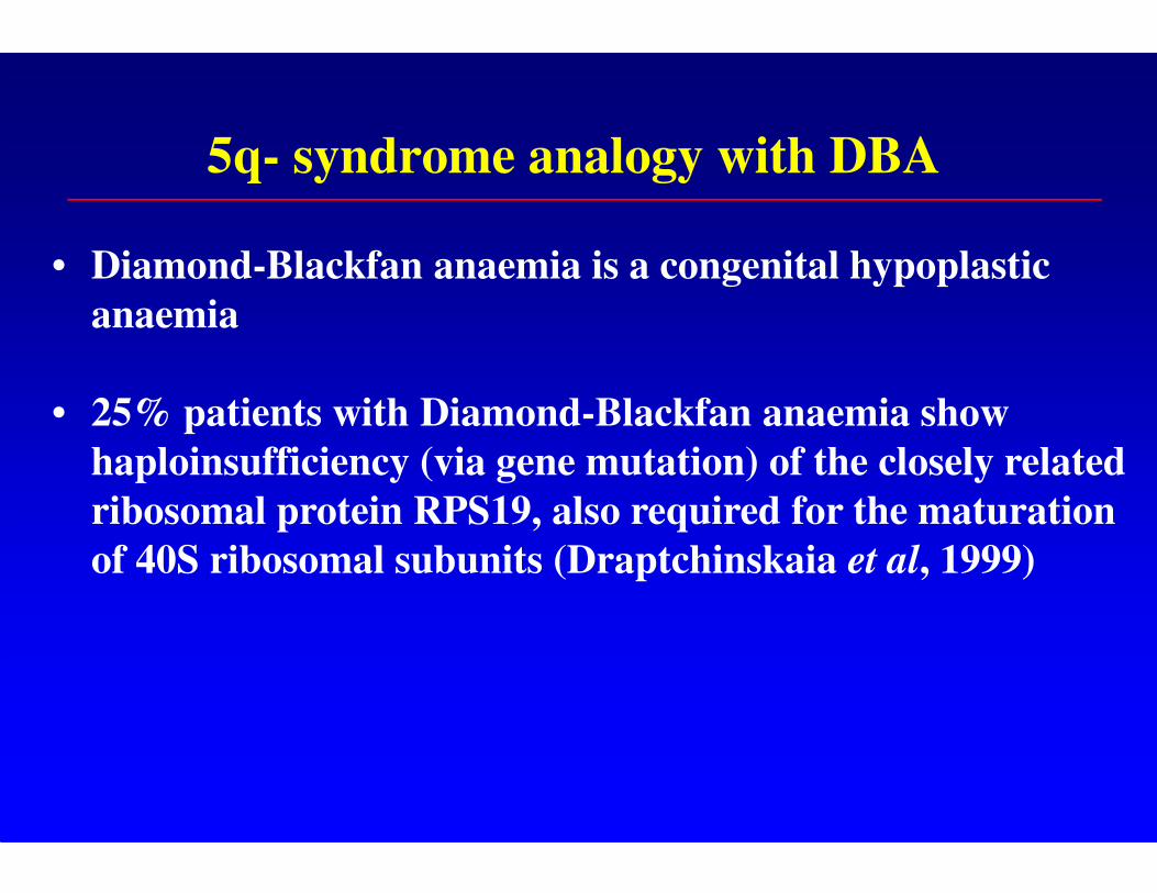
5q- syndrome analogy with DBA
• Diamond-Blackfan anaemia is a congenital hypoplastic
anaemia
• 25% patients with Diamond-Blackfan anaemia show
haploinsufficiency (via gene mutation) of the closely related
ribosomal protein RPS19, also required for the maturation
of 40S ribosomal subunits (Draptchinskaia et al, 1999)
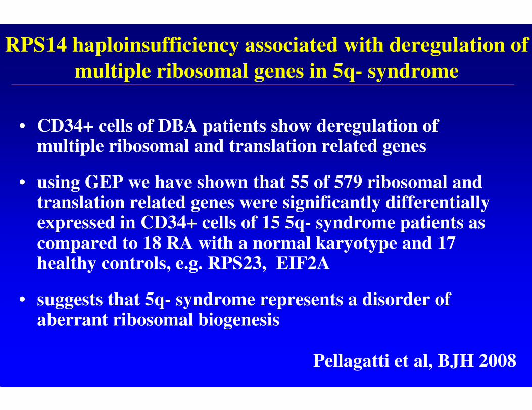
RPS14 haploinsufficiency associated with deregulation of
multiple ribosomal genes in 5q- syndrome
• CD34+ cells of DBA patients show deregulation of multiple ribosomal and translation related genes
• using GEP we have shown that 55 of 579 ribosomal and translation related genes were significantly differentially expressed in CD34+ cells of 15 5q- syndrome patients as compared to 18 RA with a normal karyotype and 17 healthy controls, e.g. RPS23, EIF2A
• suggests that 5q- syndrome represents a disorder of aberrant ribosomal biogenesis
Pellagatti et al, BJH 2008
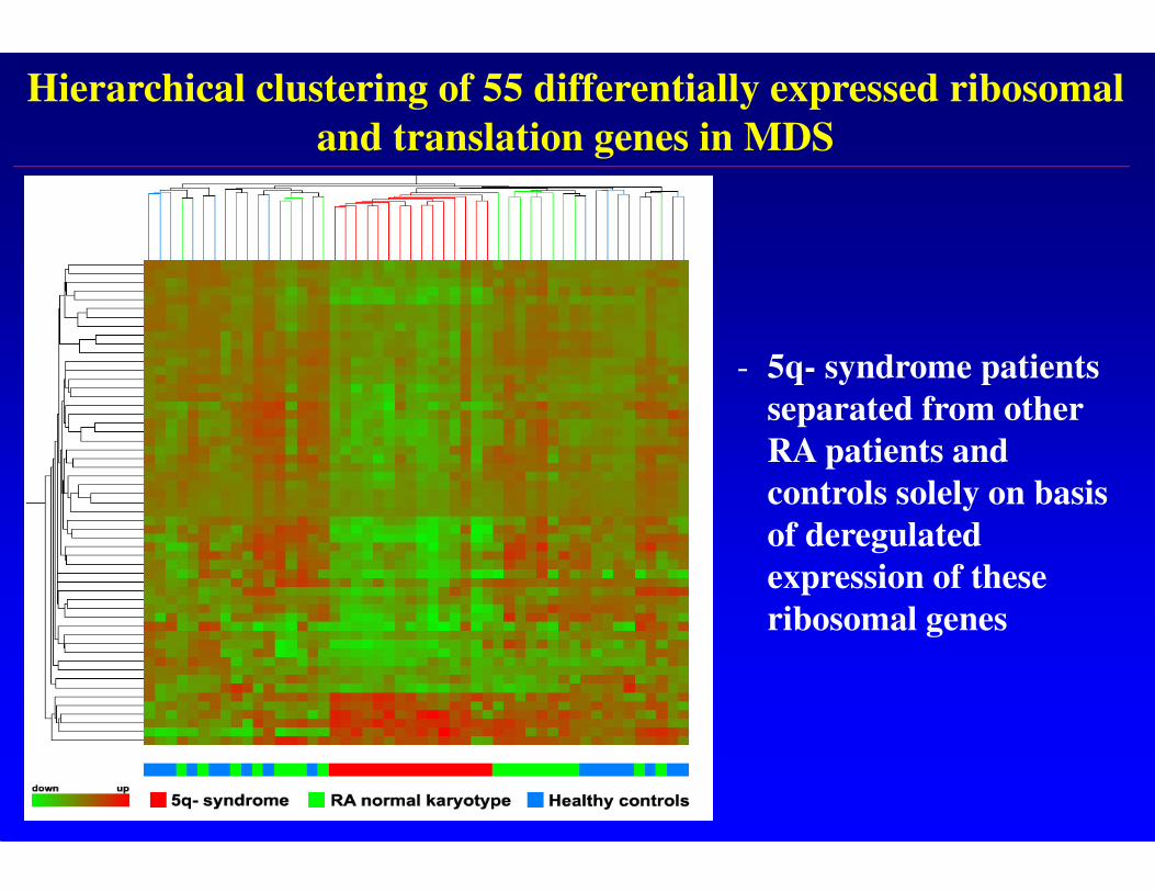
Hierarchical clustering of 55 differentially expressed ribosomal
and translation genes in MDS
- 5q- syndrome patients
separated from other
RA patients and
controls solely on basis
of deregulated
expression of these
ribosomal genes
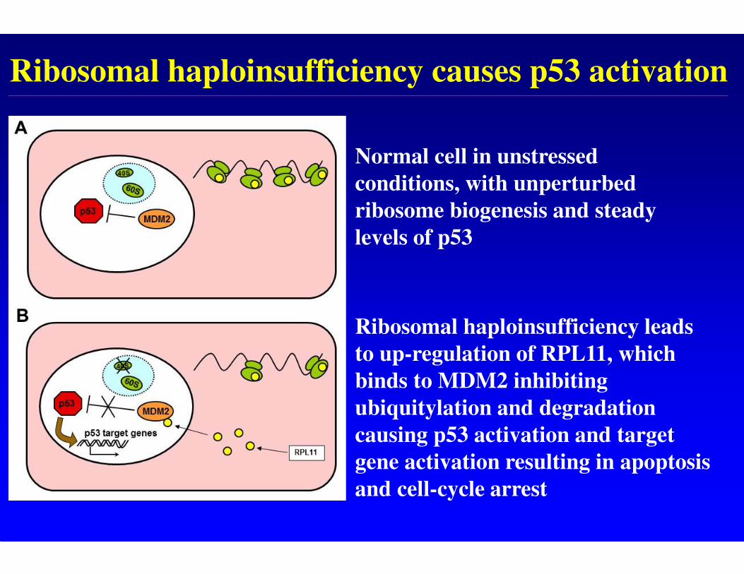
Ribosomal haploinsufficiency causes p53 activation
Normal cell in unstressed
conditions, with unperturbed
ribosome biogenesis and steady
levels of p53
Ribosomal haploinsufficiency leads
to up-regulation of RPL11, which
binds to MDM2 inhibiting
ubiquitylation and degradation
causing p53 activation and target
gene activation resulting in apoptosis
and cell-cycle arrest
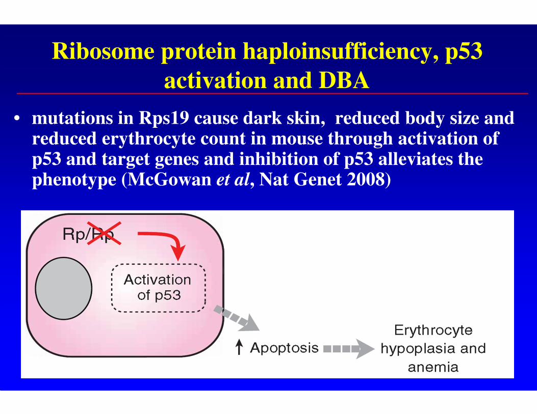
Ribosome protein haploinsufficiency, p53
activation and DBA
• mutations in Rps19 cause dark skin, reduced body size and reduced erythrocyte count in mouse through activation of p53 and target genes and inhibition of p53 alleviates the phenotype (McGowan et al, Nat Genet 2008)
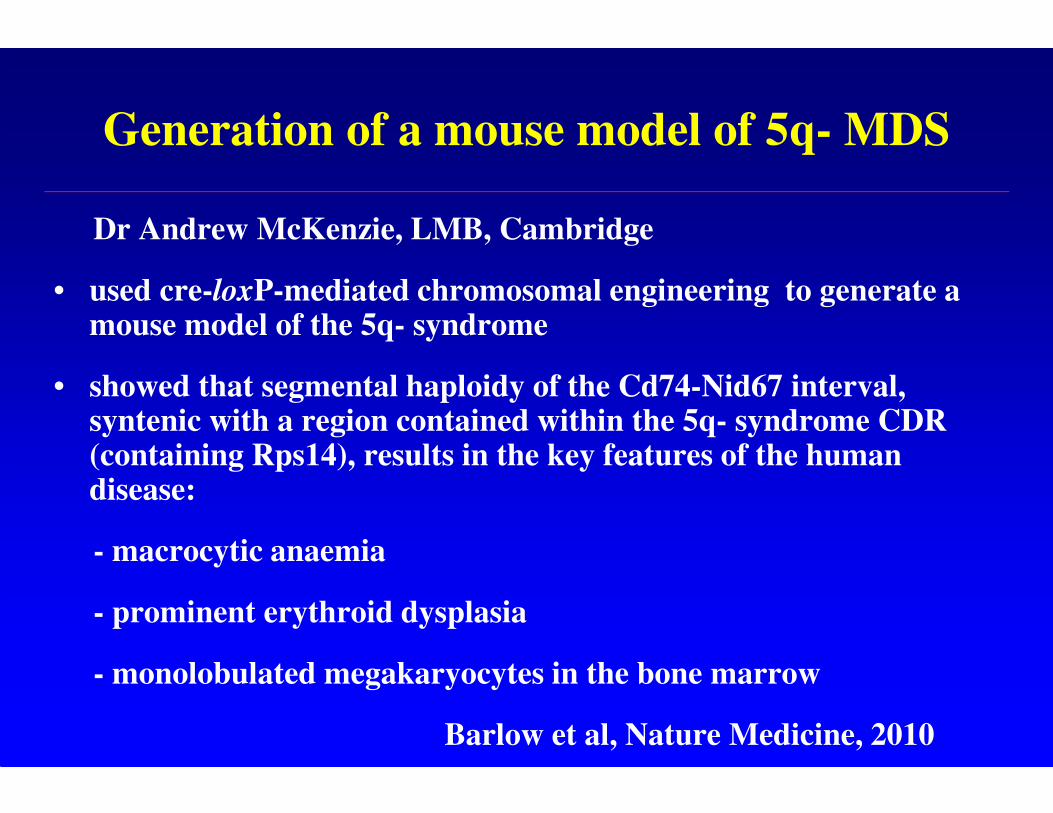
Generation of a mouse model of 5q- MDS
Dr Andrew McKenzie, LMB, Cambridge
• used cre-loxP-mediated chromosomal engineering to generate a mouse model of the 5q- syndrome
• showed that segmental haploidy of the Cd74-Nid67 interval, syntenic with a region contained within the 5q- syndrome CDR (containing Rps14), results in the key features of the human disease:
- macrocytic anaemia
- prominent erythroid dysplasia
- monolobulated megakaryocytes in the bone marrow
Barlow et al, Nature Medicine, 2010
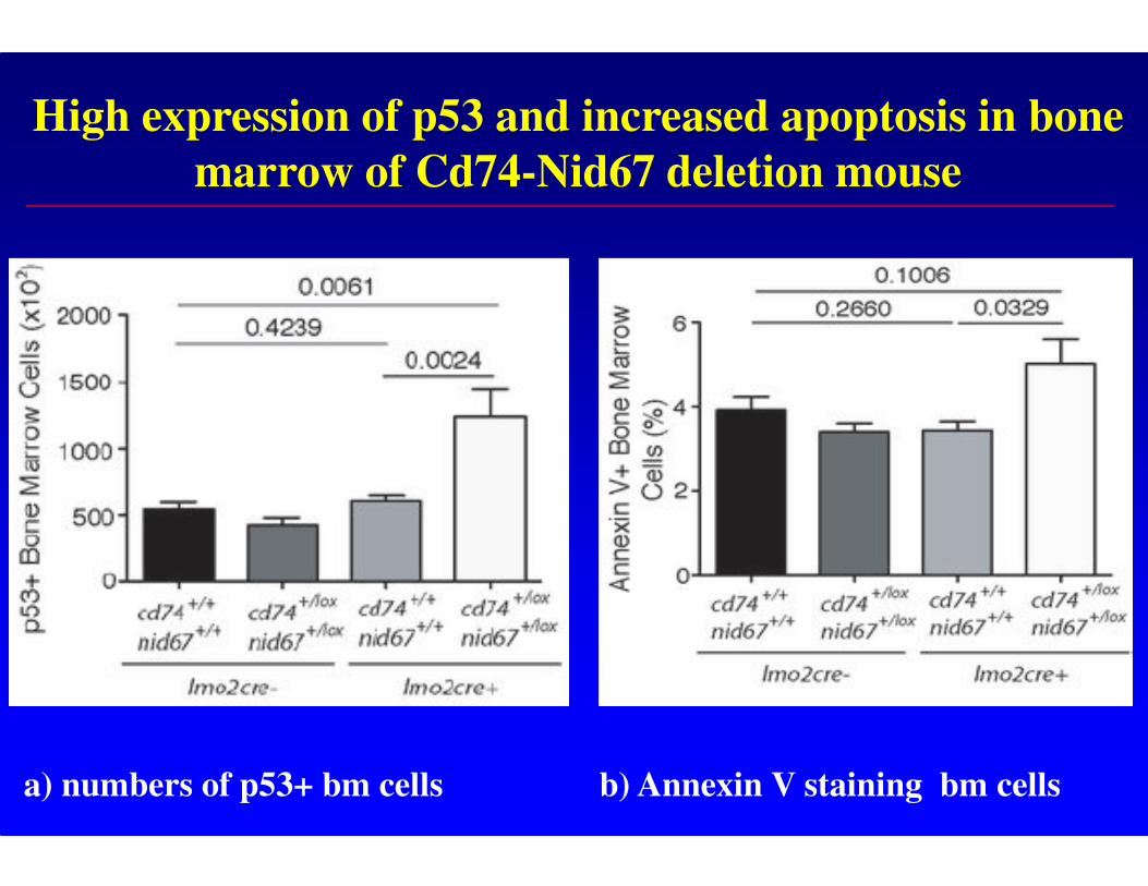
High expression of p53 and increased apoptosis in bone
marrow of Cd74-Nid67 deletion mouse
a) numbers of p53+ bm cells b) Annexin V staining bm cells
p53 and the 5q- syndrome mouse model
• the Cd74-Nid67 deletion mice were crossed with p53-/- mice -completely rescued the progenitor cell defect; restoring CMP, MEP, GMP, and HSC bone marrow populations
• suggests that a p53-dependent mechanism underlies the pathophysiology of the 5q- syndrome
Barlow et al, Nature Medicine, 2010
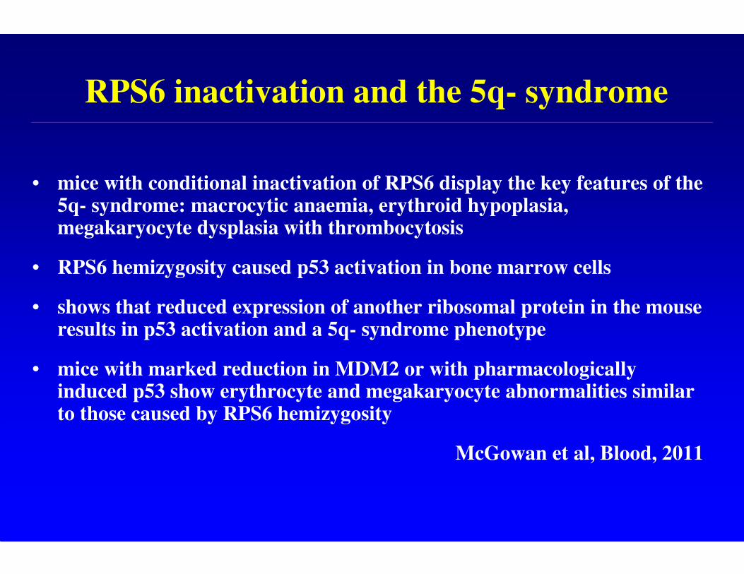
RPS6 inactivation and the 5q- syndrome
• mice with conditional inactivation of RPS6 display the key features of the 5q- syndrome: macrocytic anaemia, erythroid hypoplasia, megakaryocyte dysplasia with thrombocytosis
• RPS6 hemizygosity caused p53 activation in bone marrow cells
• shows that reduced expression of another ribosomal protein in the mouse results in p53 activation and a 5q- syndrome phenotype
• mice with marked reduction in MDM2 or with pharmacologically induced p53 show erythrocyte and megakaryocyte abnormalities similar to those caused by RPS6 hemizygosity
McGowan et al, Blood, 2011
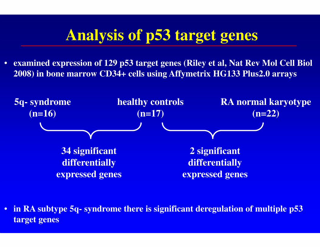
Analysis of p53 target genes
• examined expression of 129 p53 target genes (Riley et al, Nat Rev Mol Cell Biol
2008) in bone marrow CD34+ cells using Affymetrix HG133 Plus2.0 arrays
• in RA subtype 5q- syndrome there is significant deregulation of multiple p53
target genes
5q- syndrome
(n=16)
healthy controls
(n=17)
RA normal karyotype
(n=22)
34 significant
differentially
expressed genes
2 significant
differentially
expressed genes
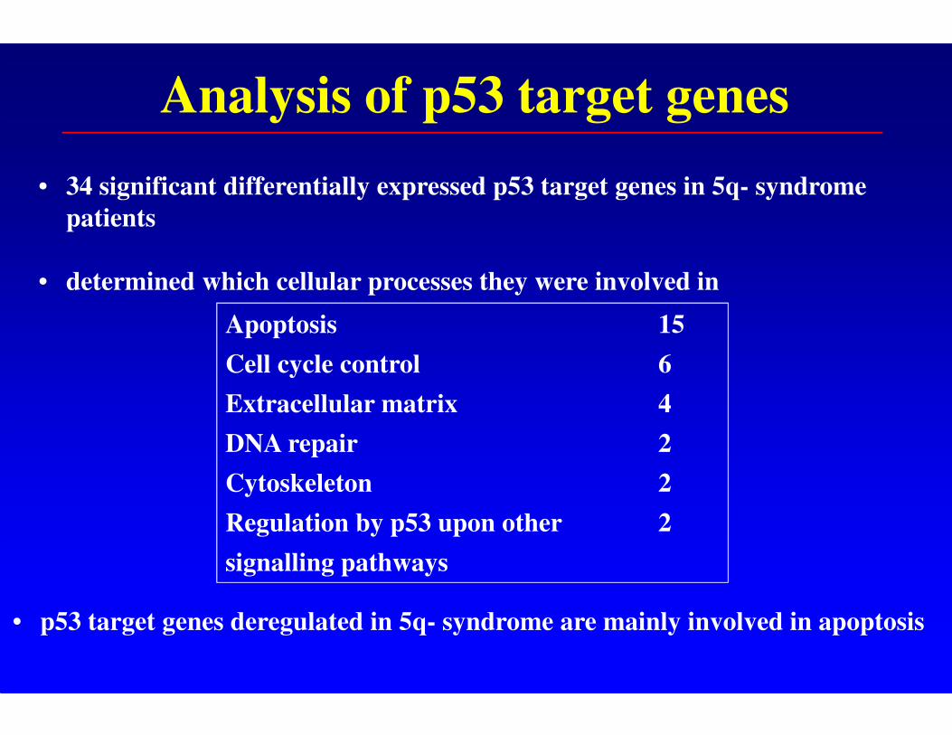
Analysis of p53 target genes
• 34 significant differentially expressed p53 target genes in 5q- syndrome
patients
• determined which cellular processes they were involved in
• p53 target genes deregulated in 5q- syndrome are mainly involved in apoptosis
Apoptosis 15
Cell cycle control 6
Extracellular matrix 4
DNA repair 2
Cytoskeleton 2
Regulation by p53 upon other 2
signalling pathways
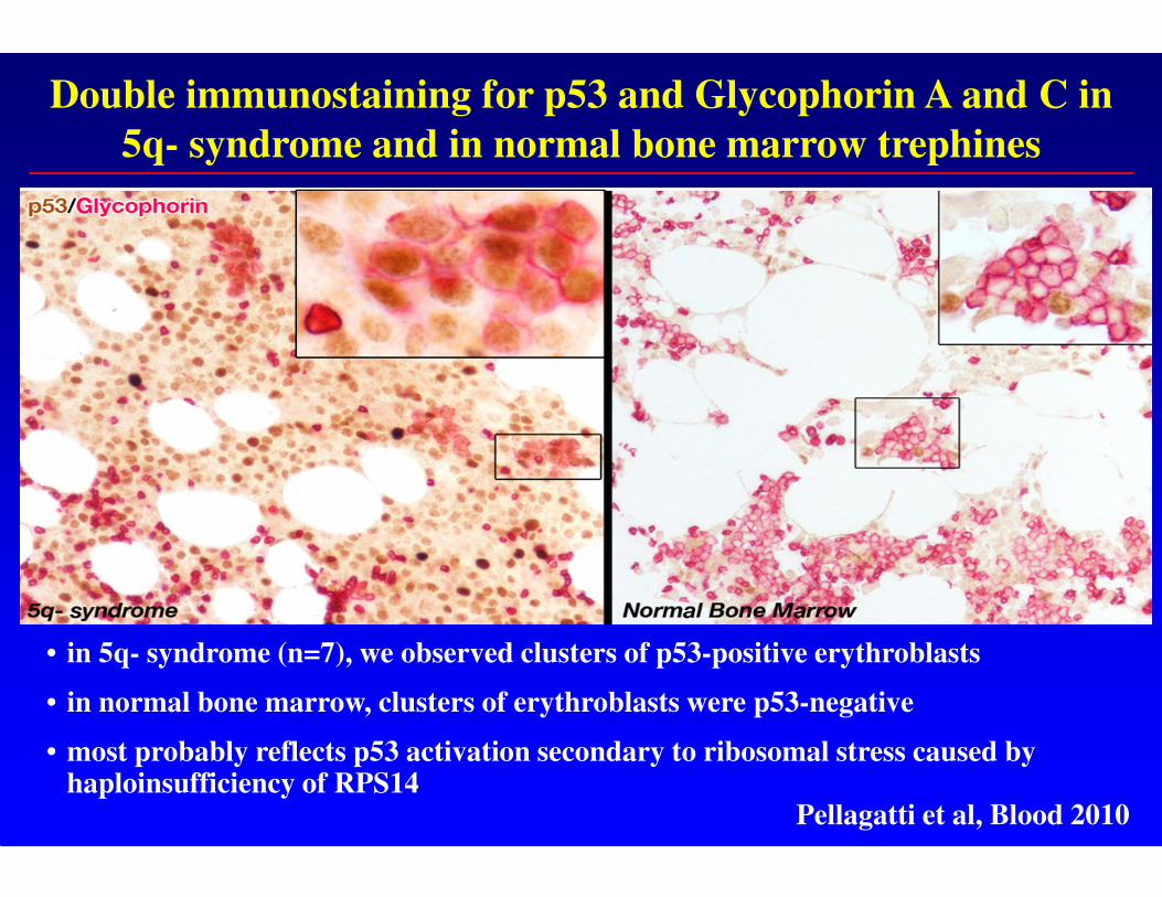
Double immunostaining for p53 and Glycophorin A and C in
5q- syndrome and in normal bone marrow trephines
• in 5q- syndrome (n=7), we observed clusters of p53-positive erythroblasts
• in normal bone marrow, clusters of erythroblasts were p53-negative
• most probably reflects p53 activation secondary to ribosomal stress caused by haploinsufficiency of RPS14
Pellagatti et al, Blood 2010
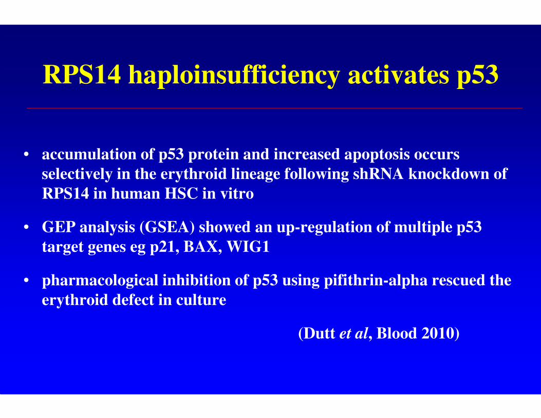
RPS14 haploinsufficiency activates p53
• accumulation of p53 protein and increased apoptosis occurs
selectively in the erythroid lineage following shRNA knockdown of
RPS14 in human HSC in vitro
• GEP analysis (GSEA) showed an up-regulation of multiple p53
target genes eg p21, BAX, WIG1
• pharmacological inhibition of p53 using pifithrin-alpha rescued the
erythroid defect in culture
(Dutt et al, Blood 2010)
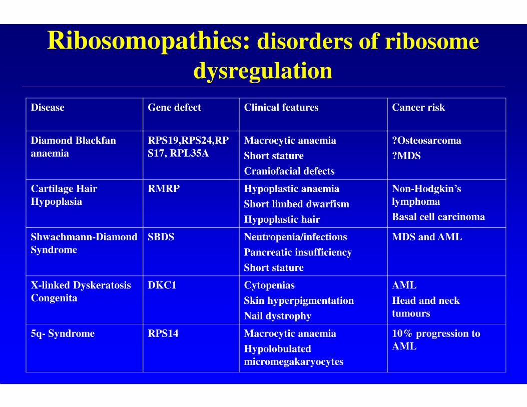
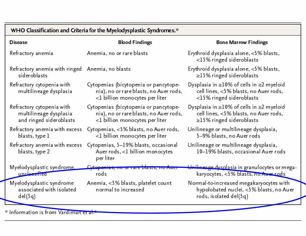
Disease Gene defect Clinical features Cancer risk
Diamond Blackfan
anaemia
RPS19,RPS24,RP
S17, RPL35A
Macrocytic anaemia
Short stature
Craniofacial defects
?Osteosarcoma
?MDS
Cartilage Hair
Hypoplasia
RMRP Hypoplastic anaemia
Short limbed dwarfism
Hypoplastic hair
Non-Hodgkin’s
lymphoma
Basal cell carcinoma
Shwachmann-Diamond
Syndrome
SBDS Neutropenia/infections
Pancreatic insufficiency
Short stature
MDS and AML
X-linked Dyskeratosis
Congenita
DKC1 Cytopenias
Skin hyperpigmentation
Nail dystrophy
AML
Head and neck
tumours
5q- Syndrome RPS14 Macrocytic anaemia
Hypolobulated
micromegakaryocytes
10% progression to
AML
Ribosomopathies: disorders of ribosome
dysregulation
Ribosome dysfunction in the 5q-
syndrome
• Introduction and mapping of the CDR
• Role of haploinsufficiency of RPS14 and p53 activation
in molecular pathogenesis
• Defective translation and treatment with L-leucine
• Cooperating genetic events in disease pathogenesis
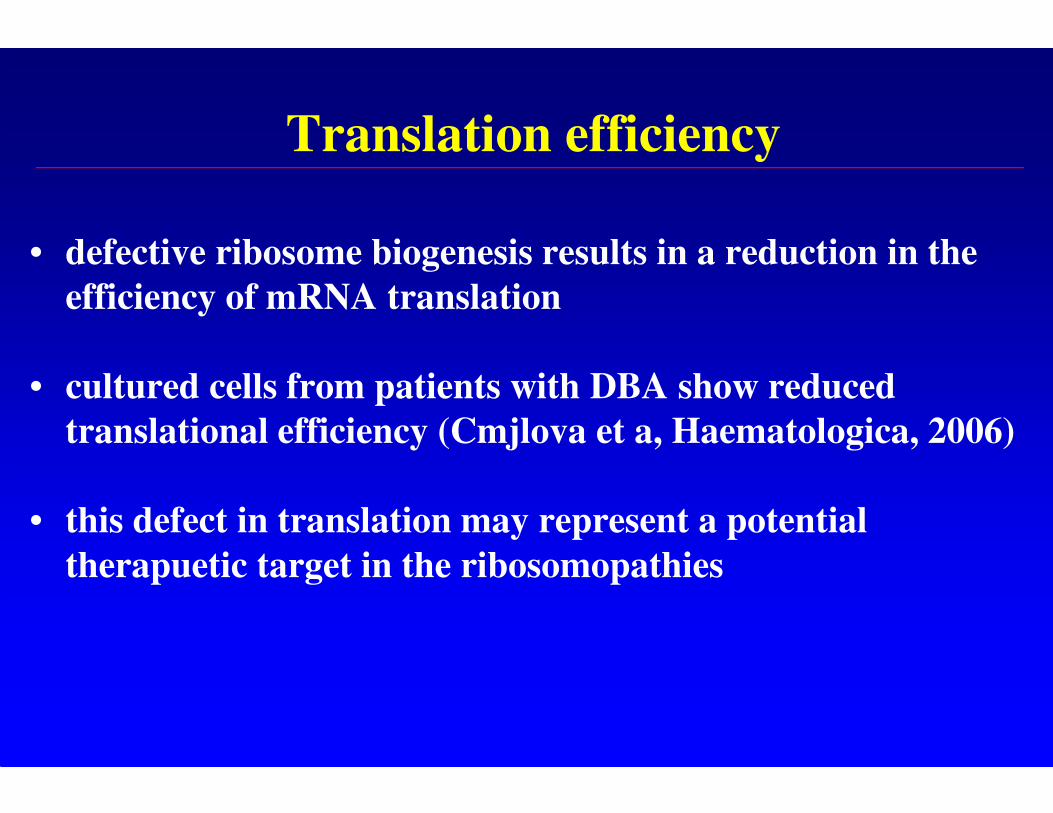
Translation efficiency
• defective ribosome biogenesis results in a reduction in the
efficiency of mRNA translation
• cultured cells from patients with DBA show reduced
translational efficiency (Cmjlova et a, Haematologica, 2006)
• this defect in translation may represent a potential
therapuetic target in the ribosomopathies
L-leucine: a translation enhancer
• L-leucine: amino acid that modulates protein synthesis by enhancing translation (activates mTOR pathway and downstream targets, activates translation signaling factors)
• L-leucine shown to improve haemoglobin levels and transfusion independence in patients with DBA (Pospisilova et al, Haematologica, 2007)
• L-leucine supplement partially rescues the erythrocyte and leukocyte numbers in RPS19-deficient mice (Jaako et al, ASH 2011-Abstract 727 Monday, December 12, 2011: 4:30 PM)
• treatment of zebrafish models of DBA and 5q- syndrome with L-leucine resulted in partial reversal of the anaemia (Virgilio et al, ASH 2010, 2011-Abstract 970 Tuesday, December 13, 2011: 8:15 AM)
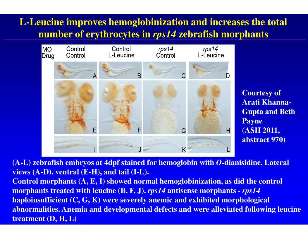
L-Leucine improves hemoglobinization and increases the total
number of erythrocytes in rps14 zebrafish morphants
(A-L) zebrafish embryos at 4dpf stained for hemoglobin with O-dianisidine. Lateral
views (A-D), ventral (E-H), and tail (I-L).
Control morphants (A, E, I) showed normal hemoglobinization, as did the control
morphants treated with leucine (B, F, J). rps14 antisense morphants - rps14
haploinsufficient (C, G, K) were severely anemic and exhibited morphological
abnormalities. Anemia and developmental defects and were alleviated following leucine
treatment (D, H, L)
Courtesy of
Arati Khanna-
Gupta and Beth
Payne
(ASH 2011,
abstract 970)
Translation efficiency in 5q- syndrome
• haploinsufficiency of RPS14 results in a pre-rRNA processing defect and in 40S subunit deficiency in human cells -it is therefore probable that translation is compromised in these cells
• aim to investigate the effects of L-leucine on erythropoiesis in the 5q- syndrome
• determine whether the use of L-leucine leads to enhanced translation and a rescue of the erythroid defect in cultured cells from 5q- syndrome patients (complementary to similar studies in 5q- mouse model)
Treatment of 5q- syndrome CD34+ cells
with L-leucine
• experiments are being performed in two stages using:
cellular model system of 5q- syndrome: lentiviral-based shRNA knockdown of RPS14 has been performed in human CD34+ cells as they differentiate into erythroid cells
5q- syndrome CD34+ cells differentiated into erythroid cellsin culture
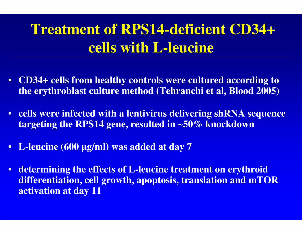
Treatment of RPS14-deficient CD34+
cells with L-leucine
• CD34+ cells from healthy controls were cultured according to the erythroblast culture method (Tehranchi et al, Blood 2005)
• cells were infected with a lentivirus delivering shRNA sequence targeting the RPS14 gene, resulted in ~50% knockdown
• L-leucine (600 µg/ml) was added at day 7
• determining the effects of L-leucine treatment on erythroid differentiation, cell growth, apoptosis, translation and mTOR activation at day 11
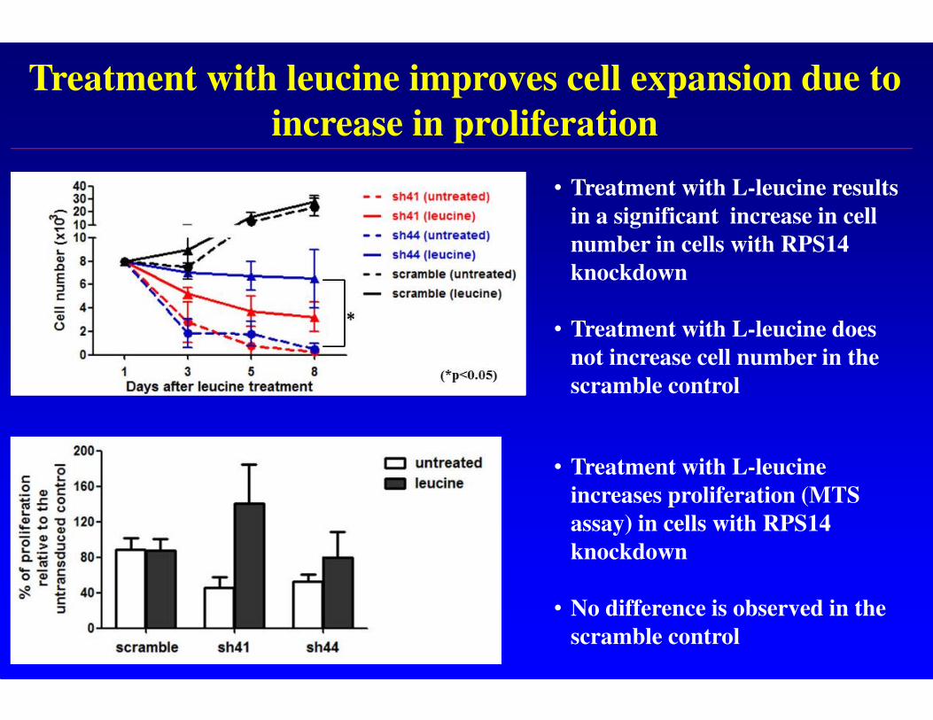
Treatment with leucine improves cell expansion due to
increase in proliferation
• Treatment with L-leucine results
in a significant increase in cell
number in cells with RPS14
knockdown
• Treatment with L-leucine does
not increase cell number in the
scramble control
• Treatment with L-leucine
increases proliferation (MTS
assay) in cells with RPS14
knockdown
• No difference is observed in the
scramble control
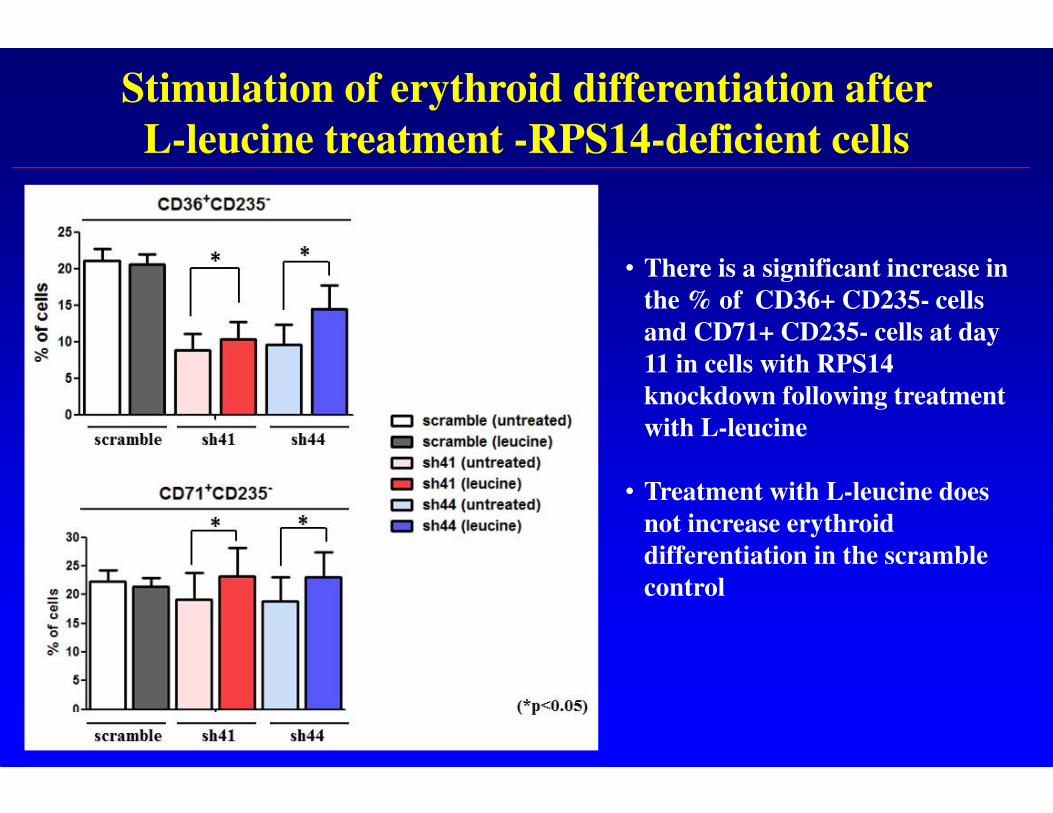
Stimulation of erythroid differentiation after
L-leucine treatment -RPS14-deficient cells
• There is a significant increase in
the % of CD36+ CD235- cells
and CD71+ CD235- cells at day
11 in cells with RPS14
knockdown following treatment
with L-leucine
• Treatment with L-leucine does
not increase erythroid
differentiation in the scramble
control
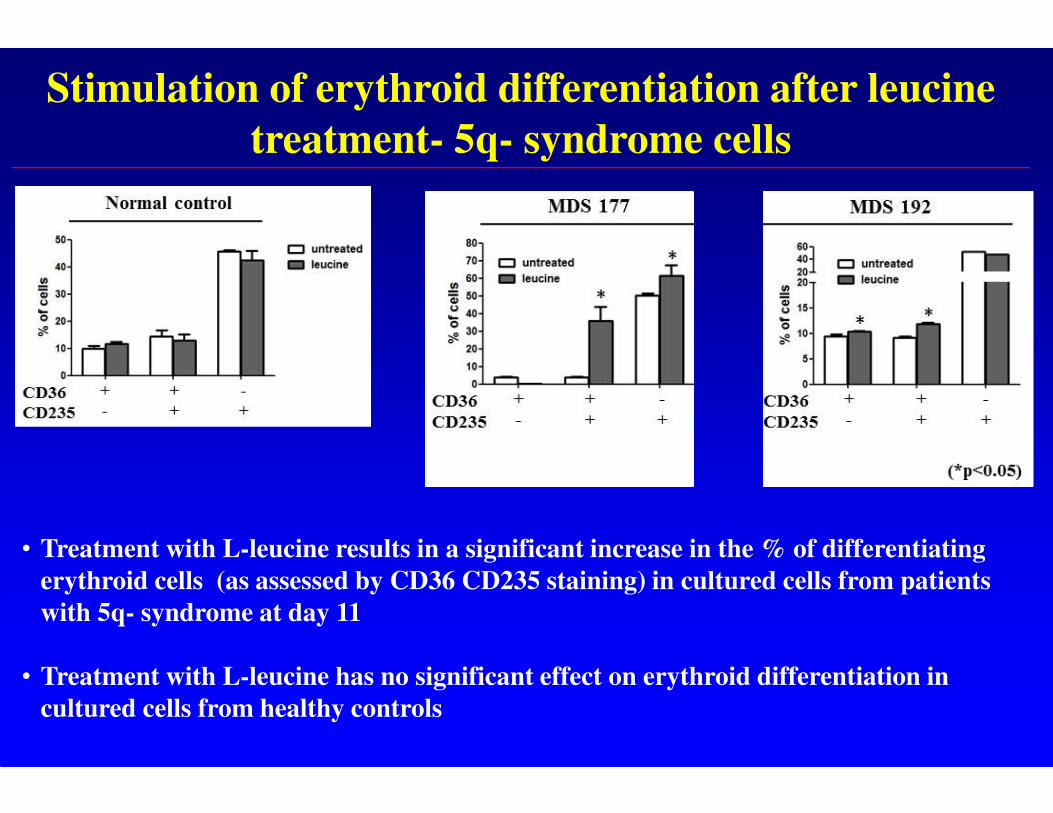
Stimulation of erythroid differentiation after leucine
treatment- 5q- syndrome cells
• Treatment with L-leucine results in a significant increase in the % of differentiating
erythroid cells (as assessed by CD36 CD235 staining) in cultured cells from patients
with 5q- syndrome at day 11
• Treatment with L-leucine has no significant effect on erythroid differentiation in
cultured cells from healthy controls
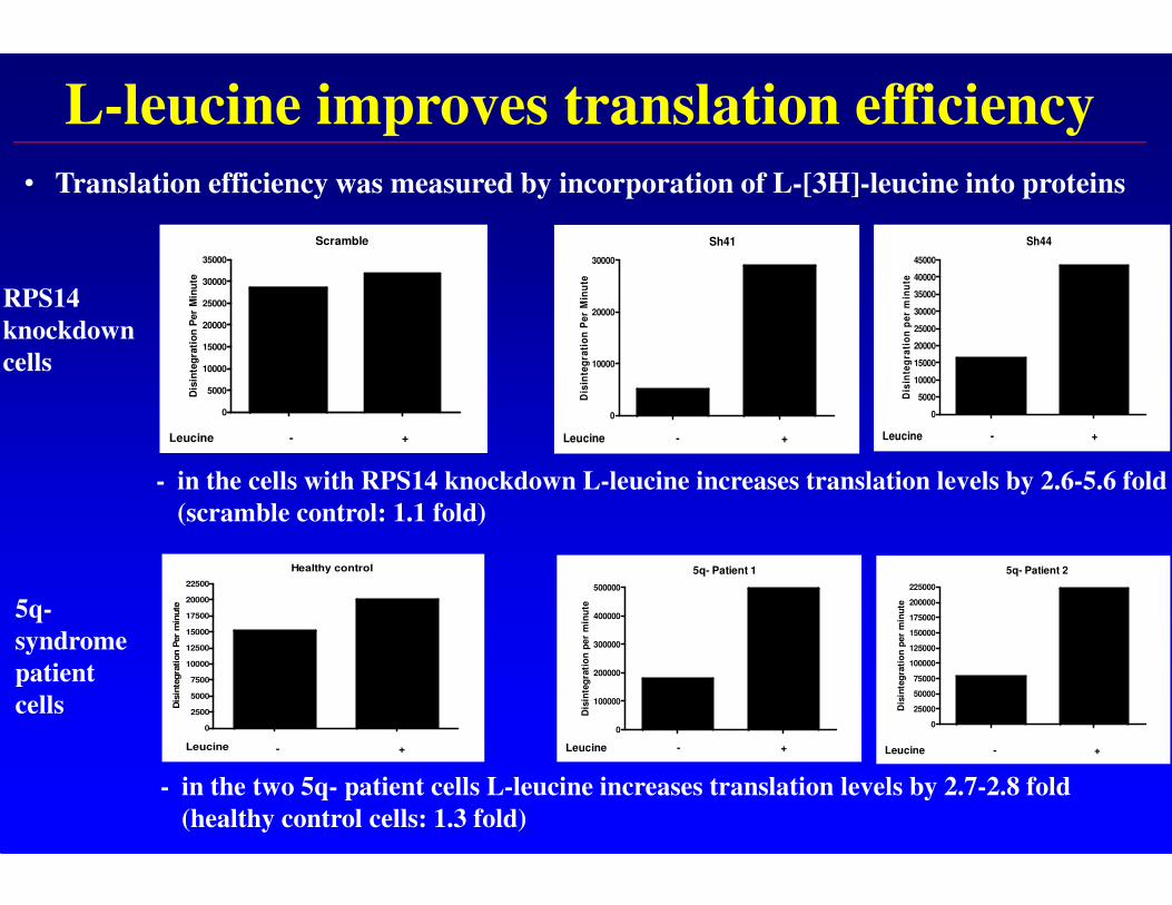
L-leucine improves translation efficiency
RPS14
knockdown
cells
5q-
syndrome
patient
cells
Scramble
0
5000
10000
15000
20000
25000
30000
35000
Leucine - +
Dis
inte
gra
tio
n P
er
Min
ute
Sh41
0
10000
20000
30000
Leucine - +
Dis
inte
gra
tio
n P
er
Min
ute
Sh44
0
5000
10000
15000
20000
25000
30000
35000
40000
45000
Leucine - +
Dis
inte
gra
tio
n p
er
min
ute
5q- Patient 2
0
25000
50000
75000
100000
125000
150000
175000
200000
225000
Leucine - +
Dis
inte
gra
tio
n p
er
min
ute
5q- Patient 1
0
100000
200000
300000
400000
500000
Leucine - +
Dis
inte
gra
tio
n p
er
min
ute
• Translation efficiency was measured by incorporation of L-[3H]-leucine into proteins
Healthy control
0
2500
5000
7500
10000
12500
15000
17500
20000
22500
Leucine - +
Dis
inte
gra
tion P
er
min
ute
- in the cells with RPS14 knockdown L-leucine increases translation levels by 2.6-5.6 fold
(scramble control: 1.1 fold)
- in the two 5q- patient cells L-leucine increases translation levels by 2.7-2.8 fold
(healthy control cells: 1.3 fold)
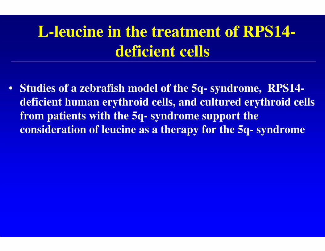
L-leucine in the treatment of RPS14-
deficient cells
• Studies of a zebrafish model of the 5q- syndrome, RPS14-
deficient human erythroid cells, and cultured erythroid cells
from patients with the 5q- syndrome support the
consideration of leucine as a therapy for the 5q- syndrome
Ribosome dysfunction in the 5q-
syndrome
• Introduction and mapping of the CDR
• Role of haploinsufficiency of RPS14 and p53 activation
in molecular pathogenesis
• Defective translation and treatment with L-leucine
• Cooperating genetic events in disease pathogenesis
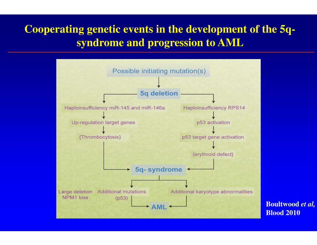
Cooperating genetic events in the development of the 5q-
syndrome and progression to AML
Boultwood et al,
Blood 2010
Acknowledgments
James Wainscoat
Andrea Pellagatti
A. H. Yip
J. Perry
C. Vuppusetty
Andrew McKenzie
Jillian Barlow
L. Drynan
M. Cazzola
L. Malcovati
E. Hellström-Lindberg
M. Jädersten
A. Giagounidis
U. Germing
S. Killick
P. Vyas
M. Calasanz
CollaboratorsLLR Unit


![Ribosome Stoichiometry: From Form to Function · Ribosome abundance: A major model, also termed the ribosome concentration hypothesis [3], that explains how ribosomes could exert](https://img.pdfslide.us/doc/110x75/60de31e56d30fc4fb30719b8/ribosome-stoichiometry-from-form-to-function-ribosome-abundance-a-major-model.jpg)