Embed Size (px)
Citation preview

IMPACT OF THE SHORT – CHAIN
FATTY ACIDS ON THE MICROBIOTA
– GUT – BRAIN AXIS
Raquel Miralles Solsona
Treball d’aprofundiment
Departament de Nutrició, Ciències de l’Alimentació i Gastronomia
Juny 2021
UNIVERSITAT DE BARCELONA
FACULTAT DE FARMÀCIA I CIÈNCIES DE L’ALIMENTACIÓ
TREBALL DE FI DE GRAU

2
This work is licenced under a Creative Commons license.

3
ABSTRACT
Currently, there are many studies that corroborate the relationship between the
microbiota and the gut – brain axis. This relationship is due through various mechanisms
including the production of some metabolites by the microbiota, such as short – chain
fatty acids. The microbiota – gut – brain axis consists in the bidirectional communication
between the microbiota, the gut and the brain where changes in the microbiota can lead
to changes in the gut and brain, and vice versa. The aim of the research is to study the
impact of short – chain fatty acids on the axis. A bibliographic research was performed
through some search engines. Studies show that short – chain fatty acids have different
effects by increasing or decreasing the quantity of certain molecules such as GLP-1,
peptide YY, insulin, glucagon, ghrelin, leptin and serotonin while they also have an
impact on the nervous system. Also is needed to emphasize its role in certain
neurological diseases such as Parkinson, autism spectrum disorder and multiple
sclerosis. However, the current evidence still needs to be strengthened by further
studies that will allow to define the mechanisms by which this relationship takes place
and confirm the impact of this type of metabolites on the organism. At the same time,
this will allow to develop new treatments for people with certain neurological diseases.
Key words: microbiota, microbiota – gut – brain axis, prebiotics, short – chain fatty acids,
neurological diseases.
RESUM
A dia d’avui són molts els estudis que corroboren la relació entre la microbiota i l’eix
intestí – cervell. Aquesta relació es dona mitjançant diversos mecanismes entre els quals
hi ha la producció de certs metabòlits per part de la microbiota, com són els àcids grassos
de cadena curta. L’eix microbiota – intestí – cervell consisteix en una comunicació
bidireccional entre la microbiota, l’intestí i el cervell on modificacions a nivell de la
microbiota poden donar lloc a canvis a nivell intestinal i cerebral i viceversa. L’objectiu
de la recerca consisteix en l’estudi de l’impacte dels àcids grassos de cadena curta sobre
l’eix. Per fer-ho s’ha realitzat una recerca bibliogràfica mitjançant alguns motors de

4
cerca. Els estudis mostren que els àcids grassos de cadena curta tenen accions a
diferents nivells incrementant o disminuint la quantitat de certes molècules com el GLP-
1, pèptid YY, insulina, glucagó, grelina, leptina i serotonina a la vegada que també tenen
un impacte sobre el sistema nerviós. Cal destacar també, el seu paper en certes malalties
neurològiques com el Parkinson, trastorn de l’espectre autista i l’esclerosis múltiple. No
obstant, l’evidència actual encara necessita ser reforçada per més estudis que permetin
acabar de definir els mecanismes pels quals es dona aquesta relació i confirmar
l’impacte d’aquest tipus de metabòlits a diferents nivells de l’organisme. A la vegada,
també permetrà generar nous tractaments per a persones que presentin certes
malalties neurològiques.
Paraules clau: microbiota, eix microbiota – intestí – cervell, prebiòtics, àcids grassos de
cadena curta, malalties neurològiques.

5
INDEX
1. Introduction............................................................................................................... 7
2. Objectives .................................................................................................................. 8
3. Materials and methods ............................................................................................. 8
4. Results ....................................................................................................................... 9
4.1. The dietary fiber and prebiotics ........................................................................ 9
4.2. Dietary fiber metabolism to short – chain fatty acids ..................................... 10
4.2.1. Synthesis, absorption and distribution of short – chain fatty acids ......... 11
4.3. The gut microbiota ........................................................................................... 12
4.4. The microbiota – gut – brain axis .................................................................... 14
4.4.1. Enteroendocrine cells ............................................................................... 14
4.5. Communication mechanisms: ......................................................................... 15
4.5.1. The neural pathway .................................................................................. 15
4.5.2. The circulatory pathway ........................................................................... 16
4.5.3. The immune pathway ............................................................................... 17
4.6. Short – chain fatty acids................................................................................... 18
4.6.1. SCFAs receptors ........................................................................................ 18
4.6.2. Effect of the short – chain fatty acids on the microbiota – gut – brain axis
20
4.7. Involvement of SCFAs in some neurological disorders .................................... 27
5. Discussion ................................................................................................................ 30
6. Conclusion ............................................................................................................... 31
7. Bibliography ............................................................................................................ 33

6
LIST OF FIGURES
Figure 1. Distinction of what is considered a prebiotic from Gibson GR. et al. (8) ........ 10
Figure 2. Schematic representation of the gut microbiota from Ghaisas S. et al. (4) .... 13
Figure 3. Effect of the SCFAs on BBB permeability from Ma Q. et al. (24)..................... 17
Figure 4. Scheme about the opposite endocrine and metabolic effects of ghrelin and
GLP-1 from Engelstoft MS. et al. (31) ............................................................................. 24
Figure 5. SCFAs involvement in the regulation of appetite and metabolism from van de
Wouw M. et al. (15). ....................................................................................................... 26
Figure 6. Interaction between microbiota, intestinal permeability and CNS from Yarandi
SS. et al. (42) ................................................................................................................... 28
LIST OF TABLES
Table 1. Location of FFAR2 and FFAR3 receptors throughout the body ...................... 200
Table 2. Main effects of the SCFAs on peptides (GLP-1 and PYY), hormones (insulin,
glucagon, leptin and ghrelin) and NT (serotonin) and their resulting physiological effect
........................................................................................................................................ 25
ABREVIATIONS
SCFA Short – Chain Fatty Acids
CNS Central Nervous System
GI Gastrointestinal
BBB Blood – Brain Barrier
VN Vagus Nerve
NT Neurotransmitters
ASD Autism Spectrum Disorder
EEC Enteroendocrine Cells
GLP-1 Glucagon-like peptide 1
PYY Peptide YY
FFAR Free Fatty Acid Receptor
NS Nervous System
ENS Enteric Nervous System
IS Immune System
LPS Lipopolysaccharide
TLR Toll-like receptors
GPCR G protein – coupled receptor
AT Adipose tissue
Treg Regulatory T cell

7
1. INTRODUCTION
This final degree project consists in conducting bibliographic research on current
scientific evidence about the relationship between short – chain fatty acids (SCFAs) and
the microbiota – gut – brain axis.
The project below includes the definition of SCFAs and the microbiota – gut – brain
axis. There are also described different mechanisms that relate both concepts and
scientific evidence about their role in certain neurological disorders.
This is an area that is increasingly being studied due to its impact on human
pathophysiology. Recent studies show how the gut microbiota influences the
functioning of the central nervous system (CNS). Through the production of hormones,
immune factors and metabolites it influences both brain behavior and cognitive
development. This fact paves the way for a possible therapeutic route (1).
In addition, the microbiota – gut – brain axis is gaining importance in the investigation
of psychiatric, neurodevelopmental, neurodegenerative and age - related disorders (2).
The microbiota has also been shown to play a key role in the regulation of energy
homeostasis and obesity. Different studies show the relationship between dysbiosis and
obesity and the role of the microbiota in eating behavior (3).
Starting from this point, it must be considered that there are several factors that can
affect the composition of the microbiota such as the type of birth, breastfeeding,
antibiotic intake, type of feeding, the presence of stressful elements, among others (2).
The SCFAs are one of the major metabolites of the intestinal microbiota and perform
various functions in humans (4). These functions and some of their mechanisms are
described below.
In order to understand the mechanisms surrounding the effects of SCFAs on this axis, it
is necessary to continue conducting further studies, and thus develop microbiota –
based intervention strategies (2).

8
2. OBJECTIVES
The main objective of this report is to conduct bibliographic research about the
evidence on the involvement of SCFAs, produced by the gut microbiota, in the
microbiota – gut – brain axis, a topic with many papers published in the last few
years. This study will be based on the understanding of different items:
- Definition of the dietary fiber and prebiotics concepts.
- Study of the synthesis of short – chain fatty acids by gut microbiota from
dietary fiber consumption.
- Study of the functioning of the microbiota – gut – brain axis.
- Analysis of the impact of short – chain fatty acids on the microbiota – gut –
brain axis.
- Research on the role of short – chain fatty acids in different neurological
disorders.
3. MATERIALS AND METHODS
This final degree project is based on an exhaustive bibliographic research. As this is a
very current topic, the work of nutritional intervention in humans is not very extensive,
so a fairly inclusive search has been carried out in order to comprise as much information
as possible and then focus on what is really useful. The main databases used were
Pubmed and Scopus, where the "5 years" and "review" filters were applied, as well as
Google Scholar to expand the information.
First, a general search was made with the terms: “microbiota AND (gut – brain axis)”,
“microbiota AND brain” and “fiber AND microbiota AND brain”. From all the papers
published, a selection of the most relevant was performed and analyzed.
From then on, a more specific search was made using terms such as “fiber AND
microbiota AND metabolism ” and “microbiota AND brain AND (short chain fatty acids)”.
It should be noted that many of the articles used were taken from the category "Similar
articles” shown in Pubmed and the own bibliography of some of the articles used.

9
4. RESULTS
4.1. The dietary fiber and prebiotics
To date, there is still no definition of dietary fiber that is unified by different organisms.
Historically, dietary fiber was considered to be those polysaccharides with a degree of
polymerization greater than 10 that are resistant to both digestion and absorption in the
gut (5). In some recent studies, those substrates with a degree of polymerization
between 3 - 9 units have been considered as they seem to have the same physiological
activities as polysaccharides with a degree greater than 10 (6).
The Regulation (UE) 1169/2011 of the European Union about the provision of food
information to consumers has defined fiber as «carbohydrate polymers with three or
more monomeric units, which are neither digested nor absorbed in the human small
intestine and belong to the following categories:
- edible carbohydrate polymers naturally occurring in the food as consumed,
- edible carbohydrate polymers which have been obtained from food raw material
by physical, enzymatic or chemical means and which have a beneficial
physiological effect demonstrated by generally accepted scientific evidence,
- edible synthetic carbohydrate polymers which have a beneficial physiological
effect demonstrated by generally accepted scientific evidence» (7).
On the other hand, the current definition of prebiotic was developed by “The
International Scientific Association for Probiotics and Prebiotics” in 2016. Prebiotic is
defined as: «Substrate that is used selectively by host microorganisms conferring a
benefit for the health». With this definition it is clear that in order to be considered
prebiotic it is necessary to demonstrate that there is a health benefit. Moreover, this
health benefit must be given through the selective use of the substrate by host
microorganisms. In addition, it also expands the concept that the substrate may not
belong to the category of carbohydrates (8).
10
Figure 1. Distinction of what is considered a prebiotic from Gibson GR. et al. (8)
Unifying both concepts, some types of dietary fiber are candidates to act as prebiotics
as they are used selectively by the microbiota and produce a beneficial effect. However,
it can be difficult to categorize dietary fiber as a prebiotic because it also depends on
some factors such as the host and whether there is a microbiota at the target site
capable of using it selectively (8). It can be concluded, therefore, that although many of
the prebiotics belong to the dietary fiber category, not all dietary fiber can be considered
prebiotic (9).
4.2. Dietary fiber metabolism to short – chain fatty acids
The physicochemical characteristics of the fiber are fermentability, solubility and
viscosity. The types of fermentable fiber include β-glucans, pectins, inulin,
fructooligosaccharides , resistant maltodextrins, resistant starch, among others (9).
Dietary fiber plays an important role in the composition, diversity and metabolism of the
microbiota that produce different metabolites which provide various beneficial
physiological effects (10). This is a turning point that defines the modulation of the
microbiota from the food pattern due to the substrate it has available and how it will
influence its growth and the metabolites produced (9).
Substrates that have not been able to be digested by human enzymes are candidates to
be metabolized by the microbiota. It must be considered that there is a difference
between the microbiota metabolic capacity according to his type of glucosidases. The
main metabolites resulting from bacterial fermentation are SCFAs and H2 and CO2

11
gases (9). The SCFAs generated are mainly acetate, propionate and butyrate. These are
mostly generated in the colon, but the concentration of SCFAs changes along the
gastrointestinal (GI) tract, being greater in the proximal colon and decreasing in the
distal colon (9,10). This decrease may be due to an increase in absorption by SLC5A81
and SLC16A12 (11).
When the amount of dietary fiber that arrives is deficient, bacteria move on to
metabolizing other substrates such as dietary or endogenous proteins and dietary
fats. These are energetically less favorable substrates for growth and, in addition, cause
a decrease in the production of SCFAs (11).
4.2.1. Synthesis, absorption and distribution of short – chain fatty acids
Dietary fiber is fermented by a variety of gut microbiota specific enzymes leading to
SCFAs as the main products. Each SCFA has its own synthesis pathway:
- Acetate: can be produced from pyruvate by the acetyl – CoA pathway and also
by the Wood – Ljungdahl pathway (11).
- Butyrate: is synthesized from two molecules of acetyl – CoA and the consequent
reduction to butyryl – CoA. Butyrate is obtained from the butyryl – CoA molecule
through the classical pathway. However, it has been shown that some bacteria
are also able to obtain butyrate from acetate and lactate (11,12).
- Propionate: its synthesis can be carried out through three pathways. From
phosphoenolpyruvate there is the acrylate pathway where lactate is reduced to
propionate and the succinate pathway where it is produced from succinate. The
third pathway is the propylene glycol one where the synthesis is carried out from
sugars of deoxyhexose type (11).
It is estimated that approximately 90–99% of SCFAs are absorbed or used by the
microbiota (9). SCFAs can diffuse through the intestinal epithelium in a non-ionized
1 SLC5A8: Sodium – coupled monocarboxylate transporter 1 2 SLC16A1: Monocarboxylate transporter 1 (MCT1)

12
form or through certain transporters such as: SLC16A1, SLC16A3 3 , SLC5A8 and,
specifically butyrate, SLC22A94 (2).
But only a small part is detected in blood circulation, mainly acetate and propionate,
unlike the butyrate which is used locally as a colonocytes energy source (9). Some
studies have shown that there is a preference in the SCFAs metabolism: butyrate >
propionate > acetate. The remaining butyrate and most of the propionate are
metabolized in the liver. The acetate, apart from being the SCFA which is found in higher
concentrations in peripheral blood, it has also been shown to be able to cross the blood
– brain barrier (BBB) as certain levels have been detected in the cerebrospinal fluid
(2,9,11).
Within the cell, SCFAs can be used as an energy source as they are metabolized primarily
by the Krebs cycle. As a result, the activity of mTOR, which acts as a sensor of the cellular
energy levels and is involved in brain physiology and behavior, is increased (2).
It has been shown that the microbiota belonging to the Bacteroidetes phylum produces
mainly acetate and propionate unlike the Firmicutes phylum which mainly produces
butyrate (13).
4.3. The gut microbiota
The microbiota is the whole set of microorganisms which are found in different parts of
the organism where a symbiotic relationship takes place. Of the whole microbiota, the
one located in the GI tract has the greatest involvement in health due to the different
functions it performs, some of them vital (4,14). The microbiome is estimated to be 100
times larger than the human genome (15).
Among all the microorganisms that inhabit the GI tract, bacteria stand out as the
majority group. In adulthood it is mainly composed by the Firmicutes and Bacteroidetes
phylum, with less from the Actinobacteria, Proteobacteria, Fusobacteria,
Verrucomicrobia and Cyanobacteria phylum (15).
3 SLC16A3: Monocarboxylate transporter 4 (MCT 4) 4 SLC22A9: Organic anion transporter 7 (OAT 7)

13
Figure 2. Schematic representation of the gut microbiota from Ghaisas S. et al. (4)
Its composition is not static as it changes throughout life due to factors such as age, diet,
lifestyle, antibiotics intake, and so on. During the first years of life, the microbiota is
conditioned by the type of birth and the food the baby receives, whether it is
breastfeeding or formulas (14). In regard to the diet, changes in the food pattern have
been seen to cause changes in the microbiota rapidly, even changes are observed 24
hours after changing a plant – based diet to a diet based on the consumption of animal
– derived foods and vice versa (13).
The current evidence shows the great involvement of the microbiota in having a good
state of health as it is associated with multiple effects that lead to the maintenance of
homeostasis. Moreover, this can play a key role in certain pathologies (4,15–17).
It has been seen that the microbiota can play a key role in regulating metabolism and
appetite control through CNS, so it has the ability to affect the eating behavior in some
eating disorders and metabolic disorders, such as obesity and malnutrition (15).

14
4.4. The microbiota – gut – brain axis
The gut – brain axis consists in the bidirectional connection between the GI tract and the
CNS that is given by afferent neurons of the spinal cord and the vagus nerve (VN) thanks
to some peptides (neuropeptide Y, cholecystokinin, ghrelin and leptin) and
neurotransmitters (NT) (dopamine, serotonin, GABA, acetylcholine and glutamate) (4).
It is a physiological system that integrates the endocrine, immune, GI system, different
neural pathways, and the brain (13).
In the recent years, due to the evidence about the microbiota impact on the axis and
the CNS, the microbiota has been incorporated into this concept, leading to what is
called the microbiota – gut – brain axis. It has also been studied as potential diagnostic
and therapeutic tool on various diseases such as Parkinson, Alzheimer, amyotrophic
lateral sclerosis, autism spectrum disorder (ASD), depressive disorder, and so on (4,17–
21). Alterations in this axis can lead to immunological, neurological, and psychiatric
illness (13).
4.4.1. Enteroendocrine cells
Related to the microbiota – gut – brain axis concept and in order to understand the
functioning of the different communication pathways and, later, the effects of the SCFAs
on the axis, it is important to talk about enteroendocrine cells (EEC). These, although
they represent only 1% of the epithelial cells of the GI tract, play a very important role.
10 types of EEC have been described and all of them act as a luminal content sensor
leading to different responses through the production of certain signaling molecules and
hormones (2,13).
Two types of cells can be highlighted: enteroendocrine L cells and enterochromaffin
cells. L enteroendocrine cells, located mostly in the distal small intestine and colon, have
an apical brush border which is in contact with the intestinal lumen. As for the
basolateral membrane, it is in contact with the vascular and lymphatic pathways
allowing secreted hormones to get into circulation rapidly (2,22).
These cells are able to secrete the "Glucagon-like peptide 1" (GLP-1) and the peptide YY
(PYY), which have an anorexigenic effect as they are involved in the regulation of
appetite. Among other substrates, these can be activated by SCFAs through the free

15
fatty acid receptor (FFAR) 2 and FFAR3 receptors that stimulate GLP-1 and PYY secretion
(2,22).
In regard to enterochromaffin cells, they are the main producers of serotonin from the
tryptophan ingested. However, there is less information about its relationship with the
microbiota (2).
4.5. Communication mechanisms:
The communication between the intestinal microbiota and the CNS is carried out
through several systems such as the autonomic nervous system (NS), enteric nervous
system (ENS), immune system (IS) and some microbiota metabolites. Bacteria are able
to activate the secretion of local NT, peptides, hormones and IS mediators. There are
three different pathways through which information is transmitted: neuronal,
circulatory and immune pathway (13,23).
4.5.1. The neural pathway
The gut is innervated by the sympathetic and parasympathetic NS where the afferent
fibers transmit information from the gut to the brain unlike the efferent ones that
project the information to the smooth muscle of the intestine (13). The signals can be
transmitted to the CNS by afferent nerves originated in the nodular and dorsal root
ganglia through the VN and the spinal sensory nerves respectively (23).
The VN belongs to the parasympathetic NS and is the main one involved in the
communication. This one ends on the intestinal mucosa and transmits information from
the intestine to the brainstem, so it is a key element of the effects that the microbiota
has on neurophysiological function (13,24). Besides, it is able to maintain homeostasis
between the gut and the brain because, among other functions, regulates the secretion
of NT in the GI system against various pathophysiological conditions (24).
Although it is known that the VN plays an important role in the microbiota – gut – brain
axis, is not entirely clear whether the microbiota or its metabolites are those who can
activate it directly. Since the nerve fibers under physiological conditions have no contact
with the lumen content, direct activation would occur as a result of an alteration in

16
intestinal permeability. Hence, because the microbiota remains in the intestinal lumen,
the communication with the afferent pathway is through indirect mechanisms (23).
The microbiota is capable of producing certain metabolites such as SCFAs, secondary
bile acids, lipopolysaccharide (LPS), among others that are neuronal modulators (23).
The GI barrier is made up of a layer of epithelial cells and avoid the lumen content to
pass through the tissue. The microbiota can secrete some products that stimulate the
EEC of the intestinal epithelium through certain receptors that are expressed on its
surface. These cells act as sensors for the lumen content and subsequently send signals
through the afferent nerves producing different neurohormones such as
cholecystokinin, GLP-1, PYY and serotonin. That allows, among other functions, to
regulate the intake, digestion and absorption of nutrients. In addition, metabolites
produced by the microbiota act as a signal of the enterochromaffin cells and regulate
serotonin synthesis that activates the VN pathway (23,24).
It is also necessary to emphasize the existence of the ENS. The ENS and CNS are in
constant communication via neural pathways. The communication between the
microbiota and the ENS is performed by metabolites such as SCFAs which are able to
cross the epithelium to act directly on the ENS. Once they reach the lamina propria, are
capable of interacting with certain receptors as Toll-like receptors (TLR) and G protein –
coupled receptor (GPCR) (25).
4.5.2. The circulatory pathway
Another way through which the microbiota and the CNS are in contact, is the circulatory
pathway through intestinal hormones, NT, inflammatory and immunological signals, and
so on (23). The EEC of the gut are able to secrete certain peptides that communicate
with the CNS through the afferent nerves in the intestine (neuronal pathway) or they
reach the brain through bloodstream. It should be noted that through circulation not
only the flow of microbial metabolites is regulated but also the information that reaches
the brain from the intestinal microbiota (13).
Unlike other metabolites such as amino acids, sugars and vitamins that are absorbed by
active mechanisms through specific transporters, microbiota metabolites can be

17
absorbed by both active and passive mechanisms. In regard to SCFAs, they can be
absorbed by monocarboxylate transporters or via diffusion. However, in case of a leaky
gut, a third mechanism is added: the paracellular via (between cells) which can lead to
an alteration in the microbiota composition and an inflammatory response (13).
Neurohormones such as serotonin, catecholamines, dopamine… are released to
circulation by neuroendocrine cells from the intestine. Among these, one of the most
studied is serotonin, which has been proved to be produced in 90% in the intestine
under microbiota regulation. However, the peripheral serotonin is not able to cross the
BBB. In contrast, SCFAs produced by the microbiota once in the bloodstream are able to
cross the BBB and reach the hypothalamus where they develop different effects (14).
Compounds capable of crossing the BBB are those with a low molecular weight, little or
no charge, and have lipid-soluble properties. Therefore, those microbial metabolites
that meet these physicochemical properties are likely to be able to diffuse into the BBB
and, as a consequence, develop their effects on the brain (24).
In addition, it has also been shown that SCFAs that are in the bloodstream can increase
the production of tight junction proteins called claudin-5 and occludin increasing the
BBB integrity that limits the access of undesirable metabolites and, at the same time,
regulates the transmission of more microbiota signals from the gut to the brain (16,24).
Figure 3. Effect of the SCFAs on BBB permeability from Ma Q. et al. (24)
4.5.3. The immune pathway
As a result of the constant communication between the NS and the IS, the effects that
the microbiota has on the NS cannot be dissociated from those it has on the IS (26).

18
The microbiota is usually found in areas where there is a greater presence of IS elements
such as immune cells, mucus, immunoglobulin A and some antimicrobial peptides. All of
them have an important function in order to maintain a homeostatic relationship
between the microbiota and the organism as they prevent microbial translocation.
However, the IS also plays an important role in axis communication (13).
The microbiota is able to secrete some molecules such as LPS which are able to get to
blood circulation and act as a promoter of innate IS. As a result, inflammatory cytokines,
which are able to cross the BBB, are secreted. However, it has been seen that the
microbiota is able to promote the secretion of non-inflammatory cytokines with a
protective role on the CNS (13,14).
Some animal studies have suggested that an absence of microbiota may result in a lower
IS response, so it could be concluded that the presence of microbiota is essential for the
proper functioning of the IS (13).
4.6. Short – chain fatty acids
SCFAs are possibly the most studied microbiota metabolites. Acetate, propionate and
butyrate make up 95% of the total. These are involved in the regulation of host –
homeostasis such as GI function, blood pressure regulation, circadian cycle,
neuroimmune function, brain function and behavior, among others. Different studies
have shown the great involvement of these compounds in the axis communication
(2,26,27).
Although the main source is the microbial fermentation of dietary fiber, there are also
endogenous sources such as protein catabolism by the microbiota and the metabolism
of long – chain fatty acids and the conversion of pyruvate to acetate by the host. Also,
small amounts can be obtained from the consumption of fermented foods (2).
4.6.1. SCFAs receptors
In 2003, two receptors, orphans until then, were identified, whose ligands were the
SCFAs. Both belong to the family of GPCRs, which includes many transmembrane
proteins where various extracellular molecules can bind. This leads to a large number of
19
physiological effects that include the regulation of the IS, autonomic NS, sensory
function (taste and smell) and the maintenance of energy homeostasis (12).
These two receptors are GPR41 and GPR43, which were later named FFAR3 and FFAR2
respectively. It should be noted that both show a preference for certain ligands
depending on his chain length. FFAR2 is more specific for acetate and propionate, unlike
FFAR3 which has a higher affinity for butyrate (12). However, some other studies also
point to propionate as a FFAR3 ligand (28).
Some studies also include the GPR109A receptor as a target only for butyrate
(2,12,25,29).
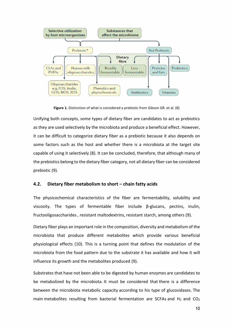
Related to the location, each receptor is located on the membranes of certain cell types
throughout the body. However, although there are some differences between studies,
they do agree on the fact that the three receptors are located in the colon, among other
cells (2,29,30).
Receptor Location Bibliography
FFAR2
Colon, immune cells, heart,
adipose tissue (AT) and skeletal
muscle
(2) Cryan JF, O’riordan KJ, Cowan CSM,
Sandhu K V., Bastiaanssen TFS, Boehme
M, et al. The microbiota-gut-brain axis.
Physiol Rev. 2019;99(4):1877-2013.
Intestinal leukocytes,
enteroendocrine cells, intestinal
epithelium and white AT.
(29) Sivaprakasam S, Prasad PD, Singh N.
Benefits of short-chain fatty acids and
their receptors in inflammation and
carcinogenesis. Pharmacol Ther.
2016;164:144-51.
Immune cells (neutrophils,
eosinophils, lymphocytes,
monocytes), pancreas (β cells),
intestinal cells (enteroendocrine L
cells and epithelial cells) and white
AT.
(30) Grundmann M, Bender E,
Schamberger J, Eitner F. Pharmacology
of free fatty acid receptors and their
allosteric modulators. Int J Mol Sci.
2021;22(4):1-38.

20
FFAR3
Colon, immune cells, heart,
peripheral NS and BBB
(2) Cryan JF, O’riordan KJ, Cowan CSM,
Sandhu K V., Bastiaanssen TFS, Boehme
M, et al. The microbiota-gut-brain axis.
Physiol Rev. 2019;99(4):1877-2013.
Enteric neurons, enteroendocrine
cells and pancreas
(29) Sivaprakasam S, Prasad PD, Singh N.
Benefits of short-chain fatty acids and
their receptors in inflammation and
carcinogenesis. Pharmacol Ther.
2016;164:144-51.
Peripheral NS, pancreas (β cells),
intestinal cells (L enteroendocrine
cells, K cells) and immune tissue
(dendritic cells and thymus)
(30) Grundmann M, Bender E,
Schamberger J, Eitner F. Pharmacology
of free fatty acid receptors and their
allosteric modulators. Int J Mol Sci.
2021;22(4):1-38.
Table 1. Location of FFAR2 and FFAR3 receptors throughout the body.
4.6.2. Effect of the short – chain fatty acids on the microbiota – gut – brain axis
SCFAs are GPCR ligands leading to many effects such as the promotion of energy and
glucose homeostasis due to their use as an energy source, the regulation of the immune
and inflammatory system responses, also hormones that control the feeling of satiety
and finally the regulation of the central and peripheral NS (16).
By means of the FFAR2, receptor they have a protective and homeostatic effect on the
colon as they are able to regulate the colonic regulatory T cell (Treg). As for the FFAR3
receptor, it has been shown to has the ability to promote beneficial metabolic effects
including the control of body weight and glucose through the gut – brain axis (25).
➔ GLP-1 and PYY peptides regulation
The main stimulus for GLP-1 and PYY secretion is food intake and digestion as an increase
in their plasma levels is observed just after and until hours after ingestion takes place
(31).

21
Since SCFAs are ligands of the endogenous FFAR2 and FFAR3 receptors of
enteroendocrine L cells, they are involved in the control of the secretion into the
bloodstream of anorexigenic intestinal peptides such as GLP-1 and PYY (3,13,15,28).
GLP-1 and PYY are involved in the regulation of appetite through both central and
peripheral pathways as they decrease the intestinal motility, regulate glucose
homeostasis and energy expenditure and, in addition, decrease appetite and food intake
(32). It should be noted that the effect of decreasing appetite and food intake by GLP-1
is through the autonomic NS and the brain (28).
In subjects supplemented with prebiotics, an increase in plasma of both peptides and
also the feeling of satiety could be observed. This could contribute to a change in
appetite and, therefore, its use as a treatment for obesity is being considered (16).
Also, it has been seen that the SCFAs regulate the production of PYY from EEC by
inhibiting the histone deacetylase (2,22). However, this effect is specific to certain
species such as humans, but not in mouse cells and, therefore, in this case, mouse is not
valid as a study model (22).
Emphasis should be made on the importance of the colon area where the SCFAs act,
since it has been seen that the infusion of acetate in the distal colon produce an increase
in circulating PYY levels unlike when it is made in the proximal colon (2).
One study showed that acetate plays a role in the central control of appetite by
regulating regions such as the hypothalamus. This effect occurs through activation of
acetyl-CoA carboxylase which leads to changes in the expression of GLP-1 and PYY.
However, it should be noted that this study was performed on a mouse model (13).
Also, there has been seen an increase of GLP-1 and PYY in the plasma due to an increase
in the propionate levels. In addition, an increase in propionate production has been seen
to lead to lower subjective appeal by individuals to high calorie foods and also low
energy intake during an ad libitum5 meal (13).
5 ad libitum: without restrictions.

22
➔ Effect on hormones: insulin, glucagon, leptin and ghrelin.
Another effect of the SCFAs is its impact on other hormones such as insulin, glucagon,
leptin and ghrelin (15). However, many more studies are still needed, especially in
humans, in order to obtain stronger evidence.
- Insulin and glucagon
The GLP-1 increases insulin secretion from the beta – pancreatic cell and decreases
glucagon secretion. This effect leads to a lower blood glucose levels as it results in lower
liver glucose production and an increased uptake by tissues (28).
In a study done on overweight adults was administered 10 grams of inulin – propionate
ester for 24 weeks. The results directed to a reduction of weight gain, intra – abdominal
AT distribution and intra – hepatocellular lipid content and also improved insulin
resistance compared to the control group who only consumed inulin (17,33).
- Leptin
Leptin is a peptide secreted by adipocytes and to a minor degree from gastric parietal
cells. This has several functions such as to decrease appetite, weight gain and adiposity.
Its receptors are found in different areas of the brain which are involved in controlling
appetite and also in the afferent nerves of the VN (34). Leptin provides the brain with
information about energy storage. By inhibiting orexigenic AgRP neurons and activation
of anorexigenic POMC neurons, leptin is able to decrease the intake and increase energy
expenditure (35).
Some studies mention that leptin is secreted due to the binding of SCFAs to the FFAR3
receptor which is in adipocytes resulting in a decreased appetite. Therefore, this effect
goes alongside GLP-1 and PYY (28).
In some in vitro studies, it has been shown that activation of FFAR3 by SCFAs on the AT
resulted in an increased leptin expression (15,35). In the same way, oral propionate
administration has been shown to increase circulating leptin levels. It should be noted,
that these studies are done in rodents (35,36).

23
Another study showed an increased leptin expression after administering an ex vivo
treatment on human AT with propionic acid6. Specifically, showed that the 1 and 3 mM
doses increased their expression by 65 and 100% respectively. On the contrary, the 10
mM concentration resulted in no significant changes (37,38).
In a study done on rodents it was seen that both acetate and propionate7 resulted in an
increase in leptin gene transcription. However, more in vivo studies are required in order
to study this relationship (39).
It should be noted that some of these studies refer to the FFAR3 receptor when the
articles detailing the location of these receptors do not indicate that this receptor is
located in AT. However, it cannot be ruled out and more studies should be done.
- Ghrelin
The ghrelin is secreted primarily in the stomach. This one is involved in regulating the
intake, body weight, adiposity, insulin secretion, glucose metabolism, stomach acid
secretion and stimulation of intestinal motility (34).
Among other receptors, ghrelin – secreting cells have on their surface the FFAR2
receptor which has SCFAs as a ligand. Their binding results in an inhibition of ghrelin
secretion (31).
Ghrelin response differs from the other gut hormones since it shows an opposite
fluctuation in plasma (32). Higher levels are obtained between meals and decrease post-
intake (31). In addition, compared to the others hormones, ghrelin has opposite effects:
stimulation of gastric emptying, hunger, glucagon secretion and inhibition of insulin
secretion and thermogenesis (32).
6 Concentrations of 0, 0.01, 0.1, 1, 3 and 10 mM (38) 7 860 μmol/L of acetate and 78 μmol/L of propionate (39)

24
Figure 4. Scheme about the opposite endocrine and metabolic effects of ghrelin and GLP-1 from
Engelstoft MS. et al. (31)
In addition, ghrelin crosses the BBB where is able to activate AgRP neurons and inhibit
POMC neurons leading to an overall appetite – boosting effect (37).
In regard to the control of ghrelin and GLP-1 secretion, GPCR ligands cause an inhibitory
and stimulatory effect respectively. This fact offers the research for possible strategies
in order to obtain both effects through the same mechanism (31).
➔ Effects on serotonin
Serotonin has several functions such as regulating satiety, anxiety, mood, stimulation of
peristalsis, secretion and vasodilation. In addition, there is also some evidence of its role
in relation to lipid and glucose metabolism (37).
On the hypothalamus, it is able to regulate food intake decreasing it by inhibiting AgRP
neurons and activating POMCs (37).
In regard to the SCFAs, it has been seen that are able to increase the synthesis and
secretion of serotonin (15,18). However, it should be noted that it is not able to cross
the BBB (37).
More than 90% of the serotonin we produce comes from the enterochromaffin cells of
the intestine where it regulates peristalsis, among other functions. From EEC human
treated with acetate and butyrate, it was observed that there was a concentration –

25
dependent increase in mRNA expression of the Tph1 gene. Tph1 is a limiting enzyme in
the serotonin synthesis. Then, is stored in granules before being released (35,40).
In addition, serotonin participates in fasting – induced adaptation as it is able to promote
lipolysis in AT and gluconeogenesis in liver. Thus, energy availability increases (35).
Also, propionate has been shown to be able to modulate serotonin secretion in the gut
and decrease its levels in the brain. This can lead to an excess of serotonin in plasma
that has been observed in children with ASD (41).
Peptides/ Hormones / NT
SCFAs effect Main physiological effects
GLP-1 ↑ - ↓ Intestinal motility
- Glucose homeostasis and energy
expenditure regulation
- ↓ Appetite and food intake
PYY ↑
Insulin ↑ (Indirect effect) - ↓ Glycemia
- Improving of insulin resistance Glucagon ↓ (Indirect effect)
Leptin ↑ - ↓ Appetite and food intake
- ↓ Weight gain and adiposity
- ↑ Energy expenditure
Ghrelin ↓ - ↓ Appetite
- Body weight, adiposity and glucose
metabolism regulation
- ↓ Stomach acid secretion, gastric
emptying and intestinal motility
- ↓ Glucagon secretion
- ↑ Insulin secretion
- ↑ Thermogenesis
Serotonin ↑ - ↑ Lipolysis in AT and gluconeogenesis
in liver → ↑ energy availability
- Regulation of satiety, anxiety, mood,
stimulation of peristalsis, secretion and
vasodilation
Table 2. Main effects of the SCFAs on peptides (GLP-1 and PYY), hormones (insulin, glucagon, leptin and
ghrelin) and NT (serotonin) and their resulting physiological effect

26
➔ Effects on the nervous system
Finally, one of the effects that SCFAs develop on the body is on the NS.
Butyrate has been shown to be able to increase the proportion of cholinergic neurons
by epigenetic mechanisms. Therefore, the effects on NS by SCFAs are not limited to
neuronal activation (15).
The FFAR3 receptor is also expressed in the ENS. When SCFAs cross the intestinal
epithelium and reach the portal vein, signaling through the FFAR3 receptors of the portal
nerves by propionate takes place. This fact leads to an increase in the activity of the
dorsal vagal complex that receives signals from the VN and the hypothalamus, which
participates in the control of appetite and metabolism (3,15).
In addition, through FFAR3 receptors located in sympathetic ganglionic neurons, SCFAs
can directly activate the sympathetic NS (42).
Also, SCFAs can cross the BBB and help maintain their integrity. SCFAs influence
neuronal signaling and NT production as they increase the expression of anorexigenic
peptides in the hypothalamus and regulate the levels of GABA, glutamate and
glutamine. Thus, they may have an impact on behavior (14,42).
Figure 5. SCFAs involvement in the regulation of appetite and metabolism from van de Wouw M. et al. (15).

27
4.7. Involvement of SCFAs in some neurological disorders
There is an increasing evidence about the relationship between microbiota and different
neurological disorders such as ASD, Parkinson, Alzheimer, and so on (4,17–21).
However, the evidence of the SCFAs in relation to these disorders is not so clear.
Some compounds produced by the microbiota such as LPS, bacterial lipoproteins,
flagellin, and CpG islands of unmethylated DNA are capable of stimulate cytokine
secretion by IS innate cells. These cytokines are able to cross the BBB and activate
microglia and certain neurons resulting in altered neuronal function that can affect
mood and behavior. Based on this fact, it is important to have in mind the role that SCFAs
play in the integrity of BBB through the production of the tight junction proteins that
limit the access of certain molecules through it (16).
GPCRs are also found in the CNS where they are involved in the regulation of
metabolism, inflammation, neurological disorders and other diseases (25). SCFAs can
cross the BBB and participate in neuronal signaling, NT production, and behavior (42).
Moreover, SCFAs are able to induce the Treg proliferation by histone modifications,
increasing acetylation and decreasing deacetylation at Foxp3+ promoter region (24,43).
Also, they have been shown to stimulate the production of retinoic acid in the intestine,
which inhibits Th17 cell differentiation and promotes the proliferation of Treg leading
to beneficial effects on neuroinflammation (12,24,30). Therefore, all these actions go in
the direction of controlling inflammation and improving the IS functioning.
Another important concept to consider is the intestinal permeability. Some products of
the microbiota have been shown to be involved in the maintenance of the intestinal
barrier. For example, SCFAs have been shown to act as trophic factors of the mucosa
and epithelial layer (3,42). Moreover, butyrate has effects on mucin production, anti –
inflammatory effects, and increase tight junction proteins leading to a better
maintenance of the intestinal barrier and reduced permeability (44).
Increased permeability can lead to translocation of the microbiota or its products such
as LPS so, IS is activated and pro – inflammatory cytokines are secreted. These, as
already mentioned, have an impact on the CNS and ENS (42).

28
Figure 6. Interaction between microbiota, intestinal permeability and CNS from Yarandi SS. et al. (42)
➔ Parkinson’s disease
The Parkinson's disease is a neurodegenerative disorder quite common among the
population. The main feature that accompanies this disorder is the loss of dopaminergic
neurons in substantia nigra and the accumulation of α – synuclein and Lewis bodies.
Recent evidence suggests that accumulation of α – synuclein starts in ENS where it is
associated to some digestive symptoms (2,24). Due to the implication of SCFAs in the
regulation of IS, certain strategies can be contemplated as an effective treatment of the
disease (24).
In these patients, a lower amount of Prevotellaceae has been found which is a genus
producer of SCFAs (43). In fact, decreased levels of SCFAs have been found in the feces
of Parkinson’s patients compared with controls (19,43).
➔ Autism spectrum disorder
As for ASD, it is a neurodevelopmental disorder that has an important genetic basis (13).
Given the neuroactive properties of SCFAs we should not stop studying the relationship
with this disorder (2).
It is also important to note that in children with ASD a higher intestinal permeability has
been observed which leads to an increase in LPS. This leads to an inflammatory state as
a result of the secretion of pro – inflammatory cytokines such as IL-6 (42).
Several preclinical studies in rodents have shown how neurotoxic doses8 of propionic
acid induce a similar behavior in ASD (2,45,46).
8 Intracerebroventricular administration of 4 μl of a solution that contains 0.26 M of propionic acid (45,46)
29
Also, some studies show the relationship between certain levels of SCFAs and the
pathogenesis of ASD. In children with this disorder, high levels of acetic acid and
propionic acid have been found in both feces and serum, as well as increased production
by the microbiota. However, the exact mechanisms by which SCFAs are involved are not
known yet (41).
So, according to these latest studies, it seems that SCFAs play a negative role in this
disorder.
On the other hand, some studies have shown that children with ASD have significantly
higher levels of Clostridium and lower levels of healthy bacteria and metabolites such as
SCFAs compared to the control group (17). Therefore, because of these contradictory
studies, the SCFAs role on this disorder is still unclear (13).
➔ Multiple Sclerosis
Multiple sclerosis is an inflammatory disease where there is a demyelination of neuronal
axons. It has been seen an abnormal immune response to the secretion of pro-
inflammatory cytokines due to an increased activity of Th1 and Th17 cells, which can
lead to infiltration of immune cells in the CNS. In these patients, this fact has been
accompanied by lower activity of Treg worsening autoimmune reactions (24). Some
studies have shown the ability of SCFAs to decrease Th17 cells and increase Treg
proliferation (12,24,30).
Also, an experimental model of multiple sclerosis showed that the administration of
propionic acid improved the disease course because a lower inflammatory state of the
CNS resulted in less neuronal demyelination (43).
Although there is growing evidence that understanding of the microbiota – gut – brain
axis could lead to strategies for the prevention and treatment of certain brain disorders,
many other studies are still needed (25).
30
5. DISCUSSION
Current evidence appears to show a relationship between SCFAs and the microbiota –
gut – brain axis through their effect at different levels.
Considering diet as one of the factors that regulates the microbiome, and this one, at
the same time, conditions the production of SCFAs, it is necessary to highlight the role
of the pattern of eating habits in everything that surrounds the microbiota – gut – brain
axis.
Following high – fat and high – sugar diets usually leads to lower fiber intake, which can
lead to dysbiosis. At the same time, this results in a decreased synthesis of some
beneficial products by the microbiota such as SCFAs (3).
Also, the antibiotics and other drugs intake are other factors that can lead to dysbiosis.
It should be noted that a state of dysbiosis can lead to negative consequences on
neurological and mental health status (27).
Although studies are still needed to correctly define the impact of SCFAs on the
microbiota – gut – brain axis, one of the effects that has been seen more clearly is the
GLP-1 increase. However, other effects like the one it has on leptin are not so clear and
more studies are needed.
The overall effect, consists in a decrease in appetite and food intake, regulation of
glucose homeostasis, energy expenditure and control of body weight.
In relation to GLP-1, currently, there are pharmacological therapies that mimic this
peptide and are indicated in people with diabetes and even one of them exclusively in
obesity (Saxenda®). Some of these active ingredients are: Dulaglutida (Trulicity®),
Exenatida (Bydureon®, Byetta®), Semaglutida (Ozempic®, Rybelsus®), Liraglutida
(Victoza®, Saxenda®) i Lixisenatida (Adlyxin®). Even though, Rybelsus ® and Adlyxin ® are
not marketed in Spain (47).
These are drugs that, despite their proven effectiveness, are not first – line treatments
in type 2 diabetes due to their higher cost and method of administration that, unlike the
oral route used in most antidiabetic drugs, these ones, except Rybelsus®, need to be
injected (48).

31
Although SCFAs effect is in the same direction as these drugs by increasing GLP-1, their
effect is currently not comparable. Among other reasons, because the production of
SCFAs from dietary fiber is highly variable depending on the person. Consumption of the
same type and amount of fiber by different subjects does not imply that it results in the
production of the same amount of SCFAs. That is because it depends, among other
things, on the type of microbiota the subject has. However, if the possibility of
consuming SCFAs directly is contemplated, more studies would be needed to analyze
the efficacy in relation to increasing GLP-1.
Another study area is their relationship with some neurological diseases like Parkinson,
ASD and multiple sclerosis. The involvement of the microbiota in these types of
disorders is becoming more important. However, more studies are needed to obtain
clear conclusions about SCFAs role.
Within this area, should be emphasized the importance of continuing with the study of
possible strategies for future treatments that may be beneficial for individuals suffering
from these types of neurological disorders.
On the other hand, it should be considered that one of the limitations of some of the
studies is that they are made in animals, mainly rodents. This fact represents an obstacle
due to the rodent’s diet differs from the human’s diet, which makes the comparison
between both microbiota problematic (4).
Most reviews show that more studies are still needed in order to define the SCFAs
impact on the axis. In relation to this, not all studies have identical results due to the
methodology used among other factors. However, they also emphasize the importance
of continuing to study this relationship.
6. CONCLUSION
After the research, it can be concluded that the SCFAs produced by the gut microbiota
have an involvement in microbiota – gut – brain axis. However, it should be emphasized
that while some of the effects are well – proven, others still require more study.

32
Also, it should be noted that the information collected was different depending on the
article consulted creating some contradictions. These, once again, leads to the
conclusion that more studies have to be performed.
Currently there is a deficient fiber intake in the population. In a study done in an adult
Spanish population between 18 and 64 years old, the fiber consumption was 12.5 ± 5.66
g / day on average. This consumption is below from the European Food Safety Authority
(EFSA) recommendation (25 g / day) (49,50).
Finally, considering the results of this research that show the potential beneficial effects
of the SCFAs, could be added one more reason why is important to increase the fiber
intake. However, as already mentioned, although most prebiotics belong to the fiber
category, it should be noted that not all fiber types act as prebiotics.

33
7. BIBLIOGRAPHY
1. Wang HX, Wang YP. Gut microbiota-brain axis. Chin Med J (Engl).
2016;129(19):2373-80.
2. Cryan JF, O’riordan KJ, Cowan CSM, Sandhu K V., Bastiaanssen TFS, Boehme M, et
al. The microbiota-gut-brain axis. Physiol Rev. 2019;99(4):1877-2013.
3. Klingbeil E, de La Serre CB. Microbiota modulation by eating patterns and diet
composition: Impact on food intake. Am J Physiol - Regul Integr Comp Physiol.
2018;315(6):R1254-60.
4. Ghaisas S, Maher J, Kanthasamy A. Gut microbiome in health and disease: Linking
the microbiome-gut-brain axis and environmental factors in the pathogenesis of
systemic and neurodegenerative diseases. Pharmacol Ther. 2016;158:52-62.
5. Korczak R, Slavin JL. Definitions, regulations, and new frontiers for dietary fiber
and whole grains. Nutr Rev. 2020;78(Suppl 1):6-12.
6. Dai FJ, Chau CF. Classification and regulatory perspectives of dietary fiber. J Food
Drug Anal. 2017;25(1):37-42.
7. EU (2011) Regulation (EU) No 1169/2011 of the European parliament and of the
Council on the provision of food information to consumers. Official Journal of the
European Union (2011) L 304 p. 18–63.
8. Gibson GR, Hutkins R, Sanders ME, Prescott SL, Reimer RA, Salminen SJ, et al.
Expert consensus document: The International Scientific Association for
Probiotics and Prebiotics (ISAPP) consensus statement on the definition and
scope of prebiotics. Nat Rev Gastroenterol Hepatol. 2017;14(8):491-502.
9. Holscher HD. Dietary fiber and prebiotics and the gastrointestinal microbiota. Gut
Microbes. 2017;8(2):172-84.
10. Han M, Wang C, Liu P, Li D, Li Y, Ma X. Dietary Fiber Gap and Host Gut Microbiota.
Protein Pept Lett. 2017;24(5):388-96.
11. Koh A, De Vadder F, Kovatcheva-Datchary P, Bäckhed F. From dietary fiber to host
physiology: Short-chain fatty acids as key bacterial metabolites. Cell.
2016;165(6):1332-45.
12. Bourassa MW, Alim I, Bultman SJ, Ratan RR. Butyrate, neuroepigenetics and the
gut microbiome: Can a high fiber diet improve brain health? Neurosci Lett.

34
2016;625:56-63.
13. Sandhu K V., Sherwin E, Schellekens H, Stanton C, Dinan TG, Cryan JF. Feeding the
microbiota-gut-brain axis: diet, microbiome, and neuropsychiatry. Transl Res.
2017;179:223-44.
14. Gomez-Eguilaz M, Ramon-Trapero JL, Perez-Martinez L, Blanco JR. El eje
microbiota-intestino-cerebro y sus grandes proyecciones [The microbiota-gut-
brain axis and its great projections]. Rev Neurol. 2019;68(3):111-117. Spanish.
15. van de Wouw M, Schellekens H, Dinan TG, Cryan JF. Microbiota-gut-brain axis:
Modulator of host metabolism and appetite. J Nutr. 2017;147(5):727-45.
16. Mohajeri MH, Brummer RJM, Rastall RA, Weersma RK, Harmsen HJM, Faas M, et
al. The role of the microbiome for human health: from basic science to clinical
applications. Eur J Nutr. 2018;57(1):1-14.
17. Vernocchi P, Del Chierico F, Putignani L. Gut microbiota profiling: Metabolomics
based approach to unravel compounds affecting human health. Front Microbiol.
2016;7:1144.
18. Heiss CN, Olofsson LE. The role of the gut microbiota in development, function
and disorders of the central nervous system and the enteric nervous system. J
Neuroendocrinol. 2019;31(5):e12684.
19. Sharon G, Sampson TR, Geschwind DH, Mazmanian SK. The Central Nervous
System and the Gut Microbiome. Cell. 2016;167(4):915-32.
20. Fung TC, Olson CA, Hsiao EY. Interactions between the microbiota, immune and
nervous systems in health and disease. Nat Neurosci. 2017;20(2):145-55.
21. Quigley EMM. Microbiota-Brain-Gut Axis and Neurodegenerative Diseases. Curr
Neurol Neurosci Rep. 2017;17(12):94.
22. Lu VB, Gribble FM, Reimann F. Free fatty acid receptors in enteroendocrine cells.
Endocrinology. 2018;159(7):2826-35.
23. Yu CD, Xu QJ, Chang RB. Vagal sensory neurons and gut-brain signaling. Curr Opin
Neurobiol. 2020;62:133-40.
24. Ma Q, Xing C, Long W, Wang HY, Liu Q, Wang RF. Impact of microbiota on central
nervous system and neurological diseases: The gut-brain axis. J
Neuroinflammation. 2019;16(1):53.
25. Liu X, Cao S, Zhang X. Modulation of Gut Microbiota-Brain Axis by Probiotics,

35
Prebiotics, and Diet. J Agric Food Chem. 2015;63(36):7885-95.
26. Bienenstock J, Kunze W, Forsythe P. Microbiota and the gut-brain axis. Nutr Rev.
2015;73(Suppl 1):28-31.
27. Dinan TG, Cryan JF. The Microbiome-Gut-Brain Axis in Health and Disease.
Gastroenterol Clin North Am. 2017;46(1):77-89.
28. Kim YA, Keogh JB, Clifton PM. Probiotics, prebiotics, synbiotics and insulin
sensitivity. Nutr Res Rev. 2018;31(1):35-51.
29. Sivaprakasam S, Prasad PD, Singh N. Benefits of short-chain fatty acids and their
receptors in inflammation and carcinogenesis. Pharmacol Ther. 2016;164:144-51.
30. Grundmann M, Bender E, Schamberger J, Eitner F. Pharmacology of free fatty acid
receptors and their allosteric modulators. Int J Mol Sci. 2021;22(4):1763.
31. Engelstoft MS, Schwartz TW. Opposite Regulation of Ghrelin and Glucagon-like
Peptide-1 by Metabolite G-Protein-Coupled Receptors. Trends Endocrinol Metab.
2016;27(9):665-75.
32. Pekmez CT, Dragsted LO, Brahe LK. Gut microbiota alterations and dietary
modulation in childhood malnutrition – The role of short chain fatty acids. Clin
Nutr. 2019;38(2):615-30.
33. Chambers ES, Viardot A, Psichas A, Morrison DJ, Murphy KG, Zac-Varghese SEK,
et al. Effects of targeted delivery of propionate to the human colon on appetite
regulation, body weight maintenance and adiposity in overweight adults. Gut.
2015;64(11):1744-54.
34. Grabauskas G, Owyang C. Plasticity of vagal afferent signaling in the gut. Med.
2017;53(2):73-84.
35. Heiss CN, Olofsson LE. Gut Microbiota-Dependent Modulation of Energy
Metabolism. J Innate Immun. 2018;10(3):163-71.
36. Xiong Y, Miyamoto N, Shibata K, Valasek MA, Motoike T, Kedzierski RM, et al.
Short-chain fatty acids stimulate leptin production in adipocytes through the G
protein-coupled receptor GPR41. Proc Natl Acad Sci U S A. 2004;101(4):1045-50.
37. van Son J, Koekkoek LL, Fleur SEL, Serlie MJ, Nieuwdorp M. The role of the gut
microbiota in the gut–brain axis in obesity: Mechanisms and future implications.
Int J Mol Sci. 2021;22(6):2993.
38. Al-Lahham SH, Roelofsen H, Priebe M, Weening D, Dijkstra M, Hoek A, et al.

36
Regulation of adipokine production in human adipose tissue by propionic acid.
Eur J Clin Invest. 2010;40(5):401-7.
39. Hernández MAG, Canfora EE, Jocken JWE, Blaak EE. The short-chain fatty acid
acetate in body weight control and insulin sensitivity. Nutrients. 2019;11(8):1943.
40. Reigstad CS, Salmonson CE, Rainey JF, Szurszewski JH, Linden DR, Sonnenburg JL,
et al. Gut microbes promote colonic serotonin production through an effect of
short-chain fatty acids on enterochromaffin cells. FASEB J. 2015;29(4):1395-403.
41. Ristori MV, Quagliariello A, Reddel S, Ianiro G, Vicari S, Gasbarrini A, et al. Autism,
gastrointestinal symptoms and modulation of gut microbiota by nutritional
interventions. Nutrients. 2019;11(11):2812.
42. Yarandi SS, Peterson DA, Treisman GJ, Moran TH, Pasricha PJ. Modulatory effects
of gut microbiota on the central nervous system: How gut could play a role in
neuropsychiatric health and diseases. J Neurogastroenterol Motil.
2016;22(2):201-12.
43. Hirschberg S, Gisevius B, Duscha A, Haghikia A. Implications of diet and the gut
microbiome in neuroinflammatory and neurodegenerative diseases. Int J Mol Sci.
2019;20(12):3109.
44. Gubert C, Kong G, Renoir T, Hannan AJ. Exercise, diet and stress as modulators of
gut microbiota: Implications for neurodegenerative diseases. Neurobiol Dis.
2020;134:104621.
45. MacFabe DF, Cain NE, Boon F, Ossenkopp KP, Cain DP. Effects of the enteric
bacterial metabolic product propionic acid on object-directed behavior, social
behavior, cognition, and neuroinflammation in adolescent rats: Relevance to
autism spectrum disorder. Behav Brain Res. 2011;217(1):47-54.
46. Shultz SR, Aziz NAB, Yang L, Sun M, MacFabe DF, O’Brien TJ.
Intracerebroventricular injection of propionic acid, an enteric metabolite
implicated in autism, induces social abnormalities that do not differ between
seizure-prone (FAST) and seizure-resistant (SLOW) rats. Behav Brain Res.
2015;278:542-8.
47. AEMPS. CIMA: Centro de información de medicamentos [Internet]. 2021 [citat 16
maig 2021]. Disponible a: https://cima.aemps.es/cima/publico/home.html
48. Cheang JY, Moyle PM. Glucagon-Like Peptide-1 (GLP-1)-Based Therapeutics:

37
Current Status and Future Opportunities beyond Type 2 Diabetes.
ChemMedChem. 2018;13(7):662-71.
49. Fundación Española de Nutrición (FEN). Ingesta y funtes alimentarias de fibra en
España: diferencias en cuanto a la prevalencia de exceso de peso y obesidad
abdominal en adutos del estudio científico ANIBES. 2015;31.
50. Dietary Reference Values for nutrients Summary report. EFSA Support Publ.
2017;14(12).