Embed Size (px)
Citation preview

Roselli et al. Genes & Nutrition (2017) 12:25 DOI 10.1186/s12263-017-0583-1
RESEARCH Open Access
Impact of supplementation with afood-derived microbial community onobesity-associated inflammation andgut microbiota composition
Marianna Roselli, Chiara Devirgiliis* , Paola Zinno, Barbara Guantario, Alberto Finamore, Rita Ramiand Giuditta PerozziAbstract
Background: Obesity is a complex pathology associated with dysbiosis, metabolic alterations, and low-gradechronic inflammation promoted by immune cells, infiltrating and populating the adipose tissue. Probioticsupplementation was suggested to be capable of counteracting obesity-associated immune and microbialalterations, based on its proven immunomodulatory activity and positive effect on gut microbial balance.Traditional fermented foods represent a natural source of live microbes, including environmental strains withprobiotic features, which could transiently colonise the gut. The aim of our work was to evaluate the impact ofsupplementation with a complex foodborne bacterial consortium on obesity-associated inflammation and gutmicrobiota composition in a mouse model.
Methods: C57BL/6J mice fed a 45% high fat diet (HFD) for 90 days were supplemented with a mixture of foodbornelactic acid bacteria derived from the traditional fermented dairy product “Mozzarella di Bufala Campana” (MBC) or withthe commercial probiotic GG strain of Lactobacillus rhamnosus (LGG). Inflammation was assessed in epididymal whiteadipose tissue (WAT) following HFD. Faecal microbiota composition was studied by next-generation sequencing.
Results: Significant reduction of epididymal WAT weight was observed in MBC-treated, as compared to LGG andcontrol, animals. Serum metabolic profiling showed correspondingly reduced levels of triglycerides and higher levels ofHDL cholesterol, as well as a trend toward reduction of LDL-cholesterol levels. Analysis of the principal leucocytesubpopulations in epididymal WAT revealed increased regulatory T cells and CD4+ cells in MBC microbiota-supplemented mice, as well as decreased macrophage and CD8+ cell numbers, suggesting anti-inflammatory effects.These results were associated with lower levels of pro-inflammatory cytokines and chemokines in WAT explants. Faecalbacterial profiling demonstrated increased Firmicutes/Bacteroidetes ratio in all mice groups following HFD.
Conclusions: Taken together, these results indicate a protective effect of MBC microbiota supplementation towardHFD-induced fat accumulation and triglyceride and cholesterol levels, as well as inflammation, suggesting a strongereffect of a mixed microbial consortium vs single-strain probiotic supplementation. The immunomodulatory activityexerted by the MBC microbiota could be due to synergistic interactions within the microbial consortium, highlightingthe important role of dietary microbes with yet uncharacterised probiotic effect.
Keywords: Chronic inflammation, Fermented dairy, Foodborne microbiota, White adipose tissue, High fat diet
* Correspondence: [email protected] and Nutrition Research Centre, Council for Agricultural Research andEconomics (CREA), Via Ardeatina 546, 00178 Rome, Italy
© The Author(s). 2017 Open Access This article is distributed under the terms of the Creative Commons Attribution 4.0International License (http://creativecommons.org/licenses/by/4.0/), which permits unrestricted use, distribution, andreproduction in any medium, provided you give appropriate credit to the original author(s) and the source, provide a link tothe Creative Commons license, and indicate if changes were made. The Creative Commons Public Domain Dedication waiver(http://creativecommons.org/publicdomain/zero/1.0/) applies to the data made available in this article, unless otherwise stated.

Roselli et al. Genes & Nutrition (2017) 12:25 Page 2 of 12
BackgroundObesity is a chronic, multifactorial disorder reachingepidemic proportions globally, affecting persons of virtu-ally all ages in both developed and developing countries[1, 2]. Promoted by a combination of genetic predispos-ition, nutritional excess, and sedentary lifestyle, obesityis primarily characterised by increased fat mass, accom-panied by development of related disorders [3–5].Expansion of the adipose organ, mainly affecting whiteadipose tissue (WAT), results in adipocyte dysfunction.WAT has been increasingly considered not only a meta-bolic organ, but also an active endocrine tissue, as it se-cretes a large number of peptide hormones calledadipokines, such as leptin and adiponectin, that operatein a complex network and actively communicate withother organs [6, 7]. Secretion by the adipose organ isdisturbed in obesity, as adipokine release is dysregulatedand associated with production of several inflammationmediators. For this reason, the adipose tissue is consideredto be a major contributor to obesity-linked low-gradechronic inflammation [8]. The inflammatory processinvolves impairment of both the innate and adaptiveimmune system and is triggered by local secretion ofinflammatory cytokines and chemokines such as tumournecrosis factor-α (TNF-α), interleukin-6 (IL-6), monocytechemoattractant protein (MCP)-1, and Regulated on Acti-vation Normal T cell Expressed and Secreted (RANTES).These mediators recruit immune cells from blood vessels,such as lymphocytes and macrophages, which in turnmassively infiltrate the adipose tissue [9]. Indeed, highlevels of inflammatory cells such as T CD8+ lymphocytesand activated M1 macrophages are found in obese WAT,accompanied by decreased levels of CD4+CD25+Foxp3+
regulatory T (Treg) cells, a key population in main-taining immunological tolerance and immune homeosta-sis [10–12]. This inflammatory status, arising locally andthen becoming systemic, triggers the onset of otherdiseases frequently associated with obesity such as themetabolic syndrome, characterised by visceral obesity,high blood pressure, insulin resistance, high circula-ting triglyceride levels, and low HDL cholesterol,leading in turn to increased risk of cardiovascular dis-eases [13–16].The gut microbiota has recently attracted much
attention as a crucial factor associated with obesity[17]. Alterations of intestinal microbial composition,in terms of bacterial phyla and classes associated withimproved energy extraction from the undigested diet-ary carbohydrate component, were identified in obesehuman subjects and animal obesity models, withconsequent impact on host metabolism and energystorage [18]. Both diet- and genetically induced obes-ity were shown to associate with imbalance in therelative proportion of Gram-negative Bacteroidetes
and Gram-positive Firmicutes, the two major phyla ofgut bacteria, with the latter prevailing in obese sub-jects [19]. However, imbalance in these two bacterialphyla is not sufficient by itself to determine the obes-ity phenotype. Other factors, such as diet, pre- andprobiotic supplementation, antibiotics, surgery, andfaecal transplantation, can impact the overall meta-bolic capacity of the gut microbiome [20]. Within thiscontext, dietary interventions aimed at promoting se-lection of beneficial intestinal microbes could repre-sent a powerful strategy to counteract obesity-associated intestinal dysbiosis. There is growing evi-dence that probiotic and/or prebiotic supplementationcan positively modulate gut microbiota, thus repre-senting important assets in the management of obes-ity [21]. The probiotic component of the gutmicrobiota can confer health benefits to the hostmainly acting on immunomodulation and positivelyinfluencing intestinal microbial balance [22]. Probioticsupplementation was therefore suggested to be ableto counteract obesity-associated immune alterationsand microbial imbalance [23–25]. As an alternative tocommercially available probiotic strains, a naturalsource of live bacteria is represented by fermentedfoods, which also confer the advantage of providingthe host with a complex microbiota containing severalenvironmental strains with potential probiotic fea-tures, such as the capability to transiently coloniseanimal and human gut and interact with the residentgut microbiota, mainly at a trophic level [26].Increasing scientific interest in fermented foods wasalso recently boosted by their possible use as modelsfor more complex microbiota such as the gut [27].The most relevant foodborne lactic acid bacteria (LAB)belong to the Lactobacillus, Lactococcus, Streptococcus,Pediococcus, and Leuconostoc genera. Several LAB speciesare also highly represented within the resident gut micro-biota of healthy humans. Lactobacillus species, in particu-lar, are abundant both in food and in the gut [28].The aim of our work was to evaluate the impact of
supplementation with a complex foodborne bacterialcommunity on obesity-associated inflammation, as wellas on gut microbiota composition. For this purpose, weused a mouse model of high fat diet (HFD)-inducedobesity, comparing the effect of supplementation with amixture of natural LAB strains derived from the trad-itional fermented dairy product “Mozzarella di BufalaCampana” (MBC) [29] and with the well-characterisedprobiotic GG strain of Lactobacillus rhamnosus (LGG).The MBC bacterial consortium was dominated byLactobacillus delbrueckii, Lactobacillus fermentum, andLeuconostoc lactis [30]. LGG was used as probiotic con-trol on the basis of its proven beneficial effects in theprevention of obesity [31, 32].

Fig. 1 Experimental design. Six-week-old C57BL/6J male micewere randomly assigned to three experimental groups (four orfive animals per group). The mice were fed a standard diet andorally supplemented daily with MBC microbiota, LGG, or PBS(CTRL). After 15 days, the mice were shifted to HFD whilecontinuing bacterial or PBS supplementation for 90 additionaldays. At the end of the experimental period, blood andepididymal WAT were collected. Faeces were sampled for gutmicrobiota analysis at the indicated time points: t0, t15, andt105. The experiment was replicated once, and the number ofmice in each group for each of the two repetitions is indicated
Roselli et al. Genes & Nutrition (2017) 12:25 Page 3 of 12
MethodsExperimental design, animals, and dietsSix-week-old C57BL/6J male mice, obtained fromCharles River Laboratories (Como, Italy), were kept at23 °C with a 12-h light-dark cycle and fed ad libitumwith a standard laboratory diet (4RF21, Mucedola,Milano, Italy, www.mucedola.it). Mice had free access tofood and water throughout the experiments. Food intakeand body weight were recorded every other day. After1 week of adaptation, animals were randomly dividedinto three groups (five mice per group) and orallysupplemented for 15 days with 1 × 10⁹ CFU/day of amixture of natural LAB strains extracted from MBC [29]or with the probiotic strain LGG. Phosphate-buffered sa-line (PBS) supplementation was used as control (CTRL).After 15 days, all mice were shifted to HFD (http://www.envigo.com/resources/data-sheets/06415.pdf, 44.8%total calories from fat, designed with similarities toResearch Diets, Inc., formula D12451 and provided byMucedola) while continuing to receive bacterial supple-mentation for 90 additional days. Due to logistic reasonsrelated to the number of animals that could be handledat the same time, the experimental design envisionedtwo rounds of treatment, 2 weeks apart from each other,in which the two groups of mice, of the same age, werefed the same batches of diets. Therefore, the secondgroup of mice was not aimed at testing reproducibility,but rather at increasing the number of treated animals.Statistical analysis of the results included all animalssubjected to the same supplementation protocol, irre-spective of their treatment within experimental period 1or 2. At the end of the experimental period, mice wereanaesthetised by intraperitoneal injection of pentobar-bital (10 mg/kg) following overnight fasting, blood wasdrawn via cardiac puncture, and epididymal WAT wasexcised, weighed, and immediately placed in ice-coldPBS under sterile conditions. Serum was prepared fromblood and stored at − 80 °C until further analysis. Faeceswere collected and stored at − 80 °C for microbiologicalanalysis at the following time points: t0 (beginning ofbacterial treatments), t15 (shift to HFD) and t105(90 days on HFD). The experimental protocol and sam-pling times are summarised in Fig. 1.
Bacterial preparationsMBC is a traditional Italian fermented cheese with PDOdesignation (Product of Designated Origin, EEC Regula-tion no. 1107). It is consumed fresh, within 2 weeksfrom production, and it contains high titres of live bac-teria [29]. To prepare the MBC microbiota, 10 g ofcheese samples were diluted in 90 ml sodium citrate so-lution (2% w/v) and homogenised in a BagMixer400(Interscience, France), as previously described [30]. Tostandardise the bacterial inoculum to be administered to
mice, the MBC homogenate was entirely used as a singleinoculum in 2 l of De Man Rogosa Sharpe (MRS)medium (Oxoid Ltd., England) and incubated at 37 °Cfor 48 h under anaerobic conditions (Anaerocult A,Merck, Germany) to obtain a final bacterial titre ofabout 1.5 × 10⁹ CFU/ml. The resulting bacterial suspen-sion was divided in aliquots containing 1 × 10⁹ CFUeach, stored at − 80 °C in 20% (v/v) glycerol, and thaweddaily for oral administration to mice, following washing,resuspension in 1× PBS, and mixing with small amountsof minced feed.The LGG strain ATCC53103 was grown, prepared,
and orally given to mice as described above for MBCmicrobiota.
Serum metabolic measurementsThe following plasma parameters were analysed: glucose(Glucose Liquid kit, Sentinel Diagnostics, Milan, Italy),HDL and LDL cholesterol (Max Discovery HDL andLDL Cholesterol Assay Kits, Bioo Scientific, Austin, TX),and triglycerides (Triglycerides Liquid kit, SentinelDiagnostics). The adiponectin was quantified by ELISA(Biorbyt, Cambridge, UK). Analyses were conducted ona subset of five samples for each treatment, due to tech-nical issues related to serum withdrawal or hemolysis.
Immune cell isolation and stainingMacrophages and lymphocytes were isolated from theepididymal WAT stromal vascular fraction (SVF), ac-cording to [33], as several populations of immune cellsare well known to reside in the SVF. The following

Roselli et al. Genes & Nutrition (2017) 12:25 Page 4 of 12
monoclonal antibodies, purchased from eBioscience(San Diego, CA), were used in this study: FITC anti-CD3(clone 500A2), PE anti-CD8 (clone 53-6.7), PE-Cy5 anti-CD4 (clone RM4-5), FITC anti-CD11b (clone M1/70),PE anti-F4/80 (clone BM8), PerCP-Cy5.5 anti-CD45(clone 30-F11), and anti-CD16/CD32 (clone 93). Briefly,1 × 106 cells, resuspended in FACS labelling buffer (PBSwith 2 mM EDTA and 1% foetal calf serum), were pre-incubated for 20 min with anti-CD16/CD32 to avoidnon-specific binding, then washed and labelled with theappropriate mixture of antibodies for 30 min, centri-fuged, and resuspended in FACS labelling buffer. Flowcytometry analysis was performed using a FACSCaliburflow cytometer (BD Biosciences, Milan, Italy). To ex-clude dead/dying cells that could non-specifically bindantibodies, leukocytes were gated according to forwardand side scatter. The percentage of T helper and cyto-toxic cells was calculated on lymphocyte gate (CD3+),whereas the CD11b+ and F4/80+ cell subsets were calcu-lated on the leukocyte gate (CD45+). Treg cell (CD4+CD25+Foxp3+) analysis was performed with a specifickit (eBioscience, San Diego, CA) staining CD4 (FITC),CD25 (PE) and transcription factor Foxp3 (PE-Cy5), ac-cording to the manufacturer’s instructions. The percent-age of CD25+Foxp3+ cells was calculated on lymphocyteCD4+ gate. For all analyses, at least 10.000 events wereacquired and analysed using the CellQuest software (BDBiosciences, Milan, Italy).
Cytokine and chemokine secretion in WAT explantsWAT explant cultures were established essentially as de-scribed by [34]. Briefly, epididymal WAT was dissected,weighed, minced, and placed into 12-well tissue cultureplates (Corning, Milan, Italy) at 120 mg/well, with either1 ml T cell activation medium (complete DMEM con-taining 3.7 g/l NaHCO3, 10% heat-inactivated foetal calfserum, 4 mM glutamine, 1% non-essential amino acids,105 U/l penicillin and 100 mg/l streptomycin, 5 ng/mlphorbol 12-myristate 13-acetate (PMA), and 1 ng/mlionomycin) or control medium (complete DMEM with-out ionomycin and PMA). All reagents were fromEuroclone (Milan, Italy), except for ionomycin andPMA, which were from Sigma (Milan, Italy). Condi-tioned media were collected after 24 h of culture at37 °C in an atmosphere of 5% CO2/95% air at 90%relative humidity and stored at − 80 °C until furtheranalysis. The levels of cytokines and chemokines wereanalysed using Bio-plex/Luminex technology (mousemagnetic Luminex screening assay, Labospace, Milan) orELISA assays (Affymetrix, eBioscience, San Diego, CA).The following cytokines and chemokines were simultan-eously detected by Luminex technology in 50 μl undilutedsamples: interferon gamma-induced protein (IP)-10, gran-ulocyte macrophage-colony stimulating factor (GM-CSF),
Regulated on Activation-Normal T cell Expressed and Se-creted (RANTES), interleukin (IL)-23, IL-4, and IL-10.The following cytokines were analysed by ELISA (100 μlsamples): tumour necrosis factor (TNF)-α, interferon(IFN)-γ, IL-17A, and IL-6. For these latter two cytokines,samples were diluted 1:500, as the readings by Luminexassays for IL-17A and IL-6 were out of range.
DNA extraction from faecal samplesTotal DNA was extracted from 80 mg faecal sampleswith QIAamp DNA Stool Mini Kit (Qiagen, Hilden,Germany) according to manufacturer’s instructions.Qiagen DNA extraction method used in this work waschosen as it was listed among the most reproduciblekits, ensuring minimal influence on next-generationsequencing (NGS) data analysis [35].
NGS analysisNGS was performed on faecal DNA samples from fouranimals for each of the three experimental groups, at thethree time points indicated in Fig. 1, namely t0, t15, andt105 (total number of samples = 36). Partial 16S rRNAgene sequences were amplified using primer pair Pro-bio_Uni and /Probio_Rev, which targets the V3 region ofthe gene and sequenced at the DNA sequencing facilityof GenProbio srl (www.genprobio.com) using a MiSeq(Illumina). Primers and protocols, including ampliconchecks, were as described in [36]. Individual sequencereads were filtered with the Illumina software to removelow quality and polyclonal sequences. All Illuminaquality-approved, trimmed, and filtered data wereexported as .fastq files and processed using a customscript based on the QIIME software suite [37]. Qualitycontrol retained sequences 140–400 bp long, with meansequence quality score > 20, and truncation at first baseif a low quality rolling 10-bp window was found.Presence of homopolymers > 7 bp and sequences withmismatched primers were omitted. To calculatedownstream diversity (alpha and beta diversity indices,UniFrac analysis), 16S rRNA operational taxonomicunits (OTUs) were defined at ≥ 97% sequence homologyusing uclust [38]. All reads were classified to the lowestpossible taxonomic rank using QIIME and a referencedataset from the SILVA database [39]. Similarities be-tween samples were calculated by unweighted UniFrac[40]. The range of similarities is calculated between thevalues 0 and 1. Principal Coordinate Analysis (PCoA)was applied using the UniFrac program.
Statistical univariate analysisValues in graphs and tables represent means ± SD. Priorto analysis, normal distribution and homogeneity of vari-ance of all variables were assumed with Shapiro-Wilk’sand Levene’s tests, respectively. Statistical significance

Roselli et al. Genes & Nutrition (2017) 12:25 Page 5 of 12
was evaluated by one-way ANOVA or by ANCOVA,followed by post hoc Tukey honestly significant differ-ence (HSD) test. Differences with P values < 0.05 wereconsidered significant. Statistical univariate analysis wasperformed with the “Statistica” software package (version5.0; Stat Soft Inc., Tulsa, OK).
Statistical multivariate analysisNon-supervised principal component analysis (PCA) ofWAT immunological profiles (leukocyte subpopulationsand cytokine/chemokine secretion) was performed withPast software, version 2.17c [41]. Data were collected ina matrix of 27 rows (number of animals) and 15columns (number of variables) and were auto-scaled bymean-centring and normalised by standard deviation.Pearson’s correlation coefficients between variables andprincipal components, as well as statistical significanceof the correlation, were also calculated.
ResultsBacterial supplementation affects epididymal WAT weightand metabolic parametersBody and WAT weight values in the three groups ofmice are shown in Table 1 in comparison with food andenergy intakes. As expected, HFD feeding inducedsignificant weight increase in all groups, leading tocomparable body weight and weight gain values by theend of the experimental period. Nevertheless, significantreduction of WAT weight (P < 0.05) was observed inMBC-treated animals, as compared to LGG and CTRLmice. Food and energy intake were similar in the threemice groups. To account for a possible influence of foodintake on WAT weight, ANCOVA analysis was per-formed, considering WAT weight as the dependent vari-able, treatment as the independent variable, and foodintake as the covariate. The results confirmed that WATweight reduction in the MBC group as compared toLGG and CTRL could not be attributable to differentialfood intake. Supplementing with the foodborne MBCmicrobiota also led to reduced serum levels of
Table 1 Body weight, epididymal WAT weight, and food andenergy intake from HFD of MBC, LGG, or CTRL mice
MBC LGG CTRL
Body weight (g)
Initial 19.19 ± 1.44 19.41 ± 1.79 18.80 ± 0.89
Final 31.75 ± 1.97 31.88 ± 2.13 30.63 ± 3.35
Gain 11.61 ± 1.94 12.47 ± 2.55 12.36 ± 2.63
WAT weight (g) 1.35*# ± 0.31 1.78 ± 0.30 1.89 ± 0.49
Food intake (g/day) 2.40 ± 0.66 2.54 ± 0.62 2.48 ± 0.63
Energy intake fromHFD (Kcal/day)
10.80 ± 0.60 11.39 ± 0.69 11.26 ± 1.05
*P < 0.05 versus CTRL (ANOVA); #P < 0.01 versus CTRL and LGG (ANCOVA)
triglycerides, coupled with higher levels of HDL choles-terol (P < 0.05 and P < 0.001, respectively), and a trendtoward decreased LDL cholesterol (P = 0.05) as com-pared to the CTRL group (Table 2). Serum metabolicparameters of LGG-treated mice displayed a similar butmilder effect, with a trend toward reduced triglyceridelevels (P = 0.05) and increased HDL-cholesterol levels(P < 0.05). No significant differences were detectedamong the three groups of mice concerning fastingglucose and adiponectin levels.
WAT immunological profiles highlight the anti-inflammatory effect of MBC microbiota supplementationFlow cytometry analysis of the main leukocyte subpopula-tions in epididymal WAT (Fig. 2) revealed increasednumbers of the immune homeostasis regulator CD4+
CD25+ Foxp3+ Treg cells (Fig. 2a, P < 0.001 vs CTRL andP < 0.01 vs LGG) and CD4+ T lymphocytes (Fig. 2b,P < 0.001 vs CTRL) in MBC microbiota-supplementedmice, accompanied by decreased pro-inflammatory CD8+
T lymphocytes (Fig. 2b, P < 0.001 vs CTRL), CD11b+ acti-vated leukocytes and F4/80+ macrophages (Fig. 2c,P < 0.001 and P < 0.01 vs CTRL, respectively), suggestingthat MBC supplementation associates with an overall anti-inflammatory effect. LGG treatment also positively affectedWAT leukocyte subpopulations in terms of increasedpercentage of Treg (P < 0.05 vs CTRL) and CD4+ cells(P < 0.001 vs CTRL) and decreased CD8+ cells (P < 0.001vs CTRL) as well as activated leukocytes (P < 0.01 vsCTRL).Leukocyte profiling of MBC-treated animals was asso-
ciated in cultured WAT explants with decreased levelsof pro-inflammatory cytokines and chemokines, such asIL-6, TNF-α and IFN-γ (P < 0.001 vs CTRL and LGG),IL-17A (P < 0.001 vs LGG), IP-10 (P < 0.01 vs LGG andP < 0.05 vs CTRL), GM-CSF, and RANTES (P < 0.05 vsCTRL). Reduced levels were also observed in WATleukocytes of LGG-supplemented mice, but they relatedto a smaller subset of pro-inflammatory cytokines,namely IL-6 and IFN-γ (P < 0.001 vs CTRL), IL-17A,
Table 2 Serum metabolic measurements in MBC, LGG, or CTRLmice
MBC LGG CTRL
Glucose (mg/dl) 107.42 ± 47.74 72.97 ± 7.64 86.91 ± 11.43
Triglycerides(mg/dl)
147.12* ± 65.91 163.21# ± 69.23 316.98 ± 142.39
HDL cholesterol(mg/dl)
155.91** ± 16.13 137.29* ± 25.21 108.86 ± 12.91
LDL cholesterol(mg/dl)
92.75# ± 19.06 105.77 ± 48.03 132.83 ± 15.09
Adiponectin(μg/ml)
16.25 ± 3.39 17.46 ± 4.87 19.23 ± 5.53
*P < 0.05 versus CTRL; **P < 0.001 versus CTRL; #P = 0.05 versus CTRL

Fig. 2 Leukocyte subpopulations in epididymal WAT. The effect of bacterial supplementation on the frequency of WAT leukocyte subpopulationswas analysed by flow cytometry. The percentage of CD25+Foxp3+ Treg cells was calculated on T lymphocyte gate (CD4+, a), CD4+ and CD8+ cellsubsets were calculated on lymphocyte gate (CD3+, b), whereas CD11b+ and F4/80+ cells were calculated on leukocyte gate (CD45+, c). Blackcolumns: MBC-supplemented mice; grey columns: LGG-supplemented; white columns: CTRL. Each column represents the mean ± SD of ninemice. Means without a common letter significantly differ
Roselli et al. Genes & Nutrition (2017) 12:25 Page 6 of 12
and RANTES (P < 0.001 and P < 0.01 vs CTRL, respect-ively) (Fig. 3). No significant differences were observedamong mice groups for the two anti-inflammatory cyto-kines IL-4 and IL-10 nor for pro-inflammatory IL-23(data not shown).Considering the dynamic and inherently multivariate
nature of the immune response, WAT immunologicalprofiles were further explored by principal componentanalysis (PCA) (Table 3). The first three principal com-ponents accounted for 64.15% of the overall variance,with individual values of 33.81, 19.47, and 10.87% forPC1, PC2, and PC3, respectively. The most informativescore plot was the PC1/PC2 shown in Fig. 4, where PC1was responsible for clearly discriminating MBC samplesfrom LGG and CTRL samples. The variables mostlycontributing to such discrimination are identified byhigher loading values on PC1 (presented in italic charac-ters in Table 3), indicating significant correlationbetween PC1 and the specific variable. In particular:PC1 shows strong significant inverse correlation withthe pro-inflammatory markers CD3CD8+ (r = − 0.813),CD11b+ (r = − 0.727), F4/80+ (r = − 0.804), IL-6 (r = −0.669), TNF-α (r = − 0.660), and GM-CSF (r = − 0.544)and significant direct correlation with the anti-
inflammatory markers CD3CD4+ (r = 0.778) andCD4CD25+ (r = 0.819). However, a tendency of the LGGand CTRL samples to separate into two distinct clustersis also observed (Fig. 4). PC2, on the other hand,discriminates a subgroup of CTRL mice showing bothpro- and anti-inflammatory features. These features arehighlighted by the most discriminative variables: thepro-inflammatory cytokines IP-10 (r = 0.842) and IFN-γ(r = 0.587) and the anti-inflammatory markers IL-4(r = 0.733) and IL-10 (r = 0.763) (Table 3).
Impact of bacterial supplementation on gut microbiotaprofilesNext-generation sequencing (NGS) of 16S rDNA fromfaecal samples of treated or control mice was used to re-trieve information on the bacterial relative abundance attime points t0, t15, and t105. Taxonomical assignmentand read abundance estimates for all detected oper-ational taxonomic units (OTUs) are reported in Fig. 5 atthe phylum level, while the corresponding profiles at thespecies level are listed in Additional file 1: Table S1. Asexpected, Bacteroidetes and Firmicutes were detected aspredominant bacterial phyla, with different relative pro-portions related to the time points analysed (Fig. 5).

Fig. 3 Cytokine and chemokine secretion in epididymal WAT explants. WAT explants were cultured in complete DMEM for 24 h in the presenceof ionomycin (1 ng/ml) and PMA (5 ng/ml). Cytokine and chemokine levels were analysed by Luminex assay or by ELISA (see the “Methods” section).Each column represents the mean ± SD of nine mice. Means without a common letter significantly differ
Roselli et al. Genes & Nutrition (2017) 12:25 Page 7 of 12
Notably, all three experimental groups displayed statisti-cally significant increase in the Firmicutes/Bacteroidetesratio at the final time point as compared to the begin-ning of the HFD treatment (t105 vs t15: P < 0.001 forMBC and LGG; P < 0.05 for CTRL). These altered ratioswere also accompanied by decreased microbial biodiver-sity, measured by the Chao1 and Shannon indices (datanot shown). Differences in the overall composition ofthe faecal bacterial community were further analysedusing the UniFrac phylogeny-based metric [40]. PrincipalCoordinates Analysis (PCoA) confirmed clustering ofbacterial species according to sampling time. The firstthree principal components accounted for 41% of theoverall variance, with individual values of 23, 10, and 8%for PC1, PC2, and PC3, respectively. The most inform-ative score plot was the PC1/PC2, shown in Fig. 6. Aclear difference was observed between the initial (t0,t15) and final (t105) time points (Fig. 6a), while no dif-ference could be observed among the three experimentalconditions when samples were grouped according tosupplementation type (Fig. 6b). However, it is worth
noting that both L. delbrueckii and Leuc. lactis species,representing two major components of the MBCmicrobiota [29, 30], were detected exclusively in faecalsamples of MBC-supplemented mice, although at verylow abundance (Additional file 1: Table S1).
DiscussionIn this work, we investigated the effects of a complexfoodborne bacterial community (MBC microbiota) onobesity-associated inflammation and gut microbiotacomposition in a HFD-induced obese mouse model. Thecultivable LAB component of MBC microbiota, selectedby growth in MRS medium, was extracted from afermented unripened cheese especially rich in live titresof LAB species [29] dominated by L. fermentum, L. del-brueckii, and Leuc. lactis [30] whose strains have oftenbeen associated with probiotic features [42]. The ration-ale for supplementing mice with the microbial consor-tium was based on the highly biodiverse nature offoodborne strains in fermented dairies, including severalLAB strains of environmental origin with beneficial,

Table 3 PCA loadings relative to the first two principalcomponents from WAT immunological profiles of MBC,LGG, and CTRL mice
PC1 PC2
CD8+ 0.3609 −0.1459
CD4+ −0.3455 0.0893
CD4CD25+ −0.3635 0.0669
CD11b+ 0.3228 −0.1137
F4/80+ 0.3569 −0.0599
GM-CSF 0.2416 −0.1880
RANTES 0.1786 −0.1534
IL-23 0.1371 −0.1230
IP-10 0.1248 0.4924
IL-4 −0.0169 0.4287
IL-10 0.0720 0.4465
IL-6 0.2973 0.3429
IL-17A 0.1223 0.0648
IFN-γ 0.2593 0.3435
TNF-α 0.2932 −0.1015
Positive or negative values indicate a direct or inverse correlation betweenvariables and PCs, respectively. Loading values associated to significantPearson’s correlation coefficients (reported in the text) are indicated in italics
Roselli et al. Genes & Nutrition (2017) 12:25 Page 8 of 12
although yet uncharacterised, features [28]. Theircombined metabolic functions and metabolites havebeen suggested to exert positive effects on host physi-ology through synergistic mechanisms, more efficientlythan single strain supplementation [43]. However, theprobiotic capacity of mixed foodborne microbial consor-tia has been gaining consideration only recently [44–46].
Fig. 4 PCA plot from epididymal WAT immunological profiles.PC1/PC2 score plot showing the distribution of samples in reducedPC1/PC2 space. The percentage variation explained by the plottedprincipal components is indicated. Symbols refer to individual mice.Red crosses: MBC-supplemented mice; blue squares: LGG-supplemented; black dots: CTRL
Moreover, most published work report supplementationwith single bacterial strains, and only few studiescompared multi-strain probiotic mixtures to investigatepossible synergistic interactions [47]. We chose to run aparallel group of mice for comparison, supplementedwith the single probiotic strain GG of Lactobacillusrhamnosus that was shown to exert positive effects onobesity-related inflammation in mice and humans [31].The obese phenotype was induced in C57BL/6J mice
by feeding a 45% HFD for 3 months, resulting in weightgain in all experimental groups irrespective of bacterialsupplementation type. Many other studies reportdecreased body weight gain following probiotic supple-mentation [31, 48]. Although we detected constantweight gain in all mice groups, decreased epididymalWAT weight was evident following oral administrationof MBC microbiota as compared to the other micegroups, as well as a more pronounced anti-inflammatoryeffect than LGG supplementation. Decreased inflam-mation and amelioration of obesity-related metabolicand immunological dysfunctions were previously ob-served with bacterial supplementation of HFD-fedmice [49, 50], but they were not accompanied by WATweight reduction. WAT is considered the main contribu-tor to development of the obesity-associated low-gradechronic systemic inflammatory state, which is charac-terised by an imbalanced cytokine network with increasedproduction of several pro-inflammatory mediators.Epididymal WAT, like other intra-abdominal WAT de-pots, is now recognised to have a more negative impacton health than subcutaneous WAT [51], and its decreasedweight following MBC supplementation further highlightsa higher efficacy of this complex microbial community insupporting healthy metabolism. The specific anti-inflammatory effects observed in our study involved de-creased levels of the pro-inflammatory cytokines IL-6 andIFN-γ and of the chemokines IP-10 and RANTES in cul-tured WAT explants of LGG-supplemented mice, whileMBC-treated animals displayed stronger decrease in theexpression of a broader panel of pro-inflammatory cyto-kines and chemokines, namely IL-6, TNF-α, IL-17A, IFN-γ, IP-10, GM-CSF, and RANTES. Other studies using sin-gle probiotic strains or multi-strain mixtures observed de-creased expression of some of these markers [48, 50, 52].IL-6 and TNF-α are the main cytokines produced by pro-inflammatory macrophages in obese adipose tissue,whereas RANTES and IP-10 are important lymphocyteand macrophage chemo-attractants [9]. IFN-γ is secretedby infiltrating CD8+ T cells, thus contributing to thecritical events driving adipose tissue inflammation [53].Regarding IL-17, it was suggested that obesity predisposesto selective expansion of the Th17 subclass of T lympho-cytes, producing high levels of IL-17 in an IL-6-dependentprocess [54]. The cytokine GM-CSF, although not
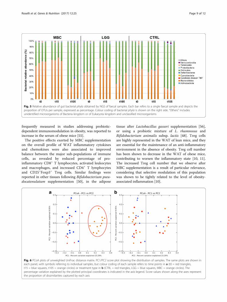
Fig. 5 Relative abundance of gut bacterial phyla obtained by NGS of faecal samples. Each bar refers to a single faecal sample and depicts theproportion of OTUs per sample, expressed as percentage. Colour coding of bacterial phyla is shown on the right side. “Others” includesunidentified microorganisms of Bacteria kingdom or of Eukaryota kingdom and unclassified microorganisms
Roselli et al. Genes & Nutrition (2017) 12:25 Page 9 of 12
frequently measured in studies addressing probiotic-dependent immunomodulation in obesity, was reported toincrease in the serum of obese mice [55].The positive effects exerted by MBC supplementation
on the overall profile of WAT inflammatory cytokinesand chemokines were also associated to improvedbalance between the major sub-populations of immunecells, as revealed by reduced percentage of pro-inflammatory CD8+ T lymphocytes, activated leukocytesand macrophages, and increased CD4+ T lymphocytesand CD25+Foxp3+ Treg cells. Similar findings werereported in other tissues following Bifidobacterium pseu-docatenulatum supplementation [50], in the adipose
Fig. 6 PCoA plots of unweighted UniFrac distance matrix. PC1/PC2 score peach panel, with symbols referring to individual samples, but colour codingt15 = blue squares, t105 = orange circles) or treatment type in b (CTRL = rpercentage variation explained by the plotted principal coordinates is indicthe proportion of dissimilarities captured by each axis
tissue after Lactobacillus gasseri supplementation [56],or using a probiotic mixture of L. rhamnosus andBifidobacterium animalis subsp. lactis [48]. Treg cellsare highly represented in the WAT of lean mice, and theyare essential for the maintenance of an anti-inflammatoryenvironment in the absence of obesity. Treg cell numberhas been shown to decrease in the WAT of obese mice,contributing to worsen the inflammatory state [10, 11].The increased Treg cell number that we observe afterMBC supplementation is a result of particular relevance,considering that selective modulation of this populationwas shown to be tightly related to the level of obesity-associated inflammation [10].
lot showing the distribution of samples. The same plots are shown inof each sample refers to time points in a (t0 = red triangles,
ed triangles, LGG = blue squares, MBC = orange circles). Theated in the axis legend. Score values shown along the axes represent

Roselli et al. Genes & Nutrition (2017) 12:25 Page 10 of 12
The anti-inflammatory effects occurring with MBCsupplementation were even more evident following PCAanalysis of the datasets, which clearly discriminated MBCsamples from LGG and CTRL samples along the firstprincipal component axis. This confirms the key role ofthe immune cell subpopulations, as well as of the cyto-kines GM-CSF, IL-6, and TNF-α, as the most importantvariables contributing to the discrimination. Separation ofthe LGG and CTRL samples into two distinct clusters washighlighted only as a trend. These effects were accompan-ied by positive changes in the expression of lipid metabol-ism biomarkers in the MBC-supplemented group, withdecreased circulating levels of triglycerides, increasedHDL-cholesterol levels, and a trend toward decreasingLDL cholesterol. Higher levels of circulating HDL choles-terol were also observed in the LGG mice group, in linewith previous reports on supplementation with single pro-biotics or mixtures [31, 48, 50].Interaction with the host metagenome is considered
an important aspect in probiotic-mediated immunestimulation [22, 57]. We analysed faecal microbiotabiodiversity in treated mice by NGS of 16S rDNA. Ourresults confirmed that gut microbiota composition wasindeed affected by HFD, leading to the establishmentof an increased Firmicutes/Bacteroidetes ratio typicalof the obesity pattern [58]. Bacterial supplementationwas not able to overcome HFD-induced effects on gutmicrobial profile, as no substantial modifications infaecal microbiota composition could be observed overtime by NGS. The overriding effect of HFD on micro-bial biodiversity was also confirmed by advancedmultivariate statistical analysis, namely Principal Coor-dinates Analysis (PCoA), revealing no specific cluster-ing of bacterial species according to supplementationtype, while highlighting a clear variation of microbialcomposition at the end of the experimental period inall mice groups. Other studies reported different ex-tent of alterations in resident gut microbiota profilefollowing probiotic treatment of HFD-fed mice [48, 50,59, 60], but the studies are not always comparable dueto different experimental designs (duration of treat-ment, percent dietary fat, etc.) and experimental ap-proaches employed for microbial profiling (i.e. NGS,qPCR). In our study, the high sensitivity of NGSallowed to detect two of the three predominant speciescharacterising the MBC-derived microbiota, namely L.delbrueckii and Leuc. lactis, although with low relativeabundance in the faecal microbiome of supplementedmice. These two species may thus be able to colonisethe gut of supplemented mice more efficiently. Gutcolonisation capacity of some components of MBC-derived microbiota was also shown in the simplemodel organism Caenorhabditis elegans [30]. On theother hand, the L. rhamnosus species that includes the
LGG strain was undetectable in faecal microbiomes ofLGG-treated mice. Conflicting results concerning LGGcolonisation capacity have been reported in theliterature. Park et al. recently observed decreasedLactobacillus relative abundances in the murine gut,including the LGG strain, following LAB supplementa-tion [59], while in another report of orally adminis-tered LGG to knockout (ApoE−/−) mice fed HFD, L.rhamnosus could be recovered by faecal dilution andplating [61]. Nevertheless, several reports indicate thatoral administration of specific bacteria can exert bene-ficial effects on the host even in the absence of colon-isation [59, 62–64].Taken together, our results suggest that supplementa-
tion with a biodiverse foodborne bacterial consortiumcan exert beneficial effects on obesity-associated inflam-mation and health-related parameters more effectivelythan single probiotic strain supplementation. A recentreport by Sonnenburg et al. clearly shows that dietaryperturbations can lead to permanent loss of specific gutbacterial taxa, due to negative selection of metabolic ac-tivities that become unnecessary under imbalanced diet-ary regimens [65]. These results point at limitations inmicrobiota resilience occurring under extreme condi-tions, such as HFD-induced obesity, where the alter-ations cannot be reversed by simple dietary interventionif not accompanied by specific bacterial supplementationaimed at restoring the lost taxa. Foodborne bacteriacould play a key role in this respect, and to the best ofour knowledge, this is among the very few reportsevaluating the impact of a complex microbial consor-tium naturally occurring in a traditional fermented foodon host physiology.
ConclusionsOur results demonstrate a stronger effect of a mixed mi-crobial consortium vs single-strain probiotic supplemen-tation in ameliorating HFD-induced inflammation in theWAT of obese mice. The present study highlights theimportance of considering complex foodborne microbialconsortia naturally occurring in fermented products forhuman consumption as potential probiotic vectors. Italso points at the importance of coupling multivariate tounivariate statistical analysis for better understanding ofthe key factors responsible for probiotic effects. Theobserved immunomodulatory activity exerted by theMBC-derived microbiota suggests synergistic interac-tions of microbial strains of environmental origin,present within the foodborne consortium. More studiesare needed to further investigate the role of dietary mi-crobes with yet uncharacterised probiotic effect, aimedalso at identifying novel, under-represented strainswhich could be unique to the foodborne microbiota.

Roselli et al. Genes & Nutrition (2017) 12:25 Page 11 of 12
Additional file
Additional file 1: Table S1. Complete results of the identification, atthe species level or, where not possible, at higher taxonomic ranks, of thesequences obtained by next-generation sequencing of faecal micesamples, expressed as percentage. Species representing the componentsof MBC microbiota are highlighted in bold. (XLSX 96 kb)
AbbreviationsCFU: Colony-forming units; CTRL: Control; GM-CSF: Granulocyte macrophage-colony stimulating factor; HFD: High fat diet; IFN: Interferon; IL: Interleukin;IP: Interferon gamma-induced protein; LAB: Lactic acid bacteria; LGG: L.rhamnosus GG; MBC: Mozzarella di Bufala Campana; MRS: De Man RogosaSharpe medium; NGS: Next-generation sequencing; OTUs: Operationaltaxonomic units; PCA: Principal component analysis; PCoA: PrincipalCoordinates Analysis; RANTES: Regulated on Activation-Normal T cellExpressed and Secreted; TNF: Tumour necrosis factor; Treg: Regulatory T cells;WAT: White adipose tissue
AcknowledgementsThe Authors wish to thank Kariklia Pascucci for her kind support in daily labwork, Dr. Andrea Ciolfi for his valuable assistance in managing the NGS data,and Dr. Fausta Natella and Dr. Gianni Pastore for their helpful suggestions onthe statistical analysis.
FundingThis work was funded in part by the Italian Ministry of Agriculture, Food &Forestry Policies (MiPAAF), with grant “MEDITO” (DM 12487/7303/11) andwith national support to the JPI-HDHL “ENPADASI” project.
Availability of data and materialsRaw NGS data are available at the EMBL-EBI European Nucleotide Archive(ENA) [66], under the study accession number PRJEB20801.
Authors’ contributionsMR, GP, and CD conceived and designed the experiments. MR, PZ, BG,AF, and CD performed the experiments. MR and CD analysed the dataand supervised all data analyses. GP contributed reagents/materials/analysis tools. MR, GP, and CD wrote the paper. RR did the animalexperiments/treatments. MR and AF performed the immunologicalanalysis. PZ, BG, and CD performed the microbiological analysis. CDanalysed the microbiota sequencing data. All authors read and approvedthe final manuscript.
Ethics approvalAll experimental procedures involving animals complied with the EuropeanGuidelines for the Care and Use of Animals for Research Purposes (Directive2010/63/EU), and protocols were approved by the Ethical Committee of theFood and Nutrition Research Center and by the National Health Ministry,General Direction of Animal Health and Veterinary Drugs (agreement no201/2015-PR).
Consent for publicationNot applicable.
Competing interestsThe authors declare that they have no competing interests.
Publisher’s NoteSpringer Nature remains neutral with regard to jurisdictional claims inpublished maps and institutional affiliations.
Received: 24 May 2017 Accepted: 13 August 2017
References1. World Health Organization. Obesity and overweight fact sheet N 311-
updated March 2011 2011, http://www.who.int/mediacentre/factsheets/fs311/en/index.html.
2. Ng M, Fleming T, Robinson M, Thomson B, Graetz N, Margono C, MullanyEC, Biryukov S, Abbafati C, Abera SF, et al. Global, regional, and nationalprevalence of overweight and obesity in children and adults during1980-2013: a systematic analysis for the Global Burden of Disease Study2013. Lancet. 2014;384:766–81.
3. Serra-Majem L, Bautista-Castano I. Etiology of obesity: two “key issues” andother emerging factors. Nutr Hosp. 2013;28(Suppl 5):32–43.
4. Crino M, Sacks G, Vandevijvere S, Swinburn B, Neal B. The influence onpopulation weight gain and obesity of the macronutrient composition andenergy density of the food supply. Curr Obes Rep. 2015;4:1–10.
5. Shabana HS. Obesity, more than a ‘cosmetic’ problem. Current knowledgeand future prospects of human obesity genetics. Biochem Genet. 2016;54:1–28.
6. Cao H. Adipocytokines in obesity and metabolic disease. J Endocrinol.2014;220:T47–59.
7. Khan M, Joseph F. Adipose tissue and adipokines: the association with andapplication of adipokines in obesity. Scientifica (Cairo). 2014;2014:328592.
8. Maury E, Brichard SM. Adipokine dysregulation, adipose tissue inflammationand metabolic syndrome. Mol Cell Endocrinol. 2010;314:1–16.
9. Huh JY, Park YJ, Ham M, Kim JB. Crosstalk between adipocytes and immunecells in adipose tissue inflammation and metabolic dysregulation in obesity.Mol Cells. 2014;37:365–71.
10. Feuerer M, Herrero L, Cipolletta D, Naaz A, Wong J, Nayer A, Lee J, GoldfineAB, Benoist C, Shoelson S, Mathis D. Lean, but not obese, fat is enriched fora unique population of regulatory T cells that affect metabolic parameters.Nat Med. 2009;15:930–9.
11. Kucharska AM, Pyrzak B, Demkow U. Regulatory T cells in obesity. Adv ExpMed Biol. 2015;866:35–40.
12. Gyllenhammer LE, Lam J, Alderete TL, Allayee H, Akbari O, Katkhouda N,Goran MI. Lower omental t-regulatory cell count is associated with higherfasting glucose and lower beta-cell function in adults with obesity. Obesity(Silver Spring). 2016;24:1274–82.
13. Alberti KG, Zimmet P, Shaw J. The metabolic syndrome—a new worldwidedefinition. Lancet. 2005;366:1059–62.
14. Schaffler A, Muller-Ladner U, Scholmerich J, Buchler C. Role of adipose tissueas an inflammatory organ in human diseases. Endocr Rev. 2006;27:449–67.
15. Sam S, Haffner S, Davidson MH, D’Agostino RB Sr, Feinstein S, Kondos G,Perez A, Mazzone T. Relation of abdominal fat depots to systemic markersof inflammation in type 2 diabetes. Diabetes Care. 2009;32:932–7.
16. Rocha VZ, Folco EJ. Inflammatory concepts of obesity. Int J Inflam. 2011;2011:529061.
17. Bell DS. Changes seen in gut bacteria content and distribution with obesity:causation or association? Postgrad Med. 2015;127:863–8.
18. Cani PD, Osto M, Geurts L, Everard A. Involvement of gut microbiota in thedevelopment of low-grade inflammation and type 2 diabetes associatedwith obesity. Gut Microbes. 2012;3:279–88.
19. Ley RE, Backhed F, Turnbaugh P, Lozupone CA, Knight RD, Gordon JI.Obesity alters gut microbial ecology. Proc Natl Acad Sci U S A.2005;102:11070–5.
20. John GK, Mullin GE. The gut microbiome and obesity. Curr Oncol Rep.2016;18:45.
21. Dahiya DK, Renuka PM, Shandilya UK, Dhewa T, Kumar N, Kumar S, PuniyaAK, Shukla P. Gut microbiota modulation and its relationship with obesityusing prebiotic fibers and probiotics: a review. Front Microbiol. 2017;8:563.
22. Sanchez B, Delgado S, Blanco-Miguez A, Lourenco A, Gueimonde M,Margolles A. Probiotics, gut microbiota, and their influence on host healthand disease. Mol Nutr Food Res. 2017;61
23. Gauffin Cano P, Santacruz A, Moya A, Sanz Y. Bacteroides uniformis CECT7771 ameliorates metabolic and immunological dysfunction in mice withhigh-fat-diet induced obesity. PLoS One. 2012;7:e41079.
24. Ji YS, Kim HN, Park HJ, Lee JE, Yeo SY, Yang JS, Park SY, Yoon HS, Cho GS,Franz CM, et al. Modulation of the murine microbiome with a concomitantanti-obesity effect by Lactobacillus rhamnosus GG and Lactobacillus sakeiNR28. Benef Microbes. 2012;3:13–22.

Roselli et al. Genes & Nutrition (2017) 12:25 Page 12 of 12
25. Takemura N, Okubo T, Sonoyama K. Lactobacillus plantarum strain no. 14reduces adipocyte size in mice fed high-fat diet. Exp Biol Med (Maywood).2010;235:849–56.
26. Shiby VK, Mishra HN. Fermented milks and milk products as functionalfoods—a review. Crit Rev Food Sci Nutr. 2013;53:482–96.
27. Wolfe BE, Dutton RJ. Fermented foods as experimentally tractable microbialecosystems. Cell. 2015;161:49–55.
28. Tamang JP, Watanabe K, Holzapfel WH. Review: diversity of microorganismsin global fermented foods and beverages. Front Microbiol. 2016;7:377.
29. Devirgiliis C, Caravelli A, Coppola D, Barile S, Perozzi G. Antibiotic resistanceand microbial composition along the manufacturing process of Mozzarelladi Bufala Campana. Int J Food Microbiol. 2008;128:378–84.
30. Zanni E, Laudenzi C, Schifano E, Palleschi C, Perozzi G, Uccelletti D,Devirgiliis C. Impact of a complex food microbiota on energy metabolism inthe model organism Caenorhabditis elegans. Biomed Res Int.2015;2015:621709.
31. Kobyliak N, Conte C, Cammarota G, Haley AP, Styriak I, Gaspar L, Fusek J,Rodrigo L, Kruzliak P. Probiotics in prevention and treatment of obesity: acritical view. Nutr Metab (Lond). 2016;13:14.
32. Kim B, Park KY, Ji Y, Park S, Holzapfel W, Hyun CK. Protective effects ofLactobacillus rhamnosus GG against dyslipidemia in high-fat diet-inducedobese mice. Biochem Biophys Res Commun. 2016;473:530–6.
33. Orr JS, Kennedy AJ, Hasty AH. Isolation of adipose tissue immune cells.J Vis Exp. 2013:e50707.
34. Strissel KJ, DeFuria J, Shaul ME, Bennett G, Greenberg AS, Obin MS. T-cellrecruitment and Th1 polarization in adipose tissue during diet-inducedobesity in C57BL/6 mice. Obesity (Silver Spring). 2010;18:1918–25.
35. Wagner Mackenzie B, Waite DW, Taylor MW. Evaluating variation in humangut microbiota profiles due to DNA extraction method and inter-subjectdifferences. Front Microbiol. 2015;6:130.
36. Milani C, Hevia A, Foroni E, Duranti S, Turroni F, Lugli GA, Sanchez B, MartinR, Gueimonde M, van Sinderen D, et al. Assessing the fecal microbiota: anoptimized ion torrent 16S rRNA gene-based analysis protocol. PLoS One.2013;8:e68739.
37. Caporaso JG, Kuczynski J, Stombaugh J, Bittinger K, Bushman FD, CostelloEK, Fierer N, Pena AG, Goodrich JK, Gordon JI, et al. QIIME allows analysis ofhigh-throughput community sequencing data. Nat Methods. 2010;7:335–6.
38. Edgar RC. Search and clustering orders of magnitude faster than BLAST.Bioinformatics. 2010;26:2460–1.
39. Quast C, Pruesse E, Yilmaz P, Gerken J, Schweer T, Yarza P, Peplies J,Glockner FO. The SILVA ribosomal RNA gene database project: improveddata processing and web-based tools. Nucleic Acids Res. 2013;41:D590–6.
40. Lozupone C, Knight R. UniFrac: a new phylogenetic method for comparingmicrobial communities. Appl Environ Microbiol. 2005;71:8228–35.
41. Hammer Ø, Harper DAT, Ryan PD. PAST: paleontological statistics softwarepackage for education and data analysis. Palaeontol Electron. 2001;4:9pp.
42. Tamang JP, Shin DH, Jung SJ, Chae SW. Functional properties ofmicroorganisms in fermented foods. Front Microbiol. 2016;7:578.
43. Bordoni A, Danesi F, Dardevet D, Dupont D, Fernandez AS, Gille D, NunesDos Santos C, Pinto P, Re R, Remond D, et al. Dairy products andinflammation: a review of the clinical evidence. Crit Rev Food Sci Nutr.2017;57:2497–525.
44. Rosa DD, Dias MM, Grzeskowiak LM, Reis SA, Conceicao LL, Peluzio MD. Milkkefir: nutritional, microbiological and health benefits. Nutr Res Rev. 2017:1–15.
45. Kim DH, Kim H, Jeong D, Kang IB, Chon JW, Kim HS, Song KY, Seo KH. Kefiralleviates obesity and hepatic steatosis in high-fat diet-fed mice bymodulation of gut microbiota and mycobiota: targeted and untargetedcommunity analysis with correlation of biomarkers. J Nutr Biochem.2017;44:35–43.
46. Choi JW, Kang HW, Lim WC, Kim MK, Lee IY, Cho HY. Kefir prevented excessfat accumulation in diet-induced obese mice. Biosci Biotechnol Biochem.2017;81:958–65.
47. Foligne B, Parayre S, Cheddani R, Famelart MH, Madec MN, Ple C, Breton J,Dewulf J, Jan G, Deutsch SM. Immunomodulation properties of multi-species fermented milks. Food Microbiol. 2016;53:60–9.
48. Alard J, Lehrter V, Rhimi M, Mangin I, Peucelle V, Abraham AL, MariadassouM, Maguin E, Waligora-Dupriet AJ, Pot B, et al. Beneficial metabolic effectsof selected probiotics on diet-induced obesity and insulin resistance inmice are associated with improvement of dysbiotic gut microbiota. EnvironMicrobiol. 2016;18:1484–97.
49. Cano PG, Santacruz A, Trejo FM, Sanz Y. Bifidobacterium CECT 7765improves metabolic and immunological alterations associated with obesityin high-fat diet-fed mice. Obesity (Silver Spring). 2013;21:2310–21.
50. Moya-Perez A, Neef A, Sanz Y. Bifidobacterium pseudocatenulatum CECT7765 reduces obesity-associated inflammation by restoring the lymphocyte-macrophage balance and gut microbiota sstructure in high-fat diet-fedmice. PLoS One. 2015;10:e0126976.
51. Wajchenberg BL, Giannella-Neto D, da Silva ME, Santos RF. Depot-specifichormonal characteristics of subcutaneous and visceral adipose tissue andtheir relation to the metabolic syndrome. Horm Metab Res. 2002;34:616–21.
52. Novotny Nunez I, Maldonado Galdeano C, de Moreno de LeBlanc A,Perdigon G. Lactobacillus casei CRL 431 administration decreasesinflammatory cytokines in a diet-induced obese mouse model. Nutrition.2015;31:1000–7.
53. Nishimura S, Manabe I, Nagasaki M, Eto K, Yamashita H, Ohsugi M, Otsu M,Hara K, Ueki K, Sugiura S, et al. CD8+ effector T cells contribute tomacrophage recruitment and adipose tissue inflammation in obesity.Nat Med. 2009;15:914–20.
54. Ahmed M, Gaffen SL. IL-17 in obesity and adipogenesis. Cytokine GrowthFactor Rev. 2010;21:449–53.
55. Boi SK, Buchta CM, Pearson NA, Francis MB, Meyerholz DK, Grobe JL, NorianLA. Obesity alters immune and metabolic profiles. New insight from obese-resistant mice on high-fat diet. Obesity (Silver Spring). 2016;24:2140–9.
56. Ukibe K, Miyoshi M, Kadooka Y. Administration of Lactobacillus gasseriSBT2055 suppresses macrophage infiltration into adipose tissue in diet-induced obese mice. Br J Nutr. 2015;114:1180–7.
57. Le Barz M, Anhe FF, Varin TV, Desjardins Y, Levy E, Roy D, Urdaci MC,Marette A. Probiotics as complementary treatment for metabolic disorders.Diabetes Metab J. 2015;39:291–303.
58. Turnbaugh PJ, Backhed F, Fulton L, Gordon JI. Diet-induced obesity is linkedto marked but reversible alterations in the mouse distal gut microbiome.Cell Host Microbe. 2008;3:213–23.
59. Park S, Ji Y, Jung HY, Park H, Kang J, Choi SH, Shin H, Hyun CK, Kim KT,Holzapfel WH. Lactobacillus plantarum HAC01 regulates gut microbiota andadipose tissue accumulation in a diet-induced obesity murine model. ApplMicrobiol Biotechnol. 2017;101:1605–14.
60. Gauffin-Cano PG, Santacruz A, Trejo FM, Sanz Y. Bifidobacterium CECT 7765improves metabolic and immunological alterations associated with obesityin high-fat diet-fed mice. Obesity (Silver Spring). 2013;21:2310–21.
61. Chan YK, Brar MS, Kirjavainen PV, Chen Y, Peng J, Li D, Leung FC, El-NezamiH. High fat diet induced atherosclerosis is accompanied with low colonicbacterial diversity and altered abundances that correlates with plaque size,plasma A-FABP and cholesterol: a pilot study of high fat diet and itsintervention with Lactobacillus rhamnosus GG (LGG) or telmisartan inApoE−/− mice. BMC Microbiol. 2016;16:264.
62. Thomas CM, Versalovic J. Probiotics-host communication: modulation ofsignaling pathways in the intestine. Gut Microbes. 2010;1:148–63.
63. Hsiao EY, McBride SW, Hsien S, Sharon G, Hyde ER, McCue T, Codelli JA,Chow J, Reisman SE, Petrosino JF, et al. Microbiota modulate behavioral andphysiological abnormalities associated with neurodevelopmental disorders.Cell. 2013;155:1451–63.
64. Bjerg AT, Sorensen MB, Krych L, Hansen LH, Astrup A, Kristensen M, NielsenDS. The effect of Lactobacillus paracasei subsp. paracasei and L. casei W8(R)on blood levels of triacylglycerol is independent of colonisation. BenefMicrobes. 2015;6:263–9.
65. Sonnenburg ED, Smits SA, Tikhonov M, Higginbottom SK, Wingreen NS,Sonnenburg JL. Diet-induced extinctions in the gut microbiota compoundover generations. Nature. 2016;529:212–5.
66. Leinonen R, Akhtar R, Birney E, Bower L, Cerdeno-Tarraga A, Cheng Y,Cleland I, Faruque N, Goodgame N, Gibson R, et al. The EuropeanNucleotide Archive. Nucleic Acids Res. 2011;39:D28–31.