Embed Size (px)
Citation preview
Impact of Malaria Preexposure on Antiparasite Cellular and HumoralImmune Responses after Controlled Human Malaria Infection
Joshua M. Obiero,a* Seif Shekalaghe,b Cornelus C. Hermsen,a Maxmillian Mpina,b Else M. Bijker,a Meta Roestenberg,a*Karina Teelen,a Peter F. Billingsley,c B. Kim Lee Sim,c Eric R. James,c Claudia A. Daubenberger,d,e Stephen L. Hoffman,c
Salim Abdulla,b Robert W. Sauerwein,a Anja Scholzena
Radboud University Medical Center, Department of Medical Microbiology, Nijmegen, The Netherlandsa; Ifakara Health Institute, Bagamoyo Research and Training Center,Bagamoyo, Tanzaniab; Sanaria Inc., Rockville, Maryland, USAc; Swiss Tropical and Public Health Institute, Department of Medical Parasitology and Infection Biology, Basel,Switzerlandd; University of Basel, Basel, Switzerlande
To understand the effect of previous malaria exposure on antiparasite immune responses is important for developing successfulimmunization strategies. Controlled human malaria infections (CHMIs) using cryopreserved Plasmodium falciparum sporozo-ites provide a unique opportunity to study differences in acquisition or recall of antimalaria immune responses in individualsfrom different transmission settings and genetic backgrounds. In this study, we compared antiparasite humoral and cellular im-mune responses in two cohorts of malaria-naive Dutch volunteers and Tanzanians from an area of low malarial endemicity, whowere subjected to the identical CHMI protocol by intradermal injection of P. falciparum sporozoites. Samples from both trialswere analyzed in parallel in a single center to ensure direct comparability of immunological outcomes. Within the Tanzaniancohort, we distinguished one group with moderate levels of preexisting antibodies to asexual P. falciparum lysate and anotherthat, based on P. falciparum serology, resembled the malaria-naive Dutch cohort. Positive P. falciparum serology at baselinewas associated with a lower parasite density at first detection by quantitative PCR (qPCR) after CHMI than that for Tanzanianvolunteers with negative serology. Post-CHMI, both Tanzanian groups showed a stronger increase in anti-P. falciparum anti-body titers than Dutch volunteers, indicating similar levels of B-cell memory independent of serology. In contrast to the Dutch,Tanzanians failed to increase P. falciparum-specific in vitro recall gamma interferon (IFN-�) production after CHMI, and in-nate IFN-� responses were lower in P. falciparum lysate-seropositive individuals than in seronegative individuals. In conclu-sion, positive P. falciparum lysate serology can be used to identify individuals with better parasite control but weaker IFN-� re-sponses in circulating lymphocytes, which may help to stratify volunteers in future CHMI trials in areas where malaria is endemic.
In 2012, Plasmodium falciparum malaria caused an estimated 207million cases and 627,000 deaths, of which 90% occurred in
children under 5 years of age and in pregnant women in sub-Saharan Africa (1). Major control efforts have been implementedwith some success (2, 3), but malaria eradication will likely requirea safe and highly protective vaccine. Subunit vaccines have thus farshown moderate efficacy at best. RTS,S is the only vaccine candi-date in phase 3 trials but, despite averting substantial numbers ofmalaria cases (4), shows only 30 to 50% reduction in clinical dis-ease after 12 months depending on both age and malaria endemic-ity and even less after 18 months (5–7). These results stress theneed for more effective second-generation vaccines. Key require-ments are not only the identification of novel immunogens butalso a better understanding of protection-related immune re-sponses. This includes the effect of previous malaria exposure onimmune responses upon reexposure or vaccination (8, 9).
During the past 3 decades, controlled human malaria infection(CHMI) trials have become an indispensable tool not only in as-sessing the efficacy of candidate vaccines (10, 11) but also in eval-uating immune responses induced by exposure to the malaria par-asite (12–15). CHMI trials have so far been performed in countrieswere malaria is not endemic in previously unexposed individuals(11, 16–19). A logical next step is to study the potential differencesin the acquisition, maintenance, or recall of immune responses inindividuals from different transmission settings and genetic back-grounds (20, 21). The availability of aseptic, purified, cryopre-served, live P. falciparum sporozoites (PfSPZs; PfSPZ Challenge)(22) opens up opportunities to carry out CHMI trials in countries
where malaria is endemic, since it bypasses the need for infectinglocal Anopheles mosquitoes with P. falciparum or importing P.falciparum-infected mosquitoes to the trial site. The first PfSPZChallenge trial in malaria-naive Dutch volunteers demonstratedan infectivity rate of 83% after intradermal injections, indepen-dent of the dose given (23). Recently, PfSPZ Challenge was usedfor the first time during a CHMI trial in healthy adult male Tan-
Received 10 December 2014 Returned for modification 26 January 2015Accepted 9 March 2015
Accepted manuscript posted online 16 March 2015
Citation Obiero JM, Shekalaghe S, Hermsen CC, Mpina M, Bijker EM, RoestenbergM, Teelen K, Billingsley PF, Sim BKL, James ER, Daubenberger CA, Hoffman SL,Abdulla S, Sauerwein RW, Scholzen A. 2015. Impact of malaria preexposure onantiparasite cellular and humoral immune responses after controlled humanmalaria infection. Infect Immun 83:2185–2196. doi:10.1128/IAI.03069-14.
Editor: J. H. Adams
Address correspondence to Robert W. Sauerwein, [email protected],or Anja Scholzen, [email protected].
* Present address: Joshua M. Obiero, University of California Irvine, Department ofMedicine, Division of Infectious Diseases, Irvine, California, USA; MetaRoestenberg, Leiden University Medical Center, Department of InfectiousDiseases, Leiden, The Netherlands.
Supplemental material for this article may be found at http://dx.doi.org/10.1128/IAI.03069-14.
Copyright © 2015, American Society for Microbiology. All Rights Reserved.
doi:10.1128/IAI.03069-14
May 2015 Volume 83 Number 5 iai.asm.org 2185Infection and Immunity
on June 20, 2018 by guesthttp://iai.asm
.org/D
ownloaded from
zanian volunteers, resulting in similar infection rates (24). As afollow-up, we here present results of the malaria-specific humoraland cellular immune responses in Tanzanians and Dutch volun-teers who were inoculated intradermally with the same number oflive PfSPZs during these CHMI studies.
MATERIALS AND METHODSHuman ethics statement. The Dutch trial (23) was approved by the Cen-tral Committee for Research Involving Human Subjects of The Nether-lands (NL31858.091.10) and registered at Clinicaltrials.gov, identifierNCT 01086917. The Tanzanian trial (24) was approved by institutionalreview boards of the Ifakara Health Institute (IHI/IRB/No25), the Na-tional Institute for Medical Research Tanzania (NIMR/HQ/R.8a/Vol.IX/1217), the Ethikkommission beider Basel (EKBB), Basel, Switzerland(EKBB 319/11), and the Tanzanian Food and Drug Administration (ref-erence no. CE.57/180/04A/50) and registered at Clinicaltrials.gov, identi-fier NCT 01540903. All study teams complied with the Declaration ofHelsinki and good clinical practice, including monitoring of data, and allvolunteers gave written informed consent.
Clinical trial design. Samples for immunological analysis were ob-tained from two CHMI trials (23, 24).
The first trial, performed at Radboud University Medical Center, Ni-jmegen, The Netherlands, was composed of 18 healthy Dutch subjectsbetween the ages of 19 and 30 years with no history of malaria. Any vol-unteer who was positive for P. falciparum serology or had resided in anarea where malaria is endemic within the previous 6 months was excludedfrom the trial. Three groups (n � 6 per group) were infected by intrader-mal injections of 2,500, 10,000, or 25,000 cryopreserved PfSPZs (NF54strain). By day 21, 15/18 volunteers had developed parasites detectable bypositive blood thick smear (TS), 5/6 in each group (23). There were nodifferences in parasite densities at diagnosis between the three dose groups(23). For immunological analysis, nine P. falciparum-positive volunteersof the 10,000 (n � 4) and 25,000 (n � 5) PfSPZ dose groups were selectedbased on availability of plasma and peripheral blood mononuclear cells(PBMCs).
The second trial was carried out in Bagamoyo, Tanzania, with volun-teers residing in Dar es Salaam (an area where malaria is hypoendemic).Twenty-four males between 20 to 35 years of age were enrolled and con-firmed to be free of parasites by real-time quantitative PCR (qPCR). Sub-jects with a self-reported history of clinical malaria in the previous 5 yearswere excluded. The volunteers were divided into two groups with 12 vol-unteers per group and infected by intradermal injections of either 10,000or 25,000 PfSPZs (NF54 strain). A total of 21/24 became both qPCR andblood smear positive by day 21 after infection (24). The three P. falci-parum-negative volunteers were excluded from analysis in the presentstudy.
PBMCs, citrate anticoagulated plasma samples from Dutch volun-teers, and serum samples from Tanzanian volunteers were collected andcryopreserved 1 day before challenge (pre-CHMI) and after treatment(post-CHMI; day 35 and day 28 after infection).
DNA extraction and qPCR analysis. A total of 5 �l Zap-Oglobin IIlytic reagent (Beckman Coulter) was added to 500 �l of EDTA blood, afterwhich the samples were mixed and stored at �80°C.
DNA extraction and quantification of parasitemia by qPCR in theDutch CHMI trial were performed in Nijmegen as described previously(25), with slight modifications. Briefly, after thawing, samples were spikedwith murine white blood cells as an extraction control, and DNA wasextracted with a MagnaPure LC isolation station. For detection of theextraction control and P. falciparum, primers for the murine albumingene and P. falciparum 18R rRNA were used as described previously (25).Additionally, the P. falciparum 18R rRNA TaqMan MGB probe AAC AATTGG AGG GCA AG– 6-carboxyfluorescein (FAM) was used.
DNA extraction and qPCR in the Tanzanian trial were carried out atthe Leiden University Medical Center, Leiden, The Netherlands, as de-scribed previously (26). Phocine herpesvirus 1 (PhHV-1) was added to the
isolation lysis buffer to serve as an internal control. For quantification ofPhHV, the primers GGGCGAATCACAGATTGAATC and GCGGTTCCAAACGTACCAA and the probe Cy5-TTTTTATGTGTCCGCCACCATCTGGATC were used.
P. falciparum (NF54 strain) standard curves for both qPCR assays wereprepared in Nijmegen by titration of ring-stage-infected red blood cells(RBC) in uninfected human blood. The two qPCR assays in both siteswere confirmed to yield the same results when quantifying the P. falcipa-rum content in sequential samples from four CHMI volunteers.
Parasite material for immunological analysis. The P. falciparumNF54 strain used in both CHMI trials is the parental strain of the 3D7clone (27). P. falciparum (NF54 strain) blood-stage parasites were cul-tured in RPMI 1640 containing 10% human A� serum and a 5% hemat-ocrit erythrocyte suspension in a semiautomated culture system and reg-ularly screened for mycoplasma contamination. For in vitro stimulationassays, asynchronous parasites harvested at a parasitemia of approxi-mately 10 to 20% were purified by centrifugation on a 63% Percoll densitygradient to obtain mature asexual stages. This resulted in concentrationsof parasitemia levels of about 80 to 90%, consisting of more than 95%schizonts/mature trophozoites. P. falciparum-infected RBC (PfRBC) werewashed twice in RPMI, cryopreserved in glycerol-containing freeze me-dium, and used upon thawing in stimulation assays. Mock-cultured unin-fected erythrocytes (uRBC) were obtained similarly and served as the control.
P. falciparum lysate for enzyme-linked immunosorbent assay (ELISA)was prepared by extracting purified schizonts/mature trophozoites with1% sodium desoxycholate and 2.5 �l phenylmethanesulfonyl fluorideprotease inhibitor for 15 min at room temperature (RT).
Recombinant and synthetic proteins. Recombinant proteins of cir-cumsporozoite protein (CSP) and liver-stage antigen 1 (LSA-1) were usedto probe humoral responses toward preerythrocytic stages, while crude P.falciparum lysates were used to assess antibody reactivity toward bloodstages. Apical membrane protein 1 (AMA-1) and exported protein 1(EXP-1) are expressed in both preerythrocytic and asexual stages.
Full-length P. falciparum NF54 CSP with repeats was produced inEscherichia coli by Gennova Biopharmaceuticals Ltd., Pune, India. A re-combinant LSA-1 construct, LSA-NRC, was expressed in E. coli, incorpo-rating the N- and C-terminal regions of the protein and two of the cen-trally placed 17-amino-acid repeats for the 3D7 LSA-1 sequence(PlasmoDB-PF3D7_1036400) (28). Both the N- and C-terminal regionsas well as the repeats are highly conserved between NF54 and 3D7. Themajor difference is the greater number of repeats, which are the primarytarget of anti-LSA-1 antibodies (29), in the NF54 sequence than in the3D7 sequence (30). Amino acids 25 to 545 of codon-optimized AMA-1 ofthe P. falciparum FVO strain were expressed in the methylotrophic yeastPichia pastoris (31, 32). A peptide covering the C-terminal amino acids 73to 162 of the integral parasitophorous vacuolar membrane protein EXP-1(Swiss-Prot Database primary accession number P04926) was chemicallysynthesized using solid-phase 9-fluorenylmethoxy carbonyl (Fmoc)chemistry and differs from the 3D7 sequence only by a single amino acidin position 160 (33).
ELISA to assess antibody reactivity. Ninety-six-well Polystyrene flat-bottom plates (Nunc Maxisorp; Thermo Scientific) were coated with 2�g/ml of CSP, EXP-1, and AMA-1, 0.25 �g/ml of LSA-1, or P. falciparumlysate at the equivalent of 20,000 PfRBC/well in phosphate-buffered saline(PBS) and incubated overnight at 4°C. Plates were blocked with 5% milkin PBS. All of the following washing steps were carried out with PBS-0.05% Tween (PBST). Using 1% milk in PBST, plasma or serum sampleswere serially diluted in duplicate starting at 1:50 to 1:800 for protein an-tigen and 1:250 to 1:4,000 for P. falciparum lysate and incubated for 3 h atroom temperature. Bound IgG was detected using horseradish peroxidase(HRP)-conjugated anti-human IgG (Thermo Scientific; diluted 1:60,000 insample buffer). Plates were developed using tetramethylbenzidine (TMB)peroxidase substrate (tebu-bio). The reaction was stopped using an equalvolume of 0.2 M H2SO4, and absorbance was measured with a spectropho-tometer plate reader at 450 nm (Anthos 2001 ELISA plate reader).
Obiero et al.
2186 iai.asm.org May 2015 Volume 83 Number 5Infection and Immunity
on June 20, 2018 by guesthttp://iai.asm
.org/D
ownloaded from
A serial dilution of a pool of sera from 100 hyperimmune Tanzanian(HIT) (20) individuals living in an area where malaria is highly endemicwas used as a reference standard and was included on each plate. Thereactivity for each antigen in undiluted HIT serum was defined as 100arbitrary units (AUs). Optical density (OD) values were converted intoAUs by using the four-parameter logistic curve fit using the AuditableData Analysis and Management System for ELISA (ADAMSEL-v1.1; http://www.malariaresearch.eu/content/software).
For each antigen, all time points of an individual volunteer were as-sayed on the same plate. To determine whether Tanzanians had a positiveP. falciparum serology (by recognition of P. falciparum lysate), the mean(�2 standard deviations [SD]) baseline antibody titer against P. falcipa-rum lysate of the Dutch volunteers was used as the cutoff for positivity.
In vitro PBMC stimulation assay to assess cellular responses. Ve-nous whole blood was collected into citrated Vacutainer CPT cell prepa-ration tubes (Becton Dickinson). PBMCs were obtained by density gradi-ent centrifugation, washed three times in cold PBS, counted, frozen at 107
cells/ml in fetal calf serum (FCS) with 10% dimethyl sulfoxide, and storedin vapor-phase nitrogen. After being thawed, PBMCs were counted andcultured at a concentration of 500,000 cells/well in a 96-well round-bot-tom plate and stimulated in duplicate at a ratio of 1:2 with 106 P. falcipa-rum NF54-infected RBC or uRBC for either 24 h or 6 days in a totalvolume of 200 �l.
Flow cytometry. Cells were stained and analyzed by flow cytometryeither directly ex vivo or after 24 h or 6 days of in vitro stimulation. Cellswere stained first for viability with LIVE/DEAD fixable Aqua dead cellstain (Invitrogen) or fixable viability dye eFluor 780 (eBioscience) andlater with three different staining panels for surface markers: for the 24-hstain, CD3 PerCP (UCH1; BioLegend), CD56-phycoerythrin (PE)(HCD56; Biolegend), anti-T-cell receptor (TCR) Pan �/�-PE (IMMU510;Beckman Coulter), CD4 Pacific Blue (OKT4; Beckman Coulter), CD8allophycocyanin (APC)-H7 (SK1; BD Pharmingen), CD45RO energycoupled dye (ECD) (UCH1; Beckman Coulter), and CD62L PeCy7(DREG56; eBioscience); for the ex vivo stain, the only changes from theprevious stain were CD3 V500 (clone SP342; BD Horizon) and CD8PerCP (RPA-T8; BioLegend); and for the third-panel 6-day stain, CD3PeCy7 (OKT3; BioLegend) and CD8 PerCP (RPA-T8; BioLegend). Forintracellular staining, cells were incubated with different monoclonal an-tibodies (MAbs) depending on the staining panels. After 30 min of incu-bation at RT, cells were washed and permeabilized with Foxp3 fix/permbuffer (eBioscience) for 30 min on ice and stained in permeabilizationbuffer (eBioscience) with IFN-� fluorescein isothiocyanate (FITC)(4S.B3; eBioscience; 24-h stain) and Foxp3 eF660 (PCH101; eBioscience;ex vivo stain) or Ki67 FITC (B56; BD Pharmingen) and Foxp3 eF660(PCH101; eBioscience; 6-day stain). Cells were collected on a CyAn ADP9-color flow cytometer (Dako/Beckman Coulter) and analyzed usingFlowJo software (Tree Star, Inc.) version 9.6. All assays were conductedwith the same batches of PfRBC and uRBC, with all time points of onevolunteer assayed in one experiment to prevent interassay variations. Nat-ural killer T cells (NKT) and gamma delta T cells (��T) were analyzed inthe same gate and henceforth are referred to as NKT-��T cells.
Statistical analysis. Statistical analysis was performed in GraphPadPrism 5. Differences within the cohorts and between time points wereanalyzed per volunteer by a Wilcoxon matched-pairs signed-rank test andthose between groups were analyzed by a Mann-Whitney U test. Therelationship between baseline antibody titers and the increase in antibodytiters was analyzed by Spearman correlation, and P values of �0.05 wereconsidered statistically significant. Cellular responses were corrected forthe background by subtracting responses to uRBC from responses toPfRBC for each sample; resulting negative values were set to zero.
RESULTSTanzanian volunteers have higher baseline antibody titers thanDutch subjects. Pre-CHMI antibody titers were significantlyhigher in Tanzanian than in the malaria-naive Dutch volunteers
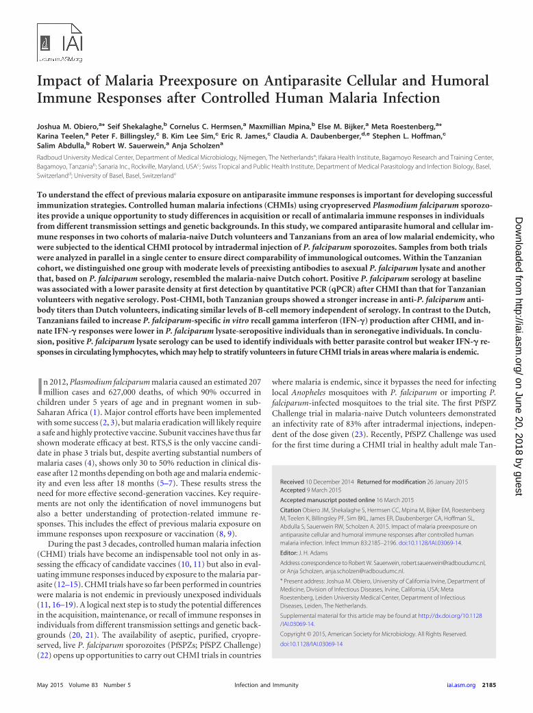
for crude P. falciparum lysate (P � 0.03), with 12/21 Tanzanianshaving titers higher than the mean (�2 SD) titers of Dutch vol-unteers. This was also true for pre-CHMI antibodies to the indi-vidual parasite antigens AMA-1 (P � 0.015; 13/21) and CSP (P �0.04; 15/21), and the same trend was found for EXP-1 (P � 0.06;8/21) (Fig. 1A). Elevated LSA-1 antibody titers were found in 5/21Tanzanians, but there was no significant difference between Dutchand Tanzanians at the group level (P � 0.16). Within the Tanzaniacohort, there was a wide range of pre-CHMI antibody responses, withsome volunteers showing only low responses, comparable to those ofthe Dutch cohort. To address whether there was a general divisioninto high and low responders to malaria antigens, we stratified Tan-zanian individuals based on their reactivity to P. falciparum lysate(containing a large number of late-liver- and blood-stage antigens)(Fig. 1B). Compared to their P. falciparum lysate-seronegative coun-terparts (n � 9), seropositive Tanzanians (n � 12) had significantlyhigher antibody titers against the cross-stage antigens AMA-1 (P �0.005; 7.6-fold higher median titer) and EXP-1 (P � 0.04; 5.4-foldhigher). The same trend was found for the sporozoite antigen CSP(P � 0.09; 2.4-fold higher) and the liver-stage antigen LSA-1 (P �0.06; 1.7-fold) (Fig. 1C).
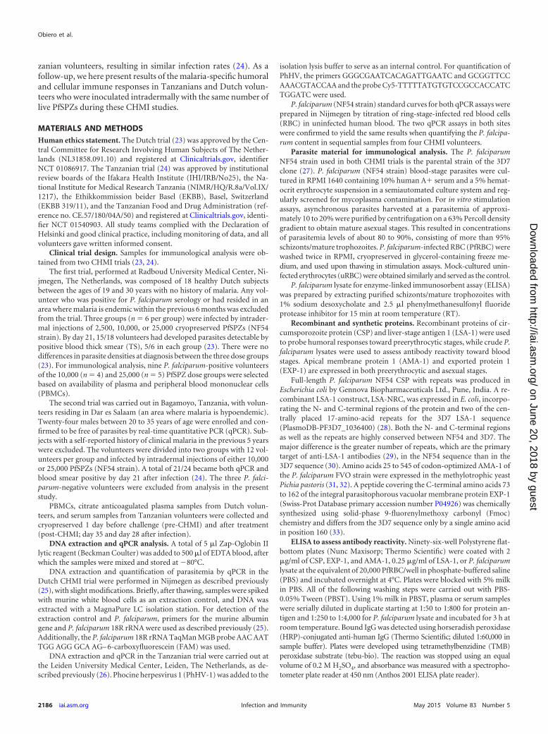
P. falciparum lysate seropositivity prior to CHMI is associ-ated with reduced initial blood-stage parasitemia. We next as-sessed whether preexisting humoral responses might be associatedwith the control of parasites in Tanzanian volunteers. In line withstronger humoral responses in the Tanzanian cohort than in theDutch cohort at baseline, Tanzanian volunteers became qPCRpositive significantly later than the Dutch volunteers (Fig. 2A),with a median prepatent period of 11.0 days (interquartile range[IQR], 11.0 to 13.5) in Tanzanians and 10.0 days (9.5 to 11.0) inDutch volunteers (P � 0.009). Similarly, prepatency by TS wasalso longer in Tanzanians (median [IQR], 13.7 days [12.75 to16.7]) than in the Dutch (12.6 days [12.3 to 14]) (P � 0.035). Thetime between detection by qPCR and by TS was comparable forboth cohorts (median [IQR] for Tanzanians, 2.6 days [2.2 to 3.15];Dutch, 3.0 days [2.0 to 3.15]; P � 0.88), but Dutch volunteers hada significantly higher peak parasite density (median number ofparasites/milliliter [IQR] for Tanzanians, 12,000 [6,800 to15,000]; Dutch, 74,000 [26,000 to 190,000]; P � 0.02), possiblydue to slight differences in the thick smear protocol between thetwo sites. Within the Tanzanian cohort, there was no significantdifference in time to qPCR-detectable parasitemia between volun-teers who were either P. falciparum lysate seropositive or serone-gative at baseline (P � 0.16) (Fig. 2B), nor was there a difference inprepatency by TS (P � 0.41) or time between detection by qPCRand TS (P � 0.15). However, seropositive Tanzanian volunteershad a significantly lower parasite load at the time of first qPCR-detectable parasitemia than their seronegative counterparts (P �0.033) (Fig. 2C). This difference remained evident, but becamesmaller, by the time the first peak in parasite load was reached (P �0.05; Fig. 2D). Across all Tanzanian volunteers, pre-CHMI anti-body titers for CSP (Pearson r � 0.45, P � 0.04), but no otherantigens, correlated significantly with prepatancy by qPCR (seeFig. S1 in the supplemental material).
P. falciparum-specific antibody responses are more effi-ciently increased in Tanzanians than in Dutch volunteers afterCHMI. Post-CHMI, antibody responses increased significantly inthe Tanzanian volunteers for P. falciparum lysate (P � 0.001;15/21 with a 3-fold increase in titers), AMA-1 (P � 0.002; 6/21),EXP-1 (P � 0.0001; 14/21), LSA-1 (P � 0.001; 5/21), and CSP
Immune Responses after Controlled Malaria Infection
May 2015 Volume 83 Number 5 iai.asm.org 2187Infection and Immunity
on June 20, 2018 by guesthttp://iai.asm
.org/D
ownloaded from
(P � 0.001; 11/21) (Fig. 3A). In contrast, the Dutch volunteersshowed significant increased titers only against EXP-1 (P � 0.004;7/9), CSP (P � 0.008; 6/9), and P. falciparum lysate (P � 0.008;3/9), while responses to LSA-1 and AMA-1 remained low andunaltered (Fig. 3B). Compared to the Dutch, Tanzanians showeda trend for stronger induction or boosting of responses based onthe overall fold increase in titers to P. falciparum lysate, AMA-1,and LSA-1 (Fig. 3C) and significantly higher absolute increases intiters for these antigens (Fig. 3D). Volunteers in both cohorts weresubjected to CHMI with either 10,000 or 25,000 PfSPZs. However,the only significant difference in antibody responses between thetwo dose groups was a slightly higher response in the 25,000 than
in the 10,000 dose group for P. falciparum lysate in Dutch volun-teers (P � 0.04) and a similar trend for the Tanzanians for CSP(P � 0.07) (see Fig. S2 in the supplemental material). Post-CHMI,P. falciparum lysate-seronegative Tanzanians had a significantlystronger fold increase in antibody titers against AMA-1 (P � 0.02)and EXP-1 (P � 0.04) than seropositive individuals, with a similartrend for P. falciparum lysate (P � 0.082) and CSP (P � 0.095),but no difference in LSA-1 responses (P � 0.80) (Fig. 3E). For theentire cohort, the fold increase of P. falciparum lysate (P � 0.01)and AMA-1 (P � 0.001) responses upon CHMI correlated nega-tively with the baseline response. The absolute increase in titers,however, was not different for any of the antigens between the two
FIG 1 Baseline malaria-specific antibody titers indicate previous exposure in Tanzanian volunteers. Antibody reactivity against crude P. falciparum lysate andthe P. falciparum antigens AMA-1, EXP-1, LSA-1, and CSP was tested prior to malaria infection. A pool of sera from 100 hyperimmune Tanzanians (HIT) wasused as a reference. Reactivity for each antigen in undiluted HIT serum was set at 100 arbitrary units (AUs). (A) Responses between Dutch (D; circles; n � 9) andTanzanian (T; squares; n � 21) cohorts were compared using a Mann-Whitney U test. (B) Tanzanian volunteers were stratified based on P. falciparum lysaterecognition at baseline as Sero� (n � 12; black box plots) or Sero� (n � 9; white box plots), using the mean � 2 SD of Dutch volunteers (gray circles) as a cutofffor positivity. (C) Pre-CHMI responses of seropositive and seronegative Tanzanians were analyzed by ELISA for individual P. falciparum antigens and comparedby Mann-Whitney U test. Scatter plots show individual data points, horizontal lines indicate the median of the group, and error bars indicate the interquartilerange (IQR). Dashed lines indicate the mean � 2 SD of antibody titers in Dutch volunteers for each respective antigen.
FIG 2 Preexposure to malaria is associated with difference in prepatency and parasitemia after CHMI. Parasitemia after CHMI was determined by qPCR. Theday of first parasite detection by qPCR after PfSPZ injection is shown for Dutch (D; gray circles; n � 9) compared to Tanzanians (T; gray squares; n � 21) (A)and for P. falciparum-seropositive (n � 12; black squares) and -seronegative (n � 9; white squares) Tanzanian volunteers (B). The parasite load (number of P.falciparum parasites per milliliter of blood) at the first day of qPCR-detectable parasitemia (C) and the time of first peak parasite density (D) are shown for P.falciparum-seropositive (n � 12; black squares) and -seronegative (n � 9; white squares) Tanzanian volunteers. Data are shown as median IQR. Groups werecompared by a Mann-Whitney U test.
Obiero et al.
2188 iai.asm.org May 2015 Volume 83 Number 5Infection and Immunity
on June 20, 2018 by guesthttp://iai.asm
.org/D
ownloaded from
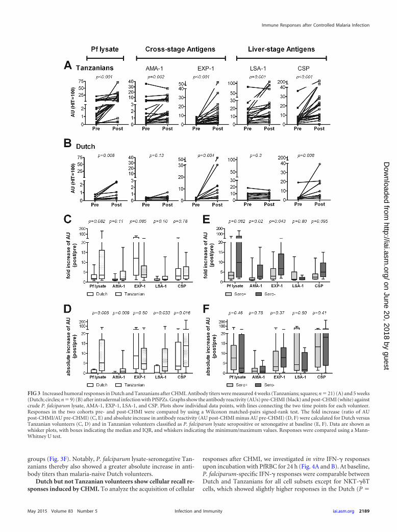
groups (Fig. 3F). Notably, P. falciparum lysate-seronegative Tan-zanians thereby also showed a greater absolute increase in anti-body titers than malaria-naive Dutch volunteers.
Dutch but not Tanzanian volunteers show cellular recall re-sponses induced by CHMI. To analyze the acquisition of cellular
responses after CHMI, we investigated in vitro IFN-� responsesupon incubation with PfRBC for 24 h (Fig. 4A and B). At baseline,P. falciparum-specific IFN-� responses were comparable betweenDutch and Tanzanians for all cell subsets except for NKT-��Tcells, which showed slightly higher responses in the Dutch (P �
FIG 3 Increased humoral responses in Dutch and Tanzanians after CHMI. Antibody titers were measured 4 weeks (Tanzanians; squares; n � 21) (A) and 5 weeks(Dutch; circles; n � 9) (B) after intradermal infection with PfSPZs. Graphs show the antibody reactivity (AUs) pre-CHMI (black) and post-CHMI (white) againstcrude P. falciparum lysate, AMA-1, EXP-1, LSA-1, and CSP. Plots show individual data points, with lines connecting the two time points for each volunteer.Responses in the two cohorts pre- and post-CHMI were compared by using a Wilcoxon matched-pairs signed-rank test. The fold increase (ratio of AUpost-CHMI/AU pre-CHMI) (C, E) and absolute increase in antibody reactivity (AU post-CHMI minus AU pre-CHMI) (D, F) were calculated for Dutch versusTanzanian volunteers (C, D) and in Tanzanian volunteers classified as P. falciparum lysate seropositive or seronegative at baseline (E, F). Data are shown aswhisker plots, with boxes indicating the median and IQR, and whiskers indicating the minimum/maximum values. Responses were compared using a Mann-Whitney U test.
Immune Responses after Controlled Malaria Infection
May 2015 Volume 83 Number 5 iai.asm.org 2189Infection and Immunity
on June 20, 2018 by guesthttp://iai.asm
.org/D
ownloaded from
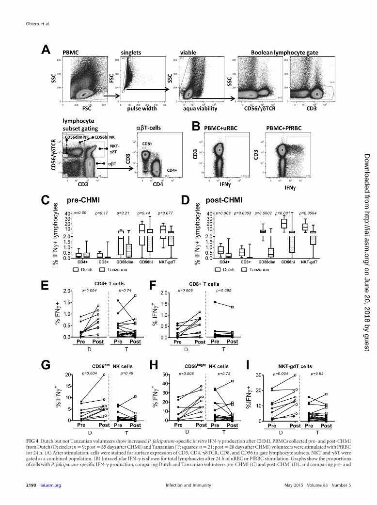
FIG 4 Dutch but not Tanzanian volunteers show increased P. falciparum-specific in vitro IFN-� production after CHMI. PBMCs collected pre- and post-CHMIfrom Dutch (D; circles; n � 9; post � 35 days after CHMI) and Tanzanian (T; squares; n � 21; post � 28 days after CHMI) volunteers were stimulated with PfRBCfor 24 h. (A) After stimulation, cells were stained for surface expression of CD3, CD4, ��TCR, CD8, and CD56 to gate lymphocyte subsets. NKT and ��T weregated as a combined population. (B) Intracellular IFN-� is shown for total lymphocytes after 24 h of uRBC or PfRBC stimulation. Graphs show the proportionsof cells with P. falciparum-specific IFN-� production, comparing Dutch and Tanzanian volunteers pre-CHMI (C) and post-CHMI (D), and comparing pre- and
Obiero et al.
2190 iai.asm.org May 2015 Volume 83 Number 5Infection and Immunity
on June 20, 2018 by guesthttp://iai.asm
.org/D
ownloaded from
0.077) (Fig. 4C). Post-CHMI, previously malaria-naive Dutchvolunteers showed significant increases in P. falciparum-specificIFN-� production by all lymphocyte subsets analyzed (CD4�, P �0.004; CD8�, P � 0.009; CD56dim, P � 0.004; CD56bright NK,P � 0.008; NKT-��T, P � 0.004) (Fig. 4E to I). In contrast, Tan-zanian volunteers showed little or no increase in IFN-�-produc-ing cells (CD4�, P � 0.74; CD8�, P � 0.065; CD56dim, P � 0.49;CD56bright NK, P � 0.75; NKT-��T, P � 0.92). In both cohortsand at both time points, CD56bright NK cells showed significantlyhigher IFN-� responses than CD56dim NK cells (Dutch pre/post-CHMI, P � 0.004; Tanzanian pre/post-CHMI, P � 0.0001). Sig-nificantly increased IFN-� responses were found in both the effec-tor (CD4�, P � 0.004; CD8�, P � 0.008) and central (CD4�, P �0.004; CD8�, P � 0.012) memory T-cell compartments in theDutch cohort, while the Tanzanians showed no such increase (seeFig. S3 in the supplemental material). As a result, post-CHMIIFN-� responses in the Dutch were significantly higher for all cellsubsets than in the Tanzanian cohort (Fig. 4D). Within the Tan-zanian cohort, there was an overall trend for higher P. falciparum-specific pre-CHMI IFN-� responses in those volunteers that hadparticularly short prepatency by qPCR (see Fig. S1 in the supple-mental material). CD8� T cells from Dutch (P � 0.04) but notTanzanian (P � 0.47) volunteers showed increased proliferativeresponses post-CHMI (see Fig. S4A in the supplemental material).The proliferation of CD4� T cells in response to PfRBC was notsignificantly altered post-CHMI in either cohort (P � 0.25 inDutch and P � 0.11 in Tanzanians) (see Fig. S4B).
Analyzing the lymphocyte subset compositions in both co-horts, we found that they were largely stable between pre- andpost-CHMI time points, with the exception of a prominent andsignificant post-CHMI increase of the NKT-��T proportion inthe Dutch volunteers and a clear decrease of CD56dim NK pro-portions in both cohorts. There was further no significant differ-ence in the proportions of CD4� CD45RO� Foxp3� T cells be-tween the two cohorts, which could have explained differences inresponsiveness (see Table S1 in the supplemental material). For
the Tanzanian cohort, we observed a significant reduction ofFoxp3� T cells post-CHMI (P � 0.008), while for the Dutch vol-unteers there was no change (P � 0.44). Stimulation with thephorbol myristate acetate (PMA) and ionomycin mitogensshowed that lymphocytes of both Dutch and Tanzanian volun-teers were fully functional and able to produce IFN-� at compa-rable levels both pre- and post-CHMI (see Fig. S4C).
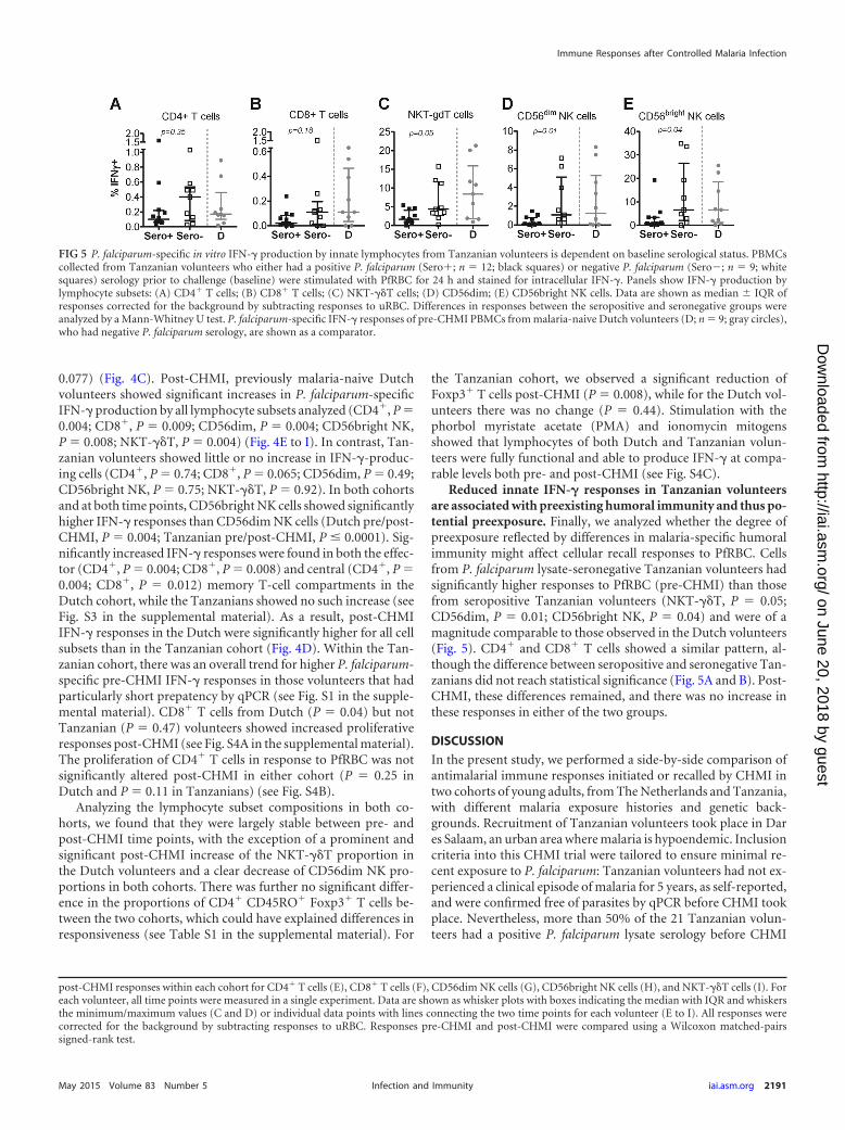
Reduced innate IFN-� responses in Tanzanian volunteersare associated with preexisting humoral immunity and thus po-tential preexposure. Finally, we analyzed whether the degree ofpreexposure reflected by differences in malaria-specific humoralimmunity might affect cellular recall responses to PfRBC. Cellsfrom P. falciparum lysate-seronegative Tanzanian volunteers hadsignificantly higher responses to PfRBC (pre-CHMI) than thosefrom seropositive Tanzanian volunteers (NKT-��T, P � 0.05;CD56dim, P � 0.01; CD56bright NK, P � 0.04) and were of amagnitude comparable to those observed in the Dutch volunteers(Fig. 5). CD4� and CD8� T cells showed a similar pattern, al-though the difference between seropositive and seronegative Tan-zanians did not reach statistical significance (Fig. 5A and B). Post-CHMI, these differences remained, and there was no increase inthese responses in either of the two groups.
DISCUSSION
In the present study, we performed a side-by-side comparison ofantimalarial immune responses initiated or recalled by CHMI intwo cohorts of young adults, from The Netherlands and Tanzania,with different malaria exposure histories and genetic back-grounds. Recruitment of Tanzanian volunteers took place in Dares Salaam, an urban area where malaria is hypoendemic. Inclusioncriteria into this CHMI trial were tailored to ensure minimal re-cent exposure to P. falciparum: Tanzanian volunteers had not ex-perienced a clinical episode of malaria for 5 years, as self-reported,and were confirmed free of parasites by qPCR before CHMI tookplace. Nevertheless, more than 50% of the 21 Tanzanian volun-teers had a positive P. falciparum lysate serology before CHMI
post-CHMI responses within each cohort for CD4� T cells (E), CD8� T cells (F), CD56dim NK cells (G), CD56bright NK cells (H), and NKT-��T cells (I). Foreach volunteer, all time points were measured in a single experiment. Data are shown as whisker plots with boxes indicating the median with IQR and whiskersthe minimum/maximum values (C and D) or individual data points with lines connecting the two time points for each volunteer (E to I). All responses werecorrected for the background by subtracting responses to uRBC. Responses pre-CHMI and post-CHMI were compared using a Wilcoxon matched-pairssigned-rank test.
FIG 5 P. falciparum-specific in vitro IFN-� production by innate lymphocytes from Tanzanian volunteers is dependent on baseline serological status. PBMCscollected from Tanzanian volunteers who either had a positive P. falciparum (Sero�; n � 12; black squares) or negative P. falciparum (Sero�; n � 9; whitesquares) serology prior to challenge (baseline) were stimulated with PfRBC for 24 h and stained for intracellular IFN-�. Panels show IFN-� production bylymphocyte subsets: (A) CD4� T cells; (B) CD8� T cells; (C) NKT-��T cells; (D) CD56dim; (E) CD56bright NK cells. Data are shown as median IQR ofresponses corrected for the background by subtracting responses to uRBC. Differences in responses between the seropositive and seronegative groups wereanalyzed by a Mann-Whitney U test. P. falciparum-specific IFN-� responses of pre-CHMI PBMCs from malaria-naive Dutch volunteers (D; n � 9; gray circles),who had negative P. falciparum serology, are shown as a comparator.
Immune Responses after Controlled Malaria Infection
May 2015 Volume 83 Number 5 iai.asm.org 2191Infection and Immunity
on June 20, 2018 by guesthttp://iai.asm
.org/D
ownloaded from
based on P. falciparum lysate ELISA, which is a standard exclusioncriterion in Dutch CHMI trials. In line with this, increased base-line antibody titers for the P. falciparum antigens CSP, LSA-1,EXP-1, and AMA-1 were found in the Tanzania cohort and par-ticularly in those with positive serology for P. falciparum lysate.This clearly indicates previous exposure to the malaria parasite inthis cohort. Asymptomatic infections, which can often occur inpeople living in areas with low malarial endemicity (34), in the 5years of no self-reported clinical malaria preceding CHMI mighthave led to a maintenance of higher antibody responses.
As might be expected, preexisting antibodies to P. falciparumantigens appeared to have an effect on the outcome of CHMI.Tanzanians had a significantly longer prepatency based on qPCRdetection than the previously malaria-naive Dutch. Furthermore,P. falciparum lysate-seropositive Tanzanians had a lower parasiteload at the time of first detection by qPCR and at the time of thefirst peak of parasite load than seronegative Tanzanians. The firstpeak of blood-stage parasitemia can be used as a proxy for parasiteliver load (16, 35). Our results might therefore indicate the emer-gence of fewer parasites from the liver and hence better control ofeither initiation or progression of liver-stage infection in preex-posed individuals within the Tanzanian cohort and in Tanzaniansthan in the Dutch cohort. In line with such an effect of preexistingantisporozoite immunity would be the observation that thoseTanzanian volunteers with a longer prepatancy by qPCR also hadhigher baseline antibody titers against the sporozoite antigen CSP.However, the first detection by qPCR in both the Dutch and Tan-zanians after intradermal PfSPZ injection was uncharacteristicallylate compared to that for infection by mosquito bite routinelyconducted in The Netherlands and elsewhere (11, 16, 18, 19, 23,24, 35). This is likely due to less efficient liver-stage infection bythis route and hence a low initial blood-stage load that reaches theqPCR detection limit only later. A lower first detectable para-sitemia in seropositive Tanzanians than in seronegative Tanzani-ans might therefore additionally be attributed to the control ofblood-stage replication prior to qPCR detection. Antibodies di-rected against AMA-1, for instance, are known to interfere withblood-stage multiplication (36) but can also confer protectionagainst liver-stage infection (37). Since recognition of both cross-stage and liver-stage antigens was stronger in the Tanzanian co-hort than in the Dutch, and particularly in the seropositive indi-viduals, both possibilities remain open. Our data, however,support antibody control of blood-stage replication only duringthe initial phase of sub-qPCR-detectable parasitemia: while para-site multiplication based on PCR data could not be directly com-pared due to the high variability of the amplification dynamics(24), there was no difference between prepatency by TS and qPCRin the two Tanzanian groups.
Upon CHMI, Dutch volunteers showed a slight but significantinduction of humoral responses against CSP, P. falciparum lysate,and EXP-1, while Tanzanians boosted responses to all antigensexamined. Reactivity to the CSP sporozoite antigen is expectedafter PfSPZ inoculation and consistent with previous findings (38,39). Increased antibody responses against P. falciparum lysate andEXP-1 likely reflect cross-stage reactivity between late-liver-stageand blood-stage merozoites, while exposure to developing liverstages (LSA-1) in naive volunteers appears insufficient to induce adetectable antibody response. Similarly, limited exposure toblood-stage-expressed AMA-1, due to early curative treatment,appears to prevent induction of detectable titers in naive volun-
teers, which is consistent with results even after multiple CHMIs(39). Consistent with preexposure, Tanzanians showed on thegroup level a greater increase in titers for P. falciparum lysate,AMA-1, and LSA-1 than the previously malaria-naive Dutch.Within the Tanzanian cohort, P. falciparum lysate-seronegativeTanzanians showed a similar absolute and accordingly muchgreater fold increase in antibody titers than their seropositivecounterparts. At baseline, these volunteers had significantly lowerresponses to most parasite antigens than their seropositive coun-terparts and largely resembled the malaria-naive Dutch cohort.The fact that this group showed a greater increase in antibodytiters than the Dutch strongly suggests that despite largely negativeP. falciparum serology, these individuals have a stable P. falci-parum-specific memory B-cell repertoire (40, 41). Given the sameincrease in absolute antibody titers, their memory B-cell reper-toire appears to be of a magnitude similar to that of the P. falcip-arum lysate-seropositive Tanzanians, despite lower circulatingplasma antibody levels.
Of note, humoral responses were assessed using a number ofrecombinant or synthetic proteins, which are not fully identical tothe sequences of these proteins expressed by the NF54 strain usedin both CHMI trials. While most antigens used in this study wereof the 3D7 sequence and thus closely resemble the NF54 sequence,AMA-1 responses were assessed using the FVO strain sequence.AMA-1 is known for its extensive antigenic polymorphism and tocause strain-specific immunity (42, 43). Nevertheless, there is asignificant antigenic overlap between AMA-1 alleles allowing forcross-reactivity across different strains, both in terms of func-tional activity and recognition, which affects mainly the magni-tude of the response (42, 44–46). Based on these data, we considerit unlikely that assessment of AMA-1 responses using the NF54 or3D7 sequence would have yielded different qualitative results.However, absolute titers would likely have been higher when us-ing the AMA-1 sequence homologous to the CHMI strain.
Another potential confounder is the fact that the P. falciparumstrains that Tanzanian volunteers were naturally exposed to priorto CHMI with P. falciparum NF54 are unknown. As any studyexamining preexposed individuals, analysis of antigen-specific re-sponses against polymorphic antigens has therefore the additionallimitation that it is difficult to match this unknown exposure his-tory. This has to especially be taken into account when investigat-ing CHMI-induced boosting of potential preexisting responses inthis cohort, which might be masked by using antigens of differentstrain origins. However, despite the fact that Tanzanians likelyexperienced exposure to a variety of P. falciparum strains prior toCHMI, there was (i) a clear division into seropositive and serone-gative individuals not just by total P. falciparum NF54 lysate butalso by all individual antigens analyzed and (ii) a clearly stron-ger increase in antibody titers in Tanzanians than in malaria-naive Dutch volunteers to several antigens, including AMA-1.We cannot exclude, however, that this might have even beenmore pronounced when using antigens of different strain ori-gins for analysis.
Although the Tanzanian volunteers in the present studyshowed evidence of humoral immune memory, parasite-specificIFN-� production by adaptive T-cell subsets was not higher thanin malaria-naive Dutch volunteers at baseline and remained un-changed after CHMI. In contrast, previously naive Dutch volun-teers showed a significant increase in IFN-� production by CD4and CD8 T-cell subsets after a primary infection, consistent with
Obiero et al.
2192 iai.asm.org May 2015 Volume 83 Number 5Infection and Immunity
on June 20, 2018 by guesthttp://iai.asm
.org/D
ownloaded from
what has been shown previously (47, 48). The lack of increasedproliferative and Th1 responses 1 month after CHMI in Tanza-nian volunteers could be partially due to immunosuppression fol-lowing exposure to blood-stage parasites during CHMI. Such T-cell immunosuppression is well described for malaria (49–58)and, although usually resolved within 2 weeks (52, 53), can persistfor more than 4 weeks (50). That Dutch volunteers are not equallyaffected by this might be due to the fact that such immunosup-pressive effects appear to be more pronounced in immune than innonimmune donors, as shown elsewhere (59). T regulatory cells(Tregs) are one potential mediator of suppressed IFN-� produc-tion by adaptive cells. Increased Treg numbers during or aftermalaria are a well-reported phenomenon (60), and malaria para-sites can enhance the suppressive activity of Tregs (61). WhileDutch and Tanzanian volunteers had similar Treg proportions,we cannot exclude that Tregs in Tanzanian volunteers might befunctionally more active, potentially due to past priming in ma-laria infections. That this apparent lack of a Th1 immune responsein the Tanzanian cohort is malaria specific is supported by the factDutch and Tanzanian volunteers showed similar Th1 responses tomitogen stimulation both pre- and post-CHMI. Nevertheless, anadditional influence of genetic background, which may explaindifferential P. falciparum-specific responses described in settingswhere malaria is endemic, cannot be excluded (62, 63).
NK cells are rapidly activated by malaria parasites, contributeto the early IFN-� response during blood-stage infection (64), andcan eliminate infected erythrocytes in vivo in a contact-dependentmanner (65). Importantly, there is a functional dichotomy: therarer CD56bright cells are more prominent in lymphatic tissuesand are superior cytokine producers, while the CD56dim subsetharbors a stronger cytotoxic potential (66–68). However, NK cellshave usually been examined as one population, and the exact con-tributions of CD56dim and CD56bright NK cell subsets to malariaimmunity thus remain to be established. An in vitro study onPBMCs from malaria-naive donors found greater IFN-� produc-tion by CD56bright than by CD56dim NK cells only in response tocytokines but not upon PfRBC stimulation (69). However, con-sistent with their generally reported greater cytotoxic potential,only CD56dim cells showed degranulation upon PfRBC stimula-tion (69). Our findings that CD56bright NK cells show a greaterIFN-� response than CD56dim NK cells to PfRBC both before andafter a primary malaria infection, as well as a memory-like effect ofthis response, are in line with findings from a previous CHMI trial(70). Tanzanian volunteers showed the same functional differencebetween the two NK cell subsets but no memory effect in eithersubset after CHMI. It was previously shown that depletion of �� Tcells abrogates memory-like IFN-� responses of innate cells oth-erwise observed after CHMI (47, 70). Therefore, the absence ofincreased PfRBC-specific �� T-cell responses post-CHMI in Tan-zania volunteers might be one reason why P. falciparum-specificIFN-� production by NK cells and other innate lymphocytes(NKT and �� T cells) was also not increased post-CHMI. The factthat P. falciparum lysate-seronegative Tanzanians had higher in-nate IFN-� responses than did seropositive Tanzanians already atbaseline is a further indication that reduced Th1 responses arelikely linked to the degree of previous malaria exposure. Of note,the proportion of CD56bright cells remained unaltered afterCHMI, while the CD56dim subset had a smaller contribution tothe PBMC compartment post-CHMI in both cohorts. It remainsto be established whether this means that, in contrast to memory-
like IFN-� production, other NK cell functions, such as migrationout of the circulation and engagement in antimalaria immuneresponses at other sites, such as the spleen, are unaffected and fullyfunctional in preexposed individuals.
Within the Tanzanian cohort, there was no association of pre-existing P. falciparum-specific IFN-� responses by T-cell subsetsor innate lymphocytes with the parasitological outcome of CHMI,i.e., prepatency or parasite load at first detection by qPCR. If at all,those with higher IFN-� responses to blood-stage PfRBC had ashorter prepatency. This does not, however, exclude that cellularresponses play a role in the prolonged prepatency of Tanzanianvolunteers and specifically those with positive P. falciparum serol-ogy. One possible reason for the lack of such an association is thatPfRBC were chosen as a stimulus for P. falciparum-specific re-sponses. This was done due to the relatively large antigenic overlapbetween blood-stage and (late) liver-stage parasites (71, 72). How-ever, responses to sporozoite and early-liver-stage antigens, whichmay be more relevant when assessing responses responsible forreducing the parasite load by targeting the earlier stages of infec-tion, are likely to be missed using PfRBC as a stimulus. Moreover,our analysis was restricted to P. falciparum-specific IFN-� produc-tion, and it is likely that other responses, for instance, degranula-tion as a proxy for cytotoxicity, may be more relevant readouts(71). Finally, the only accessible compartment for analysis of cel-lular responses in these human trials was peripheral blood. P. fal-ciparum-specific responses, and particularly those responsible forreducing liver infection, might, however, be enriched or primarilylocated in other sites, for instance, in tissue-resident memory cellsin the liver (73, 74). This might be even more pronounced inpreexposed individuals, where such a tissue-resident memorypopulation might have already been established.
Noteworthy, the differential immune responses describedherein may explain why Tanzanian volunteers reported fewerclinical symptoms, such as fever, than their Dutch counterpartsduring CHMI after PfSPZ injection (24). On the one hand, pre-existing antibody responses may reduce the initial parasite loadand mediate antidisease immunity. Additionally, the relativelylower P. falciparum-specific cellular Th1 responses observed in theTanzanian cohort than in the Dutch cohort might also be benefi-cial. Dutch volunteers preexposed to infected mosquito bites un-der chloroquine prophylaxis exhibited stronger IFN-� produc-tion and earlier clinical symptoms when reexposed to blood-stageparasites than malaria-naive volunteers during their first infection(15). Thus, a shift away from Th1 responses in preexposed volun-teers may make them less vulnerable to fever and other inflamma-tion-induced symptoms.
In conclusion, our data show that previous malaria exposure isassociated with some degree of parasite control during liver-stageor early-blood-stage infection after CHMI, humoral immunememory, and reduced antiparasite Th1 responses in the circulat-ing lymphocyte compartment. Positive P. falciparum lysate serol-ogy can be used to identify individuals with better parasite controlbut weaker peripheral blood Th1 responses, which may help tostratify volunteers in future CHMI trials in areas where malaria isendemic. However, assessment of memory B-cell responses mightbe explored as a potentially better tool than serology to definepreexposure per se. Important questions to be addressed in futurestudies include (i) which readouts other than Th1 responsesshould be used to determine immunization-induced cellular im-munity and (ii) how differences in preexisting malaria-specific
Immune Responses after Controlled Malaria Infection
May 2015 Volume 83 Number 5 iai.asm.org 2193Infection and Immunity
on June 20, 2018 by guesthttp://iai.asm
.org/D
ownloaded from
immune responses affect the outcome of whole parasite immuni-zation and vaccination approaches in areas where malaria is en-demic.
ACKNOWLEDGMENTS
We thank all the trial volunteers and the staff from the Radboud Univer-sity Medical Center, the Ifakara Health Institute/Bagamoyo Research andTraining Centre, Sanaria Inc., and the Swiss Tropical and Public HealthInstitute, all of whom made this study possible. We thank S. Singh (Gen-nova Biopharmaceuticals), D. Lanar, and G. Corradin for providing re-combinant and synthetic CSP, LSA-1, and EXP-1 proteins, respectively.We thank M. van de Vegte-Bolmer, R. Siebelink-Stoter, and W. Grau-mans for culture and isolation of PfRBC.
S.L.H. is chief executive and scientific officer at Sanaria Inc. B.K.L.S.,P.F.B., and E.R.J. are employees of Sanaria Inc., which manufactured andprovided PfSPZ Challenge, and thus do have a potential conflict of inter-est. There are no other conflicts of interest for all other authors.
This work was supported by Top Institute (TI) Pharma (grant numberT4-102), the FP7-founded European Virtual Institute of Malaria Research(EVIMalaR; grant agreement number 242095), the Tanzanian Commis-sion on Science and Technology (COSTECH), the Ifakara Health Insti-tute, and the Swiss Tropical Public Health Institute. A.S. received a long-term postdoctoral fellowship from the European Molecular BiologyOrganization (EMBO). The development and manufacturing of cryo-preserved P. falciparum sporozoites (PfSPZ Challenge) were furthersupported by Small Business Innovation Research (SBIR) (grantsR44AI058375-03, -04, -05, -05S1) from the National Institute of Allergyand Infectious Diseases at the National Institutes of Health (NIAID/NIH),USA, and through agreement number 07984 from the Program for Ap-propriate Technology in Health (PATH) Malaria Vaccine Initiative (withfunds from the Bill and Melinda Gates Foundation). The funders had norole in study design, data collection and analysis, decision to publish, orpreparation of the manuscript.
J.M.O., S.S., C.C.H., E.M.B., M.R, C.A.D., S.L.H., S.A., R.W.S., andA.S. designed the studies and experiments. S.S., E.M.B., and M.R. per-formed the clinical studies and collected clinical data. J.M.O., C.C.H.,K.T., and A.S. conducted experiments. J.M.O., C.C.H., and A.S. analyzedthe data. M.M., P.F.B., B.K.L.S., E.R.J., S.L.H., and A.S. collected/pre-pared/contributed vital reagents. J.M.O., C.C.H., R.W.S., and A.S. inter-preted the data. J.M.O., C.C.H., R.W.S., and A.S. wrote the manuscript.All authors read and approved the final manuscript.
REFERENCES1. WHO. 2013. World malaria report. WHO, Geneva, Switzerland.2. Feachem RG, Phillips AA, Hwang J, Cotter C, Wielgosz B, Greenwood
BM, Sabot O, Rodriguez MH, Abeyasinghe RR, Ghebreyesus TA, SnowRW. 2010. Shrinking the malaria map: progress and prospects. Lancet376:1566 –1578. http://dx.doi.org/10.1016/S0140-6736(10)61270-6.
3. Mendis K, Rietveld A, Warsame M, Bosman A, Greenwood B, Werns-dorfer WH. 2009. From malaria control to eradication: the WHO per-spective. Trop Med Int Health 14:802– 809. http://dx.doi.org/10.1111/j.1365-3156.2009.02287.x.
4. RTS,S Clinical Trials Partnership. 2014. Efficacy and safety of theRTS,S/AS01 malaria vaccine during 18 months after vaccination: aphase 3 randomized, controlled trial in children and young infants at11 African sites. PLoS Med 11:e1001685. http://dx.doi.org/10.1371/journal.pmed.1001685.
5. RTS,S Clinical Trials Partnership, Agnandji ST, Lell B, Fernandes JF,Abossolo BP, Methogo BG, Kabwende AL, Adegnika AA, MordmullerB, Issifou S, Kremsner PG, Sacarlal J, Aide P, Lanaspa M, Aponte JJ,Machevo S, Acacio S, Bulo H, Sigauque B, Macete E, Alonso P, AbdullaS, Salim N, Minja R, Mpina M, Ahmed S, Ali AM, Mtoro AT, HamadAS, Mutani P, Tanner M, Tinto H, D’Alessandro U, Sorgho H, Valea I,Bihoun B, Guiraud I, Kabore B, Sombie O, Guiguemde RT, OuedraogoJB, Hamel MJ, Kariuki S, Oneko M, Odero C, Otieno K, Awino N,McMorrow M, Muturi-Kioi V, Laserson KF, Slutsker L, et al. 2012. Aphase 3 trial of RTS,S/AS01 malaria vaccine in African infants. N Engl JMed 367:2284 –2295. http://dx.doi.org/10.1056/NEJMoa1208394.
6. Bejon P, White MT, Olotu A, Bojang K, Lusingu JPA, Salim N, OtsyulaNN, Agnandji ST, Asante KP, Owusu-Agyei S, Abdulla S, Ghani AC.2013. Efficacy of RTS,S malaria vaccines: individual-participant pooledanalysis of phase 2 data. Lancet Infect Dis 13:319 –327. http://dx.doi.org/10.1016/S1473-3099(13)70005-7.
7. Bojang KA, Milligan PJM, Pinder M, Vigneron L, Alloueche A, KesterKE, Ballou WR, Conway DJ, Reece WHH, Gothard P, Yamuah L,Delchambre M, Voss G, Greenwood BM, Hill A, McAdam KPWJ,Tornieporth N, Cohen JD, Doherty T. 2001. Efficacy of RTS,S/AS02malaria vaccine against Plasmodium falciparum infection in semiimmuneadult men in The Gambia: a randomised trial. Lancet 358:1927–1934.http://dx.doi.org/10.1016/S0140-6736(01)06957-4.
8. Struik SS, Riley EM. 2004. Does malaria suffer from lack of memory?Immunol Rev 201:268 –290. http://dx.doi.org/10.1111/j.0105-2896.2004.00181.x.
9. Doolan D, Dobaño C, Baird J. 2009. Acquired immunity to malaria. ClinMicrobiol Rev 22:13. http://dx.doi.org/10.1128/CMR.00025-08.
10. Sauerwein R, Roestenberg M, Moorthy V. 2011. Experimental humanchallenge infections can accelerate clinical malaria vaccine development.Nat Rev Immunol 11:57– 64. http://dx.doi.org/10.1038/nri2902.
11. Stoute JA, Slaoui M, Heppner DG, Momin P, Kester KE, Desmons P,Wellde BT, Garcon N, Krzych U, Marchand M. 1997. A preliminaryevaluation of a recombinant circumsporozoite protein vaccine againstPlasmodium falciparum malaria. RTS,S Malaria Vaccine EvaluationGroup. N Engl J Med 336:86 –91.
12. Clyde D. 1975. Immunization of man against falciparum and vivax ma-laria by use of attenuated sporozoites. Am J Trop Med Hyg 24:397– 401.
13. Hoffman S, Goh L, Luke T, Schneider I, Le T, Doolan D, Sacci J, de laVega P, Dowler M, Paul C, Gordon D, Stoute J, Church L, Sedegah M,Heppner D, Ballou W, Richie T. 2002. Protection of humans againstmalaria by immunization with radiation-attenuated Plasmodium falcipa-rum sporozoites. J Infect Dis 185:1155–1164. http://dx.doi.org/10.1086/339409.
14. Kester K, McKinney D, Tornieporth N, Ockenhouse C, Heppner D,Hall T, Krzych U, Delchambre M, Voss G, Dowler M, Palensky J,Wittes J, Cohen J, Ballou W, RTS,S Malaria Vaccine Evaluation Group.2001. Efficacy of recombinant circumsporozoite protein vaccine regimensagainst experimental Plasmodium falciparum malaria. J Infect Dis 183:640 – 647. http://dx.doi.org/10.1086/318534.
15. Bijker E, Bastiaens G, Teirlinck A, van Gemert GJ, Graumans W, van deVegte-Bolmer M, Siebelink-Stoter R, Arens T, Teelen K, Nahrendorf W,Remarque E, Roeffen W, Jansens A, Zimmerman D, Vos M, van SchaijkB, Wiersma J, van der Ven AJ, de Mast Q, van Lieshout L, Verweij J,Hermsen C, Scholzen A, Sauerwein RW. 2013. Protection against ma-laria after immunization by chloroquine prophylaxis and sporozoites ismediated by preerythrocytic immunity. Proc Natl Acad Sci U S A 110:7862–7867. http://dx.doi.org/10.1073/pnas.1220360110.
16. Roestenberg M, O’Hara GA, Duncan CJA, Epstein JE, Edwards NJ,Scholzen A, van der Ven AJAM, Hermsen CC, Hill AVS, SauerweinRW. 2012. Comparison of clinical and parasitological data from con-trolled human malaria infection trials. PLoS One 7:e38434. http://dx.doi.org/10.1371/journal.pone.0038434.
17. Engwerda CR, Minigo G, Amante FH, McCarthy JS. 2012. Experimen-tally induced blood stage malaria infection as a tool for clinical research.Trends Parasitol 28:515–521. http://dx.doi.org/10.1016/j.pt.2012.09.001.
18. Lyke KE, Laurens M, Adams M, Billingsley PF, Richman A, LoyevskyM, Chakravarty S, Plowe CV, Sim BK, Edelman R, Hoffman SL. 2010.Plasmodium falciparum malaria challenge by the bite of aseptic Anophelesstephensi mosquitoes: results of a randomized infectivity trial. PLoS One5:e13490. http://dx.doi.org/10.1371/journal.pone.0013490.
19. Hodgson SH, Ewer KJ, Bliss CM, Edwards NJ, Rampling T, AnagnostouNA, de Barra E, Havelock T, Bowyer G, Poulton ID, de Cassan S,Illingworth JJ, Douglas AD, Mange PB, Collins KA, Roberts R, Gerry S,Berrie E, Moyle S, Colloca S, Cortese R, Sinden RE, Gilbert SC, BejonP, Lawrie AM, Nicosia A, Faust SN, Hill AV. 2015. Evaluation of theefficacy of ChAd63-MVA vectored vaccines expressing CS & ME-TRAPagainst controlled human malaria infection in malaria naive individuals. JInfect Dis 211:1076 –1086. http://dx.doi.org/10.1093/infdis/jiu579.
20. Roestenberg M, McCall M, Hopman J, Wiersma J, Luty A, van GemertG, van de Vegte-Bolmer M, van Schaijk B, Teelen K, Arens T, SpaarmanL, de Mast Q, Roeffen W, Snounou G, Rénia L, van der Ven A,Hermsen C, Sauerwein R. 2009. Protection against a malaria challenge by
Obiero et al.
2194 iai.asm.org May 2015 Volume 83 Number 5Infection and Immunity
on June 20, 2018 by guesthttp://iai.asm
.org/D
ownloaded from
sporozoite inoculation. N Engl J Med 361:468 – 477. http://dx.doi.org/10.1056/NEJMoa0805832.
21. Seder RA, Chang LJ, Enama ME, Zephir KL, Sarwar UN, Gordon IJ,Holman LA, James ER, Billingsley PF, Gunasekera A, Richman A,Chakravarty S, Manoj A, Velmurugan S, Li M, Ruben AJ, Li T, EappenAG, Stafford RE, Plummer SH, Hendel CS, Novik L, Costner PJ,Mendoza FH, Saunders JG, Nason MC, Richardson JH, Murphy J,Davidson SA, Richie TL, Sedegah M, Sutamihardja A, Fahle GA, LykeKE, Laurens MB, Roederer M, Tewari K, Epstein JE, Sim BK, Ledger-wood JE, Graham BS, Hoffman SL, VRC 312 Study Team. 2013.Protection against malaria by intravenous immunization with a nonrep-licating sporozoite vaccine. Science 341:1359 –1365. http://dx.doi.org/10.1126/science.1241800.
22. Hoffman SL, Billingsley PF, James E, Richman A, Loyevsky M, Li T,Chakravarty S, Gunasekera A, Chattopadhyay R, Li M, Stafford R,Ahumada A, Epstein JE, Sedegah M, Reyes S, Richie TL, Lyke KE,Edelman R, Laurens MB, Plowe CV, Sim BK. 2010. Development of ametabolically active, non-replicating sporozoite vaccine to prevent Plas-modium falciparum malaria. Hum Vaccin 6:97–106. http://dx.doi.org/10.4161/hv.6.1.10396.
23. Roestenberg M, Bijker E, Sim B, Billingsley P, James E, Bastiaens G,Teirlinck A, Scholzen A, Teelen K, Arens T, van der Ven A, GunasekeraA, Chakravarty S, Velmurugan S, Hermsen C, Sauerwein R, Hoffman S.2013. Controlled human malaria infections by intradermal injection ofcryopreserved Plasmodium falciparum sporozoites. Am J Trop Med Hyg88:5–13. http://dx.doi.org/10.4269/ajtmh.2012.12-0613.
24. Shekalaghe S, Rutaihwa M, Billingsley PF, Chemba M, DaubenbergerCA, James E, Mpina M, Ali Juma O, Schindler T, Huber E, GunasekeraA, Manoj A, Simon B, Savarino E, Church LW, Hermsen CC, Sauer-wein RW, Plowe CV, Venkatesan M, Sasi P, Lweno O, Mutani P,Hamad A, Mohammed A, Urassa A, Mzee T, Padilla D, Ruben A, LeeSim BK, Tanner M, Abdullah S, Hoffman SL. 2014. Controlled humanmalaria infection of Tanzanians by intradermal injection of aseptic, puri-fied, cryopreserved Plasmodium falciparum sporozoites. Am J Trop MedHyg 91:471– 480. http://dx.doi.org/10.4269/ajtmh.14-0119.
25. Hermsen CC, Telgt DS, Linders EH, van de Locht LA, Eling WM,Mensink EJ, Sauerwein RW. 2001. Detection of Plasmodium falcipa-rum malaria parasites in vivo by real-time quantitative PCR. MolBiochem Parasitol 118:247–251. http://dx.doi.org/10.1016/S0166-6851(01)00379-6.
26. Adegnika AA, Verweij JJ, Agnandji ST, Chai SK, Breitling LP, Ramhar-ter M, Frolich M, Issifou S, Kremsner PG, Yazdanbakhsh M. 2006.Microscopic and sub-microscopic Plasmodium falciparum infection, butnot inflammation caused by infection, is associated with low birth weight.Am J Trop Med Hyg 75:798 – 803.
27. Walliker D, Quakyi IA, Wellems TE, McCutchan TF, Szarfman A,London WT, Corcoran LM, Burkot TR, Carter R. 1987. Genetic analysisof the human malaria parasite Plasmodium falciparum. Science 236:1661–1666. http://dx.doi.org/10.1126/science.3299700.
28. Hillier CJ, Ware LA, Barbosa A, Angov E, Lyon JA, Heppner DG, LanarDE. 2005. Process development and analysis of liver-stage antigen 1, apreerythrocyte-stage protein-based vaccine for Plasmodium falciparum.Infect Immun 73:2109 –2115. http://dx.doi.org/10.1128/IAI.73.4.2109-2115.2005.
29. Fidock DA, Gras-Masse H, Lepers JP, Brahimi K, Benmohamed L,Mellouk S, Guerin-Marchand C, Londono A, Raharimalala L, Meis JF,Langsley G, Roussilhon C, Tartar A, Druilhe P. 1994. Plasmodiumfalciparum liver stage antigen-1 is well conserved and contains potent Band T cell determinants. J Immunol 153:190 –204.
30. Zhu J, Hollingdale MR. 1991. Structure of Plasmodium falciparum liverstage antigen-1. Mol Biochem Parasitol 48:223–226. http://dx.doi.org/10.1016/0166-6851(91)90117-O.
31. Kocken C, Withers-Martinez C, Dubbeld M, van der Wel A, Hackett F,Valderrama A, Blackman M, Thomas A. 2002. High-level expression ofthe malaria blood-stage vaccine candidate Plasmodium falciparum apicalmembrane antigen 1 and induction of antibodies that inhibit erythrocyteinvasion. Infect Immun 70:4471– 4476. http://dx.doi.org/10.1128/IAI.70.8.4471-4476.2002.
32. Faber B, Remarque E, Kocken C, Cheront P, Cingolani D, Xhonneux F,Jurado M, Haumont M, Jepsen S, Leroy O, Thomas A. 2008. Produc-tion, quality control, stability and pharmacotoxicity of cGMP-producedPlasmodium falciparum AMA1 FVO strain ectodomain expressed in
Pichia pastoris. Vaccine 26:6143– 6150. http://dx.doi.org/10.1016/j.vaccine.2008.08.055.
33. Meraldi V, Nebié I, Moret R, Cuzin-Ouattara N, Thiocone A, DoumboO, Esposito F, Traoré A, Corradin G, Terenzi S. 2002. Recognition ofsynthetic polypeptides corresponding to the N- and C-terminal fragmentsof Plasmodium falciparum Exp-1 by T-cells and plasma from human do-nors from African endemic areas. Parasite Immunol 24:141–150. http://dx.doi.org/10.1046/j.1365-3024.2002.00447.x.
34. Bottius E, Guanzirolli A, Trape J-F, Rogier C, Konate L, Druilhe P.1996. Malaria: even more chronic in nature than previously thought; evi-dence for subpatent parasitaemia detectable by the polymerase chain re-action. Trans R Soc Trop Med Hyg 90:15–19. http://dx.doi.org/10.1016/S0035-9203(96)90463-0.
35. Roestenberg M, de Vlas SJ, Nieman AE, Sauerwein RW, Hermsen CC.2012. Efficacy of preerythrocytic and blood-stage malaria vaccines can beassessed in small sporozoite challenge trials in human volunteers. J InfectDis 206:319 –323. http://dx.doi.org/10.1093/infdis/jis355.
36. Arnot DE, Cavanagh DR, Remarque EJ, Creasey AM, Sowa MP, Mor-gan WD, Holder AA, Longacre S, Thomas AW. 2008. Comparativetesting of six antigen-based malaria vaccine candidates directed towardmerozoite-stage Plasmodium falciparum. Clin Vaccine Immunol 15:1345–1355. http://dx.doi.org/10.1128/CVI.00172-08.
37. Schussek S, Trieu A, Apte SH, Sidney J, Sette A, Doolan DL. 2013.Immunization with apical membrane antigen 1 confers sterile infection-blocking immunity against Plasmodium sporozoite challenge in a rodentmodel. Infect Immun 81:3586 –3599. http://dx.doi.org/10.1128/IAI.00544-13.
38. Felgner PL, Roestenberg M, Liang L, Hung C, Jain A, Pablo J, Naka-jima-Sasaki R, Molina D, Teelen K, Hermsen CC, Sauerwein R. 2013.Pre-erythrocytic antibody profiles induced by controlled human malariainfections in healthy volunteers under chloroquine prophylaxis. Sci Rep3:3549. http://dx.doi.org/10.1038/srep03549.
39. Nahrendorf W, Scholzen A, Bijker EM, Teirlinck AC, Bastiaens GJ,Schats R, Hermsen CC, Visser LG, Langhorne J, Sauerwein RW. 2014.Memory B-cell and antibody responses induced by Plasmodium falcipa-rum sporozoite immunization. J Infect Dis 210:1981–1990. http://dx.doi.org/10.1093/infdis/jiu354.
40. Ndungu FM, Lundblom K, Rono J, Illingworth J, Eriksson S, Farnert A.2013. Long-lived Plasmodium falciparum specific memory B cells in natu-rally exposed Swedish travelers. Eur J Immunol 43:2919 –2929. http://dx.doi.org/10.1002/eji.201343630.
41. Ndungu FM, Olotu A, Mwacharo J, Nyonda M, Apfeld J, Mramba LK,Fegan GW, Bejon P, Marsh K. 2012. Memory B cells are a more reliablearchive for historical antimalarial responses than plasma antibodies inno-longer exposed children. Proc Natl Acad Sci U S A 109:8247– 8252.http://dx.doi.org/10.1073/pnas.1200472109.
42. Kennedy MC, Wang J, Zhang Y, Miles AP, Chitsaz F, Saul A, Long CA,Miller LH, Stowers AW. 2002. In vitro studies with recombinant Plasmo-dium falciparum apical membrane antigen 1 (AMA1): production andactivity of an AMA1 vaccine and generation of a multiallelic response.Infect Immun 70:6948 – 6960. http://dx.doi.org/10.1128/IAI.70.12.6948-6960.2002.
43. Remarque EJ, Faber BW, Kocken CH, Thomas AW. 2008. Apical mem-brane antigen 1: a malaria vaccine candidate in review. Trends Parasitol24:74 – 84. http://dx.doi.org/10.1016/j.pt.2007.12.002.
44. Drew DR, Hodder AN, Wilson DW, Foley M, Mueller I, Siba PM, DentAE, Cowman AF, Beeson JG. 2012. Defining the antigenic diversity ofPlasmodium falciparum apical membrane antigen 1 and the requirementsfor a multi-allele vaccine against malaria. PLoS One 7:e51023. http://dx.doi.org/10.1371/journal.pone.0051023.
45. Terheggen U, Drew DR, Hodder AN, Cross NJ, Mugyenyi CK, BarryAE, Anders RF, Dutta S, Osier F, Elliott SR, Senn N, Stanisic DI, MarshK, Siba PM, Mueller I, Richards JS, Beeson JG. 2014. Limited antigenicdiversity of Plasmodium falciparum apical membrane antigen 1 supportsthe development of effective multi-allele vaccines. BMC Med 12:183. http://dx.doi.org/10.1186/s12916-014-0183-5.
46. Remarque EJ, Faber BW, Kocken CH, Thomas AW. 2008. A diversity-covering approach to immunization with Plasmodium falciparum apicalmembrane antigen 1 induces broader allelic recognition and growth inhi-bition responses in rabbits. Infect Immun 76:2660 –2670. http://dx.doi.org/10.1128/IAI.00170-08.
47. Teirlinck A, McCall MB, Roestenberg M, Scholzen A, Woestenenk R, deMast Q, van der Ven AJ, Hermsen C, Luty A, Sauerwein R. 2011.
Immune Responses after Controlled Malaria Infection
May 2015 Volume 83 Number 5 iai.asm.org 2195Infection and Immunity
on June 20, 2018 by guesthttp://iai.asm
.org/D
ownloaded from
Longevity and composition of cellular immune responses following ex-perimental Plasmodium falciparum malaria infection in humans. PLoSPathog 7:e1002389. http://dx.doi.org/10.1371/journal.ppat.1002389.
48. Teirlinck A, Roestenberg M, van de Vegte-Bolmer M, Scholzen A,Heinrichs M, Siebelink-Stoter R, Graumans W, van Gemert GJ, TeelenK, Vos M, Nganou-Makamdop K, Borrmann S, Rozier Y, Erkens MA,Luty A, Hermsen C, Sim B, van Lieshout L, Hoffman S, Visser L,Sauerwein R. 2013. NF135.C10: a new Plasmodium falciparum clone forcontrolled human malaria infections. J Infect Dis 207:656 – 660. http://dx.doi.org/10.1093/infdis/jis725.
49. Riley EM, Andersson G, Otoo LN, Jepsen S, Greenwood BM. 1988.Cellular immune responses to Plasmodium falciparum antigens in Gam-bian children during and after an acute attack of falciparum malaria. ClinExp Immunol 73:17–22.
50. Ho M, Webster HK, Looareesuwan S, Supanaranond W, Phillips RE,Chanthavanich P, Warrell DA. 1986. Antigen-specific immunosuppres-sion in human malaria due to Plasmodium falciparum. J Infect Dis 153:763–771. http://dx.doi.org/10.1093/infdis/153.4.763.
51. Chemtai AK, Okelo GB. 1989. Suppression of T-cell proliferative re-sponse in Plasmodium falciparum malaria patients—preliminary results.East Afr Med J 66:787–791.
52. Bygbjerg IC, Jepsen S, Theander TG. 1986. Lymphocyte response topurified Plasmodium falciparum antigens during and after malaria. ActaTrop 43:55– 62.
53. Theander TG, Bygbjerg IC, Andersen BJ, Jepsen S, Kharazmi A, OdumN. 1986. Suppression of parasite-specific response in Plasmodium falcip-arum malaria. A longitudinal study of blood mononuclear cell prolifera-tion and subset composition. Scand J Immunol 24:73– 81.
54. Williamson WA, Greenwood BM. 1978. Impairment of the immuneresponse to vaccination after acute malaria. Lancet i:1328 –1329.
55. Mabey DC, Brown A, Greenwood BM. 1987. Plasmodium falciparummalaria and Salmonella infections in Gambian children. J Infect Dis 155:1319 –1321. http://dx.doi.org/10.1093/infdis/155.6.1319.
56. Gunapala DE, Facer CA, Davidson R, Weir WR. 1990. In vitro analysisof Epstein-Barr virus: host balance in patients with acute Plasmodiumfalciparum malaria. I. Defective T-cell control. Parasitol Res 76:531–535.
57. Greenwood BM, Bradley-Moore AM, Bryceson AD, Palit A. 1972.Immunosuppression in children with malaria. Lancet i:169 –172.
58. Cook IF. 1985. Herpes zoster in children following malaria. J Trop MedHyg 88:261–264.
59. Riley EM, Jobe O, Blackman M, Whittle HC, Greenwood BM. 1989.Plasmodium falciparum schizont sonic extracts suppress lymphoprolifera-tive responses to mitogens and antigens in malaria-immune adults. InfectImmun 57:3181–3188.
60. Scholzen A, Minigo G, Plebanski M. 2010. Heroes or villains? T regula-tory cells in malaria infection. Trends Parasitol 26:16 –25. http://dx.doi.org/10.1016/j.pt.2009.10.004.
61. Minigo G, Woodberry T, Piera KA, Salwati E, Tjitra E, Kenangalem E,Price RN, Engwerda CR, Anstey NM, Plebanski M. 2009. Parasite-dependent expansion of TNF receptor II-positive regulatory T cells withenhanced suppressive activity in adults with severe malaria. PLoS Pathog5:e1000402. http://dx.doi.org/10.1371/journal.ppat.1000402.
62. McCall MB, Hopman J, Daou M, Maiga B, Dara V, Ploemen I, Nganou-Makamdop K, Niangaly A, Tolo Y, Arama C, Bousema JT, van derMeer JW, van der Ven AJ, Troye-Blomberg M, Dolo A, Doumbo OK,Sauerwein RW. 2010. Early interferon-gamma response against Plasmo-dium falciparum correlates with interethnic differences in susceptibility to
parasitemia between sympatric Fulani and Dogon in Mali. J Infect Dis201:142–152. http://dx.doi.org/10.1086/648596.
63. Torcia MG, Santarlasci V, Cosmi L, Clemente A, Maggi L, ManganoVD, Verra F, Bancone G, Nebie I, Sirima BS, Liotta F, Frosali F, AngeliR, Severini C, Sannella AR, Bonini P, Lucibello M, Maggi E, Garaci E,Coluzzi M, Cozzolino F, Annunziato F, Romagnani S, Modiano D.2008. Functional deficit of T regulatory cells in Fulani, an ethnic groupwith low susceptibility to Plasmodium falciparum malaria. Proc Natl AcadSci U S A 105:646 – 651. http://dx.doi.org/10.1073/pnas.0709969105.
64. Inoue S, Niikura M, Mineo S, Kobayashi F. 2013. Roles of IFN-gammaand gammadelta T cells in protective immunity against blood-stage ma-laria. Front Immunol 4:258. http://dx.doi.org/10.3389/fimmu.2013.00258.
65. Chen Q, Amaladoss A, Ye W, Liu M, Dummler S, Kong F, Wong LH,Loo HL, Loh E, Tan SQ, Tan TC, Chang KT, Dao M, Suresh S, PreiserPR, Chen J. 2014. Human natural killer cells control Plasmodium falcip-arum infection by eliminating infected red blood cells. Proc Natl Acad SciU S A 111:1479 –1484. http://dx.doi.org/10.1073/pnas.1323318111.
66. Jacobs R, Hintzen G, Kemper A, Beul K, Kempf S, Behrens G, SykoraKW, Schmidt RE. 2001. CD56bright cells differ in their KIR repertoireand cytotoxic features from CD56dim NK cells. Eur J Immunol 31:3121–3127. http://dx.doi.org/10.1002/1521-4141(2001010)31:10�3121::AID-IMMU31213.0.CO;2-4.
67. Cooper MA, Fehniger TA, Caligiuri MA. 2001. The biology of humannatural killer-cell subsets. Trends Immunol 22:633– 640. http://dx.doi.org/10.1016/S1471-4906(01)02060-9.
68. Poli A, Michel T, Theresine M, Andres E, Hentges F, Zimmer J. 2009.CD56bright natural killer (NK) cells: an important NK cell subset. Immunol-ogy 126:458–465. http://dx.doi.org/10.1111/j.1365-2567.2008.03027.x.
69. Korbel DS, Newman KC, Almeida CR, Davis DM, Riley EM. 2005.Heterogeneous human NK cell responses to Plasmodium falciparum-infected erythrocytes. J Immunol 175:7466 –7473. http://dx.doi.org/10.4049/jimmunol.175.11.7466.
70. McCall MB, Roestenberg M, Ploemen I, Teirlinck A, Hopman J, deMast Q, Dolo A, Doumbo O, Luty A, van der Ven A, Hermsen C,Sauerwein R. 2010. Memory-like IFN-� response by NK cells followingmalaria infection reveals the crucial role of T cells in NK cell activation byP. falciparum. Eur J Immunol 40:3472–3477. http://dx.doi.org/10.1002/eji.201040587.
71. Bijker EM, Teirlinck AC, Schats R, van Gemert GJ, van de Vegte-Bolmer M, van Lieshout L, IntHout J, Hermsen CC, Scholzen A, VisserLG, Sauerwein RW. 2014. Cytotoxic markers associate with protectionagainst malaria in human volunteers immunized with Plasmodium falcip-arum sporozoites. J Infect Dis 210:1605–1615. http://dx.doi.org/10.1093/infdis/jiu293.
72. Tarun AS, Peng X, Dumpit RF, Ogata Y, Silva-Rivera H, Camargo N,Daly TM, Bergman LW, Kappe SH. 2008. A combined transcriptomeand proteome survey of malaria parasite liver stages. Proc Natl Acad SciU S A 105:305–310. http://dx.doi.org/10.1073/pnas.0710780104.
73. Nganou-Makamdop K, van Gemert GJ, Arens T, Hermsen CC, Sauer-wein RW. 2012. Long term protection after immunization with P. bergheisporozoites correlates with sustained IFNgamma responses of hepaticCD8� memory T cells. PLoS One 7:e36508. http://dx.doi.org/10.1371/journal.pone.0036508.
74. Tse SW, Cockburn IA, Zhang H, Scott AL, Zavala F. 2013. Uniquetranscriptional profile of liver-resident memory CD8� T cells induced byimmunization with malaria sporozoites. Genes Immun 14:302–309. http://dx.doi.org/10.1038/gene.2013.20.
Obiero et al.
2196 iai.asm.org May 2015 Volume 83 Number 5Infection and Immunity
on June 20, 2018 by guesthttp://iai.asm
.org/D
ownloaded from