Embed Size (px)
Citation preview

Toxins 2015, 7, 3805-3817; doi:10.3390/toxins7093805
toxins ISSN 2072-6651
www.mdpi.com/journal/toxins
Article
Impact of Gastrointestinal Bacillus anthracis Infection on Hepatic B Cells
Natacha Colliou 1,2, Bikash Sahay 1,2, Mojgan Zadeh 1,2, Jennifer L. Owen 3 and
Mansour Mohamadzadeh 1,2,*
1 Department of Infectious Diseases and Pathology, College of Veterinary Medicine,
University of Florida, Gainesville, FL 32608, USA; E-Mails: [email protected] (N.C.);
[email protected] (B.S.); [email protected] (M.Z.) 2 Division of Gastroenterology, Hepatology, and Nutrition, College of Medicine,
University of Florida, Gainesville, FL 32608, USA 3 Department of Physiological Sciences, College of Veterinary Medicine, University of Florida,
Gainesville, FL 32610, USA; E-Mail: [email protected]
* Author to whom correspondence should be addressed; E-Mail: [email protected];
Tel.: +1-352-294-4117; Fax: +1-352-392-9704.
Academic Editor: Shihui Liu
Received: 27 August 2015 / Accepted: 14 September 2015 / Published: 22 September 2015
Abstract: Ingestion of Bacillus anthracis results in rapid gastrointestinal (GI) infection,
known as GI anthrax. We previously showed that during GI anthrax, there is swift
deterioration of intestinal barrier function leading to translocation of gut-associated bacteria
into systemic circulation. Additionally, we described dysfunction in colonic B cells. In
concordance with our previous studies, here, we report early migration of the Sterne strain
of B. anthracis along with other gut-resident bacteria into the infected murine liver.
Additionally, despite a global decrease in the B cell population, we observed an increase in
both B-1a and marginal zone (MZ)-like B cells. Both of these cell types are capable of
producing immunoglobulins against common pathogens and commensals, which act as a
general antibody barrier before an antigen-specific antibody response. Accumulation of these
cells in the liver was associated with an increase in chemokine expression. These data suggest
that the presence of Sterne and other commensals in the liver trigger migration of MZ-like
B cells from the spleen to the liver to neutralize systemic spread. Further research is required
to evaluate the possible cause of their failure to clear the infection within the liver, including
the potential role of dysfunctional mitogen-activated protein kinase (MAPK) signaling.
OPEN ACCESS
Toxins 2015, 7 3806
Keywords: B cells; gastrointestinal anthrax; innate lymphoid cells; liver
1. Introduction
Bacillus anthracis, a Gram-positive and spore-forming bacterium, is the causative agent of anthrax,
an infectious disease affecting all warm-blooded animals, including humans [1]. The bacterium hosts a
single chromosome and two large extra-chromosomal plasmids, pXO1 (182kb) and pXO2 (96kb), which
are essential for full virulence. The genes on the pXO1 plasmid encode components of the exotoxin
complex, responsible for clinical manifestations of the disease, whereas the pXO2 plasmid encodes
enzymes for the synthesis of a unique anti-phagocytic capsule made up of poly-D-glutamic acid [2–5].
The bacterium utilizes one of three routes to enter the body, leading to categorization of the disease into
three distinct types: cutaneous, gastrointestinal, and inhalational anthrax [6]. Most previous studies have
focused on inhalational and cutaneous anthrax [7,8]. Due to the presence of the resident gut microbiota
and unique cells in the GI tract, the disease manifests differently with GI anthrax. Previously, we reported
that B. anthracis Sterne strain-gavaged A/J mice exhibited lethal infection with a systemic spread of
pathogens, including some gut-associated bacteria due to a compromised intestinal mucosal barrier [9].
Additionally, we described impaired antibody production by innate B cell populations in the gut [10].
Herein, we describe how oral infection with B. anthracis Sterne leads to the migration of
gut-associated bacteria into the liver, with subsequent migration to other organs, suggesting a
hematogenous route of dissemination. Additionally, the impact of infection on B cells within the liver is
very different from that seen in the gut, published earlier by our laboratory [10]. Here, we report a general
depletion of B cells with hepatic infection; however, an increase in B-1a and marginal zone-like B cells
within the liver parenchyma was observed at later stages of the disease. This increase in B cells correlates
with the increased expression of B cell chemoattractants in the liver. Moreover, type 2 innate lymphoid
cells (ILC2s) increased with hepatic infection; these cells were shown to be depleted in the gut in
previous studies of GI anthrax [10].
2. Results and Discussion
2.1. B. anthracis Sterne Infection Allows Dissemination of Gut-Associated Microbes within the Liver
Due to deficiency of the complement component, C5a, the A/J strain of mice is susceptible to
infection with B. anthracis Sterne [11], which lacks the capsule that protects the bacteria against
phagocytosis [12]. Mice were gavaged with B. anthracis Sterne spores (109 CFU/mouse) to evaluate the
disease process. Two days post-infection, A/J mice began to exhibit lethargy and signs of dyspnea.
At day 14, 9/13 (69.2%) of infected mice succumbed to infection (p = 0.008) (Figure S1). We had
reported earlier that Sterne infection in mice leads to a decline in intestinal barrier function, resulting in
the systemic spread of the Sterne bacterium and gut-associated bacteria [9]; thus, we sought to evaluate
bacterial spread within the liver. We quantified the presence of B. anthracis, Enterobacteriaceae,
Bifidobacterium, and Bacteroidetes by quantitative real-time PCR (qRT-PCR) using specific primer sets
in the livers and the spleens of mice five days post-infection (Figure 1B). In the livers of the orally
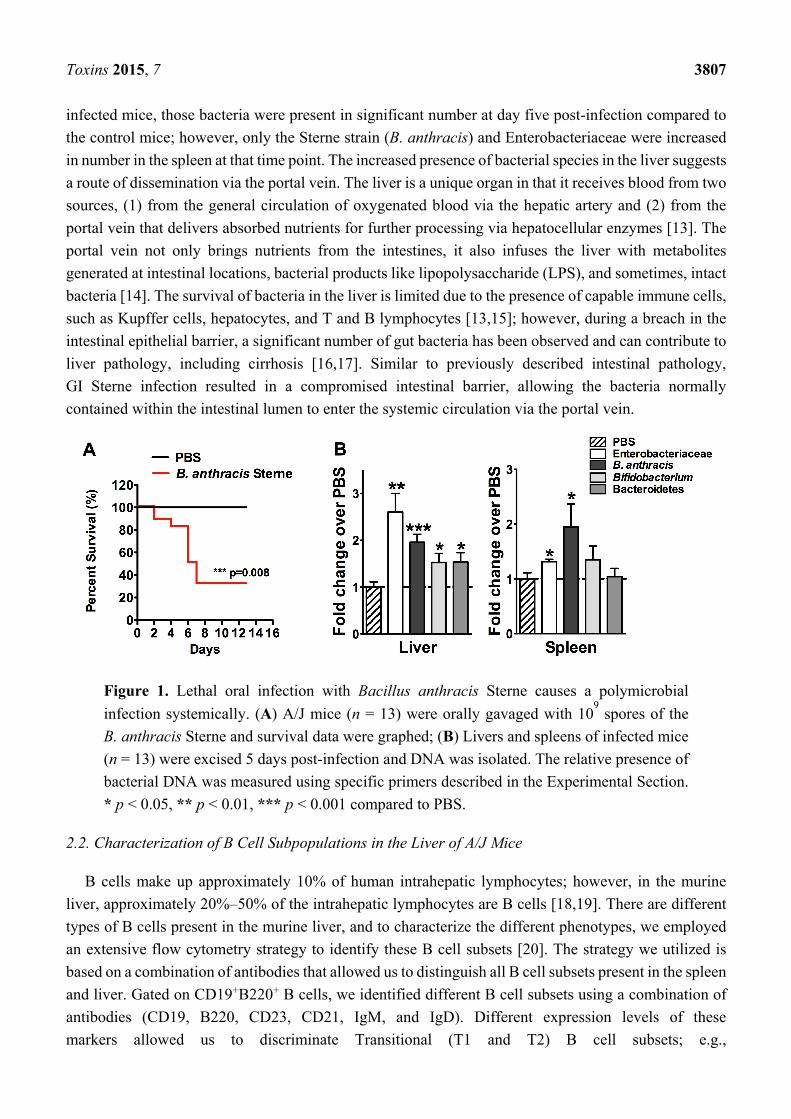
Toxins 2015, 7 3807
infected mice, those bacteria were present in significant number at day five post-infection compared to
the control mice; however, only the Sterne strain (B. anthracis) and Enterobacteriaceae were increased
in number in the spleen at that time point. The increased presence of bacterial species in the liver suggests
a route of dissemination via the portal vein. The liver is a unique organ in that it receives blood from two
sources, (1) from the general circulation of oxygenated blood via the hepatic artery and (2) from the
portal vein that delivers absorbed nutrients for further processing via hepatocellular enzymes [13]. The
portal vein not only brings nutrients from the intestines, it also infuses the liver with metabolites
generated at intestinal locations, bacterial products like lipopolysaccharide (LPS), and sometimes, intact
bacteria [14]. The survival of bacteria in the liver is limited due to the presence of capable immune cells,
such as Kupffer cells, hepatocytes, and T and B lymphocytes [13,15]; however, during a breach in the
intestinal epithelial barrier, a significant number of gut bacteria has been observed and can contribute to
liver pathology, including cirrhosis [16,17]. Similar to previously described intestinal pathology,
GI Sterne infection resulted in a compromised intestinal barrier, allowing the bacteria normally
contained within the intestinal lumen to enter the systemic circulation via the portal vein.
Figure 1. Lethal oral infection with Bacillus anthracis Sterne causes a polymicrobial
infection systemically. (A) A/J mice (n = 13) were orally gavaged with 109 spores of the
B. anthracis Sterne and survival data were graphed; (B) Livers and spleens of infected mice
(n = 13) were excised 5 days post-infection and DNA was isolated. The relative presence of
bacterial DNA was measured using specific primers described in the Experimental Section.
* p < 0.05, ** p < 0.01, *** p < 0.001 compared to PBS.
2.2. Characterization of B Cell Subpopulations in the Liver of A/J Mice
B cells make up approximately 10% of human intrahepatic lymphocytes; however, in the murine
liver, approximately 20%–50% of the intrahepatic lymphocytes are B cells [18,19]. There are different
types of B cells present in the murine liver, and to characterize the different phenotypes, we employed
an extensive flow cytometry strategy to identify these B cell subsets [20]. The strategy we utilized is
based on a combination of antibodies that allowed us to distinguish all B cell subsets present in the spleen
and liver. Gated on CD19+B220+ B cells, we identified different B cell subsets using a combination of
antibodies (CD19, B220, CD23, CD21, IgM, and IgD). Different expression levels of these
markers allowed us to discriminate Transitional (T1 and T2) B cell subsets; e.g.,
Toxins 2015, 7 3808
T1 (B220+CD19+IgDlowIgMlowCD23−CD21/35−), and T2 (CD19+IgDhighCD23highIgMhighCD21/35low).
The originally described “T2” subset contained MZ B cell precursors (MZP)
(B220+CD19+IgDhighCD21/35highCD23+IgMhigh) that differ due to the presence of CD21/35 surface
markers. T2-MZP cells also expressed CD23, which permits differentiation from MZ B cells
(B220+CD19+IgDlowCD21/35highCD23−IgMhigh). Long-lived recirculating follicular B cells (FO, also
called B-2 cells) can also be identified (B220+CD19+IgMlowCD23+CD21/35lowIgDhigh) (Figure S1).
The characterization of B-1 cells was based on the use of CD19+IgMhighCD23low(−)CD21−IgDlowCD11b+
and CD5+/− (B−1a/B−1b) staining.
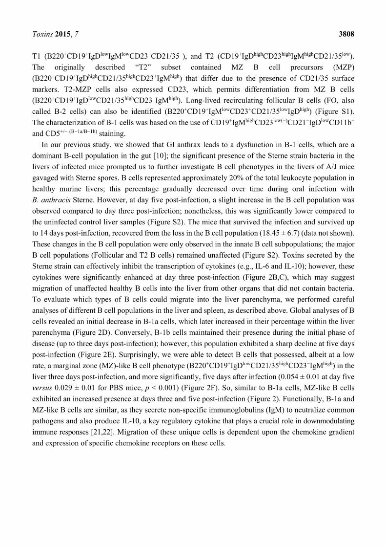
In our previous study, we showed that GI anthrax leads to a dysfunction in B-1 cells, which are a
dominant B-cell population in the gut [10]; the significant presence of the Sterne strain bacteria in the
livers of infected mice prompted us to further investigate B cell phenotypes in the livers of A/J mice
gavaged with Sterne spores. B cells represented approximately 20% of the total leukocyte population in
healthy murine livers; this percentage gradually decreased over time during oral infection with
B. anthracis Sterne. However, at day five post-infection, a slight increase in the B cell population was
observed compared to day three post-infection; nonetheless, this was significantly lower compared to
the uninfected control liver samples (Figure S2). The mice that survived the infection and survived up
to 14 days post-infection, recovered from the loss in the B cell population (18.45 ± 6.7) (data not shown).
These changes in the B cell population were only observed in the innate B cell subpopulations; the major
B cell populations (Follicular and T2 B cells) remained unaffected (Figure S2). Toxins secreted by the
Sterne strain can effectively inhibit the transcription of cytokines (e.g., IL-6 and IL-10); however, these
cytokines were significantly enhanced at day three post-infection (Figure 2B,C), which may suggest
migration of unaffected healthy B cells into the liver from other organs that did not contain bacteria.
To evaluate which types of B cells could migrate into the liver parenchyma, we performed careful
analyses of different B cell populations in the liver and spleen, as described above. Global analyses of B
cells revealed an initial decrease in B-1a cells, which later increased in their percentage within the liver
parenchyma (Figure 2D). Conversely, B-1b cells maintained their presence during the initial phase of
disease (up to three days post-infection); however, this population exhibited a sharp decline at five days
post-infection (Figure 2E). Surprisingly, we were able to detect B cells that possessed, albeit at a low
rate, a marginal zone (MZ)-like B cell phenotype (B220+CD19+IgDlowCD21/35highCD23−IgMhigh) in the
liver three days post-infection, and more significantly, five days after infection (0.054 ± 0.01 at day five
versus 0.029 ± 0.01 for PBS mice, p < 0.001) (Figure 2F). So, similar to B-1a cells, MZ-like B cells
exhibited an increased presence at days three and five post-infection (Figure 2). Functionally, B-1a and
MZ-like B cells are similar, as they secrete non-specific immunoglobulins (IgM) to neutralize common
pathogens and also produce IL-10, a key regulatory cytokine that plays a crucial role in downmodulating
immune responses [21,22]. Migration of these unique cells is dependent upon the chemokine gradient
and expression of specific chemokine receptors on these cells.
Toxins 2015, 7 3809
Figure 2. Sterne infection causes changes in the B cell population in the liver. A/J mice
(n = 43) were infected with 109 spores of B. anthracis Sterne. Liver leukocytes were isolated
from mice (day 1, n = 10; day 2, n = 10, day 3, n = 10; day 5 n = 13) at each indicated time
point using Percoll gradients, as described in the Experimental Section. Isolated leukocytes
were stained with different sets of antibodies, as depicted in the Figure S1 to define:
(A) total B cells; (B) IL-6 producing; (C) IL-10 producing B cells; (D) B-1a cells; (E) B-1b
cells; and (F) marginal zone-like (MZ-like) B cells. Data are shown as mean ± SEM.
* p < 0.05, ** <0.01, *** p < 0.001 compared with PBS-treated or day 0 mice.
2.3. Evaluation of Chemokine and Chemokine Receptors in the Liver during B. anthracis
Sterne Infection
The chemokines CXCL10; -11; -12; -13; and -16 have been implicated in either B cell trafficking or
the infiltration of other immune cells into the liver [23–27]. To evaluate whether expression of these
chemokines is altered with Sterne infection; we analyzed transcripts by qRT-PCR; which revealed
differential expression of CXCL10; -11; -12; -13; and -16 in the liver and in the spleen during Sterne
infection (Figure 3). CXCL10 and -11 (ligands for the CXCR3 receptor) were moderately expressed in
the liver; however; their expression was downregulated in the infected spleen. The expression of these
two chemokines is dependent upon interferon signaling; they then recruit inflammatory leukocytes to
the inflamed loci [28]. It is conceivable that inflammation would be increased with polymicrobial
Toxins 2015, 7 3810
infiltrates; as seen in the liver during Sterne infection. CXCL12 binds to its receptor; CXCR4; and is
secreted in response to Toll-like receptor (TLR) and cytokine signaling; to recruit inflammatory
leukocytes [29,30]; this chemokine was inhibited during Sterne infection. The classical chemokine for
B cell recruitment; CXCL13; was significantly increased during Sterne infection in the liver; however;
its expression in the spleen was moderately decreased at day five post-Sterne infection (Figure 3).
Perturbation in chemokine expression in the liver and the spleen may form a temporary chemokine
gradient for the migration of cells from a distant location; including from the spleen to the liver.
Furthermore; the downregulation of homeostatic lymphoid chemokines in the spleen; such as CXCL13;
could lead to disruption of the splenic architecture; resulting in the traffic of MZ-like B cells into
the liver.
Figure 3. Sterne infection leads to changes in chemokine production in the liver and spleen.
A/J mice (n = 43) were infected with 109 spores of B. anthracis Sterne. Total RNA was
isolated from mice (day 1, n = 10; day 2, n = 10, day 3, n = 10; day 5 n = 13) at each indicated
time point, and transcripts of CXCL10, CXCL11, CXCL12, CXCL13, CXCL16, and CCL25
were evaluated by qRT-PCR, as mentioned in the Experimental Section using gene specific
primers. Data are shown as mean ± SEM. * p < 0.05, ** <0.01, *** p < 0.001 compared with
PBS-treated or day 0 mice.

Toxins 2015, 7 3811
2.4. Sterne Infection Increases the Accumulation of Type 2 Innate Lymphoid Cells
Previously we have reported the depletion of ILC2s in the gut of Sterne-infected mice [10].
To evaluate whether a similar depletion can be seen in the liver, we evaluated the number of ILC2s in
the liver parenchyma (Figure S1). Surprisingly, instead of a depletion, we found a significant increase
in the ILC2 population during the early stage of the disease; this increase was no longer apparent at day
five post-infection (Figure 4A). Bacterial infections can be efficiently cleared by ILC3s, which secrete
IL-17 and recruit neutrophils [31]; upon investigation, these cells did not significantly change in their
percentage within the liver during infection (Figure 4B). The cytokines produced by ILC2s were also
altered during the course of infection. IL-5 and IL-13 are two major cytokines produced by ILC2s to
skew the immune response towards a Th2 phenotype. During Sterne infection, IL-5 expression was
maintained at early stages of the disease; however, at day three post-infection, its expression declined,
suggesting limited support to B cells for their survival and maintenance at Sterne-infected hepatic loci
(Figure 4C). On the other hand, IL-13 expression declined very sharply in ILC2s; nonetheless, ILC2s
recovered their IL-13 expression at day five post-infection (Figure 4D).
Figure 4. Sterne infection changes the percentage of ILC2s in infected livers. A/J mice
(n = 43) were infected with 109 spores of B. anthracis Sterne. Liver leukocytes were isolated
from mice (day 1, n = 10; day 2, n = 10, day 3, n = 10; day 5 n = 13) at each indicated time
point using Percoll gradients, as described in the Experimental Section. Isolated leukocytes
were stained with different sets of antibodies, as depicted in the Figure S1 to define:
(A) ILC2s; (B) ILC3s; (C) IL-5 producing; and (D) IL-13 producing ILC2s. Data are shown
as mean ± SEM. * p < 0.05, ** <0.01, *** p < 0.001 compared with PBS-treated or
day 0 mice.
We previously reported a decline in the number of ILC2s [10] and their secretion of cytokines in the
colons of mice infected with the Sterne strain of B. anthracis; however, here we report an increase in the
ILC2 population within the liver. ILC2s express CXCR4, CXCR6, and CCR9 to reach peripheral
sites [7,32]. The ligands for these receptors are CXCL12, CXCL16, and CCL25, respectively. We have
evaluated transcription of these chemokines in the infected livers and spleens and found increased
expression the genes (Figure 3), which suggests that these chemokines could allow for the recruitment
of ILC2s to the liver to protect the damages resulting from Sterne infection.
Toxins 2015, 7 3812
3. Experimental Section
3.1. Mice and Ethics Statement
A/J mice were obtained from the Jackson Laboratory and bred in-house at the animal facility in the
College of Veterinary Medicine at the University of Florida. Mice were provided food and water ad
libitum. Mice were sacrificed at 6–8 weeks of age in accordance with the Animal Welfare Act and the
Public Health Policy on Humane Care. All procedures were approved by the Institutional Animal Care
and Use Committee (IACUC) at the University of Florida under protocol number 201107129 and all
efforts were made to minimize animal suffering. Infected mice were monitored every 24 h and were
humanely euthanized when signs of advanced infection (e.g., difficulty breathing) were noted.
3.2. Bacillus anthracis Spore Preparation and Mouse Infections
Spores were prepared from a toxigenic, non-encapsulated strain of B. anthracis (Sterne), as described
previously [11], with the approval of the Institutional Biosafety Committee (IBC) at the University of
Florida. To calculate final concentrations, serial dilutions (1:10) were grown in triplicate on
brain-heart-infusion broth agar plates (Sterne) and colonies were subsequently counted. Mice were orally
infected with Sterne spores (109 spores/100 μL of PBS per mouse) after fasting for 4 h at the specific
time points. For the survival study, mice (n = 13) were orally infected; infected mice were monitored,
and deaths recorded during 14 days. For B cells studies, mice were orally infected; infected mice were
monitored, and sacrificed at the corresponding days for B cells analyses in the liver and in the spleen.
(n = 10 mice/group for PBS, day 1, 2 or 3 and n = 13 mice for day 5).
3.3. Quantitative Real-Time-PCR
Total genomic DNA was extracted from the hepatic and splenic tissues of the mice using gDNA
MiniPrep kit (Zymoresearch, Irvine, CA, USA), following the manufactures’ instructions. qRT-PCR
was performed on 100 ng of DNA template (SsoAdvanced SYBR® Green Supermix, Bio-Rad,
Laboratories, Inc., Hercules, CA, USA) to assess the dissemination of B. anthracis, Bifidobacterium,
Bacteroidetes, and Enterobacteriaceae (PBS = 10 and day five, n = 13). Groups were normalized to the
housekeeper Eubacteria group. A list of primers used and their sequences can be found in Table A1.
RNA was isolated from the liver and spleen using Aurum Total RNA mini Kit (Bio-Rad, Hercules,
CA, USA), and cDNA was obtained via the iScript™ Select cDNA Synthesis Kit (Bio-Rad, Hercules,
CA, USA). One µg of cDNA was used as a template for quantitative PCR via SYBR® Green on a
Bio-Rad CFX96 Real time system. mRNA levels are shown as the fold change increased over uninfected
mice, and results were normalized to the housekeeper GAPDH gene. All primers that were used are
found in Table A1 [9,10,33]. For statistical analyses, unpaired t-tests were performed using the relative
expression levels of each gene at the specified time point compared to the relative expression level of
the same gene in uninfected mice (PBS, days 1, 2, 3 post-infection: n = 10 mice/group and
day 5 post-infection, n = 13 mice/group).
Toxins 2015, 7 3813
3.4. Isolation of Immune Cells for Flow Cytometry Analyses
Livers were perfused with 10 mL of phosphate-buffered saline (PBS) via the portal vein to remove
circulating lymphocytes. Liver and spleen single-cell suspensions were prepared from whole tissue by
mechanical disruption in RPMI-1640 with 5% fetal bovine serum (FBS). After washing, the subsequent
suspension of the liver was disrupted through a 100-µm metal cell strainer and centrifuged through
40%–60% isotonic Percoll/RPMI 1640 gradient (GE-Healthcare, Uppsala, Sweden). For the spleen,
erythrocytes were lysed with ACK lysis buffer and cells were washed twice using PBS with 1% FBS.
Cells were counted and used for staining. Spleen and liver cell suspensions were stained using the
LIVE/DEAD Fixable Aqua Dead Cell Stain Kit, then washed and incubated with an Fc blocking reagent
(Miltenyi Biotec Inc., San Diego, CA, USA). A combination of different antibodies and their
corresponding isotypes were used for labeled cell suspensions. Antibodies were purchased from
eBiosciences (San Diego, CA), Biolegend (San Diego, CA, USA), and BD Pharmingen (San Diego, CA,
USA): B220 (RA3-6B2), CD19 (6D5), IgM (RMM-1), IgD (11-26C), CD23 (B3B4), CD21/35 (7E9),
CD45 (30-F11), CD11b (M1/70), CD5 (53-7.3), CD93 (AA4.1), CD90.2 (30-H12), IL33Ra (DIH9),
GATA3 (L50-823), IL-7Rα (A7R34), CD25 (PC61), ICOS (7E.17G9), CD3 (145-2C11), CD11c
(N418), RoRγt (B2D), IL6 (MP5-20F3), IL10 (JES5-16E3), IL13 (eBio13A) and IL5 (TRFK5). Lineage
staining (Lin) used for the ILCs is composed of a combination of CD3 (17A2), CD11b (M1/70),
CD11c (N418), Gr1 (RB6-8C5), CD19 (6D5) and B220 (RA3-6B2). Gating strategies are shown in the
Figure S1. The Cytofix/Cytoperm kit (BD Biosciences, San Jose, CA, USA) was used for fixation and
cellular permeabilization when intracellular staining was needed. Flow cytometric acquisition was
performed using a BD LSRFortessa™ (BD Biosciences, San Jose, CA, USA). Data were analyzed with
FlowJo software (Tree Star, Ashland, OR, USA).
3.5. Statistical Analyses
Data are represented as mean ± SEM. Statistical significance was calculated with Graphpad Prism
software (LaJolla, CA, USA) using a two-tailed t-test for two group comparisons. Values of p < 0.05
were considered significant (* p < 0.05, ** p < 0.01, *** p < 0.001).
4. Conclusions
B. anthracis Sterne infection in A/J mice causes a polymicrobial infection due to the loss of intestinal
barrier function [9]. Upon examination, data suggest that the polymicrobial infection within the liver
might cause disruption of B cell and ILC2 functions. To fight the infection and to replace dysfunctional
B cells and ILC2s, hepatic cells upregulate specific B cell and ILC2 chemokines. In the response to the
chemokines, MZ-like B cells, B-1a cells, and ILC2s migrate into the hepatic parenchyma; however, they
fail to clear the infection, potentially due to clevage of mitogen-activated protein kinases
(MAPKs) [9,34,35] by the toxins released by the Sterne strain to suppress immune cell functions.
Supplementary Materials
Supplementary materials can be accessed at: http://www.mdpi.com/2072-6651/7/9/3805/s1.

Toxins 2015, 7 3814
Acknowledgments
This work was supported by NIH R01 AI093370 (MM).
Author Contributions
Conceived and designed the experiments: N.C., B.S., M.M. Performed the experiments: N.C., M.Z.
Analyzed the data: N.C. Wrote the paper: B.S., N.C., J.L.O., and M.M.
Conflicts of Interest
The authors declare that they have no conflict of interest.
Appendix
Table A1. List of primer sequences for Real-Time PCR analyses.
Gene/Organism Sequence (5ʹ to 3ʹ) References
Bacteroides For GAGAGGAAGGTCCCCCAC [36] Bacteroides Rev CGCTACTTGGCTGGTTCAG
Bifidobacterium For CGGGTGAGTAATGCGTGACC [9] Bifidobacterium Rev TGATAGGACGCGACCCCA
Enterobacteriaceae For GTGCCAGCMGCCGCGGTAA [9] Enterobacteriaceae Rev GCCTCAAGGGCACAACCTCCAAG Bacillus anthracis For ATCACCAGAGGCAAGACACCC [37] Bacillus anthracis Rev ACCAATATCAAAGAACGACGC
Cxcl13 For CAGAATGAGGCTCAGCACAGC [9] Cxcl13 Rev CAGAATACCGTGGCCTGGAG Cxcl11 For GGCGTCAAAACATGTGACATCC [38] Cxcl11 Rev TGGCTGCATGTTCCAAGACAG Cxcl10 For TCCCTCTCGCAAGGAC [9] Cxcl10 Rev TTGGCTAAACGCTTTCAT Cxcl12 For GAGCCAACGTCAAGCATCTG [9] Cxcl12 Rev CGGGTCAATGCACACTTGTC Cxcl16 For ACCCTTGTCTCTTGCGTTCTTCCT [39] Cxcl16 Rev ATGTGATCCAAAGTACCCTGCGGT Ccl25 For CCAAGGTGCCTTTGAAGACT [33] Ccl25 Rev TCCTCCAGCTGGTGGTTACT
GAPDH For GGTGAAGGTCGGTGTGAACG [9] GAPDH Rev CTCGCTCCTGGAAGATGGTG
References
1. Beatty, M.E.; Ashford, D.A.; Griffin, P.M.; Tauxe, R.V.; Sobel, J. Gastrointestinal anthrax: Review
of the literature. Arch. Intern. Med. 2003, 163, 2527–2531.
2. Mikesell, P.; Ivins, B.E.; Ristroph, J.D.; Dreier, T.M. Evidence for plasmid-mediated toxin
production in bacillus anthracis. Infect. Immun. 1983, 39, 371–376.

Toxins 2015, 7 3815
3. Tonello, F.; Zornetta, I. Bacillus anthracis factors for phagosomal escape. Toxins 2012, 4,
536–553.
4. Lowe, D.E.; Glomski, I.J. Cellular and physiological effects of anthrax exotoxin and its relevance
to disease. Front. Cell. Infect. Microbiol. 2012, 2, 76.
5. Owen, J.L.; Yang, T.; Mohamadzadeh, M. New insights into gastrointestinal anthrax infection.
Trends Mol. Med. 2015, 21, 154–163.
6. Liu, S.; Moayeri, M.; Leppla, S.H. Anthrax lethal and edema toxins in anthrax pathogenesis.
Trends Microbiol. 2014, 22, 317–325.
7. Scanlon, S.T.; McKenzie, A.N.J. The messenger between worlds: The regulation of innate and
adaptive type-2 immunity by innate lymphoid cells. Clin. Exp. Allergy 2015, 45, 9–20.
8. Beyer, W.; Turnbull, P.C.B. Anthrax in animals. Mol. Asp. Med. 2009, 30, 481–489.
9. Lightfoot, Y.L.; Yang, T.; Sahay, B.; Zadeh, M.; Cheng, S.X.; Wang, G.P.; Owen, J.L.;
Mohamadzadeh, M. Colonic immune suppression, barrier dysfunction, and dysbiosis by
gastrointestinal bacillus anthracis infection. PLoS One 2014, 9, e100532.
10. Sahay, B.; Owen, J.L.; Zadeh, M.; Yang, T.; Lightfoot, Y.L.; Abed, F.; Mohamadzadeh, M.
Impaired colonic B-cell responses by gastrointestinal bacillus anthracis infection. J. Infect. Dis.
2014, 210, 1499–1507.
11. Welkos, S.L.; Keener, T.J.; Gibbs, P.H. Differences in susceptibility of inbred mice to bacillus
anthracis. Infect. Immun. 1986, 51, 795–800.
12. Hambleton, P.; Carman, J.A.; Melling, J. Anthrax: The disease in relation to vaccines. Vaccine
1984, 2, 125–132.
13. Crispe, I.N. The liver as a lymphoid organ. Annu. Rev. Immunol. 2009, 27, 147–163.
14. Schafer, C.; Parlesak, A.; Schutt, C.; Bode, J.C.; Bode, C. Concentrations of lipopolysaccharide-binding
protein, bactericidal/permeability-increasing protein, soluble cd14 and plasma lipids in relation to
endotoxaemia in patients with alcoholic liver disease. Alcohol. Alcohol. 2002, 37, 81–86.
15. Qi, S.; Sui, W.; Yang, M.; Chen, J.; Dai, Y. Cpg array analysis of histone h3 lysine 4 trimethylation
by chromatin immunoprecipitation linked to microarrays analysis in peripheral blood mononuclear
cells of iga nephropathy patients. Yonsei Med. J. 2012, 53, 377–385.
16. Balmer, M.L.; Slack, E.; de Gottardi, A.; Lawson, M.A.E.; Hapfelmeier, S.; Miele, L.; Grieco, A.;
van Vlierberghe, H.; Fahrner, R.; Patuto, N.; et al. The liver may act as a firewall mediating
mutualism between the host and its gut commensal microbiota. Sci. Transl Med. 2014, 6, 237ra66.
17. Wiest, R.; Garcia-Tsao, G. Bacterial translocation (bt) in cirrhosis. Hepatology 2005, 41, 422–433.
18. Holt, A.P.; Stamataki, Z.; Adams, D.H. Attenuated liver fibrosis in the absence of B cells.
Hepatology 2006, 43, 868–871.
19. Zhang, H.; Stolz, D.B.; Chalasani, G.; Thomson, A.W. Hepatic B cells are readily activated by
toll-like receptor-4 ligation and secrete less interleukin-10 than lymphoid tissue b cells.
Clin. Exp. Immunol. 2013, 173, 473–479.
20. Carsetti, R.; Rosado, M.M.; Wardmann, H. Peripheral development of b cells in mouse and man.
Immunol. Rev. 2004, 197, 179–191.
21. Kaveri, S.V.; Silverman, G.J.; Bayry, J. Natural igm in immune equilibrium and harnessing their
therapeutic potential. J. Immunol. 2012, 188, 939–945.
22. Zhang, X. Regulatory functions of innate-like b cells. Cell. Mol. Immunol. 2013, 10, 113–121.

Toxins 2015, 7 3816
23. Feng, L.; Li, L.; Liu, Y.; Qiao, D.; Li, Q.; Fu, X.Y.; Wang, H.; Lao, S.H.; Wu, C.Y. B lymphocytes
that migrate to tuberculous pleural fluid via the sdf-1/cxcr4 axis actively respond to antigens specific
for mycobacterium tuberculosis. Eur. J. Immunol. 2011, 41, 3261–3269.
24. Park, M.K.; Amichay, D.; Love, P.; Wick, E.; Liao, F.; Grinberg, A.; Rabin, R.L.; Zhang, H.W.H.;
Gebeyehu, S.; Wright, T.M.; et al. The cxc chemokine murine monokine induced by ifn-gamma
(cxc chemokine ligand 9) is made by apcs, targets lymphocytes including activated b cells, and
supports antibody responses to a bacterial pathogen in vivo. J. Immunol. 2002, 169, 1433–1443.
25. Patadia, M.; Dixon, J.; Conley, D.; Chandra, R.; Peters, A.; Suh, L.A.; Kato, A.; Carter, R.; Harris,
K.; Grammer, L.; et al. Evaluation of the presence of b-cell attractant chemokines in chronic
rhinosinusitis. Am. J. Rhinol. Allergy 2010, 24, 11–16.
26. Wang, J.; Holmes, T.H.; Cheung, R.; Greenberg, H.B.; He, X.S. Expression of chemokine receptors
on intrahepatic and peripheral lymphocytes in chronic hepatitis c infection: Its relationship to liver
inflammation. J. Infect. Dis. 2004, 190, 989–997.
27. Kunkel, E.J.; Butcher, E.C. Plasma-cell homing. Nat. Rev. Immunol. 2003, 3, 822–829.
28. Widney, D.P.; Xia, Y.R.; Lusis, A.J.; Smith, J.B. The murine chemokine cxcl11 (ifn-inducible t cell
alpha chemoattractant) is an ifn-gamma- and lipopolysaccharide-inducible glucocorticoid-attenuated
response gene expressed in lung and other tissues during endotoxemia. J. Immunol. 2000, 164,
6322–6331.
29. Karin, N. The multiple faces of cxcl12 (SDF-1alpha) in the regulation of immunity during health
and disease. J. Leukoc. Biol. 2010, 88, 463–473.
30. Klasen, C.; Ohl, K.; Sternkopf, M.; Shachar, I.; Schmitz, C.; Heussen, N.; Hobeika, E.;
Levit-Zerdoun, E.; Tenbrock, K.; Reth, M.; et al. Mif promotes B cell chemotaxis through the
receptors cxcr4 and cd74 and zap-70 signaling. J. Immunol. 2014, 192, 5273–5284.
31. Diefenbach, A. Innate lymphoid cells in the defense against infections. Eur. J. Microbiol. Immunol.
2013, 3, 143–151.
32. Griffith, J.W.; Sokol, C.L.; Luster, A.D. Chemokines and chemokine receptors: Positioning cells
for host defense and immunity. Annu. Rev. Immunol. 2014, 32, 659–702.
33. Wurbel, M.A.; McIntire, M.G.; Dwyer, P.; Fiebiger, E. Ccl25/ccr9 interactions regulate large
intestinal inflammation in a murine model of acute colitis. PLoS ONE 2011, 6, e16442.
34. Agrawal, A.; Lingappa, J.; Leppla, S.H.; Agrawal, S.; Jabbar, A.; Quinn, C.; Pulendran, B.
Impairment of dendritic cells and adaptive immunity by anthrax lethal toxin. Nature 2003, 424,
329–334.
35. Fang, H.; Xu, L.; Chen, T.Y.; Cyr, J.M.; Frucht, D.M. Anthrax lethal toxin has direct and potent
inhibitory effects on b cell proliferation and immunoglobulin production. J. Immunol. 2006, 176,
6155–6161.
36. Petnicki-Ocwieja, T.; Hrncir, T.; Liu, Y.-J.; Biswas, A.; Hudcovic, T.; Tlaskalova-Hogenova, H.;
Kobayashi, K.S. Nod2 is required for the regulation of commensal microbiota in the intestine.
Proc. Natl. Acad. Sci. USA 2009, 106, 15813–15818.
37. Makino, S.I.; Cheun, H.I.; Watarai, M.; Uchida, I.; Takeshi, K. Detection of anthrax spores from
the air by real-time pcr. Lett. Appl. Microbiol. 2001, 33, 237–240.

Toxins 2015, 7 3817
38. Roebrock, K.; Sunderkotter, C.; Munck, N.A.; Wolf, M.; Nippe, N.; Barczyk, K.; Varga, G.; Vogl,
T.; Roth, J.; Ehrchen, J. Epidermal expression of i-tac (cxcl11) instructs adaptive th2-type
immunity. FASEB J.: Off. Publ. Fed. Am. Soc. Exp. Biol. 2014, 28, 1724–1734.
39. Chen, G.; Lin, S.-C.; Chen, J.; He, L.; Dong, F.; Xu, J.; Han, S.; Du, J.; Entman, M.L.; Wang, Y.
Cxcl16 recruits bone marrow-derived fibroblast precursors in renal fibrosis. J. Am. Soc. Nephrol.
2011, 22, 1876–1886.
© 2015 by the authors; licensee MDPI, Basel, Switzerland. This article is an open access article
distributed under the terms and conditions of the Creative Commons Attribution license
(http://creativecommons.org/licenses/by/4.0/).