Embed Size (px)
DESCRIPTION
Citation preview
IMMUNOPATHOLOGY
Types of immunity: two broad categories:- Innate immunity: also called natural, or native immunity. is first line of defense. The major components of innate immunity are:o Epithelial barriers that block entry of environmental
microbes.o Phagocytic cells (mainly neutrophils and macrophages).o Natural killer (NK) cells.o Plasma proteins including the proteins of complement
system.
Adaptive immunity: also called acquired, or specific immunity. develops later after exposure to microbes. also capable of recognizing non-microbial substances
called antigens. Adaptive immunity is more powerful in combating infections. Two main types of adaptive immunity: Cell-mediated ( cellular ) immunity:o responsible for defense against intracellular microbes
( viruses and T.B ).o is mediated by T- lymphocytes.
• Humoral immunity:o protects against extracellular microbes and their
toxins ( Bacteria ).o mediated by B - lymphocytes which differentiate
into antibody-secreting cells called plasma cells and their secreted products called antibodies :
( IgM, IgD, IgG, IgA, and IgE ).
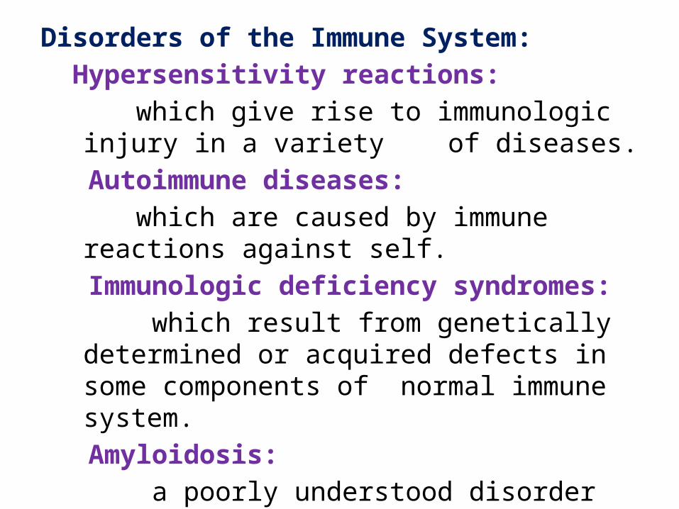
Disorders of the Immune System: Hypersensitivity reactions: which give rise to immunologic injury in a variety
of diseases. Autoimmune diseases: which are caused by immune reactions against self. Immunologic deficiency syndromes: which result from genetically determined or
acquired defects in some components of normal immune system.
Amyloidosis: a poorly understood disorder having immunologic
association.

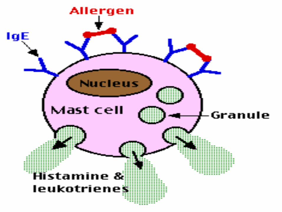
Hypersensitivity reactions: Type I Hypersensitivity reaction: Immediate hypersensitivity reaction , occurring within
minutes after combination of an antigen with antibody ( IgE ) bound to mast cells in individuals previously sensitized to that antigen .
Mast cells secrete a variety of mediators ( histamine, enzymes as proteases , cytokines as TNF ) which induce the reaction .
These reactions are often called allergy, and the antigens that elicit them called allergens.
may occur as a systemic disorder or as a local reaction.
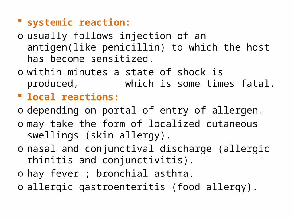
systemic reaction:o usually follows injection of an antigen(like penicillin) to
which the host has become sensitized.o within minutes a state of shock is produced, which
is some times fatal. local reactions:o depending on portal of entry of allergen.o may take the form of localized cutaneous swellings
(skin allergy).o nasal and conjunctival discharge (allergic rhinitis and
conjunctivitis).o hay fever ; bronchial asthma.o allergic gastroenteritis (food allergy).
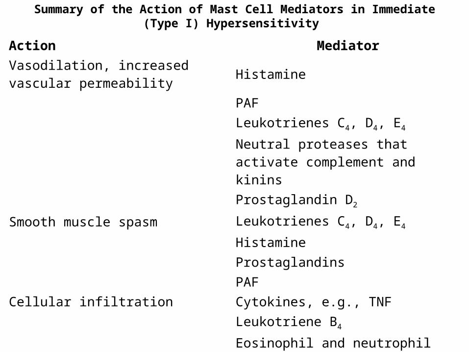
Summary of the Action of Mast Cell Mediators in Immediate (Type I) Hypersensitivity
Action MediatorVasodilation, increased vascular permeability Histamine
PAF Leukotrienes C4, D4, E4
Neutral proteases that activate complement and kinins
Prostaglandin D2
Smooth muscle spasm Leukotrienes C4, D4, E4
Histamine Prostaglandins PAFCellular infiltration Cytokines, e.g., TNF Leukotriene B4
Eosinophil and neutrophil chemotactic factors (not defined biochemically)
PAFPAF, platelet-activating factor; TNF, tumor necrosis factor.
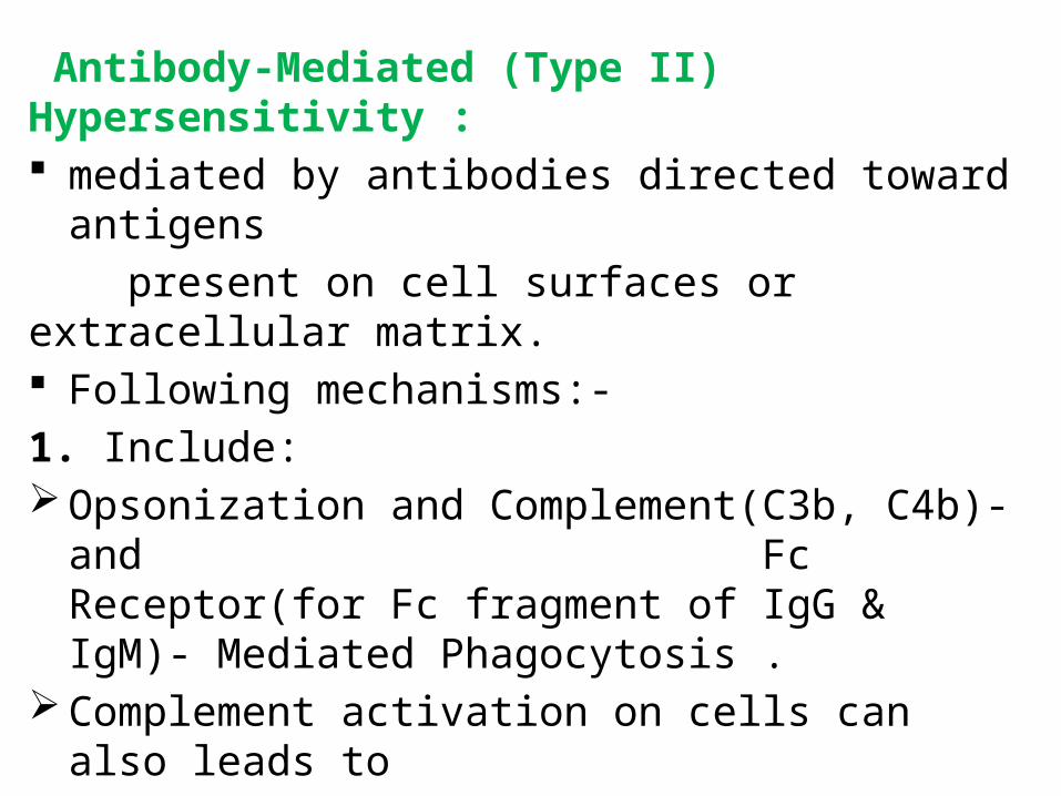
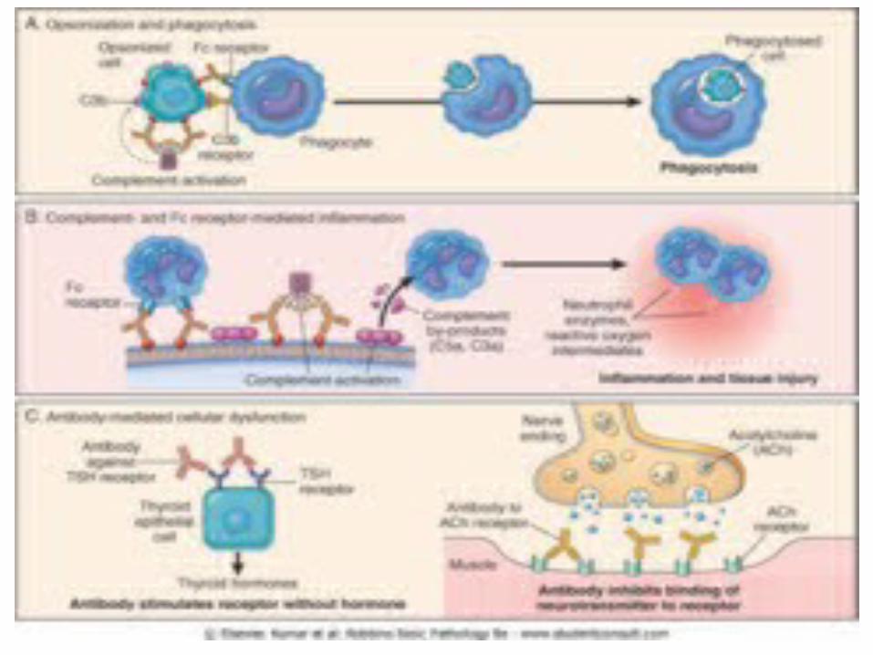
Antibody-Mediated (Type II) Hypersensitivity : mediated by antibodies directed toward antigens present on cell surfaces or extracellular matrix. Following mechanisms:-1. Include: Opsonization and Complement(C3b, C4b)- and
Fc Receptor(for Fc fragment of IgG & IgM)- Mediated Phagocytosis .
Complement activation on cells can also leads to formation of membrane attack complex (C5b-C9)which disrupts membrane integrity by "drilling holes" thereby causing osmotic lysis of cells.
Antibody-mediated destruction of cells may occur by another process called antibody-dependent cellular cytotoxicity (ADCC ); it requires the cooperation of leukocytes , and cell lysis occur without phagocytosis.
Clinically, antibody-mediated cell destruction and phagocytosis occur in following situations:
• transfusion reactions.• erythroblastosis fetalis.• autoimmune hemolytic anemia, agranulocytosis, and thrombocytopenia.• certain drug reactions.
2. Complement- and Fc Receptor-Mediated Inflammation: The deposited antibodies activate complement, such as C5a (and to lesser extent C3a and C4a), that recruit neutrophils and monocytes, as in some forms of glomerulonephritis, and vascular rejection in organ grafts. 3. Antibody-Mediated Cellular Dysfunction : antibodies directed against cell-surface receptors either impair (Ach receptor in myasthenia gravis ) , or stimulate (TSH receptor in Graves disease – thyrotoxicosis) without causing cell injury or inflammation.
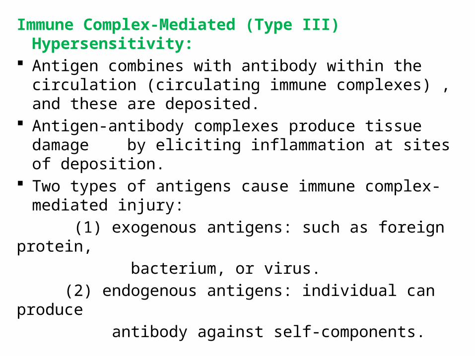
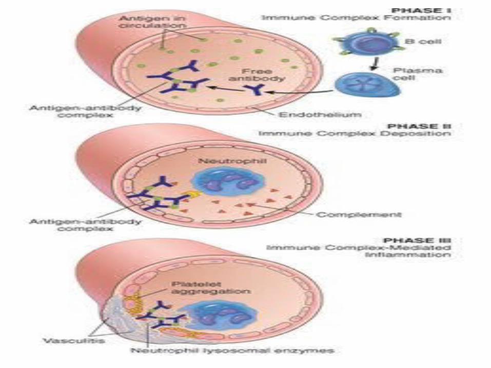
Immune Complex-Mediated (Type III) Hypersensitivity: Antigen combines with antibody within the circulation
(circulating immune complexes) , and these are deposited.
Antigen-antibody complexes produce tissue damage by eliciting inflammation at sites of deposition.
Two types of antigens cause immune complex-mediated injury:
(1) exogenous antigens: such as foreign protein, bacterium, or virus. (2) endogenous antigens: individual can produce antibody against self-components.
Immune complex-mediated diseases can be: generalized (systemic) ; or localized .
Systemic Immune Complex Disease: Acute serum sickness: Once complexes are deposited
in tissues, they initiate an acute inflammatory reaction (approximately 10 days after antigen administration), clinical features such as fever, urticaria, arthralgias, lymph node enlargement, and proteinuria appear. Local Immune Complex Disease :
Arthus reaction: a localized area of tissue necrosis resulting from acute immune complex vasculitis, usually elicited in skin.
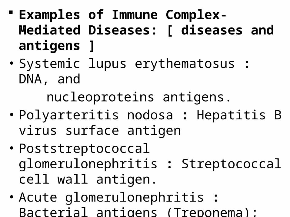
Examples of Immune Complex-Mediated Diseases: [ diseases and antigens ]
• Systemic lupus erythematosus : DNA, and nucleoproteins antigens.• Polyarteritis nodosa : Hepatitis B virus surface
antigen• Poststreptococcal glomerulonephritis :
Streptococcal cell wall antigen. • Acute glomerulonephritis : Bacterial antigens
(Treponema); parasite antigens (malaria, schistosomes); and tumor antigens.
• Reactive arthritis : Bacterial antigens (Yersinia).
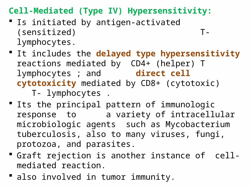
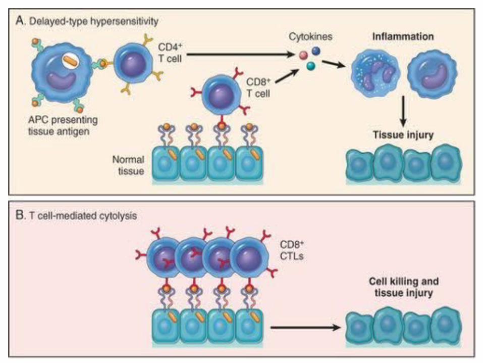
Cell-Mediated (Type IV) Hypersensitivity: Is initiated by antigen-activated (sensitized)
T-lymphocytes. It includes the delayed type hypersensitivity reactions
mediated by CD4+ (helper) T lymphocytes ; and direct cell cytotoxicity mediated by CD8+ (cytotoxic) T- lymphocytes .
Its the principal pattern of immunologic response to a variety of intracellular microbiologic agents such as Mycobacterium tuberculosis, also to many viruses, fungi, protozoa, and parasites.
Graft rejection is another instance of cell-mediated reaction.
also involved in tumor immunity.
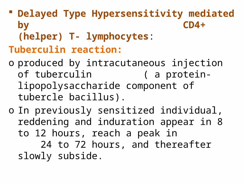
Delayed Type Hypersensitivity mediated by CD4+ (helper) T- lymphocytes:
Tuberculin reaction:o produced by intracutaneous injection of tuberculin (
a protein-lipopolysaccharide component of tubercle bacillus).
o In previously sensitized individual, reddening and induration appear in 8 to 12 hours, reach a peak in 24 to 72 hours, and thereafter slowly subside.
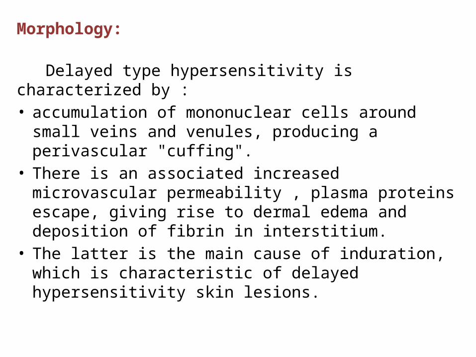
Morphology: Delayed type hypersensitivity is characterized by : • accumulation of mononuclear cells around small veins
and venules, producing a perivascular "cuffing". • There is an associated increased microvascular
permeability , plasma proteins escape, giving rise to dermal edema and deposition of fibrin in interstitium.
• The latter is the main cause of induration, which is characteristic of delayed hypersensitivity skin lesions.
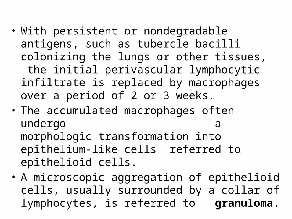
• With persistent or nondegradable antigens, such as tubercle bacilli colonizing the lungs or other tissues, the initial perivascular lymphocytic infiltrate is replaced by macrophages over a period of 2 or 3 weeks.
• The accumulated macrophages often undergo a morphologic transformation into epithelium-like cells referred to epithelioid cells.
• A microscopic aggregation of epithelioid cells, usually surrounded by a collar of lymphocytes, is referred to granuloma.
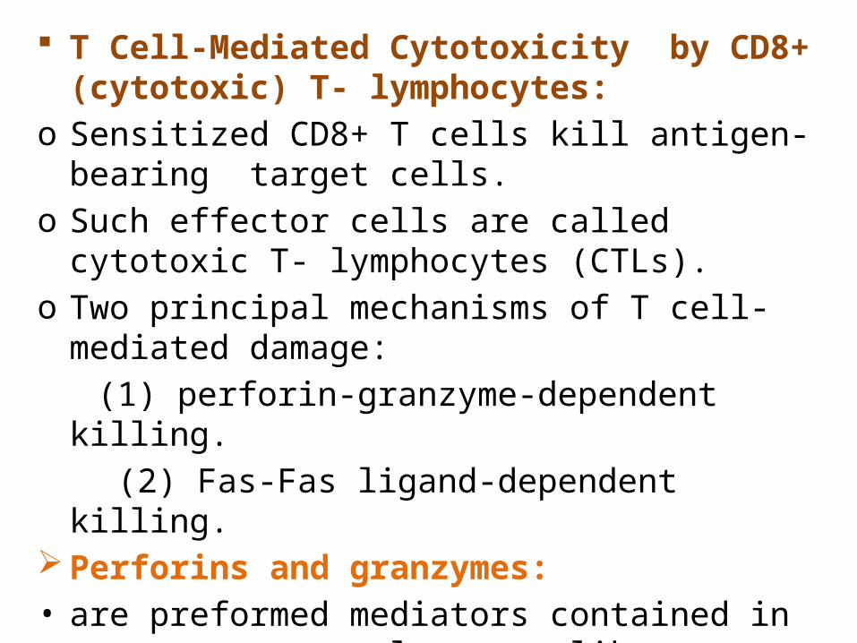
T Cell-Mediated Cytotoxicity by CD8+ (cytotoxic) T- lymphocytes:
o Sensitized CD8+ T cells kill antigen-bearing target cells.o Such effector cells are called cytotoxic T- lymphocytes
(CTLs).o Two principal mechanisms of T cell-mediated damage: (1) perforin-granzyme-dependent killing. (2) Fas-Fas ligand-dependent killing. Perforins and granzymes:• are preformed mediators contained in
lysosome-like granules of CTLs.• Perforin perforate plasma membranes of target cells ,
thus "drilling holes" into membrane.
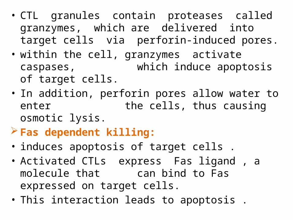
• CTL granules contain proteases called granzymes, which are delivered into target cells via perforin-induced pores.
• within the cell, granzymes activate caspases, which induce apoptosis of target cells.
• In addition, perforin pores allow water to enter the cells, thus causing osmotic lysis.
Fas dependent killing:• induces apoptosis of target cells .• Activated CTLs express Fas ligand , a molecule that
can bind to Fas expressed on target cells.• This interaction leads to apoptosis .
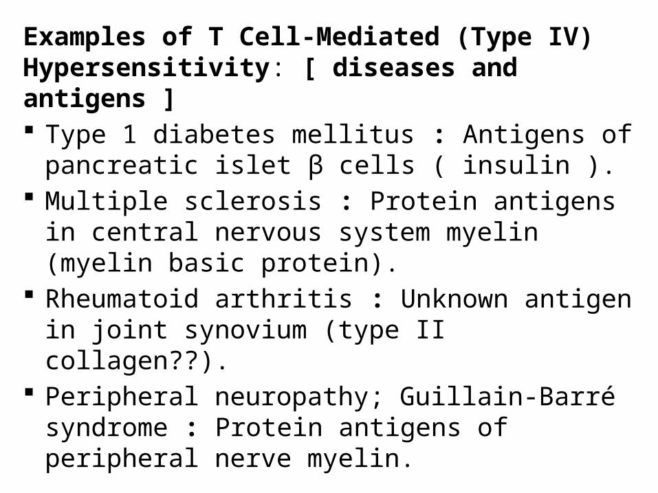
Examples of T Cell-Mediated (Type IV) Hypersensitivity: [ diseases and antigens ] Type 1 diabetes mellitus : Antigens of pancreatic
islet β cells ( insulin ). Multiple sclerosis : Protein antigens in central
nervous system myelin (myelin basic protein). Rheumatoid arthritis : Unknown antigen in joint
synovium (type II collagen??). Peripheral neuropathy; Guillain-Barré syndrome :
Protein antigens of peripheral nerve myelin.

THANK YOU