Embed Size (px)
Citation preview
Immunopathologic Studies in Relapsing Polychondritis
JERoME H. HERmAN and MARIE V. DENNIS
From the Division of Immunology, Department of Internal Medicine,University of Cincinnati Medical Center, Cincinnati, Ohio 45229
A B S T R A C T Serial studies have been performed onthree patients with relapsing polychondritis in an at-tempt to define a potential immunopathologic role fordegradation constituents of cartilage in the causationand/or perpetuation of the inflammation observed. Crudeproteoglycan preparations derived by disruptive anddifferential centrifugation techniques from human costalcartilage, intact chondrocytes grown as monolayers,their homogenates and products of synthesis providedantigenic material for investigation. Circulating antibodyto such antigens could not be detected by immunodiffu-sion, hemagglutination, immunofluorescence or comple-ment mediated chondrocyte cytotoxicity as assessed by'Cr release. Similarly, radiolabeled incorporation stud-ies attempting to detect de novo synthesis of such anti-body by circulating peripheral blood lymphocytes as as-sessed by radioimmunodiffusion, immune absorption toneuraminidase treated and untreated chondrocytes andimmune coprecipitation were negative.
Delayed hypersensitivity to cartilage constituents wasstudied by peripheral lymphocyte transformation em-ploying [3H]thymidine incorporation and the release ofmacrophage aggregation factor. Positive results wereobtained which correlated with periods of overt diseaseactivity. Similar results were observed in patients withclassical rheumatoid arthritis manifesting destructivearticular changes. This study suggests that cartilageantigenic components may facilitate perpetuation of car-tilage inflammation by cellular immune mechanisms.
INTRODUCTIONRelapsing polychondritis is a relatively rare disordercharacterized by prominent inflammation of cartilagenousstructures. Though cartilage inflammation predominates,one may observe ocular and cardiac involvement as well
This work was presented in part at the American Rheu-matism Association meeting, Washington, D. C., January1971. (1971. Arthritis Rheum. 14: 166.)
Dr. Herman is a Post-Doctoral Fellow of the ArthritisFoundation.Received for publication 1 August 1972 and in revised
form 24 October 1972.
as hematologic and serologic disturbances. There islittle insight into etiologic or pathophysiologic factorsoperative in this disease. A pathologic similarity hasbeen shown to exist between polychondritis and the ex-perimental papain (1) and hypervitaminosis-A (2) in-duced depletion of cartilaginous matrix as well as themodel of complement-dependent cartilage lysis in vitro,mediated by anti-cell membrane antibody (3, 4). How-ever, the inflammation with predominant round cellinfiltration and degeneration of chondrocytes observedin polychondritis are conspicuous differences.
It is of interest that diseases such as rheumatoid arth-ritis, Sjdgren's syndrome, and lupus errthematosus inwhich immunologic events appear to play a significantrole in the mediation of inflammation may coexist withpolychondritis. However, other than an inconclusive re-port by Dolan, Lemmon, and Teitelbaum (5), therehas been no apparent attempt to incriminate such im-munopathologic events in the pathogenesis of this disease.In the present report, serial immunologic studies per-formed on three patients with polychondritis are presentedin an attempt to define potential mechanisms operative inthe causation and/or perpetuation of the cartilage inflam-mation observed.
METHODSCartilage antigen preparation. Human cartilage antigens
were derived from two sources. The method of Malawistaand Schubert (6) employing water extraction with highspeed homogenization was utilized to secure crude proteo-glycan (PG)' from human costal cartilage. Protein poly-saccharide light (PP-L) and heavy (PP-H) derivativeswere prepared as described by Gerber, Franklin, and Schu-bert (7) based upon differential centrifugation of PG.
Chondrocytes, asepticaly isolated from human articularcartilage by collagenase digestion (8) were maintained inmonolayer culture employing Eagle's minimum essentialmedium 2 containing 20% fetal calf serum, l-glutamine, peni-
1Abbreviations used in this paper: ANA, antinuclear anti-body; MCP, metacarpophalangeal joints; PG, proteoglycan;PHA, phytohemagglutinin; PIP, proximal interphalangealjoints; PP-H, protein polysaccharide heavy derivatives;PP-L, protein polysaccharide light derivatives; SCAT,sheep cell agglutination tests; TCA, trichloroacetic acid.'Grand Island Biological Co., Grand Island, N. Y.
The Journal of Clinical Investigation Volume 52 March 1973 549
cillin, streptomycin, and mycostatin. Subculturing was un-dertaken as necessary. Studies were performed upon intactchondrocytes, their mechanical homogenates and products ofchondrocyte synthesis derived from ultrafiltration of growthmedia at time of biweekly change.
Cartilage antibody preparation. Anti-proteoglycan (anti-PG) antisera was obtained by hyperimmunizing rabbits toPG and PP-L, both of which had been pretreated with hy-aluronidase' (9) and incorporated into complete Freund'sadjuvant. Antisera was concentrated by 40% ammoniumsulfate precipitation and repeatedly absorbed with lyophi-lized normal human serum and hyaluronidase. By immuno-electrophoresis, these antisera produced two lines on reac-tion with hyaluronidase treated PG and PP-L as has beenreported previously (10). When reacted by immunodiffusionwith the chondrocyte culture media concentrate, a singleline resulted indicating the ability of these cells to synthe-size PG.
IDectection of anti-cartilage antibody. Several techniqueswere employed in an attempt to detect circulating antibodyto cartilage constituents in serial serum specimens fromthe polychondritis patients. Immunodiffusion and tanned cellhemagglutination studies were performed employing hyalu-ronidase-treated and untreated cartilage antigen prepara-tions (9). Indirect immunofluorescent techniques were un-dertaken utilizing both fixed and viable chondrocytes as asource of antigen. Slides were read on a AO Fluorolumefluorescent microscope' using BG-12 and Corning no. 5113exciter filters and an Eastman Kodak no. 15 barrier filter.'On the assumption that circulating antibody to cartilage
components might not be detectable because of specific invivo binding to antigen, an attempt was made to demon-strate such antibody in vitro by means of its synthesis byperipheral blood lymphocytes, employing ["C] amino acidincorporation technics reported previously (11). The 14C-protein synthesized de novo was chromatographically puri-fied with DEAE and G-200 chromatography into specificimmunoglobulin classes and this radio-labeled materialutilized in three types of experiments: (a) Radio-immuno-diffusion was performed in which labelled immunoglobulinand "cold" rabbit anti-human PG antibody were diffusedagainst hyaluronidase treated PG and PP-L. Radiographicpatterns were developed following 4 wk exposure to Pola-roid Type 57 Land film, ASA 3,000. (b) Having establishedan equivalence titration curve for rabbit anti-human PGand hyaluronidase treated PP-L, immune coprecipitation(11) was undertaken with labeled immunoglobulin. Resultswere contrasted with immunoglobulin synthesized by normalperipheral blood lymphocytes. Following dissolution ofwashed precipitates in 0.5N NaOH and suspension in Bray'sphosphor, liquid scintillation counting was performed usinga Mark II Liqid Scintillation System.6 (c) Specific poly-chondritis ['4C]immunoglobulin adsorption experiments wereperformed, employing suspensions of neuraminidase treatedand untreated chondrocyte preparations (12). ["C]immuno-globulins derived from normal individuals served as con-trols. In a typical experiments 5-10,000 trichloroacetic acid(TCA) precipitable counts were exposed to a 1 ml suspen-sion containing 250 X 10' chondrocytes that had been har-vested from monolayers by policing. Following 1-h rollerincubation at 370C, tubes were centrifuged at 150 g for 10min. Cell pellets were washed three times with 1 ml vol of
Worthington Biochemical Corp., Freehold, N. J.'American Optical Corp., Bedford, Mass.'Eastman Kodak Corp., Rochester, N. Y.I Nuclear-Chicago Corp., Des Plaines, Ill.
phosphate-buffered saline, the original supernates and washeswere pooled, and a portion was removed for scintillationcounting. Pellets were dissolved in 0.5NaOH and suspendedin Bray's phosphor, and liquid scintillation counting wasperformed. Percentage of counts binding to chondrocyteswas compared with counts originally added to the culturesystem as well as those recovered in supernatant portions.Antibody-mediated cytotoxicity experiments were per-
formed in which chondrocytes, labeled with sodium chro-mate, "Cr (5.0 gg/1 ml) ,7 were incubated with decomple-mented polychondritis or control sera in the presence offresh rabbit complement. Appropriate volumes of the 5'Crsolution were added to the cell suspension to give a finalradioactive level of 25 uCi/3 ml at a cell concentration of106 cells/ml. Cytotoxicity was assessed by determining iso-tope release after a 1 h incubation at 37'C. A Packardgamma scintillation system was employed (Packard Instru-ment Co., Inc., Downers Grove, Ill.).Chondrocyte DNA synthesis. To further determine the
role of humoral factors, the DNA synthesizing capacity ofchondrocytes in suspension culture was determined by pulselabeling for 16 h with [3H]thymidine' (sp. act. 11 Ci/mmol)in the presence of decomplemented polychondritis or normalsera substituted for fetal calf serum in the growth media48 h previously. Upon termination of the experiments, thecell button was pelleted by centrifuging at 150 g, DNA ex-tracted, and liquid scintillation counting performed utilizinga toluene phosphor (13). In similar experiments, chondro-cyte viability was assessed, after sera incubations, by trypanblue staining.
Cartilage antigen induced lymtphocyte blastogenesis. Theresponse of the patient's peripheral blood lymphocytes tocartilage antigens and PHA was serially assessed by [3H] thy-midine incorporation (13) and contrasted with results obtainedin 12 normal subjects and 12 patients with rheumatoid arthri-tis having radiographic evidence of destructive joint lesions.Media was supplemented by either 20% human AB+ orautologous serum, glutamine, and streptomycin. Antigens inaddition to phytohemagglutinin (0.05 ml, PHA-M) 9 con-sisted of PG (500 ,ug) both treated and untreated with hy-aluronidase, similarly processed PP-L (50 jug), chondro-cyte homogenate (500 ag), chondrocyte supernate (500 jug)and in limited experiments, viable mitomycin C treatedchondrocytes (14) which in several instances were alsoreacted with neuraminidase (12). The quantity of cartilageantigen employed was arrived at empirically based uponmaximal response of polychondritis and rheumatoid patientsand absence of stimulation in normal control cultures.Dosage range in preliminary testing consisted of the fol-lowing: PG, 5-500 ug; PP-L, 5-100 ,ug; chondrocyte ho-mogenate, 50-1,000 ,ug; chondrocyte supernate, 250-2,000,ug. Response was dose dependent, i.e., larger or smallerquantities than optimal in general associated with a negativeresponse whereas the optimal dosage gave a positive stimu-lation. This was particularly noted with the chondrocytehomogenate and supernatant antigens whereas a greaterconsistency of positivity was observed using PG and PP-Lin various dose ranges.
Tests were performed in duplicate or triplicate. At the endof an incubation period of 3 days for PHA and 6 days forcartilage antigens, a 2 h pulse labeling with [3H]thymidinewas undertaken after which DNA was extracted and liquidscintillation counting was performed. Significant stimulation
'Mallinckrodt Chemical Works, St. Louis, Mo.' Schwartz-Mann BioResearch, Orangeburg, N. Y.9Difco Laboratories, Detroit, Mich.
550 J. H. Herman and M. V. Dennis

was considered to be present if the antigen stimulated cul-ture had twice the incorporation of the control. A secondin vitro correlate of delayed hypersensitivity, sensitizedlymphocyte release of macrophage aggregation factor (15)wras evaluated serially in a single patient and its resultscorrelated with the mitogenic experiments. Comparable quan-tities of cartilage antigenic constituents were employed.
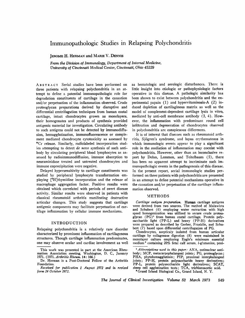
Patients. Case 1. I. M., a 55-yr-old white male, chainfood store manager, noted onset of episodic arthralgias withoccasional swelling of the knees and ankles in 1967. InJanuary 1969, the frequency of joint attacks increased withelbows, wrists, metacarpophalangeal (MCP) and proximalinterphalangeal (PIP) joints involved. 3 months subse-quently, in conjunction with a low grade temperature andmalaise, episcleritis occurred followed shortly thereafterby pain and swelling of both ears with compromise of thecanals due to collapse (Fig. 1). Audiograms demonstrateda mild high-toned hearing deficit bilaterally. Complete bloodcount, serum protein determinations, LE preparations, anti-nuclear antibody (ANA), rheumatoid factor as determinedby latex fixation and sensitized sheep cell agglutination(SCAT) tests, serum complement, VDRL, uric acid, liverfunction studies, and urinalysis were unremarkable. X-raysof the joints were negative. Westergren sedimentation rates,haptoglobulin and orosomucoid levels were elevated andelectrocardiogram demonstrated first degree heart block. Ashort trial of phenylbutazone failed to afford relief and thepatient was maintained on salicylates.Daily low dose steroid was instituted in August 1969 but
in ensuing months joint involvement became more wide-spread with arthralgias of several days duration predomi-nating. In addition the hearing deficit continued to worsenand vertigo and a stenosing tenosynovitis in the region of thedistal left forearm developed. A peripheral leukocytosis wasnoted in face of elevated levels of serum IgG and the C3component of complement. Steroids were discontinued inearly 1970 in face of clinical improvement though musculo-skeletal and auricular distress continued to a lesser degree.In May 1970, an ischemic optic neuritis developed whichwas treated with high dosage corticosteroids but neverthe-less blindness ensued in the right eye. In July 1970, thepatient experienced a left middle cerebral thrombosis withresidual right hemiparesis and an expressive aphasia. Sub-sequent treatment has consisted of steroids and azathioprinewith resultant stability in his clinical status.Case 2. A. K, a 57-yr-old white male fireman, presented
with a history of a transient 3 month episode of polyneuritisand vertigo in 1964. In 1966, mild arthralgia with occasionaljoint swelling appeared in face of elevated serum IgG, IgA,IgM, and normal complement levels. A diagnosis of possiblerheumatoid arthritis was made and short term, low dosesteroid therapy instituted. In 1967, fleeting generalizedarthralgias with large joint swelling appeared with hightemperature. The patient was anemic (hematocrit 30) withnegative Coombs' test. Bone marrow examination revealed8-10% plasma cells. The polyclonal gammopathy persisted.Symptoms largely subsided on low dose steroids but lowgrade temperature and occasional joint swelling continued.Cardiopulmonary and renal status were unremarkable.In February 1968, the patient presented with an intractable
foot ulcer with complicating osteomyelitis of the fifth digitof the right foot. He was hypertensive and had a diabeticglucose tolerance curve. Latex fixation was positive with aSCAT titer of 1: 28, serum IgA markedly elevated but ANA,LE prep, VDRL, and cryoglobulin determinations were nega-tive. In ensuing months, transient pulmonary infiltrates were
-* 9 [3 [s t " . -. X .w ! z E iN,, * -, d t :, g | , " , | r*s . i { | W. ;' g4 s :.7z$3 E N | rs... , ,i..w F..,,. ., .g R ,%. w t ....... ,.m S, . |.s . Lt in: ............... . t t ..... |., . e r
'' ,, ,, sx , v . a -w.!,, t bts s b
,,ts . ; j,@ P: : ^<] .5 *... t \. _ i
s i a3 t i v -wi g
:d's q qr # w
_iffi ; i6, *; ;$
'^{ ,; 1W;< R ., ' g > C t ', r S i n 'Me .St s* . :. t ' ... :,> ,> ffi . - Zae ,; , .......... . . S> ... .iS SI'*s;' w 'w
eS e:s '
2
.. .. X :'*-:
.: J .::
.rT f
FIGURE 1 Distortion and collapsepatient I. WI.
of the external ear in
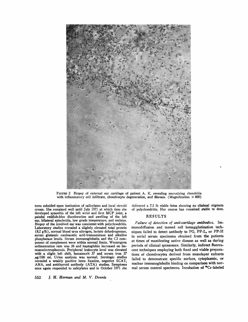
noted and poorly defined dermal lesions appeared, biopsy ofthe latter consistent with acute vasculitis. In 1969, in thepresence of pronounced peripheral vascular insufficiency, aright below-knee amputation was performed, the pathologicspecimen revealing a necrotizing vasculitis. Shortly there-after, the patient developed bilateral external auricular painand inflammation, collapse of nasal cartilage, conjunctivitis,and increased myalgia and arthralgia. Pinna biopsy demon-strated a necrotizing chondritis (Fig. 2). Treatment con-sisted of high dose steroid with resultant improvement. Inearly 1970, a left below-knee amputation was performed inface of gangrenous lesions. In July 1970, the patient experi-enced occlusion of the superior mesenteric artery with infarc-tion of the entire small bowel and succumbed. Necropsy re-vealed healed polyarteritis of the kidneys and testes, myo-carditis, and polychondritis. There was no evidence of rheu-matoid arthritis.Case 3. H. M., a 35-yr-old white female physician, pre-
sented with a history of onset in 1966 of polyarticular ar-thralgias, intermittent swelling of PIP joints, and whatappeared to be a tendinitis or tenosynovitis involving thedorsal and ventral surfaces of the wrists. Salicylates af-forded relief. Symptoms persisted to a variable degree untilApril 1971, when two months pregnant, she noted the acuteonset of painful swelling involving the right ear in associa-tion with an erythematous maculopapular eruption on theupper extremities, malaise, fever, and conjunctivitis. Symp-
Immunopathologic Studies in Relapsing Polychondritis 551
... .. .... . . .. ..: . , ' . ... . .k'.',, . ;"; ';"@,'s' "'':." . :.:. . ' S *Ast:,, s,..:. ! s'.- >i. .. . .
-. sr.. ..':. <. :.
tjUE; s .'
*.,f;g¢''' ...... '"' . :,g
NESgtf .. 't ,S;:: i;,':3R' :> !.,,,. S.. ,.....
t'..'..: ..: .::_'' S '--
e>eettY/ ::....... .... ..
s ..
.6.-,Sss.- ¢,-.x. ..
...: .. : ::.' '. . ,*E F....... .o . . . . ...: ...., + .: #
*.;i ....r 's
u
::::
::
.Xi.;.
4. w >a,sl ~ w
; '.~~~~~~~~~~~~~~~4
W-P ~ 4
wj-t *,#0*e, * tf**
pS .8%4 .. .S
j I 0 !* ,44S
!~~~~~24 S *' 5*i*s~
,,.eqS ,- ,;,, '.4
4
ji ~ ,.J .?ts
SS ., p'
b iS ._ @ @ t~~~~ S S
V ^ tilt v '
._9 :
*t"
's
..
FIGUiE: [X14>||>\-1t7X1\'iliil Lt^;II t'.il I i .tLIL Il1 LL~ltl~t, R \ KX. I i~ chicillt} 'titil/Ii\ II ndritiswith ill I tI ii Iui cit hr. cLtll it ci :i 1 tEh ,;. C1 i Iii X 400)
toms subsided upon institution of salicylates and local steroidcream. She remained well until July 1971 at which time shedeveloped synovitis of the left wrist and first MCP joint, apainful reddish-blue discoloration and swelling of the leftear, bilateral episcleritis, low grade temperature, and malaise.Biopsy of the involved ear was consistent with polychondritis.Laboratory studies revealed a slightly elevated total protein(8.2 g%), normal blood urea nitrogen, lactate dehydrogenase,serum glutamic oxaloacetic acid-transaminase and alkalinephosphatase levels. Serum immunoglobulin and the C3 com-ponent of complement were within normal limits. Westergrensedimentation rate was 36 and haptoglobin increased on im-munoelectrophoresis. Peripheral leukocyte level was elevatedwith a slight left shift, hematocrit 37 and serum iron 57,ug/100 ml. Urine analysis was normal. Serologic studiesrevealed a weakly positive latex fixation, negative SCAT,ANA, and antithyroid antibody (ATA) studies. Symptomsonce again responded to salicylates and in October 1971 she
delivered a 7.5 lb viable fetus showing no clinimal stigmataof polychondritis. Her course has remained stable to date.
RESULTSFailure of detection of anti-cartilage antibodies. Im-
munodiffusion and tanned cell hemagglutination tech-niques failed to detect antibody to PG, PP-L, or PP-Hin serial serum specimens obtained from the patientsat times of manifesting active disease as well as duringperiods of clinical quiescence. Similarly, indirect fluores-cent techniques employing both fixed and viable prepara-tions of chondrocytes derived from monolayer culturesfailed to demonstrate specific surface, cytoplasmic, or
nuclear immunoglobulin binding on comparison with nor-
mal serum control specimens. Incubation of 'Cr-labeled
552 J. H. Herman and M. V. Dennis
Ws.
.. ;.:-".,:.:-
se
chondrocytes with decomplemented polychondritis seraand a source of fresh rabbit complement did not facilitatea degree of release of label differing from normal controlsera.
Experiments designed to detect specific anti-PG anti-body synthesis by peripheral blood lymphocytes derivedfrom serial bleedings of patients I. M. and A. K. were
unsuccessful. No radioactivity was detectable in precipi-tin lines developing upon reacting labeled plus "cold"rabbit anti-cartilage antibody with hyaluronidase-treatedPG and PP-L. In immune coprecipitation experiments,no difference in precipitable radioactivity was observedafter the interaction of labeled immunoglobulin synthe-sized by polychondritis and normal peripheral bloodlymphocytes with PP-L and anti-cartilage antibody atequivalence.Immune adsorption experiments employing suspensions
of chondrocytes revealed no significant difference be-tw-een binding of polychondritis-labeled immunoglobulinsas compared with controls. Prior treatment of chondro-cytes with neuraminidase did not influence the degree ofradiolabeled adsorption.
Effect of polychondritis sera on chondrocyte viabilityand DNA synthesis. Decomplemented polychondritissera obtained during variable stages of clinical activitywere substituted for fetal calf serum in suspension cul-tures of chondrocytes 48 h before a 16 h pulse labelingwith [3H]thymidine. These studies failed to demonstrateeither enhanced or diminished DNA synthesis when sucha substitution was made. The cell viability was similar incultures augmented with polychondritis sera and fetalcalf serum.
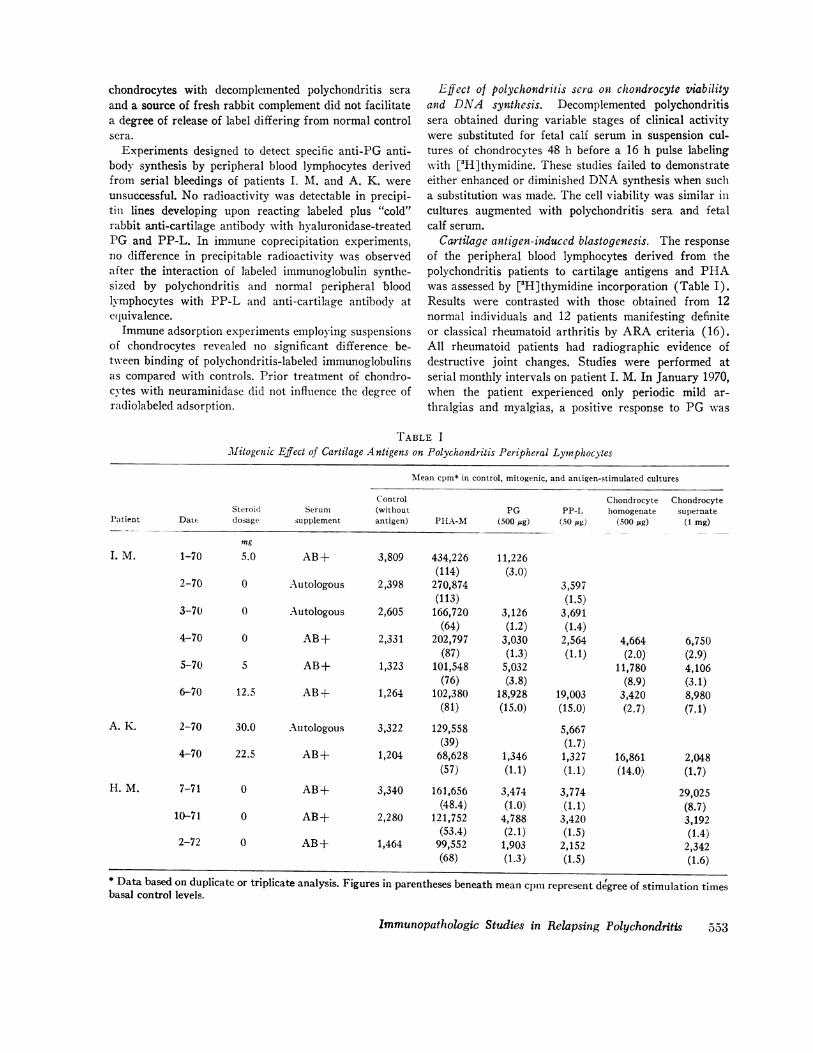
Cartilage antigen-induced blastogenesis. The responseof the peripheral blood lymphocytes derived from thepolychondritis patients to cartilage antigens and PHAwas assessed by [3H]thymidine incorporation (Table I).Results were contrasted with those obtained from 12normal individuals and 12 patients manifesting definiteor classical rheumatoid arthritis by ARA criteria (16).All rheumatoid patients had radiographic evidence ofdestructive joint changes. Studies were performed atserial monthly intervals on patient I. M. In January 1970,when the patient experienced only periodic mild ar-
thralgias and myalgias, a positive response to PG was
TABLE IMitogenic Effect of Cartilage Antigens on Polychondritis Peripheral Lymphocytes
Mean cpm* in control, mitogenic, and antigen-stimulated cultures
SteroidPatient Date dosage
I. M. 1-70
2-70
3-70
4-70
5-70
6-70 1
ControlSerum (without
supplement antigen)
mg
5.0 AB+ 3,809 434,226(114)
0 Autologous 2,398 270,874(113)
0 Autologous 2,605 166,720(64)
0 AB+ 2,331 202,797(87)
5 AB+ 1,323 101,548(76)
12.5 AB-+ 1,264 102,380(81)
ChondrocytePG PP-L, homogenate
PIIAX-M (500 mg) (.50 Mg) (500 pg)
11,226(3.0)
3,126(1.2)3,030(1.3)5,032(3.8)
18,928(15.0)
3,597(1.5)3,691(1.4)2,564 4,664(1.1) (2.0)
11,780(8.9)
19,003 3,420(15.0) (2.7)
Chondrocytesupernate
(1 mg)
6,750(2.9)4,106(3.1)8,980(7.1)
2-70 30.0 Autologous 3,322 129,558(39)
4-70 22.5 AB+ 1,204 68,628(57)
AB+
AB+
AB+
3,340 161,656(48.4)
2,280 121,752(53.4)
1,464 99,552(68)
5,667(1.7)
1,346 1,327(1.1) (1.1)
3,474(1.0)4,788(2.1)1,903(1.3)
3,774(1.1)3,420(1.5)2,152(1.5)
16,861 2,048(14.0) (1.7)
29,025(8.7)3,192(1.4)2,342(1.6)
Immunopathologic Studies in Relapsing Polychondritis
A. K.
H. M. 7-71
10-71
2-72
0
0
0
* Data based on duplicate or triplicate analysis. Figures in parentheses beneath mean cpm represent degree of stimulation timesbasal control levels.
0553
elicited, AB positive serum being employed as supple-ment. In the ensuing 2 months, negative results were ob-tained upon stimulation with PG and PP-L, an autolo-gous serum being employed as supplement. As an in-hibitor substance impairing lymphocyte transformationupon exposure to specific antigen has been described inother autologous culture systems (17-22), in subsequentcultures only AB positive sera was employed. In April,though failing to respond to PG and PP-L, the chondro-cyte homogenate and culture supernatant results werepositive. In the months of May and June, during a clini-cal exacerbation characterized by optic neuritis, syno-vitis, and fever, positive responses were obtained to allantigens tested. During this period, stimulation withviable mitomycin-treated chondrocytes and mitomycin-neuraminidase-treated cells was negative. Stimulation oflymphocytes with hyaluronidase alone failed to induceblast transformation. Two lymphocyte transformationstudies were performed on patient A. K. The initialexperiment was performed during a period of clinicalstability, the patient receiving 30 mg of prednisone daily.At this time his peripheral blood lymphocytes failed torespond to variable quantities of hyaluronidase treatedPP-L or hyaluronidase alone. 2 months before hisdeath however, the patient, though demonstrating a nega-tive response to PG, PP-L, and culture supernatant, hada positive stimulation to chondrocyte homogenate witha value 14 times base level. Three studies were performedon patient H. M. When manifesting clinical activity, apositive response to chondrocyte culture supernate wasobtained with a value 8.7 times base level. 4 months later,with symptoms controlled with salicylates, only a weeklypositive response to PG antigen was obtained followedby negative results during a subsequent inactive phase.PHA response was normal in each of the three patients.Macrophage aggregation factor was studied in pa-
tient H. M. in conjunction with the mitogenic experi-ments. A single positive aggregation response to chon-drocyte supernatant antigen was noted at time of the ini-tial bleeding, correlating with the positive blastogenicresult.
Control stimulation studies performed on 12 normaladults using a similar battery of cartilage antigens were
consistently negative except for a single low grade posi-tive response using the chondrocyte supernatant antigen(Table II). A repeat study in this individual gave anegative result. Transformation to one or more cartilageantigens was noted however, in 9 of 12 of the rheuma-toid patients. Response of 5 of 12 patients to PG was
positive with a mean incorporation of 4.0 times control;3 of 12 responded to PP-L, mean response of 6.6; 7 of12 to chondrocyte culture supernate, mean value of 9.6;4 of 12 to chondrocyte homogenate, mean response 6.3times base. A detailed evaluation of the immunologic
significance of degradation products of cartilage as re-gards causation and perpetuation of rheumatoid jointinflammation will be published independently (23).
DISCUSSION
The pathophysiologic basis underlying the developmentof the destructive cartilagenous changes observed in re-lapsing polychondritis is unknown. An explanation mustbe afforded for not only loss of metachromatic stainingof matrix but its fragmentation in conjunction withchondrocyte dissolution, mononuclear cell infiltrationand consequent fibrosis.
Involvement of a proteolytic process is inferred byexperimental studies. The capacity of lysosomal en-zymes to degrade the proteoglycans of cartilage matrixhas been shown in in vitro studies by Ziff, Gribetz, andLospalluto (24) and Weissman and Spillberg (25).The intravenous injection of crude papain (2) and theexperimental production of a state of hypervitaminosisA (3) have also induced a transient dissolution of car-tilagenous matrix secondary to loss of glycosaminogly-cans. Associated irreparable chondrolysis has been pro-duced by Lack (26) through plasmin activation bytrauma or streptococcal or staphlococcal kinases in syn-ovial fluid. Harris, DiBona, and Krane (27) have shownthat similar changes could be induced by a collagenasesynthesized by rheumatoid synoviocytes in in vitroculture experiments.Although such evidence suggests involvement of a pro-
teolvtic process in the pathogenesis of polychondritis,there is limited insight into how such enzyme activation,primarily or secondarily induced, occurs. A secondarychemical mediation is suggested by experimental studiesof ,Moskowitz et al. (28) in which components of car-tilage have been shown capable of activating Hagemanfactor and the generation of kinins. In the present study,in limited experiments we have not been able to identifya humoral factor analogous to the papain model. Seraderived at times of varying stages of clinical activityfailed to affect either chondrocyte DNA synthesis orviability.Though cartilage has been regarded as an immuno-
logically "privileged" tissue due to its lack of vascularity(29, 30), it is tempting to incriminate immunologicevents in the pathogenesis of polychondritis. If the com-
ponents of cartilage matrix are enzymatically degradedthereby allowing exposure of chondrocytes, transplanta-tion immunity is capable of being engendered with even-
tual chondrolysis (31). Furthermore, the proteoglycancomponent of cartilage matrix has been shown to beantigenic (9, 10, 32), the antigenicity appearing to re-
side at the site of union of the glycosaminoglycan andprotein moieties. Chondroitin sulfate alone is not anti-genic. In a study by Glvnn and Holborow (33), how-
554 1. H. Herman and M. V. Dennis

TABLE I IMitogenic Effect of Cartilage Antigens on Normal Peripheral Lymphocytes
Mean cpm* in control, mitogenic, and antigen-stimulated cultures
Control Chondrocyte ChondrocyteSerum (without PG PP-L homogenate supernate
Test supplement antigen) PHA-M (500 jsg) (50 jsg) (500 'Ug) (1 mg)
M. D. AB+ 1,489 93,807 2,025 1,444 1,385 2,984(63) (1.4) (1.0) (0.93) (2.0)
M. D. (repeat) AB+ 3,624 172,502 3,733 3,436 4,059 4,342(48) (1.0) (0.95) (1.0) (1.2)
J. H. AB+ 1,193 25,291 1,216 1,766 1,200 1,002(21) (1.0) (1.5) (1.0) (0.84)
M. P. AB+ 2,430 157,221 3,013 3,062 1,510 2,600(65) (1.2) (1.3) (0.62) (1.1)
D. W. AB+ 2,596 275,954 2,518 2,682 3,297 2,492(106) (0.97) (1.0) (1.3) (0.96)
M. W. AB+ 2,033 245,586 2,094 2,196 2,236 2,432(121) (1.0) (1.1) (1.1) (1.2)
B. G. AB+ 2,334 50,648 1,954 4,271 2,562(22) (0.84) (1.8) (1.1)
G. S. AB+ 6,389 228,726 8,178 5,967 11,244(36) (1.3) (0.93) (1.8)
M. K. AB+ 4,205 110,591 5,803 5,382 5,298(26) (1.4) (1.3) (1.3)
A. H. AB+ 7,929 363,148 6,954 8,718 6,185(46) (0.88) (1.1) (0.78)
J. B. AB+ 1,984 142,252 2,758 2,718 2,372(72) (1.4) (1.4) (1.2)
M. Y. AB+ 5,613 483,840 6,567 6,346 6,172(86) (1.2) (1.1) (1.1)
J. F. AB+ 3,326 155,324 3,480 3,958 3,647(47) (1.0) (1.2) (1.1)
* Data based on duplicate or triplicate analysis. Figures in parentheses beneath mean cpm represent degreeof stimulation times basal control levels.
ever, a vaccine prepared from group A P-hemolyticstreptococci in the presence of chondroitin sulfate pro-duced precipitating antibody to this polysaccharide. Rab-bits immunized systemically with this preparation de-veloped synovial lesions. Immunization with streptococcior chondroitin sulfate alone failed to produce suchchange. No comment was made as to cartilage pathol-ogy. Strobel and Seifert (34) were not able to reproducethese results.
Studies by Lloyd-Roberts (35) and Hulten andGellerstedt (36) have shown that intra-articular injec-tions of finely divided homologous or autologous hyalinecartilage could induce a synovitis, its intensity dependentupon the frequency and duration of injection. No car-
tilagenous lesions were observed. Using homogenizedsuspensions of autologous canine costal cartilage, Chris-man, Fessel, and Southwick (37) observed that pro-tracted intra-articular injection induced not only synovi-
tis but marginal exostosis and cysts. There was, how-ever, no apparent damage to articular weight bearingsurfaces as evidenced by gross or microscopic change.Considering the recognized immunogenicity of cartilageconstituents and the ability to induce systemic as wellas local immunization following intra-articular antigenicchallenge (38), it is difficult to understand why an ex-pected immune response failed to induce cartilage degra-dation. Possibly matrix integrity afforded a protectivebarrier. Such studies therefore suggest the inability of apotential primary immunologic insult to induce cartilagedamage. If immunologic events are operative, they morelikely would function in a secondary capacity, that is,function as a means of perpetuating cartilage damagesubsequent to alteration in the integrity of this tissue in-duced by other means.
Several lines of evidence suggest possible immunologicmechanisms operative in relapsing polychondritis. This
Immunopathologic Studies in Relapsing Polychondritis 555

disease not infrequently co-exists with connective tissuedisorders such as rheumatoid arthritis, systemic lupuserythematosus and Sjogren's syndrome in which im-munologic events are of acknowledged significance inpathogenesis (5). Patient A. K. in our series, appearsto be the first documented association with polyarteritisnodosa. The speculations of Christian (39) are intriguingrelative to defining an immunologically directed insultagainst proteoglycans which function as a shared con-nective tissue constituent common to cartilage, eye, andaorta, all of which may be involved in this disease proc-ess. The integrity of cartilage is dependent upon the pre-servation of its matrix making this structure more vul-nerable than the others to damage. It is of interest thatcross-reactivity has been shown to exist between strepto-coccal hyaluronate and a crude proteoglycan preparationof human cartilage (40). The frequency of an antedatedor enhanced incidence of streptococcal infections in thepolychondritis population has not been evaluated.
Dolan, Lemmon, and Teitelbaum (5) attempted to in-criminate immunopathologic events in the pathogenesisof polychondritis. Using sera derived from two patients,they employed immunofluorescence to demonstrate locali-zation of immunoglobulin at the periphery of lacunarspaces and in nuclear and cytoplasmic regions of chon-drocytes. The results were inconclusive because of con-siderable background fluorescence observed in controlpreparations. Recognizing this difficulty, we resorted touse of isolated chondrocytes for fluorescent experiments.
In the present study, we have been unable to identifyeither circulating or PBL synthesized antibody to car-tilage matrix or chondrocyte antigenic components. Itappears unlikely that physiochemical changes occurringin our lines of cultured chondrocytes are responsible forthe negative fluorescent and complement mediated cyto-toxicity results as continued synthesis of matrix con-stituents could be identified.The cartilage antigens used proved mitogenic to the
peripheral blood lymphocytes of our polychondritis pa-tients. It is to be noted that the proteoglycan prepara-tion employed was isolated by disruptive technique andcontained contaminants in addition to recognized pro-tein components of cartilage matrix. Prior to absorp-tion of anti-PG antisera with normal human serum,alpha, beta and at times even gamma globulin componentscould be identified by immunoelectrophoresis. One can-not exclude the possibility that the protein componentof proteoglycan subunit or link protein is the antigen in-ducing response. Nonetheless, the importance lies in therecognition that a constituent or constituents present innormal cartilage were able to induce such a response.Though blast transformation is not an absolute in vitro
correlate of delayed hypersensitivity, it is certainly an-other parameter of cellular immunity. In a study of the
immunologic significance of degradation products ofcartilage as regards the causation and/or perpetuation ofrheumatoid joint inflammation (23), we performed simi-lar lymphocyte stimulation studies with cartilage anti-gens and found that the results correlated almost com-pletely with other more conclusive parameters of cellularimmunity, that of liberation of macrophage aggregationand cytotoxicity factors. Release of aggregation factorwas studied in only one polychondritis patient, beingpositive at time of initial activity bu negative thereafterduring clinical quiescence.The variability noted as regards the peripheral blood
lymphocyte response to cartilage antigen is of interest.Dosage of salicylate or steroid being administered did notappear to be the responsible factor. Autologous serumpotentially contained an inhibitor to specific mitogenesis,yet did not alter PHA response. Substrates of unknowncomposition capable of inhibiting lymphocyte stimulationhave been described in the plasma of patients with vari-ous disease process (17-22). The periodic positive re-sponses observed with chondrocyte homogenate or prod-ucts of synthesis in face of negative stimulation to PGand PP-L might reflect either a quantitative antigenicphenomenon or the presence of chondrocyte constituentsof antigenic composition other than the mature proteogly-can matrix. No attempt has been made to identify thecomponents of either the chondrocyte homogenate or itssynthetic products other than detection of an antigenicconstituent reactive by Ouchterlony with absorbed anti-PG antisera.The situation presented in which lack of antigen stimu-
lated response during disease remission and the ap-pearance of such a response during exacerbation isdistinctly unusual. There are, however, few reported in-stances in which serial investigations of delayed hyper-sensitivity have been reported in human disease. It isconceivable that during period of remission, as a con-sequence of cessation of further exposure to antigen,that specifically sensitized thymus derived lymphocytesmay be at a minimum. An analogous situation may befound in experimental literature in studies of transfer ofdelayed hypersensitivity to a haptenic constituent inguinea pigs (41). Such a cellular transfer could not beachieved beyond 84 days post initial immunization butcould be accomplished a short interval after boosting ofthe donor animal.Though unable to identify antibody to cartilage com-
ponents, lymphocyte sensitization to such antigens ap-pears to be present in relapsing polychondritis as well as
in patients with seropositive rheumatoid arthritis hav-
ing radiographic evidence of destructive joint changes.This would support the hypothesis that immune mecha-nisms may be involved in the pathogenesis of polychon-dritis. It would appear unlikely that such a mechanism
556 J. H. Herman and M. V. Dennis
would function in a provocative, causal manner butcould be a basis for the perpetuation of cartilage inflam-mation once its integrity has been altered.
ACKNOWLEDGMENTSWe acknowledge the cooperation of the Department ofMedicine of the Cincinnati Veterans Administration Hospitaland Dr. N. R. Abrams for allowing study of their patients.We are grateful to Dr. Evelyn V. Hess for advice and en-couragement and for reviewing this manuscript.
This investigation was supported in part by TrainingGrant AM 05044 from the National Institute of Arthritis andMetabolic Diseases and an Arthritis Foundation ClinicalResearch Center grant.
REFERENCES1. Thomas, L., 1956. Reversible collapse of rabbit ears
after intravenous papain and prevention of recovery bycortisone. J. Exp. Med. 104: 245.
2. Thomas, L., R. T. McCluskey, J. L. Potter, and G.Weissmann. 1960. Comparison of the effects of papainand vitamin A on cartilage. I. The effect in rabbits. J.Exp. Med. 111: 705.
3. Fell, H. B., R. R. A. Coombs, and J. T. Dingle. 1966.The breakdown of embryonic (chick) cartilage and bonecultivated in the presence of complement sufficient anti-serum. I. Morphological changes, their reversibility andinhibition. Int. Arch. Allergy Appl. Immunol. 30: 146.
4. Lachmann, P. J., R. R. A. Coombs, H. B. Fell, and J.T. Dingle. 1969. The breakdown of embryonic (chick)cartilage and bone cultivated in the presence of comple-ment sufficient antiserum. III. Immunological analysis.Int. Arch. Allergy. Appi. Immunol. 36: 469.
5. Dolan, D. L., G. B. Lemmon, Jr., and S. L. Teitelbaum.1966 Relapsing polychondritis. Analytical literature re-view and studies on pathogenesis. Am. J. Med. 41: 285.
6. Malawista, I., and M. Schubert. 1958. Chondromucopro-tein: new extraction method and alkaline degradation.J. Biol. Chem. 230: 535.
7. Gerber, B. R., E. C. Franklin, and M. Schubert 1960.Ultracentrifugal fractionation of bovine nasal chondro-mucoprotein. J. Biol. Chem. 235: 2870.
8. Manning, W. K., and W. M. Bonner, Jr. 1967. Isolationand culture of chondrocytes from human adult articularcartilage. Arthritis Rheum. 10: 235.
9. Sandson, J., L. Rosenberg, and D. White. 1966. Theantigenic determinants of the protein-polysaccharides ofcartilage. J. Exp. Med. 123: 817.
10. Loewi, G., and H. Muir. 1965. The antigenicity of chon-dromucoprotein. Immunology. 9: 119.
11. Herman, J. H., J. Bradley, M. Ziff, and J. D. Smiley.1971. Response of the rheumatoid synovial membraneto exogenous immunization. J. Clin. Invest. S0: 266.
12. Ray, P. K., H. Gewurz, and R. L. Simmons. 1970.The mechanism of increased sensitivity of neuramini-dase treated cells to antibody induced cytolysis. Fed.Proc. 29: 573.
13. Goldman, J. A., A. Litwin, L. E. Adams, R. C. Krueger,and E. V. Hess. 1972. Cellular immunity to nuclearantigens in systemic lupus erythematosus. J. Clin. Invest.51: 2669.
14. Bach, F. H., and N. K. Voynow. 1966. One-way stimu-lation in mixed leukocyte cultures. Science (Wash.D. C.). 153: 545.
15. Lolekha, S., S. Dray, and S. Gotoff. 1970. Macrophageaggregation in vitro: a correlate of delayed hypersensi-tivity. J. Immunol. 104: 296.
16. Ropes, M. W., G. A. Bennet, S. Cobb, R. Jacox, andR. A. Jessar. 1959. The 1958 revision of diagnostic cri-teria for rheumatoid arthritis. Arthritis Rheum. 2: 16.
17. Paronetto, F., and H. Popper. 1970. Lymphocyte stimu-lation induced by halothane in patients with hepatitisfollowing exposure to halothane. N. Engl. J. Med. 283:277.
18. Heilman, D. H., and W. McFarland. 1966. Inhibition oftuberculin-induced mitogenesis in cultures of lympho-cytes form tuberculous donors. Int. Arch. Allergy. 30:58.
19. Levene, G. M., J. L. Turk, D. J. M. Wright, and A. G.S. Grimble. 1969. Reduced lymphocyte transformationdue to a plasma factor in patients with active syphilis.Lancet. 2: 246.
20. Silk, M. 1967. Effect of plasma from patients withcarcinoma on in vitro lymphocyte transformation. Cancer.20: 2088.
21. Knowles, M., D. Hughes, E. A. Caspary, and E. J.Field. 1968. Lymphocyte transformation in multiplesclerosis: inhibition of unstimulated thymidine uptake bya serum factor. Lancet. 2: 1207.
22. Winter, G. C. B., C. F. McCarthy, A. E. Read, andJ. M. Yoffey. 1967. Development of macrophages in phy-tohaemagglutinin cultures of blood from patients withidiopathic steatorrhoea and with cirrhosis. Br. J. Exp.Pathol. 48: 66.
23. Herman, J. H., D. W. Wiltse, and M. V. Dennis. 1972.Immunopathologic significance of cartilage degradationproducts in rheumatoid joint inflammation. ArthritisRheum. 15: 375. (Abstr.)
24. Ziff, M., H. J. Gribetz, and J. Lospalluto. 1960. Effectof leukocyte and synovial membrane extracts on carti-lage mucoprotein. J. Clin. Invest. 39: 405.
25. Weissmann, G., and I. Spilberg. 1968. Breakdown ofcartilage proteinpolysaccharide by lysosomes. ArthritisRheum. 11: 162.
26. Lack, C. H. 1959. Chondrolysis in arthritis. J. Bone Jt.Surg. B Br. Vol. 41B: 384.
27. Harris, E. D., Jr., D. R. DiBona, and S. M. Krane.1970. A mechanism for cartilage destruction in rheuma-toid arthritis. airthritis Rheum. 13: 321. (Abstr.)28. Moskowitz, R. W., H. J. Schwartz, B. Michel, 0. D.Ratnoff, and T. Astrup. 1970. Generation of kinin-likeagents by chondroitin sulfate, heparin, chitin sulfate andhuman articular cartilage: possible pathophysiologic im-plications. J. Lab. Clin. Med. 76: 790.
29. Craigmyle, M. B. L. 1960. A study of cartilage homo-grafts in rabbits sensitized by a skin homograft fromthe cartilage donor. Transplant. Bull. 26: 150.
30. Gibson, T. 1967. The transplantation of cartilage. J.Clin. Pathol. 20 (Suppl.): 513.31. Heyner, S. 1969. The significance of the intercellular
matrix in the survival of cartilage allografts. Trans-plantation. 8: 666.32. DiFerrante, N. 1964. Precipitins in the rabbit producedby protein polysaccharide from bovine nasal cartilage.Science (Wash. D. C.). 143: 250.33. Glynn, L. E., and E. J. Holborow. 1952. Conversion oftissue polysaccharides to auto-antigenis by group-A beta-haemolytic streptococci. Lancet. 2: 449.
Immunopathologic Studies in Relapsing Polychondritis 557
34. Strobel, V. W., and G. Seifert. 1961. Zur panchondritisrheumatica. Z. Rheumaforsch. 20: 247.
35. Lloyd-Roberts, G. C. 1953. The role of capsular changesin osteoarthritis of the hip joint. J. Bone Dt. Surg. 35B:627.
36. Hulten, O., and N. Gellerstedt. 1940. Uber Abnutzungs-produkte in Gelenken und ihre Resorption unter demBilde einer Synovitis detritica. Acta. Chir. Scand. 84: 1.
37. Chrisman, 0. D., J. M. Fessel, and W. 0. Southwick.1965. Experimental production of synovitis and mar-ginal articular exostoses in the knee joints of dogs.Yale J. Biol. Med. 37: 409.
38. Jasin, H. E., and M. Ziff. 1969. Immunoglobin and spe-cific antibody synthesis in a chronic inflammatory focus:antigen induced synovitis. J. Immunol. 102: 355.
39. Christian, C. L. 1970. Discussion of paper: relapsingpolychondritis with aortic involvement by Owen, D. S.,R. Irby, and E. Toone. Arthritis Rheum. 13: 880.
40. Sandson, J. L., D. Hamerman, R. Jones, R. Janis, andM. Rojkind. 1968. Immunologic and chemical similaritiesbetween the streptococcus and human connective tissue.Trans. Assoc. Am. Physicians Phila. 81: 249.
41. Phair, J. P., and F. S. Kantor. 1968. Delayed hyper-sensitivity. II. Persistence and conjugate specificity ofthe transfer reaction. J. Immunol. 100: 302.
558 J. H. Herman and M. V. Dennis