Embed Size (px)
Citation preview
Research ArticleImmunomodulatory Effects of Diterpene QuinoneDerivatives from the Roots of Horminum pyrenaicum inHuman PBMC
K. Becker,1 S. Schwaiger,2 B. Waltenberger,2 D. Fuchs ,1 C. K. Pezzei,2,3 H. Schennach,4
H. Stuppner ,2 and J. M. Gostner 5
1Division of Biological Chemistry, Biocenter, Medical University of Innsbruck, Innsbruck, Austria2Institute of Pharmacy/Pharmacognosy and Center for Molecular Biosciences Innsbruck (CMBI), University of Innsbruck,Innsbruck, Austria3Institute for Analytical Chemistry and Radiochemistry and CMBI, University of Innsbruck, Innsbruck, Austria4Central Institute of Blood Transfusion and Immunology, University Hospital of Innsbruck, Innsbruck, Austria5Division of Medical Biochemistry, Biocenter, Medical University of Innsbruck, Innsbruck, Austria
Correspondence should be addressed to D. Fuchs; [email protected] and H. Stuppner; [email protected]
Received 27 April 2017; Revised 19 August 2017; Accepted 3 October 2017; Published 14 January 2018
Academic Editor: Shane Thomas
Copyright © 2018 K. Becker et al. This is an open access article distributed under the Creative Commons AttributionLicense, which permits unrestricted use, distribution, and reproduction in any medium, provided the original work isproperly cited.
Several phytochemicals were shown to interfere with redox biology in the human system. Moreover, redox biochemistry is cruciallyinvolved in the orchestration of immunological cascades. When screening for immunomodulatory compounds, the two interferongamma- (IFN-γ-) dependent immunometabolic pathways of tryptophan breakdown via indoleamine 2,3-dioxygenase-1(IDO-1) and neopterin formation by GTP-cyclohydrolase 1 (GTP-CH-I) represent prominent targets, as IFN-γ-relatedsignaling is strongly sensitive to oxidative triggers. Herein, the analysis of these pathway activities in human peripheralmononuclear cells was successfully applied in a bioactivity-guided fractionation strategy to screen for anti-inflammatorysubstances contained in the root of Horminum (H.) pyrenaicum L. (syn. Dragon’s mouth), the only representative of themonophyletic genus Horminum. Four abietane diterpene quinone derivatives (horminone, 7-O-acetylhorminone, inuroyleanoland its 15,16-dehydro-derivative, a novel natural product), two nor-abietane diterpene quinones (agastaquinone and3-deoxyagastaquinone) and two abeo 18 (4→ 3) abietane diterpene quinones (agastol and its 15,16-dehydro-derivative) could beidentified. These compounds were able to dose-dependently suppress the above mentioned pathways with differentpotency. Beside the description of new active compounds, this study demonstrates the feasibility of integrating IDO-1and GTP-CH-I activity in the search for novel anti-inflammatory compounds, which can then be directed towards amore detailed mode of action analysis.
1. Introduction
Throughout the history of mankind, natural productextracts were used for the treatment of a broad range ofinflammation-associated symptoms such as infections, fever,or pain [1]. Nowadays, natural products are innovativesources for the development of modern therapeutic drugs
[2]. Approximately 25% of the modern medications originatefrom plants and/or phytochemicals [3]. Still, there is anenormous interest in the identification and analysis of phyto-chemicals to discover more effective agents for the treatmentof diseases or the alleviation of symptoms that are associatedwith immune activation and inflammation. Specific thera-peutic targets can be addressed with bioactivity-guided
HindawiOxidative Medicine and Cellular LongevityVolume 2018, Article ID 2980295, 10 pageshttps://doi.org/10.1155/2018/2980295

fractionation strategies, which have emerged as useful toolsin the identification of active metabolites from plant extractsand complex mixtures.
Immune activation and oxidative stress is linked with anumber of chronic diseases such as infections, autoimmunesyndromes, malignancies, neurodegenerative and cardiovas-cular disorders. The modulation of processes by whichoxidative stress induces inflammation and vice versa issuggested to be a general property of substances, whichhave anti-inflammatory, anti-aging, and health promotingeffects [4, 5].
Several plants of the Lamiaceae family, which is rich inditerpene compounds, are known for their antioxidant and/or anti-inflammatory activities, including sage, thyme, rose-mary, and lavender [6]. After an initial screening, a dichloro-methane extract of the roots of Horminum pyrenaicum L.(syn.: Pyrenean Dead-nettle or Dragon’s mouth), a Europeanrepresentative of the Lamiaceae family, was chosen forbioguided isolation.
Genetic studies positioned the genus Horminum withinthe tribe Mentheae into a close relation to the genera Prunellaand Cleonia [7]. H. pyrenaicum is the only representativeof this monophyletic genus. It grows on chalky and stonysoils as well as on rough pastures in a height around 1400till 2500m and is domestic in the south of the Alps and inthe Pyrenees [8]. It is a perennial herbaceous plant, whichis able to reach 10 to 30 cm height with a strong ligneoustap-root and a plain or weakly branched rhizome [9]. Knownconstituents of H. pyrenaicum are diterpenes like horminone(=7-hydroxyroyleanone) and 7-O-acetylhorminone in theaerial plant parts as well as agastaquinone, coleon U 12-methylether and 3-deoxyagastaquinone. So far, the lattercompound is only described from this species, and it iscontained in the sub-aerial plant parts [10].
Quinones are ubiquitous in nature and derivatives ofquinones are common components of biologically relevantmolecules such as K vitamins or compounds that are directlyinvolved in oxidative metabolism like coenzyme Q10.
The diterpene derivatives (phytocannabinoids) present inCannabis plants (Cannabis sativa L., Cannabis indica Lam.)were previously reported to exert potent anti-inflammatoryand immunomodulatory effects, which could also bedemonstrated in an in vitro setting of mitogen-inducedperipheral blood mononuclear cells (PBMC), by applyingneopterin formation and tryptophan breakdown as sensi-tive and convenient readouts [11]. This in vitro modelhas been used to monitor immunomodulatory activitiesfor a broad variety of phytochemicals, drugs, or plantextracts [12].
Briefly, PBMC are isolated from whole blood, treatedwith test substances and stimulated with mitogens such asphytohaemagglutinin (PHA), which efficiently induce therelease of interferon-γ (IFN-γ) and initiate T helper (Th)type 1 responses. Consequently, tryptophan breakdown andneopterin formation via the enzymes indoleamine 2,3-dioxy-genase-1 (IDO-1) and GTP-cyclohydrolase 1 (GTP-CH-I) isinitiated [13, 14]. The measurement of these biomarkersallows evaluation and quantification of the immunomodula-tory properties of compounds in an in vitro setting, where the
interplay between human T-cells and monocytes/macro-phages is mimicked.
Activation of the biochemical pathways of tryptophancatabolism and neopterin formation are central events dur-ing the cellular (Th1-type) immune reaction [15]. Dysregula-tion and/or overactivation of these pathways is associatedwith infections, cardiovascular diseases, neurodegenerativedisorders, with many different types of cancers, and withthe process of aging [16–19]. The Th1-type cytokine IFN-γis the major inducer of these and many other pro-inflammatory signaling cascades [20]. IDO-1 catalyzes therate-limiting step in the conversion of the essential aminoacid tryptophan into kynurenine [14, 21]. The kynurenineto tryptophan ratio (Kyn/Trp) is indicative for the activityof IDO-1, when additional inflammation markers such asneopterin are present [22]. Neopterin is formed by theenzyme GTP-CH-I in response to IFN-γ, and has beenapplied as biomarker of Th1-type immune response in sev-eral in vitro and clinical settings [23]. High Kyn/Trp and highneopterin levels usually coincide in patients suffering fromvarious Th1-related immunopathologies [19].
Within this study, bioactive diterpene quinones derivedfrom H. pyrenaicum were identified through a bioactivity-guided separation and isolation process, by focusing onimmunomodulatory, preferentially anti-inflammatory prop-erties. The bioactivity screen was based on the test systemdescribed above, using mitogen-stimulated human PBMCand tryptophan breakdown as well as neopterin formation,two important immunometabolic pathways of the cellularimmune response, as readout.
2. Material and Methods
2.1. General Experimental Procedures. All reagents used wereof analytical grade and were purchased from Sigma Aldrich(Vienna, Austria). HPLC solvents were of gradient grade.Technical grade solvents were distilled before use. Waterwas produced by reverse osmosis followed by distillation.Optical rotations were measured using a Perkin-Elmer(Wellesley, MA) 341 polarimeter. 1D- and 2D–NMR spectrawere acquired with a Bruker (BrukerBiospin, Rheinstetten,Germany) Ultrashield plus 600 spectrometer or a BrukerDRX 300 spectrometer using CDCl3 or deuterated acetoni-trile (containing 0.03% TMS) (Euriso-Top, Saint-Aubin,France). Chemical shift values were referenced to the residualsolvent signals. HPLC analyses were carried out using an HP1050 system (Agilent, Waldbronn, Germany) equipped withautosampler, DAD, and column thermostat. Separationswere performed on a Phenomenex (Torrance, CA) Luna3μm Phenyl-Hexyl column (150× 3.00mm). A mobilephase consisting of 0.02% TFA and 1% THF in H2O (v/v)(solvent A) and 25% THF in MeOH (v/v) (solvent B) wasemployed with gradient elution (0min, 40% B; 5min, 40%B; 15min, 65% B; 40min, 80% B; 50min, 99% B; 70min,99% B). The detection wavelength was 280nm, and thethermostat was set at 45°C. The injection volume was10μL; the flow rate was 0.3mL/min. For LC-ESI-MS exper-iments, the HPLC system was coupled to a Bruker (BrukerDaltonics, Bremen, Germany) Esquire 3000plus iontrap,
2 Oxidative Medicine and Cellular Longevity

replacing solvent A with a solution of 0.1% acetic acid and0.9% formic acid in H2O (v/v). The MS parameters were asfollows: splitless; ESI negative and positive mode; sprayvoltage −4.5 kV; nebulizer gas 30 psi; drying gas flow rate10.00 L/min; m/z range 50–1500; capillary temperature350°C. For high-speed countercurrent chromatography(HSCCC) separations a P.C. Inc. (Potomac, MD) series 690multilayer (triple) coil HSCCC instrument with a Gilson(Villiers-le-Bel, France) pump system (model 302/803 C)was used. Semipreparative HPLC separations were carriedout on a Dionex (Dionex Softron, Germering, Germany) sys-tem fitted with a P580 pump, a ASI-100 autosampler, a UVD170U detector, a Gilson 206 fraction collector, and a Phe-nomenex Synergi Polar-RP 80A column (150× 4.6mm i.d.,4μm). A solvent system of H2O (solvent A) and 25% THFin MeOH (solvent B) was used for isocratic elution with35% A and 65% B. The detection wavelength was 280nm,the temperature 45°C, the injection volume 25μL, and theflow rate was 1mL/min. Sephadex LH-20 (Pharmacia Bio-tech, Uppsala, Sweden) and silica gel (VWR, Darmstadt,Germany) were used as stationary phases for CC. Thin layerchromatography (TLC) was carried out on silica gel 60 F254plates (VWR, Darmstadt, Germany) using suitable mobilephases, and detection was performed at UV 254 and 366 aswell as after derivatization with vanillin/H2SO4 (1% w/v and5% v/v methanolic solutions, resp.). Preparative TLC wasperformed using silica gel 60 F254 PLC plates (Merck,Darmstadt, Germany).
2.2. Plant Material and Extraction. H. pyrenaicum was col-lected at the southeast of the Sellajoch (1930m altitude),Italy, at the 22nd of August 2003. A voucher specimen(UA-030822_A1) was deposited at the Herbarium of theInstitute of Pharmacy/Pharmacognosy, University of Inns-bruck, Austria. The air-dried root material (842 g) wasgrounded and defatted by exhaustive maceration with petro-leum ether at room temperature yielding 4.62 g of red-brownpetroleum ether extract (0.55%). For a prescreen for potentialbiological activity in the cell-based assay system describedbelow, 25.0 g of the defatted plant material was re-extractedby sonication for 15 minutes with 200mL dichloromethane(DCM). This procedure was repeated three-times, yielding341mg after evaporation of the solvent (1.36%). Forcompound isolation, 750 g of the dried plant material werere-extracted by Soxhlet extraction with dichloromethane for24 hours at room temperature, yielding 14.1 g of extract.
2.3. Fractionation for Activity Localization. The DCM-extract(pretrial, 200mg) was subjected to fractionation on aSephadex-LH20 column (Pharmacia Biotech No. 17–0090-01, 2 cm× 70 cm) using a DCM-acetone mixture (85 : 15, v/v)as eluent. Eight fractions (HOR-a1 toHOR-a8) were collectedfor further testing in the cell-based screening system.
2.4. Isolation of Diterpene Quinones. 10.6 g of the dichloro-methane extract was subjected to silica gel column chroma-tography (Silicagel 60, grain size 40–63μm, Merck No.1.09385) using a gradient of hexane to DCM as mobile phase.A part of the fraction (195.2mg) eluted at 580mL to 650mL
with hexane/DCM (95+ 5), was used for further separation.The obtained fraction was purified by HSCCC using a systemof heptane/MeCN/DCM (10+6+ 3, v/v/v) using the upperphase as mobile phase (head to tail) with a flow-rate of1.00mL/min, 800 rpm, in a 230mL coil. The eluate was com-bined to 14 fractions. Fraction 5 (eluted at 270–315mL)contained 7.8mg of pure compound 8. Fraction 10 (elutedat 595–680mL) was purified by preparative TLC usingchloroform as mobile phase, yielding 3.6mg of compound3. The same procedure was applied to fraction 9 (eluted at535–590mL) revealing 4.1mg of compound 2. Fraction 7(eluted at 340–415mL) was further purified by semi-preparative HPLC; 2.0mg of compound 6 and 3.5mg ofcompound 7 could be purified in this way. Fraction 8 (elutedat 420–530mL) was further purified by Sephadex-LH20 CCusing DCM/acetone (85+ 15, v/v) as mobile phase yielding8.9mg of compound 1 as well as a fraction (13mg) contain-ing compounds 4 and 5. Due to the small amount of theobtained mixture, the structure elucidation of compounds 4and 5 was performed by means of LC-SPE-NMR [24]. There-fore, the compound mixture (13.2mg) was dissolved in1.00mL methanol. The solution was injected into the HPLCsystem (ten times 20μL) and separated on a PhenomenexSynergy Polar RP 80A column (150× 4.2mm, 4μm) withan isocratic mixture of 40% water and 60% methanol con-taining 25% THF at 40°C and a flow rate of 0.80mL/min.The corresponding peaks (compound 4 at 33.5min,0.08mg; compound 5 at 35.9min, 0.20mg) were trapped onSPE-cartridges, dried and transferred into a Bruker AvanceII 600 NMR system with deuterated acetonitrile.
NMR data of 15,16-dehydroinuroyleanol (4) indeuterated acetonitrile: δC (150.90MHz, in ppm), δH(600.13MHz, in ppm, multiplicity, coupling constants J inHz in brackets): 1: 36.8, 3.31 td (6.2/3.4), 1.32m, 2H; 2:19.4, 1.77 td (3.6/13.9), 1.55 dtd (3.7/7.5/7.8/11.4), 2H; 3:41.6, 1.48m, 1.30m, 2H; 4: 33.9; 5: 50.3, 1.81 dd (2.6/14.8),1H; 6: 36.1 2.71 dd (14.8/17.1), 2.56 dd (2.5/17.2), 2H; 7:207.2; 8: 112.6; 9: 137.7; 10: 40.9; 11: 138.6; 12: 153.2; 13:123.0; 14: 156.4; 15: 138.5; 16: 118.0, 5.34 td (1.7/3.4), 4.95br s, 2H; 17: 23.1, 2.06 s, 3H; 18: 32.9, 0.94 s, 3H; 19: 21.6,0.97 s, 3H; 20: 17.4, 1.37 s, 3H; OCH3: 61.4, 3.77 s, 3H, OHat 11: 6.85 s; OH at 14: 13.29 s.
NMR data of 3-deoxyagastaquinone (8) in CDCl3: δC(75.47MHz, in ppm), δH (300.13MHz, in ppm, multiplicity,coupling constants J in Hz in brackets): 1: 124.4, 7.72 d (10.0),1H; 2: 131.6, 6.22 dt (5.0, 10.0), 1H; 3: 37.6, 2.25 dd (2.0, 4.0),2H; 4: 34.9; 5: 155.7; 6: 120.5, 7.18 d, 1H; 7: 161.6; 8: 112.8; 9:125.2; 10: 127.6; 11: 183.6; 12: 159.8; 13: 137.4; 14: 190.3; 15:24.1, 3.39 sept (7.0), 1H; 16: 20.3, 1.27 d (7.0), 3H; 17: 20.3,1.27 d (7.0), 3H; 18: 28.4, 1.28 s, 3H; 19: 28.4, 1.28 s, 3H;OH: 13.19 s; OCH3: 60.7, 4.04 s.
2.5. Cell Culture and PBMC Isolation. The study was per-formed in accordance with the Declaration of Helsinki.PBMC were isolated from healthy donors who gave writtenconsent that their donated blood may be used for scientificpurposes, in case when it was not selected for transfusion.The PBMC were isolated by density gradient centrifugationas described earlier [12]. After isolation, the cells were
3Oxidative Medicine and Cellular Longevity

washed three times with phosphate buffered saline contain-ing 1μM ethylenediaminetetraacetic acid and subsequentlythey were maintained in RPMI 1640 (Sigma Aldrich, Vienna,Austria) supplemented with 2mM L-glutamine, 10% heat-inactivated fetal calf serum (Biochrom, Germany) and50μg/mL gentamicin (Lonza-BioWhittaker, USA), at 37°Cin a humidified atmosphere containing 5% CO2.
The cells were seeded in 48-well plates (1.5× 10⁶ cells/mL/well) and treated either with increasing concentrations of theextract, the individual fractions or the pure compounds, dis-solved in DMSO. The PBMC were either stimulated with10μg/mL of the mitogen lectin phytohaemagglutin (PHA)after 30 minutes of treatment, or were left unstimulated. Inaddition to PHA, the stimulatability of the PBMC wascontrolled for each experiment by treatment with the lectinconcanavalin A (ConA) at a concentration of 10μg/mL. After48 h of incubation, cell culture supernatants were collectedand stored at −20°C until analysis. In addition, 1-methyl-D-tryptophan (Sigma-Aldrich, Vienna, Austria) was used as apostive control for the suppression of IDO-1 acitivity [25].
2.6. Measurements of Tryptophan and KynurenineConcentrations. Briefly, the HPLC analysis of tryptophanand kynurenine was performed on a ProStar Varian system(USA) using rp-18 columns (Merck, Germany) and acetatebuffer as eluent (flow-rate: 0.9mL/min) according to theprotocol described earlier [26, 27]. 3-Nitro-L-tyrosine (SigmaAldrich, Austria) was used as an internal standard. Kynure-nine and tryptophan standards were purchased from Sigma-Aldrich (Austria). Kynurenine and 3-nitro-L-tyrosine weredetected by UV-absorbance at 360nmwavelength (ShimadzuSPD-6A UV detector, Austria), tryptophan was detected byits fluorescence with an excitation wavelength of 286 nmand an emission wavelength of 366nm (ProStar 360 detector,Varian, USA). The Kyn/Trp was calculated, which is anestimate of IDO-1 activity [12] and expressed in μmol Kyn /mmol Trp. The sensitivity of the measurements was0.5μmol/L kynurenine and 0.1μmol/L tryptophan.
2.7. Neopterin Measurements.Neopterin concentrations weremeasured by ELISA (BRAHMS, Germany), according tothe manufacturers’ instructions with the detection limitof 2 nmol/L.
2.8. IC50 Calculations and Statistical Analysis. The half max-imal (50% inhibitory) concentration (IC50) was calculated byusing the CalcuSyn software (Biosoft, UK) according theconcept of Chou and Talalay [28]. The Statistical Packagefor the Social Sciences (SPSS, version 21, Chicago, USA)was used for the analysis of data with Friedman andWilcoxonsigned-rank tests, as not all data showed normal distribution.P-values below 0.05 were considered to indicate significance.Results are expressed as percent of unstimulated and PHA-stimulated control cells and are shown as mean ± standarderror of the mean (SEM).
3. Results
3.1. Tryptophan, Kynurenine, and Neopterin Metabolismin PBMC. On average (mean± SEM), concentrations of
29.9± 0.8μmol/L tryptophan and 1.2± 0.2μmol/L kynure-nine weremeasured in the PBMC supernatants from differentdonors kept in culture for 48h, resulting in a Kyn/Trp of40.1± 6.1. Neopterin concentrations were 3.5± 0.2 nmol/L(n = 7). After stimulationwith the lectins phytohemagglutininand concanavalinA, bothneotperin, kynurenine andKyn/Trplevels raised significantly compared to the untreated control,while tryptophan concentrations decreased (Figure 1).
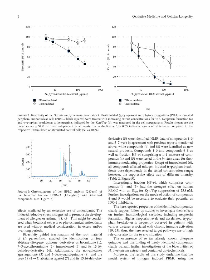
3.2. Bioactivity-Guided Fractionation. The DCM extract ofH.pyrenaicum roots was separated by Sephadex-LH20 columnchromatography to obtain 8 fractions (HOR-a1 to HOR-a8). Fractions with similar compound spectra were pooled.Fractions HOR-a3, HOR-a4, HOR-a6, HOR-a7, and HOR-a8 were tested for their immunomodulatory activity in thePBMC cell model to guide the subsequent isolation ofbioactive constituents. Several fractions contained bioactivecompounds, which dose-dependently suppressed the targetpathways (see supplemental figure S1). The half maximalinhibitory concentrations (IC50) for neopterin formationand tryptophan breakdown of the most active fractionsHOR-a3, HOR-a4, and HOR-a6 can be found in Table 1.The strongest suppressive effect on both mitogen inducedpathway was obtained with fraction HOR-a3, for whichneopterin formation was suppressed to 50% at a treatmentconcentration of 63.5μg/mL and tryptophan breakdownwas reduced to 50% with 7.02μg/mL HOR-a3 (Figure 2).
In unstimulated cells, there was a minor but significantsuppressive effect on both pathways upon exposure toHOR-a3, however at higher treatment concentrations only.Due to the stronger relative effect on Kyn/Trp compared toneopterin formation, the tryptophan breakdown pathwaywas selected as primary decision making-parameter forfurther subfractionation of the most active fraction HOR-a3.
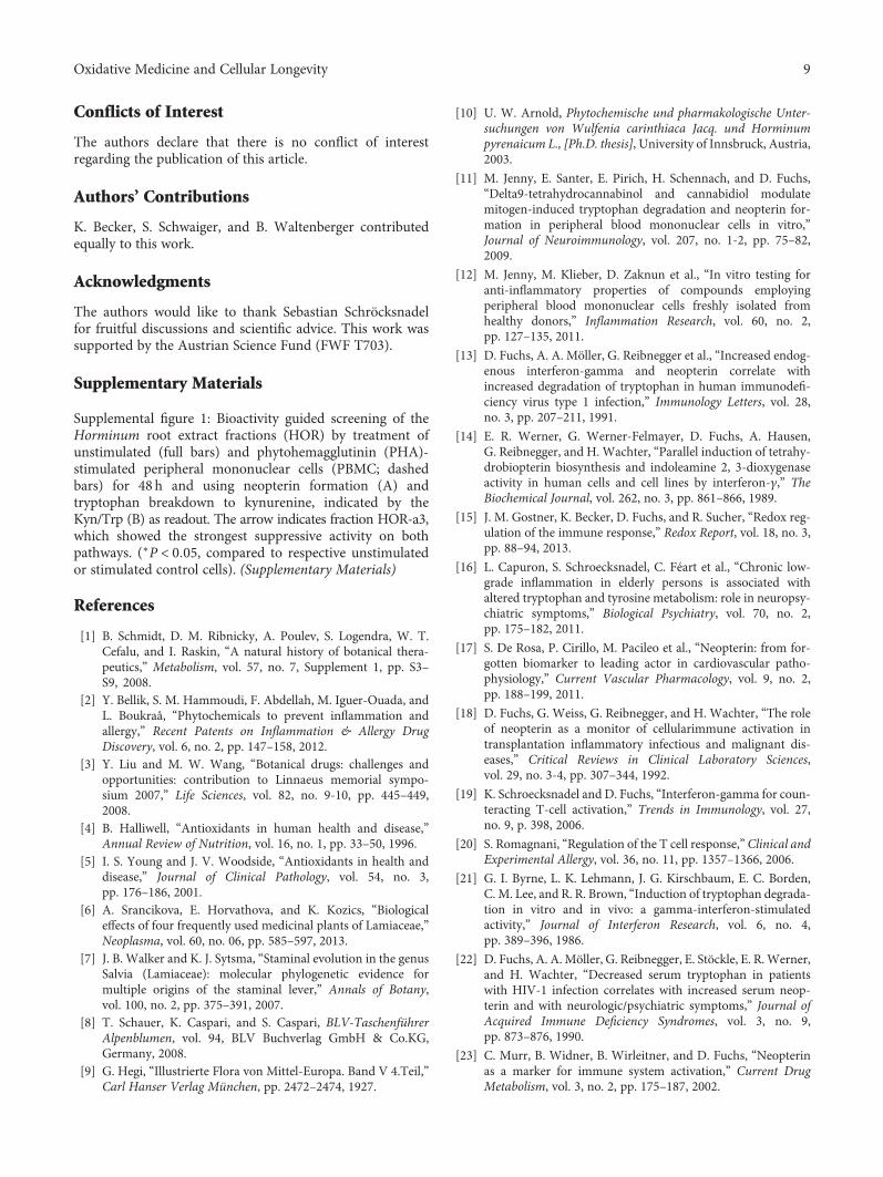
3.3. Isolation of Bioactive Diterpene Quinones. Aiming at theisolation of the major compounds present in the bioactivefraction HOR-a3, 10.6 g of the DCM extract of the root mate-rial ofH. pyrenaicumwas separated by means of silica gel andSephadex LH 20 column chromatography, HSCCC, as well assemi-preparative HPLC (Figure 3). This enabled the isolationof six compounds (Figure 4). They were identified by massspectrometry, 1- and 2-D NMR, and comparison of the spec-tral data with literature as the diterpene quinones horminone(1) [29, 30], 7-O-acetylhorminone (2) [31, 32], agastaqui-none (3) [33], inuroyleanol (6) [34, 35], agastol (7) [36, 37],and 3-deoxyagastaquinone (8). Compound 8 was identifiedas a new natural product.
In addition to these diterpene quinones, of which onlycompounds 1 and 2 are known constituents of H. pyrenai-cum, the bioguided separation process yielded fraction HP-r4 comprising two major compounds, which were difficultto separate with the used chromatographic techniques.Therefore, in this case, we did not follow the classicalapproach, that is, separation and isolation, NMR measure-ment and identification, and biological testing of the purecompounds, but another approach, that is, testing of thecompound mixture and subsequent identification of thecompounds by LC-SPE-NMR according to a procedure
4 Oxidative Medicine and Cellular Longevity

described by Sturm et al. [24]. One of the compounds com-prised in the mixture was identified as 15,16-dehydroagastol(5) [38]. The compound has already been isolated fromAgastache rugosa [36]. However, it represents a newlyidentified compound in the speciesH. pyrenaicum. By meansof 1D and 2D–NMR-spectroscopy, the second constituent offraction HP-r4 was identified as 15,16-dehydroinuroyleanol(4), which is a new natural compound (Figure 4).
Consequently, the isolated diterpenchinones 1–3 and 6–8as well as fraction HP-r4 comprising a mixture of com-pounds 4 and 5 were tested on their ability to influenceTh1-type immune responses.
3.4. Anti-Inflammatory Effects of the Isolated Compounds. Allof the known and identified compounds in this study exceptinuroyleanol (6) were able to influence tryptophan break-down in stimulated PBMC at micromolar concentrationsand in a dose-dependent manner, however, with differentintensities (Table 2, Figure 5).
Strongest influences exerted the equimolar compoundmixture HP-r4 (1 : 1 mixture of (4) 15,16-dehydroinuroylea-nol and (5) 15,16-dehydroagastol), followed by (3) agastaqiu-none> (2) 7-O-acetylhorminone > (8) 3-deoxyagastaquinone> (7) agastol > (1) horminone as indicated by the IC50 values(Table 2, Figure 5).
An inhibition of neopterin formation in PHA-stimulatedPBMC was observed for HP-r4 and compounds (1), (2), (8),while compounds (7) showed minor and (3) and (6) showedno dose-dependent effect (data not shown). However, due tosample limitation, neopterin formation could not be deter-mined for compounds (1) and (3) with sufficient repetitions.
4. Conclusion
This study reports the isolation process of H. pyreanicumcompounds in a bio-guided way, where fractions with highimmunomodulating properties were used to direct furthersubfractionation processes. This guided separation ofbioactive compounds successfully led to the identification ofpotent diterpene quinone derivatives, which were able tosuppress central Th1-type immunometabolic pathways. Avariety of phytochemicals were shown to interfere withIDO-1 activity previously, for example, the antioxidants res-veratrol [12], the lavender oil constituents (−)-linalool,(+)-α-pinene and (+)-limonene [39], the lignans arctigeninand trachelogenin [40] or the benzylisoquinoline alkaloidberberine [41]. In addition, commonly used anti-inflammatory medications were shown to interfere with theIDO-1 and GTP-CH-I activity, for example, acetylsalicylicacid (aspirin) [42]. These studies further confirm the impor-tance of these pathways to be investigated in mode of actionstudies of phytochemicals. Furthermore, it should be consid-ered that the suppressive effect can be either indirect by thegeneration of a more reductive milieu due to the antioxidantnature of several phytochemicals, thereby counteracting Tcell activation leading to less IFN-γ production [43], ordirectly as has been shown for example for arctigenin and tra-chelogenin that interact with the binding site of the enzyme[44]. The investigation of neopterin formation gives a furtherhint towards the mode of action, as in human macrophagesneopterin is produced at the expense of tetrahydrobiopterin(BH4), an essential cofactor for several monooxygenases [45].
Inflammation and immune activation play an importantrole in the pathogenesis of a variety of diseases and are ofimportance especially in chronic conditions, when theinduction of regulatory T-cells (Treg) counteracts the ini-tial immune activation cascade and contributes to themaintenance of an immunosuppressed state, which is asso-ciated with fatal outcome, for example, in patients withcardiovascular disease but also in cancer. This adversesituation is well indicated by increased neopterin andKyn/Trp levels in patients that are associated withincreased mortality [46, 47]. Hence, the regulatory influ-ence of plant compounds could be of importance to nor-malize the immunological situation in such patients.Central immunological cascades are redox sensitive, thustargeting these processes could be a successful strategy.However, several recent studies point towards the adverse
⁎ ⁎ ⁎⁎
⁎ ⁎
⁎⁎
1
10
100
1000
10000
100000
Neo(nmol/L)
Trp(�휇mol/L)
Kyn(�휇mol/L)
Kyn/Trp(�휇mol/mmol)
UnstimulatedCon A (10 �휇g/mL) PHA (10 �휇g/mL)
Figure 1: Concentrations of neopterin (Neo), tryptophan (Trp),kynurenine (Kyn) as well as the Kyn/Trp, a measure ofIDO-1 activity, in the supernatant of unstimulated PBMC andcells stimulated with 10 μg/mL concanavalin A (Con A) orphytohaemagglutinin (PHA) for 48 h. Results shown are the meanvalues ± SEM of seven independent experiments run in duplicates(∗p < 0 05, compared to unstimulated cells; please note log-scale).
Table 1: Half maximal inhibitory concentration (IC50) ofHorminum pyrenaicum extract sub-fractions (HOR-a3 to HOR-a6) that had the largest effect on neopterin formation and IDO-1activity, as indicated by the kynurenine to tryptophan ratio inphytohemagglutinin stimulated PBMC. More details are shown inthe supplemental figure S1.
Neopterin formationIC50 [μg/mL]
IDO-1 activityIC50 [μg/mL]
HOR-a3 63.8 7.02
HOR-a4 121 18.4
HOR-a6 159 34.0
5Oxidative Medicine and Cellular Longevity
effects mediated by an excessive use of antioxidants. Theinduced reductive stress is suggested to promote the develop-ment of allergies or asthma [48, 49]. This might be consid-ered when botanical extracts or phytochemical antioxidantsare used without medical consideration, in excess and/orover long periods.
Bioactivity guided fractionation of the root materialof H. pyrenaicum, enabled the identification of fourabietane-diterpene quinone derivatives as horminone (1),7-O-acetylhorminone (2), inuroyleanol (6) and its 15,16-dehydro-derivative (4). Additionally, the nor-abietanesagastaquinone (3) and 3-deoxyagastaquinone (8), and theabeo 18 (4→ 3) abietanes agastol (7) and its 15,16-dehydro-
derivative (5) were identified. NMR-data of compounds 1–3and 5–7 were in agreement with previous reports mentionedabove, while compounds (4) and (8) were identified as newnatural products. Compounds 1–3 and compounds 6–8 aswell as fraction HP-r4 comprising a 1 : 1 mixture of com-pounds (4) and (5) were tested in the in vitro assay for theirimmune-modulating properties. Except of inuroyleanol (6),all compounds affected mitogen-induced tryptophan break-down dose-dependently in the tested concentration range;however, the suppressive effect was of different intensity(Table 2, Figure 5).
Interestingly, fraction HP-r4, which comprises com-pounds (4) and (5), had the strongest effect on humanPBMC with an IC50 for Kyn/Trp suppression of 25.8μM.Further investigations on the mode of action of compounds4 and 5 would be necessary to evaluate their potential asIDO-1 inhibitors.
The here reported properties of the identified compoundsclearly support follow-up studies to investigate their effectson further immunological cascades, including neopterinformation. Higher neopterin levels and accelerated trypto-phan breakdown is frequently observed in patients withvarious diseases associated with chronic immune activation[19, 23], thus, the here selected target pathways are of highrelevance also for the in vivo situation.
The occurrence of so far already known diterpenequinones and the finding of newly identified compoundsclearly warrant further investigations of the bioactivities ofH. pyrenaicum extracts and contained phytochemicals.
Moreover, the results of this study underline that themodel system of mitogen induced PBMC using the
0
20
40
60
80
100
120
1 10 1000100H. pyrenaicum DCM extract (�휇g/mL)
PHA-stimulatedUnstimulated
⁎
⁎
⁎
⁎⁎
Neo
pter
in (%
of c
ontro
l)
(a)
0
20
40
60
80
100
120
1 10 100 1000
Kyn/
Trp
(% o
f con
trol)
H. pyrenaicum DCM extract (�휇g/mL)
PHA-stimulatedUnstimulated
⁎⁎
⁎
⁎
⁎
(b)
Figure 2: Bioactivity of the Horminum pyrenaicum root extract: Unstimulated (grey squares) and phytohemagglutinin (PHA)-stimulatedperipheral mononuclear cells (PBMC; black squares) were treated with increasing extract concentrations for 48 h. Neopterin formation (a)and tryptophan breakdown to kynurenine, indicated by the Kyn/Trp (b), was measured in the cell supernatants. Results shown are themean values ± SEM of three independent experiments run in duplicates. ∗p < 0 05 indicates significant differences compared to therespective unstimulated or stimulated control cells (set as 100%).
(min)10 20 30 40 50 60
(mAU
)
0
100
200
300
4001
2
345
6
7
8
Figure 3: Chromatogram of the HPLC analysis (280 nm) ofthe bioactive fraction HOR-a3 (1.0mg/mL) with identifiedcompounds (see Figure 4).
6 Oxidative Medicine and Cellular Longevity

O
O
O
OHO
HO
O
OH
OH
HO
O
OH
OH
O
O
O
OH
Horminone (1) 7-O-Acetylhorminone (2)
Agastaquinone (3) 15,16-Dehydroinuroyleanol (4)
15,16-Dehydroagastol (5) Inuroyleanol (6)
Agastol (7) 3-Deoxyagastaquinone (8)
H H
O
OH
O
OH
H
HO
O
OH
O
O
OH
O
O
H
HO
O
OH
O
O
Figure 4: Identified constituents of the roots of Horminum pyrenaicum. Black, blue and violet colors represent abietane-diterpene quinones,nor-abietane diterpene quinones, and abeo 18 (4→ 3) abietane diterpene quinones, respectively.
7Oxidative Medicine and Cellular Longevity

immunometabolic pathways of tryptophan breakdown andneopterin formation, is a reliable screening approach for abio-guided fractionation and isolation of immunomodula-tory compounds. By using human PBMC from healthydonors, the aspect of crosstalk between different immunecompetent cells, most importantly between T-cells andmonocytes/macrophages is taken into account [12]. More-over, by using unstimulated and mitogen-stimulatedconditions, immune activating and anti-inflammatoryproperties can be analyzed in parallel. As both neopterinand Kyn/Trp are biomarkers, which have been used forthe monitoring of several human diseases, there is a close
connection to the in vivo situation. Due to the limitationsof every in vitro system, further research will be needed toextrapolate the findings to the human system. In thisregard, the combination of the here reported pathway-focused strategy with an unbiased functional genomicsapproach could be advantageous in building an interactionmodel of the compounds of interests with other importantimmunological pathways, as well as to identify potentialunwanted off target effects [50]. In conclusion, the herereported approach justifies future studies for an in depthanalysis of the immunomodulatory properties of thereported diterpene quinones.
Table 2: Half maximal inhibitory concentration (IC50) of the isolated compounds on IDO-1 activity, as indicated by the kynurenine totryptophan ratio in phytohemagglutinin stimulated PBMC, calculated according Chou and Talalay [28] (n.d.: concentration-dependentactivity could not be determined in the tested concentration range; cursive: estimated values, only limited repetitions were possible due tosample limitations).
IDO-1 activityIC50 [μM]
(1) Horminone 346
(2) 7-O-Acetylhorminone 71.7
(3) Agastaqiunone 46.3
HP-r4 (1 : 1 mixture of (4) 15,16-dehydroinuroyleanol and (5) 15,16-dehydroagastol) 25.8
(6) Inuroyleanol n.d.
(7) Agastol 165
(8) 3-Deoxyagastaquinone 79.5
1-Methyl-D-tryptophan (positive control) 9.3
⁎⁎
⁎
⁎
⁎
⁎
0
20
40
60
80
100
120
140
1 10 100 1000Compounds (�휇M)
(1) Horminone(2) 7-O-Acetylhorminone
(3) AgastaquinoneHPr4 (mix of (4) and (5))
Kyn/
Trp
(% o
f PH
A co
ntro
l)
(a)
0
20
40
60
80
100
120
140
160
180
1 10 100 1000Compounds (�휇M)
⁎
⁎
⁎
⁎
(6) Inuroyleanol(7) Agastol(8) 3-Deoxyagastaquinone
Kyn/
Trp
(% o
f PH
A co
ntro
l)
(b)
Figure 5: Inhibition of indoleamine 2,3-dioxygenase, as indicated by Kyn/Trp, in mitogen-stimulated PBMC by the identified compounds.PBMC treated with PHA alone were used as a control for each experiment (set as 100%). (A) (1) horminone (n = 1), (2) 7-O-acetylhorminone(n = 3), (3) agastaqiunone (n = 2) and HP-r4, which is an equimolar mixture of the compounds (4) 15,16-dehydroinuroyleanol and (5)15,16-dehydroagastol (the average molecular weight was used for calculations; n = 3) and (B) (6) inuroyleanol (n = 3), (7) agastol (n = 3)and (8) 3-deoxyagastaquinone (n = 3). The number of replicates is indicated in brackets behind each compound. It was not possible torepeat the experiments in all cases due to the low amount of substance yield.
8 Oxidative Medicine and Cellular Longevity
Conflicts of Interest
The authors declare that there is no conflict of interestregarding the publication of this article.
Authors’ Contributions
K. Becker, S. Schwaiger, and B. Waltenberger contributedequally to this work.
Acknowledgments
The authors would like to thank Sebastian Schröcksnadelfor fruitful discussions and scientific advice. This work wassupported by the Austrian Science Fund (FWF T703).
Supplementary Materials
Supplemental figure 1: Bioactivity guided screening of theHorminum root extract fractions (HOR) by treatment ofunstimulated (full bars) and phytohemagglutinin (PHA)-stimulated peripheral mononuclear cells (PBMC; dashedbars) for 48 h and using neopterin formation (A) andtryptophan breakdown to kynurenine, indicated by theKyn/Trp (B) as readout. The arrow indicates fraction HOR-a3,which showed the strongest suppressive activity on bothpathways. (∗P < 0 05, compared to respective unstimulatedor stimulated control cells). (Supplementary Materials)
References
[1] B. Schmidt, D. M. Ribnicky, A. Poulev, S. Logendra, W. T.Cefalu, and I. Raskin, “A natural history of botanical thera-peutics,” Metabolism, vol. 57, no. 7, Supplement 1, pp. S3–S9, 2008.
[2] Y. Bellik, S. M. Hammoudi, F. Abdellah, M. Iguer-Ouada, andL. Boukraâ, “Phytochemicals to prevent inflammation andallergy,” Recent Patents on Inflammation & Allergy DrugDiscovery, vol. 6, no. 2, pp. 147–158, 2012.
[3] Y. Liu and M. W. Wang, “Botanical drugs: challenges andopportunities: contribution to Linnaeus memorial sympo-sium 2007,” Life Sciences, vol. 82, no. 9-10, pp. 445–449,2008.
[4] B. Halliwell, “Antioxidants in human health and disease,”Annual Review of Nutrition, vol. 16, no. 1, pp. 33–50, 1996.
[5] I. S. Young and J. V. Woodside, “Antioxidants in health anddisease,” Journal of Clinical Pathology, vol. 54, no. 3,pp. 176–186, 2001.
[6] A. Srancikova, E. Horvathova, and K. Kozics, “Biologicaleffects of four frequently used medicinal plants of Lamiaceae,”Neoplasma, vol. 60, no. 06, pp. 585–597, 2013.
[7] J. B. Walker and K. J. Sytsma, “Staminal evolution in the genusSalvia (Lamiaceae): molecular phylogenetic evidence formultiple origins of the staminal lever,” Annals of Botany,vol. 100, no. 2, pp. 375–391, 2007.
[8] T. Schauer, K. Caspari, and S. Caspari, BLV-TaschenführerAlpenblumen, vol. 94, BLV Buchverlag GmbH & Co.KG,Germany, 2008.
[9] G. Hegi, “Illustrierte Flora von Mittel-Europa. Band V 4.Teil,”Carl Hanser Verlag München, pp. 2472–2474, 1927.
[10] U. W. Arnold, Phytochemische und pharmakologische Unter-suchungen von Wulfenia carinthiaca Jacq. und Horminumpyrenaicum L., [Ph.D. thesis], University of Innsbruck, Austria,2003.
[11] M. Jenny, E. Santer, E. Pirich, H. Schennach, and D. Fuchs,“Delta9-tetrahydrocannabinol and cannabidiol modulatemitogen-induced tryptophan degradation and neopterin for-mation in peripheral blood mononuclear cells in vitro,”Journal of Neuroimmunology, vol. 207, no. 1-2, pp. 75–82,2009.
[12] M. Jenny, M. Klieber, D. Zaknun et al., “In vitro testing foranti-inflammatory properties of compounds employingperipheral blood mononuclear cells freshly isolated fromhealthy donors,” Inflammation Research, vol. 60, no. 2,pp. 127–135, 2011.
[13] D. Fuchs, A. A. Möller, G. Reibnegger et al., “Increased endog-enous interferon-gamma and neopterin correlate withincreased degradation of tryptophan in human immunodefi-ciency virus type 1 infection,” Immunology Letters, vol. 28,no. 3, pp. 207–211, 1991.
[14] E. R. Werner, G. Werner-Felmayer, D. Fuchs, A. Hausen,G. Reibnegger, and H. Wachter, “Parallel induction of tetrahy-drobiopterin biosynthesis and indoleamine 2, 3-dioxygenaseactivity in human cells and cell lines by interferon-γ,” TheBiochemical Journal, vol. 262, no. 3, pp. 861–866, 1989.
[15] J. M. Gostner, K. Becker, D. Fuchs, and R. Sucher, “Redox reg-ulation of the immune response,” Redox Report, vol. 18, no. 3,pp. 88–94, 2013.
[16] L. Capuron, S. Schroecksnadel, C. Féart et al., “Chronic low-grade inflammation in elderly persons is associated withaltered tryptophan and tyrosine metabolism: role in neuropsy-chiatric symptoms,” Biological Psychiatry, vol. 70, no. 2,pp. 175–182, 2011.
[17] S. De Rosa, P. Cirillo, M. Pacileo et al., “Neopterin: from for-gotten biomarker to leading actor in cardiovascular patho-physiology,” Current Vascular Pharmacology, vol. 9, no. 2,pp. 188–199, 2011.
[18] D. Fuchs, G. Weiss, G. Reibnegger, and H. Wachter, “The roleof neopterin as a monitor of cellularimmune activation intransplantation inflammatory infectious and malignant dis-eases,” Critical Reviews in Clinical Laboratory Sciences,vol. 29, no. 3-4, pp. 307–344, 1992.
[19] K. Schroecksnadel and D. Fuchs, “Interferon-gamma for coun-teracting T-cell activation,” Trends in Immunology, vol. 27,no. 9, p. 398, 2006.
[20] S. Romagnani, “Regulation of the T cell response,” Clinical andExperimental Allergy, vol. 36, no. 11, pp. 1357–1366, 2006.
[21] G. I. Byrne, L. K. Lehmann, J. G. Kirschbaum, E. C. Borden,C. M. Lee, and R. R. Brown, “Induction of tryptophan degrada-tion in vitro and in vivo: a gamma-interferon-stimulatedactivity,” Journal of Interferon Research, vol. 6, no. 4,pp. 389–396, 1986.
[22] D. Fuchs, A. A.Möller, G. Reibnegger, E. Stöckle, E. R.Werner,and H. Wachter, “Decreased serum tryptophan in patientswith HIV-1 infection correlates with increased serum neop-terin and with neurologic/psychiatric symptoms,” Journal ofAcquired Immune Deficiency Syndromes, vol. 3, no. 9,pp. 873–876, 1990.
[23] C. Murr, B. Widner, B. Wirleitner, and D. Fuchs, “Neopterinas a marker for immune system activation,” Current DrugMetabolism, vol. 3, no. 2, pp. 175–187, 2002.
9Oxidative Medicine and Cellular Longevity

[24] S. Sturm, C. Seger, M. Godejohann, M. Spraul, andH. Stuppner, “Conventional sample enrichment strategiescombined with high-performance liquid chromatography-solid phase extraction-nuclear magnetic resonance analysisallows analyte identification from a single minuscule Corydalissolida plant tuber,” Journal of Chromatography. A, vol. 1163,no. 1-2, pp. 138–144, 2007.
[25] S. Lob, A. Konigsrainer, R. Schafer, H. G. Rammensee,G. Opelz, and P. Terness, “Levo- but not dextro-1-methyl tryp-tophan abrogates the IDO activity of human dendritic cells,”Blood, vol. 111, no. 4, pp. 2152–2154, 2008.
[26] A. Laich, G. Neurauter, B. Widner, and D. Fuchs, “More rapidmethod for simultaneous measurement of tryptophan andkynurenine by HPLC,” Clinical Chemistry, vol. 48, no. 3,pp. 579–581, 2002.
[27] B. Widner, E. R. Werner, H. Schennach, H. Wachter, andD. Fuchs, “Simultaneous measurement of serum tryptophanand kynurenine by HPLC,” Clinical Chemistry, vol. 43,no. 12, pp. 2424–2426, 1997.
[28] T. C. Chou and P. Talalay, “Quantitative analysis of dose-effectrelationships: the combined effects of multiple drugs orenzyme inhibitors,” Advances in Enzyme Regulation, vol. 22,pp. 27–55, 1984.
[29] M. M. Janot and P. Potier, “Identification of a quinoneisolated from Horminum pyrenaicum as 7-hydroxyroylea-none,” Annales Pharmaceutiques Francaises, vol. 22, no. 5,pp. 387–395, 1964.
[30] L. T. Jonathan, C.-T. Chen, J. M. Pezzuto, H. H. S. Fong, andN. R. Farnsworth, “7-O-methylhorminone and other cytotoxicditerpene quinones from Lepechinia bullata,” Journal of Natu-ral Products, vol. 52, no. 3, pp. 571–575, 1989.
[31] M. M. Abdul-Ghani, “Diterpene quinones from Salvia viridishorminium,” Alexandria Journal of Pharmaceutical Sciences,vol. 18, no. 1, pp. 31–33, 2004.
[32] O. E. Edwards, G. Feniak, and M. Los, “Diterpenoid quinonesof Inula royleana,” Canadian Journal of Chemistry, vol. 40,no. 8, pp. 1540–1546, 1962.
[33] H.-K. Lee, S.-R. Oh, and J.-I. Kim, “Agastaquinone, a newcytotoxic diterpenoid quinone from Agastache rugosa,” Jour-nal of Natural Products, vol. 58, no. 11, pp. 1718–1721,1995.
[34] S. V. Bhat, P. S. Kalyanaraman, H. Kohl, and N. J. De Souza,“Inuroyleanol and 7-ketoroyleanone, two novel diterpenoidsof Inula royleana,” Tetrahedron, vol. 31, no. 8, pp. 1001–1004, 1975.
[35] B. Fraga, C. Diaz, E. GA, and A. Gonzales-Coloma, “Diter-penes from Salvia broussonetii transformed roots and theirinsecticidal activity,” Journal of Agricultural and Food Chemis-try, vol. 53, no. 13, pp. 5200–5206, 2005.
[36] H. K. Lee, J. Soon, S. R. Oh, J. Kim, Y. Kim, and C. O. Lee,“Diterpenoids from the roots of Agastache rugosa and theircytotoxic activities,” Saengyak Hakhoechi, vol. 25, no. 4,pp. 319–327, 1994.
[37] Z. M. Zou, D. Q. Yu, and P. Z. Cong, “Diterpenoids from theroots of Agastache rugosa,” Journal of Chinese PharmaceuticalSciences, vol. 6, no. 3, pp. 115–118, 1997.
[38] Z. M. Zou and P. Z. Cong, “Chemical constituents from rootsof Agastache rugosa,” Yaoxue Xuebao, vol. 26, no. 12, pp. 906–910, 1991.
[39] J. M. Gostner, M. Ganzera, K. Becker et al., “Lavender oil sup-presses indoleamine 2,3-dioxygenase activity in human
PBMC,” BMC Complementary and Alternative Medicine,vol. 14, no. 1, p. 503, 2014.
[40] S. Kuehnl, S. Schroecksnadel, V. Temml et al., “Lignans fromCarthamus tinctorius suppress tryptophan breakdown viaindoleamine 2,3-dioxygenase,” Phytomedicine, vol. 20, no. 13,pp. 1190–1195, 2013.
[41] C. J. Yu, M. F. Zheng, C. X. Kuang, W. D. Huang, and Q. Yang,“Oren-gedoku-to and its constituents with therapeutic poten-tial in Alzheimer's disease inhibit indoleamine 2, 3-dioxygenase activity in vitro,” Journal of Alzheimer's Disease,vol. 22, no. 1, pp. 257–266, 2010.
[42] K. Schroecksnadel, C. Winkler, B. Wirleitner, H. Schennach,and D. Fuchs, “Aspirin down-regulates tryptophan degrada-tion in stimulated human peripheral blood mononuclear cellsin vitro,” Clinical and Experimental Immunology, vol. 140,no. 1, pp. 41–45, 2005.
[43] K. Schroecksnadel, B. Fischer, H. Schennach, G. Weiss, andD. Fuchs, “Antioxidants suppress Th1-type immune responsein vitro,” Drug Metabolism Letters, vol. 1, no. 3, pp. 166–171,2007.
[44] V. Temml, S. Kuehnl, D. Schuster, S. Schwaiger, H. Stuppner,and D. Fuchs, “Interaction of Carthamus tinctorius lignan arc-tigenin with the binding site of tryptophan-degrading enzymeindoleamine 2,3-dioxygenase,” FEBS Open Bio, vol. 3, no. 1,pp. 450–452, 2013.
[45] E. R. Werner, G. Werner-Felmayer, D. Fuchs et al., “Tetrahy-drobiopterin biosynthetic activities in human macrophages,fibroblasts, THP-1, and T 24 cells. GTP-cyclohydrolase I isstimulated by interferon-gamma, and 6-pyruvoyl tetrahydrop-terin synthase and sepiapterin reductase are constitutivelypresent,” The Journal of Biological Chemistry, vol. 265, no. 6,pp. 3189–3192, 1990.
[46] T. B. Grammer, D. Fuchs, B. O. Boehm, B. R. Winkelmann,and W. Maerz, “Neopterin as a predictor of total and cardio-vascular mortality in individuals undergoing angiography inthe Ludwigshafen risk and cardiovascular health study,” Clin-ical Chemistry, vol. 55, no. 6, pp. 1135–1146, 2009.
[47] G.Weinlich, C.Murr, L. Richardsen, C.Winkler, and D. Fuchs,“Decreased serum tryptophan concentration predicts poorprognosis in malignant melanoma patients,” Dermatology,vol. 214, no. 1, pp. 8–14, 2007.
[48] J. D. Milner, D. M. Stein, R. McCarter, and R. Y. Moon, “Earlyinfant multivitamin supplementation is associated withincreased risk for food allergy and asthma,” Pediatrics,vol. 114, no. 1, pp. 27–32, 2004.
[49] D. Zaknun, S. Schroecksnadel, K. Kurz, and D. Fuchs, “Poten-tial role of antioxidant food supplements preservatives andcolorants in the pathogenesis of allergy and asthma,” Interna-tional Archives of Allergy and Immunology, vol. 157, no. 2,pp. 113–124, 2012.
[50] J. M. Gostner, O. A. Wrulich, M. Jenny, D. Fuchs, andF. Ueberall, “An update on the strategies in multicomponentactivity monitoring within the phytopharmaceutical field,”BMC Complementary and Alternative Medicine, vol. 12,no. 1, p. 18, 2012.
10 Oxidative Medicine and Cellular Longevity

Stem Cells International
Hindawiwww.hindawi.com Volume 2018
Hindawiwww.hindawi.com Volume 2018
MEDIATORSINFLAMMATION
of
EndocrinologyInternational Journal of
Hindawiwww.hindawi.com Volume 2018
Hindawiwww.hindawi.com Volume 2018
Disease Markers
Hindawiwww.hindawi.com Volume 2018
BioMed Research International
OncologyJournal of
Hindawiwww.hindawi.com Volume 2013
Hindawiwww.hindawi.com Volume 2018
Oxidative Medicine and Cellular Longevity
Hindawiwww.hindawi.com Volume 2018
PPAR Research
Hindawi Publishing Corporation http://www.hindawi.com Volume 2013Hindawiwww.hindawi.com
The Scientific World Journal
Volume 2018
Immunology ResearchHindawiwww.hindawi.com Volume 2018
Journal of
ObesityJournal of
Hindawiwww.hindawi.com Volume 2018
Hindawiwww.hindawi.com Volume 2018
Computational and Mathematical Methods in Medicine
Hindawiwww.hindawi.com Volume 2018
Behavioural Neurology
OphthalmologyJournal of
Hindawiwww.hindawi.com Volume 2018
Diabetes ResearchJournal of
Hindawiwww.hindawi.com Volume 2018
Hindawiwww.hindawi.com Volume 2018
Research and TreatmentAIDS
Hindawiwww.hindawi.com Volume 2018
Gastroenterology Research and Practice
Hindawiwww.hindawi.com Volume 2018
Parkinson’s Disease
Evidence-Based Complementary andAlternative Medicine
Volume 2018Hindawiwww.hindawi.com
Submit your manuscripts atwww.hindawi.com