Embed Size (px)
Citation preview
For research purposes only.Not for use in humans.Prices subject to change. FAX: 303.730.1966 • WEB: www.novusbio.com 2
IMMUNOLOGYCATALOG OF ANTIBODIES FOR
1
Table of ContentsInnate Immunity ................... 2-8
Cytokines .......................... 2-4
Chemokines ...................... 4-6
Toll-Like Receptors ............. 7-8
Adaptive Immunity .............9-13
B Cells ............................9-10
T Cells .........................11-12
MHCs .................................13
Immunoglobulins .................13
CD Cell Markers ....................14
Autoimmune Disorders ....15-17
In The News ..........................18
Application KeyELISA - Elisa
FACS - Fluorescent Activated Cell Sorting
Func - Functional Assay
ICC - Immunocytochemistry
IF - Immunofluorescence
IHC - Immunohistochemistry
IHC-Fr - Immunohistochemistry Frozen
IHC-P - Immunohistochemistry Paraffin
IP - Immunoprecipitation
IVA - In Vitro Assay
RIA - Radioimmunoassay
RI - Radioimmunodiffusion
WB - Western Blot
Reactivity KeyBb - Baboon
Bv - Bovine
Ca - Canine
Eq - Equine
Fe - Feline
Gp - Guinea Pig
Hu - Human
Immunology Research
Mk - Monkey
Mu - Mouse
Po - Porcine
Rb - Rabbit
Rt - Rat
Sh - Sheep
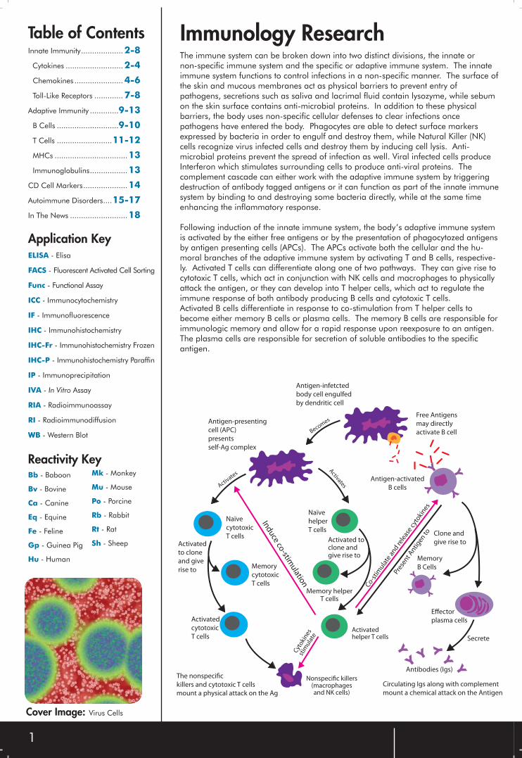
The immune system can be broken down into two distinct divisions, the innate or non-specific immune system and the specific or adaptive immune system. The innate immune system functions to control infections in a non-specific manner. The surface of the skin and mucous membranes act as physical barriers to prevent entry of pathogens, secretions such as saliva and lacrimal fluid contain lysozyme, while sebum on the skin surface contains anti-microbial proteins. In addition to these physical barriers, the body uses non-specific cellular defenses to clear infections once pathogens have entered the body. Phagocytes are able to detect surface markers expressed by bacteria in order to engulf and destroy them, while Natural Killer (NK) cells recognize virus infected cells and destroy them by inducing cell lysis. Anti-microbial proteins prevent the spread of infection as well. Viral infected cells produce Interferon which stimulates surrounding cells to produce anti-viral proteins. The complement cascade can either work with the adaptive immune system by triggering destruction of antibody tagged antigens or it can function as part of the innate immune system by binding to and destroying some bacteria directly, while at the same time enhancing the inflammatory response.
Following induction of the innate immune system, the body’s adaptive immune system is activated by the either free antigens or by the presentation of phagocytozed antigens by antigen presenting cells (APCs). The APCs activate both the cellular and the hu-moral branches of the adaptive immune system by activating T and B cells, respective-ly. Activated T cells can differentiate along one of two pathways. They can give rise to cytotoxic T cells, which act in conjunction with NK cells and macrophages to physically attack the antigen, or they can develop into T helper cells, which act to regulate the immune response of both antibody producing B cells and cytotoxic T cells.Activated B cells differentiate in response to co-stimulation from T helper cells to become either memory B cells or plasma cells. The memory B cells are responsible for immunologic memory and allow for a rapid response upon reexposure to an antigen. The plasma cells are responsible for secretion of soluble antibodies to the specific antigen.
Antigen-infetcted
body cell engulfed
by dendritic cell
BecomesAntigen-presenting
cell (APC)
presents
self-Ag complex
Activate
s
Naïve
cytotoxic
T cellsActivated
to clone
and give
rise toMemory
cytotoxic
T cells
Activated
cytotoxic
T cells
Ind
uce co
-stimu
lation
Activates
Naïve
helper
T cells
Activated toclone andgive rise to
Memory helperT cells
Cyt
oki
nes
stim
ula
te
Nonspecific killers(macrophagesand NK cells)
Activatedhelper T cells
Co
-sti
mu
late
an
d re
leas
e cy
toki
nes
Pres
ent A
nti
gen
to
Y
YY
Y
Y
YY
Y
Free Antigens
may directly
activate B cell
Antigen-activated
B cells
Clone and
give rise to
Memory
B Cells
Effector
plasma cells
SecreteY
Y Y
YYY
Antibodies (Igs)The nonspecific
killers and cytotoxic T cells
mount a physical attack on the Ag
Circulating Igs along with complement
mount a chemical attack on the Antigen
Cover Image: Virus Cells
2
Innate ImmunityInnate immunity is the defense mechanism that attacks an infection at onset. It does not adapt to specific pathogens to provide long-lasting protection as the adaptive immune system does. Most infectious agents that penetrate the body’s outer epithelial surfaces are quickly eliminated by the innate immune response preventing the appearance of disease symptoms. The word innate
implies genetically determined mechanisms. Innate immunity functions in a two part mechanism. First, the pathogen is recognized by soluble proteins and cell-surface receptors. Serum proteins of the complement system are activated to covalently bind the pathogen. Next, effectors cells (phagocytic white blood cells) are recruited to engulf the pathogen via endocytosis and to destroy it in the phagosome.
Cytokines are signaling proteins produced by various cells for use in cellular communication in order to regulate immunity, inflammation and hematopoiesis. They act by binding membrane receptors, which then
activate second messengers in order to alter gene expression. Cytokines interact via autocrine or paracrine action, although some work by endocrine action as well.
Cytokines
Tumor Necrosis Factors (TNFs) are a family of cytokines that trigger apoptosis. TNF alpha is mainly secreted by macrophages and causes apoptosis of certain tumor cells lines. It also can stimulate cell proliferation and
differentiation under certain conditions. TNF alpha and TNF beta are closely related as they share the same receptors and have similar cellular actions.
Tumor Necrosis Factors
NB100-75387 TNF alpha Chicken Polyclonal ELISA, WB Hu, Mu, Rt
NBP1-05081 TNF alpha Mouse Monoclonal ELISA Hu
NB600-587 TNF alpha (2C8) Rabbit Polyclonal ELISA, IHC, IP, RI, WB Hu, Mk
NB100-64741 TNF alpha (MP9-20A4) Rat Monoclonal ELISA, FACS, IHC-Fr Hu
NB200-445 TNF alpha Mouse Monoclonal ELISA, IHC-Fr, IHC-P Hu
NB110-57620 TNF alpha (EP1085Y) Rabbit Monoclonal WB Hu
NB100-78165 TNF beta (359-238-8) Mouse Monoclonal ELISA Hu
NB100-78166 TNF beta (359-81-11) Mouse Monoclonal FACS, ICC, IHC Hu
NB100-78167 TNF beta (359-81-11), Biotin Mouse Monoclonal ELISA, FACS, ICC Hu
NB600-822 TNF beta Rabbit Polyclonal ELISA, WB Hu
Catalog# Product Host Type Application Species TNF alpha AntibodyNB100-75387
Western blot analysis on E coli-derived fusion protein usingNB100-75387.
Species: Hu, Mu, RtApplications: ELISA, WB
Do you need to freshen up your IHC, RNAi,
Western Blot or ELISA skills?
A little fuzzy on the theory?
Have students to teach?
Check out our How To Series:Go to www.novusbio.com/support
and click on Downloads to download our four presentations.
How To Series CDs
r
3
NB120-14306 IL-1 alpha Chicken Polyclonal IHC, WB Hu, Mu, Rt
NB100-78122 IL-1 alpha (364-3B3-14) Mouse Monoclonal ELISA, FACS, ICC, IHC Hu
NB110-57118 IL-1R1 (EP409Y) Rabbit Monoclonal FACS, IP, WB Hu, Mu, Rt
NB100-78216 IL-1 beta (JK1B-1) Mouse Monoclonal ELISA, FACS, ICC Hu
NB110-57112 IL-1 beta (EP407Y) Rabbit Monoclonal WB Hu
NB600-633 IL-1 beta Rabbit Polyclonal ELISA, FACS, IHC-Fr, IHC-P, IP, WB Hu
NB110-60926 IL-2 Mouse Monoclonal IHC Hu
NB100-78124 IL-2 (MQ1-17H12) Rat Monoclonal ELISA, FACS, ICC, IHC, IP Hu, Mk, Bb
NB200-506 IL-2 Rabbit Polyclonal WB Po
NB100-79977 IL-3R beta Rabbit Polyclonal IHC, WB Hu, Mu, Rt
NB100-78127 IL-3 (BVD3-1F9) Rat Monoclonal FACS, ICC, IHC, IP, WB Hu
NB110-8389 IL-3 (2F2) Mouse Monoclonal ELISA Hu
H00003565-A01 IL-4 Mouse Polyclonal ELISA, WB Hu
NB200-505 IL-4 (BVD4-1D11) Rat Monoclonal ELISA, FACS, IHC, WB Mu
NB100-78175 IL-5 (TRFK5) Rat Monoclonal ELISA, FACS, ICC, IHC, WB Hu, Mu, Gp
NB600-1131 IL-6 Rabbit Polyclonal ELISA, WB Hu
NB100-2809 IL-6R Goat Polyclonal ELISA, WB Hu
NBP1-02757 IL-6 Mouse Monoclonal ELISA, IHC-P, WB Hu
H00003569-B01 IL-6 Mouse Polyclonal ELISA, IF, IHC-P, WB Hu
H00003574-B01 IL-7 Mouse Polyclonal ELISA, WB Hu
NB110-85536 IL-7R alpha Rabbit Polyclonal ELISA, IHC, WB Hu, Mu, Rt
NB110-57119 IL-8 (EP117Y) Rabbit Monoclonal WB Hu
NB100-78220 IL-8 (JK8-1) Mouse Monoclonal ELISA, FACS, ICC, IP Hu
H00003578-B02 IL-9 Mouse Polyclonal ELISA, WB Hu
NB110-57114 IL-10 (EP1115Y) Rabbit Monoclonal WB Hu, Mu, Rt
NB100-78143 IL-10 (JES3-9D7) Rat Monoclonal ELISA, FACS, ICC, IHC, WB Hu, Bb
H00003588-B01 IL10-R beta Mouse Polyclonal ELISA, WB Hu
H00003590-M01 IL-11R alpha (2D4-F4) Mouse Monoclonal ELISA, WB Hu
H00003589-M01 IL-11 (3A2) Mouse Monoclonal ELISA, IHC-P Hu
NB100-61661 IL-12 beta/IL-12 p40 Goat Polyclonal ELISA, WB Hu
NB100-78185 IL-12 (C15.6) Rat Monoclonal ELISA, FACS, ICC, IHC, IP, WB Mu
NB200-594 IL-13 (JES10-5A2) Rat Monoclonal ELISA, FACS, ICC, IHC-Fr, WB Hu
NB110-57115 IL-15 (EP433Y) Rabbit Monoclonal WB Hu
NB200-620 IL-15 Rabbit Polyclonal WB Mu
NBP1-04306 IL-16 (J2F10) Mouse Monoclonal ELISA, WB Hu
H00003603-B01 IL-16 Mouse Polyclonal ELISA, WB Hu
NB100-78211 IL-17 alpha (TC11-18H10.1) Rat Monoclonal ELISA, FACS, ICC, WB Mu
NB200- 201 IL-18 Binding Protein Rabbit Polyclonal WB Hu
Interleukins are a diverse group of cytokines. IL-1 and IL-6 are released by macrophages and work with TNF alpha to induce inflammation at the onset of infection. IL-2 is produced by activated T cells and primarily functions in the adaptive immune response.
IL-3 is a hematopoietic factor that promotes growth and differentiation of blood cells. CD4 TH2 cells secrete IL-4, IL-10 and IL-13 which function in antibody production. IL-12 is released by various immune cells and activates NK cells.
Interleukins
Catalog# Product Host Type Application Species IL-2 AntibodyNB110-60926
Species: HuApplications: IHC
Staining of lymphocytes in PBMNC cultures after PMA and ionomycin stimulation usingNB110-60926.
IL-3R beta AntibodyNB100-79977
Species: Hu, Mu, RtApplications: IHC, WB
Immuno-histochemical analysis on human spleen usingNB100-79977.
IL-6 AntibodyNBP1-02757
Species: HuApplications: ELISA, IHC-P, WB
Immuno-histochemical analysis of human tonsil using NBP1-02757.
IL-1 beta (EP407Y) AntibodyNB110-57112
Species: HuApplications: WB
Western blot analysis on recombinantprotein usingNB110-57112.
IL-6 AntibodyH00003569-B01
Species: HuApplications: ELISA, IF, IHC-P, WB
Immuno-fluorescence of purified H00003569-B01on HeLa cell.
IL-12 beta/IL-12 p40 AntibodyNB100-61661
Species: HuApplications: ELISA, WB
Western blotanalysis on humanliver lysateusing NB100-61661.
IL-10 (EP1115Y) AntibodyNB110-57114
Western blot analysis on recombinant protein usingNB110-57114.
Species: Hu, Mu, RtApplications: WB
NEW
IL-9 AntibodyH00003578-B02
Species: HuApplications: ELISA, WB
Western Blot analysis in transfected 293T cell line using H00003578-B02.
Read more about IL-1’s implication
in Rhuematoid Arthritis on page 17.
4
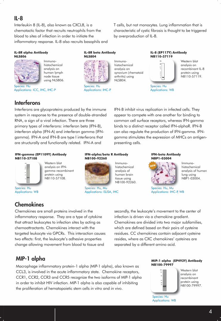
Interleukin 8 (IL-8), also known as CXCL8, is a chemotactic factor that recruits neutrophils from the blood to sites of infection in order to initiate the inflammatory response. IL-8 also recruits basophils and
T cells, but not monocytes. Lung inflammation that is characteristic of cystic fibrosis is thought to be triggered by overproduction of IL-8.
IL-8
IL-8R alpha AntibodyNLS806
Immuno-histochemical analysis onhuman lymph node tissueusing NLS806.
Species: HuApplications: ICC, IHC, IHC-P
IL-8R beta AntibodyNLS804
Immuno-histochemical analysis on synovium (rhematoid arthritis) using NLS804.
Species: HuApplications: IHC-P
IL-8 (EP117Y) AntibodyNB110-57119
Western blot analysis onrecombinant IL-8 protein using NB110-57119.
Species: HuApplications: WB
Interferons are glycoproteins produced by the immune system in response to the presence of double-stranded RNA, a sign of a viral infection. There are three primary types of interferons: interferon beta (IFN-B), interferon alpha (IFN-A) and interferon gamma (IFN-gamma). IFN-A and IFN-B are type I interferons that are structurally and functionally related. IFN-A and
IFN-B inhibit virus replication in infected cells. They appear to compete with one another for binding to common cell surface receptors, whereas IFN-gamma binds to a distinct receptor called IFN-alphaR. IFN-B can also regulate the production of IFN-gamma. IFN-gamma stimulates the expression of MHCs on antigen-presenting cells.
Interferons
IFN-gamma (EP1109Y) AntibodyNB110-57108
Western blot analysis on IFN-gamma recombinant protein using NB110-57108.
Species: HuApplications: WB
IFN-alpha/beta R AntibodyNB100-92260
Immuno-histochemical analysis of human brain tissue using NB100-92260.
Species: Hu, MuApplications: ELISA, IHC
IFN-beta AntibodyNBP1-03004
Immuno-histochemical analysis of human lung using NBP1-03004.
Species: Hu, MuApplications: IHC-P, WB
NEW
Chemokines are small proteins involved in the inflammatory response. They are a type of cytokine that attract leukocytes to infection sites by acting as chemoattractants. Chemokines interact with the targeted leukocyte via GPCRs. This interaction causes two effects: first, the leukocyte’s adhesive properties change allowing movement from blood to tissue and
secondly, the leukocyte’s movement to the center of infection is driven via a chemokine gradient. Chemokines are divided into two major subfamilies, which are defined based on their pairs of cysteineresidues. CC chemokines contain adjacent cysteine resides, where as CXC chemokines’ cysteines are separated by a different amino acid.
Chemokines
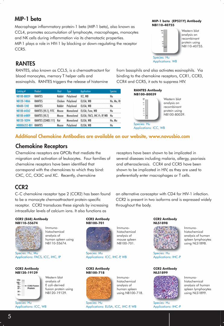
MIP-1 alphaMacrophage inflammatory protein-1 alpha (MIP-1 alpha), also known as CCL3, is involved in the acute inflammatory state. Chemokine receptors, CCR1, CCR2, CCR3 and CCR5 recognize the two isoforms of MIP-1 alpha in order to inhibit HIV infection. MIP-1 alpha is also capable of inhibiting the proliferation of hematopoietic stem cells in vitro and in vivo.
MIP-1 alpha (EP493Y) AntibodyNB100-79997
Western blotanalysis onrecombinantprotein usingNB100-79997.
Species: HuApplications: WB
5
NB100-80039 RANTES Rabbit Polyclonal ICC, WB Hu
NB120-14066 RANTES Chicken Polyclonal ELISA, WB Hu, Mu, Rt
NB600-1242 RANTES Rabbit Polyclonal ELISA, WB Hu
NB100-64262 RANTES (VL1), FITC Mouse Monoclonal ELISA, Func, WB Hu
NB100-64809 RANTES (VL1) Mouse Monoclonal ELISA, FACS, IHC-Fr, IP, WB Hu
NB120-10394 RANTES (53405.111) Rat Monoclonal ELISA, WB Hu, Mu
H00006352-B01 RANTES Mouse Polyclonal ELISA, WB Hu
RANTES, also known as CCL5, is a chemoattractant for blood monocytes, memory T helper cells and eosinophils. RANTES triggers the release of histamine
from basophils and also activates eosinophils. Via binding to the chemokine receptors, CCR1, CCR3, CCR4 and CCR5, it acts to suppress HIV.
RANTES
Catalog# Product Host Type Application Species RANTES AntibodyNB100-80039
Western blot analysis on recombinant protein usingNB100-80039.
Species: HuApplications: ICC, WB
Chemokine receptors are GPCRs that mediate the migration and activation of leukocytes. Four families of chemokine receptors have been identified that correspond with the chemokines to which they bind: CXC, CC, CX3C and XC. Recently, chemokine
receptors have been shown to be implicated in several diseases including malaria, allergy, psoriasis and atherosclerosis. CCR4 and CCR5 have been shown to be implicated in HIV, as they are used to preferentially enter macrophages or T cells.
Chemokine Receptors
Additional Chemokine Antibodies are available on our website, www.novusbio.com
C-C chemokine receptor type 2 (CCR2) has been found to be a monocyte chemoattractant protein-specific receptor. CCR2 transduces these signals by increasing intracellular levels of calcium ions. It also functions as
an alternative coreceptor with CD4 for HIV-1 infection. CCR2 is present in two isoforms and is expressed widely throughout the body.
CCR2
MIP-1 betaMacrophage inflammatory protein-1 beta (MIP-1 beta), also known as CCL4, promotes accumulation of lymphocyte, macrophages, monocytes and NK cells during inflammation via its chemotactic properties. MIP-1 plays a role in HIV-1 by blocking or down-regulating the receptor CCR5.
MIP-1 beta (EP521Y) AntibodyNB110-40755
Western blot analysis on recombinant protein using NB110-40755.
Species: HuApplications: WB
CCR2 (E68) AntibodyNB110-55674
Immuno-histochemical analysis of human spleen using NB110-55674.
Species: Hu, MuApplications: FACS, ICC, IHC, IP
CCR2 AntibodyNB100-701
Immuno-histochemical analysis of mouse spleenNB100-701.
Species: MuApplications: ICC, IHC-P, WB
CCR2 AntibodyNLS1898
Immuno-histochemical analysis of human spleen lymphocytesusing NLS1898.
Species: HuApplications: IHC-P
CCR2 AntibodyNB120-19129
Western blot analysis of E coli-derived fusion protein usingNB120-19129.
Species: HuApplications: ICC, WB
CCR2 AntibodyNB100-718
Immuno-histochemical analysis of human spleenusing NB100-718.
Species: HuApplications: ELISA, ICC, IHC-P, WB
CCR2 AntibodyNLS1899
Immuno-histochemical analysis of humanspleen lymphocytesusing NLS1899.
Species: HuApplications: IHC-P
6
NB100-702 CCR1 Rabbit Polyclonal ELISA, ICC, IHC-P, WB Hu, Mu
NB110-55674 CCR2 (E68) Rabbit Monoclonal FACS, ICC, IHC, IP Hu, Mu
NB100-1631 CXCR2 (48311.211) Mouse Monoclonal FACS Hu
NB110-55675 CCR3 (Y31) Rabbit Monoclonal ICC, IHC, WB Hu, Mu
NLS1374 CXCR3 Rabbit Polyclonal IHC, IHC-P Hu
NBP1-00770 CXCR3 Rabbit Polyclonal ELISA, WB Hu
NBP1-02364 CCR4 Rabbit Polyclonal IHC Hu
NB100-55749 CXCR4 Rabbit Polyclonal ICC, IF, WB Hu, Mu
NLS1376 CXCR4 Rabbit Polyclonal IHC, IHC-P, WB Hu
NB100-531 CCR5 Rabbit Polyclonal ELISA, FACS, Func, IVA Hu, Mk
NB100-78048 CCR5 (R-C10) Mouse Monoclonal ELISA, FACS, ICC, IP, WB Hu
NB100-78049 CCR5 (R-C10), Biotin Mouse Monoclonal ELISA, FACS, ICC Hu
NB100-56333 CXCR5 Rabbit Polyclonal WB Hu
NLS1384 CXCR5 Rabbit Polyclonal IHC-P Hu
NBP1-04271 CCR6 (4C6) Mouse Monoclonal ELISA, WB Hu
NLS1102 CXCR6 Rabbit Polyclonal IHC, IHC-P Hu
NB110-55678 CCR7 (E271) Rabbit Monoclonal FACS, ICC, IHC, WB Hu
NB100-712 CCR7 Goat Polyclonal ELISA, FACS, ICC, IHC, WB Hu, Mu, Mk, Bv
NB110-55680 CCR7 (Y59) Rabbit Monoclonal ICC, IHC, IP, WB Hu, Mu, Rt
NBP1-02360 CCR8 Rabbit Polyclonal IHC Hu
NB110-55681 CCR8 (E76) Rabbit Monoclonal ICC, WB Hu, Mu
NB110-55682 CCR8 (E77) Rabbit Monoclonal FACS, ICC, IP, WB Hu, Rt
NB100-708 CCR9 Goat Polyclonal ELISA, FACS, ICC, IHC-P, WB Mu
NB110-55683 CCR9 (E99) Rabbit Monoclonal FACS, ICC, WB Hu, Mu
NB100-707 CCR10 Goat Polyclonal ELISA, ICC, IHC, WB Hu
NB100-56319 CCR10 Rabbit Polyclonal WB Hu
NB100-58971 CCR10 Rabbit Polyclonal IHC, IHC-P Hu
NB100-705 CCR11 Goat Polyclonal ELISA, ICC, IHC, WB Hu, Mu
NB100-55762 CX3CR1 Rabbit Polyclonal IHC-P, WB Hu, Mu, Rt
C-C chemokine receptor type 5 (CCR5), also known as CD195, is a receptor for various inflammatory chemokines, including MIP-1 alpha, MIP-1 beta and
RANTES. CCR5 may play a role in the control of granulocytic lineage proliferation or differentiation. It also is a major coreceptor for human HIV infection.
CCR5
CCR5 AntibodyNB100-714
Immuno-histochemical analysis on human peripheral blood leukocytes using NB100-714.
Species: HuApplications: ELISA, ICC, IHC-P, WB
CCR5 (NT) AntibodyNB100-55914
Immuno-histochemical analysis on human lymph node tissue using NB100-55914.
Species: HuApplications: IHC, WB
CCR5 AntibodyNB100-66572
Immuno-histochemical analysis on human lymph node tissue using NB100-66572.
Species: HuApplications: IHC-P, WB
[CCR5 NB100-531] Khurana, S., et al. Identification of a linear peptide recognized by monoclonal antibody 2D7 capable of generating CCR5-specific antibodies with human immunodeficiency virus-neutralizing activity. J. Virology. 79(11): 6791-6800, 2005.
Catalog# Product Host Type Application Species CXCR3 AntibodyNLS1374
Species: HuApplications: IHC, IHC-P
Immuno-histochemical analysisof human tonsil using NLS1374.
CCR4 AntibodyNBP1-02364
Species: HuApplications: IHC
Immuno-histochemical analysisof human spleen using NBP1-02364.
CXCR6 AntibodyNLS1102
Species: HuApplications: IHC, IHC-P
Immuno-histochemical analysis of tonsil(interfollicular zone)using NLS1102.
CCR7 (E271) AntibodyNB110-55678
Species: HuApplications: FACS, ICC, IHC, WB
Immuno-fluorescent analysis of A431 cells using NB110-55678.
CCR8 AntibodyNBP1-02360
Immuno-histochemical analysis of human tonsil using NBP1-02360.
Species: HuApplications: IHC
CCR9 AntibodyNB100-708
Immuno-histochemical analysis of mousespleen usingNB100-708.
Species: MuApplications: ELISA, FACS, ICC, IHC-P, WB
CX3CR1 AntibodyNB100-55762
Immuno-histochemical analysis of human heart tissue using NB100-55762.
Species: Hu, Mu, RtApplications: IHC-P, WB
NEW
NEW
7
NB110-86979 TLR1 Rabbit Polyclonal ELISA, IHC, WB Hu, Mu
NB100-56563 TLR1 Rabbit Polyclonal FACS, WB Hu, Rt
NB120-14275 TLR2 Chicken Polyclonal WB Hu, Mu, Rt
NB200-580 TLR2 (TL2.1) Mouse Monoclonal FACS, IHC, IP, WB Hu
NB100-77885 TLR2 (TL2.1), Biotin Mouse Monoclonal FACS Hu
NB100-77886 TLR2 (TL2.1), FITC Mouse Monoclonal FACS Hu
NB120-1655 TLR2 Goat Polyclonal ELISA, ICC, IHC-P Hu
NB200-536 TLR2 Rabbit Polyclonal WB Hu, Mu, Rt, Mk
NB100-56042 TLR2 Rabbit Polyclonal WB Hu
NB100-56056 TLR2 (TL2.1), Biotin Mouse Monoclonal FACS, IP Hu
NBP1-03016 TLR2 Rabbit Polyclonal ELISA, IHC-P, WB Hu, Mu
NB100-56586 TLR2 Rabbit Polyclonal WB Mu
NBP1-03023 TLR2 Rabbit Polyclonal IHC-P, WB Hu, Mu
NBP1-04929 TLR3 Chicken Polyclonal WB Hu, Mu, Rt
NB110-93591 TLR3 Rabbit Polyclonal WB Mu, Rt, Eq, Bv
NB100-77956 TLR3 (TLR-104) Mouse Monoclonal FACS, ICC, WB Hu
NB100-77629 TLR4/MD2 (MTS510) Rat Monoclonal FACS, ICC, IP Mu
NB100-56581 TLR4 Rabbit Polyclonal ICC, IHC, WB Hu, Mu
H00007099-B01P TLR4 Mouse Polyclonal ELISA, WB Hu
NB600-728 TLR4 (HTA125), PE Mouse Monoclonal FACS Hu, Mk
NBP1-03144 TLR5 Rabbit Polyclonal IHC-P, WB Hu, Mu, Rt
NB200-571 TLR5 (85B152.5), FITC Mouse Monoclonal FACS, WB Hu, Mu
NBP1-02709 TLR6 Rabbit Polyclonal IHC-P, WB Hu
H00010333-M14 TLR6 (1E5) Mouse Monoclonal ELISA, WB Hu
NBP1-04349 TLR7 (4F4) Mouse Monoclonal ELISA, WB Hu
NBP1-04930 TLR7 Chicken Polyclonal WB Hu, Mu, Rt
NB100-80844 TLR7 Rabbit Polyclonal ICC, IHC, WB Hu, Mu
H00051284-M07 TLR7 (4G6) Mouse Monoclonal ELISA, WB Hu
NB110-10920 TLR8 (44C143) Mouse Monoclonal FACS, WB Hu
NB110-87039 TLR9 Rabbit Polyclonal ELISA, IHC, WB Hu, Mu, Rt
NBP1-03091 TLR9 Rabbit Polyclonal IHC-P, WB Hu, Mu, Rt
H00054106-M03 TLR9 (1E8) Mouse Monoclonal ELISA, IF, WB Hu
NBP1-02881 TLR10 Rabbit Polyclonal IHC-P, WB Hu
NB110-86981 TLR11 Rabbit Polyclonal ELISA, IHC, WB Hu
NB100-56742 TLR12 Rabbit Polyclonal FACS, WB Mu
NB100-56189 TLR13 Rabbit Polyclonal ELISA Mu, Rt
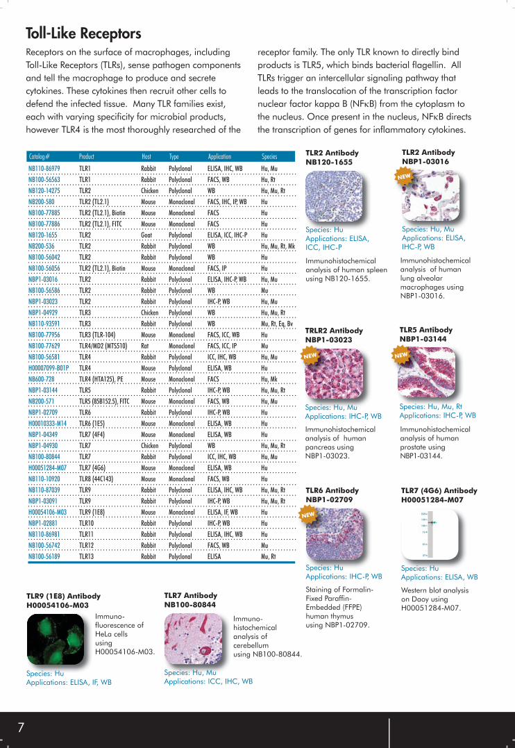
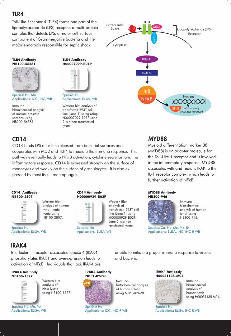
Toll-Like ReceptorsReceptors on the surface of macrophages, including Toll-Like Receptors (TLRs), sense pathogen components and tell the macrophage to produce and secrete cytokines. These cytokines then recruit other cells to defend the infected tissue. Many TLR families exist, each with varying specificity for microbial products, however TLR4 is the most thoroughly researched of the
receptor family. The only TLR known to directly bind products is TLR5, which binds bacterial flagellin. All TLRs trigger an intercellular signaling pathway that leads to the translocation of the transcription factor nuclear factor kappa B (NFκB) from the cytoplasm to the nucleus. Once present in the nucleus, NFκB directs the transcription of genes for inflammatory cytokines.
Catalog# Product Host Type Application Species TLR2 AntibodyNB120-1655
Species: HuApplications: ELISA, ICC, IHC-P
Immunohistochemical analysis of human spleen using NB120-1655.
TLR9 (1E8) AntibodyH00054106-M03
Immuno-fluorescence of HeLa cellsusingH00054106-M03.
Species: HuApplications: ELISA, IF, WB
TLR7 AntibodyNB100-80844
Species: Hu, MuApplications: ICC, IHC, WB
Immuno-histochemical analysis of cerebellumusing NB100-80844.
TLR7 (4G6) AntibodyH00051284-M07
Species: HuApplications: ELISA, WB
Western blot analysis on Daoy usingH00051284-M07.
Immunohistochemical analysis of human lung alveolar macrophages using NBP1-03016.
TLR5 AntibodyNBP1-03144
Species: Hu, Mu, RtApplications: IHC-P, WB
Immunohistochemical analysis of human prostate using NBP1-03144.
NEW
TLR6 AntibodyNBP1-02709
Species: HuApplications: IHC-P, WB
Staining of Formalin-Fixed Paraffin-Embedded (FFPE) human thymus using NBP1-02709.
NEW
TRLR2 AntibodyNBP1-03023
Species: Hu, MuApplications: IHC-P, WB
Immunohistochemical analysis of human pancreas using NBP1-03023.
NEW
TLR2 AntibodyNBP1-03016
Species: Hu, MuApplications: ELISA, IHC-P, WB
NEW
TLR4 AntibodyNB100-56581
Immuno-histochemical analysis of normal prostate sections using NB100-56581.
Species: Hu, MuApplications: ICC, IHC, WB
TLR4Toll-Like Receptor 4 (TLR4) forms one part of the lipopolysaccharide (LPS) receptor, a multi-protein complex that detects LPS, a major cell-surface component of Gram-negative bacteria and the major endotoxin responsible for septic shock.
TLR4 AntibodyH00007099-B01P
Western Blot analysis of transfected 293T cell line (Lane 1) using using H00007099-B01P. Lane 2 is a non-transfected lysate.
Species: HuApplications: ELISA, WB
CD14 AntibodyNB100-2807
Western blot analysis of human lymph node lysate usingNB100-2807.
Species: HuApplications: ELISA, WB
CD14 AntibodyH00000929-B02P
Western Blot analysis of transfected 293T cell line (Lane 1) using H00000929-B02P. Lane 2 is a non-ransfected lysate.
Species: HuApplications: ELISA, WB
CD14
Interleukin-1 receptor associated kinase 4 (IRAK4) phosphorylates IRAK1 and overexpression leads to activation of NFκB. Individuals that lack IRAK4 are
unable to initiate a proper immune response to viruses and bacteria.
IRAK4
IRAK4 AntibodyH00051135-M04
Species: HuApplications: ELISA, IHC-P, WB
Immuno-histochemical analysis ofhuman testisusing H00051135-M04.
IRAK4 AntibodyNBP1-02628
Immuno-histochemical analysis of human spleen using NBP1-02628.
Species: HuApplications: ICC, IHC-P, WB
IRAK4 AntibodyNB100-1527
Species: Hu, Mu, MkApplications: ELISA, WB
Western blot analysis of Hela lysate using NB100-1527.
CD14 binds LPS after it is released from bacterial surfaces and cooperates with MD2 and TLR4 to mediate the immune response. This pathway eventually leads to NFκB activation, cytokine secretion and theinflammatory response. CD14 is expressed strongly on the surface of monocytes and weakly on the surface of granulocytes. It is also ex-pressed by most tissue macrophages.
MYD88 Myeloid differentiation marker 88 (MYD88) is an adapter molecule for the Toll-Like 1 receptor and is involved in the inflammatory response. MYD88 associates with and recruits IRAK to the IL-1 receptor complex, which leads to further activation of NFκB.
MYD88 AntibodyNB300-946
Immuno-histochemical analysis of human tonsil using NB300-946.
Species: Ca, Hu, Mu, Mk, RtApplications: ELISA, IHC, IHC-P, WB
CD14MD2
MYD88
TLR4Extracellular
Space Lipopolysaccharide (LPS)
Receptor
IRAK4
TRAF6
Cytoplasm
IκB
NFκB
NFκB
Nucleus
Inflammatory
Cytokines Produced
NEW
9
Adaptive ImmunityThe unique function of the adaptive immune response provides the body with the ability to defend itself against specific invaders. Through complex mechanisms of antigen recognition, the body can ward off infection by attacking only specific pathogens while leaving the surrounding tissues alone. The body produces
countless types of immunoglobulins and T cellreceptors in response to infection. These remain latent in the system for many years after initial infection, thusallowing the body to effectively defeat diseases in the event of subsequent reexposure.
B CellsB cells produce soluble immunoglobulins that recognize specific antigens. Upon binding to their target, these tags allow for the recruitment of phagocytes and the destruction of the pathogen.
Immunoglobulins are capable of binding an intact pathogen in extracellular space, specifically targeting carbohydrate or amino acid groups on the surface of the invading molecule.
CD40 AntibodyNB500-548
Species: HuApplications: FACS
Surface staining of human peripheral blood cells using NB500-548.
IL-5 AntibodyNB100-78133
Sandwich ELISAof NB100-78133paired withNB100-78134.
Species: Hu, MkApplications: ELISA, FACS, ICC, IHC, WB
IL-4 AntibodyNB100-79978
Species: HuApplications: ICC, WB
Western blot analysis of human tonsil lysate using NB100-79978.
These cell types perform a critical role in the activation of B cells. Without co-stimulation, there is often not enough signal strength to cause the naïve B
cells to differentiate. Helper/effector T cells use cytokines and direct interaction with B cells to promote B cell proliferation.
CD4 TH2 Helper Cells
CD81 (1D6) AntibodyNB100-65805
Species: HuApplications: FACS, IHC-P, IP
Staining of human peripheral blood lymphocytes using NB100-65805.
CD19 (LT19) AntibodyNB500-338
Surface staining of human peripheral blood cells using NB500-338.
Species: HuApplications: FACS, IP
Another important part of B cell activation is the B cell coreceptor, which aids in binding the antigen and aligning both the receptor and coreceptor. This
alignment increases the relative proximity of cytoplasmic tyrosine kinases with CD19, which upon phos-phorylation begins to mobilize internal signaling factors.
B Cell Coreceptors
CD21 (LT21) AntibodyNB100-77958
Species: HuApplications: FACS
Human peripheral blood lymphocytes stained with NB100-77958.
CD81 (6D5) AntibodyNB100-77600
Species: MuApplications: FACS, IP, WB
C57BL/6 mouse splenocytes stained with NB100-77600.
CD19 AntibodyNB100-75635
Species: HuApplications: WB
Western Blot analysis on E coli-derived fusion protein using NB100-75635.
CD19 (MB19-1) AntibodyNB100-77382
C57BL/6 mouse splenocytes stained with NB100-77382.
Species: MuApplications: FACS, IP
10
TLR6 AntibodyNB 110-93544
Species: Hu, Mu, Rt, Mk, ShApplications: WB
Western blot analysis in Jurkat whole cell extract using NB110-93544.
TLR4 AntibodyNB100-56581
Immuno-histochemical analysis of Ramos using NB100-56581.
Species: Hu, MuApplications: ICC, IHC, WB
CD14 AntibodyNB500-444
Species: Hu, MkApplications: ELISA, FACS, IP, WB
Surface staining of human peripheral blood cells using NB500-444.
Thymus-independent (TI) antigens are capable of binding B cell receptors and activating naïve B cells without the assistance of CD4 T cells. These antigens
are also capable of binding other receptors on the B cell surface, such as TLRs, to activate B cell proliferation.
TI Antigens
The numerous steps involved in the rearrangement of immunoglobulin genes are affected by changes in production of certain protein products that regulate
successful development of B cells. The resulting rearrangements in immunoglobulin genes allow for the diverse production of immunoglobulins.
Immunoglobulin-Gene Rearrangement
VPREB3 (4H8) AntibodyH00029802-M08
Species: HuApplications: ELISA, WB
Western Blot analysis in transfected 293T cell line usingH99929802-M08.
TdT AntibodyNB120-15242
Immuno-histochemical analysis of human thymus using NB120-15242. Species: Hu
Applications: IHC, IHC-P
RAG2 AntibodyH00005897-B01
Species: HuApplications: ELISA, IF, WB
Immunofluorescence on HeLa cell usingH00005897-B01.
IKZF2 AntibodyH00022807-B01P
Species: HuApplications: ELISA, WB
Immunofluorescence on HeLa cellsusingH00022807-B01P.
Btk (EP420Y) [Tyr223] AntibodyNB100-79907
Western blot analysis on Ramos cell lysates using NB100-79907.
Species: HuApplications: IP, WB
CD45R (RA3-6B2) AntibodyNB100-77420
Species: Mu, Hu, FeApplications: FACS, IHC, IP
C57BL/6 mouse splenocytes stained with NB100-77420.
Antibody Grants
Grant Award Date: 1 Award selected on the 15th of every month. Awardees will receive a 0.2 mg test sample of affinity
purified rabbit sera. (Typical antibody production takes 4-5 months). If the product works and you supply the necessary
documentation, you will receive 2 mgs of affinity purified antibody in exchange for product feedback. Novus reserves the
right to sell the antibody produced by the grant. Submit by the end of the month to be selected in the following month’s
drawing by fax (below) or email ([email protected]).
WANT YOUR ANTIBODY PRODUCED FOR FREE?Visit our website, www.novusbio.com and fill out the Antibody Grant
Sheet for a chance to receive 2 mgs of FREE antibody!
11
T Cell Receptors (TCRs) are membrane-bound glycoprotein complexes generally found on the surface
of T cells. They are similar to a single antigen-binding portion of immunoglobulins formed in B cells.
T Cell Receptors
CD8 AntibodyNB600-1051
Species: HuApplications: IHC-P
Immuno-histochemical analysis of human tonsil stained using NB600-1051.
CD4 (EDU-2)AntibodyNB500-565
Surface staining of human peripheral blood cells using NB500-565.
Species: HuApplications: FACS
TCR alpha/beta (R73) AntibodyNB100-77720
Species: Rt, MkApplications: FACS, IHC-Fr, WB
LOU rat splenocytes stained with NB100-77720.
The movement of naïve T cells into secondary lymphoid tissue requires interaction of adhesion molecules on the surface of the T cells with adhesion molecules on the surface of endothelial cells.
Molecules, such as selectins and vascular addressins, aid in homing of the T cells, while integrins strengthen the bond once the T cell and endothelium have come into contact.
Adhesion Molecules
CD11 alpha (MEM-25) AntibodyNB500-328
Species: HuApplications: FACS, IP
Surface staining of human peripheral blood cells usingNB500-328.
CD34 (QBEND-10) AntibodyNB100-1934
Immuno-histochemical analysis of human tonsil usingNB100-1934.
Species: Hu, MkApplications: IHC-P, IP, WB
CD62L (DREG-56) AntibodyNB100-77803
Species: Hu, BvApplications: FACS
Human peripheral blood lymphocytes stained usingNB100-77803.
CD2 (RPA-2.10) AntibodyNB100-77733
Species: Hu, Bb, Mk, Sh, PoApplications: FACS
Human peripheral blood lymphocytes stained usingNB100-77733.
CD102 (3C4) AntibodyNB100-77471
C57BL/6 mouse splenocytes stained using NB100-77471.
Species: MuApplications: FACS, IHC-Fr, IP
ICAM-1 (EP1442Y) AntibodyNB110-57106
Species: HuApplications: ICC, IHC, WB
Immuno-histochemical analysis of human tonsil using NB110-57106.
Activation of naïve T cells requires a co-stimulatory signal from an antigen-presenting cell (APC). Expression of the required activation molecules only occurs in the
event of an infection which activates the innate immune system causing upregulation of the B7 genes.
Activating Proteins
CD86 (EP1158Y) AntibodyNB110-55488
Species: Hu, Mu, RtApplications: FACS, ICC, IHC, IP, WB
Immuno-histochemical analysis of human tonsil usingNB110-55488.
CD80 (EP1155Y) AntibodyNB110-55564
Immuno-histochemical analysis of human tonsil using NB110-55564.
Species: Hu, Mu, RtApplications: FACS, ICC, IHC, WB
CD28 (JJ319) AntibodyNB100-77718
Species: RtApplications: FACS, IP
LOU rat splenocytes stained using NB100-77718.
T CellsThese lymphocytes are an important part of the adaptive immune system. They are functionally different from B cells because they bind short peptides that have been assembled in a MHC molecule. This
response to antigen processing makes T cells antigen- specific. Unlike B cells that produce soluble antibodies, T cells have a more diverse role which often includes interaction with other cell types.
12
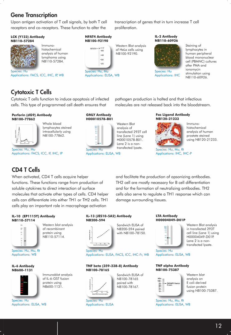
Upon antigen activation of T cell signals, by both T cell receptors and co-receptors. These function to alter the
transcription of genes that in turn increase T cell proliferation.
Gene Transcription
IL-2 AntibodyNB110-60926
Species: HuApplications: IHC
Staining of lymphocytes in human peripheral blood mononuclear cell (PBMNC) cultures after PMA and ionomycin stimulation usingNB110-60926.
NFAT4 AntibodyNB100-92190
Western Blot analysis of HeLa cells usingNB100-92190.
Species: Hu, MuApplications: ELISA, WB
LCK (Y123) AntibodyNB110-57284
Species: HuApplications: FACS, ICC, IHC, IP, WB
Immuno-histochemical analysis of human lymphoma using NB110-57284.
Cytotoxic T cells function to induce apoptosis of infected cells. This type of programmed cell death ensures that
pathogen production is halted and that infectious molecules are not released back into the bloodstream.
Cytotoxic T Cells
Fas Ligand AntibodyNB120-21233
Species: Hu, Mu, RtApplications: IHC, IHC-P
Immuno-histochemical analysis of human prostate stained using NB120-21233.
GNLY AntibodyH00010578-B01
Western Blot analysis of transfected 293T cell line (Lane 1) using H00010578-B01. Lane 2 is a non-transfected lysate.Species: Hu
Applications: ELISA, WB
Perforin (dG9) AntibodyNB100-77862
Species: Hu, MuApplications: FACS, ICC, IF, IHC, IP
Whole blood lymphocytes stained intracellularly usingNB100-77862.
When activated, CD4 T cells acquire helper functions. These functions range from production of soluble cytokines to direct interaction of surface molecules that activate other types of cells. CD4 helper cells can differentiate into either TH1 or TH2 cells. TH1 cells play an important role in macrophage activation
and facilitate the production of opsonizing antibodies. TH2 cell are mostly necessary for B cell differentiation and for the formation of neutralizing antibodies. TH2 cells also serve to regulate a TH1 response which can damage surrounding tissues.
CD4 T Cells
LTA AntibodyH00004049-D01P
Species: HuApplications: ELISA, WB
Western Blot analysis in transfected 293T cell line (Lane 1) usingH00004049-D01P.Lane 2 is a non-transfected lysate.
IL-13 (JES10-5A2) AntibodyNB200-594
Sandwich ELISA of NB200-594 pairedwith NB100-78150.
Species: HuApplications: ELISA, FACS, ICC, IHC-Fr, WB
IL-10 (EP1115Y) AntibodyNB110-57114
Species: Hu, Mu, RtApplications: WB
Western blot analysis of recombinant protein using NB110-57114.
TNF alpha AntibodyNB100-75387
Species: Hu, Mu, RtApplications: ELISA, WB
Western blotanalysis onE coli-derived fusion protein using NB100-75387.
TNF beta (359-238-8) AntibodyNB100-78165
Sandwich ELISA ofNB100-78165paired withNB100-78167.
Species: HuApplications: ELISA
IL-6 AntibodyNB600-1131
Species: HuApplications: ELISA, WB
Immunoblot analysis of IL-6-GST fusion protein using NB600-1131.
13
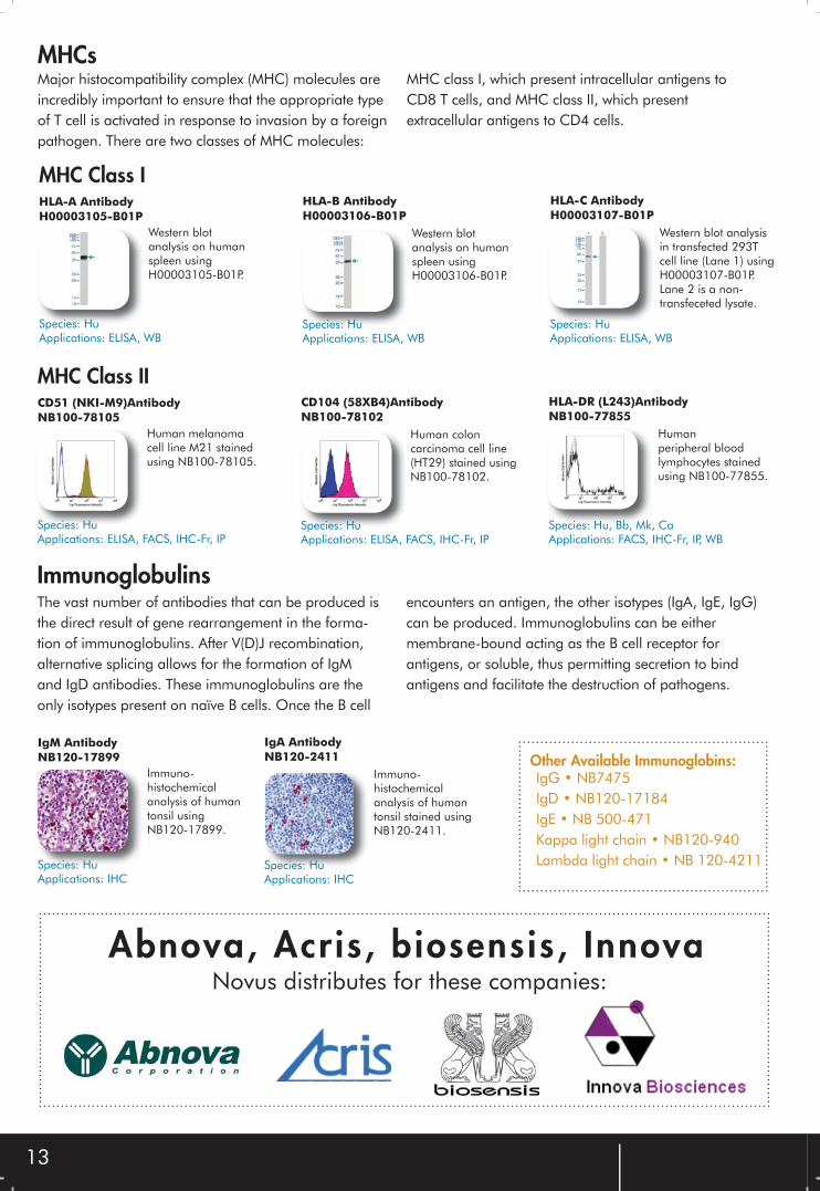
Major histocompatibility complex (MHC) molecules are incredibly important to ensure that the appropriate type of T cell is activated in response to invasion by a foreign pathogen. There are two classes of MHC molecules:
MHC class I, which present intracellular antigens to CD8 T cells, and MHC class II, which present extracellular antigens to CD4 cells.
MHCs
HLA-C AntibodyH00003107-B01P
Species: HuApplications: ELISA, WB
Western blot analysis in transfected 293T cell line (Lane 1) using H00003107-B01P.Lane 2 is a non-transfeceted lysate.
HLA-B AntibodyH00003106-B01P
Western blot analysis on human spleen usingH00003106-B01P.
Species: HuApplications: ELISA, WB
HLA-A AntibodyH00003105-B01P
Species: HuApplications: ELISA, WB
Western blot analysis on human spleen usingH00003105-B01P.
MHC Class I
HLA-DR (L243)AntibodyNB100-77855
Species: Hu, Bb, Mk, CaApplications: FACS, IHC-Fr, IP, WB
Human peripheral blood lymphocytes stained using NB100-77855.
CD104 (58XB4)AntibodyNB100-78102
Human colon carcinoma cell line (HT29) stained usingNB100-78102.
Species: HuApplications: ELISA, FACS, IHC-Fr, IP
CD51 (NKI-M9)AntibodyNB100-78105
Species: HuApplications: ELISA, FACS, IHC-Fr, IP
Human melanoma cell line M21 stained using NB100-78105.
MHC Class II
The vast number of antibodies that can be produced is the direct result of gene rearrangement in the forma-tion of immunoglobulins. After V(D)J recombination, alternative splicing allows for the formation of IgM and IgD antibodies. These immunoglobulins are the only isotypes present on naïve B cells. Once the B cell
encounters an antigen, the other isotypes (IgA, IgE, IgG) can be produced. Immunoglobulins can be either membrane-bound acting as the B cell receptor for antigens, or soluble, thus permitting secretion to bind antigens and facilitate the destruction of pathogens.
Immunoglobulins
IgA AntibodyNB120-2411
Immuno-histochemical analysis of human tonsil stained using NB120-2411.
Species: HuApplications: IHC
IgM AntibodyNB120-17899
Species: HuApplications: IHC
Immuno-histochemical analysis of human tonsil usingNB120-17899.
IgG • NB7475IgD • NB120-17184 IgE • NB 500-471Kappa light chain • NB120-940Lambda light chain • NB 120-4211
Other Available Immunoglobins:
Abnova, Acris, biosensis, InnovaNovus distributes for these companies:
14
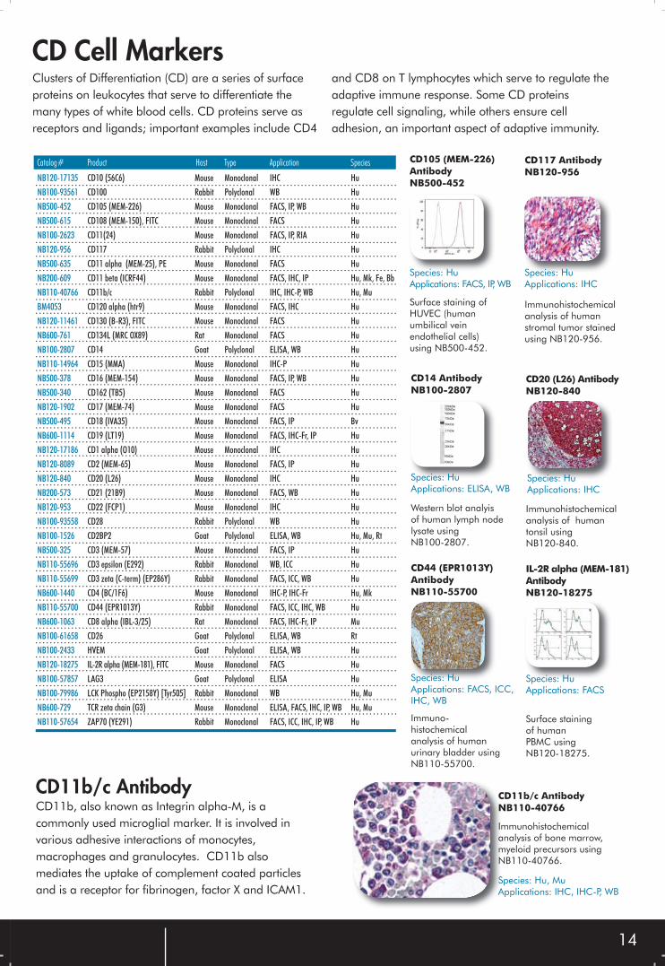
CD11b, also known as Integrin alpha-M, is a commonly used microglial marker. It is involved in various adhesive interactions of monocytes, macrophages and granulocytes. CD11b also mediates the uptake of complement coated particles and is a receptor for fibrinogen, factor X and ICAM1.
NB120-17135 CD10 (56C6) Mouse Monoclonal IHC Hu
NB100-93561 CD100 Rabbit Polyclonal WB Hu
NB500-452 CD105 (MEM-226) Mouse Monoclonal FACS, IP, WB Hu
NB500-615 CD108 (MEM-150), FITC Mouse Monoclonal FACS Hu
NB100-2623 CD11(24) Mouse Monoclonal FACS, IP, RIA Hu
NB120-956 CD117 Rabbit Polyclonal IHC Hu
NB500-635 CD11 alpha (MEM-25), PE Mouse Monoclonal FACS Hu
NB200-609 CD11 beta (ICRF44) Mouse Monoclonal FACS, IHC, IP Hu, Mk, Fe, Bb
NB110-40766 CD11b/c Rabbit Polyclonal IHC, IHC-P, WB Hu, Mu
BM4053 CD120 alpha (htr9) Mouse Monoclonal FACS, IHC Hu
NB120-11461 CD130 (B-R3), FITC Mouse Monoclonal FACS Hu
NB600-761 CD134L (MRC OX89) Rat Monoclonal FACS Hu
NB100-2807 CD14 Goat Polyclonal ELISA, WB Hu
NB110-14964 CD15 (MMA) Mouse Monoclonal IHC-P Hu
NB500-378 CD16 (MEM-154) Mouse Monoclonal FACS, IP, WB Hu
NB500-340 CD162 (TB5) Mouse Monoclonal FACS Hu
NB120-1902 CD17 (MEM-74) Mouse Monoclonal FACS Hu
NB500-495 CD18 (IVA35) Mouse Monoclonal FACS, IP Bv
NB600-1114 CD19 (LT19) Mouse Monoclonal FACS, IHC-Fr, IP Hu
NB120-17186 CD1 alpha (O10) Mouse Monoclonal IHC Hu
NB120-8089 CD2 (MEM-65) Mouse Monoclonal FACS, IP Hu
NB120-840 CD20 (L26) Mouse Monoclonal IHC Hu
NB200-573 CD21 (21B9) Mouse Monoclonal FACS, WB Hu
NB120-953 CD22 (FCP1) Mouse Monoclonal IHC Hu
NB100-93558 CD28 Rabbit Polyclonal WB Hu
NB100-1526 CD2BP2 Goat Polyclonal ELISA, WB Hu, Mu, Rt
NB500-325 CD3 (MEM-57) Mouse Monoclonal FACS, IP Hu
NB110-55696 CD3 epsilon (E292) Rabbit Monoclonal WB, ICC Hu
NB110-55699 CD3 zeta (C-term) (EP286Y) Rabbit Monoclonal FACS, ICC, WB Hu
NB600-1440 CD4 (BC/1F6) Mouse Monoclonal IHC-P, IHC-Fr Hu, Mk
NB110-55700 CD44 (EPR1013Y) Rabbit Monoclonal FACS, ICC, IHC, WB Hu
NB600-1063 CD8 alpha (IBL-3/25) Rat Monoclonal FACS, IHC-Fr, IP Mu
NB100-61658 CD26 Goat Polyclonal ELISA, WB Rt
NB100-2433 HVEM Goat Polyclonal ELISA, WB Hu
NB120-18275 IL-2R alpha (MEM-181), FITC Mouse Monoclonal FACS Hu
NB100-57857 LAG3 Goat Polyclonal ELISA Hu
NB100-79986 LCK Phospho (EP2158Y) [Tyr505] Rabbit Monoclonal WB Hu, Mu
NB600-729 TCR zeta chain (G3) Mouse Monoclonal ELISA, FACS, IHC, IP, WB Hu, Mu
NB110-57654 ZAP70 (YE291) Rabbit Monoclonal FACS, ICC, IHC, IP, WB Hu
CD Cell MarkersClusters of Differentiation (CD) are a series of surface proteins on leukocytes that serve to differentiate the many types of white blood cells. CD proteins serve as receptors and ligands; important examples include CD4
and CD8 on T lymphocytes which serve to regulate the adaptive immune response. Some CD proteins regulate cell signaling, while others ensure cell adhesion, an important aspect of adaptive immunity.
Catalog# Product Host Type Application Species CD105 (MEM-226)AntibodyNB500-452
Species: HuApplications: FACS, IP, WB
Surface staining of HUVEC (human umbilical vein endothelial cells) using NB500-452.
CD117 AntibodyNB120-956
Species: HuApplications: IHC
Immunohistochemical analysis of human stromal tumor stained using NB120-956.
CD14 AntibodyNB100-2807
Species: HuApplications: ELISA, WB
Western blot analyis of human lymph node lysate usingNB100-2807.
CD20 (L26) AntibodyNB120-840
Immunohistochemical analysis of human tonsil usingNB120-840.
Species: HuApplications: IHC
CD44 (EPR1013Y) AntibodyNB110-55700
Species: HuApplications: FACS, ICC, IHC, WB
Immuno-histochemicalanalysis of human urinary bladder using NB110-55700.
IL-2R alpha (MEM-181) AntibodyNB120-18275
Surface staining of human PBMC usingNB120-18275.
Species: HuApplications: FACS
CD11b/c AntibodyNB110-40766
Species: Hu, MuApplications: IHC, IHC-P, WB
Immunohistochemicalanalysis of bone marrow, myeloid precursors using NB110-40766.
CD11b/c Antibody
15
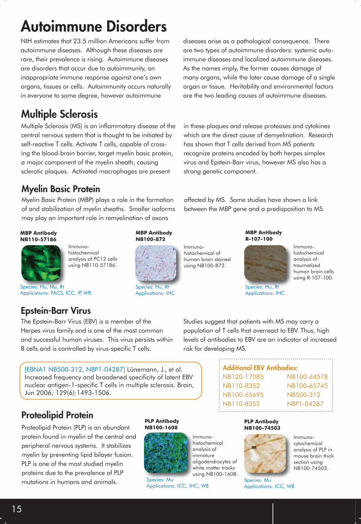
Autoimmune DisordersNIH estimates that 23.5 million Americans suffer from autoimmune diseases. Although these diseases are rare, their prevalence is rising. Autoimmune diseases are disorders that occur due to autoimmunity, an inappropriate immune response against one’s own organs, tissues or cells. Autoimmunity occurs naturally in everyone to some degree, however autoimmune
diseases arise as a pathological consequence. There are two types of autoimmune disorders: systemic auto-immune diseases and localized autoimmune diseases. As the names imply, the former causes damage of many organs, while the later cause damage of a single organ or tissue. Heritability and environmental factors are the two leading causes of autoimmune diseases.
Multiple Sclerosis (MS) is an inflammatory disease of the central nervous system that is thought to be initiated by self-reactive T cells. Activate T cells, capable of cross-ing the blood-brain barrier, target myelin basic protein, a major component of the myelin sheath, causing sclerotic plaques. Activated macrophages are present
in these plaques and release proteases and cytokines which are the direct cause of demyelination. Research has shown that T cells derived from MS patients recognize proteins encoded by both herpes simplex virus and Epstein-Barr virus, however MS also has a strong genetic component.
Multiple Sclerosis
MBP AntibodyR-107-100
Species: Hu, RtApplications: IHC
Immuno-histochemical analysis of traumatizedhuman brain cellsusing R-107-100.
MBP AntibodyNB100-872
Immuno-histochemical ofhuman brain stained using NB100-872.
Species: Hu, RtApplications: IHC
MBP AntibodyNB110-57186
Species: Hu, Mu, RtApplications: FACS, ICC, IP, WB
Immuno-histochemical analysis of PC12 cells using NB110-57186.
Myelin Basic Protein (MBP) plays a role in the formation of and stabilization of myelin sheaths. Smaller isoforms may play an important role in remyelination of axons
affected by MS. Some studies have shown a link between the MBP gene and a predisposition to MS.
Myelin Basic Protein
NB120-17085 NB110-8352NB100-65695 NB110-8352
NB100-64578NB100-65745NB500-312NBP1-04287
Proteolipid ProteinPLP AntibodyNB100-1608
Immuno-histochemical analysis of immature oligodendrocytes of white matter tracks using NB100-1608.
Species: MuApplications: ICC, IHC, WB
PLP AntibodyNB100-74503
Immuno-cytochemical analysis of PLP in mouse brain thick section using NB100-74503.
Species: MuApplications: ICC, WB
Proteolipid Protein (PLP) is an abundant protein found in myelin of the central and peripheral nervous systems. It stabilizes myelin by preventing lipid bilayer fusion. PLP is one of the most studied myelin proteins due to the prevalence of PLP mutations in humans and animals.
The Epstein-Barr Virus (EBV) is a member of the Herpes virus family and is one of the most common and successful human viruses. This virus persists within B cells and is controlled by virus-specific T cells.
Studies suggest that patients with MS may carry a population of T cells that overreact to EBV. Thus, high levels of antibodies to EBV are an indicator of increased risk for developing MS.
Epstein-Barr Virus
Additional EBV Antibodies:[EBNA1 NB500-312, NBP1-04287] Lünemann, J., et al. Increased frequency and broadened specificity of latent EBV nuclear antigen-1-specific T cells in multiple sclerosis. Brain,Jun 2006; 129(6):1493-1506.
Type 1 Diabetes is a disease in which the immune system destroys insulin-producing beta cells in the pancreas. Current research shows that this attack is mediated by killer T cells. Apoptosis of beta cells occurs
several months prior to symptomatic onset, thus type 1 diabetes is sometimes referred to as a silent killer. Studies show that B lymphocytes also play a role in pathogenesis, however their role is less clear.
Type 1 Diabetes
Insulin is a polypeptide hormone that enhances membrane transport of glucose and other molecules, as well as promoting glycogen storage, formation of
triglycerides and synthesis of proteins and nucleic acids. Deficiencies in insulin result in type 1 diabetes.
Insulin
Insulin (D3E7 (5B6/6)) AntibodyNB100-64697
Species: Rt, HuApplications: ELISA, IHC-Fr, IHC-P
Immuno-histochemicalanalysis ofhuman pancreas using NB100-64697.
Insulin (C-PEP-01) AntibodyNB500-413
Immuno-histochemical analysis of human pancreas using NB500-413.
Species: HuApplications: ELISA, ICC, IHC, RIA
Insulin (E2E3) AntibodyNB120-9569
Species: Bv, Hu, Po, Rt, RbApplications: ELISA, ICC, IHC, WB
Immuno-histochemical analysis of human pancreas using NB120-9569.
IA2 IA2 AntibodyNBP1-00134
Western blot analylsis on NIH 3T3 lysate usingNBP1-00134.
Species: Hu, Mu, RtApplications: ELISA, WB
IA2 AntibodyH00005798-B01
Western Blot analysis ontransfected 293T cell line (Lane 1)usingH00005798-B01.Lane 2 is a non-transfected lysate.Species: Hu
Applications: ELISA, WB
IA2 belongs to the protein tyrosine phosphatase family. IA2 is an autoantigen reactive with type 1 diabetes patients’ sera, thus it may be a potential target of autoimmunity in type 1 diabetes.
CD137CD137 belongs to the tumor necrosis factor receptor family and is expressed on activated T cells. The functions of CD137 in T cells include regulating activation, proliferation and apoptosis. Studies suggest that CD137 plays a significant role in the development of and genetic predisposition to type 1 diabetes. CD137 antibody therapy can suppress the development of type 1 diabetes in mice.
CD137 AntibodyNB100-92367
Western blot analysis on extracts from K562 cells using NB100-92367.
Species: HuApplications: ELISA, WB
GADGlutamic acid decarboxylase (GAD) catalyzes the conversion of glutamate to GABA, a major inhibitory neurotransmitter in the CNS. GAD exists as two isoforms, GAD65 and GAD67. GAD65 is thought to be the major autoantigen and an autoreactive T cell target in type 1 diabetes.
GAD65 AntibodyNB120-15299
Immuno-histochemicalanalysis of human pancreas using NB120-15299.
Species: Hu, RtApplications: IHC
GAD1/2 AntibodyNB100-92033
Immuno-histochemicalanalysis of human lung carcinoma tissue using NB100-92033.
Species: Hu, MuApplications: ELISA, IHC, WB
NEW
[IA2 NBP1-00134] Buzzetti R, et al. Non Insulin Requiring Autoimmune Diabetes Study Group. High titer of autoantibodies to GAD identifies a specific phenotype of adult-onset autoimmune diabetes. Diabetes Care. 2007 Apr;30(4):932-938.
16
Rhuematoid Arthritis
TNFTumor necrosis factor (TNF) is a cytokine produced by macrophages that causes the inflammation associated with RA. Medications currently available to combat RA function by binding TNF and preventing it from functioning.
TNF alpha AntibodyNB100-80057
Western blot analysis on recombinant protein using NB100-80057.
Species: HuApplications: WB
TNF alpha AntibodyNB100-75387
Western blotanalysis on E coli derived fusion protein using NB100-75387.
Species: Hu, Mu, RtApplications: ELISA, WB
Interleukin-1 (IL-1) is a pivotal cytokine involved in the pathogenesis of RA. IL-1 alpha and IL-1 beta bind the same cell surface receptor, have 25% amino acid sequence identity, and elicit similar biological responses. IL-1 works with IL-6 and TNF alpha to
induce early onset inflammatory responses. IL-1 also activates chondrocytes to stimulate cartilage break-down, activates osteoclasts to initiate bone resoprtion, and induces fibroblast proliferation to initiate synovial pannus formation.
IL-1
IL-1 beta AntibodyH00003553-B01
Species: HuApplications: ELISA, WB
Western Blot analysis of transfected 293T cell line usingH00003553-B01.
IL-1 beta AntibodyNB110-57112
Western blot analysis of onrecombinant protein using NB110-57112.
Species: HuApplications: WB
IL-1 beta AntibodyNB600-633
Species: Hu, MkApplications: ELISA, FACS, IHC-Fr, IHC-P, IP, WB
Western blot analysisof anti-IL1β usingNB600-633.
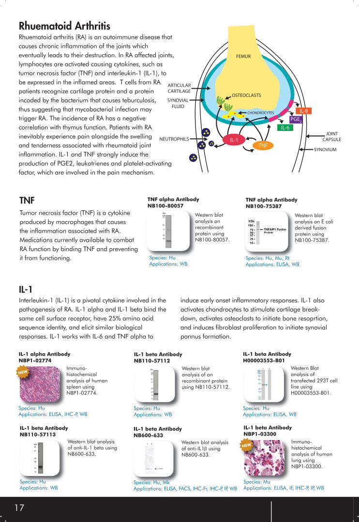
IL-6
PGE2
IL-8
ARTICULAR
CARTILAGE
SYNOVIAL
FLUID
NEUTROPHILS
OSTEOCLASTS
CHONDROCYTES
JOINT
CAPSULE
SYNOVIUM
FEMUR
TNFIL-1
Rhuematoid arthritis (RA) is an autoimmune disease that causes chronic inflammation of the joints which eventually leads to their destruction. In RA affected joints, lymphocytes are activated causing cytokines, such as tumor necrosis factor (TNF) and interleukin-1 (IL-1), to be expressed in the inflamed areas. T cells from RA patients recognize cartilage protein and a protein incoded by the bacterium that causes teburculosis, thus suggesting that mycobacterial infection may trigger RA. The incidence of RA has a negative correlation with thymus function. Patients with RA inevitably experience pain alongside the swelling and tenderness associated with rheumatoid jointinflammation. IL-1 and TNF strongly induce the production of PGE2, leukotrienes and platelet-activating factor, which are involved in the pain mechanism.
IL-1 beta AntibodyNBP1-03300
Species: MuApplications: ELISA, IF, IHC-P, IP, WB
Immuno-histochemical analysis of human lung using NBP1-03300.
IL-1 beta AntibodyNB110-57113
Species: HuApplications: WB
Western blot analysis of anti-IL-1 beta usingNB600-633.
IL-1 alpha AntibodyNBP1-02774
Immuno-histochemical analysis of human spleen using NBP1-02774.
Species: HuApplications: ELISA, IHC-P, WB
NEW
NEW
17
18
I N T H E N E W S1. [CD36 NB110-59724] During A, Doraiswamy S, and Harrison EH. Xanthophylls are preferentially taken up compared with beta-carotene by retinal cells via a SRBI-dependent mechanism. J Lipid Res 2008;49(8):1715-1724.
2. [Cathepsin F NB100-1784] Maubach G, Lim MCC, and Zhuo L. Nuclear Cathepsin F Regulates Activation Markers in Rat Hepatic Stellate Cells. Mol Biol Cell 2008;19(10):4238-4248.
3. [Enterokinase Cleavage Site NB600-345] Stahnke B, Thepen T, Stocker M, et al. Granzyme B-H22(scFv), a human immunotoxin targeting CD64 in acute myeloid leukemia of monocytic subtypes. Mol Cancer Ther 2008;7(9):2924-2932.
4. [Defensin beta-3 NB200-117] Akiyama M, Sakai K, Takayama C, et al. CGI-58 Is an alpha/beta-Hydrolase within Lipid Transporting Lamellar Granules of Differentiated Keratinocytes. Am J Pathol 2008;173(5):1349-1360.
5. [HLA-DMB H00003109-M01] Callahan MJ, Nagymanyoki Z, Bonome T, et al. Increased HLA-DMB Expression in the Tumor Epithelium Is Associated with Increased CTL Infiltration and Improved Progno-sis in Advanced-Stage Serous Ovarian Cancer. Clin Cancer Res 2008;14(23):7667-7673.
8. [IL-18 Binding Protein NB200-201] He Z, Lu L, Altmann C, et al. Interleukin-18 binding protein transgenic mice are protected against ischemic acute kidney injury. Am J Physiol Renal Physiol 2008;295(5):F1414-1421.
9. [TLR2 NB200-580] Henning LN, Azad AK, Parsa KVL, et al. Pulmonary Surfactant Protein A Regulates TLR Expression and Activity in Human Macrophages. J Immunol 2008;180(12):7847-7858.
10. [Nurr1 NB110-40415] Luo Y, Xing F, Guiliano R, et al. Identification of a Novel Nurr1-Interacting Protein. J Neurosci 2008;28(37):9277-9286.
11. [MART-1 NB110-57185] Massoumi R, Kuphal S, Hellerbrand C, et al. Down-regulation of CYLD expression by Snail promotes tumor progression in malignant melanoma. J Exp Med 2009;206(1):221-232.
12. [Clostridium difficile Toxin A NB600-1066] Dissanayake SK, Olkhanud PB, O’Connell MP, et al. Wnt5A Regulates Expression of Tumor-Associated Antigens in Melanoma via Changes in Signal Transducers and Activators of Transcription 3 Phosphorylation. Cancer Res 2008;68(24):10205-10214.
13. [TARDBP NB110-55376] Geser F, Brandmeir NJ, Kwong LK, et al. Evidence of Multisystem Disor-der in Whole-Brain Map of Pathological TDP-43 in Amyotrophic Lateral Sclerosis. Arch Neurol. May 1, 2008; 65(5):636-41.
14. [TARDBP H00023435-M01] Weihl CC, Temiz P, Miller SE, et al. TDP-43 accumulation in inclusion body myopathy muscle suggests a common pathogenic mechanism with frontotemporal dementia. J Neurol Neurosurg Psychiatry. 2008 Oct;79(10):1186-9.

Daily product updates!www.novusbio.comTOLL FREE: 888.506.6887 • PHONE: 303.730.1950
OR
DE
RIN
G


![ProductDataSheet FullSize v4 - Novus BiologicalsFlow Cytometry: HIF-1 alpha Antibody [NB100-449] - HeLa cells were treated for 15 hrs with 200 uM CoCl2, fixed in PFA, and permeabilized](https://img.pdfslide.us/doc/110x75/5ea8f11b9def324a2d25735d/productdatasheet-fullsize-v4-novus-biologicals-flow-cytometry-hif-1-alpha-antibody.jpg)