Embed Size (px)
Citation preview

1
Immunological Principles Guiding the Rational
Design of Particles for Vaccine Delivery
Katelyn T. Gause,1 Adam K. Wheatley,2 Jiwei Cui,1,† Yan Yan,1 Stephen J. Kent,2 and
Frank Caruso1,*
1ARC Centre of Excellence in Convergent Bio-Nano Science and Technology, and the
Department of Chemical and Biomolecular Engineering, The University of Melbourne,
Parkville, Victoria 3010, Australia
2ARC Centre of Excellence in Convergent Bio-Nano Science and Technology, and the
Department of Microbiology and Immunology, The University of Melbourne, at the Peter
Doherty Institute for Infection and Immunity, Parkville, Victoria 3010, Australia
*AUTHOR EMAIL ADDRESS: [email protected]

2
ABSTRACT
Despite the immense public health successes of immunization over the past century,
effective vaccines are still lacking for globally important pathogens such as human
immunodeficiency virus (HIV), malaria, and tuberculosis (TB). Exciting recent advances
in immunology and biotechnology over the past few decades have facilitated a shift from
empirical to rational vaccine design, opening possibilities for improved vaccines. Some
of the most important advancements include: (i) the purification of subunit antigens with
high safety profiles, (ii) the identification of innate pattern recognition receptors (PRRs)
and cognate agonists responsible for inducing immune responses, and (iii) developments
in nano- and microparticle fabrication and characterization techniques. The latter
advances now allow highly tunable physicochemical properties of particle-based
vaccines, including composition, size, shape, surface characteristics, and degradability.
We propose that enhanced collaborative efforts between immunology and materials
science will give rise to next-generation vaccines. This process will be significantly aided
by a greater understanding of the immunological principles guiding vaccine antigenicity,
immunogenicity, and efficacy. With specific emphasis on PRR-targeted adjuvants and
particle physicochemical properties, this review aims to provide an overview of the
current literature to guide and focus rational particle-based vaccine design efforts.
KEYWORDS: adjuvant; vaccine particles; codelivery; antigen presentation; pattern
recognition receptors; antigen presenting cell; lymph node trafficking; subunit antigen;
TLR; NLR
3
VOCABULARY
Antigen, unique molecule (e.g., protein, peptide, polysaccharide) that is specifically
recognized by the adaptive immune system; adjuvant, a component (e.g., alum, PRR
agonist) or characteristic (e.g., particle-based delivery system) of a vaccine formulation
that enhances the quality or quantity of the induced immune response; pattern
recognition receptor (PRR), cellular receptors that recognize pathogen- and danger-
associated molecular patterns (PAMPs and DAMPs); toll-like receptor (TLR), PRRs on
the cell surface membrane (TLR1, TLR2, TLR4, TLR5, TLR6) that recognize bacterial
products such as lipopolysaccharide, lipoteichoic acids, lipoproteins, and flagellum and
on the endosomal membrane (TLR3, TLR7, TLR8, TLR9) that recognize viral nucleic
acids, which can be accessed during viral replication or upon intracellular degradation;
nucleotide-binding oligomerization domain-like receptors (NOD-like receptor,
NLR), PRRs in the cytoplasm. NLRP3 senses cellular damage and stress. NOD receptors
recognize bacterial peptidoglycan (PGN); agonist, a molecule that specifically interacts
with a cellular receptor (e.g., PRR) to activate a physiological response, such as an
immune response; endocytosis, active cellular internalization that can occur via a variety
of cell surface receptors, such as PRRs.

4
Since the introduction of the smallpox vaccine by Edward Jenner in 1798, vaccines
have been created to protect against a range of infectious diseases.1 The eradication of
smallpox was announced by the World Health Organization (WHO) in 1979, poliovirus
is now nearing global eradication, and measles is controlled in most parts of the world.
With the exception of safe water, vaccination is considered the most effective health
intervention ever developed.2 Despite successes to date, safe and efficacious vaccines are
still lacking for many important chronic human pathogens, such as malaria, tuberculosis
(TB), and human immunodeficiency virus (HIV).
Most current vaccines are derived from either live-attenuated or inactivated pathogens
or toxins (i.e., toxoid). Live-attenuated vaccines contain pathogens that have been
weakened through selective propagation (i.e., multiple passages in non-human hosts) to
reduce their replicative fitness and prevent onward transmission. Administration of these
vaccines typically results in mild to asymptomatic infection, but generates long-lived
immunity similar to that observed in individuals who recover from natural infection.
However, live-attenuated vaccines have the potential to cause disease, especially in
individuals with compromised immune systems. Inactivated and toxoid vaccines contain
pathogens or toxins, respectively, that are inactivated by heat or chemical (e.g.,
formaldehyde) treatment. Inactivated and toxoid vaccines are potentially safer than live-
attenuated vaccines, but material derived from pathogens inherently contains microbial
components that can increase the risks of unwanted side effects, such as excessive
inflammation. Batch-to-batch variation and pathogens with difficult or problematic
culturing protocols are additional disadvantages associated with live-attenuated,
inactivated, and toxoid vaccines. Enabled by advances in bioinformatics (i.e.,

5
immunoinformatics)3,4 recombinant DNA technologies, and genetic engineering, the
development of protein and peptide subunit antigens has opened possibilities for
rationally designing safer vaccines for a wider range of applications including cancer and
chronic infections.5-7 However, in their purified, soluble form, protein and peptide
antigens are poorly immunogenic; that is, immunization generally does not induce
responses that are sufficient to result in protective immunity. This is because: (i)
immunostimulating microbial components are not present in these purified antigens, and
(ii) diffusion and clearance of soluble material inhibits the required local concentration of
antigen necessary for immune response induction. Particulate systems are inherently
more immunogenic than soluble systems, thus subunit antigens require particle-based
delivery systems and adjuvants to induce immune responses.6,8
In addition to subunit antigen-based vaccines, most vaccines require adjuvants to
induce sufficient immune responses (“adjuvare” is Latin for “to help”).9 Currently
licensed vaccines are formulated with either aluminum salts (e.g., aluminum
oxohydroxide, aluminum hydroxyphosphate) (also known as “alum”) or oil-in-water
emulsions, which act as both particulate vaccine delivery vehicles and
immunostimulants.10,11 Both alum and emulsion adjuvants were empirically identified
and the mechanisms of vaccine enhancement remain poorly defined.12,13 However, these
adjuvants boost immune responses and in particular, neutralizing antibodies, which are a
correlate of protection for most human pathogens for which there are currently licensed
vaccines.14 For several major pathogens such as malaria, TB, and HIV, effective vaccines
have been elusive and traditional approaches of vaccine development have either failed or
have been too weakly protective to be widely useful.15-17 Recent advances in

6
biotechnology and a greater understanding of the immunological basis for effective
vaccination may facilitate the rational design of next-generation vaccines;18 particularly
the identification of immunopotentiating molecules that specifically activate pattern
recognition receptors (PRRs) on innate immune cells, which could form the basis of
advanced adjuvant formulations,19 and highly tunable particle-based delivery systems for
precise delivery of antigens and adjuvants in vivo.20 This review provides an overview of
vaccine immunology as it relates to PRR activation and the effects of vaccine particle
physicochemical properties on the quality and magnitude of immune responses to
immunization. Two classes of PRRs with significant potential as targets for next-
generation adjuvants are highlighted: toll-like receptors (TLRs) and NOD-like receptors
(NLRs). Additionally, important recent studies that have elucidated the effects of particle
size, shape, surface characteristics, and degradability on the efficacy of particle-based
vaccines are discussed in detail. The overarching aim of this review is to contextualize
how adjuvant and particle characteristics can be modularly engineered to achieve desired
immunization outcomes.
OVERVIEW OF THE GENERATION OF PROTECTIVE IMMUNE RESPONSES BY
VACCINATION
Vaccine Administration and Trafficking. The majority of currently utilized vaccines
are administered intramuscularly (i.e., direct injection into the skeletal muscle), a route
associated with low reactogenicity, which is highly favorable for licensure. Tissue
damage at the site of administration triggers local innate immune responses (e.g.,

7
cytokine and chemokine secretion) by muscle cells and muscle-resident immune cells
(reviewed in Liang et al.21). This leads to local inflammation and the infiltration of
immune cells from the circulation to the site of injection, particularly neutrophils and
antigen presenting cells (APCs) such as monocytes/macrophages and dendritic cells
(DCs), a subset of immune cells highly specialized for antigen capture and presentation.
DCs, both migratory and those resident within the muscle, efficiently capture antigen
from the extracellular environment via endocytosis (e.g., phagocytosis,
macropinocytosis), which can occur via a variety of cell surface receptors,22-24 including
PRRs that recognize pathogen- and danger-associated molecular patterns (PAMPs and
DAMPs, respectively) (Figure 1a).19 Internalization of antigen and the engagement of
PRRs induce DC maturation, upregulatation of antigen processing machinery,25 and
presentation of intracellularly degraded antigen fragments on the cell surface by
complexation with major histocompatibility complex (MHC) molecules (Figure 1a). In
addition, DC maturation drives changes in the expression patterns of surface chemokine
receptors (e.g., CCR7), which results in migration out of the muscle to lymphoid organs
via the blood or lymphatic system.26 Some vaccine material may also traffic to lymph
nodes via convective flow from the interstitium without assistance from migratory APCs
(Figure 1b).27
Priming of Adaptive Immune Responses in Lymph Nodes. Lymph nodes are located in
anatomically strategic positions to sample antigens and facilitate adaptive immune
responses, which are dependent upon two important subsets of lymphocytes, T cells and
B cells (Figure 1c). Within lymphoid tissues, T cells and B cells localize to two
functionally partitioned areas termed the T cell zones and B cell zones. Mature DCs

8
arriving from the tissues enter the T cell zone, where T cell recognition via the T cell
receptor (TCR) of intracellularly processed antigen presented in the context of MHC
drives the activation of antigen-specific naïve T cells (often termed signal I; T cell
signaling reviewed in Mantegazza et al.28). Alternatively, antigens that have entered the
lymph nodes without internalization and trafficking by DCs at the injection site may be
phagocytosed and processed by sub-capsular sinus (SCS) macrophages.29,30 If sufficiently
small, antigen may also directly diffuse into the T cell zone via conduits established by
fibroblastic reticular cells,31,32 where lymph-node resident DCs can internalize and
present antigens to T cells.33 DCs simultaneously express co-stimulatory signals on the
cell surface (i.e., CD80/CD86) (signal II), and a cocktail of secreted cytokines (signal III)
that act in concert to fine tune the activation and differentiation program of responding T
cells, thereby tailoring the host immune response to the nature of the pathogen.34,35 Two
common types of T cells have been delineated based upon differing glycoprotein co-
receptor components of the TCR, either CD4+ or CD8+ (Figure 1c). DC-mediated
activation of CD4+ and CD8+ T cells triggers proliferation and differentiation into
immune effectors, which act both directly and indirectly to clear infections and prevent
disease. In addition, proliferating T cells have the capacity to differentiate into long-lived
populations of cells primed for rapid response to secondary exposure, the immunological
memory that is a hallmark of adaptive immunity.
CD8+ T cells recognize antigen peptide fragments (~8-9 amino acids) in the context of
MHC class I, which is ubiquitously expressed by every host cell and predominantly used
to present antigens localized within the cytoplasm. Endocytosed material can also be
presented via MHC class I, a process termed “cross-presentation” (Figure 1a). The

9
cellular mechanisms that enable cross-presentation may include several overlapping
pathways (reviewed in Joffre et al.36). Materials within the endosomes can be degraded
into peptide fragments, allowing import into the endoplasmic reticulum (ER) and
presentation via classical MHC class I pathways.36 Alternatively, degraded peptides can
be imported directly back into phagosomes (vacuolar pathway) for MHC I loading and
transport to the cell surface (reviewed in Ma et al.37). Activated antigen-specific CD8+ T
cells (i.e., cytotoxic T lymphocytes, CTLs) leave lymphoid sites and actively seek out
and kill infected cells displaying cognate peptides via MHC I on the cell surface. This
cytotoxic/cytolytic ability is crucial in the maintenance of effective immune control
against intracellular pathogens and cancer.38 CD4+ T cells recognize peptides (9-20
amino acids) complexed with MHC class II molecules, which are mainly expressed by
professional APCs (i.e., DCs, macrophages/monocytes, B cells) MHC class II
presentation is mainly used for extracellular antigens endocytosed and degraded in
endosomal/lysosomal compartments (reviewed in Roche et al.39). Activated CD4+ T
cells, or T helper (Th) cells, provide critical support to many aspects of the immune
response, including CTL and serum antibody responses.40 While numerous specialized
subsets of Th cells are recognized in the literature, such as Th1 (IFN-γ-producing), Th2
(IL-4- and IL-5-producing), Th17, and regulatory T cells (Treg), the CD4+ T cell
compartment displays incredible plasticity, both in terms of phenotype (i.e., surface
marker expression) and function (i.e., cytokine and chemokine secretion) (reviewed in
Oestreich et al.41).
Unlike T cells, B cells can directly recognize antigens via localized immunoglobulins
(Igs) on the cell surface called B cell receptors (BCRs) (signal I; B cell signaling

10
reviewed in Yuseff et al.42). While B cells can encounter antigens in the periphery,
coincidental interactions are likely rare events. Instead, antigens trafficked to B cell zones
(follicles) are retained for extended time periods by a network of follicular DCs (FDCs).43
This temporal and spacial co-localization significantly increases the likelihood of naïve B
cells to engage with their cognate antigen. At least two major pathways of antigen
delivery to FDCs have been identified. Antigens are captured by SCS macrophages and
imported from the SCS into the B cell folllicle.44,45 Here, antigen can either be recognised
by cognate B cells, or relayed by non-cognate B cells to FDCs via a mechanism
dependent upon complement and complement receptor 2 (CD21).46 Alternatively, protein
antigens with a hydrodynamic radius around 4-5 nm (Mw ~70 kDa) may diffuse directly
via conduits from the SCS to the B cell follicle.47 BCR binding to cognate antigens
triggers internalization, B cell activation, upregulation of antigen processing machinery,
and presentation of degraded antigens via MHC class II.48,49 Activated B cells migrate to
the T cell zone/B cell zone border where TCR:MHC II interactions with antigen-specific
CD4+ T cells leads to the provision of T cell “help” via CD40:CD40 ligand (CD40L)
signalling (signal II).50,51 This in turn promotes the upregulation of transcription factor
Bcl-6 in both B cells and T cells,52-54 driving the formation of germinal centers in B cells,
which are specialized foci of B cell proliferation and maturation (reviewed in Victora et
al.55). Germinal centers function as the site of BCR diversification and enable the process
of affinity maturation, whereby B cells are selected for high affinity binding to cognate
antigens by sequential rounds of proliferation and competition for limited CD40L-
dependent help from T follicular helper (Tfh) cells.56 B cells exiting germinal centers can
differentiate into long lived memory B cells that circulate in the periphery. A subset of

11
generally high affinity B cells selected in the germinal center initate a differentiation
program toward plasma cells, which are highly specialized for the secretion of antibodies,
the soluble secreted forms of the BCR. Plasma cells migrate via the bloodstream and take
up long-term residence within bone marrow niches where they can provide a stable and
long-term source of serum antibodies, for some antigens up to the lifetime of the host.
Antibodies can mediate direct neutralization of pathogens and/or the clearance of infected
cells via mechanisms such as antibody-dependent cellular cytotoxicity (ADCC)57 or
antibody-dependent phagocytosis (ADP).58,59
Recapitulating the complex coordination of immune cells required for the generation
of an efficacious adaptive immune response is a challenge for vaccine development.
However, an ever expanding understanding into the immunological principles driving
vaccine immunogenicity creates opportunities to harness complex immune systems with
rationally designed, next-generation vaccines and thereby maximize protective potential.

12
Figure 1. Vaccine administration and induction of innate and adaptive immune
responses. a) Vaccines are administered via intramuscular (most common), intradermal,
oral, and mucosal routes, where they encounter local immune cells such as neutrophils,
monocytes, macrophages, and dendritic cells (DCs), a subset of antigen presenting cells
(APCs) highly specialized for antigen capture and presentation. Upon internalization,
vaccine particles can activate PRRs on the cell surface (e.g., TLR1, TLR2, TLR4, TLR5,
TLR6, TLR11), endosome (e.g., TLR3, TLR7, TLR8, TLR9), and cytoplasm (e.g., NOD1,
NOD2, NLR3). Captured vaccines are degraded with endosomal/lysosomal
compartments into peptide fragments, which are sucsequently presented on the cell
surface upon major histocompatibility complex (MHC) molecules. b) Internalization of
antigen and the engagement of pattern recognition receptors (PRRs) induces DC
maturation, which facilitates migration out of the muscle to lymphoid organs via the
blood or lymphatic system. Small vaccine particles (~20-30 nm) can effectively traffic to

13
lymph nodes via convective flow without assistance from migratory APCs at the site of
administration; whereas larger particles are more likely to be retained at the injection
site and require transport into the lymph nodes by migratory APCs. c) DCs activate
CD8+ and CD4+ T cells via MHC class I and II presentation, respectively. Activated
CD8+ T cells can differentiate into cytotoxic T lymphocytes (CTLs), which are crucial
for control against intracellular pathogens and cancer. Activated CD4+ T cells can
differentiate into T helper (Th) cells, such as Th1 (IFN-γ-producing), Th2 (IL-4- and IL-
5-producing), Th17, or regulatory T cells (Treg) that provide critical support to other
immune cells, such as CTLs, via complementary cytokine secretion, and to serum
antibody responses, via CD40:CD40 ligand co-stimulation of antigen-specific B cells.
MODULATION OF VACCINE IMMUNE RESPONSES BY CELLULAR
RECEPTORS FOR MICROBIAL COMPONENTS
A critical role of the innate immune system is to scan foreign material and relay
critical information to the adaptive immune system to modulate the strength and quality
of protective immunity.19,60,61 In general, this occurs through activation of PRRs. A range
of PRR agonists are now under intense investigation for use as adjuvants that target
specific innate immune cell recognition pathways.62,63 For example, Monophosphoryl
lipid A (MPL) was the first PRR agonist approved for use in human vaccines and many
others are undergoing preclinical and clinical trials.64,65 MPL is a derivative of lipid A
from Salmonella minnesota R595 that is detoxified by mild hydrolytic treatment. MPL
has been formulated with alum in an adjuvant called AS04 that is licensed for use in HPV

14
and HBV vaccines. AS04 is considered safe and more effective than alum,66 thus
solidifying the potential of PRR agonist-based adjuvants. Studies clearly indicate that
activation of PRRs has varied and complex effects on the outcome of immunization,19
which can be exploited in rational adjuvant design. It should be noted that improved
adjuvants using PRR and other approaches often results in increased local and systemic
side effects (increased reactogenicity). Although some side effects will be tolerable in the
setting of a high risk of acquisition of severe diseases, an important goal of PRR-adjuvant
vaccine research is to improve immunogenicity without unacceptable increases in side
effects. TLR and NOD-based approaches are among promising adjuvants in this regard.
Toll-like Receptors. Toll-like receptors (TLRs) are the most extensively characterized
PRRs with 10 and 13 TLRs identified in humans and mice, respectively. TLRs on the cell
surface (TLR1, TLR2, TLR4, TLR5, TLR6) recognize bacterial products such as
lipopolysaccharide (LPS), lipoteichoic acids, lipoproteins, and flagellum. Endosomal
TLRs (TLR3, TLR7, TLR8, TLR9) recognize viral or bacterial nucleic acids, which can
be accessed during viral replication or upon intracellular degradation. TLR activation
mainly polarizes Th1-biased adaptive immune responses;19,35,67-69 however, TLR2
activation has been shown to polarize Th2-biased immune responses.70,71 There is also a
clear trend in several studies showing that endosomal TLR signaling enhances cross
presentation and CD8+ T cell responses,72-79 and that surface TLRs can actually suppress
CD8+ T cell responses.79
Activation of multiple TLRs can result in synergy or inhibition of immune responses
via intracellular crosstalk, the mechanisms of which have been reviewed in detail.80-82
Various reports have shown that TLR pathways that use the adapter molecule, MyD88

15
(all TLRs except TLR3), can synergize with TLR pathways that signal through the
adapter molecule, TRIF (TLR3 and TLR4) in the induction of innate inflammatory
responses,83-86 Th1 polarization capacity,84 and antibody responses.87 Zhu et al.
demonstrated that the combination of TLR3, TLR9, and TLR2/6 ligands induced CD8+ T
cell responses with synergistically enhanced functional avidity compared with single and
paired ligands; however, the number of activated CD8+ T cells was not significantly
different.88 Additionally, immunization with the triple ligand combination signficantly
enhanced protection against viral challenge compared with single and paired ligands.
Overall, the study demonstrated that even though MyD88-dependent pathways are not
synergistic as a pair, when costimulated with TRIF-dependent TLR3, protection can be
enhanced through the quality, and not quantity, of the CD8+ T cell responses.
NOD-like Receptors. Up to 22 NOD-like receptors (NLRs) have been identified in
humans. Although the triggers and functions of many NLRs remain unknown, NOD1,
NOD2, and NLRP3 are the best characterized.89-91 NLRP3 is a widely studied NLR that
senses cellular damage and stress.92 NLRP3 (and some other NLRs) activate multiprotein
complexes called inflammasomes that facilitate the production and release of
inflammatory cytokines, IL-1β and IL-18.93 Activation of the transcription factor nuclear
factor-κB (NF-κB) induces transcription for pro-IL-1β while pro-IL-18 is constitutively
expressed and increases in expression upon cellular activation. Activated inflammasomes
then recruit caspase-1, which is a cysteine-aspartic acid protease that cleaves and
activates pro-IL-1β and pro-IL-18 into their bioactive forms.94,95 It has been shown that
alum adjuvants and other particulates (e.g., nanoparticles and microparticles) activate the

16
NLRP3 inflammasome through lysosomal destabilization, which causes leakage of
proteolytic enzymes into the cytosol.96-98
NOD receptors (i.e., NOD1, NOD2) recognize peptidoglycan (PGN). NOD2 detects
muramyl dipeptide (MDP), which is a motif common to both Gram-positive and Gram-
negative bacterial PGN.99 Notably, MDP is also recognized by NLRP3.100 NOD1
specifically detects γ-glutamyl diaminopimelic acid (iE-DAP), a breakdown product of
PGN, which is found almost exclusively in Gram-negative bacteria.101,102 Immunization
with NOD1 and NOD2 agonists (FK156 and MDP, respectively) with the model protein
antigen ovalbumin (OVA) was shown to induce strongly polarized Th2 adaptive immune
responses and no CD8+ T cell responses.103,104 CFA is heat killed mycobacteria that
contains agonists for both TLRs and NODs. The same studies showed that optimal Th1,
Th2, and CD8+ T cell responses to CFA relied on NOD1 and NOD2 signaling, indicating
that NOD signaling can facilitate TLR-driven Th1 and CD8+ T cell responses.103,104 In
contrast, NOD signaling due to PGN contaminants in LPS (TLR4 agonist) was recently
found to inhibit cross presentation.105 Another recent study found that immunization with
NOD1 and NOD2 agonists resulted in enhanced cross-presentation in vitro and CD8+ T
cell responses in vivo.106 Thus, the role of NOD signaling in activating CD8+ T cell
responses remains largely unclear, both in the presence and absence of TLR
costimulation.
Recently, Pavot et al. reported an investigation of a NOD/TLR adjuvant system using
a chimeric ligand containing a NOD2 and TLR2 agonist.107 The chimeric ligand
synergistically enhanced Th1-polarized IgG1 and IgG2a production following
subcutaneous administration; while single ligands did not signficantly enhance the

17
antibody response. Several studies have also demonstrated enhanced and synergistic
activation induced by signaling between TLRs and NOD receptors.85,107-111 In our recent
study, using a particle-based system, we showed that NOD2 activation played different
roles in modulating the adaptive immune response depending on coactivation of TLR9.112
Specially, NOD2 activation alone resulted in Th2-polarized CD4+ T cell and serum
antibody responses; however, in the presence of TLR9 costimulation, there was an
enhancement of Th1-polarized CD4+ T cell and serum antibody responses compared
with TLR9 stimulation alone. Notably, NOD2 coactivation also abrogated the CD8+ T
cell response observed in groups where TLR9 alone was activated.
PARTICLE-BASED VACCINE DELIVERY SYSTEMS
Particulate systems are inherently more immunogenic than soluble systems (e.g.,
cross-presentation efficiency113-115), as nano- and microparticles mimic the size,
geometry, and properties that the immune system recognizes. Thus, delivery of subunit
antigens using particle-based delivery systems can lead to significant improvements in
immmunogencity.6,8 Virus-like particles (VLPs) were the first subunit antigen- and
nanoparticle-based vaccines to reach the market with the FDA approval of the
recombinant hepatitis B surface antigen (HBsAg) vaccine in 1986.116,117 VLPs are self-
assembling nanoparticles composed of viral capsid proteins that mimic viral structure but
do not contain genetic material. There are now four VLP vaccines on the market:
GlaxoSmithKline (GSK)’s Engerix® for hepatitis B virus (HBV) and Cervavix® for
human papillomavirus (HPV), and Merck and Co., Inc.’s Recombivax HB® for HBV and

18
Gardasil® for HPV. There are also many other VLP vaccines currently undergoing
preclinical and clinical development.118
In addition to VLPs, many other types of particles are under investigation for subunit
antigen delivery, including those based on lipids, synthetic polymers, natural polymers,
and inorganic materials.8,119-121 Liposomes are the most widely implemented particle-
based system in the clinic and on the market so far. Liposomes are comprised of
concentric phospholipid bilayers that contain hydrophilic domains in the interior and
exterior and hydrophobic domains in the lipid bilayer.122 Two liposomal vaccine systems
are currently approved for use in humans: Crucell’s Inflexal V® for seasonal influenza123
and Epaxal® for hepatitis A.124
Aside from the inherent immunogenicity associated with particulate structure, the
properties of particulate delivery systems can be engineered to enhance immune
responses through controlled composition (e.g., targeting and/or immunostimulating
ligands, multiple antigens125) and physicochemical properties (e.g., size, shape, surface
properties, degradability).20,126,127 It is clear that particle properties influence immune
responses;20,126-131 however, a more complete understanding of how to engineer intrinsic
particle properties to optimize and/or tune the vaccination outcome is required. The
following sections describe studies elucidating the impacts of particle properties on
various types of immune responses that are relevant to the outcome of vaccination (i.e.,
innate immune cell activation, MHC class I antigen presentation, MHC class II antigen
presentation, lymph node trafficking, CD4+ T cell responses, CD8+ T cell responses, and
B cell responses).

19
Influence of Particle Size. Particle size plays a significant role in vaccine efficacy due
to its influence on lymph node trafficking and localization,27,132-135 adaptive immune
responses,136,137 and cross-presentation.138,139 Studies have suggested that vaccine
particles approximately 20-30 nm in size can effectively traffic to lymph nodes within 2 h
via convective flow from the interstitium without assistance from migratory APCs at the
site of administration; whereas larger particles are more likely to be retained at the
injection site and require transport into the lymph nodes by migratory APCs.27 For
example, Reddy et al. showed that 20-25 nm particles entered the dermal lymphatic
capillary network and localized in lymph nodes more efficiently than 45 or 100 nm
particles.132,133 Size has also been shown to influence the cellular distribution of particles
within the lymph node. For example, Manolova et al. showed that upon injection into
mice, 20 nm polystyrene beads localized in the SCS and B cell areas while larger
particles were excluded from the SCS and found in areas more distal from B cell
follicles.27 In contrast, other studies employing state-of-the-art visualization techniques
have suggested that small (40 nm), intermediate (200 nm), and large (1 μm) particles can
all directly access lymph nodes via the afferent lymphatics,33 as can bacteria and viruses
during infection.140,141 Therefore, the influence of injection site, local hydrodynamic
forces, and particle size on the initiation of immune responses to particle-based vaccines
require further investigation.
In terms of immunogenicity, Fifis et al. found using different sizes of polystyrene
beads with conjugated OVA (20, 40, 100, 200, 500, 1000, 2000 nm), that 40 nm beads
induced the highest IFN-γ-producing CD4+ and CD8+ T cell responses and IgG
production following intradermal immunization.136 Compared with 20 nm and 1000 nm

20
beads, 40 nm beads were associated with a significantly higher percentage of lymph node
cells. Out of the OVA-positive lymph node cells, 40 nm beads associated mostly with
DCs, whereas 1000 nm beads associated mostly with macrophages. Additionally, 40 nm
beads protected against tumor challenge more effectively than 1 μm beads and soluble
OVA. A follow-up study compared OVA-conjugated polystyrene beads in a narrower
size range (20, 40, 49, 67, 93, 101, 123 nm), showing optimal IFN-γ-producing CD4+
and CD8+ T cell responses upon intradermal immunization with 40 and 49 nm particles
(Figure 2a,b).142 Interestingly, the study also demonstrated significantly higher IL-4-
producing CD4+ T cell activation in response to larger beads (93, 101, 123 nm) (Figure
2c). Notably, the study showed minimal differences in IgG production and dominance in
the IgG1 isotype across the range of particle sizes. The findings demonstrate the
possibility of tuning particle size to polarize CD4+ T cell responses. Another study
recently compared the antibody responses induced by gold nanoparticles conjugated with
antigenic peptides of 2, 5, 8, 12, 17, 37, or 50 nm, showing that 8 nm nanoparticles
induced the highest levels of antibody production, while the 37 and 50 nm nanoparticles
were ineffective.137

21
Figure 2. Impact of particle size on T cell immunogenicity in vivo. OVA-conjugated
polystyrene particles 40 and 49 nm in diameter induce CD8+ T cell (a) and IFN-γ-
producing CD4+ T cell responses (b); whereas 93, 101, and 123 nm particles induce IL-
4-producing CD4+ T cell responses (c). Adapted from ref 142. Copyright 2007 American
Chemical Society.

22
Regarding the effect of particle size on cross-presentation efficiency, studies indicate
that decreased particle size is correlated with increased efficiency of cross-
presentation.138,139 For example, Hirai et al. compared the cross-presentation efficiency of
DCs pulsed with different sized silica particles (70, 100, 300, 1000 nm) and OVA.138 The
study showed that 70 and 100 nm particles enhanced antigen localization in the cytosol
from endosomes and induced cross-presentation, while 300 and 1000 nm particles did
not.
Influence of Particle Shape. Studies have indicated that shape may critically influence
the efficacy of particle-based systems used for drug and vaccine delivery.143,144 It is
known that shape plays an important role in cellular uptake, as demonstrated in studies
showing enhanced internalization of spherical particles compared with particles with high
aspect ratios.145-147 Sharma et al. reported that internalization was dependent on cell
membrane binding, where longer particles were more efficiently attached, but
internalization was inhibited by size.145 Another study recently showed that rods
exhibited higher specific uptake and lower nonspecific uptake compared with spheres
conjugated with targeting antibodies.148 Niikura et al. showed that gold rods (40 x 10 nm)
were taken up more efficiently than spheres (20 and 40 nm) and cubes (40 x 40 x 40 nm)
in both mouse macrophages and DCs.149 TEM images showed that 20 nm spheres and
rods escaped endosomes and localized in the cytoplasm following uptake while 40 nm
spheres and cubes remained in endocytic compartments. Additionally, only rods induced
significant levels of IL-1β and IL-18 secretion in DCs, indicating activation of the
inflammasome, probably through lysosomal rupture during endosomal escape. On the
other hand, 40 nm spheres and cubes induced significant TNF-α, IL-6, IL-12, and GM-
23
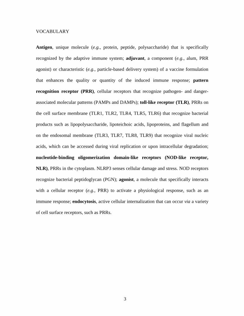
CSF secretion. In vivo, 40 nm spheres coated with the West Nile Virus envelope protein
induced the highest total IgG production in mice compared with rods, cubes and 20 nm
spheres. The study showed an inverse relationship between the specific surface area (total
surface area per particle volume) and antibody production and TNF-α secretion (Figure
3). As the specific surface area depends on both size and shape, the study indicates that
both of these parameters are crucial in determining the immune response.
Figure 3. Impact of surface area on immunogenicity. (a) Antibody production or (b)
TNF-α secretion by DCs shown as a function of the specific surface area (total surface

24
area per particle volume) of a given particle vaccine. 20 nm spheres (blue), 40 nm
spheres (red), cubes (green), rods (orange). Adapted from ref 149. Copyright 2013
American Chemical Society.
Influence of Particle Charge. It is well known that positive surface charge enhances
internalization by cells via electrostatic attractive forces between particles and negatively
charged cell membranes.150,151 Positively charged particles are also exploited for
enhancing immune responses at mucosal tissues,152-154 which is required to induce
mucosal immunity necessary for pathogens that enter at mucosal surfaces. Following
pulmonary immunization, Thomas et al. found that positively charged polyethyleneimine
(PEI)-modified PLGA microspheres induced higher antibody and T cell responses
compared with unmodified particles.152 Fromen et al. compared OVA-conjugated
hydrogel nanoparticles that varied in charge but had constant size, shape, and antigen
loading.153 Pulmonary immunization with cationic nanoparticles enhanced systemic and
lung antibody titers, germinal center B-cell expansion, and increased CD4+ T cell
activation in lung draining lymph nodes compared with anionic nanoparticles.
Additionally, DCs treated ex vivo with cationic nanoparticles induced enhanced T cell
proliferation, expression of MHCII, T cell costimulatory molecules, and cytokine
secretion compared with anionic nanoparticles or soluble OVA. Recently, Stary et al.
showed that by delivering UV-inactivated Chlamydia trachomatis (UV-Ct) and R848
(resiquimod), a TLR7/8 agonist, via charge switching nanoparticles antigen presentation
was redirected to immunogenic DCs, whereas UV-Ct on its own is presented by
tolerogenic DCs, causing an exacerbation of host susceptibility in conventional and

25
humanized mice.154 These particles had a cationic charge below pH 6.5 (allowing
conjugation with negatively charged UV-Ct) and a slight negative charge at physiological
pH 7.4.
Influence of Particle Hydrophobicity. Seong and Matzinger proposed that
hydrophobicity was one of the signals recognized by the innate immune system.155,156 In
agreement with this notion, various studies have correlated hydrophobic particle
properties with enhanced immune responses.157,158 For example, Moyano et al. recently
showed that increasing hydrophobicity of surface attached ligands on gold nanoparticles
was correlated with upregulation of proflammatory cytokine gene expression.157 In
another study, the effect of microparticle hydrophobicity was evaluated in vitro and in
vivo using particles that were constant in size and morphology but were made from
polymers that differed in hydrophobicity: poly(D, L-lactic acid) (PLA), poly(D, L-lactic-
co-glycolic acid) (PLGA), and poly(monomethoxypolyethylene glycol-co-D, L-lactide)
(mPEG-PLA).158 The study correlated the increased hydrophobicity of PLA
microparticles with increased cellular internalization and upregulation of MHCII and
CD86 expression in DCs in vitro and significantly elevated IFN-γ- and IL-4-producing T
cell responses following subcutaneous immunization. Thomas et al. demonstrated that
carboxylated nanoparticles induced activated complement in situ and enhanced antibody
production and T cell responses in vivo compared with hydroxylated surfaces.159
Shahbazi et al. showed enhanced immunostimulatory effects in vitro and in vivo using
nanoparticles with high levels of C-H structures on the surface compared to those with
nitrogen and oxygen.160

26
A series of studies by the Narasimhan group studied the complex immunological
effects of polyanhydride nanoparticles with varied chemistry and hydrophobicity using
copolymers based on sebacic acid (SA), 1,6-bis-(p-carboxyphenoxy)hexane (CPH), and
1,8-bis-(p-carboxyphenoxy)-3,6-dioxaocatane (CPTEG). The least hydrophobic particles
(i.e., SA-rich) were shown to be more efficiently internalized by DCs than the more
hydrophobic particles (i.e., CPH-rich).161 Additionally, the more hydrophobic particles
did not induce the production of IL-6, IL-1β, of TNF-α by DCs, but did induce
expression of MHC II and CD86. On the other hand, the less hydrophobic particles
induced production of higher amounts of secreted cytokines but no expression of surface
markers. The molecular descriptors responsible for DC activation patterns were
determined using informatics analysis, finding number of backbone oxygen moieties,
percentage of hydroxyl end groups, polymer hydrophobicity, and number of akyl ethers
to be the most important.162 The relationship between particle chemistry and the kinetics
and maturation of the induced humoral response upon pulmonary immunization of
particles containing F1-V antigen was also examined.163 The least hydrophobic particles
(20:80 CPH:SA) degraded the fastest and more rapidly induced an antibody response.
CPH-rich formulations (20:80 CPTEG:CPH, 50:50 CPTEG:CPH) degraded more slowly,
persisted in the lungs for at least 63 days, and induced higher antibody titers with a
greater breadth of epitope specificity. It was hypothesized that the induction of longer
lived plasma cells was due to the slow and continuous release of antigen as well as a
more inflammatory environment assumed to be induced by the hydrophobic character of
the particles.

27
ADVANTAGES OF PARTICLE-BASED VACCINES OVER TRADITIONAL
FORMULATIONS
High Density Array of Vaccine Antigens. In contrast to T cell responses, which require
APC intermediaries to initiate a primary immune response, B cells have the capacity to
directly engage vaccine antigens. Subunit antigens do not effectively induce an antibody
response when injected in their free, soluble state because B cells have evolved to
recognize dense, highly repetitive epitope arrangements on the surfaces of pathogens
(e.g., viruses, flagellum) or alternatively, arrayed epitopes bound in immune complexes
on the surface of FDCs. Highly repetitive arrays of epitopes in vaccines can efficiently
crosslink BCRs and trigger potent B cell activation, resulting in enhanced B cell
responses. The density and conformation of the encountered antigen can significantly
modulate subsequent immunity. A major advantage of particle-based vaccines is the
ability to finely control these aspects of antigen delivery. For example, Kanekiyo et al.
showed that an epitope presented by self-assembling nanoparticles of ferritin (octahedral
cage consisting of 24 subunits) or encapsulin (icosahedron made of 60 identical subunits)
resulted in significantly enhanced antibody titres compared with the soluble epitope.164
Using VLPs with covalently attached epitopes of different density, Jegerlehner et al.
showed that the magnitude of antibody responses was significantly correlated with
epitope density.165 The study showed that 60 epitopes per particle spaced 5-10 nm apart
drove maximal humoral immune responses following immunization of mice. Paus et al.
showed that antigen density on sheep red blood cell conjugates was crucial for activating
the extrafollicular plasma B cell response but not the germinal center response.166 Some
small moieties (termed “haptens”) are not immunogenic unless conjugated to a larger

28
carrier (usually protein). This is especially relevant for bacterial polysaccharides, which
require protein conjugates for vaccine efficacy, such as those used in medically important
Haemophilus influenzae type B, meningococcal, or pneumococcal vaccines. While
nanoparticles can directly substitute for the protein carrier in some cases to increase the
immunogenicity of haptens,167 protein-based nanoparticles may offer the ability to act as
effective protein carriers for hapten-based vaccines.
Codelivery of Adjuvants and Immunomodulatory Agents. Immunostimulating ligands
can be simultaneously delivered with vaccine antigens to enhance vaccine efficacy, with
co-packaging of both a means to maximize delivery to the same immune cells in vivo and
thereby limit off target adjuvant affects. This is particularly important for the safety of
PRR agonists, as it spatially constrains the action of PRR agonists and avoids nonspecific
inflammatory responses. A number of studies have shown that the attachment of
immunomodulatory agents, such as PRR ligands,87,112 DC-targeting antibodies,168 ER-
targeting peptides (for enhancing cross-presentation),169 and PEG,170 can enhance and
tune immune responses. Ligands can be incorporated into particles by encapsulation,
physical adsorption, or covalent conjugation.171-173 Covalent conjugation is the preferred
method for incorporating PRRs agonist and other biofunctional ligands due to
controllability over ligand density and orientation. A variety of coupling techniques have
been established for ligand conjugation.174
Recently, studies have emerged demonstrating copackaging of multiple PRR agonists
within a single particle.87,112,175 Using a particle-based delivery system, Kasturi et al.
found that immunization of mice with synthetic nanoparticles containing antigens and
TLR4 (MPL) and TLR7 (R837) ligands induced synergistic increases in antibody

29
production that depended on direct TLR4 and TLR7 activation on the same B cell (Figure
4).87 Notably, however, human B cells do not constitutively express TLR4, and so the
implications of TLR4/7 co-signaling are not clear for human vaccines. In our recent
study, a mesoporous silica-templated protein antigen (OVA) particle was covalently
conjugated with either NOD2, TLR9, or a combination of both ligands leading to
qualitatively and quantitiavely different innate and adaptive immune responses.113
Figure 4. Codelivery of MPL and R837 drives TLR4 and TLR7 activation, respectively,
on the same B cell, leading to synergistic antibody production. a) B cell-deficient mice

30
(μMT mice) reconstituted with B cells from TRIF-/-, MyD88-/-, TLR4-/-, and/or TLR7-/-
mice. b) Synergy is replenished in μMT mice reconstituted with B cells from wild-type
mice. c) Antibody responses are diminished in μMT mice reconstituted with B cells from
TLR4-/- mice, TLR7-/- mice, or a 1:1 mixture of both. d) Antibody responses are
diminished in μMT mice reconstituted with B cells from TRIF-/- or MyD88-/- mice. e)
CD4+ T cell responses are substantially reduced in μMT mice reconstituted with B cells
from TRIF-/- or MyD88-/- mice. Adapted from ref 87. Copyright 2011 Macmillan
Publishers Ltd.
The density of surface ligands, has also been correlated with particle
immunogenicity.176 OVA-containing PLGA nanoparticles functionalized with avidin-
palmitic acid were surface modified with varying amounts of biotinylated anti-DEC-205
monoclonal antibodies (Figure 5).176 The amount of IL-10 produced by DCs in vitro and
IL-10 and IL-5 produced by CD4+ T cells upon restimulation in vitro increased with
ligand density. These results were shown to be independent of DC uptake. Particles were
also used to boost the primary immune response to OVA in CFA to determine whether
this trend was reproduced in vivo. The results showed that IL-10 and IL-5 secretion by
splenocytes restimulated with OVA also increased with increasing ligand density. This
effect was shown to be due to variations in receptor crosslinking.

31
Figure 5. Antibodies targeting antigen to immune cells PRRs influences immune
responses in vitro and in vivo. a) OVA-encapsulated PLGA particles with anti-DEC205
monoclonal antibody conjugated via avidin-biotin; b) IL-10 secretion from DCs
incubated with indicated particles or soluble OVA with DEC205 conjugate; c) IL-10
secretion from OVA-specific CD4+ OTII T cells incubated with DCs from (b) for 72 h; d-
e) IL-10 and IL-5 secretion from whole splenocytes restimulated with OVA following
booster immunization with indicated groups; f) IgG1 titre following intraperitoneal
immunization with indicated groups. Adapted from ref 176. Copyright 2011 Elsevier.
Controlled Rates of Intracellular Cargo Release. For the generation of CD8+ T cell
responses, particle-based antigens must be cross-presented by APCs via MHC class I.

32
Thus, the controlled release of encapsulated antigens upon intracellular degradation is a
widely implemented approach to enhance cross-presentation. Various strategies have
been proposed for engineering intracellular stimuli-responsive release mechanisms in
particles such as systems based on pH,177-179 redox,180-182 and enzymatic activity.183-185 A
study by Howland et al. demonstrated the dependence of antigen release kinetics on
MHC class I presentation efficiency, using yeast cells with surface-displayed model
antigen peptides constructed by fusing peptides to receptors on the yeast cell membrane
via disulfide bonds.186 Release kinetics were manipulated by including linkers of varying
proteolytic degradability. When the yeasts were incubated with DCs, the pattern of cross-
presentation was similar to the pattern of protease cleavage, indicating that faster antigen
release within the phagosome results in more efficient cross-presentation. The study also
showed that antigen released beyond 25 min did not significantly contribute to cross-
presentation, suggesting a limited window for productive intracellular antigen release,
and that antigen released after 25 min may be mostly degraded by lysosomal proteases. In
another study, Broaders et al. compared antigen presentation induced by dextran
microparticles with tunable degradation rates based on modification of the dextran with
acetal groups (Figure 6).187 Acid-catalyzed hydrolysis of the acetals regenerates native
dextran and acetone and methanol by-products. The study showed that particles that
degraded more rapidly (i.e., low acetalation) induced significantly better MHC class I and
MHC class II antigen presentation.

33
Figure 6. Enhanced MHC class I and class II antigen presentation is correlated with
rapid intracellular antigen release kinetics. Adapted from ref 187. Copyright 2009
National Academy of Sciences.
Also using acetalated dextran particles with encapsulated polyIC (TLR3 ligand), Peine
et al. found that low acetalation (i.e., rapid degradation) was correlated with enhanced
cytokine secretion (i.e., IL-1β, IL-2, IL-6, TNF-α, IFN-γ) by a DC-like cell line.188 In
contrast, IL-12 showed an inverse correlation. Although the reasons behind this trend are
not clear, the study indicates that the release rate of PRR agonists in particle-based
systems influences T cell-polarizing inflammatory responses.
CONCLUSIONS AND OUTLOOK
Particle-based systems have tremendous potenetial for enhancing vaccine immunity,
with the option of targeting in vivo and the codelivery of multiple antigens and adjuvant
ligands. Several recent studies have emerged elucidating key parameters that govern
vaccination outcome by particle-based systems. As our understanding of these principles

34
grows, the rational improvement of synthetic particle-based vaccines will rely on elegant
studies that focus on filling crucial knowledge gaps.
Vaccine formulations that enhance Th1 responses, CD8+ T cell responses, and
mucosal immunity are currently highly sought after for effective immunization against
pathogens for which there are not currently licensed vaccines. Thus, developing
improved approaches for polarizating CD4+ T cell differentiation, enhancing cross-
presentation, and navigating the mucosal barrier are currently the focus of many efforts.
To meet these goals, a clearer understanding of how to rationally formulate particle-based
vaccines will be needed. As induced immune responses are a complex interplay of many
particle characteristics, as well as other immunization conditions (e.g., route of
administration, booster injections, age and health of recipient), accurate predictions of
vaccination outcomes will likely require multiparameter models, which have recently
emerged for correlating particle properties with blood protein adsorption, cellular
internalization, and cell viability.189,190 It is expected that these types of multiparameter
models will provide important insights moving forward. The rational design of particles
for highly specific and robust immunity provides an exciting path for the generation of
vaccines for which effective immunization schemes are currently lacking.
AUTHOR INFORMATION
Corresponding Author
*E-mail: [email protected]
Present Addresses

35
†Key Laboratory of Colloid and Interface Chemistry of Ministry of Education, and the
School of Chemistry and Chemical Engineering, Shandong University, Jinan 250100,
China
Notes
The authors declare no competing financial interest.
ACKNOWLEDGMENT
This research was conducted and funded by the Australian Research Council (ARC)
Centre of Excellence in Convergent Bio-Nano Science and Technology (project number
CE140100036). This work was also supported by the ARC under the Australian Laureate
Fellowship (F.C., FL120100030).
REFERENCES
1. Plotkin, S. History of Vaccination. Proc. Natl. Acad. Sci. U.S.A. 2014, 111, 12283-12287.
2. Ehreth, J. The Value of Vaccination: A Global Perspective. Vaccine 2003, 21, 4105-4117.
3. Patronov, A.; Doytchinova, I. T-cell Epitope Vaccine Design by Immunoinformatics. Open Biol. 2013, 3, 120139.
4. Sirskyj, D.; Diaz-Mitoma, F.; Golshani, A.; Kumar, A.; Azizi, A. Innovative Bioinformatic Approaches for Developing Peptide-Based Vaccines Against Hypervariable Viruses. Immunol. Cell Biol. 2011, 89, 81-89.
5. Moyle, P. M.; Toth, I. Modern Subunit Vaccines: Development, Components, and Research Opportunities. ChemMedChem 2013, 8, 360-376.

36
6. Foged, C. Subunit Vaccines of the Future: The Need for Safe, Customized and Optimized Particulate Delivery Systems. Ther. Delivery 2011, 2, 1057-1077.
7. Purcell, A. W.; McCluskey, J.; Rossjohn, J. More than One Reason to Rethink the Use of Peptides in Vaccine Design. Nat. Rev. Drug Discov. 2007, 6, 404-414.
8. Skwarczynski, M.; Toth, I. Recent Advances in Peptide-Based Subunit Nanovaccines. Nanomedicine 2014, 9, 2657-2669.
9. Murphy, K.; Travers, P.; Walport, M.; Janeway, C. In Janeway's Immunobiology. Garland Science: New York, 2012.
10. O'Hagan, D. T.; Ott, G. S.; De Gregorio, E.; Seubert, A. The Mechanism of Action of MF59 – An Innately Attractive Adjuvant Formulation. Vaccine 2012, 30, 4341-4348.
11. HogenEsch, H. Mechanism of Immunopotentiation and Safety of Aluminum Adjuvants. Front. Immunol. 2013, 3, 406.
12. Pasquale, A.; Preiss, S.; Silva, F.; Garçon, N. Vaccine Adjuvants: From 1920 to 2015 and Beyond. Vaccines 2015, 3, 320-343.
13. O'Hagan, D. T.; Fox, C. B. New Generation Adjuvants – From Empiricism to Rational Design. Vaccine 2015, 33, B14-B20.
14. Bergmann-Leitner, E.; Leitner, W. Adjuvants in the Driver’s Seat: How Magnitude, Type, Fine Specificity and Longevity of Immune Responses are Driven by Distinct Classes of Immune Potentiators. Vaccines 2014, 2, 252-296.
15. Rerks-Ngarm, S.; Pitisuttithum, P.; Nitayaphan, S.; Kaewkungwal, J.; Chiu, J.; Paris, R.; Premsri, N.; Namwat, C.; de Souza, M.; Adams, E.; Benenson, M.; Gurunathan, S.; Tartaglia, J.; McNeil, J. G.; Francis, D. P.; Stablein, D.; Birx, D. L.; Chunsuttiwat, S.; Khamboonruang, C.; Thongcharoen, P.; Robb, M. L.; Michael, N. L.; Kunasol, P.; Kim, J. H.; Investigators, M.-T. Vaccination with ALVAC and AIDSVAX to Prevent HIV-1 Infection in Thailand. N. Engl. J. Med. 2009, 361, 2209-2220.
16. Olotu, A.; Fegan, G.; Wambua, J.; Nyangweso, G.; Leach, A.; Lievens, M.; Kaslow, D. C.; Njuguna, P.; Marsh, K.; Bejon, P. Seven-Year Efficacy of RTS, S/AS01 Malaria Vaccine among Young African Children. N. Engl. J. Med. 2016, 374, 2519-2529.
17. Andersen, P.; Woodworth, J. S. Tuberculosis Vaccines – Rethinking the Current Paradigm. Trends Immunol. 2014, 35, 387-395.
18. Jones, L. H. Recent Advances in the Molecular Design of Synthetic Vaccines. Nat. Chem. 2015, 7, 952-960.
19. Palm, N. W.; Medzhitov, R. Pattern Recognition Receptors and Control of Adaptive Immunity. Immunol. Rev. 2009, 227, 221-233.

37
20. Moon, J. J.; Huang, B.; Irvine, D. J. Engineering Nano- and Microparticles to Tune Immunity. Adv. Mater. 2012, 24, 3724-3746.
21. Liang, F.; Lore, K. Local Innate Immune Responses in the Vaccine Adjuvant-Injected Muscle. Clin. Transl. Immunol. 2016, 5, e74.
22. Jiang, W.; Swiggard, W. J.; Heufler, C.; Peng, M.; Mirza, A.; Steinman, R. M.; Nussenzweig, M. C. The Receptor DEC-205 Expressed by Dendritic Cells and Thymic Epithelial Cells is Involved in Antigen Processing. Nature 1995, 375, 151-155.
23. Schreibelt, G.; Klinkenberg, L. J.; Cruz, L. J.; Tacken, P. J.; Tel, J.; Kreutz, M.; Adema, G. J.; Brown, G. D.; Figdor, C. G.; de Vries, I. J. The C-type Lectin Receptor CLEC9A Mediates Antigen Uptake and (Cross-)Presentation by Human Blood BDCA3+ Myeloid Dendritic Cells. Blood 2012, 119, 2284-2292.
24. Koppel, E. A.; van Gisbergen, K. P.; Geijtenbeek, T. B.; van Kooyk, Y. Distinct Functions of DC-SIGN and its Homologues L-SIGN (DC-SIGNR) and mSIGNR1 in Pathogen Recognition and Immune Regulation. Cell. Microbiol. 2005, 7, 157-165.
25. Cella, M.; Sallusto, F.; Lanzavecchia, A. Origin, Maturation and Antigen Presenting Function of Dendritic Cells. Curr. Opin. Immunol. 1997, 9, 10-16.
26. Clatworthy, M. R.; Aronin, C. E.; Mathews, R. J.; Morgan, N. Y.; Smith, K. G.; Germain, R. N. Immune Complexes Stimulate CCR7-Dependent Dendritic Cell Migration to Lymph Nodes. Nat. Med. 2014, 20, 1458-1463.
27. Manolova, V.; Flace, A.; Bauer, M.; Schwarz, K.; Saudan, P.; Bachmann, M. F. Nanoparticles Target Distinct Dendritic Cell Populations According to their Size. Eur. J. Immunol. 2008, 38, 1404-1413.
28. Mantegazza, A. R.; Magalhaes, J. G.; Amigorena, S.; Marks, M. S. Presentation of Phagocytosed Antigens by MHC Class I and II. Traffic 2013, 14, 135-152.
29. Chtanova, T.; Han, S. J.; Schaeffer, M.; van Dooren, G. G.; Herzmark, P.; Striepen, B.; Robey, E. A. Dynamics of T cell, Antigen-Presenting Cell, and Pathogen Interactions During Recall Responses in the Lymph Node. Immunity 2009, 31, 342-355.
30. Asano, K.; Nabeyama, A.; Miyake, Y.; Qiu, C. H.; Kurita, A.; Tomura, M.; Kanagawa, O.; Fujii, S.; Tanaka, M. CD169-Positive Macrophages Dominate Antitumor Immunity by Crosspresenting Dead Cell-Associated Antigens. Immunity 2011, 34, 85-95.
31. Bajenoff, M.; Germain, R. N. B-cell Follicle Development Remodels the Conduit System and Allows Soluble Antigen Delivery to Follicular Dendritic Cells. Blood 2009, 114, 4989-4997.
32. Sixt, M.; Kanazawa, N.; Selg, M.; Samson, T.; Roos, G.; Reinhardt, D. P.; Pabst, R.; Lutz, M. B.; Sorokin, L. The Conduit System Transports Soluble Antigens from the

38
Afferent Lymph to Resident Dendritic Cells in the T cell Area of the Lymph Node. Immunity 2005, 22, 19-29.
33. Gerner, M. Y.; Torabi-Parizi, P.; Germain, R. N. Strategically Localized Dendritic Cells Promote Rapid T cell Responses to Lymph-Borne Particulate Antigens. Immunity 2015, 42, 172-185.
34. Chen, L. P.; Flies, D. B. Molecular Mechanisms of T cell Co-stimulation and Co-inhibition. Nat. Rev. Immunol. 2013, 13, 227-242.
35. Pasare, C.; Medzhitov, R. Toll-Dependent Control Mechanisms of CD4 T cell Activation. Immunity 2004, 21, 733-741.
36. Joffre, O. P.; Segura, E.; Savina, A.; Amigorena, S. Cross-Presentation by Dendritic Cells. Nat. Rev. Immunol. 2012, 12, 557-569.
37. Ma, W. B.; Zhang, Y.; Vigneron, N.; Stroobant, V.; Thielemans, K.; van der Bruggen, P.; Van den Eynde, B. J. Long-Peptide Cross-Presentation by Human Dendritic Cells Occurs in Vacuoles by Peptide Exchange on Nascent MHC Class I Molecules. J. Immunol. 2016, 196, 1711-1720.
38. Williams, M. A.; Bevan, M. J. Effector and Memory CTL Differentiation. Annu. Rev. Immunol. 2007, 25, 171-92.
39. Roche, P. A.; Furuta, K. The Ins and Outs of MHC Class II-Mediated Antigen Processing and Presentation. Nat. Rev. Immunol. 2015, 15, 203-216.
40. Zhu, J. F.; Yamane, H.; Paul, W. E. Differentiation of Effector CD4 T cell Populations. Annu. Rev. Immunol. 2010, 28, 445-489.
41. Oestreich, K. J.; Weinmann, A. S. Master Regulators or Lineage-Specifying? Changing Views on CD4+ T Cell Transcription Factors. Nat. Rev. Immunol. 2012, 12, 799-804.
42. Yuseff, M. I.; Pierobon, P.; Reversat, A.; Lennon-Dumenil, A. M. How B cells Capture, Process and Present Antigens: A Crucial Role for Cell Polarity. Nat. Rev. Immunol. 2013, 13, 475-486.
43. Allen, C. D.; Cyster, J. G. Follicular Dendritic Cell Networks of Primary Follicles and Germinal Centers: Phenotype and Function. Semin. Immunol. 2008, 20, 14-25.
44. Junt, T.; Moseman, E. A.; Iannacone, M.; Massberg, S.; Lang, P. A.; Boes, M.; Fink, K.; Henrickson, S. E.; Shayakhmetov, D. M.; Di Paolo, N. C.; van Rooijen, N.; Mempel, T. R.; Whelan, S. P.; von Andrian, U. H. Subcapsular Sinus Macrophages in Lymph Nodes Clear Lymph-Borne Viruses and Present Them to Antiviral B cells. Nature 2007, 450, 110-114.

39
45. Carrasco, Y. R.; Batista, F. D. B cells Acquire Particulate Antigen in a Macrophage-Rich Area at the Boundary between the Follicle and the Subcapsular Sinus of the Lymph Node. Immunity 2007, 27, 160-171.
46. Phan, T. G.; Grigorova, I.; Okada, T.; Cyster, J. G. Subcapsular Encounter and Complement-Dependent Transport of Immune Complexes by Lymph Node B cells. Nat. Immunol. 2007, 8, 992-1000.
47. Roozendaal, R.; Mempel, T. R.; Pitcher, L. A.; Gonzalez, S. F.; Verschoor, A.; Mebius, R. E.; von Andrian, U. H.; Carroll, M. C. Conduits Mediate Transport of Low-Molecular-Weight Antigen to Lymph Node Follicles. Immunity 2009, 30, 264-276.
48. Chesnut, R. W.; Grey, H. M. Antigen Presentation by B cells and its Significance in T-B Interactions. Adv. Immunol. 1986, 39, 51-94.
49. Lanzavecchia, A. Antigen Uptake and Accumulation in Antigen-Specific B cells. Immunol. Rev. 1987, 99, 39-51.
50. Lederman, S.; Yellin, M. J.; Inghirami, G.; Lee, J. J.; Knowles, D. M.; Chess, L. Molecular Interactions Mediating T-B Lymphocyte Collaboration in Human Lymphoid Follicles. Roles of T cell-B-cell-Activating Molecule (5c8 Antigen) and CD40 in Contact-Dependent Help. J. Immunol. 1992, 149, 3817-3826.
51. Klaus, S. J.; Berberich, I.; Shu, G.; Clark, E. A. CD40 and its Ligand in the Regulation of Humoral Immunity. Semin. Immunol. 1994, 6, 279-286.
52. Nurieva, R. I.; Chung, Y.; Martinez, G. J.; Yang, X. O.; Tanaka, S.; Matskevitch, T. D.; Wang, Y. H.; Dong, C. Bcl6 Mediates the Development of T Follicular Helper Cells. Science 2009, 325, 1001-1005.
53. Fukuda, T.; Yoshida, T.; Okada, S.; Hatano, M.; Miki, T.; Ishibashi, K.; Okabe, S.; Koseki, H.; Hirosawa, S.; Taniguchi, M.; Miyasaka, N.; Tokuhisa, T. Disruption of the Bcl6 Gene Results in an Impaired Germinal Center Formation. J. Exp. Med. 1997, 186, 439-448.
54. Ye, B. H.; Cattoretti, G.; Shen, Q.; Zhang, J.; Hawe, N.; de Waard, R.; Leung, C.; Nouri-Shirazi, M.; Orazi, A.; Chaganti, R. S.; Rothman, P.; Stall, A. M.; Pandolfi, P. P.; Dalla-Favera, R. The BCL-6 Proto-Oncogene Controls Germinal-Centre Formation and Th2-Type Inflammation. Nat. Genet. 1997, 16, 161-170.
55. Victora, G. D.; Nussenzweig, M. C. Germinal Centers. Annu. Rev. Immunol. 2012, 30, 429-457.
56. Victora, G. D.; Schwickert, T. A.; Fooksman, D. R.; Kamphorst, A. O.; Meyer-Hermann, M.; Dustin, M. L.; Nussenzweig, M. C. Germinal Center Dynamics Revealed by Multiphoton Microscopy with a Photoactivatable Fluorescent Reporter. Cell 2010, 143, 592-605.

40
57. Jegaskanda, S.; Reading, P. C.; Kent, S. J. Influenza-Specific Antibody-Dependent Cellular Cytotoxicity: Toward a Universal Influenza Vaccine. J. Immunol. 2014, 193, 469-475.
58. Kramski, M.; Parsons, M. S.; Stratov, I.; Kent, S. J. HIV-Specific Antibody Immunity Mediated through NK Cells and Monocytes. Curr. HIV Res. 2013, 11, 388-406.
59. Nimmerjahn, F.; Gordan, S.; Lux, A. FcγR Dependent Mechanisms of Cytotoxic, Agonistic, and Neutralizing Antibody Activities. Trends Immunol. 2015, 36, 325-336.
60. Iwasaki, A.; Medzhitov, R. Regulation of Adaptive Immunity by the Innate Immune System. Science 2010, 327, 291-295.
61. Iwasaki, A.; Medzhitov, R. Control of Adaptive Immunity by the Innate Immune System. Nat. Immunol. 2015, 16, 343-353.
62. Coffman, R. L.; Sher, A.; Seder, R. A. Vaccine Adjuvants: Putting Innate Immunity to Work. Immunity 2010, 33, 492-503.
63. Olive, C. Pattern Recognition Receptors: Sentinels in Innate Immunity and Targets of New Vaccine Adjuvants. Expert Rev. Vaccines 2012, 11, 237-256.
64. Steinhagen, F.; Kinjo, T.; Bode, C.; Klinman, D. M. TLR-Based Immune Adjuvants. Vaccine 2011, 29, 3341-3355.
65. Duthie, M. S.; Windish, H. P.; Fox, C. B.; Reed, S. G. Use of Defined TLR Ligands as Adjuvants within Human Vaccines. Immunol. Rev. 2011, 239, 178-196.
66. Giannini, S. L.; Hanon, E.; Moris, P.; Van Mechelen, M.; Morel, S.; Dessy, F.; Fourneau, M. A.; Colau, B.; Suzich, J.; Losonksy, G.; Martin, M.-T.; Dubin, G.; Wettendorff, M. A. Enhanced Humoral and Memory B Cellular Immunity Using HPV16/18 L1 VLP Vaccine Formulated with the MPL/Aluminium Salt Combination (AS04) Compared to Aluminium Salt Only. Vaccine 2006, 24, 5937-5949.
67. Iwasaki, A.; Medzhitov, R. Toll-Like Receptor Control of the Adaptive Immune Responses. Nat. Immunol. 2004, 5, 987-995.
68. Schnare, M.; Barton, G. M.; Holt, A. C.; Takeda, K.; Akira, S.; Medzhitov, R. Toll-Like Receptors Control Activation of Adaptive Immune Responses. Nat. Immunol. 2001, 2, 947-950.
69. Gavin, A. L.; Hoebe, K.; Duong, B.; Ota, T.; Martin, C.; Beutler, B.; Nemazee, D. Adjuvant-Enhanced Antibody Responses in the Absence of Toll-Like Receptor Signaling. Science 2006, 314, 1936-1938.
70. Dillon, S.; Agrawal, A.; Van Dyke, T.; Landreth, G.; McCauley, L.; Koh, A.; Maliszewski, C.; Akira, S.; Pulendran, B. A Toll-Like Receptor 2 Ligand Stimulates Th2

41
Responses In Vivo, via Induction of Extracellular Signal-Regulated Kinase Mitogen-Activated Protein Kinase and c-Fos in Dendritic Cells. J. Immunol. 2004, 172, 4733-4743.
71. Redecke, V.; Hacker, H.; Datta, S. K.; Fermin, A.; Pitha, P. M.; Broide, D. H.; Raz, E. Cutting Edge: Activation of Toll-Like Receptor 2 Induces a Th2 Immune Response and Promotes Experimental Asthma. J. Immunol. 2004, 172, 2739-2743.
72. Datta, S. K.; Raz, E. Induction of Antigen Cross-Presentation by Toll-Like Receptors. Springer Semin. Immunopathol. 2005, 26, 247-255.
73. Datta, S. K.; Redecke, V.; Prilliman, K. R.; Takabayashi, K.; Corr, M.; Tallant, T.; DiDonato, J.; Dziarski, R.; Akira, S.; Schoenberger, S. P.; Raz, E. A Subset of Toll-Like Receptor Ligands Induces Cross-Presentation by Bone Marrow-Derived Dendritic Dells. J. Immunol. 2003, 170, 4102-4110.
74. Jelinek, I.; Leonard, J. N.; Price, G. E.; Brown, K. N.; Meyer-Manlapat, A.; Goldsmith, P. K.; Wang, Y.; Venzon, D.; Epstein, S. L.; Segal, D. M. TLR3-Specific Double-Stranded RNA Oligonucleotide Adjuvants Induce Dendritic Cell Cross-Presentation, CTL Responses, and Antiviral Protection. J. Immunol. 2011, 186, 2422-2429..
75. Schulz, O.; Diebold, S. S.; Chen, M.; Naslund, T. I.; Nolte, M. A.; Alexopoulou, L.; Azuma, Y. T.; Flavell, R. A.; Liljestrom, P.; Sousa, C. R. E. Toll-Like Receptor 3 Promotes Cross-Priming to Virus-Infected Cells. Nature 2005, 433, 887-892.
76. Maurer, T.; Heit, A.; Hochrein, H.; Ampenberger, F.; O'Keeffe, M.; Bauer, S.; Lipford, G. B.; Vabulas, R. M.; Wagner, H. CpG-DNA Aided Cross-Presentation of Soluble Antigens by Dendritic Cells. Eur. J. Immunol. 2002, 32, 2356-2364.
77. Schwarz, K.; Storni, T.; Manolova, V.; Didierlaurent, A.; Sirard, J. C.; Rothlisberger, P.; Bachmann, M. F. Role of Toll-Like Receptors in Costimulating Cytotoxic T cell Responses. Eur. J. Immunol. 2003, 33, 1465-1470.
78. Oh, J. Z.; Kurche, J. S.; Burchill, M. A.; Kedl, R. M. TLR7 Enables Cross-Presentation by Multiple Dendritic Cell Subsets through a Type I IFN-Dependent Pathway. Blood 2011, 118, 3028-3038.
79. Mandraju, R.; Murray, S.; Forman, J.; Pasare, C. Differential Ability of Surface and Endosomal TLRs to Induce CD8 T cell Responses In Vivo. J. Immunol. 2014, 192, 4303-4315.
80. Kawai, T.; Akira, S. The Role of Pattern-Recognition Receptors in Innate Immunity: Update on Toll-Like Receptors. Nat. Immunol. 2010, 11, 373-384.
81. Lee, M. S.; Kim, Y. J. Signaling Pathways Downstream of Pattern-Recognition Receptors and Their Cross Talk. Annu. Rev. Biochem. 2007, 76, 447-480.

42
82. Trinchieri, G.; Sher, A. Cooperation of Toll-Like Receptor Signals in Innate Immune Defence. Nat. Rev. Immunol. 2007, 7, 179-190.
83. Zhu, Q.; Egelston, C.; Vivekanandhan, A.; Uematsu, S.; Akira, S.; Klinman, D. M.; Belyakov, I. M.; Berzofsky, J. A. Toll-Like Receptor Ligands Synergize through Distinct Dendritic Cell Pathways to Induce T cell Responses: Implications for Vaccines. Proc. Natl. Acad. Sci. U.S.A. 2008, 105, 16260-16265.
84. Napolitani, G.; Rinaldi, A.; Bertoni, F.; Sallusto, F.; Lanzavecchia, A. Selected Toll-Like Receptor Agonist Combinations Synergistically Trigger a T Helper Type 1-Polarizing Program in Dendritic Cells. Nat. Immunol. 2005, 6, 769-776.
85. Timmermans, K.; Plantinga, T. S.; Kox, M.; Vaneker, M.; Scheffer, G. J.; Adema, G. J.; Joosten, L. A. B.; Netea, M. G. Blueprints of Signaling Interactions between Pattern Recognition Receptors: Implications for the Design of Vaccine Adjuvants. Clin. Vaccine Immunol. 2013, 20, 427-432.
86. Bagchi, A.; Herrup, E. A.; Warren, H. S.; Trigilio, J.; Shin, H. S.; Valentine, C.; Hellman, J. MyD88-Dependent and MyD88-Independent Pathways in Synergy, Priming, and Tolerance between TLR Agonists. J. Immunol. 2007, 178, 1164-1171.
87. Kasturi, S. P.; Skountzou, I.; Albrecht, R. A.; Koutsonanos, D.; Hua, T.; Nakaya, H. I.; Ravindran, R.; Stewart, S.; Alam, M.; Kwissa, M.; Villinger, F.; Murthy, N.; Steel, J.; Jacob, J.; Hogan, R. J.; Garcia-Sastre, A.; Compans, R.; Pulendran, B. Programming the Magnitude and Persistence of Antibody Responses with Innate Immunity. Nature 2011, 470, 543-U136.
88. Zhu, Q.; Egelston, C.; Gagnon, S.; Sui, Y. J.; Belyakov, I. M.; Klinman, D. M.; Berzofsky, J. A. Using 3 TLR Ligands as a Combination Adjuvant Induces Qualitative Changes in T cell Responses Needed for Antiviral Protection in Mice. J. Clin. Invest. 2010, 120, 607-616.
89. Krishnaswamy, J. K.; Chu, T.; Eisenbarth, S. C. Beyond Pattern Recognition: NOD-Like Receptors in Dendritic Cells. Trends Immunol. 2013, 34, 224-233.
90. Kanneganti, T. D.; Lamkanfi, M.; Nunez, G. Intracellular NOD-Like Receptors in Host Defense and Disease. Immunity 2007, 27, 549-559.
91. Geddes, K.; Magalhaes, J. G.; Girardin, S. E. Unleashing the Therapeutic Potential of NOD-Like Receptors. Nat. Rev. Drug Discov. 2009, 8, 465-479.
92. Tschopp, J.; Schroder, K. NLRP3 Inflammasome Activation: The Convergence of Multiple Signalling Pathways on ROS Production? Nat. Rev. Immunol. 2010, 10, 210-215.
93. Martinon, F.; Mayor, A.; Tschopp, J. The Inflammasomes: Guardians of the Body. Annu. Rev. Immunol. 2009, 27, 229-265.

43
94. Latz, E.; Xiao, T. S.; Stutz, A. Activation and Regulation of the Inflammasomes. Nat. Rev. Immunol. 2013, 13, 397-411.
95. Guo, H. T.; Callaway, J. B.; Ting, J. P. Y. Inflammasomes: Mechanism of Action, Role in Disease, and Therapeutics. Nat. Med. 2015, 21, 677-687.
96. Hornung, V.; Bauernfeind, F.; Halle, A.; Samstad, E. O.; Kono, H.; Rock, K. L.; Fitzgerald, K. A.; Latz, E. Silica Crystals and Aluminum Salts Activate the NALP3 Inflammasome through Phagosomal Destabilization. Nat. Immunol. 2008, 9, 847-856.
97. Lunov, O.; Syrovets, T.; Loos, C.; Nienhaus, G. U.; Mailander, V.; Landfester, K.; Rouis, M.; Simmet, T. Amino-Functionalized Polystyrene Nanoparticles Activate the NLRP3 Inflammasome in Human Macrophages. ACS Nano 2011, 5, 9648-9657.
98. Yang, E. J.; Kim, S.; Kim, J. S.; Choi, I. H. Inflammasome Formation and IL-1β Release by Human Blood Monocytes in Response to Silver Nanoparticles. Biomaterials 2012, 33, 6858-6867.
99. Girardin, S. E.; Boneca, I. G.; Viala, J.; Chamaillard, M.; Labigne, A.; Thomas, G.; Philpott, D. J.; Sansonetti, P. J. Nod2 is a General Sensor of Peptidoglycan through Muramyl Dipeptide (MDP) Detection. J. Biol. Chem. 2003, 278, 8869-8872.
100. Martinon, F.; Agostini, L.; Meylan, E.; Tschopp, J. Identification of Bacterial Muramyl Dipeptide as Activator of the NALP3/Cryopyrin Inflammasome. Curr. Biol. 2004, 14, 1929-1934.
101. Girardin, S. E.; Boneca, I. G.; Carneiro, L. A. M.; Antignac, A.; Jehanno, M.; Viala, J.; Tedin, K.; Taha, M. K.; Labigne, A.; Zahringer, U.; Coyle, A. J.; Bertin, J.; Sansonetti, P. J.; Philpott, D. J. Nod1 Detects a Unique Muropeptide from Gram-Negative Bacterial Peptidoglycan. Science 2003, 300, 1584-1587.
102. Chamaillard, M.; Hashimoto, M.; Horie, Y.; Masumoto, J.; Qiu, S.; Saab, L.; Ogura, Y.; Kawasaki, A.; Fukase, K.; Kusumoto, S.; Valvano, M. A.; Foster, S. J.; Mak, T. W.; Nunez, G.; Inohara, N. An Essential Role for NOD1 in Host Recognition of Bacterial Peptidoglycan Containing Diaminopimelic Acid. Nat. Immunol. 2003, 4, 702-707.
103. Fritz, J. H.; Le Bourhis, L.; Sellge, G.; Magalhaes, J. G.; Fsihi, H.; Kufer, T. A.; Collins, C.; Viala, J.; Ferrero, R. L.; Girardin, S. E.; Philpott, D. J. Nod1-Mediated Innate Immune Recognition of Peptidoglycan Contributes to the Onset of Adaptive Immunity. Immunity 2007, 26, 445-459.
104. Magalhaes, J. G.; Fritz, J. H.; Le Bourhis, L.; Sellge, G.; Travassos, L. H.; Selvanantham, T.; Girardin, S. E.; Gommerman, J. L.; Philpott, D. J. Nod2-Dependent Th2 Polarization of Antigen-Specific Immunity. J. Immunol. 2008, 181, 7925-7935.

44
105. Wagner, C. S.; Cresswell, P. TLR and Nucleotide-Binding Oligomerization Dmain-like Receptor Signals Differentially Regulate Exogenous Antigen Presentation. J. Immunol. 2012, 188, 686-693.
106. Asano, J.; Tada, H.; Onai, N.; Sato, T.; Horie, Y.; Fujimoto, Y.; Fukase, K.; Suzuki, A.; Mak, T. W.; Ohteki, T. Nucleotide Oligomerization Binding Domain-Like Receptor Signaling Enhances Dendritic Cell-Mediated Cross-Priming In Vivo. J. Immunol. 2010, 184, 736-745.
107. Pavot, V.; Rochereau, N.; Resseguier, J.; Gutjahr, A.; Genin, C.; Tiraby, G.; Perouzel, E.; Lioux, T.; Vernejoul, F.; Verrier, B.; Paul, S. Cutting Edge: New Chimeric NOD2/TLR2 Adjuvant Drastically Increases Vaccine Immunogenicity. J. Immunol. 2014, 193, 5781-5785.
108. Fritz, J. H.; Girardin, S. E.; Fitting, C.; Werts, C.; Mengin-Lecreulx, D.; Caroff, M.; Cavaillon, J. M.; Philpott, D. J.; Adib-Conquy, M. Synergistic Stimulation of Human Monocytes and Dendritic Cells by Toll-Like Receptor 4 and NOD1- and NOD2-Activating Agonists. Eur. J. Immunol. 2005, 35, 2459-2470.
109. Tada, H.; Aiba, S.; Shibata, K. I.; Ohteki, T.; Takada, H. Synergistic Effect of Nod1 and Nod2 Agonists with Toll-Like Receptor Agonists on Human Dendritic Cells to Generate Interleukin-12 and T Helper Type 1 Cells. Infect. Immun. 2005, 73, 7967-7976.
110. Takada, H.; Uehara, A. Enhancement of TLR-Mediated Innate Immune Responses by Peptidoglycans through NOD Signaling. Curr. Pharm. Des. 2006, 12, 4163-4172.
111. Uehara, A.; Yang, S.; Fujimoto, Y.; Fukase, K.; Kusumoto, S.; Shibata, K.; Sugawara, S.; Takada, H. Muramyldipeptide and Diaminopimelic Acid-Containing Desmuramylpeptides in Combination with Chemically Synthesized Toll-Like Receptor Agonists Synergistically Induced Production of Interleukin-8 in a NOD2- and NOD1-Dependent Manner, Respectively, in Human Monocytic Cells in Culture. Cell Microbiol. 2005, 7, 53-61.
112. Gause, K. T.; Yan, Y.; O'Brien-Simpson, N. M.; Cui, J.; Lenzo, J. C.; Reynolds, E. C.; Caruso, F. Codelivery of NOD2 and TLR9 Ligands via Nanoengineered Protein Antigen Particles for Improving and Tuning Immune Responses. Adv. Funct. Mater. 2016, 26, 7526-7536.
113. Kovacsovicsbankowski, M.; Clark, K.; Benacerraf, B.; Rock, K. L. Efficient Major Histocompatibility Complex Class-I Presentation of Exogenous Antigen upon Phagocytosis by Macrophages. Proc. Natl. Acad. Sci. U.S.A. 1993, 90, 4942-4946.
114. Harding, C. V.; Song, R. Phagocytic Processing of Exogenous Particulate Antigens by Macrophages for Presentation by Class-I MHC Molecules. J. Immunol. 1994, 153, 4925-4933.

45
115. Storni, T.; Bachmann, M. F. Loading of MHC Class I and II Presentation Pathways by Exogenous Antigens: A Quantitative In Vivo Comparison. J. Immunol. 2004, 172, 6129-6135.
116. Buonaguro, L.; Tagliamonte, M.; Tornesello, M. L.; Buonaguro, F. M. Developments in Virus-Like Particle-Based Vaccines for Infectious Diseases and Cancer. Expert Rev. Vaccines 2011, 10, 1569-1583.
117. Roldao, A.; Mellado, M. C. M.; Castilho, L. R.; Carrondo, M. J. T.; Alves, P. M. Virus-Like Particles in Vaccine Development. Expert Rev. Vaccines 2010, 9, 1149-1176.
118. Kushnir, N.; Streatfield, S. J.; Yusibov, V. Virus-Like Particles as a Highly Efficient Vaccine Platform: Diversity of Targets and Production Systems and Advances in Clinical Development. Vaccine 2012, 31, 58-83.
119. Smith, D. M.; Simon, J. K.; Baker, J. R. Applications of Nanotechnology for Immunology. Nat. Rev. Immunol. 2013, 13, 592-605.
120. Zhao, L.; Seth, A.; Wibowo, N.; Zhao, C.-X.; Mitter, N.; Yu, C.; Middelberg, A. P. J. Nanoparticle Vaccines. Vaccine 2014, 32, 327-337.
121. Peek, L. J.; Middaugh, C. R.; Berkland, C. Nanotechnology in Vaccine Delivery. Adv. Drug Deliv. Rev. 2008, 60, 915-928.
122. Giddam, A. K.; Zaman, M.; Skwarczynski, M.; Toth, I. Liposome-Based Delivery System for Vaccine Candidates: Constructing an Effective Formulation. Nanomedicine 2012, 7, 1877-1893.
123. Herzog, C.; Hartmann, K.; Kuenzi, V.; Kuersteiner, O.; Mischler, R.; Lazar, H.; Glueck, R. Eleven Years of Inflexal (R) V-a Virosomal Adjuvanted Influenza Vaccine. Vaccine 2009, 27, 4381-4387.
124. Bovier, P. A. Epaxal (R): A Virosomal Vaccine to Prevent Hepatitis A Infection. Expert Rev. Vaccines 2008, 7, 1141-1150.
125. Tan, S. W.; Sasada, T.; Bershteyn, A.; Yang, K. Y.; Ioji, T.; Zhang, Z. P. Combinational Delivery of Lipid-Enveloped Polymeric Nanoparticles Carrying Different Peptides for Anti-Tumor Immunotherapy. Nanomedicine 2014, 9, 635-647.
126. Bachmann, M. F.; Jennings, G. T. Vaccine Delivery: A Matter of Size, Geometry, Kinetics and Molecular Patterns. Nat. Rev. Immunol. 2010, 10, 787-796.
127. Xiang, S. D.; Scholzen, A.; Minigo, G.; David, C.; Apostolopoulos, V.; Mottram, P. L.; Plebanski, M. Pathogen Recognition and Development of Particulate Vaccines: Does Size Matter? Methods 2006, 40, 1-9.
128. Hubbell, J. A.; Thomas, S. N.; Swartz, M. A. Materials Engineering for Immunomodulation. Nature 2009, 462, 449-460.

46
129. Leleux, J.; Roy, K. Micro and Nanoparticle-Based Delivery Systems for Vaccine Immunotherapy: An Immunological and Materials Perspective. Adv. Healthc. Mater. 2013, 2, 72-94.
130. Irvine, D. J.; Swartz, M. A.; Szeto, G. L. Engineering Synthetic Vaccines Using Cues from Natural Immunity. Nat. Mater. 2013, 12, 978-990.
131. De Koker, S.; Lambrecht, B. N.; Willart, M. A.; van Kooyk, Y.; Grooten, J.; Vervaet, C.; Remon, J. P.; De Geest, B. G. Designing Polymeric Particles for Antigen Delivery. Chem. Soc. Rev. 2011, 40, 320-339.
132. Reddy, S. T.; Rehor, A.; Schmoekel, H. G.; Hubbell, J. A.; Swartz, M. A. In Vivo Targeting of Dendritic Cells in Lymph Nodes with Poly(propylene sulfide) Nanoparticles. J. Controlled Release 2006, 112, 26-34.
133. Reddy, S. T.; van der Vlies, A. J.; Simeoni, E.; Angeli, V.; Randolph, G. J.; O'Neill, C. P.; Lee, L. K.; Swartz, M. A.; Hubbell, J. A. Exploiting Lymphatic Transport and Complement Activation in Nanoparticle Vaccines. Nat. Biotechnol. 2007, 25, 1159-1164.
134. Li, X. R.; Sloat, B. R.; Yanasarn, N.; Cui, Z. R. Relationship between the Size of Nanoparticles and Their Adjuvant Activity: Data from a Study with an Improved Experimental Design. Eur. J. Pharm. Biopharm. 2011, 78, 107-116.
135. Oussoren, C.; Zuidema, J.; Crommelin, D. J. A.; Storm, G. Lymphatic Uptake and Biodistribution of Liposomes after Subcutaneous Injection.: II. Influence of Liposomal Size, Lipid Composition and Lipid Dose. Biochim. Biophys. Acta, Biomembr. 1997, 1328, 261-272.
136. Fifis, T.; Gamvrellis, A.; Crimeen-Irwin, B.; Pietersz, G. A.; Li, J.; Mottram, P. L.; McKenzie, I. F. C.; Plebanski, M. Size-Dependent Immunogenicity: Therapeutic and Protective Properties of Nano-Vaccines Against Tumors. J. Immunol. 2004, 173, 3148-3154.
137. Chen, Y. S.; Hung, Y. C.; Lin, W. H.; Huang, G. S. Assessment of Gold Nanoparticles as a Size-Dependent Vaccine Carrier for Enhancing the Antibody Response Against Synthetic Foot-and-Mouth Disease Virus Peptide. Nanotechnology 2010, 21, 195101.
138. Hirai, T.; Yoshioka, Y.; Takahashi, H.; Ichihashi, K.; Yoshida, T.; Tochigi, S.; Nagano, K.; Abe, Y.; Kamada, H.; Tsunoda, S.; Nabeshi, H.; Yoshikawa, T.; Tsutsumi, Y. Amorphous Silica Nanoparticles Enhance Cross-Presentation in Murine Dendritic Cells. Biochem. Biophys. Res. Commun. 2012, 427, 553-556.
139. Mant, A.; Chinnery, F.; Elliott, T.; Williams, A. P. The Pathway of Cross-Presentation is Influenced by the Particle Size of Phagocytosed Antigen. Immunology 2012, 136, 163-175.

47
140. Kastenmuller, W.; Torabi-Parizi, P.; Subramanian, N.; Lammermann, T.; Germain, R. N. A Spatially-Organized Multicellular Innate Immune Response in Lymph Nodes Limits Systemic Pathogen Spread. Cell 2012, 150, 1235-1248.
141. Hickman, H. D.; Takeda, K.; Skon, C. N.; Murray, F. R.; Hensley, S. E.; Loomis, J.; Barber, G. N.; Bennink, J. R.; Yewdell, J. W. Direct Priming of Antiviral CD8(+) T Cells in the Peripheral Interfollicular Region of Lymph Nodes. Nat. Immunol. 2008, 9, 155-165.
142. Mottram, P. L.; Leong, D.; Crimeen-Irwin, B.; Gloster, S.; Xiang, S. D.; Meanger, J.; Ghildyal, R.; Vardaxis, N.; Plebanski, M. Type 1 and 2 Immunity Following Vaccination is Influenced by Nanoparticle Size: Formulation of a Model Vaccine for Respiratory Syncytial Virus. Mol. Pharm. 2007, 4, 73-84.
143. Champion, J. A.; Katare, Y. K.; Mitragotri, S. Particle Shape: A New Design Parameter for Micro- and Nanoscale Drug Delivery Carriers. J. Controlled Release 2007, 121, 3-9.
144. Venkataraman, S.; Hedrick, J. L.; Ong, Z. Y.; Yang, C.; Ee, P. L. R.; Hammond, P. T.; Yang, Y. Y. The Effects of Polymeric Nanostructure Shape on Drug Delivery. Adv. Drug Deliv. Rev. 2011, 63, 1228-1246.
145. Sharma, G.; Valenta, D. T.; Altman, Y.; Harvey, S.; Xie, H.; Mitragotri, S.; Smith, J. W. Polymer Particle Shape Independently Influences Binding and Internalization by Macrophages. J. Controlled Release 2010, 147, 408-412.
146. Champion, J. A.; Mitragotri, S. Role of Target Geometry in Phagocytosis. Proc. Natl. Acad. Sci. U.S.A. 2006, 103, 4930-4934.
147. Champion, J. A.; Mitragotri, S. Shape Induced Inhibition of Phagocytosis of Polymer Particles. Pharm. Res. 2009, 26, 244-249.
148. Barua, S.; Yoo, J. W.; Kolhar, P.; Wakankar, A.; Gokarn, Y. R.; Mitragotri, S. Particle Shape Enhances Specificity of Antibody-Displaying Nanoparticles. Proc. Natl. Acad. Sci. U.S.A. 2013, 110, 3270-3275.
149. Niikura, K.; Matsunaga, T.; Suzuki, T.; Kobayashi, S.; Yamaguchi, H.; Orba, Y.; Kawaguchi, A.; Hasegawa, H.; Kajino, K.; Ninomiya, T.; Ijiro, K.; Sawa, H. Gold Nanoparticles as a Vaccine Platform: Influence of Size and Shape on Immunological Responses in Vitro and in Vivo. ACS Nano 2013, 7, 3926-3938.
150. Foged, C.; Arigita, C.; Sundblad, A.; Jiskoot, W.; Storm, G.; Frokjaer, S. Interaction of Dendritic Cells with Antigen-Containing Liposomes: Effect of Bilayer Composition. Vaccine 2004, 22, 1903-1913.
151. Foged, C.; Brodin, B.; Frokjaer, S.; Sundblad, A. Particle Size and Surface Charge Affect Particle Uptake by Human Dendritic Cells in an in Vitro Model. Int. J. Pharm. 2005, 298, 315-322.

48
152. Thomas, C.; Gupta, V.; Ahsan, F. Influence of Surface Charge of PLGA Particles of Recombinant Hepatitis B Surface Antigen in Enhancing Systemic and Mucosal Immune Responses. Int. J. Pharm. 2009, 379, 41-50.
153. Fromen, C. A.; Robbins, G. R.; Shen, T. W.; Kai, M. P.; Ting, J. P. Y.; DeSimone, J. M. Controlled Analysis of Nanoparticle Charge on Mucosal and Systemic Antibody Responses Following Pulmonary Immunization. Proc. Natl. Acad. Sci. U.S.A. 2015, 112, 488-493.
154. Stary, G.; Olive, A.; Radovic-Moreno, A. F.; Gondek, D.; Alvarez, D.; Basto, P. A.; Perro, M.; Vrbanac, V. D.; Tager, A. M.; Shi, J. J.; Yethon, J. A.; Farokhzad, O. C.; Langer, R.; Starnbach, M. N.; von Andrian, U. H. A Mucosal Vaccine Against Chlamydia Trachomatis Generates Two Waves of Protective Memory T cells. Science 2015, 348.
155. Seong, S. Y.; Matzinger, P. Hydrophobicity: An Ancient Damage-Associated Molecular Pattern that Initiates Innate Immune Responses. Nat. Rev. Immunol. 2004, 4, 469-478.
156. Matzinger, P. The Danger Model: A Renewed Sense of Self. Science 2002, 296, 301-305.
157. Moyano, D. F.; Goldsmith, M.; Solfiell, D. J.; Landesman-Milo, D.; Miranda, O. R.; Peer, D.; Rotello, V. M. Nanoparticle Hydrophobicity Dictates Immune Response. J. Am. Chem. Soc. 2012, 134, 3965-3967.
158. Liu, Y.; Yin, Y.; Wang, L. Y.; Zhang, W. F.; Chen, X. M.; Yang, X. X.; Xu, J. J.; Ma, G. H. Surface Hydrophobicity of Microparticles Modulates Adjuvanticity. J. Mat. Chem. B 2013, 1, 3888-3896.
159. Thomas, S. N.; van der Vlies, A. J.; O'Neil, C. P.; Reddy, S. T.; Yu, S. S.; Giorgio, T. D.; Swartz, M. A.; Hubbell, J. A. Engineering Complement Activation on Polypropylene Sulfide Vaccine Nanoparticles. Biomaterials 2011, 32, 2194-2203.
160. Shahbazi, M. A.; Fernandez, T. D.; Makila, E. M.; Le Guevel, X.; Mayorga, C.; Kaasalainen, M. H.; Salonen, J. J.; Hirvonen, J. T.; Santos, H. A. Surface Chemistry Dependent Immunostimulative Potential of Porous Silicon Nanoplatforms. Biomaterials 2014, 35, 9224-9235.
161. Goodman, J. T.; Ramirez, J. E. V.; Boggiatto, P. M.; Roychoudhury, R.; Pohl, N. L. B.; Wannemuehler, M. J.; Narasimhan, B. Nanoparticle Chemistry and Functionalization Differentially Regulates Dendritic Cell-Nanoparticle Interactions and Triggers Dendritic Cell Maturation. Part. Part. Syst. Charact. 2014, 31, 1269-1280.
162. Petersen, L. K.; Ramer-Tait, A. E.; Broderick, S. R.; Kong, C. S.; Ulery, B. D.; Rajan, K.; Wannemuehler, M. J.; Narasimhan, B. Activation of Innate Immune Responses in a Pathogen-Mimicking Manner by Amphiphilic Polyanhydride Nanoparticle Adjuvants. Biomaterials 2011, 32, 6815-6822.

49
163. Haughney, S. L.; Ross, K. A.; Boggiatto, P. M.; Wannemuehler, M. J.; Narasimhan, B. Effect of Nanovaccine Chemistry on Humoral Immune Response Kinetics and Maturation. Nanoscale 2014, 6, 13770-13778.
164. Kanekiyo, M.; Bu, W.; Joyce, M. G.; Meng, G.; Whittle, J. R. R.; Baxa, U.; Yamamoto, T.; Narpala, S.; Todd, J. P.; Rao, S. S.; McDermott, A. B.; Koup, R. A.; Rossmann, M. G.; Mascola, J. R.; Graham, B. S.; Cohen, J. I.; Nabel, G. J. Rational Design of an Epstein-Barr Virus Vaccine Targeting the Receptor-Binding Site. Cell 2015, 162, 1090-1100.
165. Jegerlehner, A.; Storni, T.; Lipowsky, G.; Schmid, M.; Pumpens, P.; Bachmann, M. F. Regulation of IgG Antibody Responses by Epitope Density and CD21-Mediated Costimulation. Eur. J. Immunol. 2002, 32, 3305-3314.
166. Paus, D.; Phan, T. G.; Chan, T. D.; Gardam, S.; Basten, A.; Brink, R. Antigen Recognition Strength Regulates the Choice between Extrafollicular Plasma Cell and Germinal Center B cell Differentiation. J. Exp. Med. 2006, 203, 1081-1091.
167. Ishii, N.; Fitrilawati, F.; Manna, A.; Akiyama, H.; Tamada, Y.; Tamada, K. Gold Nanoparticles Used as a Carrier Enhance Production of Anti-Hapten IgG in Rabbit: A Study with Azobenzene-Dye as a Hapten Presented on the Entire Surface of Gold Nanoparticles. Biosci., Biotechnol., Biochem. 2008, 72, 124-131.
168. Mintern, J. D.; Percival, C.; Kamphuis, M. M. J.; Chin, W. J.; Caruso, F.; Johnston, A. P. R. Targeting Dendritic Cells: The Role of Specific Receptors in the Internalization of Polymer Capsules. Adv. Healthcare Mater. 2013, 2, 940-4.
169. Sneh-Edri, H.; Likhtenshtein, D.; Stepensky, D. Intracellular Targeting of PLGA Nanoparticles Encapsulating Antigenic Peptide to the Endoplasmic Reticulum of Dendritic Cells and Its Effect on Antigen Cross-Presentation in Vitro. Mol. Pharm. 2011, 8, 1266-1275.
170. De Koker, S.; Cui, J.; Vanparijs, N.; Albertazzi, L.; Grooten, J.; Caruso, F.; De Geest, B. G. Engineering Polymer Hydrogel Nanoparticles for Lymph Node-Targeted Delivery. Angew. Chem. Int. Ed. 2016, 55, 1334-1339.
171. Di Marco, M.; Shamsuddin, S.; Razak, K. A.; Aziz, A. A.; Devaux, C.; Borghi, E.; Levy, L.; Sadun, C. Overview of the Main Methods Used to Combine Proteins with Nanosystems: Absorption, Bioconjugation, and Encapsulation. Int. J. Nanomed. 2010, 5, 37-49.
172. Black, M.; Trent, A.; Tirrell, M.; Olive, C. Advances in the Design and Delivery of Peptide Subunit Vaccines with a Focus on Toll-Like Receptor Agonists. Expert Rev. Vaccines 2010, 9, 157-173.
173. Krishnamachari, Y.; Salem, A. K. Innovative Strategies for Co-delivering Antigens and CpG Oligonucleotides. Adv. Drug Deliv, Rev. 2009, 61, 205-217.

50
174. Demento, S. L.; Siefert, A. L.; Bandyopadhyay, A.; Sharp, F. A.; Fahmy, T. M. Pathogen-Associated Molecular Patterns on Biomaterials: A Paradigm for Engineering New Vaccines. Trends Biotechnol. 2011, 29, 294-306.
175. Li, A. V.; Moon, J. J.; Abraham, W.; Suh, H. Y.; Elkhader, J.; Seidman, M. A.; Yen, M. M.; Im, E. J.; Foley, M. H.; Barouch, D. H.; Irvine, D. J. Generation of Effector Memory T Cell-Based Mucosal and Systemic Immunity with Pulmonary Nanoparticle Vaccination. Sci. Transl. Med. 2013, 5.
176. Bandyopadhyay, A.; Fine, R. L.; Demento, S.; Bockenstedt, L. K.; Fahmy, T. M. The Impact of Nanoparticle Ligand Density on Dendritic-Cell Targeted Vaccines. Biomaterials 2011, 32, 3094-3105.
177. Kwon, Y. J.; James, E.; Shastri, N.; Frechet, J. M. J. In Vivo Targeting of Dendritic Cells for Activation of Cellular Immunity Using Vaccine Carriers Based on pH-Responsive Microparticles. Proc. Natl. Acad. Sci. U.S.A. 2005, 102, 18264-18268.
178. Wilson, J. T.; Keller, S.; Manganiello, M. J.; Cheng, C.; Lee, C. C.; Opara, C.; Convertine, A.; Stayton, P. S. pH-Responsive Nanoparticle Vaccines for Dual-Delivery of Antigens and Immunostimulatory Oligonucleotides. ACS Nano 2013, 7, 3912-3925.
179. Molino, N. M.; Anderson, A. K. L.; Nelson, E. L.; Wang, S. W. Biomimetic Protein Nanoparticles Facilitate Enhanced Dendritic Cell Activation and Cross-Presentation. ACS Nano 2013, 7, 9743-9752.
180. Sexton, A.; Whitney, P. G.; Chong, S.-F.; Zelikin, A. N.; Johnston, A. P. R.; De Rose, R.; Brooks, A. G.; Caruso, F.; Kent, S. J. A Protective Vaccine Delivery System for In Vivo T cell Stimulation using Nanoengineered Polymer Hydrogel Capsules. ACS Nano 2009, 3, 3391-3400.
181. De Rose, R.; Zelikin, A. N.; Johnston, A. P. R.; Sexton, A.; Chong, S.-F.; Cortez, C.; Mulholland, W.; Caruso, F.; Kent, S. J. Binding, Internalization, and Antigen Presentation of Vaccine-Loaded Nanoengineered Capsules in Blood. Adv. Mater. 2008, 20, 4698-4703.
182. Chong, S.-F.; Sexton, A.; De Rose, R.; Kent, S. J.; Zelikin, A. N.; Caruso, F. A Paradigm for Peptide Vaccine Delivery Using Viral Epitopes Encapsulated in Degradable Polymer Hydrogel Capsules. Biomaterials 2009, 30, 5178-5186.
183. De Koker, S.; De Geest, B. G.; Singh, S. K.; De Rycke, R.; Naessens, T.; Van Kooyk, Y.; Demeester, J.; De Smedt, S. C.; Grooten, J. Polyelectrolyte Microcapsules as Antigen Delivery Vehicles to Dendritic Cells: Uptake, Processing, and Cross-Presentation of Encapsulated Antigens. Angew. Chem. Int. Ed. 2009, 48, 8485-8489.
184. Dierendonck, M.; De Koker, S.; Cuvelier, C.; Grooten, J.; Vervaet, C.; Remon, J.-P.; De Geest, B. G. Facile Two-Step Synthesis of Porous Antigen-Loaded Degradable Polyelectrolyte Microspheres. Angew. Chem. Int. Ed. 2010, 49, 8620-8624.

51
185. De Koker, S.; Naessens, T.; De Geest, B. G.; Bogaert, P.; Demeester, J.; De Smedt, S.; Grooten, J. Biodegradable Polyelectrolyte Microcapsules: Antigen Delivery Tools with Th17 Skewing Activity After Pulmonary Delivery. J. Immunol. 2010, 184, 203-211.
186. Howland, S. W.; Wittrup, K. D. Antigen Release Kinetics in the Phagosome are Critical to Cross-Presentation Efficiency. J. Immunol. 2008, 180, 1576-1583.
187. Broaders, K. E.; Cohen, J. A.; Beaudette, T. T.; Bachelder, E. M.; Frechet, J. M. J. Acetalated Dextran is a Chemically and Biologically Tunable Material for Particulate Immunotherapy. Proc. Natl. Acad. Sci. U.S.A. 2009, 106, 5497-5502.
188. Peine, K. J.; Bachelder, E. M.; Vangundy, Z.; Papenfuss, T.; Brackman, D. J.; Gallovic, M. D.; Schully, K.; Pesce, J.; Keane-Myers, A.; Ainslie, K. M. Efficient Delivery of the Toll-like Receptor Agonists Polyinosinic:Polycytidylic Acid and CpG to Macrophages by Acetalated Dextran Microparticles. Mol. Pharm. 2013, 10, 2849-2857.
189. Walkey, C. D.; Olsen, J. B.; Song, F. Y.; Liu, R.; Guo, H. B.; Olsen, D. W. H.; Cohen, Y.; Emili, A.; Chan, W. C. W. Protein Corona Fingerprinting Predicts the Cellular Interaction of Gold and Silver Nanoparticles. ACS Nano 2014, 8, 2439-2455.
190. Liu, R.; Jiang, W.; Walkey, C. D.; Chan, W. C. W.; Cohen, Y. Prediction of Nanoparticles-Cell Association Based on Corona Proteins and Physicochemical Properties. Nanoscale 2015, 7, 9664-9675.

52
Table of Contents graphic:

Minerva Access is the Institutional Repository of The University of Melbourne
Author/s:Gause, KT;Wheatley, AK;Cui, J;Yan, Y;Kent, SJ;Caruso, F
Title:Immunological Principles Guiding the Rational Design of Particles for Vaccine Delivery
Date:2017-01-01
Citation:Gause, K. T., Wheatley, A. K., Cui, J., Yan, Y., Kent, S. J. & Caruso, F. (2017). ImmunologicalPrinciples Guiding the Rational Design of Particles for Vaccine Delivery. ACS NANO, 11 (1),pp.54-68. https://doi.org/10.1021/acsnano.6b07343.
Persistent Link:http://hdl.handle.net/11343/127235