Embed Size (px)
Citation preview

Immunohistochemical markers to differentiate oral precancerous and cancerous lesion: an integrated tissue-based microscopic analysis
Sanjit Mukherjee1, Atul Katarkar1, Jay Gopal Ray2 and Keya Chaudhuri1
1 Molecular & Human Genetics Division, CSIR-Indian Institute of Chemical Biology, Kolkata-700032, India 2 Oral & Maxillofacial Pathology, Dr. R. Ahmed Dental College & Hospital, Kolkata-700014, India
Despite advances in defining the biology of oral carcinomas, there has been little progress in determining markers that distinguish preinvasive cellular proliferations. In the present scenario immunohistochemical methods enjoy rapid growth in its application in paraffin embedded sections with the ultimate goal of integrating tissue-based analysis with proteomic information. Though a considerable number of studies exists exploring the potency of a large number of markers to diagnose various oral precancerous lesions and to differentiate it from carcinoma, none has been translated into diagnostic immunohistochemical panel to be widely used in medical laboratories. Based upon our previous work and present study, we propose a panel staining along with an analysis using color deconvolution plugin of the Image J software that can analyze the staining intensity to differentiate oral carcinogenesis at various stages. The proposed markers include antioxidant markers like NADP(H) quinone Oxidoreductase, neoangiogenic markers like VEGF, MMP 2 & 9, and cellular proliferation markers like CCND1 and CyA. We have also shown successful application of this panel staining in understanding the non-cancerous nature of a commonly known carcinoma; verrucous carcinoma. In conclusion, the above protein markers along with a simple analysis shows promise in diagnostic immunohistochemistry and might provide additional information to the oral pathologists in determining the status of oral carcinogenesis.
Keywords: oral cancer, precancer, immunohistochemistry, antioxidant markers, neoangiogenic markers, cellular proliferation markers
1. Introduction:
Oral cancer may arise at different sites of the oral cavity with diverse predisposing factors, prevalence and treatment outcomes. In many cases oral cancers are preceded by potentially malignant lesions and conditions such as Leukoplakia and Submucous fibrosis. It is regarded as the sixth most common cancer reported globally with an annual incidence of over 300,000 cases, of which 62% arise in developing countries. The rate of incidence of oral cancer varies in different regions of the world. In India it is 20 per 100,000 population to 10 per 100,000 in the U.S., and less than 2 per 100,000 in the Middle East and accounts for over 30% of all cancers in India (1). This variation in numbers is due to various predisposing factors mainly tobacco and processed arecanut products. Recently, a trend has been observed towards increased incidence of oral cancer among young adults. Inclination towards various tobacco habits at a very early stage in life either through availability or through imitation of a known individual is increasing the cancer incidence at a very early stage of life (2, 3). Though oral cavity is accessible for visual examination, and oral cancers and premalignant lesions have well-defined clinical diagnostic features, oral cancers are typically detected in their advanced stages. In fact, in India, 60-80% of patients present with advanced disease as compared to 40% in developed countries (4) so the overall survival is also reduced. Early detection would not only improve the cure rate, but it would also lower the cost and morbidity associated with treatment. Presently, though histopathology is the primary tool for determining the progression of oral cancer, it may be less confirmatory in differentiating between closely related non-malignant and malignant states and fails in providing information about the severity and changes at the molecular level (5). One such case is oral verrucous carcinoma. The present literature delivers confusing diagnosis in relation to its cancerous nature, mainly based upon oral inspection and histopathology. In such cases a secondary confirmatory test is required. Immunohistochemistry finds its importance here. Since first reported in 1940’s (6), immunolocalisation of tumor marker proteins in paraffin embedded tissues, has become a crucial technique and has been used as a critical diagnostic tool in research as well as clinical investigations. It has apparent advantage over traditionally used special enzyme staining techniques that identify only a limited number of proteins, enzymes and tissue structures. With increasing number of tumor marker proteins being identified, these are used as molecular markers to determine the severity of necrosis (6). These have increased the importance of immunohistochemistry in the field of cancer detection such as defining metastatic tumors (7). Among such tumor markers VEGF (vascular endothelial growth factor), MMP2&9 (matrix metalloproteinases) are frequently used in pathological research to detect neoangiogenesis and metastasis (8, 9). On the other hand enzymes such as NQO1 (NADPH quinone oxidoreductases) and SOD (superoxide dismutase) are used as oxidative stress markers (10, 11). Through last decade, the optical pathology has revolutionised by the introduction of digital cameras producing dynamic images and has been successfully applied in differentiating stages of oral premalignant lesions (12, 13). The slide scanners produce whole slide images (WSI, also called digital or virtual slides) that combine the advantages of images from live cameras (whole slide access) and digital cameras (high resolution). In combination with this automated image analysis the creation of programs and application of several
Current Microscopy Contributions to Advances in Science and Technology (A. Méndez-Vilas, Ed.)
© 2012 FORMATEX 433

algorithms to WSI (computing might take a long time because of WSI resolution) for detection of regions of interest (14). Software for computerized quantification of immunohistochemically stained WSI to improve the objective assessment of the immunoreactivity is also available. Advent of a new color deconvolution algorithm provides a robust and flexible method for objective immunohistochemical analysis of samples stained with up to three different stains (15), using a laboratory microscope and standard RGB camera setup, followed by intensity measurement by java-based open source image analysis software Image J v1.44. This plugin was used to quantify the intensity of DAB staining and determining the expression of these tumor marker proteins. The present article describes a combinatorial application of these proteins and the intensity analysis technique in differentiating some cases of oral potentially malignant conditions as well as a unique example of verrucous carcinoma has been presented how immunohistochemistry can be applied in differentiating between malignancy and nonmalignancy.
2. Material and methods:
2.1. Selection of patients
Clinically diagnosed cases of 5 oral leukoplakia, 5 oral submucous fibrosis, 2 oral verrucous carcinoma and 10 oral squamous cell carcinoma recruited from patients at OPD of Dept of Oral & Maxillofacial pathology of Dr. R Ahmed Dental College & Hospital, Kolkata, India were included in the present study. Oral biopsy tissues were obtained from the site of the lesion and tissues from 3 patients who reported with some teeth problem were also obtained. These tissue samples were collected with informed consent after a through oral examination and confirmed negative mucosal infection and served as control or normal tissue. Written consent was obtained from each patient and the study was approved by an Institutional ethics review committee. All the patients had a history of use of panmasala & gutkha, smoking or alcohol intake. Confirmations of the cases were done by histopathology.
2.2. Histopathology
Collected biopsy samples were washed thoroughly with normal saline and formalin fixed. These were then embedded in paraffin, thin sections (5-7 μm) were obtained using a microtome, collected on slides and finally stained with eosin and hematoxylin. The pathological changes were observed under light microscope under different magnification.
2.3. Immunohistochemistry
Immunohistochemistry was performed as described previously (16). Briefly, Paraffin embedded tissue sections (4 μm) of each of normal (N), leukoplakia (L), Oral submucous fibrosis (O) and oral squamous cell carcinoma (C), and verrucous carcinoma (VC) obtained were cut and stained by the NovolinkTM Max Polymer detection system (Novocastra TM, UK ). The sections were subsequently incubated with optimally diluted primary antibodies. These were mouse mAb NQO1 (Cell Signaling technology, USA), Monoclonal Anti-human VEGF165b Antibody (R&D systems, USA) (1:25), Human MMP-2 Affinity Purified pAb (R & D Systems, USA) (1:25) Human MMP-9 Polyclonal Ab (R & D Systems, USA) (1:25). Sections were further incubated with the substrate/chromogen 3’, 3’diaminobenzidine (DAB). Slides were observed under LEICA DM 3000 microscope and results were interpreted for differential diagnosis of pathophysiological processes.
2.4. IHC stain scoring
The stain intensity was scored using ImageJ 1.45s, a National Institute of Health Java based image analyzing software. The image was processed using the plugin of ColourDeconvulation (15) to separate into distinct layers based on the stain i.e. separate the image of the section with respect to the haematoxylin stain Fig 2 (seen as a blue image) and 3’,3’-diaminobenzidine (DAB) stain (seen as a brown image). The brown DAB image is further used for the calculation. The brown DAB image was converted to binary image and grey value measured.
2.5. Statistical analysis
The mean±standard error of mean was calculated. Then Student’s unpaired t-test was performed to observe the difference between the oral pathological states. Resulting p values were noted and p<0.05 was considered as significant.
Current Microscopy Contributions to Advances in Science and Technology (A. Méndez-Vilas, Ed.)
© 2012 FORMATEX 434

3. Results:
Immunolocalization of VEGF, NQO1, MMP2, MMP9 cyclin A and cyclin D: The immunolocalization of various molecular markers were tested to understand the expression and distribution of these proteins in the diseased conditions selected in the present study in comparison to normal tissue (Fig. 1) and (16).
Figure 1. Comparative immunolocalization of tumor marker proteins in serial sections obtained from normal (N), oral leukoplakia (LPK), oral submucous fibrosis (OSF), oral squamous cell carcinoma (OSCC) and verrucous carcinoma (VCA). A. Cyclin A and B. Cyclin D1. (other figures can be viewed from our previous reports (16).
Figure 2. (A) Photomicrograph of a oral tissue slide that is stained with DAB (brown), hematoxylin (blue). Color-deconvolution results separating the contributions of hematoxylin and DAB (D) to the original image followed by a binary image conversion and quantification by Image J software.
Current Microscopy Contributions to Advances in Science and Technology (A. Méndez-Vilas, Ed.)
© 2012 FORMATEX 435
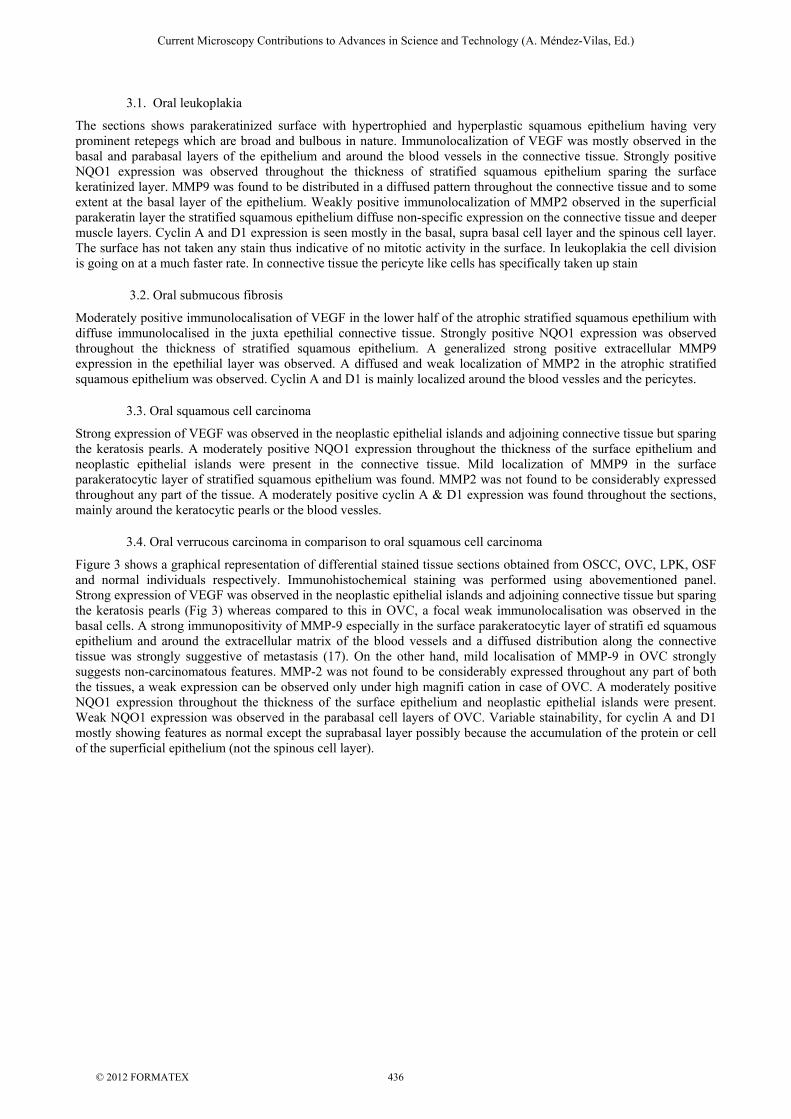
3.1. Oral leukoplakia
The sections shows parakeratinized surface with hypertrophied and hyperplastic squamous epithelium having very prominent retepegs which are broad and bulbous in nature. Immunolocalization of VEGF was mostly observed in the basal and parabasal layers of the epithelium and around the blood vessels in the connective tissue. Strongly positive NQO1 expression was observed throughout the thickness of stratified squamous epithelium sparing the surface keratinized layer. MMP9 was found to be distributed in a diffused pattern throughout the connective tissue and to some extent at the basal layer of the epithelium. Weakly positive immunolocalization of MMP2 observed in the superficial parakeratin layer the stratified squamous epithelium diffuse non-specific expression on the connective tissue and deeper muscle layers. Cyclin A and D1 expression is seen mostly in the basal, supra basal cell layer and the spinous cell layer. The surface has not taken any stain thus indicative of no mitotic activity in the surface. In leukoplakia the cell division is going on at a much faster rate. In connective tissue the pericyte like cells has specifically taken up stain
3.2. Oral submucous fibrosis
Moderately positive immunolocalisation of VEGF in the lower half of the atrophic stratified squamous epethilium with diffuse immunolocalised in the juxta epethilial connective tissue. Strongly positive NQO1 expression was observed throughout the thickness of stratified squamous epithelium. A generalized strong positive extracellular MMP9 expression in the epethilial layer was observed. A diffused and weak localization of MMP2 in the atrophic stratified squamous epithelium was observed. Cyclin A and D1 is mainly localized around the blood vessles and the pericytes.
3.3. Oral squamous cell carcinoma
Strong expression of VEGF was observed in the neoplastic epithelial islands and adjoining connective tissue but sparing the keratosis pearls. A moderately positive NQO1 expression throughout the thickness of the surface epithelium and neoplastic epithelial islands were present in the connective tissue. Mild localization of MMP9 in the surface parakeratocytic layer of stratified squamous epithelium was found. MMP2 was not found to be considerably expressed throughout any part of the tissue. A moderately positive cyclin A & D1 expression was found throughout the sections, mainly around the keratocytic pearls or the blood vessles.
3.4. Oral verrucous carcinoma in comparison to oral squamous cell carcinoma
Figure 3 shows a graphical representation of differential stained tissue sections obtained from OSCC, OVC, LPK, OSF and normal individuals respectively. Immunohistochemical staining was performed using abovementioned panel. Strong expression of VEGF was observed in the neoplastic epithelial islands and adjoining connective tissue but sparing the keratosis pearls (Fig 3) whereas compared to this in OVC, a focal weak immunolocalisation was observed in the basal cells. A strong immunopositivity of MMP-9 especially in the surface parakeratocytic layer of stratifi ed squamous epithelium and around the extracellular matrix of the blood vessels and a diffused distribution along the connective tissue was strongly suggestive of metastasis (17). On the other hand, mild localisation of MMP-9 in OVC strongly suggests non-carcinomatous features. MMP-2 was not found to be considerably expressed throughout any part of both the tissues, a weak expression can be observed only under high magnifi cation in case of OVC. A moderately positive NQO1 expression throughout the thickness of the surface epithelium and neoplastic epithelial islands were present. Weak NQO1 expression was observed in the parabasal cell layers of OVC. Variable stainability, for cyclin A and D1 mostly showing features as normal except the suprabasal layer possibly because the accumulation of the protein or cell of the superficial epithelium (not the spinous cell layer).
Current Microscopy Contributions to Advances in Science and Technology (A. Méndez-Vilas, Ed.)
© 2012 FORMATEX 436

Figure3. Graphical representation of variable staining intensity as measured by Image J software. Normal (N), oral leukoplakia (LPK), oral submucous fibrosis (OSF), oral squamous cell carcinoma (OSCC) and verrucous carcinoma (VCA).
4. Discussion:
Previously we have reported a diagnostic procedure intervening the morphological variation of epithelial cells of various oral precancerous conditions along with a novel artificial neural network based CAD technique to identify the progressive stages of the oral precancerous condition as OSF based on microscopic images of TEM (12, 16) and light microscopy. We have also demonstrated the efficacy of the above immunohistochemical panel staining in differentiating and understanding the precancerous nature and their malignant progression, and have put forward the importance of immunohistochemistry as a confirmatory test to showcase the carcinogenic status of VC. The hallmarks of carcinogenesis are neoangiogenesis and degradation of the extracellular matrix, which helps in tumour growth, survival and metastasis of neoplastic cells. In this process, vascular endothelial growth factor (VEGF) is regarded as one of the major contributing factor in angiogenesis whereas the matrix metalloproteinases (MMPs) (a family of zinc containing endopeptidases) causes degradation of various components of the extracellular matrix and helps in metastasisation. VEGF is a potent mitogen and chemoattractant for endothelial cells and induces the release of MMP-2, MMP-9 which, in turn, regulates the angiogenic switch that can occur very early in some cancers, even before malignant progression, with increased vessel density seen in precancerous lesions. On the other hand, superoxide dismutase (SOD) and nicotinamide adenine dinucleotide phosphate: quinone oxidoreductase 1 (NQO1), a cytosolic enzyme detected mainly in epithelial cells catalyses the two-electron reduction of quinone compounds and prevents the generation of reactive oxygen species, thus protecting cells from oxidative damage and carcinogenesis. Important role of Cyclin A and D1 has been reported in progression of cancers of lung, breast, oral etc. The cyclin D1 proto-oncogene is an important regulator of G1 to S phase progression in many different cell types. Cyclin A is particularly interesting among the cyclin family because it can activate two different cyclin-dependent kinases (CDKs) and functions in both S phase and mitosis. Especially a number of therapeutic agents have been observed to induce cyclin D1 degradation in vitro which has made the later a useful avenue for therapeutic intervention (17). During the last decade histopathology gained hugely from the progress of software development and formulation of new algorithms. The innovation of digital pathology has opened new challenges where whole-slide examination on computer screens has become possible for several applications in pathology. This strategy should base upon three steps, firstly a general histopathology should be obtained, the progression should be monitored using a good panel of tumor marker proteins histochemically and finally enumerating the intensity using the color deconvolution plugin of Image J software. Application of these strategies should be implied by pathologists to differentiate diseases where general observation fails. We have proposed the panel of marker proteins and provided an example how effective this is in case of solving the age-old diagnostic dilemma of differentiating OVC with OSCC. So to conclude in this study we propose NADP(H) quinone Oxidoreductase, neoangiogenic markers like VEGF, MMP 2 & 9, and cellular proliferation markers like CCND1, CyA followed by image analysis as a promising panel to distinguish as well as to understand the biology of oral squamous cell carcinogenesis.
Current Microscopy Contributions to Advances in Science and Technology (A. Méndez-Vilas, Ed.)
© 2012 FORMATEX 437

5. References:
[1] Sankaranarayan R, Masuyer E, Swaminathan R, Ferley J, Whelan S (1998); Head and neck cancer: a global perspective on epidemiology and prognosis. Anticancer Res 18:4779-86,
[2] Elango JK, Gangadharan P, Sumithra S, Kuriakose MA (2006). Trends of head and neck cancers in urban and rural India. Asian Pac J Cancer Prev 7:108-12.
[3] Sankaranarayanan R, Ramadas K, Thomas G, Muwonge R, Thara S, Mathew B, Rajan B (2005). Effect of screening on oral cancer mortality in Kerala, India: a cluster-randomised control trial; Trivandrum Oral Cancer Screening Study Group. Lancet 365:1927-33.
[4] Subramanian S, Sankaranarayanan R, Bapat B, Somnathan T, Thomas G Mathew B, Vinoda J, Ramdas K (2009). Cost-effectiveness of oral cancer screening: results from a cluster randomized controlled trial in India. Bull World Health Organ 87:200-206,
[5] Crook T, Perry AR, Osen P et al (2002). Molecular and cellular pathology of cancer. In Souhami RL, Tannock I, Hohenberger P, Eds. Oxford textbook of oncology, 2nd edition,. Oxford University press Inc. New York; 227-240.
[6] Hayat M.A. Handbook of immunohistochemistry and in situ hybridization of human carcinomas: Academic Press, 2005 [7] Halliday A. Idikio (2010). Immunohistochemistry in diagnostic surgical pathology: contributions of protein life-cycle, use of
evidencebased methods and data normalization on interpretation of immunohistochemical stains Int J Clin Exp Pathol; 3(2):169- 176.
[8] Raica M, Cimpean AM, Ribatti D (2009). Angiogenesis in pre-malignant conditions. Eur J Cancer. Jul; 45(11):1924-34. [9] Rosenthal EL, Matrisian LM (2006). Matrix metalloproteinases in head and neck cancer. Head Neck. Jul; 28(7):639-48. [10] Siegel D, Franklin W A and Ross D (1998). Immunohistochemical detection of NAD (P) H: quinone oxidoreductase in human
lung and lung tumors. Clinical Cancer Research September 4; 2065 [11] Terakado N, Shintani S, Nakahara Y, Mihara M, Tomizawa K, Suzuki K, Taniguchi N, Matsumura T (2000). Expression of
Cu,Zn-SOD, Mn-SOD and GST-pi in oral cancer treated with preoperative radiation therapy. Oncol Rep. Sep-Oct;7(5):1113-7 [12] Paul, R.R., Mukherjee, A., Datta, P.K., Banerjee, S., Pal, M, Chatterjee, Chaudhuri, K., 2005. Novel wavelet-neural
networkbased pathological stage detection technique for an oral precancerous condition. J. Clin.Pathol. 58, 932–938. [13] Pal M , Ray Chaudhuri S, Jadav A, Banerjee S, Paul R R, Dutta P K, Ghosh B, Chatterjee J , Chaudhuri K (2008). Quantitative
dimensions of histopathological attributes and status of GSTM1–GSTT1 in oral submucous fibrosis Tissue cell Dec; 40(6):425-35.
[14] Al-Janabi, S., Huisman, A. and Van Diest, P. J. (2012), Digital pathology: current status and future perspectives. Histopathology, 61: 1–9. doi: 10.1111/j.1365-2559.2011.03814.x
[15] Ruifrok AC, Johnston DA (2001) Quantification of histochemical staining by color deconvolution. Anal Quant Cytol Histol 23:291– 299
[16] Mukherjee S, Ray JG and Chaudhuri K. (2010). Microscopic analysis of histological and immunohistochemical sections to differentiate normal, precancer and cancerous oral squamous epithelial tissues.. Microscopy Book Series titled "Microscopy: Science, Technology, Applications and Education” Ed. Antonio Méndez-Vilas and Jesús Díaz Álvarez, Published by Formatex Research Center, Volume 2 ISBN (13): 978-84-614-6190-5; pp. 993-1000.
[17] Ray JG, Mukherjee S, Pattanayak (Mohanty) S and Chaudhuri, K (2010). Oral Verrucous Carcinoma-A misnomer? Immunohistochemistry based comparative study of two cases. . BMJ Case Reports; doi:10.1136/bcr.11.2010.3479
[18] Deshpande A, Sicinski P and Hinds PW (2005) Cyclins and cdks in development and cancer: a perspective Oncogene 24, 2909–2915.
Current Microscopy Contributions to Advances in Science and Technology (A. Méndez-Vilas, Ed.)
© 2012 FORMATEX 438


![Verrucous Carcinoma of Larynx Presenting as a Hairy Wl ...downloads.hindawi.com/journals/dte/1997/864231.pdf254 M. KAWAIDAet al. [5] Ferlito,A.,Reacher,G.(1980).Ackerman’stumor(verrucous](https://img.pdfslide.us/doc/110x75/609dc55f3c323e327e0d8670/verrucous-carcinoma-of-larynx-presenting-as-a-hairy-wl-254-m-kawaidaet-al.jpg)