Embed Size (px)
Citation preview

Immunohistochemical Analyses of Focal Adhesion KinaseExpression in Benign and Malignant Human Breast andColon Tissues: Correlation with Preinvasive andInvasive Phenotypes1
William G. Cance,2 Janet E. Harris,Mary V. Iacocca, Elizabeth Roche, XiHui Yang,Jinli Chang, Stephen Simkins, and LiHui XuDepartments of Surgery [W. G. C., J. E. H., E. R., X. Y., J. C., L. X.]and Pathology [M. V. I.] and Lineberger Comprehensive CancerCenter, University of North Carolina at Chapel Hill School ofMedicine, Chapel Hill, North Carolina 27599-7210, and Departmentof Microbiology, Pathology, and Parasitology, College of VeterinaryMedicine, North Carolina State University, Raleigh, North Carolina27695 [S. S.]
ABSTRACTThe focal adhesion kinase (FAK) is a protein tyrosine
kinase linked to signaling events between cells and the ex-tracellular matrix. Studies at the Western blot level havedemonstrated up-regulation of FAK expression in invasivebreast and colon cancers. To assess p125FAK expression atthe cellular level, we developed monoclonal antibodies thatspecifically detected FAK in formalin-fixed, paraffin-embedded tissue sections and analyzed the levels of FAKexpression in human breast and colon tissues. Monoclonalantibody 4.47 demonstrated FAK-specific focal adhesionstaining by immunofluorescence assays on BT-474 breastcancer cells and detected aMr 125,000 protein by bothWestern blotting and immunoprecipitation analyses. Usingimmunohistochemical techniques, the expression of p125FAK
was analyzed in 36 normal and 43 preinvasive or invasivehuman breast and colon tissues from individual patients.FAK was weakly expressed in most benign breast epitheliumbut was up-regulated at moderate or strong levels in 14 of 18invasive breast carcinomas. In seven samples of ductal car-cinoma-in situ, FAK was overexpressed. Borderline-to-weakexpression of FAK was detected in the normal colonic epi-thelium. In the invasive colon cancers, FAK was overex-pressed at moderate or strong levels in 13 of 15 tumors.Furthermore, FAK expression was up-regulated in areas of
dysplastic, premalignant colon epithelium. These resultsprovide the first evidence at the cellular level that FAKexpression is variably overexpressed in breast and coloncancer and suggest that up-regulation occurs at an earlystage of tumorigenesis.
INTRODUCTIONThe FAK3 is a protein tyrosine kinase that is a critical
mediator of signaling events between cells and their extracellu-lar matrix. Originally identified in v-src-transformed chick em-bryo fibroblasts (1), FAK has been linked to integrin-signalingpathways (2–5), cellular motility (6), and apoptosis (7–9). FAKnot only functions as a kinase but also serves as a scaffoldingprotein for the recruitment of other Src-homology 2 (SH2) andSrc-homology 3 (SH3)-containing signaling molecules (10). Intumor cells, FAK is thought to have a dual function both inpromoting tumor cell adhesion and in acting as a survival signalto inhibit apoptosis as a tumor develops anchorage-independentgrowth properties.4
Although several studies have demonstrated up-regulationof FAK expression as normal breast and colon tissues becometransformed and develop into primary, invasive carcinomas(11–14), the significance of these findings has remained con-troversial (15). Some studies, in fact, have not demonstratedp125FAK overexpression in human breast cancer cells (16).Thus, the significance of FAK expression in human tumorsremains to be determined. Many of the analyses have reliedlargely on blotting methods to demonstrate mRNA and p125FAK
overexpression, with the inherent problems of being unable todistinguish tumor cells from surrounding stromal contaminants.In addition, blotting techniques do not allow the comparison ofFAK expression at the cellular level between preinvasive le-sions, such as DCIS or dysplastic colon polyp epithelium andinvasive carcinoma.
Most of the available anti-FAK antibodies are unsuitablefor large studies of p125FAK expression in human tumors be-cause they do not readily detect p125FAK on FFPE tissue sec-tions. For example, our polyclonal V39 anti-FAK antibodyspecifically recognized p125FAK on Western blots but was not a
Received 10/11/99; revised 3/29/00; accepted 4/4/00.The costs of publication of this article were defrayed in part by thepayment of page charges. This article must therefore be hereby markedadvertisementin accordance with 18 U.S.C. Section 1734 solely toindicate this fact.1 Supported by National Cancer Institute Grant CA65910. W. G. C. is arecipient of the George H. A. Clowes, Jr., Memorial Research CareerDevelopment Award from the American College of Surgeons.2 To whom requests for reprints should be addressed, at University ofNorth Carolina at Chapel Hill, Department of Surgery, CB No. 7210,Chapel Hill, NC 27599-7210. Phone: (919) 966-5221; Fax: (919) 966-8806; E-mail: [email protected].
3 The abbreviations used are: FAK, focal adhesion kinase; FRNK,FAK-related nonkinase; FFPE, formalin-fixed, paraffin-embedded;DCIS, ductal carcinomain situ; GST, glutathioneS-transferase; DAB,3–39diaminobenzidine.4 L-H. Xu, X. Yang, C.A. Bradham, D.A. Brenner, R.J. Craven, andW.G. Cance. The focal adhesion kinase suppresses transformation-associated, anchorage-independent apoptosis in human breast cancercells, submitted for publication.
2417Vol. 6, 2417–2423, June 2000 Clinical Cancer Research
Research. on May 29, 2017. © 2000 American Association for Cancerclincancerres.aacrjournals.org Downloaded from

suitable reagent for immunohistochemistry. For these reasons,we have developed monoclonal antibodies against the NH2
terminus of FAK that recognize p125FAK by immunohistochem-ical analyses of FFPE specimens. This has allowed us to directlycompare the levels of FAK expression between benign andmalignant cells. In this report, we have characterized the ex-pression of p125FAK in matched samples of normal and malig-nant breast and colon tissues from individual patients and havedemonstrated, for the first time at the immunohistochemicallevel, up-regulation of p125FAK in both preinvasive and invasivebreast and colon cancer.
MATERIALS AND METHODSDevelopment of Anti-p125FAK Monoclonal Antibody
4.47. To develop a FAK-specific monoclonal antibody, wedeveloped an immunogen directed at the NH2 terminus of themolecule to avoid consensus sequences within the kinase do-main as well as cross-reactivity with the autonomously ex-pressed COOH-terminal fragment of FAK, FRNK (17). Wecreated a GST fusion protein containing amino acids 1–423 ofFAK, immunized mice, and we performed fusions according tostandard techniques (18). A total of 1600 supernatant sampleswere initially screened by ELISA for FAK immunoreactivity.
Positive clones were then screened by indirect immunofluores-cence assays on BT-474 breast cancer cells as described below.Finally, clones were analyzed by immunohistochemistry onFFPE tissue sections, as described below. We created threeFAK-specific monoclonal antibodies, two of which recognizedp125FAK on FFPE sections. In this report, we have focused onmonoclonal antibody 4.47 (available from Upstate Biotechnol-ogy, Inc., Lake Placid, NY).
Immunofluorescence, Western Blotting, and Immuno-precipitation Analyses. Indirect immunofluorescence mi-croscopy was performed on BT-474 breast cancer cells usingstandard techniques (19). FAK2/2 knockout embryo fibro-blasts (kindly provided by Steven Hanks, Vanderbilt University,Nashville, TN) were used as a negative control in these assaysto demonstrate p125FAK specificity. Lysates of paired normaland tumor samples were prepared as described (12), and 50mgof cell lysate were analyzed for FAK expression by Westernblotting using anti-FAK 4.47 monoclonal antibody (1:50 dilu-tion; 0.3mg).
For immunoprecipitation, 500mg of tissue lysate wereincubated with 10mg of anti-FAK 4.47 monoclonal antibody inthe presence of protein G plus protein A agarose (Calbiochem,San Diego, CA). The precipitated proteins were resolved on
Fig. 1 Characterization of monoclonal antibody 4.47 for specific p125FAK immunoreactivity.A, left, indirect immunofluorescence on BT-474 breastcancer cells demonstrates intense staining at focal adhesions;middle, the FAK2/2 knockout fibroblasts demonstrate no focal adhesion staining with4.47, despite having intact focal adhesions by paxillin staining (right), thus demonstrating the specificity of the antibody.B, Western blot analysescomparing antibodies 4.47 and C20 on lysates of RD rhabdomyosarcoma cells, BT-474 cells, and FAK2/2fibroblasts. An intenseMr 125,000 bandis demonstrated in the RD and BT-474 cells, with no reactivity in the FAK knockout fibroblasts.C, immunoprecipitation analyses demonstrating thatantibody 4.47 immunoprecipitates aMr 125,000 protein from a RD cell lysate, detected by both 4.47 and C20. The murine IgG heavy chain (IgGHC)is seen in the 4.47 lane.
2418FAK Expression in Benign and Malignant Breast and Colon Tissues
Research. on May 29, 2017. © 2000 American Association for Cancerclincancerres.aacrjournals.org Downloaded from

SDS-PAGE and immunoblotted using the anti-FAK 4.47 mono-clonal antibody or anti- FAK polyclonal antibody (C20; SantaCruz Biotechnology, Santa Cruz, CA). Proteins were visualizedusing the enhanced chemiluminescence detection system (Am-ersham, Arlington Heights, IL; Ref. 20).
Tissue Samples and Immunohistochemistry Assays.Fresh frozen as well as FFPE breast and colon tissue sampleswere obtained from University of North Carolina-LinebergerComprehensive Cancer Center Tissue Procurement and Analy-sis Facility as well as the University of North Carolina Depart-ment of Pathology. FFPE tissue was obtained from 21 patientswith breast carcinoma, 2 patients with villoglandular polyps ofthe colon, and 15 patients with colon carcinoma. All of theH&E-stained sections were reviewed, and areas of benign epi-thelium, dysplasia within polyps, and colon and breast carci-noma were identified. Paired normal and neoplastic/tumor sam-ples were examined in 36 of these patients.
Immunohistochemistry was performed as described previ-ously (21), with the following modifications. After deparafiniza-tion, rehydration, and quenching of endogenous peroxidase ac-tivity, mercuric pigments were removed by incubating sectionsin Auto/Iodine (Fischer Scientific, Pittsburgh, PA) for 1 min.
Slides were incubated in Redusol (Fischer Scientific) two timesfor 2 min. each. The hydration process was completed by rinsingthree times for 3 min in 13Automation Buffer (Fischer Scien-tific).
Heat-induced epitope recovery was used as sections weresubmerged in 200 ml of 13Antigen Retrieval Citra Buffer(Biogenex) and steam-heated in a standard steamer (Black andDecker) for 30 min. Sections were blocked in normal horseserum for 15 min and then incubated with anti-FAK 4.47 mono-clonal antibody (1:50 dilution; 0.3mg) overnight at 4°C. Controlsections were incubated with a comparable concentration (1:37;0.3 mg) of isotype-matched monoclonal antibody, MOPC-21(Sigma, St. Louis, MO).
The sections were washed extensively in AutomationBuffer and then incubated with biotinylated goat antimouse IgGfollowed by avidin peroxidase using the Vectastatin ABC elitekit (Vector Laboratories, Burlingame, CA). The chromogenicreaction was performed with DAB, toned with the DAB enhanc-ing solution (Vector Laboratories).
Immunohistochemistry Scoring. A single pathologist(M. V. I. ) scored each tissue section for FAK expression. Epi-thelium from each section was scored for staining intensity
Table 1 Immunohistochemical analyses of p125FAK expression in breast epithelium: benign, DCIS, and invasive cancer
Patient Tissue typeStainingintensity
% positivecells Tissue type
Stainingintensity
% positivecells
1 Benign 0 0 Inv.a ductal 1 5
2 Benign 0 0 Inv. ductal 11 60
3 Benign NA NA Inv. ductal 111 90
4 Benign 1 20 Inv. ductal 1111 95
5 Benign 1 25 Inv. ductal 111 75
6 Benign 0 0 Inv. ductal 111 90
7 Benign 1 50 Inv. ductal 111 90
8 Benign 11 25 Inv. ductal 1111 95
9 Benign 11 20 Inv. ductal 111 90
10 Benign 111 75 Inv. ductal 1111 100
11 Benign 0 0 Inv. ductal 11 30
12 Benign 11 30 DCIS 111 50
13 Benign 1111 80 DCIS 1111 100
14 Benign 11 50 DCIS 1111 95
15 Benign 11 75 Inv. lobular 111 90
16 Benign 111 75 DCIS 1111 95Inv. ductal 1111 95
17 Benign 11 25 DCIS 111 50Inv. ductal 111 50
18 Benign 0 0 DCIS 111 90Inv. ductal 111 90
19 Benign 0 0 DCIS 11 50Inv. ductal 11 50
20 Benign 0 0 Mixed Inv.lobular and ductal
1111 90
21 Benign 0 0 Mixed Inv.lobular and ductal
1111 100
a Inv., invasive; NA, not applicable;1, borderline;11, weak;111, moderate;1111, strong.
2419Clinical Cancer Research
Research. on May 29, 2017. © 2000 American Association for Cancerclincancerres.aacrjournals.org Downloaded from

based on the following scoring system: 0, none; 11, borderline;21, weak; 31, moderate; 41, strong. Staining characteristics ofcellular localization (cytoplasm, nucleus, membrane, combina-tion thereof) and overall distribution (homogeneous throughoutcell population, unifocal, heterogeneous, or multifocal) wererecorded.
RESULTSCharacterization of Anti-p125FAK Monoclonal Anti-
body 4.47. To demonstrate immunoreactivity of this mono-clonal antibody at the focal adhesions, indirect immunofluores-cence assays were performed on BT-474 breast cancer cells.Monoclonal antibody 4.47 localized p125FAK specifically to thefocal adhesions of these cells (Fig. 1A). In contrast, staining ofthe FAK2/2 knockout embryo fibroblasts with this antibodydid not detect specific localization of p125FAK at the focaladhesions (Fig. 1A), although these cells demonstrated intactfocal adhesions when probed for the focal adhesion protein,paxillin (Fig. 1A). In addition, staining BT-474 cells with theantibody preincubated with GST-FAK-NH2 terminal immuno-gen (data not shown) abrogated the specific localization ofp125FAK, further suggesting the specific immunoreactivity ofthe antibody.
To determine whether the 4.47 antibody recognized theMr
125,000 FAK protein in other confirmations, western blot andimmunoprecipitation analyses were performed using the RDrhabdomyosarcoma cell lysate. Western blot analyses were per-formed as described below, detecting aMr 125,000 protein that
comigrated with the p125FAK detected by using a commerciallyavailable anti-FAK polyclonal antibody, C20 (Fig. 1B). Immu-noprecipitation analyses demonstrated that this 4.47 monoclonalantibody specifically recognized p125FAK in a native confirma-tion (Fig. 1C) as compared with the anti-FAK C20 antibody.The specificity of 4.47 antibody was further demonstrated byblocking studies where immunoblotting or immunoprecipitationwas abrogated when the antibody was preincubated with theGST-FAK-NH2 terminal immunogen (data not shown).
p125FAK Is Overexpressed in Noninvasive DCIS andInvasive Breast Cancer. In the 20 samples of benign breastepithelium that were obtained from patients with breast cancer,there was little detectable p125FAK expression (Table 1). Eightpatients had no detectable FAK expression in their benign ductsand lobules, and 9 patients had only borderline-to-weak expres-sion. Only three samples demonstrated moderate or strong FAKimmunoreactivity in the benign epithelium. Intriguingly, thebenign ducts and lobules adjacent to a site of DCIS or invasivecarcinoma frequently demonstrated p125FAK immunoreactivity,whereas the benign tissue remote from the cancer usually hadminimal to undetectable FAK expression. In contrast, p125FAK
was detected at varying levels in all of the breast carcinomaspecimens from the 21 patients that we tested. The significantup-regulation of FAK expression in these breast cancer tissues isdemonstrated in the benign breast tissue and infiltrating ductalcarcinoma from patient 4 (Fig. 2A). Here, a benign breast lobule(Fig. 2A,NOR) lacks significant FAK expression, whereas thecarcinoma (Fig. 2A,INV) demonstrates strong (41) FAK ex-
Fig. 2 FAK overexpression in invasive human breast cancer and DCIS.A, paired samples of normal breast (NOR) and invasive breast cancer (INV)from the same patient demonstrate no detectable FAK expression in the normal tissue. In contrast, the invasive breast cancer cells (INV) demonstrateintense membrane and cytoplasmic immunoreactivity for p125FAK. Note the absence of immunoreactivity in the surrounding tumor-infiltratinglymphocytes (immunohistochemistry,3600).B, in this single FFPE tissue section, the focus of DCIS (DCIS) expresses p125FAK at a slightly lowerlevel than the adjacent focus of infiltrating ductal carcinoma (INV;left; 3200). These two areas are shown at higher magnification (3600) todemonstrate this difference (DCIS,middle; INV, right).
2420FAK Expression in Benign and Malignant Breast and Colon Tissues
Research. on May 29, 2017. © 2000 American Association for Cancerclincancerres.aacrjournals.org Downloaded from
pression in both the cytoplasm and the cytoplasmic membraneof the tumor cells. The intensity of FAK expression in themalignant, invasive epithelium ranged from 11 to 41, with 14of the 18 samples expressing moderate or strong (31 or 41)FAK immunoreactivity. However, not the entire population oftumor cells in these specimens overexpressed p125FAK. Asnoted in Table 1, there was a variability in percentage of tumorcells demonstrating immunoreactivity against FAK, rangingfrom 5 to 100% of the tumor cells.
The invasive breast carcinoma specimens represented avariety of cell types, including pure invasive ductal, mixedductal and lobular, and pure invasive lobular carcinomas. Inaddition, seven of the samples contained DCIS either by itself(patients 12, 13, and 14), or adjacent to an infiltrating carcinoma(patients 16, 17, 18, and 19). Intriguingly, each of these DCISsamples demonstrated intense FAK overexpression, comparablewith their invasive counterparts. This is shown in a tissuesection from patient 18 (Fig. 2B), where the DCIS (Fig. 2B,DCIS) and the invasive carcinoma (Fig. 2B,INV) both show 31staining intensity in 90% of cells. These results suggest thatoverexpression of FAK is an early event in breast carcinoma andis not restricted to fully invasive tumors.
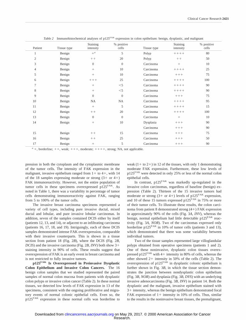
p125FAK Is Overexpressed in Preinvasive DysplasticColon Epithelium and Invasive Colon Cancers. The 16benign colon samples that we studied represented the pairedsamples of normal colon mucosa from patients with dysplasticcolon polyps or invasive colon cancer (Table 2). In these normaltissues, we detected low levels of FAK expression in 13 of thespecimens, consistent with the ongoing proliferative and migra-tory events of normal colonic epithelial cells. Even so, thep125FAK expression in these normal cells was borderline to
weak (11to 21) in 12 of the tissues, with only 1 demonstratingmoderate FAK expression. Furthermore, these low levels ofp125FAK were detected in only 25% or less of the normal colonepithelial cells.
In contrast, p125FAK was markedly up-regulated in theinvasive colon carcinomas, regardless of baseline (benign) ex-pression (Table 2). Thirteen of the 15 invasive tumors hadmoderate or strong (31or 41) levels of p125FAK expression,and 10 of these 15 tumors expressed p125FAK in 75% or moreof their tumor cells. To illustrate these results, the colon carci-noma from patient 8 demonstrated strong (41) FAK expressionin approximately 90% of the cells (Fig. 3A, INV), whereas thebenign, normal epithelium had little detectable p125FAK reac-tivity (Fig. 3A, NOR). Two of the carcinomas expressed onlyborderline p125FAK in 10% of tumor cells (patients 3 and 13),which demonstrated that there was some variability betweenindividual tumors.
Two of the tissue samples represented large villoglandularpolyps obtained from operative specimens (patients 1 and 2).One of these noninvasive, dysplastic colon tissues overex-pressed p125FAK with 41 intensity in 80% of cells, whereas theother showed 21intensity in 50% of the cells (Table 2). Theoverexpression of p125FAK in dysplastic colonic epithelium isfurther shown in Fig. 3B, in which the tissue section demon-strates the junction between nondysplastic colon epithelium(Fig. 3B,NOR) and dysplasia (Fig. 3B,DYS) with an underlyinginvasive adenocarcinoma (Fig. 3B, INV) in patient 14. Both thedysplastic and the malignant, invasive epithelium stained with31 intensity, whereas the benign epithelium demonstrated focalFAK expression of 11intensity in 10% of cells. Thus, similarto the results in the noninvasive breast tissues, the premalignant,
Table 2 Immunohistochemical analyses of p125FAK expression in colon epithelium: benign, dysplastic, and malignant
Patient Tissue typeStainingintensity
% positivecells Tissue type
Stainingintensity
% positivecells
1 Benign 1a 5 Polyp 1111 80
2 Benign 11 20 Polyp 11 50
3 Benign 0 0 Carcinoma 1 10
4 Benign 1 10 Carcinoma 1111 25
5 Benign 1 10 Carcinoma 111 75
6 Benign 111 25 Carcinoma 1111 100
7 Benign 1 ,5 Carcinoma 1111 90
8 Benign 1 ,5 Carcinoma 1111 90
9 Benign 0 0 Carcinoma 111 75
10 Benign NA NA Carcinoma 1111 95
11 Benign 1 5 Carcinoma 1111 15
12 Benign 11 20 Carcinoma 1111 100
13 Benign 0 0 Carcinoma 1 10
14 Benign 1 10 Dysplasia 111 90
Carcinoma 111 90
15 Benign 11 15 Carcinoma 111 75
16 Benign 11 25 Carcinoma 111 50
17 Benign 1 5 Carcinoma 111 90a 1, borderline;11, weak;111, moderate;1111, strong; NA, not applicable.
2421Clinical Cancer Research
Research. on May 29, 2017. © 2000 American Association for Cancerclincancerres.aacrjournals.org Downloaded from

dysplastic colon epithelia seemed to overexpress FAK beforeacquiring the fully malignant and invasive phenotypes.
DISCUSSIONThese results provide definitive evidence, at the cellular
level, that the expression of p125FAK is up-regulated as normalbreast and colon tissues undergo malignant transformation. Thelevels of FAK overexpression in breast and colon tumor cellsrepresent a higher degree of overexpression than some of theother tyrosine kinases known to be overexpressed in humantumors, such as Src in colon cancer (22, 23). Furthermore, theresults demonstrate that FAK overexpression seems to be anearly event in the development of breast and colon cancer,occurring in some tissues before the tumor has acquired its fullinvasive potential.
It is intriguing to speculate on possible reasons for thesehigh levels of FAK overexpression during tumorigenesis. FAKhas been shown to be a survival signal for anchorage-dependentcells (8, 24), and inhibition of FAK expression in tumor cellshas been shown to result in cell death (7, 19). Thus, it is
conceivable that tumor cells up-regulate FAK expression tomaintain survival as they progress through the anchorage-dependent and anchorage-independent phases of growth, inva-sion, and metastasis.
In this study, we have examined paired samples of normaland neoplastic tissues from individual patients. This has givenus the advantage of directly comparing levels of FAK expres-sion within the same genetic background. In addition, the indi-vidual tissue sections that contained a combination of normal,preinvasive, and invasive breast and colon tissues have allowedus to compare FAK expression in these pathological typeswithout the potential variability of immunostaining betweendifferent slides. As a caveat to these results, it has been shownthat molecular genetic aberrations occur in normal ducts adja-cent to breast cancers (25). This raises the possibility that thenoninvasive lesions adjacent to the invasive cancers may con-tain such genetic abnormalities that could have an influence onFAK expression. Indeed, in benign ducts and lobules adjacent totumors, up-regulation of FAK expression was seen.
The inconsistency of studies examining the levels of FAK
Fig. 3 FAK overexpression in invasive human colon cancer and colon dysplasia.A, this paired sample of normal colon (NOR) and invasive coloncancer (INV) from the same patient demonstrates little detectable p125FAK immunoreactivity in the normal colon mucosa. In contrast, the invasivecolon cancer demonstrates intense FAK immunostaining in the cytoplasm of the tumor cells (immunohistochemistry,3400).B, this individual FFPEsection demonstrates normal colon mucosa (NOR), with adjacent dysplastic mucosa (DYS), surrounded by a colon cancer that has invaded underneaththe normal mucosa (INV;3200). Inset in the left panel, the junction between normal and invasive tumor, which is shown at a higher magnification(3600) in theright panel, demonstrates the difference in levels of p125FAK expression.
2422FAK Expression in Benign and Malignant Breast and Colon Tissues
Research. on May 29, 2017. © 2000 American Association for Cancerclincancerres.aacrjournals.org Downloaded from

expression in human tumors may, in part, be explained byproblems with existing antibodies recognizing p125FAK onFFPE tissue sections. Although some investigators have beenable to use the prototype anti-FAK 2A7 antibody (26) in im-munohistochemical analyses (27), other investigators have notseen comparable results, which leads to conclusions thatp125FAK is not up-regulated in breast tumor cells (16). Notably,the 2A7 antibody was raised against avian FAK bound to Src inan immunoprecipitate, in contrast to 4.47, which was raisedspecifically against human FAK. Furthermore, 2A7 recognizes aCOOH-terminal epitope in both FAK and FRNK, whereas 4.47recognizes a FAK-specific epitope in the NH2 terminus. Wehave not had reproducible success using 2A7 or other commer-cially available monoclonal antibodies for immunohistochemi-cal analyses (data not shown) and, thus, have sought to producemonoclonal antibodies against p125FAK that were suitable forFFPE analyses. The development of this immunohistochemicalreagent, antibody 4.47, will allow large studies of FAK expres-sion in population-based archival samples to be performed todetermine whether there is diagnostic and prognostic signifi-cance to the levels of p125FAK expression.
In summary, these results are the first definitive immuno-histochemical evidence that FAK expression is up-regulated inbreast and colon cancer. The 4.47 antibody can be used to studyFAK in other tumor systems to determine whether these corre-lations are, in fact, common among different types of tumors.Finally, this dramatic up-regulation of FAK in tumors, com-bined with the relative lack of expression in normal breast andcolon tissues, suggests that FAK is a rational target for breastand colon cancer therapeutics.
ACKNOWLEDGMENTSWe thank Lynn Dressler, David Cowan, and the Tissue Procure-
ment and Analysis Core Facility of the University of North CarolinaLineberger Comprehensive Cancer Center for immunohistochemistryadvice and tissue specimens. We thank Rolf Craven and Shelton Earpfor critical review of the manuscript.
REFERENCES1. Schaller, M. D., Borgman, C. A., Cobb, B. S., Vines, R. R., Reyn-olds, A. B., and Parsons, J. T. p125FAK a structurally distinctive protein-tyrosine kinase associated with focal adhesions. Proc. Natl. Acad. Sci.USA, 89: 5192–5196, 1992.2. Kornberg, L., Earp, H. S., Parsons, J. T., Schaller, M., and Juliano,R. L. Cell adhesion or integrin clustering increases phosphorylation ofa focal adhesion-associated tyrosine kinase. J. Biol. Chem.,267:23439–23442, 1992.3. Lipfert, L., Haimovich, B., Schaller, M. D., Cobb, B. S., Parsons,J. T., and Brugge, J. S. Integrin-dependent phosphorylation and activa-tion of the protein tyrosine kinase p125FAK in platelets. J. Cell Biol.,119: 905–912, 1992.4. Schlaepfer, D. D., Hanks, S. K., Hunter, T., and van der Geer, P.Integrin-mediated signal transduction linked to Ras pathway by GRB2binding to focal adhesion kinase. Nature (Lond.),372: 786–791, 1994.5. Guan, J. L. Role of focal adhesion kinase in integrin signaling. Int.J. Biochem. Cell Biol.,29: 1085–1096, 1997.6. Cary, L. A., Chang, J. F., and Guan, J. L. Stimulation of cellmigration by overexpression of focal adhesion kinase and its associationwith Src and Fyn. J. Cell Sci.,109: 1787–1794, 1996.7. Xu, L. H., Owens, L. V., Sturge, G. C., Yang, X., Liu, E. T., Craven,R. J., and Cance, W. G. Attenuation of the expression of the focal
adhesion kinase induces apoptosis in tumor cells. Cell Growth Differ.,7: 413–418, 1996.
8. Frisch, S. M., Vuori, K., Ruoslahti, E., and Chan-Hui, P. Y. Controlof adhesion-dependent cell survival by focal adhesion kinase. J. CellBiol., 134: 793–799, 1996.
9. Hungerford, J. E., Compton, M. T., Matter, M. L., Hoffstrom, B. G.,and Otey, C. A. Inhibition of p125FAK in cultured fibroblasts results inapoptosis. J. Cell Biol.,135: 1383–1390, 1996.
10. Sieg, D. J., Hauck, C. R., and Schlaepfer, D. D. Required role offocal adhesion kinase (FAK) for integrin-stimulated cell migration.J. Cell Sci.,112: 2677–2691, 1999.
11. Weiner, T. M., Liu, E. T., Craven, R. J., and Cance, W. G.Expression of focal adhesion kinase gene and invasive cancer. Lancet,342: 1024–1025, 1993.
12. Owens, L. V., Xu, L., Craven, R. J., Dent, G. A., Weiner, T. M.,Kornberg, L., Liu, E. T., and Cance, W. G. Overexpression of the focaladhesion kinase (p125FAK) in invasive human tumors. Cancer Res.,55:2752–2755, 1995.13. Owens, L. V., Xu, L., Dent, G. A., Yang, X., Sturge, G. C., Craven,R. J., and Cance, W. G. Focal adhesion kinase as a marker of invasivepotential in differentiated human thyroid cancer. Ann. Surg. Oncol.,3:100–105, 1996.14. Han, N. M., Fleming, R. Y., Curley, S. A., and Gallick, G. E.Overexpression of focal adhesion kinase (p125FAK) in human colorectalcarcinoma liver metastases: independence from c-src or c-yes activation.Ann. Surg. Oncol.,4: 264–268, 1997.15. Schaller, M. D. The focal adhesion kinase. J. Endocrinol.,150:1–7,1996.16. Glukhova, M., Koteliansky, V., Sastre, X., and Thiery, J. P. Adhe-sion systems in normal breast and in invasive breast carcinoma. Am. J.Pathol.,146: 706–716, 1995.17. Schaller, M. D., Borgman, C. A., and Parsons, J. T. Autonomousexpression of a noncatalytic domain of the focal adhesion-associatedprotein tyrosine kinase p125FAK. Mol. Cell. Biol., 13: 785–791, 1993.18. Cance, W. G., Wells, S. A., Jr., Dilley, W. G., Welch, M. J., Otsuka,F. L., and Davie, J. M. Human parathyroid antigen: characterization andlocalization with monoclonal antibodies. Proc. Natl. Acad. Sci. USA,83: 6112–6116, 1986.19. Xu, L. H., Yang, X., Craven, R. J., and Cance, W. G. The COOH-terminal domain of the focal adhesion kinase induces loss of adhesionand cell death in human tumor cells. Cell Growth Differ.,9: 999–1005,1998.20. Cance, W. G., Craven, R. J., Bergman, M., Xu, L., Alitalo, K., andLiu, E. T. Rak, a novel nuclear tyrosine kinase expressed in epithelialcells. Cell Growth Differ.,5: 1347–1355, 1994.21. Cance W. G. Brennan, M. F., Dudas, M. E., Huang, C. M., andCordon-Cardo, C. Altered expression of the retinoblastoma gene prod-uct in human sarcomas. N. Engl. J. Med.,323: 1457–1462, 1990.22. Cartwright, C. A., Meisler, A. I., and Eckhart, W. Activation of thep60c-src protein kinase is an early event in colonic carcinogenesis. Proc.Natl. Acad. Sci. USA,87: 558–562, 1990.23. Iravani, S., Mao, W., Fu, L., Karl, R., Yeatman, T., Jove, R., andCoppola, D. Elevated c-Src protein expression is an early event incolonic neoplasia. Lab. Investig.,78: 365–371, 1998.24. Ilic, D., Almeida, E. A. C., Schlaepfer, D. D., Dazin, P., Aizawa, S.,and Damsky, C. H. Extracellular matrix survival signals transduced byfocal adhesion kinase suppress p53-mediated apoptosis. J. Cell Biol.,143: 547–560, 1998.25. Deng, G., Lu, Y., Zlotnikov, G., Thor, A. D., and Smith, H. S. Lossof heterozygosity in normal tissue adjacent to breast carcinomas. Sci-ence (Washington DC),274: 2057–2059, 1996.26. Kanner, S. B., Reynolds, A. B., Vines, R. R., and Parsons, J. T.Monoclonal antibodies to individual tyrosine-phosphorylated proteinsubstrates of oncogene-encoded tyrosine kinases. Proc. Natl. Acad. Sci.USA, 87: 3328–3332, 1990.27. Kornberg, L. J. Focal adhesion kinase expression in oral cancers.Head Neck,20: 634–639, 1998.
2423Clinical Cancer Research
Research. on May 29, 2017. © 2000 American Association for Cancerclincancerres.aacrjournals.org Downloaded from

2000;6:2417-2423. Clin Cancer Res William G. Cance, Janet E. Harris, Mary V. Iacocca, et al. Tissues: Correlation with Preinvasive and Invasive PhenotypesExpression in Benign and Malignant Human Breast and Colon Immunohistochemical Analyses of Focal Adhesion Kinase
Updated version
http://clincancerres.aacrjournals.org/content/6/6/2417
Access the most recent version of this article at:
Cited articles
http://clincancerres.aacrjournals.org/content/6/6/2417.full.html#ref-list-1
This article cites 27 articles, 18 of which you can access for free at:
Citing articles
/content/6/6/2417.full.html#related-urls
This article has been cited by 55 HighWire-hosted articles. Access the articles at:
E-mail alerts related to this article or journal.Sign up to receive free email-alerts
Subscriptions
Reprints and
To order reprints of this article or to subscribe to the journal, contact the AACR Publications
Permissions
To request permission to re-use all or part of this article, contact the AACR Publications
Research. on May 29, 2017. © 2000 American Association for Cancerclincancerres.aacrjournals.org Downloaded from

![Ductal Adenocarcinoma Ex Pleomorphic Adenoma of the ... · lesions [2, 5]. Carcinoma ex pleomorphic adenoma (Ca ex PA) is a rare transformation of a benign primary PA to a malignant](https://img.pdfslide.us/doc/110x75/60bd399bb7acaf776f026cd1/ductal-adenocarcinoma-ex-pleomorphic-adenoma-of-the-lesions-2-5-carcinoma.jpg)