Embed Size (px)
Citation preview
ImmunohematologyJOURNA L O F B LOOD GROUP S E RO LOGY AND EDUCAT I ON
V O L U M E 2 4 , N U M B E R 1 , 2 0 0 8
ImmunohematologyJOURNA L O F B L OOD G ROU P S E RO L OGY AND E DU C AT I O N
VO L U M E 2 4 , N U M B E R 1 , 2 0 0 8
C O N T E N T S
1R E P O R T
Summary of the Caribbean subregional workshop on quality in immunohematology,a collaborative effort to improve international blood transfusion services
J.R. CRUZ, D.M. HARMENING,AND S.J. NANCE
4R E V I E W
Technical issues in neonatal transfusionsS.R. SLOAN
10R E V I E W
Neonatal red cell transfusionsC.A. LITTY
15R E V I E W
Neonatal and infant platelet transfusionsD.A. SESOK-PIZZINI AND D. FRIEDMAN
20R E V I E W
Neonatal plasma transfusionsP.T. PISCIOTTO
27COMMUN I C AT I O N S
L E T T E R T O T H E E D I T O R S
Prevalence ofYt(a–) in Hispanic blood donorsE.TOSSAS, M. BAXTER,M.E. REID, D. CHARLES-PIERRE,AND C. LOMAS-FRANCIS
28E R R AT UM
Vol. 23,No. 1, 2007
29ANNOUNC EM EN T S
32ADV E R T I S E M E N T S
35I N S T RU C T I O N S F O R AU T HO R S
EDITORS-IN-CHIEF MANAGING EDITORSandra Nance,MS,MT(ASCP)SBB Cynthia Flickinger,MT(ASCP)SBB
Philadelphia, Pennsylvania Philadelphia, Pennsylvania
Connie M.Westhoff,MT(ASCP)SBB, PhDPhiladelphia, Pennsylvania
TECHNICAL EDITOR SENIOR MEDICAL EDITORChristine Lomas-Francis,MSc Geralyn M.Meny,MD
NewYork City, NewYork Philadelphia, Pennsylvania
ASSOCIATE MEDICAL EDITORSDavid Moolton,MD Ralph R.Vassallo,MD
Philadelphia, Pennsylvania Philadelphia, Pennsylvania
EDITORIAL BOARD
EMERITUS EDITORIAL BOARDDelores Mallory,MT(ASCP)SBB
Supply, North Carolina
EDITORIALASSISTANT PRODUCTIONASSISTANTJudith Abrams Marge Manigly
COPY EDITOR PROOFREADER ELECTRONIC PUBLISHERMary L.Tod Lucy Oppenheim Paul Duquette
Immunohematology is published quarterly (March, June, September, and December) by theAmerican Red Cross,National Headquarters,Washington,DC 20006.
Immunohematology is indexed and included in Index Medicus and MEDLINE on the MEDLARS system. The contents are also cited in theEBASE/Excerpta Medica and Elsevier BIOBASE/Current Awareness in Biological Sciences (CABS) databases.
The subscription price is $40.00 (U.S.) and $50.00 (foreign) per year.
Subscriptions, Change of Address, and Extra Copies:Immunohematology, P.O.Box 40325, Philadelphia, PA 19106
Or call (215) 451-4902Web site:www.redcross.org/pubs/immuno
Copyright 2008 byTheAmerican National Red CrossISSN 0894-203X
Patricia Arndt,MT(ASCP)SBBPomona, California
James P.AuBuchon,MDLebanon, New Hampshire
Martha R.Combs,MT(ASCP)SBBDurham, North Carolina
Geoffrey Daniels, PhDBristol, United Kingdom
Anne F. Eder,MDWashington, District of Columbia
George Garratty, PhD, FRCPathPomona, California
Brenda J.Grossman,MDSt. Louis, Missouri
W. John Judd, FIBMS,MIBiolAnn Arbor, Michigan
Christine Lomas-Francis,MScNewYork City, NewYork
Gary Moroff, PhDRockville, Maryland
John J.Moulds,MT(ASCP)SBBShreveport, Louisiana
Paul M.Ness,MDBaltimore, Maryland
Joyce Poole, FIBMSBristol, United Kingdom
Mark Popovsky,MDBraintree, Massachusetts
Marion E.Reid, PhD, FIBMSNewYork City, NewYork
S.Gerald Sandler,MDWashington, District of Columbia
Jill R. Storry, PhDLund, Sweden
David F. Stroncek,MDBethesda, Maryland
I M M U N O H E M A T O L O G Y, VO L U M E 2 4 , N U M B E R 1 , 2 0 0 8 1
In 1999, the Pan American Health Organization(PAHO),after consulting with the Directors of the BloodBanks in the Caribbean countries and with theCoordinators of the National Blood Programs of theLatin American countries, prepared an action plan forimproving the safety of blood transfusion in the regionof the Americas. It clearly expressed (1) the need toassure the quality of blood bank services in all sectors,(2) the requirement for blood banks to participate inprograms of external evaluation of performance,and (3)a subregional approach that was warranted in theCaribbean countries because of the small number ofblood banks. The plan of action was approved by theDirecting Council of PAHO in October 1999.1 As part ofthe initiative to strengthen blood services,the CaribbeanEpidemiology Center (CAREC), with technical andfinancial support from PAHO and the collaboration ofthe AABB, prepared the Caribbean Regional Standardsfor Blood Banks and Transfusion Services.2 Thesestandards have specific sections that detail therequirements for compatibility testing and externalquality assessments.
To bring the plan of action into operation and tofacilitate compliance with the Caribbean RegionalStandards, PAHO requested that the United KingdomNational External Quality Assessment Scheme (UKNEQAS) for Blood Transfusion Laboratory Practice
Summary of the Caribbeansubregional workshop on qualityin immunohematology, acollaborative effort to improveinternational blood transfusionservicesJ.R. CRUZ, D.M. HARMENING,AND S.J. NANCE
R E P O R T
Table 1. Participation in external evaluation of performance
Country Centers participating in external evaluation
Anguilla 1
Antigua and Barbuda 1
Aruba 1
Bahamas 2
Barbados 1
Belize 1
British Virgin Islands 1
Cayman Islands 1
Curacao 0
Dominica 1
Grenada 1
Guyana 1
Haiti* 1
Jamaica 2
Montserrat 1
St.Kitts and Nevis 2
St. Lucia 1
St.Vincent and the Grenadines 1
Suriname 1
Trinidad andTobago 3
Turks and Caicos Islands 1
*Haiti started participating in 2005.
2 I M M U N O H E M A T O L O G Y, VO L U M E 2 4 , N U M B E R 1 , 2 0 0 8
J.R. CRUZ ET AL.
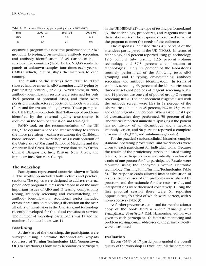
organize a program to assess the performance in ABOgrouping, D typing, crossmatching, antibody screening,and antibody identification of 25 Caribbean bloodservices in 20 countries (Table 1). UK NEQAS sends thepanels of unknown samples four times each year toCAREC, which, in turn, ships the materials to eachcountry.
The results of the surveys from 2002 to 20053
showed improvement inABO grouping and D typing byparticipating centers (Table 2). Nevertheless, in 2005,antibody identification results were returned for only49.5 percent of potential cases, and there werepersistent unsatisfactory reports for antibody screening(four) and for crossmatching (seven). These promptedthe UK NEQAS to conclude that“follow-up of problemsidentified by the external quality assessments isrequired, in the form of education and training.”3
PAHO took on the recommendation of the UKNEQAS to organize a hands-on,wet workshop to addressthe more prevalent weaknesses among the Caribbeanblood services. The workshop was a collaboration ofthe University of Maryland School of Medicine and theAmerican Red Cross. Reagents were donated by Ortho-Clinical Diagnostics, Inc., Raritan, New Jersey, andImmucor, Inc.,Norcross,Georgia.
The WorkshopParticipants represented countries shown in Table
1. The workshop included both lectures and practicalsessions. The topics were designed to address externalproficiency program failures with emphasis on the mostimportant issues of ABO and D testing, compatibilitytesting, antibody screening and crossmatching, andantibody identification. Additional topics includederrors in transfusion medicine,a discussion on the over-all safety of transfusion in theAmericas, and technologyrecently developed for the blood transfusion service.The number of workshop participants was 17 and thenumber of contact hours was 14.
BaseliningAt the start of the workshop, the participants were
surveyed using electronic ResponseCard keypads(courtesy of Turning Technologies LLC, Youngstown,OH) to ascertain: (1) how many laboratories participate
in the UK NEQAS,(2) the type of testing performed,and(3) the technology, procedures, and reagents used intheir laboratories. The responses were used to adjustthe program to meet the needs of the audience.
The responses indicated that 64.7 percent of theattendees participated in the UK NEQAS. In terms oftechnology,37.5 percent reported using gel technology,12.5 percent tube testing, 12.5 percent columntechnology, and 37.5 percent a combination oftechnologies. Only 27 percent of the laboratoriesroutinely perform all of the following tests: ABOgrouping and D typing, crossmatching, antibodyscreening, and antibody identification. In terms ofantibody screening,43 percent of the laboratories use athree-vial set (not pooled) of reagent screening RBCs,and 14 percent use one vial of pooled group O reagentscreening RBCs. The enhancement reagents used withthe antibody screen were LISS in 42 percent of thelaboratories, albumin in 25 percent, PEG in 25 percent,and other reagents in 8 percent. When asked what typeof crossmatches they performed, 50 percent of thelaboratories reported immediate spin (IS) if the patienthas no history of an alloantibody and a negativeantibody screen, and 50 percent reported a completecrossmatch (IS, 37°C, and anti-human globulin).
For the practical sessions,clinical histories,samples,standard operating procedures, and worksheets weregiven to each participant for individual work. Becausethe results of the proficiency survey indicated testingfailures, the participants were individually proctored ata ratio of one proctor for four participants. Results weretabulated using the anonymous vote-in electronictechnology (TurningPoint,Turning Technologies;Table3). The response cards allowed instant tabulation ofresults. Root causes of the problems were shared byproctors, and the rationale for the tests, results, andinterpretations were discussed collectively. During thefirst practical session there were 64 reportingopportunities, 48 (75%) of which were correct, with 9nonresponses (Table 3).
As further preventive action and future education,acopy of the book Modern Blood Banking andTransfusion Practices,4 D.M. Harmening, editor, wasgiven to each participant. To facilitate mentoring andproblem solving,e-mail addresses of the primary facultywere distributed.
EvaluationEleven (65%) of 17 participants graded the overall
quality of the workshop as Excellent. All the comments
Table 2. Error rates (%) among participating centers, 2002–2005
Test 2002–03 2003–04 2004–05
ABO 2.5 0.0 0.5
D 4.3 2.3 0.6
I M M U N O H E M A T O L O G Y, VO L U M E 2 4 , N U M B E R 1 , 2 0 0 8 3
Caribbean immunohematology workshop
for improvement were suggestions to extend theduration of the workshop to allow for more discussionand interaction with the professors. As indicated byparticipant responses, we believe that the workshopachieved the objective of improving knowledge andcompetencies among the participants; it also renovatedtheir motivation for the work they do. Furthermore,opportunities for future clinical discussions, consul-tations, and referrals were also established.
DiscussionThe need for this workshop was precipitated by the
scores in the external quality assessment usingproficiency samples. Although the scores had improvedsomewhat over time, the failure rate was too large todepend solely on time to improve it. As a result of thescores in the different areas, lectures, surveys, and wetworkshop samples were developed to directly impactthe problems.
Once the workshop was designed and approved,theparticipants were invited and supported in travelaccommodations.
Survey data obtained at the beginning of theworkshop indicated further areas for concentration andconfirmed the topics were appropriate. Interim evalu-ations as to topic area development and the directiveswere used to allow course correction during theworkshop.
The proof of the value of the workshop and theapplicability to the participants will be unknown untilbetter scores in the external evaluation are observed.Success will also rely on the participants sharing theirknowledge and training with other staff. Sixty-fivepercent of the participants evaluated the workshop asexcellent,with thirty-five percent rating the program asgood.
AcknowledgmentsThe training workshop held at the University of
Maryland School of Medicine had the financial supportof the SpanishAgency for International Cooperation andwas the result of the work carried out by severalpartners:the UK NEQAS as the organizer of the programfor external evaluation of performance; CAREC as theCaribbean liaison and referral center for both PAHO andthe UK NEQAS; the American Red Cross; the Universityof Maryland;Ortho-Clinical Diagnostics,Inc.(Raritan,NJ);and Immucor, Inc., (Norcross, GA). The authors thankDr. Richard Benjamin, Tony Casina, Dr. Scott Chesla,Deidre Parsons, and Kenrick Semple for theirparticipation.
References1. Pan American Health Organization. 41st DirectingCouncil.Document CD41/13.Strengthening BloodBanks in the Region of the Americas. San Juan,Puerto Rico, 27 September–1 October, 1999.
2. Caribbean Epidemiology Center. CaribbeanRegional Standards (CRS) for Blood Banks andTransfusion Services. 1st ed. Port of Spain, 2001.
3. White J. UK National External Quality AssessmentScheme for BloodTransfusion Laboratory Practice.Report of the EQA for Blood Group Serology PAHOSponsored Laboratories,October 2004–September2005. January 2006.
4. Harmening DM. Modern blood banking andtransfusion practices, 5th ed. Philadelphia: F.A.Davis, 2005.
Jose R. Cruz, DSc, (corresponding author) RegionalAdvisor Laboratory and Blood Services, PanAmericanHealth Organization, Technology and Health ServicesDelivery Area, 525 23rd Street NW, Washington, DC20037; Denise M. Harmening, PhD, MT(ASCP),Professor, Department of Medical & ResearchTechnology, University of Maryland School ofMedicine, 100 Penn Street AHB RM 440B, Baltimore,MD 21201;Sandra J.Nance, MS, MT(ASCP)SBB, SeniorDirector, Immuno-hematology Reference Laboratories,Biomedical Services Operations, American Red Cross,700 Spring Garden Street, Philadelphia, PA 19123.
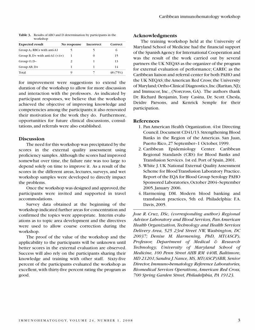
Table 3. Results of ABO and D determination by participants in theworkshop
Expected result No response Incorrect Correct
GroupA2 RBCs with anti-A1 5 5 6
Group B,D+ with anti-A1 (<4+) 1 0 15
Group O,D– 2 1 13
GroupAB,D+ 1 1 14
Total 9 7 48 (75%)
4 I M M U N O H E M A T O L O G Y, VO L U M E 2 4 , N U M B E R 1 , 2 0 0 8
Neonatal transfusions provide challenges at several steps in theprocess. Neonates are often transfused with relatively small volumesat slow flow rates from syringes,whereas at other times they requirerelatively massive transfusions or exchange transfusions. To facilitatethese specialized transfusions, blood banks often modify theirprocedures to provide small volumes of blood components that aresometimes dispensed in syringes or to reconstitute whole blood forexchange transfusions. Hospitals must implement policies andprocedures to ensure that the blood components are transfusedsafely when using these specialized techniques for infants.Nevertheless, some issues remain in many hospitals, such as thedifficulty in safely warming blood components for neonataltransfusions and the difficulties in using approved labels for smallcontainers that are sometimes prepared at the bedside.Immunohematology 2008;24:4–9.
Key Words: neonatal transfusions, syringes, aliquots,exchange transfusions, concentrating blood compo-nents,washing blood components
Neonatal Transfusion VolumesThe large variation in volumes transfused to
neonates impacts the processes for preparing bloodcomponents in the blood bank and for transfusing thepatient at the bedside. Neonates can receive very smallsimple transfusions or larger transfusions,some of whichare considered massive.
Large transfusions including massive transfusionscan occur in one of several settings. Exchange trans-fusions, which are most frequently performed forneonatal hyperbilirubinemia,usually consist of exchang-ing two blood volumes with reconstituted whole blood.Some surgical procedures result in substantial hemor-rhage, necessitating large and sometimes massivetransfusions. Other scenarios in which neonatescommonly receive massive transfusions include thosewhen the patient is placed on an extracorporeal circuitsuch as a cardiopulmonary bypass circuit or anextracorporeal membrane oxygenation circuit.
An entire massive transfusion for a neonate may beobtained from 1 unit because the volume in 1 RBC unit
may be more than the blood volume of the neonate.Very small premature infants have blood volumes of upto 100 mL/kg, which means, for example that a 1500-gpremature infant has a blood volume of up to 150 mL,which is less than the volume of 1 RBC unit.
Although “massive transfusions” for an infant arerelatively small and may be less than 1 unit of RBCs,standard simple transfusions can be extremely small.Indeed, an entire simple transfusion usually consists ofless than 50 mL. A standard transfusion of RBCs orplasma or platelets usually consists of 5 to 15 mL/kg. Forexample, in the 1500-g premature infant, an entiresimple transfusion would be approximately 10 mL.
Venous AccessTransfusions can be performed via a large-bore
central catheter, a small-bore central catheter, or aperipheral venous catheter. A large-bore central catheteris used for extracorporeal circuits in which the“transfusion” is an integral part of the circuit. Otherneonates sometimes have small-bore central catheters.However,some people prefer to reserve these cathetersfor nutrient solutions or drugs because of concernsabout clogging a small-bore central catheter with ablood transfusion.1 Hence,small peripheral needles areoften used for transfusions.
Rapid transfusion through narrow peripheralneedles can cause small amounts of hemolysis in somesituations.2,3 Although the resulting hemolysis may bemeasurable and even cause hemoglobinuria in rarecircumstances, it is rarely if ever clinically significant.The most potentially dangerous effect of hemolysis atthe needle would be the effect of transfusing freepotassium. However,more potassium is liberated fromRBCs during storage than is released via hemolysis at theneedle, and it would be extremely unlikely that enoughpotassium would be transfused through a small-boreneedle to induce dangerous hyperkalemia.2
Technical issues in neonataltransfusionsS.R. SLOAN
R E V I E W
I M M U N O H E M A T O L O G Y, VO L U M E 2 4 , N U M B E R 1 , 2 0 0 8 5
Neonatal transfusion technical issues
Blood WarmersUnfortunately, no blood warmers available in the
United States are specifically designed for neonataltransfusions. There are some disadvantages associatedwith using blood warmers designed for adulttransfusions for neonatal transfusions. One disadvantageis that the volume of fluid required for the bloodwarmer is often larger than the volume to be transfused,which means that some of the blood component willoften be wasted. Another disadvantage associated withsome blood warmers is that because the flow rate forneonatal transfusions is relatively slow, the blood mayreequilibrate to ambient room temperature after passingthrough the blood warmer but before entering thepatient’s circulation. However, most simple neonataltransfusions do not need to be warmed.
Transfusion Techniques—Controlling FlowRate
Except for transfusions that are an integral part of acircuit, most neonatal transfusions are administeredfrom a syringe. Although the syringe plunger may bemanually pressed, most simple transfusions areadministered using a syringe pump to control thetransfusion rate,which is usually approximately 5 to 15mL/kg per hour.
SyringesUnlike most transfusions, neonatal transfusions
usually require transfer of a blood component into asyringe before the actual transfusion. This step oftenoccurs at the patient’s bedside,but some hospital bloodbanks transfer blood components into syringes for selectneonatal transfusions.
When a relatively large volume of a unit(approximately one half) might be transfused,the bloodbank often prepares the unit using procedures identicalto those used for most transfusions of larger patients;blood is dispensed in a standard blood bag. Thetransfusionist might then transfer aliquots of the unitinto a syringe at the patient’s bedside so that thetransfusion rate and volume can be controlled. Thetransfusionist can connect the blood bag to a syringethrough a three-way stopcock, with the other port onthe stopcock connected to a catheter that enters thepatient’s circulation. Using this setup, the bloodcomponent can be filtered while it is transferred fromthe bag to the syringe using a blood administration set;then the stopcock is readjusted, and the blood
component is then transfused from the syringe. Onerisk associated with this approach is that RBCs couldhemolyze if forced through an improperly adjustedstopcock, but this rarely causes clinically significantproblems.
Some blood banks transfer blood components intosyringes for clinical areas such as the neonatal intensivecare unit. The blood bank may simply transfer the bloodinto a syringe using the same connection devices thatwould be used by the clinical area. The disadvantage ofthis approach is that the remainder of the bloodcomponent will have a reduced shelf life because thesystem has been opened and is susceptible tocontamination. AABB Standards limits the shelf life ofsuch a unit to 24 hours from the time the unit is entered.4
If the blood bank has a sterile docking device, it canconnect a blood administration set to the tubing that ispart of the blood unit. The blood bank then transfers aportion of the blood component into a syringe,seals offthe tubing,and disconnects the blood administration setfrom the blood bag. This allows the remainder of theblood component to be stored and used at a later date.5
It is advisable to minimize the time that platelets arestored in syringes,which,unlike platelet storage bags,aremade of gas-impermeable plastic. Platelets are meta-bolically active, and platelets stored in syringes becomedepleted of oxygen and use anaerobic metabolism.6,7
This results in the generation of lactic acid and decreasesthe pH of the platelet component.6–8 This effect happensmore rapidly in conditions that favor greater metabolicactivity, including storage at 37°C and storage of volume-reduced platelets.6,7 Although storage in syringes for upto 6 hours results in platelets that are generallyacceptable by FDA standards and that appear acceptableafter passage through a syringe, it is advisable to avoidstorage at 37°C and to minimize storage times andstorage of volume-reduced platelets.7,8
Satellite BagsSome hospital transfusion services dispense blood
components in satellite bags instead of, or in additionto, syringes. Syringe aliquoting is most useful when thetransfusion service has a sterile connection device toattach the blood administration set to a bloodcomponent unit. Without a sterile connection device,the blood administration set must be spiked into theblood component unit, which opens the system,increasing the risk of contamination and limiting theshelf life of the remaining unit to no more than anadditional 24 hours byAABB Standards.4
6 I M M U N O H E M A T O L O G Y, VO L U M E 2 4 , N U M B E R 1 , 2 0 0 8
S.R. SLOAN
A variety of satellite bag systems are available, all ofwhich are designed to provide aliquots of the originalblood component units in smaller bags. These systems,such as some“pediatric packs”provided by some bloodcenters,contain several (usually six) satellite blood bagscontaining aliquots of the original RBC unit. When theyare prepared by a blood center, sterility of the system ismaintained and the RBC units have the same outdate asa nonaliquoted RBC unit. Similar systems, such as thePedi-Pak (Genesis BPS, Hackensack, NJ) are available tohospital transfusion services. With these systems,staff inthe transfusion service can aliquot a unit into smallerbags,either with or without a sterile connection device.Without the sterile connection device, the shelf life ofthe unit is decreased,but this may still be useful becauseit allows 1 unit to be used for several babies in 1 day orseveral aliquots of 1 unit to be used for one baby whorequires multiple transfusions during 1 day.
Satellite bag systems are also available for plasma.Most commonly, a unit of plasma is divided into 4 to 6smaller subunits before freezing, and blood supplierscan often provide this. The transfusion service thenthaws a small dose of plasma when needed. The unitcan be thawed in the same warmers used for standardunits, but the thawing times differ and each laboratoryshould determine the thawing times for the differenttypes of frozen plasma units that are used. When usinga standard plasma warmer device, the thawing timedepends on the dimensions of the unit and is especiallydependent on the unit’s thickness. Hence,smaller unitssometimes take more time to thaw than larger units.
Alternatively, a plasma unit can be aliquoted intosatellite bags after thawing, using a sterile connectiondevice. This might be useful if no small frozen plasmaunits are available and several pediatric doses areneeded. Additionally, if one infant is expected to needseveral plasma doses,donor exposure can be limited bytransfusing with aliquots from the same thawed unit.
FilteringAll blood components should be filtered near the
time of transfusion, as required by accreditingorganizations such as the AABB.9 This filtering step,which is in addition to optional filtering to reduceleukocyte concentration, is designed to remove anyaggregates that may develop during storage of bloodcomponents. Although this filtering is generallyperformed when the blood component is transfused,themost common exception occurs with neonataltransfusions, in which the blood component is often
filtered when it is transferred into a syringe. Thisexception is allowed because it is extremely difficult toappropriately prime a catheter and blood filter and thenprecisely control the transfusion volume flow rate whenthe syringe is connected to a filter during the transfusion.Because filtering is often most conveniently performedwhen the blood component is transferred into a syringe,the blood bank may filter the blood component.
Some facilities use blood administration setsespecially designed for neonatal transfusions. These setsare designed to minimize the priming volume and bloodlosses that occur in standard blood administration sets.Indeed, it is not unusual for up to 40 mL to be lost in thestandard blood administration set. Neonatal bloodadministration sets have smaller filters and narrowercatheters that are often shorter to minimize thesevolumes. Additionally, manufacturers such as CharterMedical, Ltd.,Winston-Salem, North Carolina, sell setswith various configurations, such as sets with a syringeor syringes preattached to facilitate transfer into syringeswithin a sterile closed system.5 With these systems,volume losses can be minimized to 1 to 3 mL for eachaliquot that is prepared from a unit, and some bloodbanks have reported preparing more than 20 aliquotsfrom 1 unit of a blood component.
IrradiationCellular blood components, including RBCs, whole
blood, and platelets, may be irradiated to preventtransfusion-associated GVHD. The shelf life and functionof platelets is not affected by irradiation.10–13 However,RBC units are affected by irradiation, which should beconsidered in blood bank policies and procedures.
Potassium is released from RBCs during storage,andthis process is substantially hastened by irradiation.14 Tominimize exposure to extracellular potassium, the unitshould be irradiated as close to the time of transfusionas possible if the unit will be used for a large and rapidtransfusion.15,16 This is recommended because casereports suggest that neonates are particularlysusceptible to transfusion-associated hyperkalemia,which occurs,although rarely,when they are transfusedwith RBC units or whole blood that contains elevatedpotassium concentrations in the supernatant.17 If it isnot possible to provide freshly irradiated units, eitherbecause of a lack of inventory of appropriate units orbecause the hospital transfusion service lacks anirradiator, then the units used for large transfusions canbe washed to remove most of the extracellularpotassium.
I M M U N O H E M A T O L O G Y, VO L U M E 2 4 , N U M B E R 1 , 2 0 0 8 7
Neonatal transfusion technical issues
Washing and Concentrating BloodComponents
Although the methods used to wash bloodcomponents for neonates are the same as those used towash blood components for older patients, a fewpossible indications for washing are more common inneonates. Case reports suggest that transfusion ofplasma-containing components increases hemolysisowing to T-cell activation in neonates, and hencewashing may be indicated for these patients.18 Althoughtransfused potassium is not problematic for standardsimple transfusions, it may be problematic for largetransfusions for very young patients, and RBC units aresometimes washed to reduce extracellular potassium.Although this is effective for the first 2 to 3 hours afterwashing, potassium can rapidly rise after washing,and 18 hours after washing irradiated RBC units,the extracellular potassium concentration can exceed15 mEq/L.19
Although there is little evidence that concentratedRBCs or platelets are beneficial for neonatal transfusions,some transfusion services provide concentrated RBCsand platelets. A dose of RBCs concentrated to a hemato-crit of approximately 68% can be obtained by removingthe aliquot from an RBC unit that has been hangingupside down in the refrigerator for 72 hours.20
Alternatively,some have developed methods to concen-trate RBCs to more than 80 percent by centrifuging theunit at 4000 × g for 4 minutes and transferring a portionof the concentrated RBCs to a satellite bag.21,22
Platelets can be concentrated by centrifugation,removal of some supernatant, and resuspending theremaining platelets.23 However, the additionalmanipulation of platelets required to wash or volume-reduce platelets causes platelet loss,can cause clumping,and may induce some platelet dysfunction.
Exchange TransfusionsSpecial considerations apply to a neonatal exchange
transfusion,which involves a massive transfusion for aninfant. The methods used to prepare the bloodcomponent and perform the exchange are designed tominimize the risk involved with the procedure.
Two blood volumes are usually exchanged, with aneonate’s blood volume ranging from 85 mL/kg for fullterm infants to 100 mL/kg for very low birth weightinfants. Most frequently, the neonate’s blood is ex-changed with reconstituted whole blood with ahematocrit of 40 to 60% that is prepared from RBC unitsand plasma.24,25 Often the first step in preparing
reconstituted whole blood is to remove the supernatantfrom the RBC unit, especially if the RBC unit containsan additive. Although this can be accomplished bywashing the unit in an automated blood washer, amodified method in which normal saline solution isadded, the unit is centrifuged, and most of thesupernatant is removed removes sufficient amounts ofsupernatant for this purpose. When the RBC unit isready for reconstitution, the hematocrit of the unit ismeasured, and the amount of plasma needed to dilutethe RBC unit to the desired final hematocrit iscalculated.
The exchange transfusion is sometimes performedthrough simultaneous withdrawal and transfusionthrough two sites of vascular access.26,27 However, thediscontinuous method is more frequently used. With thediscontinuous method, a stopcock is used to alternatetransfusion and withdrawal of blood through one site.28
With the discontinuous method,usually no more than 5mL/kg of blood is withdrawn and replaced during eachreplacement cycle. With either method,a blood warmeris usually recommended, and the unit should be keptwell mixed throughout the procedure.
LabelsBags and syringes of blood used for transfusion need
to be labeled, and all FDA-licensed and registeredfacilities and allAABB-accredited facilities will soon needto be in full compliance with ISBT standards.29,30 Theseregulations apply regardless of where the aliquot isprepared. In most cases,the labels for aliquots will needto be provided by the hospital transfusion service evenif the aliquot is prepared at the bedside. Because labelsneed to be in compliance with specific size require-ments, facilities are developing labels and techniquesthat permit the labels to be used with syringes andsyringe pumps. For example, some labels may need tobe folded back to permit visualization of the bloodcomponent in a syringe.
SummaryTwo issues result in the use of special techniques
when preparing and transfusing blood components forneonates. First, neonates can be especially susceptibleto some transfusion risks such as metabolic abnormal-ities and fluid shifts. Hence, blood banks and clinicalareas avoid transfusing fluids containing nonphysiologicelectrolyte concentrations and using rapid transfusionrates whenever possible. Second,standard supplies andequipment are designed for transfusion of larger
8 I M M U N O H E M A T O L O G Y, VO L U M E 2 4 , N U M B E R 1 , 2 0 0 8
S.R. SLOAN
patients. In some cases, hospitals need to useequipment that is not well designed for neonataltransfusions, such as blood warmers. In other cases,equipment and supplies for neonatal transfusions areavailable, such as satellite bags, pediatric filters, syringepumps, and syringes. In these cases, hospitals mustconsider the impact that these systems have onworkflow and the potency and purity of transfusedblood components.
References1. Wong EC, Schreiber S, Criss VR, et al. Feasibility ofred blood cell transfusion through small borecentral venous catheters used in neonates. PediatrCrit Care Med 2004;5:69–74.
2. Miller MA, Schlueter AJ.Transfusions via hand-heldsyringes and small-gauge needles as risk factors forhyperkalemia.Transfusion 2004;44:373–81.
3. Pisciotto P, Wong E. Technical considerations/mechanical devices. In: Hillyer C, Luban N, StraussR,eds.Handbook of pediatric transfusion medicine.San Diego: Elsevier Academic Press, 2004:121–30.
4. 24th Edition Blood Bank/Transfusion ServiceStandards Program Unit,Standards for Blood Banksand Transfusion Services,AABB, 2006:53, standard5.1.8A.
5. Roseff SD. Pediatric blood collection andtransfusion technology. In: Herman JH, Manno CS,eds. Pediatric transfusion therapy. Bethesda:AABBPress, 2002:217–48.
6. Pisciotto PT, Snyder EL,Napychank PA,Hopfer SM.In vitro characteristics of volume-reduced plateletconcentrate stored in syringes.Transfusion 1991;31:404–8.
7. Pisciotto PT, Snyder EL, Snyder JA, et al. In vitrocharacteristics of white cell-reduced single-unitplatelet concentrates stored in syringes.Transfusion1994;34:407–11.
8. Strasser EF,Stachel DK,Schwarzkopf P,et al.Plateletfunction in variable platelet split productsintended for neonatal transfusion. Transfusion2006;46:757–65.
9. 24th Edition Blood Bank/Transfusion ServiceStandards Program Unit,Standards for Blood Banksand Transfusion Services,AABB, 2006:48, standard5.19.9.
10. Moroff G, George VM, Siegl AM, Luban NL. Theinfluence of irradiation on stored platelets.Transfusion 1986;26:453–6.
11. Read EJ, Kodis C, Carter CS, Leitman SF.Viability ofplatelets following storage in the irradiated state.Apair-controlled study.Transfusion 1988;28:446–50.
12. Sweeney JD, Holme S, Moroff G. Storage ofapheresis platelets after gamma radiation.Transfusion 1994;34:779–83.
13. van der Meer PF, Pietersz RN. Gamma irradiationdoes not affect 7-day storage of platelet concen-trates.Vox Sang 2005;89:97–9.
14. Hume HA, Preiksaitis JB. Transfusion associatedgraft-versus-host disease,cytomegalovirus infectionand HLA alloimmunization in neonatal andpediatric patients.Transfus Sci 1999;21:73–95.
15. Roseff SD, Eder A, Hume H, et al. Pediatrictransfusion; a physician’s handbook, 2nd ed.Bethesda:AABB Press, 2006:208.
16. Strauss RG. Routinely washing irradiated red cellsbefore transfusion seems unwarranted.Transfusion1990;30:675–7.
17. HallTL,BarnesA,Miller JR,et al.Neonatal mortalityfollowing transfusion of red cells with high plasmapotassium levels.Transfusion 1993;33:606–9.
18. Eder AF, Manno CS. Does red-cell T activationmatter? Br J Haematol 2001;114:25–30.
19. Weiskopf RB, Schnapp S, Rouine-Rapp K, et al.Extracellular potassium concentrations in redblood cell suspensions after irradiation andwashing.Transfusion 2005;45:1295–301.
20. SherwoodWC, Donato T, Clapper C,Wilson S.Theconcentration of AS-1 RBCs after inverted gravitysedimentation for neonatal transfusions. Trans-fusion 2000;40:618–9.
21. Strauss RG,Burmeister LF, Johnson K,et al.AS-1 redcells for neonatal transfusions: a randomized trialassessing donor exposure and safety.Transfusion1996;36:873–8.
22. Strauss RG,Villhauer PJ, Cordle DG.A method tocollect, store and issue multiple aliquots of packedred blood cells for neonatal transfusions.Vox Sang1995;68:77–81.
23. Moroff G, Friedman A, Robkin-Kline L, et al.Reduction of the volume of stored plateletconcentrates for use in neonatal patients.Transfusion 1984;24:144–6.
24. Goodstein M. Neonatal red cell transfusion. In:Herman JH, Manno CS, eds. Pediatric transfusiontherapy.Bethesda:AABB Press, 2002:39–91.
I M M U N O H E M A T O L O G Y, VO L U M E 2 4 , N U M B E R 1 , 2 0 0 8 9
Neonatal transfusion technical issues
25. AABB Technical Manual Program Unit. Neonataland pediatric transfusion practice. In: Brecher ME,ed. Technical manual. Bethesda: AABB Press,2005:557–80.
26. Wallerstein H.Treatment of severe erythroblastosisby simultaneous removal and replacement of theblood of the newborn infant. Science 1946;103:583–4.
27. Goldman SL, Tu HC. Automated method forexchange transfusion:a newmodification.J Pediatr1983;102:119–21.
28. Diamond LK, Allen FH Jr, Thomas WO Jr.Erythroblastosis fetalis. VII. Treatment withexchange transfusion. N Engl J Med 1951;244:39–49.
29. 24th Edition Blood Bank/Transfusion ServiceStandards Program Unit,Standards for Blood Banksand Transfusion Services,AABB, 2006:12, standard5.1.6.3.1.
30. FDA,Center for Biologics Evaluation and Research.Guidance for industry: recognition and use of astandard for uniform blood and blood componentcontainer labels. U.S. Department of Health andHuman Services, 2006:21.
Steven R. Sloan, MD, PhD, Medical Director PediatricTransfusion Medicine, Joint Program in TransfusionMedicine and Department of Laboratory Medicine,Children’s Hospital, Boston, 300 LongwoodAve., Bader406, Boston, MA 02115.
Attention: State Blood Bank Meeting OrganizersIf you are planning a state meeting and would like copies of Immunohematology for distribution,pleasecontact Cindy Flickinger,Managing Editor, 4 months in advance, by fax or e-mail at (215) 451-2538 [email protected].
10 I M M U N O H E M A T O L O G Y, VO L U M E 2 4 , N U M B E R 1 , 2 0 0 8
This review discusses RBC transfusion in the neonatal age group andexplores how one institution arrived at current common practice.Special considerations such as CMV infectious risk and GVHD arediscussed. Immunohematology 2008;24:10–14.
The neonatal intensive care unit (NICU) patientpopulation is one of the patient groups in the hospitalmost heavily transfused with RBCs. All infantsexperience a normal decline in hemoglobin concen-tration during the first weeks of life; however, this isproblematic in premature infants because of adiminished output of erythropoietin in response toanemia. In addition, these patients,who often have lowbirth weights, need close monitoring of blood gases,electrolytes, and other laboratory variables, whichcontributes substantially to the transfusion requirementas a result of the small circulating RBC volume. Althoughthese tests are often impossible to do without, there issome help that can be provided to the phlebotomists torelieve the blood loss volume from being even greater.The infant younger than 4 months of age isimmunologically immature, which makes RBCalloimmunization exceedingly rare, so some of theserologic testing performed in the blood bank for otherage groups can be abbreviated. This 4-month period isoften defined in the blood bank as the neonatal periodbecause of this distinction. In other medicalsubspecialties the term neonate may describe differentage ranges. For this patient population the reversegrouping and the crossmatch that are seen in routineblood bank practice can be waived. This saves therepeated drawing of a crossmatch specimen every 3days. Initial testing must determine ABO group and Dtype and include a screen of the serum (infant’s ormother’s) for unexpected antibodies. If there areantibodies present in the serum, blood that testsnegative for the corresponding antigen can be providedwithout crossmatch.1
In the start of the 1990s the sick infant whounderwent multiple transfusions was typically exposedto 9 or 10 different donors.2 These transfusions weredispensed in small amounts or “aliquots”of the original
unit because of the small size of the patient,but even sothe number of donors increased the risk of certaintransfusion-related complications, such as infectiousrisks, which are proportional to donor exposure.Neonatal transfusion practices have changed since then,not in reducing the total volume of blood transfused,butin decreasing the number of donor exposures. At theoutset of the decade the rise in potassium that was seenin RBC units stored for any length of time was feared byneonatologists because of the risk that a posttransfusionrise in serum potassium to abnormal levels would causefatal arrhythmias. Studies of small-volume transfusionsin neonates that compared units of RBCs stored untiltheir expiration date with fresher units showed thatposttransfusion potassium concentrations did not riseto abnormal levels and were not a clinical issue.3–5 It isimportant to note that the transfusions being discussedwere small-volume (15 mL/kg),slow transfusions. Large-volume, rapid transfusions performed in this age groupcan occur in surgery, exchange transfusion, andextracorporeal membrane oxygenation. When thepotassium load cannot distribute itself throughout thetotal blood volume quickly enough, it may result inarrhythmia. This was described in 1993 in a neonatewho received older RBCs as a rapid transfusion incardiac surgery, and died of cardiac arrest.6 The RBCswere stored in CPDA-1; today more commonly usedadditive solutions have a better potassium profile.However, it is still prudent in a rapid or large-volumetransfusion setting to use the freshest RBCs possible asopposed to units that are close to their expiration date,although in a small-volume, slow transfusion setting,minimizing donor exposures is more important than ageof the RBCs.
Another change in practice occurring at the sametime, as alluded to previously, was the use of additivesolutions,which were new to transfusion services. Thesesolutions increased RBC storage to 42 days because theywere better for RBC metabolism and decreasedhemolysis. The first additive solution in widespread usewas AS-1 (Adsol), which contained additional dextrose
Neonatal red cell transfusionsC.A. LITTY
R E V I E W
I M M U N O H E M A T O L O G Y, VO L U M E 2 4 , N U M B E R 1 , 2 0 0 8 11
Neonatal red cell transfusions
and mannitol. Mannitol may cause an osmotic diuresis,which again was worrisome to neonatologists because ofthe potential consequences. This was a theoretical risk,and in 1991 before controlled studies appeared Lubanet al.7 addressed this eloquently by calculating theamount of supernatant fluid present in a small-volumetransfusion and the volume of these additives actuallytransfused to the infant. In fact the concentration ofmannitol actually transfused per kilogram wassubstantially less than would be needed to cause anosmotic diuresis.7 Indeed, this was borne out whencontrolled studies began to appear. A study comparingAS-1 units with CPDA-1 units showed that the patientsreceiving AS-1 actually had an improved glucosehomeostasis in that the amount of hypoglycemia seenafter neonatal transfusions was reduced. (Hypoglycemiaseen with neonatal transfusions is usually a function ofhigh-glucose fluids being discontinued during atransfusion to use the current intravenous access owingto the difficulty of obtaining multiple access sites in thispatient group.) Also, urine output, pH, and serumelectrolytes were not significantly different.8
With time many centers accepted the use of AS-1units with no negative consequences; however, bloodcenters began to purchase bag sets from manufacturerswho used AS-3 (Nutricel). This product differed fromAS-1 in the presence of phosphate rather than mannitol.Again,a study confirmed that in clinical use the additivesolution did not cause harm to the neonates transfused.9
Use of all group O,D– or only group O,D– and groupO D+ for neonates stemmed from the practice of usingonly fresh RBCs for transfusion. This way the unit couldbe used for many neonates before it reached its 5- or 7-day expiration date. The problem with this method isthat it depletes group O units from the blood supply,when they are already used excessively (i.e., traumapatients). When the practice changed to using dedicatedunits many centers switched toABO-specific RBCs. Thechangeover was not complete, unfortunately, and thisexacerbates group O shortages in many regions. Someblood bank workers advocate not switching to ABO-specific units in a patient of any age until a secondsample is drawn for confirmation as a safety measure.Whether this will become the practice in neonates, inwhom blood draws should be kept at a minimum, isunknown,but obligating this entire group of patients toreceive only group O RBC units will certainly have animpact on the supply in the community at large.
A consideration in the choice of blood in the NICUis that the multiply transfused preterm infant of a CMV-
seronegative mother is at an increased risk oftransfusion-transmitted CMV infection. CMV infectionhas a highly variable clinical picture and can beasymptomatic, severe, or fatal. There is often sepsisassociated with hepatosplenomegaly, abnormal bloodcounts, and pneumonitis.
Blood products can be tested for CMV antibodies,and seronegative products can then be provided for use.Because CMV is an organism that is associated withWBCs, providing WBC-reduced cellular blood compo-nents is an appropriate way to reduce the risk.10–13 Withthe increasing use of leukoreduction this method hasbecome well accepted as a CMV safe alternative, butthere are some physicians who will still insist on CMV-seronegative blood. Because most communities havehigh rates of seropositivity in the donor population, it isunacceptable to waste seronegative blood on sero-positive patients in a situation in which only CMV-testedblood products are requested. Many hospitals find iteasier to provide leukoreduced blood to all than to testall mothers and provide selective blood products.
Transfusion-associated graft-versus-host disease (TA-GVHD) occurs when an immunocompromised patientis transfused with blood from an immunologicallycompetent donor. The donor T lymphocytes can thenproliferate unimpeded and engraft. Fever follows atabout 4 weeks (versus 10 days in an adult) and rash atabout 30 days (versus 12 days in an adult);14 liver andgastrointestinal involvement and severe cytopeniaensue. The pancytopenia differs from the GVHD seenafter bone marrow transplant because in TA-GVHD thebone marrow is part of the host, thus it is affected also.This accounts for the very high fatality rate seen in thelatter,attributable to hemorrhage and infection. There isalso a longer course of infection from transfusion todeath in neonates than adults. Several theories as to themechanism of these differences are discussed in thethorough literature review from Japan of Ohto andAnderson.14
Irradiation of blood components is the only methodto render the T lymphocyte nonmitogenic and preventthe reaction. Although leukoreduction reduces thenumber of WBC greatly, there is no known thresholdbelow whichTA-GVHDwill not occur;therefore, it is notan adequate method for prevention.
The infants who are at risk for TA-GVHD havespecific risks other than being an infant. These includeimmunodeficiency disorder, intrauterine transfusionfollowed by postnatal exchange transfusion, severeprematurity and low birth weight, and family members
12 I M M U N O H E M A T O L O G Y, VO L U M E 2 4 , N U M B E R 1 , 2 0 0 8
C.A. LITTY
providing directed donations.13 Therefore a blanketpolicy of irradiating blood for all infants or NICUpatients is not required. Some hospitals, however,mayopt to do this. A number of factors should go into thisconsideration. A hospital with a high-level NICU, withvery premature or sick infants,and a blood bank that hasits own irradiator and can easily irradiate beforetransfusion for infants who are at sufficient risk is mostlikely to find a policy to irradiate RBCs for all neonatesuseful. This is more efficient than expecting clinical staffto provide birth weight and clinical history so that eachpatient’s risk can be determined and will avoid missingthe one patient who falls through the cracks. Hospitalswithout this high-risk population or an irradiator of theirown will have to wait for irradiated components to besent specially from the blood center. If they are not usedas originally planned then the issue of storage lesioncomes into play. (Irradiated RBCs have a shorter shelflife, 28 days, because of increased storage lesion.)Considering the wait time and the decreased shelf life ofunits, it would be more efficient to determine the actualrisk of each patient than to set a broad policy.
RBCs are supplied to replace oxygen carryingcapacity, but the improvement in oxygen offloading atthe tissue level and its effects on patient outcomecannot be measured. Therefore we do not actually knowwhether we are improving the patient’s condition. It isimportant for clinicians to believe there is a benefitbefore transfusing and not just have a knee-jerk reactionto a number on a lab report, which is all too common.However, conflicting evidence provides reason fordebate among clinicians.
Iatrogenic losses were discussed earlier;however, toquantify, these losses should be replaced when 10percent of the blood volume has been phlebotomized.Anemia is harder to define in this age group because ofthe changing normal values. At this institution, thehemoglobin range on the first day of life is 16.5 to 21.5g/dL. This declines throughout the next few months,and at 3 months of age a hemoglobin of 10.4 g/dL is thelower limit of normal. This is called the physiologicanemia of infancy. As this change takes place HbF isreplaced by HbA,which has a lower affinity for oxygen,and thus releases it for tissue consumption more
efficiently. In preterm infants the hemoglobin levels arelower at any given point and the decline is morepronounced, and the switch over to HbA is affected bythe degree of prematurity. There is even controversyover defining the signs of anemia in this age group,withtachycardia, tachypnea, bradycardia, recurrent apnea,and poor weight gain being used.15,16
Table 1 summarizes guidelines by the AABBPediatric Hemotherapy Committee. It is interesting tonote that these are from the mid-1990s; more recent,more restrictive guidelines are available from Britain andare summarized inTable 2.17
Two recent studies provide a glimpse of theconflicting opinions. A study from 2005 thatrandomized 100 preterm infants to a restrictive or liberaltransfusion group showed there may be harm topatients in the restrictive group.18 The liberal groupreceived transfusions for hematocrits less than 46%,andthe restrictive group used 34% as the cutoff fortransfusion. However, these thresholds were adjustedlower as patients progressed through three stages ofclinical condition. Infants in the restrictive group hadmore intraparenchymal brain hemorrhage, periventric-ular leukomalacia,and apnea. In 2006,a larger study waspublished.19 It was called the PINT study for prematureinfants in need of transfusion. It also used a low andhigh threshold for hemoglobin, which changed forclinical condition, starting with 10 to 11.5 g/dL versus12 to 13.5 g/dL. Differences in each group dependedon whether or not there was respiratory support. Therewere 451 infants enrolled in this study,and there was nosignificant evidence of benefit to the high transfusionthreshold.
An exception to using hemoglobin values as an aidto determining transfusion threshold is seen when anewborn suffers from HDN. In HDN the mother hasformed IgG antibodies to an antigen on the fetal RBCs.It can be the D of the Rh system or another RBC antigen.There is immune-mediated hemolysis resulting inanemia, but a further problem exists related to thebilirubin levels. This is because at birth the newborn’sliver is not mature enough to conjugate the large
Table 1. RBC indications for infants younger than 4 months of age15,16
Hb < 13 g/dL Severe pulmonary or cyanotic heartdisease, heart failure
Acute loss of 10% blood volume Phlebotomy or other cause
Hb < 8 g/dL Stable neonate with clinicalmanifestations of anemia
Table 2. RBC transfusion thresholds for infants younger than 4 months ofage17
Anemia in the first 24 hours Hb = 12 g/dL
Cumulative blood loss in 1 week NICU 10% blood volume
Neonate receiving intensive care Hb = 12 g/dL
Acute blood loss 10% blood volume
Chronic oxygen dependency Hb = 11 g/dL
Late anemia, stable patient Hb = 7 g/dL
I M M U N O H E M A T O L O G Y, VO L U M E 2 4 , N U M B E R 1 , 2 0 0 8 13
Neonatal red cell transfusions
amounts of bilirubin produced by the hemolysis.Unconjugated bilirubin presents a great danger to thedeveloping central nervous system. Exchange trans-fusions are performed with reconstituted whole bloodcreated by removing the additive solution from RBCsand combining with thawed FFP. The infant’s blood isremoved in aliquots followed by transfusion of aliquotsof reconstituted whole blood. It is best to use areconstituted product with a hematocrit of 45 ± 5%.Theplasma portion of this product is necessary to replacethe infant’s plasma, which has high levels ofunconjugated bilirubin and maternal antibody. Inaddition the process removes IgG-coated RBCs beforethey have been hemolyzed, and the RBCs treat theanemia. The RBCs chosen must be compatible with themother’s serum, which means they lack the antigencorresponding to the maternal antibody. Typically groupAB FFP is used to reconstitute. The need for irradiationshould be evaluated as discussed in a previous section.
The levels of bilirubin that lead to this procedurewill vary from hospital to hospital based on differingnormal ranges,usually more than 25 mg/dL,but the rateof rise of bilirubin is used along with the level to decidewhen an exchange transfusion is appropriate. WhenHDN is not severe enough to necessitate exchangetransfusion, treatment consists of phototherapy, whichexposes the skin to a specific wavelength of light thatconverts the bilirubin into a more soluble form that canbe excreted without conjugation.
The transfusing physician should always be awarethat the well-defined risks that are documented in thetextbooks are not the only risks to transfusion. Theremay be consequences to transfusion that are hard toshow because of the complexities of the illness of thesick, transfused population. There also may be donorcharacteristics specific to one region that are not widelyseen. One example is lead in the environment. A donorexposed to lead may be a source of lead exposure to atransfusion recipient. This was shown in a study from1991 to 1992.20 Posttransfusion increases of lead wereseen in 19 premature infants in relation to the amountof blood they received in Oakland,California. Whetherthis would be seen in other locations or in the presentdecade is unknown.
Another interesting and more recent study lookedat the association of RBC transfusions and necrotizingenterocolitis (NEC).21 This is a serious acquiredgastrointestinal disorder seen in low birthweightneonates. A small group of stable, growing prematureneonates developed NEC within 48 hours of transfusion.
Whether there are host-specific or RBC storagecharacteristics that influence this risk is unknown.
HCV look-back is a good example of futureconsequences to what seems like a life-savingintervention today. The first tests for HCV appeared in1990,and a second-generation test came out in 1992. In1998 the FDA recommended HCV look-back to all bloodestablishments. This meant that donors who testedpositive for HCV had previously negative or untesteddonations traced so that recipients could be found andtested in case the virus had been transmitted by theearlier transfusion. Among patients whose donors werelater found to be HCV-positive,children represented 10to 20 percent of those who acquired posttransfusionhepatitis C.22 A recent study from Alaska looked at allpatients who were transfused while in the NICU asopposed to the FDA-recommended look-back describedhere, which only tested recipients whose donors laterwere tested positive.23 In this study of 216 screenedpatients,7 (3%) were hepatitis C antibody–positive;6 ofwhich were also hepatitis C virus-RNA positive.
Some of their lives may very well have been savedby the transfusions, but it is certainly something thetransfusing physician should be thinking about whenconsidering whether or not to transfuse. The risk ofserious consequences that may manifest themselvesmany years in the future is only worth taking if there isa real benefit from the transfusion.
Those in the transfusion medicine community canhelp patients even though they are not the professionalswriting orders at the bedside. From the technologistsin the blood bank to the medical directors,every chanceto educate the clinicians should be seized. Much ofwhat we know about risks of transfusion has beendescribed in the last 15 to 20 years. This is not coveredin the medical school curriculum.
This can be said for all patients, but it is especiallytrue in the infant who has the most years of life aheadof all transfused patients. Although the currenthealthcare team of transfusing physicians and bloodbank workers will probably not be involved in thepatient’s care after 15 years, the patient may be dealingwith a transfusion-transmitted illness we cannot evenimagine today.
References1. Silva MA, ed. Standards for blood banks andtransfusion services,24th ed.Bethesda:AABB,2007.
2. Sacher RA,Luban NLC,Strauss RG.Current practiceand guidelines for the transfusion of cellular blood
14 I M M U N O H E M A T O L O G Y, VO L U M E 2 4 , N U M B E R 1 , 2 0 0 8
C.A. LITTY
components in the newborn. Transfus Med Rev1989;3:39–54.
3. Patten E, Robbins M, Vincent J, et al. Use of redblood cells older than five days for neonataltransfusions. J Perinatol 1991;11:37–40.
4. Lee DA,SlagleTA,JacksonTM,et al.Reducing blooddonor exposures in low birth weight infants by theuse of older, unwashed packed red blood cells. JPediatr 1995;126:280–6.
5. Liu EA, Mannino FL, Lane TA. Prospective,randomized trial of the safety and efficacy of alimited donor exposure transfusion program forpremature neonates. J Pediatr 1994;125:92–6.
6. HallTL,BarnesA,Miller JR,et al.Neonatal mortalityfollowing transfusion of red cells with high plasmapotassium levels.Transfusion 1993;33:606–9.
7. Luban NLC,Strauss RG,Hume HA.Commentary onthe safety of red cells preserved in extended-storage media for neonatal transfusions.Transfusion1991;31:229–35.
8. Goodstein MH, Locke RG, Wlodarczyk D, et al.Comparison of two preservation solutions forerythrocyte transfusions in newborn infants. JPediatr 1993;123:783–8.
9. Strauss RG, Burmeister LF, Johnson K, et al.Feasibility and safety of AS-3 red blood cells forneonatal transfusions. J Pediatr 2000;136:215–9.
10. Gilbert GL,Hayes K,Hudson H,et al. Prevention oftransfusion–acquired cytomegalovirus infection ininfants by blood filtration to remove leukocytes.Lancet 1989;333:1228–31.
11. Strauss RG. Selection of white cell-reduced bloodcomponents during early infancy.Transfusion 1993;33:352–7.
12. Delage G.Transfusion-transmitted infections in thenewborn.Transfus Med Rev 1995;9:271–6.
13. Strauss RG.Data-driven blood banking practices forneonatal RBC transfusions. Transfusion 2000;40:1528–40.
14. Ohto H,Anderson KC.Posttransfusion graft-versus-host disease in Japanese newborns. Transfusion1996;36:117–23.
15. Stehling L, Luban NLC, Anderson KC, et al.Guidelines for blood utilization review.Transfusion1994;34:438–48.
16. Hume H, Bard H. Small volume red blood celltransfusion for neonatal patients.Transfus Med Rev1995;9:187–99.
17. Gibson BES, Todd A, Roberts I, et al. Transfusionguidelines for neonates and older children. Br JHaematol 2004;124:433–53.
18. Bell EF, Stauss RG,Widness JA, et al. Randomizedtrial of liberal versus restrictive guidelines for redblood cell transfusion in preterm infants.Pediatrics2005;115:1685–91.
19. Kirplani H, Whyte RK, Andersen C, et al. Thepremature infants in need of transfusion (PINT)study:a randomized,controlled trial of a restrictive(low) versus liberal (high) transfusion threshold forextremely low birth weight infants. J Pediatr 2006;149:301–7.
20. Bearer CF,O’Riordan MA,Powers R.Lead exposurefrom blood transfusion to premature infants. JPediatr 2000;137:549–54.
21. Mally P, Golombek SG, Mishra R, et al.Associationof necrotizing enterocolitis with elective packedred blood cell transfusions in stable growingpremature neonates.Am J Perinatol 2006;23:451–8.
22. Aach RD, Yomtovian RA, Hack M. Neonatal andpediatric posttransfusion hepatitis C: a look backand a look forward. Pediatrics 2000;105:836–42.
23. Cagle HH, Jacob J, Homan CE, et al. Results of ageneral hepatitis C lookback program for personswho received blood transfusions in a neonatalintensive care unit between January 1975 and July1992.Arch Pediatr Adolesc Med 2007;161:125–30.
Cathy A. Litty, MD, Director of Transfusion Medicine,St. Christopher’s Hospital for Children, Department ofPathology and Laboratory Medicine, Erie Ave at FrontSt, Philadelphia, PA 19134.
Phone, Fax, and Internet Information: If you have any questions concerning Immunohematology, Journalof Blood Group Serology and Education, or the Immunohematology Methods and Procedures manual,contact us by e-mail at [email protected]. For information concerning the National ReferenceLaboratory for Blood Group Serology, including theAmerican Rare Donor Program,please contact Sandra Nance,by phone at (215) 451-4362, by fax at (215) 451-2538, or by e-mail at [email protected]
I M M U N O H E M A T O L O G Y, VO L U M E 2 4 , N U M B E R 1 , 2 0 0 8 15
Neonates are defined as infants younger than 28days old, and in general many transfusion protocols arestandardized to represent infants younger than 4 monthsof age. Often the distinction is made between pre-mature neonates and term neonates, and institutionalprotocols may be established on the basis of age or birthweight. When this is important in terms of transfusionprotocols, premature neonates, neonates, and infantswill be described separately.
Indications for Neonatal and Infant PlateletTransfusions
Healthy term infants are born with the same plateletcount as adults, whereas premature infants may haveplatelet counts that are on the lower end of the adultnormal range (150 × 109/L to 450 × 109/L). When theplatelet count is less than normal, this is an indicationfor investigation and possibly treatment that mayinclude platelet transfusions. Many disorders are asso-ciated with thrombocytopenia in neonates, and aninvestigation for infections, drug exposures, auto-immunity or alloimmunity, thromboses, neoplasms, andgenetic conditions is important for the evaluation of theneed for treatment or transfusion.
The degree of thrombocytopenia in newbornsvaries, although thrombocytopenia is the mostcommonly reported hematologic abnormality inneonatal intensive care units (NICU). According toCastle et al.,1 75 percent of sick neonates will have atransient thrombocytopenia by day 2 of life,which willnadir by day 4 and return to normal counts by day 10 oflife in 86 percent of neonates. These neonates may notrequire a transfusion. However, approximately 50percent of hospitalized neonates will have a plateletcount decrease to less than 100 × 109/L,and 20 percentwill have a decrease to less than 50 × 109/L.1,2 Theseneonates may require platelet transfusion. There isongoing controversy over the indications, trigger, and
dosing for platelet transfusions in these patients,and thispresents a clinical challenge owing to the limited datafrom controlled clinical trials.
Platelet Transfusion GuidelinesThe decision to transfuse is often considered in
terms of treatment of severe bleeding in an unstableneonate or prophylaxis to prevent bleeding in a morestable neonate. The AABB Pediatric HemotherapyCommittee, by developing a practice consensus, wasone of the first groups to develop guidelines forneonatal platelet transfusion for the purposes ofauditing.3 AABB members who were surveyed for thestudy noted that the platelet transfusion criteria for sickpremature infants was too liberal at 100 × 109/L incontrast to the medical practice at many institutions totransfuse at less than 50 × 109/L. After that consensussurvey, other published guidelines emerged for infants.These guidelines recommended that stable neonateswith a platelet count of less than 50 × 109/L with activebleeding or an invasive procedure with productionfailure may require platelet transfusion. Neonates withplatelet counts less than 30 × 109/L as a result of failureof platelet production,but no identified bleeding or riskof bleeding,may also require platelet transfusion. In rareinstances, guidelines for neonates may more closelyresemble guidelines for older children and adults, inwhom transfusions are recommended for plateletcounts of 5 to 10 × 109/L. A sick unstable neonate maybe transfused if the platelet count drops below 100 ×109/L with active bleeding or when an invasiveprocedure is anticipated in patients with sepsis,disseminated intravascular coagulation, or othermechanism of platelet consumption.4,5
The sick premature infant is at especially high riskfor an intracranial hemorrhage (ICH) from a low plateletcount or poor platelet function. These infants (youngerthan 37 weeks) have decreased plasma coagulation
Neonatal and infant platelettransfusionsD.A. SESOK-PIZZINI AND D. FRIEDMAN
R E V I E W
16 I M M U N O H E M A T O L O G Y, VO L U M E 2 4 , N U M B E R 1 , 2 0 0 8
D.A. SESOK-PIZZINI AND D. FRIEDMAN
proteins and poor platelet function compared witholder children and adults. In addition, these prematureneonates have an underdeveloped subependymalmatrix that puts them at increased risk for an ICH,particularly in the first 3 to 5 days after birth.6 Amulticenter prospective, randomized controlled trialinvestigated whether early use of platelet concentratesreduced the incidence or extension of ICH in sickneonates.2 This study concluded that neonates whowere transfused when their platelet count decreased toless than 150 × 109/L did not show a significantdifference in a new, or extension of an, ICH comparedwith the untransfused control group. Infants in thecontrol group received a transfusion when their plateletcounts were less than 50 × 109/L or the infant wasbleeding. However, the study group did show an overallreduction in the use of plasma and RBCs compared withthe controls. This study would suggest that prophylactictransfusion at a higher trigger value does not impactclinical outcome for risk of ICH.
More recent guidelines for platelet transfusionrecommended a lower trigger value of less than 20 ×109/L or 30 × 109/L for platelet transfusions in terminfants.7,8 The rationale for the lower trigger value isthat most serious bleeding caused by thrombocyto-penia occurs in the first days of life. Therefore,patientsseverely thrombocytopenic from sepsis or necrotizingenterocolitis beyond the first few days of life rarely havemajor hemorrhage. Other recommendations given area higher trigger level of less than 50 × 109/L for patientswho are at highest risk for hemorrhage caused byclinical instability or very low birth weight (less than1000 g). Lastly, a trigger value of less than 50 × 109/Lwould be indicated for neonates with major bleedingfrom a pulmonary, gastrointestinal, or renal source. Inreviewing studies of contemporary platelet transfusionpractice in neonates, Murray and Roberts9 noted thatthere were a variety of platelet triggers used in differentneonatal intensive care units and that thrombo-cytopenic neonates were 10 times more likely to diethan neonates who did not receive transfusions. DelVecchio et al.10 also concluded that neonates whoreceive more than four platelet transfusions had a riskof death 29.9 times that of neonates who did notreceive a transfusion. This is most likely attributableto the underlying disease and severity of theclinical condition resulting in the thrombocytopenia.The lack of definitive criteria for prophylacticplatelet transfusions in this population caused theauthors to conclude that alternative treatments for
thrombocytopenia are needed. These treatments mayinclude hemopoietic growth factors. Clinical trials areneeded to investigate the use of recombinantthrombopoietin to increase platelet production inthrombocytopenic neonates.
Selection of Platelet ComponentsPlatelet components may be platelet pheresis
(single-donor platelets or apheresis platelets) or platelets(whole blood–derived platelets). Both platelet compo-nents are currently stored for 5 days,but they differ withrespect to their platelet counts and risk for adverseeffects. Apheresis platelets are prepared using anautomated collection device from a single donor andcollected in volumes of 200 to 400 mL,which includesplatelets, donor plasma, anticoagulants, leukocytes, anda few RBCs. Apheresis platelets that are leukocytereduced should contain less than 5 × 10⁶ WBCs andgreater than 3 × 10¹¹ platelets. In contrast,whole blood–derived platelets are prepared from centrifuged wholeblood to a final concentrated volume of about 50 mL.Whole blood–derived platelets also contain donorplasma, anticoagulant, leukocytes, and RBCs. Wholeblood–derived platelets that are leukocyte reduced haveless than 5 × 10⁶ WBCs and greater than 5.5 × 10¹⁰
platelets.11,12
The standard dose for neonates is based on bodyweight and is the same for either apheresis platelets orwhole blood–derived platelets at 5 to 10 mL/kg for anincrease of 50,000/ L in the platelet count. Somecenters calculate doses based on random donor units ortheir equivalents.13 In some institutions, dose is basedon the platelet count from the component, and thealiquot given to the neonate is adjusted accordingly. Ifapheresis platelets are used for platelet transfusions, theplatelets may be separated into smaller aliquots todecrease donor exposures and platelet wastage. Thealiquot or platelet syringe is considered an open system,and the platelets will expire within 4 hours. As long asthe separation into a syringe is done with a sterileconnecting device, the expiration time of the “motherunit” should not change.11,12 However, the viability ofthe platelets may be affected by altering the volumewithin the storage bag. Transfusion services preparingaliquots from apheresis platelets should be aware ofthe manufacturer requirements for platelet storage.This practice of providing platelet aliquots fromapheresis platelets is controversial because of theaccuracy of the platelet component in volume only andnot in final platelet concentration.
I M M U N O H E M A T O L O G Y, VO L U M E 2 4 , N U M B E R 1 , 2 0 0 8 17
Neonatal and infant platelet transfusions
Both platelets and apheresis platelets require thesame storage conditions, but the testing for bacterialcontamination remains disparate between the twocomponents. Platelet pheresis components routinelyundergo sampling for bacterial contamination viaculture methods. In the case of whole blood–derivedplatelets,bacterial testing is often done by pH testing orother methods less sensitive than those used forapheresis platelets. In the future, changes in standardpooling practices for whole blood–derived platelets andimproved testing methods may provide the mostsensitive and specific bacterial testing for both plateletcomponents.
Risk of Platelet Transfusion in the Neonateand Infant
The infectious risk for each component may beperceived as different for neonatal transfusions becauseof the ability to transfuse many more aliquots fromapheresis platelets compared with whole blood–derivedplatelets. This provides a mechanism for donor limi-tation with regard to platelet transfusion and thereforeexposes the neonatal patient to less infectious risk.Strict adherence to sterile technique for the aliquotpreparation is necessary to avoid bacterial contami-nation during the aliquot manufacturing process.
In addition to infectious risk, the neonate isexposed to other risks of transfusion with the use ofeither whole blood–derived platelets or apheresisplatelets. Hemolytic risks from ABO-incompatibleplasma may be significant in the neonate owing tosmall blood volumes, although a precise risk estimatefor neonates is not available. Accrediting agencies suchas the College of American Pathologists and AABBrequire protocols to ensure safety ofABO-incompatibletransfusions.11,14 If possible, infants should receiveonly platelets that are ABO-group specific orcompatible with the infant’s plasma. WhenABO-groupspecific or compatible platelets are not available, theinstitution may elect to modify the component toremove plasma and reduce the risk for hemolysis.These modifications include saline replacement,volume reduction, and washing.12 Depending on theinstitution, all or some of these modifications may beavailable. With each modification, less platelets arerecovered and some will become activated,which canadversely affect the efficacy of the transfusion. Untilthe platelet supply is sufficient for demand, thesemodifications are necessary for platelet transfusionsupport in the neonate.
Neonates may require additional special compo-nents as a result of the underlying disease causing thethrombocytopenia. Neonates with neonatal alloimmunethrombocytopenia will require special platelets that lackthe platelet-specific antigen implicated in the disease.These antigen-negative platelets may be available from amaternal source or another allogeneic donor. It is alsoconceivable that maternal HLA antibodies passivelyacquired in the neonate may impact response to platelettransfusions. Transfusion-transmitted CMV is of concernin the premature neonate because of the risk of seriousCMV disease in premature infants. Components withreduced risk for CMV transmission are either negativewhen tested for CMV antibodies or leukocyte reduced.Patients at risk for severe CMV infection include thosewith congenital immunodeficiency disorders or AIDS,hematopoietic progenitor cell transplant recipients,organ allograft transplant recipients, premature infantsduring infancy, cancer patients undergoing intensechemotherapy, and recipients of intrauterine trans-fusion.5 Irradiation of platelet components to preventtransfusion-associated GVHD is also indicated forpremature infants. The requirement for CMV-negativeand irradiated platelets in the term neonate is morecontroversial.
Protocols for Neonatal and Infant PlateletTransfusion at The Children’s Hospital ofPhiladelphia
At The Children’s Hospital of Philadelphia (CHOP),we support a very large infant intensive care unit with60 NICU and 15 cardiac intensive care unit (CICU) beds.Our NICU and CICU neonates and infants receiveirradiated, “CMV-safe” leukocyte-reduced apheresisplatelets when a transfusion is required. ABO and Dtype-specific or compatible apheresis platelet aliquotsare selected. IfABO group-specific or compatible is notavailable, saline replacement is performed to removeABO-incompatible plasma. Saline suspension requirescentrifugation to remove plasma followed by the sterileaddition of 0.9% normal saline, usually back to thestarting volume. These components are placed back onthe rotator for approximately 1 hour to ensure propermixing. Aliquots are manufactured in the blood bankwith the use of a sterile connecting device. The syringealiquots expire within 4 hours as an open system, andthe remaining platelets are stored in the originalcontainer with no change in expiration time. Duringblood shortages,whole blood–derived platelets are usedfor transfusion. Our present policy calls for irradiation
18 I M M U N O H E M A T O L O G Y, VO L U M E 2 4 , N U M B E R 1 , 2 0 0 8
D.A. SESOK-PIZZINI AND D. FRIEDMAN
of all platelet products in inventory, but it excludesirradiation of RBC components for term neonates insurgery unless there is a specific medical indication forirradiation.
CHOP provides D type–specific or compatible plate-lets and would recommend RhIG administration in theevent a female neonate with D– RBCs received D+components. Our policy reinforces concerns raisedwith the reported case from Brigham and Women’sHospital where an infant, at 17 weeks of age,developedanti-D from a platelet transfusion. This case reportsuggests that only a small amount of D+ RBCs may causeantibody formation in an infant just beyond 4 months ofage.15 Further data need to be collected to determinethe sensitization risk of D+ platelet transfusion in D–infants around 4 months of age.
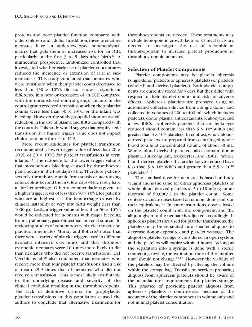
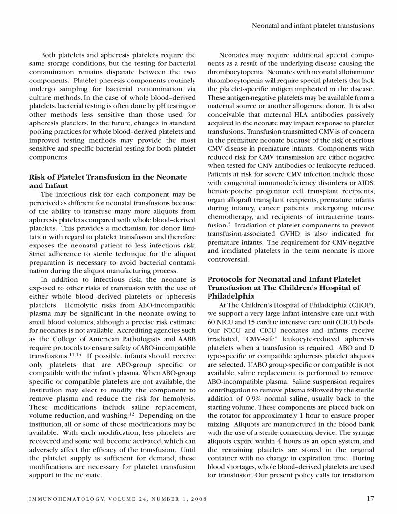
At CHOP, we have surveyed our institutional platelettriggers in the NICU, and our data are similar to thosepublished by Del Vecchio et al.,10 who noted a range ofplatelet transfusion triggers. Figure 1 displays the plateletcount that immediately preceded the transfusion in 909platelet transfusion events in our NICU. A trigger plateletcount value of less than 100 × 109/L would account forabout 90 percent of the platelet transfusions ad-ministered in the CHOP NICU. However, according topublished guidelines, a more stringent trigger value ofless than 50 × 109/L may have been considered in someof these patients. Figure 2 shows that the majority ofplatelet transfusions in neonates occurred in the firstweek of life. It appears that the majority of the platelettransfusions were given in accordance with publishedguidelines. As we begin to look at our institutional dataand outcomes, we may better understand the risks andoutcomes of our NICU trigger values,and our guidelinesmay change accordingly.
Neonates and infants on extracorporeal membraneoxygenation (ECMO) are at considerable risk forthrombocytopenia. Platelets are given at startup and atcircuit change to avoid the thrombocytopenia generallyassociated with the procedure as a result of plateletsbinding to the tubing or activated within the ECMOcircuit. Additional dilutional effects from the massivetransfusion of RBCs and plasma also factor into the riskfor thrombocytopenia. Approximately 10 to 35 percentof neonates receiving ECMO will have a hemorrhagicevent.16 Patient platelet counts need to be closelymonitored during ECMO. Adjustments are required toallow a higher transfusion of platelet dose based onweight. At CHOP, ECMO infants and neonates aretransfused platelets to a maximum volume of 40 mL/kg.
ConclusionsNeonatal and infant platelet transfusions are given
in a prophylactic or therapeutic setting to thrombo-cytopenic neonates and infants,either to reduce the riskof bleeding or for active bleeding. Because thrombo-cytopenia in a neonate can be caused by a variety offactors, it is important for the hospital to developindications, guidelines, and thresholds for platelettransfusions. Neonates and infants receiving trans-fusions are at additional risk for other adverse reactionsto blood products, including infection, immunologiceffects, transfusion-related acute lung injury, and fluidoverload, among others. Institutional protocols shoulddefine the need for irradiation and CMV-negativecomponents in infants, neonates, and prematureneonates. Standardization of practice, when possible,allows for better process control and patientmanagement.
Fig. 1. Frequency distribution of 909 pretransfusion platelet counts ininfants who received platelet transfusions while admitted to TheChildren’s Hospital of Philadelphia NICU between July 1,2006,andJune 30, 2007.
Fig. 2. Frequency distribution of patient ages at the time of platelettransfusion (N = 909) in infants who received platelet transfusionswhile admitted to The Children’s Hospital of Philadelphia NICUbetween July 1, 2006, and June 30, 2007.
I M M U N O H E M A T O L O G Y, VO L U M E 2 4 , N U M B E R 1 , 2 0 0 8 19
Neonatal and infant platelet transfusions
References1. Castle V,Andrew M, Kelton J, et al. Frequency andmechanism of neonatal thrombocytopenia. JPediatr 1986;108:749–55.
2. Andrew M, Vegh P, Caco C, et al. A randomized,controlled trial of platelet transfusions inthrombocytopenic premature infants. J Pediatr1993;123:285–91.
3. Strauss RG, Blanchette VS, Hume H, et al. Nationalacceptability of American Association of BloodBanks Pediatric Hemotherapy Committee guide-lines for auditing pediatric transfusion practices.Transfusion 1993;33:168–71.
4. Roseff SD, Luban NLC, Manno CS. Guidelines forassessing appropriateness of pediatric transfusion.Transfusion 2002:42:1398–413.
5. Roseff SC.Pediatric transfusion:A physician’s hand-book. 2nd ed.Bethesda,MD:AABB Press, 2006.
6. Strauss RG.Transfusion therapy for neonates.Am JDis Child 1991;145:904–11.
7. Murray NA, Howarth LJ, McCloy MP, et al. Platelettransfusion in the management of severe thrombo-cytopenia in neonatal intensive care unit patients.Transfus Med 2002;12:35–41.
8. Gibson BE, Todd A, Roberts I, et al. Transfusionguidelines for neonates and older children. Br JHaematol 2004;124:433–53.
9. Murray NA, Roberts IA. Neonatal transfusionpractice.Arch Dis Child Fetal Neonatal Ed 2004;89:F101–7.
10. DelVecchioA,Sola MC,Theriaque DW,et al.Platelettransfusion in the neonatal intensive care unit:factors predicting which patients will requiremultiple transfusions.Transfusion 2001:41;803–8.
11. The AABB Standards Program Committee, ed.AABB standards for blood banks and transfusionservices. 24th ed.Bethesda,MD:AABB, 2006.
12. Brecher ME, ed. Technical manual. 15th ed.Bethesda,MD:AABB, 2005.
13. Hillyer CD, Strauss RG, Luban NLC. Handbook ofpediatric transfusion medicine. San Diego, CA:Elsevier, 2004.
14. College of American Pathologists Commission onLaboratoryAccreditation LaboratoryAccreditationProgramTransfusion Medicine Checklist.Availableat: http://www.cap.org/apps/docs/laboratory_accreditation/checklists/transfusion_medicine_october2006.pdf. Last accessed March 17, 2008.
15. Haspel RL,Walsh L,Sloan SR.Platelet transfusion inan infant leading to formation of anti-D:implications for immunoprophylaxis.Transfusion2004;44:747–9.
16. Festa CJ, Feng AK, Bigos D.Transfusion support inpediatric surgery, trauma, and the intensive careunit. In: Herman JH, Manno CS, eds. Pediatrictransfusion therapy. Bethesda, MD: AABB Press,2002.
Deborah A. Sesok-Pizzini, MD, MBA (correspondingauthor), Medical Director, Blood Bank andTransfusion Medicine, The Children’s Hospital ofPhiladelphia, Assistant Professor, Department ofClinical Pathology and Laboratory Medicine, andDavid Friedman, MD, Associate Medical Director,Blood Bank and Transfusion Medicine, The Children’sHospital of Philadelphia, Assistant Professor,Department of Hematology, University ofPennsylvania School of Medicine, 34th Street and CivicCenter Boulevard, Philadelphia, PA 19104-4399.
20 I M M U N O H E M A T O L O G Y, VO L U M E 2 4 , N U M B E R 1 , 2 0 0 8
Neonatal plasma transfusionsP.T. PISCIOTTO
R E V I E W
Sick neonates are among the more heavilytransfused groups of patients in the hospital setting.1
Transfusion therapy for neonates began early in the 20thcentury. One of the first controlled trials compared theuse of intravenous versus intramuscular administrationof whole blood in infants with hemorrhagic disease ofthe newborn (vitamin K deficiency bleeding).2 The useof plasma components in neonates for hemostaticabnormalities coincided with the availability of theseblood components for all patients in the 1950s. Ingeneral, plasma transfusions are administered lessfrequently to neonates than RBC transfusions.3,4
Inasmuch as the physiologic state of the hemostaticsystem in newborns is different compared with that inolder children and adults, it is important to have a goodunderstanding of the developmentally unique aspect ofthis system to appreciate the therapeutic goals of plasmareplacement therapy.5 Although there are guidelines forneonatal transfusion as well as recommendations toassess transfusion criteria, most of these are derivedfrom consensus opinion because there are limitedevidence-based data from controlled clinical trials.6,7
Developmental HemostasisNormal hemostasis is the physiologic mechanism by
which the body controls bleeding and preventsthrombotic events. This results from a dynamic balancebetween the formation of thrombin, fibrin deposition,thrombin inhibition, and fibrinolysis. The process canbe viewed in three overlapping stages, starting with theformation of a platelet plug, which involves theendothelial layer of the vessel wall and platelets,followed by the interaction of platelets and plasmaprocoagulant proteins (coagulation factors) to form astable fibrin clot limited to the area of vascular injury(by action of anticoagulant proteins). The final stageresults in the breakdown of the fibrin clot and repair ofthe vascular damage (fibrinolytic system).
Plasma coagulation proteins do not cross theplacenta. Synthesis of these proteins begins in the firsttrimester of pregnancy. Although the basic coagulation,
anticoagulation, and fibrinolytic pathways are the samein neonates as in older children and adults, theconcentration of many of the coagulation proteinsvaries greatly. Concentrations are selectively andsignificantly different and dependent on both thegestational and postnatal age of the infant.5 Severalfactors, including fibrinogen; factorsVIII,V,and XIII; andvonWillebrand factor, achieve adult levels at birth, butmost other hemostatic proteins are decreased. Thealtered state is the result of multiple mechanismsincluding decreased production as a result of theimmature nature of the neonatal liver, increasedclearance of the proteins, consumption at the time ofbirth, and synthesis of proteins that have decreasedfunctional activity.5 In full-term infants these factors areapproximately 50 percent of adult levels and even lowerin healthy premature infants.8,9 As a result of thedecreased production of these factors, the generation ofthrombin,which is the key enzyme in the formation offibrin, may be decreased or delayed.10 Despite thesefindings, healthy infants are hemostatically stable. Thestandard laboratory tests of coagulation function,however—the prothrombin time and activated partialthromboplastin time—are greater than the adultreference ranges, reflecting the reduction of thesemultiple procoagulant proteins. Therefore, it is impor-tant to refer to established reference ranges for agewhen evaluating hemostatic integrity and potential needfor transfusion in the neonate. The newborn hemostaticsystem matures rapidly, with variables reaching adultlevels usually by 6 months. This process is acceleratedin premature infants, who show similar levels ofcoagulation proteins to term infants by 6 months afterbirth. As this is a dynamic maturing system multiplereference ranges,reflecting the gestational and postnatalage of the infant, are necessary. The ranges publishedbyAndrew et al.5 are the most comprehensive and oftenare used as guidelines.11 It is important to remember,however, that these ranges were established usingdifferent reagents and assay systems for measuringcoagulation function than are available today.
I M M U N O H E M A T O L O G Y, VO L U M E 2 4 , N U M B E R 1 , 2 0 0 8 21
Neonatal plasma transfusions
The plasma concentration of the anticoagulants,which include anti-thrombin III;heparin cofactor II;andthe vitamin K–dependent factors,protein C and proteinS,are also decreased. This appears to be compensated inpart by the enhanced thrombin inhibition via theincreased levels of α2-macroglobulin (α2-M). In terms ofthe fibrinolytic system in the newborn there is adecrease in the concentration of plasminogen andtherefore the generation of plasmin, which is the keyenzyme responsible for breakdown of fibrin.5 Despitethese developmental differences,the hemostatic systemin the healthy infant is largely physiologic, providingprotection from both bleeding and thrombosis becausethe balance between procoagulants,anticoagulants,andthe fibrinolytic system is maintained. The concern,however, is that there is very little reserve capacity andany additional pathologic insult, such as birth asphyxia,which is associated with disseminated intravascularcoagulation (DIC) in the neonate, may contribute tomorbidity in the sick and premature infant.
Bleeding in the Neonatal PeriodBleeding complications from coagulation abnormal-
ities in the newborn are usually the result of an acquireddefect in hemostasis,although congenital disorders maybe present,particularly in association with an iatrogenicchallenge.11 The clinical presentation of bleeding in theneonate may present at various sites, and the conse-quences are different compared with those of olderchildren and adults. Oozing from the umbilicus orperipheral blood sampling sites,bleeding into the scalpresulting in large cephalohematomas, or bleeding fromcircumcision are some of the typical ways bleeding maypresent in the neonate. Often,however, the first sign isan intracranial hemorrhage (ICH), and even a minorbleed can result in shock.
Disseminated Intravascular CoagulationThe incidence of DIC is high in the neonate,
particularly the premature infant. DIC is an acquiredcoagulation disorder in which both the coagulation andfibrinolytic systems are activated, resulting in theformation of microthrombi and a bleeding tendencyowing to the consumption of certain coagulationfactors. This occurs as a secondary event to anotherdisease entity. In the neonate this can include manyconditions such as sepsis, birth asphyxia, acidosis,respiratory distress syndrome (RDS), necrotizingenterocolitis, infection, and shock.12 Because theseinfants are already starting with lower levels of
procoagulant proteins, the increased consumption ofthese proteins, in conjunction with consumption ofplatelets,can result in a profound coagulopathy early inthe disease process. The clinical spectrum of DIC in theneonate can range from an asymptomatic low-gradecompensated DIC to fulminant DIC characterized bybleeding requiring replacement therapy. Although thereare no randomized studies looking at the efficacy ofreplacement therapy for bleeding associated with DIC,its use in this clinical situation is generally recom-mended in guideline documents.6,7,11 The latter maynecessitate not only replacement of procoagulantproteins and naturally occurring inhibitors with plasmatransfusion but also platelet transfusions. Cryoprecip-itate, which contains a higher concentration of factorVIII and fibrinogen per unit volume of FFP, may beparticularly useful in the presence of hypofibrino-genemia. There has been some evidence of beneficialeffects with the use of antithrombin and protein Cconcentrates in adults with DIC associated withmultiorgan failure.13 There has been limited clinicalexperience with their use in neonates, and thereforefurther clinical trials would be warranted for definitiverecommendation regarding their use as treatment. It isimportant to remember,however, that if the underlyingcondition is not corrected the hemostatic improvementwill only be temporary. Therefore one of the mostimportant aspects of managing DIC is taking aggressivemeasures to reverse the underlying disease process.
Vitamin K DeficiencyHemorrhagic disease of the newborn was described
in 1894,14 but was not linked to vitamin K deficiencyuntil the mid-1900s. The vitamin K–dependent factorsas mentioned previously are physiologically low at birthand functionally inactive in the presence of vitamin Kdeficiency. Vitamin K is required for modifying thecoagulation proteins II, VII, IX, and X and theanticoagulant proteins C and S. Placental transfer of thevitamin is poor, and neonates have limited stores ofvitamin K at birth. Because intestinal bacteria producevitamin K2,a contributing factor for decreased vitamin Kin neonates may be the presence of a sterile gut in thefirst few days of life. This can be further aggravated ininfants who are breast-fed,because vitamin K content isalso limited in breast milk. In most countries prophy-lactic vitamin K1 (phytonadione, which is present ingreen leafy vegetables) is given after birth to reduce therisk of hemorrhagic disease. There are actually threeclassifications of hemorrhagic disease of the newborn,
22 I M M U N O H E M A T O L O G Y, VO L U M E 2 4 , N U M B E R 1 , 2 0 0 8
P.T. PISCIOTTO
which are based on the clinical presentation and riskfactors.15 Early hemorrhagic disease is the rarest, withthe onset of bleeding occurring within the first 24hours. Bleeding is variable,but can be serious, includingICH. Early onset is usually associated with medications,such as anticonvulsants, taken during pregnancy thatcan cross the placenta and interfere with vitamin Kmetabolism.16 In an attempt to reduce early hemor-rhagic disease, strategies for the prophylactic use ofantenatal vitamin K have been suggested.17 Classichemorrhagic disease of the newborn presents between2 and 5 days of life with purpura, mucosal membranebleeding, bleeding from the umbilicus, or gastro-intestinal bleeding in an infant who otherwise appearswell. Presentation with ICH appears to be relatively lesscommon. The classic form of hemorrhagic disease isusually associated with infants who are being breast-fed,but may also occur if there are feeding problems. A lateform of hemorrhagic disease may also occur in thenewborn, in which the onset of bleeding occurs afterthe first week of life,peaking between 2 and 8 weeks oflife. Although breast-feeding and inadequate prophylaxiswith vitamin K are frequently documented, there are avariety of diseases that can compromise the supply ofvitamin K that must be considered. Unlike those withclassic hemorrhagic disease of the newborn, theseinfants often present with ICH,which can be associatedwith significant morbidity (neurologic) and mortality.The treatment of vitamin K deficiency will be dictatedby the clinical situation. The vitamin K–dependentproteins increase within a few hours after theadministration of a parenteral dose of vitamin K. In thepresence of significant bleeding that may be lifethreatening, plasma in addition to vitamin K can begiven to correct the hemostatic defect.6,7,11
Liver DiseaseAlthough some degree of hepatic impairment is not
uncommon in the neonatal period and may occur inconjunction with sepsis, hypoxia, and total parenteralnutrition, fulminant hepatic failure is an uncommonevent. Viral infections, metabolic disorders, storagedisorder, and shock are some recognized causes ofneonatal hepatic failure.18 The coagulopathy of liverfailure is the result of several processes, but reducedsynthesis of procoagulant proteins plays an importantrole in a setting in which there already are physio-logically reduced levels of many coagulation proteins.Other contributing factors are decreased clearance ofactivated coagulation factors by the liver, activation of
fibrinolysis, impaired utilization of vitamin K, andthrombocytopenia. In the presence of clinical bleedingsome temporary benefit may be achieved by thereplacement of coagulation proteins with plasma andcryoprecipitate. Management of the coagulopathy,however,would essentially be supportive until recoveryof hepatic function could be achieved. If there isevidence of cholestasis, the administration of vitamin Ksupplements may be helpful.
Coagulopathy Associated WithCardiopulmonary Bypass andExtracorporeal Membrane Oxygenation
A substantial coagulopathy occurs during cardio-pulmonary bypass (CPB), which is the result of severaldifferent contributing factors. Neonates are at higher riskfor increased bleeding, related in part to the immaturenature of their coagulation system. In addition the oftencomplex repairs that are needed can require longer CPBtime or deeper hypothermia.19–22 Hypothermia (a meansof cerebral protection), which is often induced duringCPB,can alter hemostasis by negatively affecting plateletfunction as well as inhibiting serine protease activity.During CPB a hemodilution occurs, which is morepronounced in the neonate than in larger pediatric andadult patients. This is the result of the small intravascularvolume of the neonate relative to the volume of thebypass circuit. After the onset of CPB mean plasmaconcentrations of procoagulant proteins and inhibitorsare reduced by approximately 50 percent of pre-CPBvalues.19 There is a similar reduction in the fibrinolyticproteins. All of these changes have a significant effect onthe generation of thrombin. In addition to the hemo-dilution, and despite systemic heparinization, there isevidence that both the coagulation and fibrinolyticsystems are activated by the CPB circuit, which mayresult in a low-grade consumptive coagulopathy. Adilutional thrombocytopenia also occurs, and plateletfunction is impaired. Various strategies have been usedto decrease the morbidity of CPB and transfusionrequirements. Some of these have included minimizingthe amount of exposure to the CPB circuit by keepingthe duration of CPB as short as possible and minia-turizing the circuit to limit the artificial surface area towhich the blood is exposed.21,22 This miniaturizationalso results in smaller priming volumes and lesshemodilution. The use of ultrafiltration during or imme-diately after CPB to remove excess fluid has helped tomaintain the patient’s hemoglobin through hemocon-centration, attenuate the dilutional coagulopathy, and
I M M U N O H E M A T O L O G Y, VO L U M E 2 4 , N U M B E R 1 , 2 0 0 8 23
Neonatal plasma transfusions
reduce postoperative bleeding.22,23 The use of aprotinin,which is a serine protease inhibitor used to preventfibrinolysis as well as contact activation, has not shownconsistent results in the pediatric setting and warrantsfurther investigation as to its risk-benefit ratio.21 A recentprospective randomized controlled trial in 200 infantsless than 1 year of age compared the use of fresh wholeblood (less than 48 hours) with reconstituted bloodconsisting of one unit of RBCs in additive solution andone unit of thawed FFP for priming the bypass circuitand for adding to the bypass circuit when the patientswere rewarmed at the end of surgery.24 Fresh wholeblood did not confer a clinical or biochemical advantageover reconstituted blood. However, an increased lengthof stay in the intensive care unit and increased peri-operative fluid overload were associated with primingthe circuit with fresh whole blood.
Extracorporeal membrane oxygenation (ECMO) is amodified heart-lung machine, which provides gasexchange to support patients who have severe butpotentially reversible respiratory or cardiac failure.Some of the differences from patients requiring CPB arethat these patients are maintained at normal bodytemperature, undergo extrathoracic cannulation fromrelatively accessible vessels, and because there is nolarge venous reservoir in the circuit, require only partialanticoagulation with heparin.25 The time course forECMO support,however,can be days to weeks. As withCPB, coagulation complications of ECMO can besignificant as the patient is heparinized, and theassociated activation and consumption of procoagulantfactors and platelets related to the exposure of blood toartificial surfaces is also present. If ECMO is initiatedafter open heart surgery, the coagulation effects areadditive. The number of transfusions required variesconsiderably based on individual patient characteristicsas well as variation related to the institutional protocoland ECMO technique. There have been no clinical trialslooking at the influence of transfusion protocols onclinical outcome. Approximately 15 percent of neonateson ECMO sustain an ICH.25
Intracranial HemorrhageThe commonest form of ICH in the premature infant
is a periventricular-intraventricular hemorrhage (IVH).The incidence is around 15 to 20 percent for infants lessthan 32 weeks’ gestation.26 The majority occur withinthe first 72 hours of life. Although low birth weight andincreasing prematurity are associated with IVH,additional risk factors are RDS, intrapartum asphyxia,
and vaginal delivery. The etiology is incompletelyunderstood. Some of the factors believed to contributeto IVH include alterations in cerebral blood flow,fragilityof the immature germinal matrix vessels, and hypoxia-induced endothelial damage. These appear to be moresignificant than impaired hemostasis. A randomizedcontrol trial of prophylactic FFP carried out in the 1980sdid show a reduced risk of IVH documented by cranialultrasound.27 However,there was no substantial changein the coagulation profile after the administration of FFP,which suggested that the benefit seen might have beensecondary to circulatory stabilization after volumeexpansion. A subsequent large prospective randomizedcontrol study undertaken by the Northern NeonatalNursing Initiative Trial Group, comparing the infusionof FFP, gelatin, or glucose in 776 preterm infants lessthan 32 weeks’gestation within 2 hours of birth,showedno evidence that any of the interventions had any effecton death or disability, followed out 2 years.28,29 Currentevidence therefore does not support the routine use ofprophylactic FFP in preterm infants at risk for IVH.7
Indications and ContraindicationsFFP has been transfused to neonates for a variety of
reasons that may not have had any proven benefit.National surveys of transfusion practice have shown thatalthough neonates receive relatively few FFP transfusionsas compared with RBC transfusions, a percentage ofhospitals responded that some of the frequent uses ofFFP were either to treat hypovolemia, as fluid replace-ment during partial exchange transfusion,or as adjuvanttherapy for sepsis.3,4 Hypovolemia as the most frequentindication for transfusion of plasma was reported by 11percent of respondents in one survey and 41 percent inanother. These surveys reflect practice in the late 1980sand early 1990s. There have been no recent surveys todetermine whether there has been a shift in transfusionpractice. A more recent report of transfusion practice ina single institution showed that a significant shifttoward appropriate use of plasma occurred only afterimplementation of a computer-based audit system thatincluded an educational component as follow-up.30
Before the initiation of the audit system approximately 8percent of the plasma transfusions administered toneonates would have been classified as not beingappropriate because there was no evidence of acoagulopathy. This was reduced to approximately 1percent after an educational effort.
The underlying principle that should govern theappropriateness of plasma transfusion in the neonate is
24 I M M U N O H E M A T O L O G Y, VO L U M E 2 4 , N U M B E R 1 , 2 0 0 8
the same as that in older patients: plasma transfusionshould be given for the treatment of clinically significantbleeding, or to prevent bleeding before an invasiveprocedure, when there is a decrease in one or morecoagulation factors, and a safer more appropriatealternative to treatment does not exist.6,7 Some of thedifficulty in the neonate is trying to determine what rolean immature hemostatic system plays in the etiology ofa bleed. This is the case with IVH in which randomizedclinical trials have been helpful.
Plasma ComponentsThere are several plasma components available that
are used for coagulation factor replacement. Plasmafrom whole blood collected in either CPDA-1, CPD, orCP2D that is separated by centrifugation and frozenwithin 8 hours is labeled as FFP and if frozen within 24hours as “Plasma Frozen Within 24 Hours After Phle-botomy” (FP24). Frozen plasma is thawed usually in awater bath and should be either transfused immediatelyor stored at 1 to 6°C for up to 24 hours. If these frozenproducts are thawed and not used within 24 hours theyare relabeled as “Thawed Plasma” and need to betransfused within 5 days. The processing of FP24 mayresult in slightly lower concentration of factors V andVIII. However, based on in vitro data of the functionalactivity of these factors, they still are within the normalreference ranges, supporting the use of these productsfor the accepted indications for plasma transfusion.31–33
The concentrations of coagulation factors in thawedplasma from FFP which is stored at 1 to 6°C untilexpiration (day 5) are stable with the exception offactor VIII activity, which is reduced by 35 to 41percent.34 FFP can also be collected by apheresistechnology. In this case the anticoagulant is eitherACDor sodium citrate,and the plasma is frozen within 6 to 8hours.
Dose and AdministrationNo specific compatibility testing is required before
transfusion of frozen plasma components. However,because plasma contains isohemagglutinins, the com-ponent must be ABO compatible with the recipient’sRBCs. Hemolytic reactions attributable to incompatibleplasma transfusions have been reported in theneonate.35 If the recipient’s ABO type is not available,then group AB plasma can be administered. BecauseRh-alloimmunization rarely occurs as a result of Rh(D)mismatch of frozen plasma components,Rh(D) compat-ibility is not essential. After the frozen plasma is thawed,
aliquots can be prepared using a sterile connectingdevice and transfer bags if multiple doses are needed.
A guide for replacement therapy is to achieve a“minimal hemostatic level” based on gestational andpostnatal age of the infant.36 There have been no studieslooking at the recovery of coagulation factors or clear-ance time after FFP administration in a sick neonate.A common dosing regimen has been 10 to 20 mL/kg.This theoretically should result in a rise in coagulationfactors of approximately 20 percent immediately afterinfusion.The dose and frequency,however,will dependon the clinical situation and plasma concentration thatis being targeted. Posttransfusion monitoring of thecoagulation status is important to determine optimaltherapy.
SummaryAlthough there has been an overall trend toward
reduction of transfusions in neonates, limited data fromrandomized clinical trials to guide practice exist. Theneonatal hemostatic system is immature at birth,placingsick premature infants, who may have minimal reservecapacity, at potential increased risk for bleedingcomplications. In general, there is agreement thatplasma should be transfused to correct deficiencies inthe face of bleeding. What is needed,however, is a betterunderstanding of when these deficiencies do or do notaffect bleeding risk and outcome to further providemore evidence-based approaches to transfusionpractice.
References1. Stauss RG.Transfusion therapy in neonates.Am JDis Child 1991;145:904–11.
2. Sanford HN, Leslie EI. Blood coagulation factors inhemorrhagic disease of the newborn and the valueof intramuscular injections of father’s and mother’sblood. J Pediatr 1938;12:16–20.
3. Strauss RG, Levy GJ, Sotelo-Avila C, et al. Nationalsurvey of neonatal transfusion practices: II. Bloodcomponent therapy. Pediatrics 1993;91:530–6.
4. Hume H,BlanchetteV,Strauss RG,Levy GJ.A surveyof Canadian neonatal blood transfusion practices.Transfus Sci 1997;18:71–80.
5. Andrew M,Paes B,Johnston M.Development of thehemostatic system in the neonate and younginfant. Am J Pediatric Hematol Oncol 1990;12:95–104.
P.T. PISCIOTTO
I M M U N O H E M A T O L O G Y, VO L U M E 2 4 , N U M B E R 1 , 2 0 0 8 25
Neonatal plasma transfusions
6. Roseff SD, Luban NLC, Manno CS. Guidelines forassessing appropriateness of pediatric transfusion.Transfusion 2002;42:1398–413.
7. British Committee for Standards in Haematology.Transfusion guidelines for neonates and olderchildren.Br J Haematol 2004;124:433–53.
8. Andrew M, Paes B,Milner R, et al. Development ofthe human coagulation system in the full-terminfant. Blood 1987;70:165–7.
9. Andrew M, Paes B,Milner R, et al. Development ofthe human coagulation system in the healthypremature infant. Blood 1988;72:1651–7.
10. Andrew M, Schmidt B, Mitchell L, et al.Thrombingeneration in newborn plasma is criticallydependent on the concentration of prothrombin.Thrombin Haemost 1990;63:27–30.
11. Williams MD,Chalmers EA,Gibson BES.Guideline:the investigation and management of neonatalhemostasis and thrombosis. Br J Haematol 2002;119:295–309.
12. Shirahata A, ShirakawaY,Murakami C.Diagnosis ofDIC in very low birth weight infants. SeminThromb Hemost 1998;24:467–71.
13. Lee WL, Downey GP. Coagulation inhibitors insepsis and disseminated intravascular coagulation.Intensive Care Med 2000;26:1701–6.
14. Townsend CW. The hemorrhagic disease of thenewborn.Arch Pediatr 1894;11:559–65.
15. Greer FR.Vitamin K deficiency and hemorrhage ininfancy.Clin Perinatol 1995;22:759–77.
16. Astedt B.Antenatal drugs affecting vitamin K statusof the fetus and newborn. Semin Thromb Hemo-stasis 1995;21:364–70.
17. Cornelissen M, Steegers-Theunissen R, Kollee L, etal. Supplementation of vitamin K in pregnantwomen receiving anti-convulsant therapy preventsneonatal vitamin K deficiency.Am J Obstet Gynecol1993;168:884–8.
18. Shneider BL. Neonatal liver failure Curr OpinPediatr 1996;8:495–501.
19. Kern FH, Morana NJ, Sears JJ, Hickey PR.Coagulation defects in neonates during cardio-pulmonary bypass. Ann Thorac Surg 1992;54:541–6.
20. Chambers L,Cohen D,Davis J.Transfusion patternsin pediatric open heart surgery. Transfusion1996;36:150–4.
21. Barcelona SL,ThompsonAA,Cote CJ.Intraoperativepediatric blood transfusion therapy: a review of
common issues.Part II: transfusion therapy,specialconsiderations, and reduction of allogenic bloodtransfusions. Pediatr Anesth 2005;15:814–30.
22. Shen I, Giacomuzzi C, Ungerleider RM. Currentstrategies for optimizing the use of cardio-pulmonary bypass in neonates and infants. AnnThorac Surg 2003;75(Suppl):S729–34.
23. Friesen RH, Campbell DN, Clarke DR,TornabeneMA. Modified ultrafiltration attenuates dilutionalcoagulopathy in pediatric open heart operations.AnnThorac Surg 1997;64:1787–9.
24. Mou SS, Giroir BP, Molitor-Kirsch EA, et al. Freshwhole blood versus reconstituted blood for pumppriming in heart surgery in infants. N Engl J Med2004;351:1635–44.
25. Wolfson PJ. The development and use ofextracorporeal membrane oxygenation in neo-nates.AnnThorac Surg 2003;76(Suppl):S2224–9.
26. OhW,Fanaroff AA,Veter J, et al.Neonatal mortalityand morbidity in very low birth weight infants: aseven year trend analysis of the Neonatal NetworkData. Pediatr Res 1996;39:235.
27. Beverly DW, Chance GW, Inwood MJ, Schaus M,O’Keefe B. Intraventricular haemorrhage andhaemostsis defects.Arch Dis Child 1984;59:444–8.
28. NNNI Trial Group.A randomized trial comparingthe effect of prophylactic intravenous fresh frozenplasma, gelatin or glucose on early mortality andmorbidity in preterm infants. Eur J Pediatr1996;155:580–8.
29. NNNITrail Group.Randomised trial of prophylacticearly fresh-frozen plasma or gelatin in pretermbabies: outcome at 2 years. Lancet 1996;348:229–32.
30. Petaja J,Anderson S, Syrjala M.A simple automatedaudit system for following and managing practicesof platelet and plasma transfusions in a neonatalintensive care unit.Transfus Med 2004;14:281–8.
31. O’Neill EM,Royley J,Hansson-Wicher M,et al.Effectof 24-hour whole blood storage on plasma clottingfactors.Transfusion 1999;39:488–91.
32. Smith JF,Ness PM,Moroff G,Luban NLC.Retentionof coagulation factors in plasma frozen after ex-tended holding at 1–6°C.Vox Sang 2000;78:28–30.
33. Cardigan R, Lawrie AS, Mackie IJ,Williamson LM.The quality of fresh-frozen plasma produced fromwhole blood stored at 4°C overnight.Transfusion2005;45:1342–8.
26 I M M U N O H E M A T O L O G Y, VO L U M E 2 4 , N U M B E R 1 , 2 0 0 8
P.T. PISCIOTTO
34. Downes KA, Wilson E, Yomtovian R. Serialmeasurement of clotting factors in thawed plasmastored for 5 days.Transfusion 2001;41:570.
35. Duguid JK, Minards J, Bolton-Maggs PHB.Incompatible plasma transfusions and haemolysisin children.BMJ 1999;318:176–7.
36. Andrew M,Brooker LA.Blood component therapyin neonatal hemostatic disorders.Transfus Med Rev1995;9:231–50.
Patricia T. Pisciotto, MD, Chief Medical Officer,American Red Cross Biomedical Services, NortheastDivision, 209 Farmington Avenue, Farmington, CT06032; and Professor, Department Pathology andLaboratory Medicine, University of Connecticut Schoolof Medicine, 263 FarmingtonAvenue, Farmington, CT06030.
Notice to Readers: All articles published,including
communications and book reviews, reflect the
opinions of the authors and do not necessarily
reflect the official policy of theAmerican Red Cross.

I M M U N O H E M A T O L O G Y, VO L U M E 2 4 , N U M B E R 1 , 2 0 0 8 27
C O M M U N I C A T I O N S
Letter to the Editors
Prevalence of Yt(a–) in Hispanic blood donors
The Yt blood group system consists of a high-prevalence antigen (Yta) and a low-prevalence antigen(Ytb), which give rise to three phenotypes:Yt(a+b–),Yt(a+b+),andYt(a–b+).1 TheYt(a–) phenotype is moreprevalent in Israeli Jews (1 in 69),Arabs (1 in 45), andDruse (1 in 40)2 than it is in Northern Europeans,Canadians, and African Americans (1 in 500), or inJapanese (none in 5000).3 This phenotype has not beenfound in testing cohorts of Japanese, Inuits,Thais, andNative Americans.3 The Yta antigen is quiteimmunogenic, and anti-Yta is not infrequently made byYt(a–) persons exposed toYt(a+) RBCs. Sometimes,butnot always, anti-Yta causes reduced in vivo survival oftransfusedYt(a+) RBCs. For this reason,physicians oftenrequestYt(a–) blood components for transfusion to theirpatients with anti-Yta.3 In spite of our ongoing testing ofAfricanAmerican and Caucasian donors with anti-Yta, inour region there is still a relative shortage ofYt(a–) blooddonors. Thus, we extended our testing to includeHispanic donors, and the results are presented here.
RBCs from blood donors who self-identified asbeing African American, Caucasian, or Hispanic werescreened with anti-Yta in IgG gel cards (Micro TypingSystems, Inc., Ortho-Clinical Diagnostics, Raritan, NJ).Nonreactive RBCs were retested by the sametechnologist using the same anti-Yta but by IAT in testtubes. Repeat nonreactive samples were then tested bya second technologist using a different source of anti-Yta by the IAT in test tubes. The results of our testing areshown inTable 1.
The prevalence of Yt(a–) in Hispanic donors wassimilar to that in Caucasian donors. The prevalence inCaucasians was similar to published figures. Hispanicsare, therefore,a valuable ethnicity to screen for this rarephenotype. We identified nearly three times as manyYt(a–) donors in the Hispanic and Caucasian donors as
in African American donors. The number of Yt(a–)AfricanAmerican donors we found was about a quarterof that expected from reports in the literature.
Edith TossasLaboratory of Immunohematology
NewYork Blood Center45-01Vernon Boulevard
Long Island City, NY 11101
Michael Baxter, BSLaboratory of Immunohematology
NewYork Blood Center45-01Vernon Boulevard
Long Island City, NY 11101
Marion E.Reid, PhDLaboratory of Immunohematology
NewYork Blood Center310 East 67th StreetNewYork, NY 10065
Daisy Charles-Pierre, BSLaboratory of Immunohematology
NewYork Blood Center45-01Vernon Boulevard
Long Island City, NY 11101
Christine Lomas-Francis, MSc, FIMBSLaboratory of Immunohematology
NewYork Blood Center45-01Vernon Boulevard
Long Island City, NY 11101
References1. Reid ME, Lomas-Francis C. Blood group antigenfactsbook,2nd ed.San Diego:Academic Press,2004.
2. Levene C,Bar-Shany S,Manny N,et al.TheYt bloodgroups in Israeli Jews,Arabs,and Druse.Transfusion1987;27:471–4.
3. Issitt PD,Anstee DJ.Applied blood group serology,4th ed. Durham, NC: Montgomery ScientificPublications, 1998.
Table 1. Results of screening donors with anti-Yta
Donors Number tested Number Yt(a–) Prevalence
Hispanic 9933 13 1 in 764 (0.13%)
Caucasian 2077 3 1 in 692 (0.14%)
AfricanAmerican 10,622 5 1 in 2124 (0.05%)
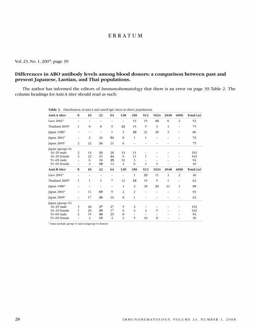
28 I M M U N O H E M A T O L O G Y, VO L U M E 2 4 , N U M B E R 1 , 2 0 0 8
E R R A T U M
Vol. 23,No. 1, 2007;page 39
Differences in ABO antibody levels among blood donors: a comparison between past andpresent Japanese, Laotian, and Thai populations.
The author has informed the editors of Immunohematology that there is an error on page 39,Table 2. Thecolumn headings for Anti-A titer should read as such:
Table 2. Distribution of anti-A and anti-B IgG titers in three populations
Anti-A titer 8 16 32 64 128 256 512 1024 2048 4096 Total (n)
Laos 2001* – – – – – 13 15 16 6 2 52
Thailand 2005* 1 8 8 9 23 15 5 3 3 – 75
Japan 1986* – – – 1 3 39 21 20 2 – 86
Japan 2001* – 2 16 51 8 1 1 – – – 79
Japan 2005* 2 22 24 21 6 – – – – – 75
Japan (group O)16–29 male 2 14 33 28 14 11 – – – – 10216–29 female 2 22 23 34 9 11 1 – – – 10251–69 male – 6 18 35 32 3 – – – – 9451–69 female – 2 18 14 4 6 2 3 – – 49
Anti-B titer 8 16 32 64 128 256 512 1024 2048 4096 Total (n)
Laos 2001* – – – – – 1 23 11 3 2 40
Thailand 2005* 1 1 1 7 12 19 15 5 1 – 62
Japan 1986* – – – – 1 2 28 32 22 3 88
Japan 2001* – 11 69 9 2 2 – – – – 93
Japan 2005* – 17 20 16 8 1 – – – – 62
Japan (group O)16–29 male 3 28 37 27 5 2 – – – – 10216–29 female 1 26 39 17 6 4 4 5 – – 10251–69 male 2 15 40 29 8 – – – – – 9451–69 female – 4 15 4 3 5 10 8 – – 49
* Data include group O and nongroup O donors
I M M U N O H E M A T O L O G Y, VO L U M E 2 4 , N U M B E R 1 , 2 0 0 8 29
IMMUNOHEMATOLOGY IS ON THE WEB!
www.redcross.org/pubs/immuno
For more information or to send an e-mail
message“To the editor”
Notice to Readers: Immunohematology, Journalof Blood Group Serology and Education, isprinted on acid-free paper.
A N N O U N C E M E N T S
Monoclonal antibodies available at no charge:The NewYork Blood Center has developed a wide range of monoclonal antibodies (both murine and humanized)that are useful for donor screening and for typing RBCs with a positive DAT. These include anti-A1, -M, -s, -U, -D,-Rh17, -K, -k, -Kpa, -Jsb, -Fya, -Fy3, -Fy6, -Wrb, -Xga, -CD99, -Dob, -H, -Ge2, -Ge3, -CD55 (both SCR2/3 and SCR4), -Oka, -I,and anti-CD59. Most of the antibodies are murine IgG and require the use of anti-mouse IgG for detection (Anti-K,-k, and -Kpa). Some are directly agglutinating (Anti-A1, -M, -Wrb and -Rh17) and a few have been humanized into theIgM isoform (Anti-Jsb). The antibodies are available at no charge to anyone who requests them. Please visit ourWebsite for a complete list of available monoclonal antibodies and the procedure for obtaining them.
For additional information,contact:Gregory Halverson,NewYork Blood Center,310 East 67th Street,NewYork,NY10021 / e-mail: [email protected] (phone 212-570-3026, FAX: 212-737-4935) or visit the Web site athttp://www.nybloodcenter.org >research >immunochemistry >current list of monoclonal antibodies available.
Specialist in Blood Bank (SBB) ProgramThe Department of Transfusion Medicine, National Institutes of Health, is accepting applications for its 1-yearSpecialist in Blood Bank Technology Program. Students are federal employees who work 32 hours/week. Thisprogram introduces students to all areas of transfusion medicine including reference serology,cell processing,HLA,and infectious disease testing. Students also design and conduct a research project. NIH is an Equal OpportunityOrganization. Application deadline is December 31, 2008 for the July 2009 class. See www.cc.nih.gov/dtm >education for brochure and application. For further information contact Karen M. Byrne at (301) 451-8645 [email protected]
Meetings!
April 11–13 American Red Cross Immunohematology Reference Laboratory (IRL)Conference 2008
The American Red Cross Immunohematology Reference Laboratory (IRL) Conference 2008 will be held April 11through 13, 2008, at the Chaparral Suites Resort, in Scottsdale,Arizona. For more information, contact CindyFlickinger at (215) 451-4909 or [email protected].
May 8–9 Heart of America Association of Blood Banks (HAABB)The spring meeting of the Heart of AmericaAssociation of Blood Banks (HAABB) will be held May 8 and 9,2008, atthe Embassy Suites on the Country Club Plaza in Kansas City,Missouri. For more information, refer to theWeb siteat http://www.haabb.org.
30 I M M U N O H E M A T O L O G Y, VO L U M E 2 4 , N U M B E R 1 , 2 0 0 8
A N N O U N C E M E N T S C O N T ’ D
MSc in Transfusion and Transplantation Sciences
Are you working in NHS or National Blood Service and looking for training?This course could be for you.
Applications are invited from medical or science graduates to study for the MSc in Transfusion and Trans-plantation Sciences. The course is run jointly by The Department of Cellular & Molecular Medicine, Uni-versity of Bristol and the Bristol Institute of Transfusion Sciences.
The course starts in October 2008 and can be studied full-time for 1 year or part-time over 2 or 3 yearsby block release.
The course aims to develop your interest, knowledge and understanding of the theory, practical tech-niques and research relating to the science of transplantation and transfusion medicine.
For example,
• How is blood processed?
• When do we give platelet transfusions?
• How is tissue engineering being used to replace heart valves?
• What causes haemolytic anaemia?
• How do we reduce the transfusion related risk of HIV and vCJD?
Teaching combines informal lectures, tutorials, practical laboratory experience and a research project withthe bias on transfusion.
The lecture units are: Haemopoiesis, Immunology, Platelets and coagulation, Blood groups, Haematologi-cal diseases, Blood donation, Blood components, Clinical transfusion, Transfusion transmitted infections,Stem cell transplantation, Solid organ transplantation and Tissue engineering.
The course is accredited by The Institute of Biomedical Sciences and directed byProfessor David Anstee and Dr Tricia Denning-Kendall.
For further details visit:
http://www.blood.co.uk/ibgrl/MSc/MScHome.htmor contact:Dr Tricia Denning-Kendall,University of Bristol, Geoffrey Tovey Suite,National Blood Service, Southmead Rd Bristol, BS10 5ND, England.TEL 0117 9912093, E-MAIL [email protected]
I M M U N O H E M A T O L O G Y, VO L U M E 2 4 , N U M B E R 1 , 2 0 0 8 31
A N N O U N C E M E N T S C O N T ’ D
DEPARTMENT OF CLINICAL LABORATORY SCIENCES
SCHOOL OF ALLIED HEALTH PROFESSIONS
VIRGINIA COMMONWEALTH UNIVERSITY
Faculty Position
The Department of Clinical Laboratory Sciences at Virginia Commonwealth University invites
applications for a full-time, 12 month, tenure-track faculty position. The Department, located
on the MCV Campus of VCU, is one of nine departments in the School of Allied Health
Professions. VCU is a large urban, research-extensive institution with a richly diverse university
community and commitment to multicultural opportunities. The Department offers both B.S.
and M.S. degree programs in Clinical Laboratory Sciences and provides the CLS specialty track
in the Ph.D. program in Health Related Sciences.
The successful candidate will be responsible for teaching clinical immunology and
immunohematology/blood banking courses on campus and on-line at the undergraduate and
graduate levels, interacting with clinical faculty at affiliated clinical sites, and student mentoring.
Also expected are scholarly activities and research, university service responsibilities, and
professional activities.
Applicants must have a Master’s degree (Ph.D. preferred), national certification as a generalist
in the clinical laboratory, clinical or college teaching experience, excellent interpersonal and
written and oral communication skills, and demonstrated scholarly productivity. Preference will
be given to applicants with specialist certification in blood banking and a record of active
participation in professional societies.
Salary and rank will be commensurate with education and experience.
Review of applications will begin immediately and continue until the position is filled. Send a
letter of interest, curriculum vitae, and the names of three references to:
William Korzun, Ph.D.
Department of Clinical Laboratory Sciences
Virginia Commonwealth University
PO Box 980583, Richmond, VA 23298-0583.
“Virginia Commonwealth University is an equal opportunity/affirmative action employer. Women,minorities and persons with disabilities are encouraged to apply.”
32 I M M U N O H E M A T O L O G Y, VO L U M E 2 4 , N U M B E R 1 , 2 0 0 8
A D V E R T I S E M E N T S
Blood Group Antigens & AntibodiesA guide to clinical relevance & technical tips
BYMARION E. REID AND CHRISTINE LOMAS-FRANCIS
The authors are using royalties generated from the sale of this pocketbook for educationalpurposes to mentor people in the joys of immunohematology as a career. They will accomplishthis in the following ways:
• Sponsor workshops, seminars, and lectures• Sponsor students to attend a meeting• Provide copies of the pocketbook
(See www.sbbpocketbook.com)
The book, which costs $25, can be ordered in two ways:
1. Order online from the publisher at: www.sbbpocketbook.com
2. Order from the authors, who will sign the book. Send a check, made payable to“New York Blood Center” and indicate “Pocketbook” on the memo line, to:
Marion Reid, Laboratory of ImmunochemistryNew York Blood Center310 East 67th StreetNew York, NY 10021Please include the recipient’s complete mailing address.
About the book
This compact “pocketbook” from the authors of the Blood Group Antigen FactsBook is a mustfor anyone who is involved in the laboratory or bedside care of patients with blood groupalloantibodies.
The book contains clinical and technical information about the nearly 300 ISBT recognizedblood group antigens and their corresponding antibodies. The information is listed inalphabetical order for ease of finding—even in the middle of the night. Included in the book isinformation relating to:
• Clinical significance of antibodies in transfusions and HDN.• Number of compatible donors that would be expected to be found in testing 100
donors. Variations in different ethnic groups are given.• Characteristics of the antibodies and optimal technique(s) for their detection.• Technical tips to aid their identification.• Whether the antibody has been found as an autoantibody.
I M M U N O H E M A T O L O G Y, VO L U M E 2 4 , N U M B E R 1 , 2 0 0 8 33
A D V E R T I S E M E N T S C O N T ’ D
NATIONAL REFERENCE LABORATORY FORBLOOD GROUP SEROLOGY
Immunohematology ReferenceLaboratory
AABB,ARC,NewYork State, and CLIA licensed(215) 451-4901—24-hr. phone number
(215) 451-2538—Fax
American Rare Donor Program(215) 451-4900—24-hr. phone number
(215) 451-2538—[email protected]
Immunohematology(215) 451-4902—Phone, business hours
(215) 451-2538—[email protected]
Quality Control of Cryoprecipitated-AHF(215) 451-4903—Phone, business hours
(215) 451-2538—Fax
National Platelet Serology Reference
Laboratory
Diagnostic testing for:
• Neonatal alloimmune thrombocytopenia (NAIT)
• Posttransfusion purpura (PTP)
• Refractoriness to platelet transfusion
• Heparin-induced thrombocytopenia (HIT)
• Alloimmune idiopathic thrombocytopenia
purpura (AITP)
Medical consultation available
Test methods:
• GTI systems tests
—detection of glycoprotein-specific platelet
antibodies
—detection of heparin-induced antibodies (PF4
ELISA)
• Platelet suspension immunofluorescence test
(PSIFT)
• Solid phase red cell adherence (SPRCA) assay
• Monoclonal antibody immobilization of platelet
antigens (MAIPA)
• Molecular analysis for HPA-1a/1b
For information, e-mail: [email protected]
or call:
Maryann Keashen-Schnell
(215) 451-4041 office
(215) 451-4205 laboratory
Sandra Nance
(215) 451-4362
American Red Cross Blood Services
Musser Blood Center
700 Spring Garden Street
Philadelphia, PA 19123-3594
CLIA LICENSED
Reference and Consultation Services
Antibody identification and problem resolution
HLA-A,B,C, and DR typing
HLA-disease association typing
Paternity testing/DNA
For information regarding our services, contact
Mehdizadeh Kashi at (503) 280-0210, or write to:
Pacific Northwest Regional Blood Services
ATTENTION: Tissue Typing Laboratory
American Red Cross
3131 North Vancouver
Portland, OR 97227
CLIA LICENSED,ASHI ACCREDITED
34 I M M U N O H E M A T O L O G Y, VO L U M E 2 4 , N U M B E R 1 , 2 0 0 8
A D V E R T I S E M E N T S C O N T ’ D
IgA/Anti-IgA Testing
IgA and anti-IgA testing is available to do thefollowing:• Identify patients with anti-IgA• Investigate anaphylactic reactions• Confirm IgA-deficient donors
Our ELISA assay for IgA detects antigen to0.05 mg/dL.
For information on charges and samplerequirements, call (215) 451-4909, e-mail:
[email protected],or write to:
American Red Cross Blood ServicesMusser Blood Center
700 Spring Garden StreetPhiladelphia, PA 19123-3594
ATTN: Cindy Flickinger
CLIA LICENSED
National Neutrophil Serology ReferenceLaboratory
Our laboratory specializes in granulocyte antibodydetection and granulocyte antigen typing.
Indications for granulocyte serology testinginclude:• Alloimmune neonatal neutropenia (ANN)• Autoimmune neutropenia (AIN)• Transfusion related acute lung injury (TRALI)
Methodologies employed:• Granulocyte agglutination (GA)• Granulocyte immunofluorescence by flowcytometry (GIF)
• Monoclonal antibody immobilization ofneutrophil antigens (MAINA)
TRALI investigations also include:• HLA (PRA) Class I and Class II antibody detection
For further information contact:
Neutrophil Serology Laboratory(651) 291-6797
Randy Schuller(651) 291-6758
American Red Cross Blood ServicesNeutrophil Serology Laboratory
100 South Robert StreetSt. Paul, MN 55107
CLIA LICENSED
Donor IgA Screening
• Effective tool for screening large volumes ofdonors
• Gel diffusion test that has a 15-year proventrack record:– Approximately 90 percent of all donorsidentified as IgA deficient by are confirmedby the more sensitive testing methods
For information regarding charging and samplerequirements, call Kathy Kaherl at:
(860) 678-2764, e-mail: [email protected] write to:
Reference LaboratoryAmerican Red CrossConnecticut Region209 Farmington Ave.Farmington, CT 06032
CLIA LICENSED
I M M U N O H E M A T O L O G Y, VO L U M E 2 4 , N U M B E R 1 , 2 0 0 8 35
ImmunohematologyJOURNAL OF BLOOD GROUP SEROLOGYAND EDUCATION
Instructions to theAuthors
I. GENERAL INSTRUCTIONSBefore submitting a manuscript, consult current issues of
Immunohematology for style. Double-space throughout the manuscript.Number the pages consecutively in the upper right-hand corner, beginningwith the title page.
II. SCIENTIFIC ARTICLE, REVIEW, OR CASE REPORT WITHLITERATURE REVIEWA. Each component of the manuscript must start on a new page in thefollowing order:1. Title page2. Abstract3. Text4. Acknowledgments5. References6. Author information7. Tables8. Figures
B. Preparation of manuscript1. Title page
a. Full title of manuscript with only first letter of first word capitalized(bold title)
b. Initials and last name of each author (no degrees; all CAPS), e.g., M.T.JONES, J.H. BROWN, AND S.R. SMITH
c. Running title of ≤40 characters, including spacesd. Three to ten key words
2. Abstracta. One paragraph, no longer than 300 wordsb. Purpose, methods, findings, and conclusion of study
3. Key wordsa. List under abstract
4. Text (serial pages): Most manuscripts can usually, but not necessarily,be divided into sections (as described below). Survey results and reviewpapers may need individualized sectionsa. Introduction
Purpose and rationale for study, including pertinent backgroundreferences
b. Case Report (if indicated by study)Clinical and/or hematologic data and background serology/molecular
c. Materials and MethodsSelection and number of subjects, samples, items, etc. studied anddescription of appropriate controls, procedures, methods, equipment,reagents, etc. Equipment and reagents should be identified inparentheses by model or lot and manufacturer’s name, city, and state.Do not use patient’s names or hospital numbers.
d. ResultsPresentation of concise and sequential results, referring to pertinenttables and/or figures, if applicable
e. DiscussionImplication and limitations of the study, links to other studies; ifappropriate, link conclusions to purpose of study as stated inintroduction
5. Acknowledgments: Acknowledge those who have made substantialcontributions to the study, including secretarial assistance; list any grants.
6. Referencesa. In text, use superscript, Arabic numbers.b. Number references consecutively in the order they occur in the text.
7. Tablesa. Head each with a brief title; capitalize the first letter of first word (e.g.,
Table 1. Results of . . .) use no punctuation at the end of the title.
b. Use short headings for each column needed and capitalize first letterof first word. Omit vertical lines.
c. Place explanation in footnotes (sequence: *, †, ‡, §, ¶, **, ††).8. Figures
a. Figures can be submitted either by e-mail or as photographs (5″ × 7″glossy).
b. Place caption for a figure on a separate page (e.g. Fig. 1 Results of. . . ),ending with a period. If figure is submitted as a glossy, place firstauthor’s name and figure number on back of each glossy submitted.
c. When plotting points on a figure, use the following symbols if possible:�� � �� � �� �.
9. Author informationa. List first name, middle initial, last name, highest degree, position held,
institution and department, and complete address (including ZIPcode) for all authors. List country when applicable.
III. EDUCATIONAL FORUMA. All submitted manuscripts should be approximately 2000 to 2500words with pertinent references. Submissions may include:1. An immunohematologic case that illustrates a sound investigative
approach with clinical correlation, reflecting appropriate collaboration tosharpen problem solving skills
2. Annotated conference proceedingsB. Preparation of manuscript1. Title page
a. Capitalize first word of title.b. Initials and last name of each author (no degrees; all CAPs)
2. Texta. Case should be written as progressive disclosure and may include the
following headings, as appropriatei. Clinical Case Presentation: Clinical information and differential
diagnosisii. Immunohematologic Evaluation and Results: Serology and
molecular testingiii. Interpretation: Include interpretation of laboratory results,
correlating with clinical findingsiv. Recommended Therapy: Include both transfusion and
nontransfusion-based therapiesv. Discussion: Brief review of literature with unique features of this
casevi. Reference: Limited to those directly pertinentvii. Author information (see II.B.9.)viii. Tables (see II.B.7.)
IV. LETTER TO THE EDITORA. Preparation1. Heading (To the Editor)2. Title (first word capitalized)3. Text (written in letter [paragraph] format)4. Author(s) (type flush right; for first author: name, degree, institution,
address [including city, state, Zip code and country]; for other authors:name, degree, institution, city and state)
5. References (limited to ten)6. Table or figure (limited to one)
Send all manuscripts by e-mail to [email protected]
36 I M M U N O H E M A T O L O G Y, VO L U M E 2 4 , N U M B E R 1 , 2 0 0 8
What is a certified Specialist in Blood Banking (SBB)?• Someone with educational and work experience qualifications who successfully passes the American Society for Clinical Pathology
(ASCP) board of registry (BOR) examination for the Specialist in Blood Banking.• This person will have advanced knowledge, skills, and abilities in the field of transfusion medicine and blood banking.
Individuals who have an SBB certification serve in many areas of transfusion medicine:• Serve as regulatory, technical, procedural, and research advisors• Perform and direct administrative functions • Develop, validate, implement, and perform laboratory procedures• Analyze quality issues, preparing and implementing corrective actions to prevent and document issues• Design and present educational programs• Provide technical and scientific training in blood transfusion medicine• Conduct research in transfusion medicine
Who are SBBs?Supervisors of Transfusion Services Managers of Blood Centers LIS Coordinators EducatorsSupervisors of Reference Laboratories Research Scientists Consumer Safety OfficersQuality Assurance Officers Technical Representatives Reference Lab Specialist
Why be an SBB?Professional growth Job placement Job satisfaction Career advancement
How does one become an SBB?• Attend a CAAHEP-accredited Specialist in Blood Bank Technology Program OR• Sit for the examination based on criteria established by ASCP for education and experience
Fact #1: In recent years, the average SBB exam pass rate is only 38%.Fact #2: In recent years, greater than 73% of people who graduate from CAAHEP-accredited programs pass the SBB exam.
Conclusion:The BEST route for obtaining an SBB certification is to attend a CAAHEP-accredited Specialist in Blood Bank Technology Program
Becoming a Specialist in Blood Banking (SBB)
Contact the following programs for more information:On site oror On line
Program Contact Name Phone Contact Email Contact Website Program
Walter Reed Army Medical Center William Turcan 202-782-6210 [email protected] www.militaryblood.dod.mil On site
American Red Cross, Southern CA Region Michael Coover 909-859-7496 [email protected] none On site
ARC-Central OH Region Joanne Kosanke 614-253-2740 x2270 [email protected] none On site
Blood Center of Southeastern Wisconsin Lynne LeMense 414-937-6403 [email protected] www.bcw.edu On site
Community Blood Center/CTS Dayton, OH Nancy Lang 937-461-3293 [email protected] http://www.cbccts.org/education/sbb.htm On line
Gulf Coast School of Blood Bank Technology Clare Wong 713-791-6201 [email protected] www.giveblood.org/education/distance/htm On line
Hoxworth Blood Center, Univ. of Cincinnati Susan Wilkinson 513-558-1275 [email protected] www.hoxworth.org On site
Indiana Blood Center Jayanna Slayten 317-916-5186 [email protected] www.indianablood.org On line
Johns Hopkins Hospital Jan Light 410-955-6580 [email protected] http://pathology2.jhu/department/divisions/ On sitetranfusion/sbb.cfm
Medical Center of Louisiana Karen Kirkley 504-903-3954 [email protected] none On site
NIH Clinical Center Department of Karen Byrne 301-496-8335 [email protected] www.cc.nih.gov/dtm On siteTransfusion Medicine
Rush University Veronica Lewis 312-942-2402 [email protected] www.rushu.rush.edu/health/dept.html On line
Transfusion Medicine Center at Marjorie Doty 727-568-5433 x 1514 [email protected] www.fbsblood.org On lineFlorida Blood Services
University of Texas Health Science Center Linda Myers 210-731-5526 [email protected] www.uthscsa.edu On siteat San Antonio
University of Texas Medical Branch Janet Vincent 409-772-3055 [email protected] www.utmb.edu/sbb On lineat Galveston
University of Texas SW Medical Center Barbara Laird-Fryer 214-648-1785 [email protected] http://telecampus.utsystem.edu On line
Additional Information can be found by visiting the following websites: www.ascp.org, www.caahep.org and www.aabb.org
Revised August 2007

Musser Blood Center700 Spring Garden StreetPhiladelphia, PA 19123-3594
(Place Label Here)

ImmunohematologyThe Journal of Blood Group Serology and Education
published quarterly by The American National Red CrossSubscription Application
�� United States—$40 per year* �� Outside United States—$50 per year*SBB/BB students free for 1 year with letter of validation
NAME ________________________________________________________________ DEGREE(S) ______________________
ORGANIZATION __________________________________________________________________________________________
DEPT./DIV. ____________________________________________________________________________________________
STREET __________________________________________________________________________________________________
CITY, STATE, ZIP CODE __________________________________________________________________________________
�� Check if home address used �� Check enclosed
�� VISA Acct. No. ______________________________ 3-Digit Code: ______ Exp. Date: __________
�� MasterCard Acct. No. ________________________ 3-Digit Code: ______ Exp. Date: __________*Make check payable in U.S. dollars to THE AMERICAN RED CROSS. Mail this card in an envelopeaddressed to: Immunohematology, P.O. Box 40325, Philadelphia, PA 19106. THIS FORM MUST ACCOMPANY PAYMENT. 02/2008

ImmunohematologyMethods and Procedures
�� $70 United States* �� $60 students and orders of 5 or more (United States)* �� $85 foreign*
NAME DEGREE(S)
ORGANIZATION
DEPT./DIV.
STREET
CITY STATE COUNTRY (FOREIGN)
ZIP CODE
�� Check enclosed.
�� VISA Acct. No. ______________________________ 3-Digit Code: ______ Exp. Date: __________
�� MasterCard Acct. No. ________________________ 3-Digit Code: ______ Exp. Date: __________*Make check payable in U.S. dollars to THE AMERICAN RED CROSS. Mail this card in an envelopeaddressed to: Immunohematology, P.O. Box 40325, Philadelphia, PA 19106. THIS FORM MUST ACCOMPANY PAYMENT. 02/2008